Article

Regulation of the Later Stages of Nodulation Stimulated by IPD3/CYCLOPS Transcription Factor and Cytokinin in Pea Pisum sativum L.

by
Elizaveta S. Rudaya
1,†,
Polina Yu. Kozyulina
1,†,
Olga A. Pavlova
1,
Alexandra V. Dolgikh
1,
Alexandra N. Ivanova
2,3 and
Elena A. Dolgikh
1,*
1
All-Russia Research Institute for Agricultural Microbiology, Podbelsky chausse 3, Pushkin, 196608 St. Petersburg, Russia
2
Komarov Botanical Institute RAS, Prof. Popov St., 2, 197376 St. Petersburg, Russia
3
Faculty of Biology, St. Petersburg State University, Universitetskaya Emb. 7-9, 199034 St. Petersburg, Russia
*
Author to whom correspondence should be addressed.
These authors contributed equally to this work.
Plants 2022, 11(1), 56; https://doi.org/10.3390/plants11010056
Submission received: 25 November 2021 / Revised: 15 December 2021 / Accepted: 22 December 2021 / Published: 25 December 2021
(This article belongs to the Special Issue Development of the Legume Root Nodules)

Abstract

:
The IPD3/CYCLOPS transcription factor was shown to be involved in the regulation of nodule primordia development and subsequent stages of nodule differentiation. In contrast to early stages, the stages related to nodule differentiation remain less studied. Recently, we have shown that the accumulation of cytokinin at later stages may significantly impact nodule development. This conclusion was based on a comparative analysis of cytokinin localization between pea wild type and ipd3/cyclops mutants. However, the role of cytokinin at these later stages of nodulation is still far from understood. To determine a set of genes involved in the regulation of later stages of nodule development connected with infection progress, intracellular accommodation, as well as plant tissue and bacteroid differentiation, the RNA-seq analysis of pea mutant SGEFix--2 (sym33) nodules impaired in these processes compared to wild type SGE nodules was performed. To verify cytokinin’s influence on late nodule development stages, the comparative RNA-seq analysis of SGEFix--2 (sym33) mutant plants treated with cytokinin was also conducted. Findings suggest a significant role of cytokinin in the regulation of later stages of nodule development.

1. Introduction

Signal exchange between legume plants and rhizobial bacteria from the Rhizobiaceae family results in the development of specialized organs on roots, the nitrogen-fixing nodules. Nod factors are perceived by the complexes of transmembrane LysM-receptor-like kinases in the root epidermal cells, which trigger symbiosis development. In legumes with the determinate type of nodules such as Lotus japonicus, the complex of LjNFR5/LjNFR1 receptors perceives Nod factors [1,2,3], while in legumes with indeterminate nodule type, such as Medicago truncatula and Pisum sativum L., several receptor complexes can be involved in the control of early plant responses to Nod factors and infection development [4,5,6,7,8,9]. Receptor complex MtNFP/MtLYK3, MtLYK4 is involved in the control of infection process in M. truncatula [5,6], whereas in pea P. sativum L. the complex consists of PsSym10/PsSym37 and, probably, PsSym2 (PsLykX) [8,9,10]. PsSym2 (PsLykX) is required for strain-specific inoculation of pea cultivars from Afghanistan and Iran (Sym2A) and related to specific blocking of the infection process [10,11]. Early responses can be controlled by the PsSym10/PsK1 receptor complex in pea [1,9,12], while it remains unknown whether the MtNFP can function in complex with an additional receptor kinase in M. truncatula at the early stages of symbiosis. Subsequently, the signal is transmitted into the cell by receptor kinase with leucine-rich (LRR) repeats in the extracellular domain MtDMI2/PsSym19/LjSYMRK [13,14]. Signal transduction involves mevalonates synthesized by the enzyme 3-hydroxy-3-glutaryl coenzyme A reductase 1 (MtHMGR1) activated by MtDMI2/PsSym19/LjSYMRK. Cation channels MtDMI1/PsSym8/LjPOLLUX and LjCASTOR [15,16], the components of nuclear pore NUP85, NUP133, and NENA [17,18,19], and calcium channels MtCNGC15a,b,c/PsCNGC15a,b,c are recruited at the next stages and induce calcium spiking in the nucleus and perinuclear space [20]. In addition, the ATP-dependent calcium ATPase MtMCA8/PsMCA8/LjMCA8 is also involved in calcium exchange, promoting the removal of calcium from the nucleus [21]. Changes in nuclear calcium concentration can be perceived by calcium, calmodulin-dependent protein kinase CCaMK (MtDMI3/PsSYM9/LjCCaMK), to activate subsequent stages connected with infection and organogenesis in symbiosis [22,23,24]. Therefore, the signal cascade triggered by Nod factors leads to the realization of two genetic programs in nodule development: infection process and organogenesis [25].
Slightly different regulators may be involved in the regulation of the infection process and organogenesis and many of them remain unknown. Under the influence of СCaMK, phosphorylation of its target, the coiled-coil MtIPD3/PsSym33/LjCYCLOPS transcription factor, occurs followed by its activation [26,27,28,29]. In turn, the MtIPD3/PsSym33/LjCYCLOPS transcription factor activates the NIN gene by binding with a specific sequence in the promoter of this gene, the CYC-box [29,30]. NIN transcription factor is essential for rhizobial entry via infection threads in the epidermis [31,32,33,34,35]. In addition, the MtIPD3/PsSym33/LjCYCLOPS can stimulate the transcription factors from the GRAS family (MtNSP1-MtNSP2/PsNSP1-PsNSP2/LjNSP1-LjNSP2) and MYB family (IPN2), which support the signal transduction in the epidermis [36,37,38,39,40]. They can form complex and activate the NIN gene expression independently, through binding to another IPN2-RE responsive element in the promoter of this gene [40]. ERN1 (ERF Required for nodulation 1) transcription factor is also activated in response to calcium spiking in root hairs [41,42,43]. Both the NSP1-NSP2 complex and ERN1 transcription factors are essential for triggering the full-level expression of the Enod11 gene encoding proline-rich cell wall protein necessary for infection development [44]. In addition, recent studies suggest that regulators of plant response to gibberellins, the DELLA proteins, interact with NSP2-NSP1 to induce the expression of ERN1 [45,46,47]. DELLA proteins can also function as a bridge linking IPD3/CYCLOPS and NSP2-NSP1 [46]. Finally, the NIN transcription factor may activate its downstream targets related to infection development: the components of heterotrimeric CCAAT-box-binding factor complex, the nuclear factor Y proteins (NF-Ys) [48,49,50]. This activation leads to the development of the infection program in epidermis cells, where the NIN, NF-YA1, and ERN1 transcription factors bind with a whole complex of additional regulators based on Chip-seq analysis [30].
In legumes with an indeterminate type of nodules, the activation of the organogenesis is related to pericycle, endodermis, and root inner cortex cells remote from the epidermis. This activation involves mobile regulators, which are activated under the influence of the NIN transcription factor in the epidermis and move into the cells of the cortex [30]. Presumably, they can be transcription factors or microRNAs, but these specific regulators have not been identified yet. However, it was shown that these unknown factors affect the accumulation of cytokinin in pericycle cells, and the regulator of the cytokinin response, the B-type RR transcription factor, which additionally stimulates the expression of the NIN gene in pericycle cells upon binding to the remote CE-box in the promoter of this gene [32]. In turn, the NIN transcription factor binds to the promoters of the genes encoding NF-YA1 and NF-YB1 subunits of the heterotrimeric complex of CCAAT- box-binding factors, which, through the MtLBD16/LjASL18 regulator and STY (Short internodes/Stylish) transcription factors, activate the expression of genes encoding enzymes involved in auxin biosynthesis such as YUCCA [33,51]. Auxin stimulates the subsequent proliferation of pericycle and endodermal cells followed by coordinated mitotic activation of cortical cells and nodule primordium formation. Therefore, the primary changes in cytokinin level and activation of the NIN gene in pericycle and endodermis cells may precede auxin accumulation in these cells and later in inner root cortical cells, which leads to primordium development. It was shown that the transcription factor MtKNOX3/PsKNOX3 stimulates cytokinin biosynthesis in developing nodules through IPT3, and LOG1, LOG2 enzymes [52]. Moreover, NIN up-regulates the expression of the CRE1 gene encoding cytokinin receptor [53], which is required for the perception of cytokinin at the initial stages of nodulation but may also be involved in the control of later stages of nodule development [54].
In contrast to the early stages, the subsequent stages related to nodule differentiation remain less studied. Recently, we have shown that the accumulation of cytokinin at later stages may significantly impact nodule development. This conclusion was based on a comparative analysis of cytokinin localization between wild-type pea and ipd3/cyclops mutants [55]. However, the role of cytokinin at these later stages of nodulation is still far from understood. Here, we present data from the comparative transcriptomic analysis of the wild type and ipd3/cyclops mutant nodules SGEFix--2 (sym33). SGEFix--2 (sym33) mutant, which carries defective gene encoding IPD3/CYCLOPS transcription factor, has “locked” infection threads inside the nodule with very occasional bacterial release. This results in non-infected nodules and disturbance in subsequent plant tissue and bacteroid differentiation [56,57,58,59]. We have also estimated the effect of cytokinin treatment on this mutant using transcriptomic analysis.

2. Results

2.1. Effect of Exogenously Applied Cytokinin on the Morphology and Structural Characteristics of SGEFix--2 (sym33) Mutant Nodules

At the first stage, the effect of treatment with 10 µM synthetic cytokinin (6-benzyl amino purine) BAP on sym33 pea mutant plants was estimated. It was previously shown that concentrations of cytokinin BAP in the range of 1–10 µM may stimulate the nodulation in M. sativa and pea plants, while inhibition of nodule formation was shown in the range of 20–25 µM due to the stimulation of ethylene in response to treatment [60,61].
Pea plants were harvested 14 days after inoculation (14 dai) and morphological parameters were measured. Primary and lateral roots length, as well as a number of nodules, were recorded (Figure 1A–C). Analysis revealed the increased number of nodules per plant in the SGEFix--2 (sym33) mutant treated with 10 µM BAP (Figure 1B). At the same time, no effect on nodule number was shown for wild-type plants of cv. SGE after BAP treatment (Figure 1C). Although the cytokinin may inhibit the root development, no significant negative effect on primary root and lateral roots length was detected in wild type and mutant after 10 µM BAP application (Figure 1A). Therefore, this demonstrates that cytokinin treatment may have a stimulating effect on nodule formation in sym33 mutant. We hypothesized that cytokinin may up-regulate the expression of genes related to further stages of nodule development in sym33 mutant.

2.2. Histological Analysis of Nodules in Wild Type and sym33 Mutant Plants Treated with Cytokinin

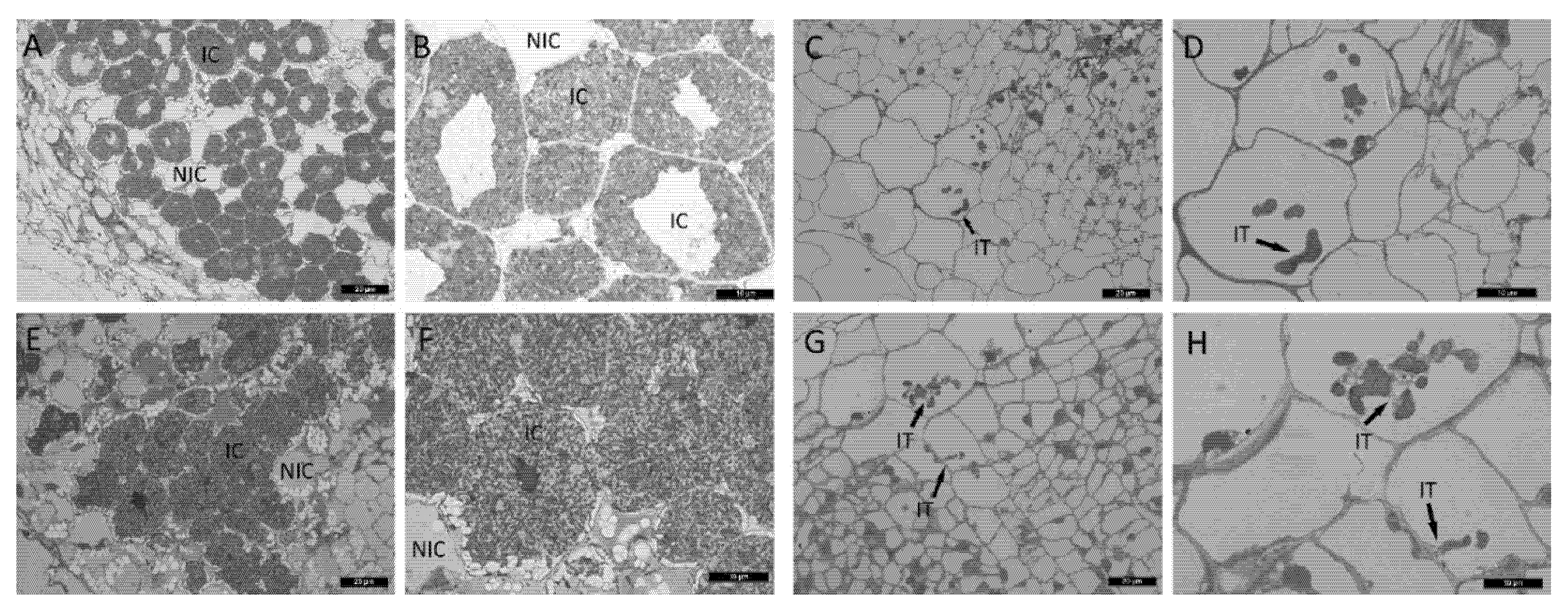
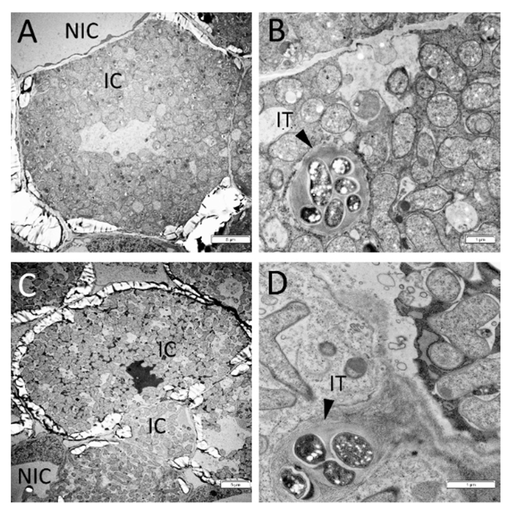
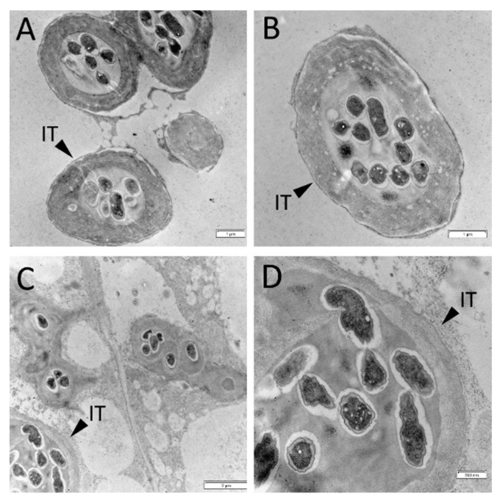
To verify the influence of cytokinin on morphological features of nodule development in wild type and mutant, light microscopy and transmission electron microscopy (TEM) were used. No significant effect of cytokinin on wild-type nodules was found (Figure 2E,F). The development of infection threads and bacteroids did not change noticeably in response to cytokinin treatment (Figure 2E,F and Figure 3). SGEFix--2 (sym33) mutant has “locked” infection threads inside the nodule and blocking in bacterial release[56,57,58,59], which was also shown in our experiments (Figure 2C,D). Although we found that the cytokinin stimulated the additional formation of nodules in sym33 mutant, the development of infection threads and bacterial accommodation seems to be similarly impaired in nodules of mutant plants nontreated or treated with cytokinin (Figure 2C,D,G,H). At the same time, TEM analysis showed that the development of bacteria in infection threads in the nodules of sym33 mutant treated with cytokinin may be blocked and degradation of bacteria was sometimes visible (Figure 4).

2.3. Comparative Analysis of Expression Patterns between Cv. SGE Wild Type and SGEFix--2 (Sym33) Mutant Nodules Using a Transcriptomic Approach

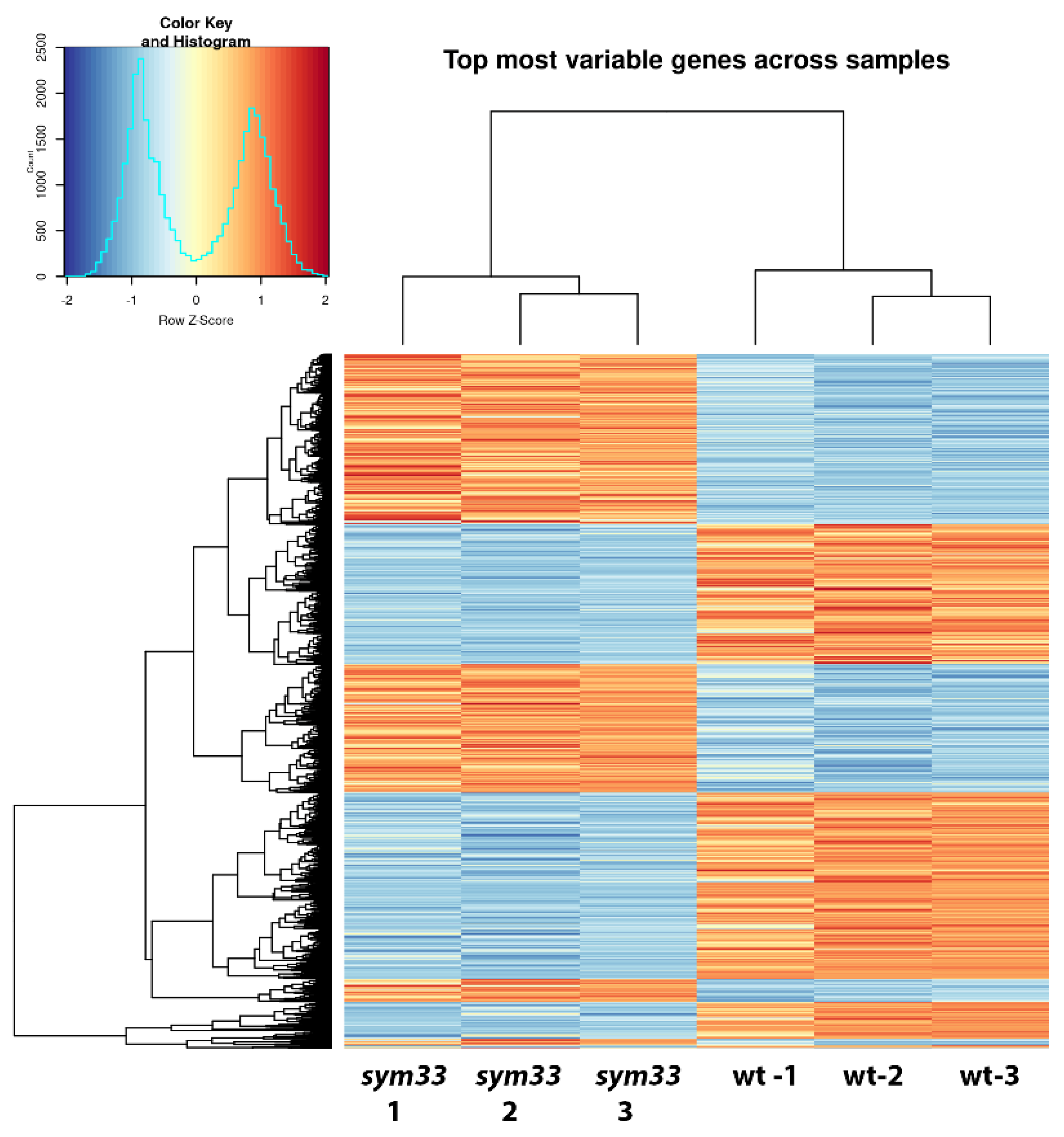
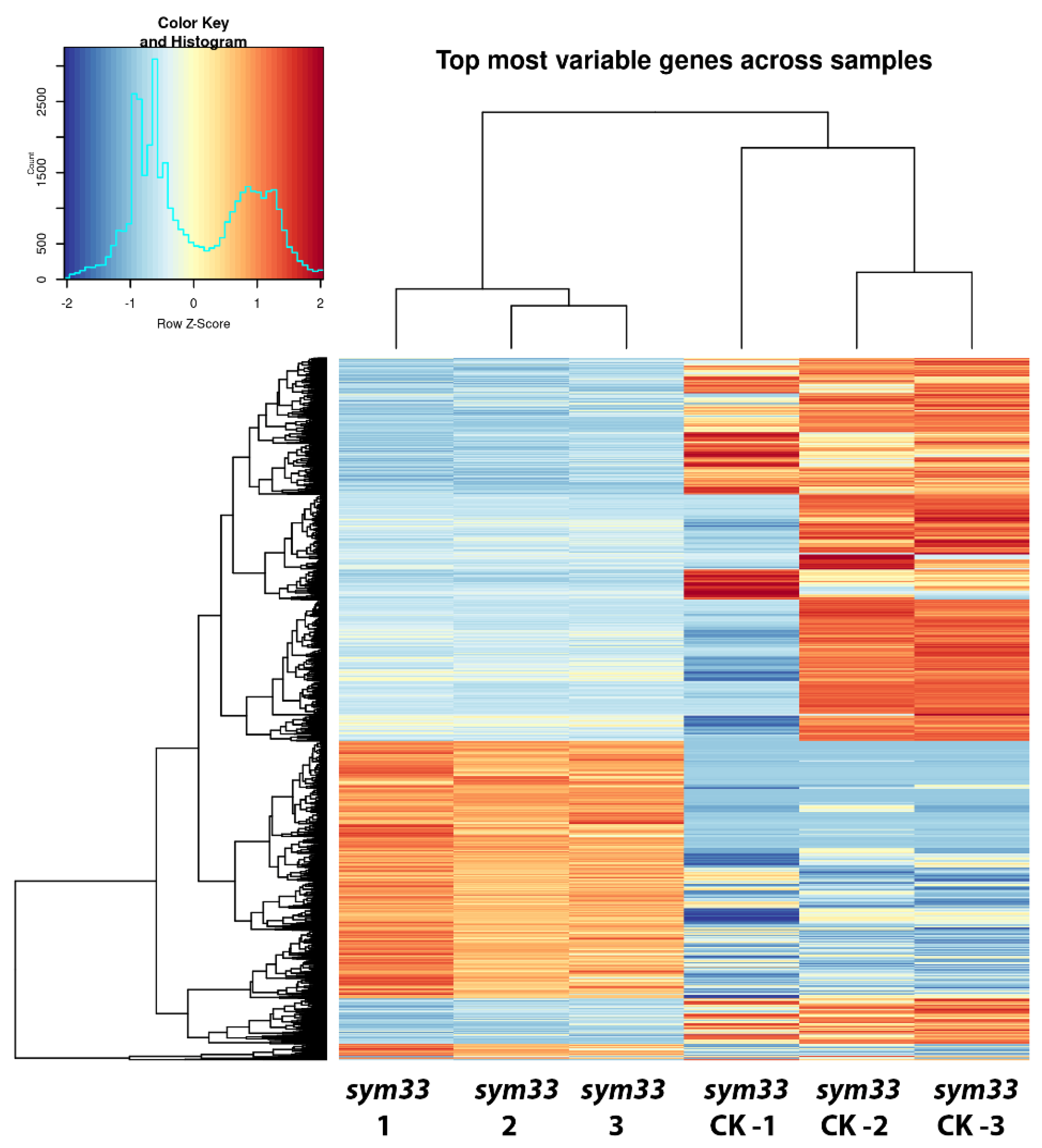
Given that we found an increase in the number of SGEFix-2 (sym33) mutant nodules after treatment with cytokinin, as well as morphological changes in them, we decided to analyze the gene expression changes associated with these effects. We have compared the patterns of differential expressed genes between cv. SGE wild-type nodules and SGEFix-2 (sym33) mutant nodules (Figure 5) as well as between nodules of SGEFix-2 (sym33) mutant plants without treatment and treated with cytokinin (Figure 5). Using a cutoff threshold equal to log2 fold change value > 2 and p adjusted value < 0.05, we discovered that about 1365 genes were up-regulated in wild type SGE nodules compared with sym33 mutant nodules, while about 2906 transcripts were up-regulated in sym33 mutant in response to cytokinin treatment compared to unstimulated mutant (Figure S1). Moreover, 538 genes were similarly up-regulated in both sets (Figure 6). Since 538 genes represent about 40% of the total number of up-regulated genes in wild-type nodules compared with mutant (1365 up-regulated), we suggest that cytokinin plays an important role in the regulation of late stages of nodule development. These results are in line with our previous findings demonstrating significant differences in the content of cytokinin between wild-type and sym33 mutant nodules [55].

2.4. Functional Activity of Differentially Expressed Genes between cv. SGE Wild Type and SGEFix--2 (sym33) Mutant Based on Gene Ontology Analysis

We used gene ontology (GO) set enrichment analysis to find out which biological processes were impacted in mutant nodules in comparison with wild-type nodules and which functions were affected in Fix- nodules by cytokinin treatment (Figure 7 and Figure 8). Analysis of gene ontology annotated pathways activated in the nodules of wild type and sym33 mutant showed the stimulation of the functional activity of genes encoding the enzymes degrading the storage polysaccharides such as starch as well as involved in the biosynthesis of proteins, amino acids, and nucleic acids (Figure 7). Significant activation of genes encoding proteins regulating transport processes through various membranes was also shown. This reflects the difference between effective wild-type cv. SGE and ineffective SGEFix--2 (sym33) mutant nodules. Stimulation of processes of calcium-dependent phospholipid binding suggests that signal transduction pathways activated by calcium and secondary metabolites may be involved in the regulation of effective symbiotic nodules. Stimulation of activity of pathways regulating the redox processes was also shown predominantly in effective nodules of wild type.
Analysis revealed that genes up-regulated in mutant’s nodules after cytokinin treatment are significantly enriched in processes associated with stress response (Figure 8). Among these processes, the highest rate of change was shown for ‘ribonuclease T2 activity’ which is associated with response to pathogens and abiotic stress in plants [62], and the ‘oxygen binding’ process which is important for plant stress signaling [63]. It is important to point out that genes that are up-regulated in wild nodules compared to mutant nodules are also enriched in ‘oxygen binding’.
We also found that the ‘potassium ion binding’ process was impacted by cytokinin treatment and down-regulated genes were significantly enriched in it. According to the recently published data [64], symbiosomes and vacuoles of infected cells are characterized by defects in K+ balance which is associated with mislocation of some plant ion channels. Aa detailed search among differentially expressed (DE) genes showed that several genes encoding potassium transporters and channels (Psat4g076320, Psat0s3462g0080) were significantly down-regulated in mutant nodules after cytokinin treatment. Similarly, the expression of Psat4g07632 and several other genes associated with ‘potassium ion binding’ processes were also decreased in wild-type nodules as compared to mutant nodules.

3. Discussion

It was previously shown that in M. truncatula ipd3 and L. japonicus cyclops mutants the nodules remain non-infected due to the blocking of bacterial release from the infection threads and subsequent intracellular accommodation [26,29,65,66]. Similarly, pea mutant SGEFix--2 (sym33) defective in IPD3/CYCLOPS transcription factor was impaired in the bacterial release from infection threads that resulted in non-infected nodules and disturbance in subsequent plant tissue and bacteroid differentiation [56,57,58,59]. Such mutants may be considered as a useful model for the investigation of later stages of nodule formation related to the differentiation of plant tissues and bacteroids. In addition, an altered cytokinin pattern in sym33 mutants [55] may point towards the importance of cytokinin in the regulation of these later stages of nodulation.
To determine a set of genes involved in the regulation of later stages of nodule development connected with infection progress, intracellular accommodation, as well as plant tissue and bacteroid differentiation, the RNA-seq analysis of pea mutant SGEFix--2 (sym33) nodules impaired in these processes compared to wild type SGE nodules was performed. To verify cytokinin’s influence on late nodule development stages, the comparative RNA-seq analysis of SGEFix--2 (sym33) mutant plants treated with cytokinin was also conducted.
Comparative analysis of the wild type and pea sym33 mutant nodules revealed the significant up-regulation of the genes encoding IPD3/CYCLOPS (Psat2g142200), NIN (Psat2g001120), KNOX3 (Psat6g028400), EFD (Psat7g259160), and NSP1 (Psat0s741g0280) transcription factors as well as A-type cytokinin response regulators RR11 (Psat4g192080) in wild type nodules compared with sym33 mutant (Table S1), which is in line with our previous findings [55]. NIN gene (Psat2g001120) encoding transcription factor and a number of genes encoding A-type cytokinin response regulators RR11 and RR4 (Psat4g192080, Psat0s644g0040) were found among up-regulated genes in nodules of cytokinin treated sym33 mutant (Table S2). Since the increased expression of these genes may be considered as markers of cytokinin stimulation, this indicates the expected effect of cytokinin treatment on pea mutant plants.
About 40% of the total number of up-regulated genes in wild-type nodules (compared to untreated sym33) matched with up-regulated genes in sym33 mutant upon cytokinin treatment (Figure S1). Taken together with the changes in nodule number that occurred in sym33 mutant after cytokinin stimulation, these findings suggest a significant role of cytokinin in the regulation of later stages of nodule development. Although the expression of 538 genes was altered, we can discuss only a few in this article. Among them, the genes encoding NSP1 (Psat0s741g0280), NIN (Psat2g001120), EFD (Psat7g259160), and RSD (Psat3g136520) transcription factors were shown to be up-regulated in wild type nodules compared with nodules of sym33 mutant as well as in sym33 mutant nodules in response to cytokinin treatment (Tables S1 and S2), thus suggesting that these transcription factors may be involved in the regulation of late stages of nodule development and induced by cytokinin. Indeed, it was recently shown that the NIN transcription factor does not only play a key role during root nodule initiation, but is also involved in the regulation of later stages of nodule development [67]. Here, we showed that stimulation of NIN expression as well as NSP1, EFD, and RSD at these stages may be connected with cytokinin treatment.
The RSD (Regulator of symbiosome differentiation, Psat3g136520) gene is involved in the regulation of the bacteroid formation. The level of RSD expression was higher in wild-type nodules compared to mutant nodules and up-regulated in response to cytokinin treatment of sym33 mutant. The RSD transcription factor is required for suppression of defense responses in nodules during bacteria accommodation [68]. It controls the expression of gene encoding VAMP721a (Vesicle-associated membrane protein 721a) protein, which is important for vesicular transport. The close homolog of this protein in Arabidopsis is also involved in the repression of plant immunity. In the nodules of rsd mutant plants, the bacteroids degrade rapidly followed by brown pigment accumulation, indicating activation of the plant defense reactions [69]. Using nin-16 weak allele mutant, it was shown that the RSD expression level was sufficient to suppress the development of defense reactions and early senescence in the invasion zone, but not sufficient in the fixation zone of nodules, where the level of NIN gene expression was significantly less. This suggests the importance of the NIN transcription factor for regulation of RSD expression at later stages and switching the programs controlling the development of defense reactions in different zones of nodules [67].
SEUSS-like protein (Psat6g039480) is a transcriptional adaptor in plants that provides the assembly of multimeric regulatory complexes. Gene encoding SEUSS-like protein was activated in wild-type nodules compared to sym33 mutant nodules and in response to cytokinin treatment (Tables S1 and S2). The effect of SEUSS on the stem, leaf, and root development as well as the formation of reproductive tissues was shown in Arabidopsis and other plants [70]. It was suggested that this regulator may be connected with the determination of tissue specificity in plants. Within the developing flower, for example, the SEUSS protein complex physically interacts with a few MADS domain DNA-binding proteins (APETALA1, SHORT VEGETATIVE PHASE1, and AGAMOUS-LIKE24) that regulate subsequent organ development [71,72].
GATA transcription factor (Psat4g097200) was found to be significantly up-regulated in wild-type nodules compared to sym33 mutant nodules. It is also activated in response to cytokinin treatment in sym33 mutant nodules. In addition, another GATA transcription factor, HAN1-like (Psat6g020240), was significantly stimulated by cytokinin. Some GATA transcription factors were shown to be involved in symbiosis development in legume plants. The MtHAN1 and MtHAN2 genes encoding GATA transcription factors in M. truncatula are mainly expressed in the nodule primordia and in the infection zone in mature nodules, suggesting the participation of these transcription factors in nodulation [73]. The expression of genes encoding NCR peptides, responsible for bacteroid differentiation, is shown to be suppressed in han1 han2 double mutant [74]. Hence, HANs may regulate nodule formation by influencing the expression and processing of small peptides including NCRs.
Indeed, NCR (Nodule cysteine-rich) secreted peptides are activated in response to rhizobial inoculation or Nod factor perception and are mainly involved in the control of bacteroid differentiation in legume plants [74,75]. An increased transcription for a set of genes encoding NCR peptides (Psat0s3945g0040, Psat2g028680, Psat2g027600, Psat2g122960, Psat0s2086g0040) was found in both sets (Tables S1 and S2). NCR genes may be expressed at different stages of symbiosis development. However, the expression of most of them is related to the stages when the bacteroids become mature and start fixing nitrogen, suggesting that these “late” genes are mainly involved in maintaining the functional activity of bacteroids, but not their initial development [76]. Here, it was shown for the first time that cytokinin may induce NCR genes related to later stages of nodule function.
Gene encoding NF-YC-like (Psat3g010480) protein was activated in wild-type nodules compared with sym33 mutant nodules and up-regulated in response to cytokinin treatment. Components of heterotrimeric CCAAT-box-binding factor complex, the nuclear factor Y proteins (NF-Ys), composed of three subunits, NF-YA, NF-YB, and NF-YC, play an important role in plant development. NF-YA1 and NF-YA2 play an important role at early stages of rhizobial infection, but also at later stages of symbiosis, regulating nodule organogenesis in M. truncatula [77,78,79,80]. In addition, M. truncatula MtNF-YC1 and MtNF-YC2 genes are required for nodule organogenesis [81]. An important role of NF-YA1 and NF-YB1 subunits was shown in symbiotic nodule formation in Phaseolus vulgaris [82]. Further analysis should be performed to verify the role of pea NF-YC1 in the regulation of nodule formation.
A few genes encoding cyclins (Psat2g083400) and cyclin-like F-box proteins (Psat1g007080, Psat7g162280) showed an increased level of expression in wild-type nodules compared to sym33 mutant nodules and activation in response to cytokinin treatment in sym33 mutant nodules. Cyclin-like F-box proteins are one of the main components of the SCF (SKP1 (S-phase-kinase-associated protein 1), Cullin-1, F-box protein) complex that belongs to the family of E3 ubiquitin ligase. Cyclin-like F-box proteins participate in the regulation of the cell cycle associated with target protein degradation [83]. Therefore, the cyclin-like F-box proteins are mainly localized in the tissues consisting of actively dividing cells. As an example, the accumulation of transcripts of F-box proteins was found in the nodule primordia of M. truncatula [83]. F-box proteins may be involved in the determination of tissue specification during plant development [84]. At the same time, some F-box-like proteins can act as negative regulators of plant resistance to pathogens [85]. microRNA MiR1134 is known to suppress the F-box/RNI/FBD-like domain protein in plant defense response regulation [86].
Transcription factors of AP2/ERF (APETALA2/Ethylene responsive factor) superfamily regulate plant development and response to stress. AP2/ERF transcription factors are classified into four families, including the AP2, ERF, RAV (AP2/B3 domain), and Soloist families [87]. The gene Psat4g225040 encoding transcription factor from the AP2 family was significantly activated in wild-type nodules compared with sym33 mutant nodules as well as in response to cytokinin treatment. The AP2/ERF transcription factors, such as ERN1 and ERN2, were shown to be essential for rhizobial infection and early stages of nodulation in legumes of legume plants [42,88]. Another AP2/ERF NNC1 (Nodule number control 1) may be a positive regulator of nodule senescence genes in mature nodules and can be silenced through miR172c as it was shown in P. vulgaris [89]. NNC1 directly targets and represses the early nodulin gene ENOD40 that plays a key role in nodulation [89].
MADS-domain/AGL transcription factors belong to a large family of regulators involved in the development of plant organs. It was previously shown that these transcription factors are involved in the development of root architecture [90]. The action of MADS-box transcription factors to control the legume root and nodule architecture is suggested to be carried out through interaction with the NIN transcription regulator [91]. Gene encoding MADS-domain/AGL transcription factors (Psat4g046280) was induced in wild-type nodules compared to sym33 mutant nodules, as well as in nodules of sym33 mutant in response to cytokinin treatment in our experiments. Further analysis should elaborate on their role in nodule development regulation.
It is known that the cytokinin/auxin ratio is strictly controlled and plays an important role in nodule development during the legume-rhizobia symbiosis [92]. We found that the cytokinin treatment may stimulate the expression of SHI/STY (Short internode/stylish) (Psat0s133g0120) gene, the closest homolog of STY1 from L. japonicus. SHI/STY transcription factors participate in the development of the generative organs and are important for the correct specification of tissue identity and differentiation in plants [93,94,95]. As SHI/STY may fulfill a similar function in various plants and mosses, we can talk about a conservative role of SHI/STY in tissue. Their participation in the development of nodules in Medicago was shown to be connected to the regulation of YUCCA enzymes, involved in the local biosynthesis of auxin. YUCCA1 (Psat6g030600) gene was also found among differentially expressed genes in nodules of sym33 mutant treated with cytokinin. Moreover, up-regulation in wild type and cytokinin treated mutant was shown for a set of genes encoding auxin-regulated proteins. Since the SHI/STY may be the direct regulators of auxin biosynthesis, they are important for the early stages of nodule organogenesis but seem to be also required for subsequent nodule development [96]. In L. japonicus, the SHI/STY was found to promote the differentiation of nodule structure, including an increase in cell size and the formation of vascular bundles [97].
In our experiments, the treatment of wild-type plants with cytokinin did not result in visible morphological changes of nodule structural features as the light microscopy and TEM showed. However, treatment of sym33 mutant plants with cytokinin according to the results of TEM analysis may induce partial degradation of bacteria in the infection threads. Moreover, it may stimulate the activation of callose and starch deposition in the cells of mutant as analysis of gene ontology pathways showed. Although cytokinin stimulated the additional formation of nodules in sym33 mutant, it seems that the development of bacteria inside infection threads in these nodules was disturbed. Comparative transcriptomic analysis of the wild type and mutant mature nodules showed stimulation of defense reactions, but this stimulation was stronger in nodules of cytokinin treated sym33 mutant compared with non-treated. As an example, the gene (Psat7g031360) encoding RING-H2 zinc finger protein was up-regulated in wild-type nodules compared with sym33 mutant nodules, but its expression level was significantly increased in response to cytokinin treatment of mutant plants. RING-H2 zinc finger protein and E3 ubiquitin-protein ligase are shown to be positive regulators of plant resistance to stress [98]. All the above-mentioned suggest that cytokinin may be an important regulator of plant resistance to infection. This balance is tightly regulated and may be disturbed by cytokinin regulating defense reactions in plants.
Therefore, transcriptomic profiling of wild-type and mutant nodules, as well as nodules in plants treated with cytokinin, resulted in the identification of a number of differentially expressed genes. This demonstrates the importance of the IPD3/CYCLOPS transcription factor for the regulation of later stages related to nodule differentiation and its interplay with cytokinin at these stages. We found many genes that may be involved in the regulation of the intracellular accommodation of bacteria and subsequent plant tissue and bacteroid differentiation among them. Further analysis should be performed to verify the role of these regulators in the control of late stages of nodule morphogenesis in pea.

4. Materials and Methods

4.1. Bacterial Strains and Inoculation

The Rhizobium leguminosarum biovar viciae CIAM 3841 strain was cultured at 28 °C on TY agar medium [99] supplemented with 0.6 mg/mL streptomycin. For the preparation of inoculum, bacteria were incubated in liquid B- medium [100]. The optical density of the suspension at 600 nm (OD600) was adjusted to 0.5–0.7.

4.2. Plant Material and Growth Conditions

Pisum sativum L. SGE cultivar and SGEFix--2 (sym33-3) mutant seeds from a collection of ARRIAM (St. Petersburg, Russia) were sterilized with sulfuric acid for 10 min, washed with sterile water 4 times, transferred to plates with 0.8% water agar, and germinated at room temperature in the dark for 4–5 days. After germination, the seedlings were transferred to the pots with vermiculite, saturated with Jensen’s medium, and grown in a climatic chamber (Binder, Germany) at 21 °C with cycles of 16 h light/8 h dark and humidity 64%. Pea seedlings were inoculated with 2 mL of R. leguminosarum bv. viciae CIAM 3841 per plant on the next day after transfer. One day after, half of the plants were treated with 6-BAP, at a final concentration of 10 μm and a volume of 50 mL per pot (diluted with sterile distilled water). Treatment was carried out every other day, and control plants were watered in the same volume with sterile distilled water. For gene expression analysis, the nodules were harvested after 14 days.

4.3. Material Fixation

The nodules were fixed in a freshly prepared solution of 2% paraformaldehyde (Sigma-Aldrich, MO, USA) in 1/3 of the concentration of MTSB buffer (50 mM PIPES (pH 6.9) (Amresco, OH, USA); 5 mM MgSO4 × 7H2O; 5 mM ethylene glycol-bis (β-aminoethyl ether)—N, N, N‘, N’-tetraacetic acid (Sigma, MO, USA) with the addition of 0.25% glutaraldehyde (solution Grade I, 25% in H2O. Sigma-Aldrich, MO, USA), 0,3% Twin-20 (Amresco, OH, USA), 0,3% Triton-X-100 (Amresco, OH, USA). For optimal penetration of the fixative, the air from the tissue was evacuated three times for 7 min at 0.9 bar using a ME 1 vacuum pump (Vacuubrand, Germany) and left overnight at +4 °C. Then, the material was washed with PBS buffer (0.137 M NaCl, 0.0027 KCl, 0.01 M Na2HPO4, 0.0018 M KH2PO4, pH 7.4).

4.4. Light and Transmission Electron Microscopy

The nodules were fixed in solution of 2.5% (v/v) glutaraldehyde (EMS, Hatfield, PA, USA) and 2% (w/v) formaldehyde (EMS, Hatfield, PA, USA) in 0.1 M cacodylate buffer (pH 7.4) overnight and postfixed in ice-cold 2% OsO4 (EMS, Hatfield, PA, USA) in the same buffer for 1.5 h. Samples were dehydrated in ethanol series and acetone and embedded in EmBed 812 epoxy resin (EMS, Hatfield, PA, USA). Semithin (1 μm) and ultrathin sections were made using ultratome EM UC7 (Leica, Vienna, Austria). Semithin sections were stained with 20% solution of Epoxy tissue stain with toluidine blue and basic fuchsin (EMS, Hatfield, PA,USA) in 50% ethanol and observed under light microscope DM2500 (Leica, Vienna, Austria). Ultrathin sections were stained on grids with 2% uranyl acetate water solution and Reynold’s lead citrate. Images were taken using the transmission electron microscope JEM-1400 (Jeol, Tokyo, Japan) equipped with a side camera Veleta (Olympus, Tokyo, Japan) at 80 kV.

4.5. Isolation of RNA and Preparation of Libraries

According to the manufacturer’s protocol, the total RNA was isolated from the nodules of inoculated or treated with cytokinin plants using the PureZol reagent (Bio-Rad Laboratories, Hercules, CA, USA). To remove genomic DNA, DNAseI treatment (Thermo Fisher Scientific, Waltham, MA, USA) was used.
To prepare libraries for the experiment with SGE Fix--2 (sym33-3), we used NEBNext® Poly (A) mRNA Magnetic Isolation Module (E7490) purification kit, NEBNext® Ultra ™ II Directional RNA Library Prep Kit for Illumina® (E7760), and the NEBNext® Multiplex Oligos primer set for Illumina® (Index Primers Set 2) (E7500S) (New England Biolabs, Rowley, MA, USA). At all stages, the material was cleaned using AMPure XP beads (Beckman Coulter, Brea, CA, USA).

4.6. Illumina Sequencing and Data Analysis

Three groups of libraries representing three independent biological experiments (4–5 plants for one replication of one treatment option) were sequenced on a NovaSeq 6000 (Illumina, San Diego, CA, USA) with single-end 100 bp reads according to the manufacturer’s protocol. The resulting reads were trimmed off Illumina adapters. Additionally, read ends were trimmed by sequencing quality (phred33 score < 20) using Trimmomatic [101]. To obtain read counts per both transcript and gene, fastq files were further processed with RSEM package [102] using transcriptome generated from Pisum sativum genome v1a [103]) as a reference. The differential expression was performed on the resulting raw count matrix of 9 individual samples using the DESeq2 R package [104]. Transcripts were considered to be differentially expressed with the adjusted p-value < 0.05 and absolute value of log2 (fold change) > 2. Gene ontology [105] gene set enrichment analysis was executed using the GSEABase R package [106] with cutoff values: odds ratio > 2, and p-value < 0.05.

4.7. Statistical Analysis

Statistical analysis of root length and number of nodules was based on the results of three independent experiments (4–5 plants per one variant were used). The error bars represent the mean ± SEM of three repeats. The asterisks indicate statistically significant differences based on Student’s t-test (* p < 0.05; ** p < 0.01).

Supplementary Materials

The following materials are available online at https://www.mdpi.com/article/10.3390/plants11010056/s1, Table S1: Analysis of differential gene expression in pea nodules of cv. SGE wild type (wt) and SGEFix--2 (sym33) mutant; Table S2: Analysis of differential gene expression in pea nodules of SGEFix--2 (sym33) mutant plants untreated or treated with cytokinin; Figure S1: The graphical output of differential gene expression in pea nodules of cv. SGE wild type and SGEFix--2 (sym33) mutant as well as in pea nodules of SGEFix--2 (sym33) mutant plants untreated or treated with cytokinin based on Venna diagram using cutoff threshold equal log2 fold change value > 2 and p adjusted value < 0.05.

Author Contributions

Investigation, writing—original draft preparation, methodology, E.S.R.; software analysis, methodology, P.Y.K.; investigation, O.A.P. and A.V.D.; investigation, microscopy, A.N.I.; conceptualization, writing—review and editing, supervision, E.A.D. All authors have read and agreed to the published version of the manuscript.

Funding

The article was made with the support of the Ministry of Science and Higher Education of the Russian Federation in accordance with agreement № 075-15-2020-920 date 16 November 2020, on providing a grant in the form of subsidies from the Federal budget of the Russian Federation. The grant was provided for state support for the creation and development of a World-class Scientific Center “Agrotechnologies for the Future”.

Institutional Review Board Statement

Not applicable.

Informed Consent Statement

Not applicable.

Data Availability Statement

Not applicable.

Acknowledgments

The research was performed using equipment of the Core Centrum “Genomic Technologies, Proteomics and Cell Biology” in ARRIAM.

Conflicts of Interest

The authors declare they have no competing interests.

References

  1. Madsen, E.B.; Madsen, L.H.; Radutoiu, S.; Olbryt, M.; Rakwalska, M.; Szczyglowski, K.; Sato, S.; Kaneko, T.; Tabata, S.; Sandal, N.; et al. A receptor kinase gene of the LysM type is involved in legumeperception of rhizobial signals. Nature 2003, 425, 637–640. [Google Scholar] [CrossRef]
  2. Radutoiu, S.; Madsen, L.H.; Madsen, E.B.; Jurkiewicz, A.; Fukai, E.; Quistgaard, E.M.H.; Albrektsen, A.S.; James, E.K.; Thirup, S.; Stougaard, J. LysM domains mediate lipochitin-oligosaccharide recognition and Nfr genes extend the symbiotic host range. EMBO J. 2007, 26, 3923–3935. [Google Scholar] [CrossRef] [PubMed] [Green Version]
  3. Broghammer, A.; Krusell, L.; Blaise, M.; Sauer, J.; Sullivan, J.T.; Maolanon, N.; Vinther, M.; Lorentzen, A.; Madsen, E.B.; Jensen, K.J.; et al. Legume receptors perceive the rhizobial lipochitin oligosaccharide signal molecules by direct binding. Proc. Natl. Acad. Sci. USA 2012, 109, 13859–13864. [Google Scholar] [CrossRef] [PubMed] [Green Version]
  4. Amor, B.B.; Shaw, S.L.; Oldroyd, G.E.D.; Maillet, F.; Penmetsa, R.V.; Cook, D.; Long, S.R.; Denarie, J.; Gough, C. The NFP locus of Medicago truncatula controls an early step of Nod factor signal transduction upstream of a rapid calcium flux and root hair deformation. Plant J. 2003, 34, 495–506. [Google Scholar] [CrossRef] [Green Version]
  5. Limpens, E.; Franken, C.; Smit, P.; Willemse, J.; Bisseling, T.; Geurts, R. LysM domain receptor kinases regulating rhizobial Nod factor-induced infection. Science 2003, 302, 630–633. [Google Scholar] [CrossRef] [PubMed]
  6. Smit, P.; Limpens, E.; Geurts, R.; Fedorova, E.; Dolgikh, E.; Gough, C.; Bisseling, T. Medicago LYK3, an Entry Receptor in Rhizobial Nodulation Factor Signaling. Plant Physiol. 2007, 145, 183–191. [Google Scholar] [CrossRef] [PubMed] [Green Version]
  7. Bensmihen, S.; de Billy, F.; Gough, C. Contribution of NFP LysM domains to the recognition of Nod factors during the Medicago truncatula/Sinorhizobium meliloti symbiosis. PLoS ONE 2011, 6. [Google Scholar] [CrossRef] [PubMed] [Green Version]
  8. Zhukov, V.; Radutoiu, S.; Madsen, L.H.; Rychagova, T.; Ovchinnikova, E.; Borisov, A.; Tikhonovich, I.; Stougaard, J. The Pea Sym37 Receptor Kinase Gene Controls Infection-Thread Initiation and Nodule Development. Mol. Plant-Microbe Interact. 2008, 21, 1600–1608. [Google Scholar] [CrossRef] [PubMed] [Green Version]
  9. Kirienko, A.N.; Porozov, Y.B.; Malkov, N.V.; Akhtemova, G.A.; Le Signor, C.; Thompson, R.; Saffray, C.; Dalmais, M.; Bendahmane, A.; Tikhonovich, I.A.; et al. Role of a receptor-like kinase K1 in pea Rhizobium symbiosis development. Planta 2018, 248, 1101–1120. [Google Scholar] [CrossRef] [PubMed]
  10. Sulima, A.S.; Zhukov, V.A.; Afonin, A.A.; Zhernakov, A.I.; Tikhonovich, I.A.; Lutova, L.A. Selection Signatures in the First Exon of Paralogous Receptor Kinase Genes from the Sym2 Region of the Pisum sativum L. Genome. Front. Plant Sci. 2017, 8. [Google Scholar] [CrossRef] [Green Version]
  11. Geurts, R.; Heidstra, R.; Hadri, A.E.; Downie, J.A.; Franssen, H.; Van Kammen, A.; Bisseling, T. SYM2 of pea is involved in a nodulation factor-perception mechanism that controls the infection process in the epidermis. Plant Physiol. 1997, 115, 351–359. [Google Scholar] [CrossRef] [PubMed] [Green Version]
  12. Kirienko, A.N.; Vishnevskaya, N.A.; Kitaeva, A.B.; Shtark, O.Y.; Kozyulina, P.Y.; Thompson, R.; Dalmais, M.; Bendahmane, A.; Tikhonovich, I.A.; Dolgikh, E.A. Structural variations in LysM domains of LysM-RLK psK1 may result in a different effect on Pea–Rhizobial symbiosis development. Int. J. Mol. Sci. 2019, 20, 1624. [Google Scholar] [CrossRef] [PubMed] [Green Version]
  13. Endre, G.; Kereszt, A.; Kevei, Z.; Mihacea, S.; Kaló, P.; Kiss, G.B. A receptor kinase gene regulating symbiotic nodule development. Nature 2002, 417, 962–966. [Google Scholar] [CrossRef] [PubMed]
  14. Stracke, S.; Kistner, C.; Yoshida, S.; Mulder, L.; Sato, S.; Kaneko, T.; Tabata, S.; Sandal, N.; Stougaard, J.; Szczyglowski, K.; et al. A plant receptor-like kinase required for both bacterial and fungal symbiosis. Nature 2002, 417, 959–962. [Google Scholar] [CrossRef]
  15. Ané, J.M.; Kiss, G.B.; Riely, B.K.; Penmetsa, R.V.; Oldroyd, G.E.D.; Ayax, C.; Lévy, J.; Debellé, F.; Baek, J.M.; Kalo, P.; et al. Medicago truncatula DMI1 Required for Bacterial and Fungal Symbioses in Legumes. Science 2004, 303, 1364–1367. [Google Scholar] [CrossRef] [Green Version]
  16. Charpentier, M.; Martins, T.V.; Granqvist, E.; Oldroyd, G.E.D.; Morris, R.J. The role of DMI1 in establishing Ca2+ oscillations in legume symbioses. Plant Signal. Behav. 2013, 8, e2289. [Google Scholar] [CrossRef] [PubMed] [Green Version]
  17. Kanamori, N.; Madsen, L.H.; Radutoiu, S.; Frantescu, M.; Quistgaard, E.M.H.; Miwa, H.; Downie, J.A.; James, E.K.; Felle, H.H.; Haaning, L.L.; et al. A nucleoporin is required for induction of Ca2+ spiking in legume nodule development and essential for rhizobial and fungal symbiosis. Proc. Natl. Acad. Sci. USA 2006, 103, 359–364. [Google Scholar] [CrossRef] [Green Version]
  18. Saito, K.; Yoshikawa, M.; Yano, K.; Miwa, H.; Uchida, H.; Asamizu, E.; Sato, S.; Tabata, S.; Imaizumi-Anraku, H.; Umehara, Y.; et al. Nucleoporin85 is required for calcium spiking, fungal and bacterial symbioses, and seed production in Lotus japonicus. Plant Cell 2007, 19, 610–624. [Google Scholar] [CrossRef] [PubMed] [Green Version]
  19. Groth, M.; Takeda, N.; Perry, J.; Uchid, H.; Dräxl, S.; Brachmann, A.; Sato, S.; Tabata, S.; Kawaguchi, M.; Wang, T.L.; et al. NENA, a Lotus japonicus homolog of Sec13, is required for rhizodermal infection by arbuscular mycorrhiza fungi and rhizobia but dispensable for cortical endosymbiotic development. Plant Cell 2010, 22, 2509–2526. [Google Scholar] [CrossRef] [PubMed] [Green Version]
  20. Chiasson, D.M.; Haage, K.; Sollweck, K.; Brachmann, A.; Dietrich, P.; Parniske, M. A quantitative hypermorphic CNGC allele confers ectopic calcium flux and impairs cellular development. Elife 2017, 6, e25012. [Google Scholar] [CrossRef] [Green Version]
  21. Capoen, W.; Sun, J.; Wysham, D.; Otegui, M.S.; Venkateshwaran, M.; Hirsch, S.; Miwa, H.; Downie, J.A.; Morris, R.J.; Ane, J.-M.; et al. Nuclear membranes control symbiotic calcium signaling of legumes. Proc. Natl. Acad. Sci. USA 2011, 108, 14348–14353. [Google Scholar] [CrossRef] [PubMed] [Green Version]
  22. Lévy, J.; Bres, C.; Geurts, R.; Chalhoub, B.; Kulikova, O.; Duc, G.; Journet, E.-P.; Ané, J.-M.; Lauber, E.; Bisseling, T.; et al. A Putative Ca2+ and Calmodulin-Dependent Protein Kinase Required for Bacterial and Fungal Symbioses. Science 2004, 303, 1361–1364. [Google Scholar] [CrossRef] [PubMed] [Green Version]
  23. Mitra, R.M.; Shaw, S.L.; Long, S.R. Six nonnodulating plant mutants defective for Nod factor-induced transcriptional changes associated with the legume-rhizobia symbiosis. Proc. Natl. Acad. Sci. USA 2004, 101, 10217–10222. [Google Scholar] [CrossRef] [Green Version]
  24. Gleason, C.; Chaudhuri, S.; Yang, T.; Muñoz, A.; Poovaiah, B.W.; Oldroyd, G.E.D. Nodulation independent of rhizobia induced by a calcium-activated kinase lacking autoinhibition. Nature 2006, 441, 1149–1152. [Google Scholar] [CrossRef]
  25. Oldroyd, G.E.D.; Downie, J.A. Coordinating Nodule Morphogenesis with Rhizobial Infection in Legumes. Annu. Rev. Plant Biol. 2008, 59, 519–546. [Google Scholar] [CrossRef]
  26. Messinese, E.; Mun, J.-H.; Yeun, L.H.; Jayaraman, D.; Rougé, P.; Barre, A.; Lougnon, G.; Schornack, S.; Bono, J.-J.; Cook, D.R.; et al. A Novel Nuclear Protein Interacts With the Symbiotic DMI3 Calcium- and Calmodulin-Dependent Protein Kinase of Medicago truncatula. Mol. Plant-Microbe Interact. 2007, 20, 912–921. [Google Scholar] [CrossRef] [PubMed] [Green Version]
  27. Yano, K.; Yoshida, S.; Muller, J.; Singh, S.; Banba, M.; Vickers, K.; Markmann, K.; White, C.; Schuller, B.; Sato, S.; et al. CYCLOPS, a mediator of symbiotic intracellular accommodation. Proc. Natl. Acad. Sci. USA 2008, 105, 20540–20545. [Google Scholar] [CrossRef] [Green Version]
  28. Limpens, E.; Bisseling, T. CYCLOPS: A new vision on rhizobium-induced nodule organogenesis. Cell Host Microbe 2014, 15, 127–129. [Google Scholar] [CrossRef] [Green Version]
  29. Singh, S.; Katzer, K.; Lambert, J.; Cerri, M.; Parniske, M. CYCLOPS, A DNA-binding transcriptional activator, orchestrates symbiotic root nodule development. Cell Host Microbe 2014, 15, 139–152. [Google Scholar] [CrossRef] [Green Version]
  30. Liu, C.W.; Breakspear, A.; Guan, D.; Cerri, M.R.; Jackson, K.; Jiang, S.; Robson, F.; Radhakrishnan, G.V.; Roy, S.; Bone, C.; et al. NIN acts as a network hub controlling a growth module required for rhizobial infection. Plant Physiol. 2019, 179, 1704–1722. [Google Scholar] [CrossRef] [Green Version]
  31. Schauser, L.; Roussis, A.; Stiller, J.; Stougaard, J. A plant regulator controlling development of symbiotic root nodules. Nature 1999, 402, 191–195. [Google Scholar] [CrossRef] [PubMed]
  32. Liu, J.; Rutten, L.; Limpens, E.; Van Der Molen, T.; Van Velzen, R.; Chen, R.; Chen, Y.; Geurts, R.; Kohlen, W.; Kulikova, O.; et al. A remote cis-regulatory region is required for nin expression in the pericycle to initiate nodule primordium formation in medicago truncatula. Plant Cell 2019, 31, 68–83. [Google Scholar] [CrossRef] [PubMed] [Green Version]
  33. Soyano, T.; Shimoda, Y.; Kawaguchi, M.; Hayashi, M. A shared gene drives lateral root development and root nodule symbiosis pathways in Lotus. Science 2019, 366, 1021–1023. [Google Scholar] [CrossRef] [PubMed]
  34. Marsh, J.F.; Rakocevic, A.; Mitra, R.M.; Brocard, L.; Sun, J.; Eschstruth, A.; Long, S.R.; Schultze, M.; Ratet, P.; Oldroyd, G.E.D. Medicago truncatula NIN is essential for rhizobial-independent nodule organogenesis induced by autoactive calcium/calmodulin-dependent protein kinase. Plant Physiol. 2007, 144, 324–335. [Google Scholar] [CrossRef] [PubMed] [Green Version]
  35. Kosuta, S.; Held, M.; Hossain, M.S.; Morieri, G.; MacGillivary, A.; Johansen, C.; Antolín-Llovera, M.; Parniske, M.; Oldroyd, G.E.D.; Downie, A.J.; et al. Lotus japonicus symRK-14 uncouples the cortical and epidermal symbiotic program. Plant J. 2011, 67, 929–940. [Google Scholar] [CrossRef]
  36. Kaló, P.; Gleason, C.; Edwards, A.; Marsh, J.; Mitra, R.M.; Hirsch, S.; Jakab, J.; Sims, S.; Long, S.R.; Rogers, J.; et al. Nodulation signaling in legumes requires NSP2, a member of the GRAS family of transcriptional regulators. Science 2005, 308, 1786–1789. [Google Scholar] [CrossRef]
  37. Smit, P.; Raedts, J.; Portyanko, V.; Debellé, F.; Gough, C.; Bisseling, T.; Geurts, R. NSP1 of the GRAS protein family is essential for rhizobial nod factor-induced transcription. Science 2005, 308, 1789–1791. [Google Scholar] [CrossRef] [PubMed] [Green Version]
  38. Hirsch, S.; Oldroyd, G.E.D. GRAS-domain transcription factors SCR/SHR—GRAS Proteins Involved in Root Radial Patterning NSP1/NSP2—Formation of GRAS Domain Complex at the DNA. Plant Signal. Behav. 2009, 4, 698–700. [Google Scholar] [CrossRef] [Green Version]
  39. Dolgikh, E.A.; Leppyanen, I.V.; Osipova, M.A.; Savelyeva, N.V.; Borisov, A.Y.; Tsyganov, V.E.; Geurts, R.; Tikhonovich, I.A. Genetic dissection of Rhizobium-induced infection and nodule organogenesis in pea based on ENOD12A and ENOD5 expression analysis. Plant Biol. 2011, 13, 285–296. [Google Scholar] [CrossRef]
  40. Xiao, A.; Yu, H.; Fan, Y.; Kang, H.; Ren, Y.; Huang, X.; Gao, X.; Wang, C.; Zhang, Z.; Zhu, H.; et al. Transcriptional regulation of NIN expression by IPN2 is required for root nodule symbiosis in Lotus japonicus. New Phytol. 2020, 227, 513–528. [Google Scholar] [CrossRef]
  41. Miwa, H.; Sun, J.; Oldroyd, G.E.D.; Downie, J.A. Analysis of Nod-factor-induced calcium signaling in root hairs of symbiotically defective mutants of Lotus japonicus. Mol. Plant-Microbe Interact. 2006, 19, 914–923. [Google Scholar] [CrossRef] [PubMed] [Green Version]
  42. Middleton, P.H.; Jakab, J.; Penmetsa, R.V.; Starker, C.G.; Doll, J.; Kaló, P.; Prabhu, R.; Marsh, J.F.; Mitra, R.M.; Kereszt, A.; et al. An ERF transcription factor in Medicago truncatula that is essential for nod factor signal transduction. Plant Cell 2007, 19, 1221–1234. [Google Scholar] [CrossRef] [Green Version]
  43. Cerri, M.R.; Frances, L.; Laloum, T.; Auriac, M.C.; Niebel, A.; Oldroyd, G.E.D.; Barker, D.G.; Fournier, J.; de Carvalho-Niebel, F. Medicago truncatula ERN transcription factors: Regulatory interplay with NSP1/NSP2 GRAS factors and expression dynamics throughout rhizobial infection. Plant Physiol. 2012, 160, 2155–2172. [Google Scholar] [CrossRef] [PubMed] [Green Version]
  44. Cerri, M.R.; Frances, L.; Kelner, A.; Fournier, J.; Middleton, P.H.; Auriac, M.C.; Mysore, K.S.; Wen, J.; Erard, M.; Barker, D.G.; et al. The symbiosis-related ERN transcription factors act in concert to coordinate rhizobial host root infection. Plant Physiol. 2016, 171, 1037–1054. [Google Scholar] [CrossRef] [PubMed]
  45. Fonouni-Farde, C.; Tan, S.; Baudin, M.; Brault, M.; Wen, J.; Mysore, K.S.; Niebel, A.; Frugier, F.; Diet, A. DELLA-mediated gibberellin signalling regulates Nod factor signalling and rhizobial infection. Nat. Commun. 2016, 7. [Google Scholar] [CrossRef] [PubMed]
  46. Jin, Y.; Liu, H.; Luo, D.; Yu, N.; Dong, W.; Wang, C.; Zhang, X.; Dai, H.; Yang, J.; Wang, E. DELLA proteins are common components of symbiotic rhizobial and mycorrhizal signalling pathways. Nat. Commun. 2016, 7, 12433. [Google Scholar] [CrossRef]
  47. Dolgikh, A.V.; Kirienko, A.N.; Tikhonovich, I.A.; Foo, E.; Dolgikh, E.A. The DELLA Proteins Influence the Expression of Cytokinin Biosynthesis and Response Genes During Nodulation. Front. Plant Sci. 2019, 10, 432. [Google Scholar] [CrossRef] [PubMed]
  48. Soyano, T.; Kouchi, H.; Hirota, A.; Hayashi, M. Nodule inception directly targets NF-Y subunit genes to regulate essential processes of root nodule development in Lotus japonicus. PLoS Genet. 2013, 9, e1003352. [Google Scholar] [CrossRef] [Green Version]
  49. Laloum, T.; Baudin, M.; Frances, L.; Lepage, A.; Billault-Penneteau, B.; Cerri, M.R.; Ariel, F.; Jardinaud, M.F.; Gamas, P.; De Carvalho-Niebel, F.; et al. Two CCAAT-box-binding transcription factors redundantly regulate early steps of the legume-rhizobia endosymbiosis. Plant J. 2014, 79, 757–768. [Google Scholar] [CrossRef] [PubMed]
  50. Zanetti, M.E.; Blanco, F.A.; Beker, M.P.; Battaglia, M.; Aguilar, O.M. A C Subunit of the Plant Nuclear Factor NF-Y Required for Rhizobial Infection and Nodule Development Affects Partner Selection in the Common Bean–Rhizobium etli Symbiosis. Plant Cell 2010, 22, 4142–4157. [Google Scholar] [CrossRef] [PubMed] [Green Version]
  51. Suzaki, T.; Ito, M.; Yoro, E.; Sato, S.; Hirakawa, H.; Takeda, N.; Kawaguchi, M. Endoreduplication-mediated initiation of symbiotic organ development in Lotus japonicus. Development 2014, 141, 2441–2445. [Google Scholar] [CrossRef] [Green Version]
  52. Azarakhsh, M.; Kirienko, A.N.; Zhukov, V.A.; Lebedeva, M.A.; Dolgikh, E.A.; Lutova, L.A. KNOTTED1-LIKE HOMEOBOX 3: A new regulator of symbiotic nodule development. J. Exp. Bot. 2015, 66, 7181–7195. [Google Scholar] [CrossRef] [PubMed] [Green Version]
  53. Vernié, T.; Kim, J.; Frances, L.; Ding, Y.; Sun, J.; Guan, D.; Niebel, A.; Gifford, M.L.; de Carvalho-Niebel, F.; Oldroyd, G.E.D. The NIN transcription factor coordinates diverse nodulation programs in different tissues of the Medicago truncatula root. Plant Cell 2015, 27, 3410–3424. [Google Scholar] [CrossRef] [PubMed] [Green Version]
  54. Plet, J.; Wasson, A.; Ariel, F.; Le Signor, C.; Baker, D.; Mathesius, U.; Crespi, M.; Frugier, F. MtCRE1-dependent cytokinin signaling integrates bacterial and plant cues to coordinate symbiotic nodule organogenesis in Medicago truncatula. Plant J. 2011, 65, 622–633. [Google Scholar] [CrossRef] [PubMed]
  55. Dolgikh, E.A.; Kusakin, P.G.; Kitaeva, A.B.; Tsyganova, A.V.; Kirienko, A.N.; Leppyanen, I.V.; Dolgikh, A.V.; Ilina, E.L.; Demchenko, K.N.; Tikhonovich, I.A.; et al. Mutational analysis indicates that abnormalities in rhizobial infection and subsequent plant cell and bacteroid differentiation in pea (pisum sativum) nodules coincide with abnormal cytokinin responses and localization. Ann. Bot. 2020, 125, 905–923. [Google Scholar] [CrossRef] [PubMed]
  56. Tsyganov, V.E.; Morzhina, E.V.; Stefanov, S.Y.; Borisov, A.Y.; Lebsky, V.K.; Tikhonovich, I.A. The pea (Pisum sativum L.) genes sym33 and sym40 control infection thread formation and root nodule function. Mol. Gen. Genet. 1998, 259, 491–503. [Google Scholar] [CrossRef] [PubMed]
  57. Tsyganov, V.E.; Seliverstova, E.V.; Voroshilova, V.A.; Tsyganova, A.V.; Pavlova, Z.B.; Lebskii, V.K.; Borisov, A.Y.; Brewin, N.J.; Tikhonovich, I.A. Double mutant analysis of sequential functioning of pea (Pisum sativum L.) genes Sym13, Sym33, and Sym40 during symbiotic nodule development. Russ. J. Genet. Appl. Res. 2011, 1, 343–348. [Google Scholar] [CrossRef]
  58. Voroshilova, V.A.; Boesten, B.; Tsyganov, V.E.; Borisov, A.Y.; Tikhonovich, I.A.; Priefer, U.B. Effect of mutations in Pisum sativum L. genes blocking different stages of nodule development on the expression of late symbiotic genes in Rhizobium leguminosarum bv. viciae. Mol. Plant Microbe Interact. 2001, 14, 471–476. [Google Scholar] [CrossRef] [Green Version]
  59. Voroshilova, V.A.; Demchenko, K.N.; Brewin, N.J.; Borisov, A.Y.; Tikhonovich, I.A. Initiation of a legume nodule with an indeterminate meristem involves proliferating host cells that harbour infection threads. New Phytol. 2009, 181, 913–923. [Google Scholar] [CrossRef]
  60. Bauer, P.; Ratet, P.; Crespi, M.D.; Schultze, M.; Kondorosi, A. Nod factors and cytokinins induce similar cortical cell division, amyloplast deposition and MsEnod12A expression patterns in alfalfa roots. Plant J. 1996, 10, 91–105. [Google Scholar] [CrossRef]
  61. Lorteau, M.A.; Ferguson, B.J.; Guinel, F.C. Effects of cytokinin on ethylene production and nodulation in pea (Pisum sativum) cv. Sparkle. Physiol. Plant. 2001, 112, 421–428. [Google Scholar] [CrossRef]
  62. Tsyganov, V.E.; Belimov, A.A.; Borisov, A.Y.; Safronova, V.I.; Georgi, M.; Dietz, K.-J.; Tikhonovich, I.A. A Chemically Induced New Pea (Pisum sativum) Mutant SGECd t with Increased Tolerance to, and Accumulation of, Cadmium. Ann. Bot. 2007, 99, 227–237. [Google Scholar] [CrossRef]
  63. Singh, S.P.; Thomason, P.A.; Lilla, S.; Schaks, M.; Tang, Q.; Goode, B.L.; Machesky, L.M.; Rottner, K.; Insall, R.H. Cell–substrate adhesion drives Scar/WAVE activation and phosphorylation by a Ste20-family kinase, which controls pseudopod lifetime. PLoS Biol. 2020, 18, e3000774. [Google Scholar] [CrossRef]
  64. Schmidt, R.R.; Weits, D.A.; Feulner, C.F.J.; Van Dongen, J.T. Oxygen sensing and integrative stress signaling in plants. Plant Physiol. 2018, 176, 1131–1142. [Google Scholar] [CrossRef] [Green Version]
  65. van Velzen, R.; Holmer, R.; Bu, F.; Rutten, L.; van Zeijl, A.; Liu, W.; Santuari, L.; Cao, Q.; Sharma, T.; Shen, D.; et al. Comparative genomics of the nonlegume Parasponia reveals insights into evolution of nitrogen-fixing rhizobium symbioses. Proc. Natl. Acad. Sci. USA 2018, 115, E4700–E4709. [Google Scholar] [CrossRef] [Green Version]
  66. Ovchinnikova, E.; Journet, E.P.; Chabaud, M.; Cosson, V.; Ratet, P.; Duc, G.; Fedorova, E.; Liu, W.; Op Den Camp, R.; Zhukov, V.; et al. IPD3 controls the formation of nitrogen-fixing symbiosomes in pea and Medicago spp. Mol. Plant-Microbe Interact. 2011, 24, 1333–1344. [Google Scholar] [CrossRef] [Green Version]
  67. Liu, J.; Rasing, M.; Zeng, T.; Klein, J.; Kulikova, O.; Bisseling, T. NIN is essential for development of symbiosomes, suppression of defence and premature senescence in Medicago truncatula nodules. New Phytol. 2021, 230, 290–303. [Google Scholar] [CrossRef] [PubMed]
  68. Berrabah, F.; Ratet, P.; Gourion, B. Multiple steps control immunity during the intracellular accommodation of rhizobia. J. Exp. Bot. 2015, 66, 1977–1985. [Google Scholar] [CrossRef] [PubMed] [Green Version]
  69. Sinharoy, S.; Torres-Jerez, I.; Bandyopadhyay, K.; Kereszt, A.; Pislariu, C.I.; Nakashima, J.; Benedito, V.A.; Kondorosi, E.; Udvardi, M.K. The C2H2 transcription factor REGULATOR OF SYMBIOSOME DIFFERENTIATION represses transcription of the secretory pathway gene VAMP721a and promotes symbiosome development in Medicago truncatula. Plant Cell 2013, 25, 3584–3601. [Google Scholar] [CrossRef] [Green Version]
  70. Bao, F.; Azhakanandam, S.; Franks, R.G. SEUSS and SEUSS-LIKE transcriptional adaptors regulate floral and embryonic development in arabidopsis. Plant Physiol. 2010, 152, 821–836. [Google Scholar] [CrossRef] [Green Version]
  71. Gregis, V.; Sessa, A.; Colombo, L.; Kater, M.M. AGL24, SHORT VEGETATIVE PHASE, and APETALA1 redundantly control AGAMOUS during early stages of flower development in Arabidopsis. Plant Cell 2006, 18, 1373–1382. [Google Scholar] [CrossRef] [Green Version]
  72. Sridhar, V.V.; Surendrarao, A.; Liu, Z. Erratum: APETALA1 and SEPALLATA3 interact with SEUSS to mediate transcription repression during flower development. Development 2006, 133, 3496. [Google Scholar] [CrossRef] [Green Version]
  73. Xu, Y.; Wang, H.; Lu, Z.; Wen, L.; Gu, Z.; Zhang, X.; Yu, G.; Wang, H.; Zhou, C.; Han, L. Developmental Analysis of the GATA Factor HANABA TARANU Mutants in Medicago truncatula Reveals Their Roles in Nodule Formation. Front. Plant Sci. 2021, 12, 597. [Google Scholar] [CrossRef]
  74. Pan, H.; Wang, D. Nodule cysteine-rich peptides maintain a working balance during nitrogen-fixing symbiosis. Nat. Plants 2017, 3, 17048. [Google Scholar] [CrossRef]
  75. Guefrachi, I.; Nagymihaly, M.; Pislariu, C.I.; Van de Velde, W.; Ratet, P.; Mars, M.; Udvardi, M.K.; Kondorosi, E.; Mergaert, P.; Alunni, B. Extreme specificity of NCR gene expression in Medicago truncatula. BMC Genom. 2014, 15, 712. [Google Scholar] [CrossRef] [Green Version]
  76. Mergaert, P. Differentiation of symbiotic nodule cells and their rhizobium endosymbionts. Adv. Bot. Res. 2020, 94, 149–180. [Google Scholar]
  77. Combier, J.P.; Frugier, F.; De Billy, F.; Boualem, A.; El-Yahyaoui, F.; Moreau, S.; Vernié, T.; Ott, T.; Gamas, P.; Crespi, M.; et al. MtHAP2-1 is a key transcriptional regulator of symbiotic nodule development regulated by microRNA169 in Medicago truncatula. Genes Dev. 2006, 20, 3084–3088. [Google Scholar] [CrossRef] [Green Version]
  78. Combier, J.P.; De Billy, F.; Gamas, P.; Niebel, A.; Rivas, S. Trans-regulation of the expression of the transcription factor MtHAP2-1 by a uORF controls root nodule development. Genes Dev. 2008, 22, 1549–1559. [Google Scholar] [CrossRef] [Green Version]
  79. Laloum, T.; De Mita, S.; Gamas, P.; Baudin, M.; Niebel, A. CCAAT-box binding transcription factors in plants: Y so many? Trends Plant Sci. 2013, 18, 157–166. [Google Scholar] [CrossRef]
  80. Laporte, P.; Lepage, A.; Fournier, J.; Catrice, O.; Moreau, S.; Jardinaud, M.F.; Mun, J.H.; Larrainzar, E.; Cook, D.R.; Gamas, P.; et al. The CCAAT box-binding transcription factor NF-YA1 controls rhizobial infection. J. Exp. Bot. 2014, 65, 481–494. [Google Scholar] [CrossRef] [Green Version]
  81. Baudin, M.; Laloum, T.; Lepage, A.; Rípodas, C.; Ariel, F.; Frances, L.; Crespi, M.; Gamas, P.; Blanco, F.A.; Zanetti, M.E.; et al. A phylogenetically conserved group of nuclear factor-Y transcription factors interact to control nodulation in legumes. Plant Physiol. 2015, 169, 2761–2773. [Google Scholar] [CrossRef]
  82. Rípodas, C.; Castaingts, M.; Clúa, J.; Villafañe, J.; Blanco, F.A.; Zanetti, M.E. The PvNF-YA1 and PvNF-YB7 subunits of the heterotrimeric nf-y transcription factor influence strain preference in the Phaseolus vulgaris–rhizobium etli symbiosis. Front. Plant Sci. 2019, 10, 221. [Google Scholar] [CrossRef]
  83. Iantcheva, A.; Boycheva, I.; Vassileva, V.; Revalska, M.; Zechirov, G. Cyclin-like F-box protein plays a role in growth and development of the three model species Medicago truncatula, Lotus japonicus, and Arabidopsis thaliana. Res. Rep. Biol. 2015, 6, 117–130. [Google Scholar] [CrossRef] [Green Version]
  84. Zühl, L.; Volkert, C.; Ibberson, D.; Schmidt, A. Differential activity of F-box genes and E3 ligases distinguishes sexual versus apomictic germline specification in Boechera. J. Exp. Bot. 2019, 70, 5643–5657. [Google Scholar] [CrossRef] [Green Version]
  85. Bonhomme, M.; André, O.; Badis, Y.; Ronfort, J.; Burgarella, C.; Chantret, N.; Prosperi, J.M.; Briskine, R.; Mudge, J.; Debéllé, F.; et al. High-density genome-wide association mapping implicates an F-box encoding gene in Medicago truncatula resistance to Aphanomyces euteiches. New Phytol. 2014, 201, 1328–1342. [Google Scholar] [CrossRef]
  86. Vivek, A.T. In silico identification and characterization of microRNAs based on EST and GSS in orphan legume crop, Lens culinaris medik. (Lentil). Agri Gene 2018, 8, 45–56. [Google Scholar] [CrossRef]
  87. Shu, Y.; Liu, Y.; Zhang, J.; Song, L.; Guo, C. Genome-wide analysis of the AP2/ERF superfamily genes and their responses to abiotic stress in Medicago truncatula. Front. Plant Sci. 2016, 6, 1247. [Google Scholar] [CrossRef] [Green Version]
  88. Andriankaja, A.; Boisson-Dernier, A.; Frances, L.; Sauviac, L.; Jauneau, A.; Barker, D.G.; De Carvalho-Niebel, F. AP2-ERF transcription factors mediate nod factor-dependent Mt ENOD11 activation in root hairs via a novel cis-regulatory motif. Plant Cell 2007, 19, 2866–2885. [Google Scholar] [CrossRef] [Green Version]
  89. Íñiguez, L.P.; Nova-Franco, B.; Hernández, G. Novel players in the AP2-miR172 regulatory network for common bean nodulation. Plant Signal. Behav. 2015, 10, e1062957. [Google Scholar] [CrossRef] [Green Version]
  90. Alvarez-Buylla, E.R.; García-Ponce, B.; de la Paz Sánchez, M.; Espinosa-Soto, C.; García-Gómez, M.L.; Piñeyro-Nelson, A.; Garay-Arroyo, A. MADS-box genes underground becoming mainstream: Plant root developmental mechanisms. New Phytol. 2019, 223, 1143–1158. [Google Scholar] [CrossRef] [Green Version]
  91. Ayra, L.; del Rocio Reyero-Saavedra, M.; Isidra-Arellano, M.C.; Lozano, L.; Ramírez, M.; Leija, A.; Fuentes, S.I.; Girard, L.; Valdés-López, O.; Hernández, G. Control of the Rhizobia Nitrogen-Fixing Symbiosis by Common Bean MADS-Domain/AGL Transcription Factors. Front. Plant Sci. 2021, 12, 679463. [Google Scholar] [CrossRef]
  92. Reid, D.E.; Nadzieja, M.; Novak, O.; Heckmann, A.B.; Sandal, N.; Stougaard, J. Cytokinin biosynthesis promotes cortical cell responses during nodule development. Plant Physiol. 2017, 175, 361–375. [Google Scholar] [CrossRef]
  93. Eklund, D.M.; Thelander, M.; Landberg, K.; Ståldal, V.; Nilsson, A.; Johansson, M.; Valsecchi, I.; Pederson, E.R.A.; Kowalczyk, M.; Ljung, K.; et al. Homologues of the Arabidopsis thaliana SHI/STY/LRP1 genes control auxin biosynthesis and affect growth and development in the moss Physcomitrella patens. Development 2010, 137, 1275–1284. [Google Scholar] [CrossRef] [Green Version]
  94. Gomariz-Fernández, A.; Sánchez-Gerschon, V.; Fourquin, C.; Ferrándiz, C. The role of SHI/STY/SRS genes in organ growth and carpel development is conserved in the distant eudicot species Arabidopsis thaliana and Nicotiana benthamiana. Front. Plant Sci. 2017, 8, 814. [Google Scholar] [CrossRef] [Green Version]
  95. Estornell, L.H.; Landberg, K.; Cierlik, I.; Sundberg, E. SHI/STY genes affect pre- and post-meiotic anther processes in auxin sensing domains in arabidopsis. Front. Plant Sci. 2018, 9, 150. [Google Scholar] [CrossRef] [Green Version]
  96. Shrestha, A.; Zhong, S.; Therrien, J.; Huebert, T.; Sato, S.; Mun, T.; Andersen, S.U.; Stougaard, J.; Lepage, A.; Niebel, A.; et al. Lotus japonicus Nuclear Factor YA1, a nodule emergence stage-specific regulator of auxin signalling. New Phytol. 2020, 229, 1535–1552. [Google Scholar] [CrossRef]
  97. Hossain, M.S.; Shrestha, A.; Zhong, S.; Miri, M.; Austin, R.S.; Sato, S.; Ross, L.; Huebert, T.; Tromas, A.; Torres-Jerez, I.; et al. Lotus japonicus NF-YA1 plays an essential role during nodule differentiation and targets members of the SHI/STY gene family. Mol. Plant-Microbe Interact. 2016, 29, 950–964. [Google Scholar] [CrossRef] [Green Version]
  98. Kim, M.H.; Cho, J.S.; Park, E.J.; Lee, H.; Choi, Y.I.; Bae, E.K.; Han, K.H.; Ko, J.H. Overexpression of a poplar Ring-H2 zinc finger, Ptxerico, confers enhanced drought tolerance via reduced water loss and ion leakage in Populus. Int. J. Mol. Sci. 2020, 21, 9454. [Google Scholar] [CrossRef]
  99. Orosz, L.; Sváb, Z.; Kondorosi, Á.; Sik, T. Genetic studies on Rhizobiophage 16-3. Mol. Gen. Genet. MGG 1973, 125, 341–350. [Google Scholar] [CrossRef]
  100. van Brussel, A.A.; Tak, T.; Wetselaar, A.; Pees, E.; Wijffelman, C. Small leguminosae as test plants for nodulation of Rhizobium leguminosarum and other rhizobia and agrobacteria harbouring a leguminosarum sym-plasmid. Plant Sci. Lett. 1982, 27, 317–325. [Google Scholar] [CrossRef]
  101. Bolger, A.M.; Lohse, M.; Usadel, B. Trimmomatic: A flexible trimmer for Illumina sequence data. Bioinformatics 2014, 30, 2114–2120. [Google Scholar] [CrossRef] [Green Version]
  102. Li, B.; Dewey, C.N. RSEM: Accurate transcript quantification from RNA-Seq data with or without a reference genome. BMC Bioinform. 2011, 12, 323. [Google Scholar] [CrossRef] [Green Version]
  103. Kreplak, J.; Madoui, M.A.; Cápal, P.; Novák, P.; Labadie, K.; Aubert, G.; Bayer, P.E.; Gali, K.K.; Syme, R.A.; Main, D.; et al. A reference genome for pea provides insight into legume genome evolution. Nat. Genet. 2019, 51, 1411–1422. [Google Scholar] [CrossRef]
  104. Love, M.I.; Huber, W.; Anders, S. Moderated estimation of fold change and dispersion for RNA-seq data with DESeq2. Genome Biol. 2014, 15, 550. [Google Scholar] [CrossRef] [Green Version]
  105. Boccacci, P.; Beltramo, C.; Sandoval Prando, M.A.; Lembo, A.; Sartor, C.; Mehlenbacher, S.A.; Botta, R.; Torello Marinoni, D. Gene ontology: Tool for the unification of biology. Mol. Breed. 2015, 25, 25–29. [Google Scholar] [CrossRef]
  106. Morgan, A.M.; Falcon, S.; Gentleman, R. GSEABase: Gene Set Enrichment Data Structures and Methods, R Package Version 1.56.0; 2021. Available online: http://www.bioconductor.org (accessed on 25 October 2021).
Figure 1. Analysis of root length (A) and the number of nodules in SGEFix--2 (sym33) mutant (B) and wild-type cv. SGE (C) plants 2 weeks after inoculation with Rhizobium leguminosarum bv. viciae CIAM1026 and treatment with 10 μM 6-BAP (cytokinin, CK). The graphs show the results of three independent experiments (4–5 plants per one variant were used). The error bars represent the mean ± SEM of three repeats. The asterisks indicate statistically significant differences based on Student’s t-test (** p < 0.01).
Plants 11 00056 g001
Figure 2. Light microscopy images of 2-week-old nontreated nodules of wild-type (A,B) and sym33 mutant (C,D) and treated with 10 μM 6-BAP (cytokinin) wild-type (E,F) and sym33 mutant (G,H). IC—infected cells, NIC—noninfected cells, IT—infection threads. (A,C,E,G) images have 40× magnification. (B,D,F,H) images have 100× magnification. Arrowheads indicate infection threads. Scale bars are 20 μm (A) and 10 μm (B).
Plants 11 00056 g002
Figure 3. Transmission electron microscopy images of 2-week-old wild-type nodules nontreated (A,B) or treated (C,D) with 10 μM 6-BAP (cytokinin) plants. IC—infected cells, NIC—noninfected cells, IT—infection thread. Arrowheads indicate infection threads. Scale bars are 5 μm (A,C) and 1 μm (B,D).
Plants 11 00056 g003
Figure 4. Transmission electron microscopy images of 2-week-old nodules of sym33 mutant nontreated (A,B) or treated (C,D) with 10 μM 6-BAP (cytokinin). IT—infection thread. Scale bars are 2 μm (C), 1 μm (A,B), and 500 nm (D).
Plants 11 00056 g004
Figure 5. Differential gene expression in pea nodules of cv. SGE wild type and SGEFix--2 (sym33) mutant. Heatmap represents top differentially expressed transcripts in wild-type nodules to sym33 mutant nodules as normalized counts after variance stabilizing transformation of raw read counts per transcript. The data of three independent biological repeats were combined for each variant—sym33 and wild type (wt) nodules.
Plants 11 00056 g005
Figure 6. Differential gene expression in pea nodules of SGEFix--2 (sym33) mutant plants untreated or treated with 10 µM BAP (cytokinin). Heatmap represents top differentially expressed transcripts in nodules of sym33 mutant plants non-treated and treated with cytokinin (CK) as normalized counts after variance stabilizing transformation of raw read counts per transcript. The data of three independent biological repeats were combined for each variant—sym33 nodules without treatment and sym33 after cytokinin (CK) treatment.
Plants 11 00056 g006
Figure 7. Gene ontology pathways that are overrepresented in differentially expressed genes in wild-type nodules compared to sym33 mutant nodules (up-and down-regulated separately).
Plants 11 00056 g007
Figure 8. Gene ontology pathways that are overrepresented in differentially expressed genes in nodules of untreated sym33 mutant plants compared to cytokinin treated (up-and down-regulated separately).
Plants 11 00056 g008
Publisher’s Note: MDPI stays neutral with regard to jurisdictional claims in published maps and institutional affiliations.