Article

A Case of Plant Vaccination: Enhancement of Plant Immunity against Verticillium dahliae by Necrotized Spores of the Pathogen

by
Eirini G. Poulaki
,
Maria Frantzeska Triviza
,
Marius Malai
and
Sotirios E. Tjamos
*
Laboratory of Plant Pathology, Agricultural University of Athens, 75 Iera Odos str., 11855 Athens, Greece
*
Author to whom correspondence should be addressed.
Plants 2022, 11(13), 1691; https://doi.org/10.3390/plants11131691
Submission received: 27 May 2022 / Revised: 18 June 2022 / Accepted: 24 June 2022 / Published: 26 June 2022

Abstract

:
The soil-borne fungus Verticillium dahliae is causing a devastating vascular disease in more than 200 species of dicotyledonous plants. The pathogen attacks susceptible plants through the roots, colonizes the plant vascular system, and causes the death of aerial tissues. In this study, we used Arabidopsis and eggplants to examine the plant protective and immunization effects of autoclaved V. dahliae spores against V. dahliae. We observed that the application of V. dahliae autoclaved spores in eggplants and Arabidopsis resulted in enhanced protection against V. dahliae, since the disease severity and pathogen colonization were lower in the plants treated with V. dahliae autoclaved spores when compared to controls. In addition, upregulation of the defense related genes PR1 and PDF1.2 in the Arabidopsis plants treated with the V. dahliae autoclaved spores was revealed. Furthermore, pathogenicity experiments in the Arabidopsis mutant cerk1, defective in chitin perception, revealed a loss of protection against V. dahliae in the cerk1 treated with the V. dahliae autoclaved spores. The participation of the chitin receptor CERK1 is evident in Arabidopsis immunization against V. dahliae using autoclaved spores of the pathogen.

1. Introduction

Plants possess an innate immune system comprised of pathogen associated molecular pattern (PAMP)-triggered immunity (PTI) and effector-triggered immunity (ETI). PAMPs are conserved molecular patterns in pathogens, alarming plants of the presence of potential intruders in their close vicinity [1]. Chitin, as one of the most conserved domains in the fungal cell wall, is a well-studied PAMP that could be sensed by plants, which in turn activate their defense mechanisms [2]. Chitin is a polysaccharide [β-1,4-N-acetylglucosamine (GlcNAc)n], which can be degraded to smaller parts, oligosaccharides, due to the action of plant chitinases [3]. Subsequently, the generated chitin fragments can be recognized by a chitin perception system resulting in the activation of PTI [2]. The plant chitin-sensing system comprises of receptors belonging to the lysin motif (LysM) domain family, including pattern recognition receptors (PRRs), receptor-like kinases (RLKs), and receptor-like proteins (RLPs) [4]. In Arabidopsis thaliana, the LysM-RLK chitin receptor CERK1 has been identified to bind N-acetylated chitin fragments, triggering the downstream plant defense [5,6].
An intriguing aspect of plant defense is the phenomenon of priming. This is a plant immunological state triggered by pathogen derived compounds, effectors, beneficial microbes, and various synthetic/natural compounds which enable plants to display a timely and robust activation of plant defenses upon detection of a biotic or abiotic stress [7,8,9,10]. In these respects, various biocontrol agents have been used to trigger induced systemic resistance in plants against various plant pathogens, mainly against plant pathogens where no effective chemical controls exist, such as Verticillium dahliae [11].
The soilborne fungal pathogen V. dahliae infects over 200 dicotyledon plant species, causing significant losses in plant yield, mostly in temperate zones [12,13]. The pathogen infects plants through the root system and spreads through the plant causing leaf wilting, chlorosis, yellowing, and tissue discoloration, eventually leading to plant death. The endophytic lifestyle of the pathogen and the production of long-living dormant structures, microsclerotia, make the pathogen inaccessible to chemical compounds. In addition, the scarcity of resistance genes in plant species poses a further limitation to Verticillium disease control [14]. Therefore, most research has focused on the development of biocontrol-based disease management strategies. However, these strategies are time consuming and impose a microbial input in the soil environment which may have detrimental effects to the indigenous microbiome and could be ineffective since the introduced microorganisms have to overcome various biotic and abiotic stresses under field conditions.
With these considerations, we investigated the potential of autoclaved V. dahliae spores to prime Arabidopsis thaliana and eggplants against V. dahliae. The priming effect of necrotized pathogen spores has not attracted any attention until now and to our best knowledge, this is the first published study describing the efficacy of necrotized pathogen structures to trigger the plant defense mechanism against a pathogen. The autoclaved V. dahliae spores were applied to the plants either as a root drenching or stem/leaf injecting inoculum. The observed plant protective activity of the V. dahliae spores led us to further investigate (i) the expression levels of the plant defense genes, PR1 and PDF1.2, marker genes of the salicylic acid (SA) and ethylene/jasmonate (ET/JA) dependent defenses [15], respectively and (ii) the participation of the CERK1 receptor using the A. thaliana mutant cerk1-2 [16].

2. Results

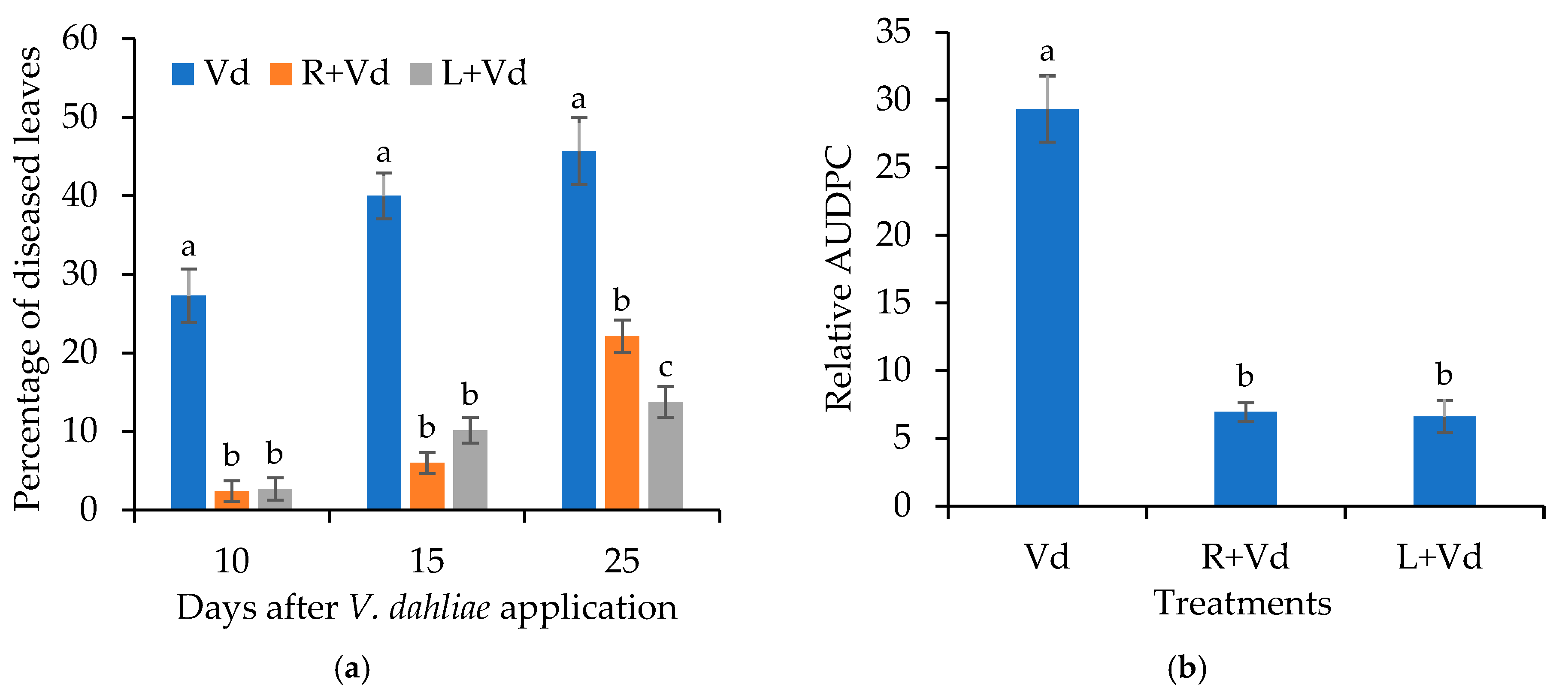
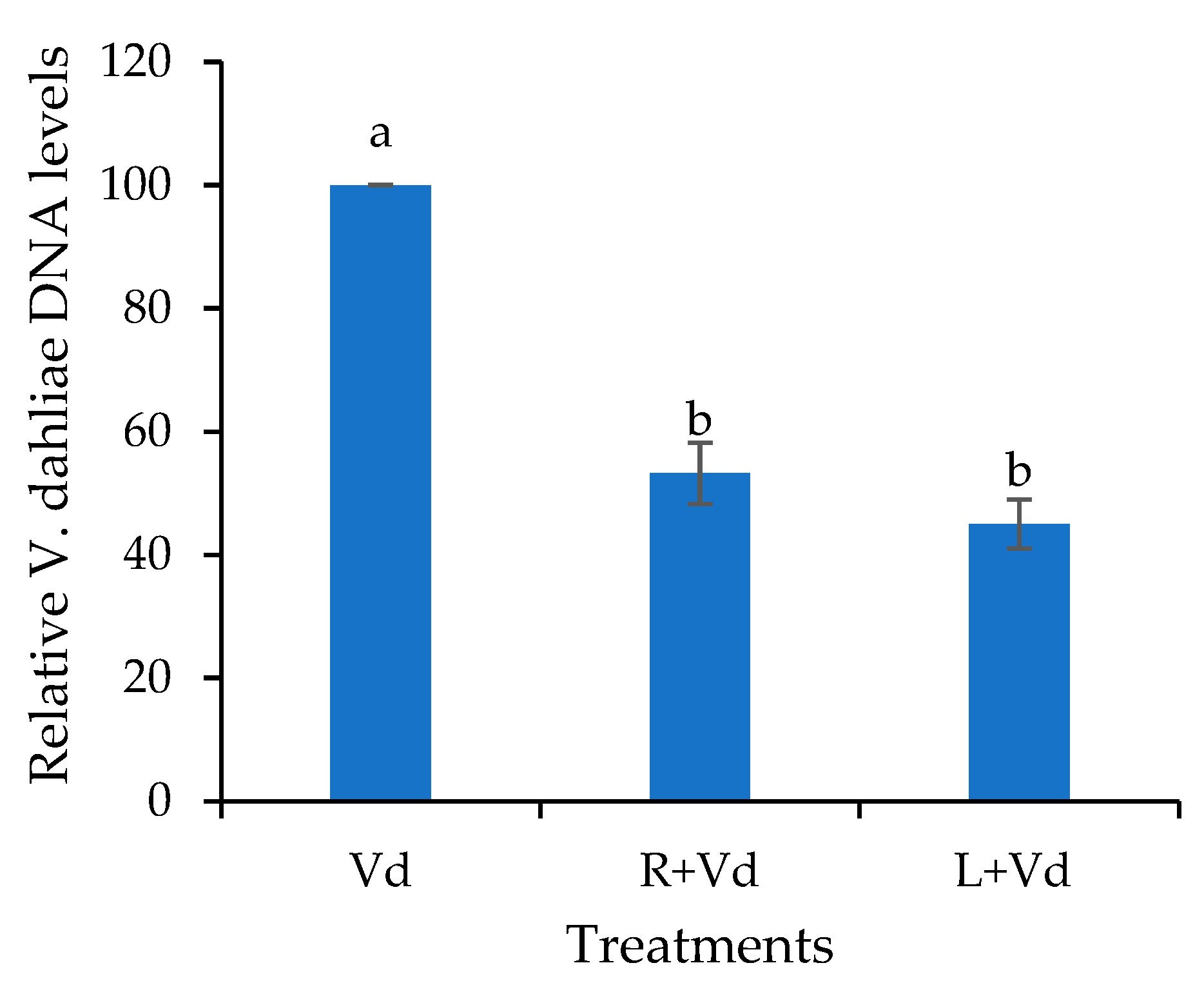
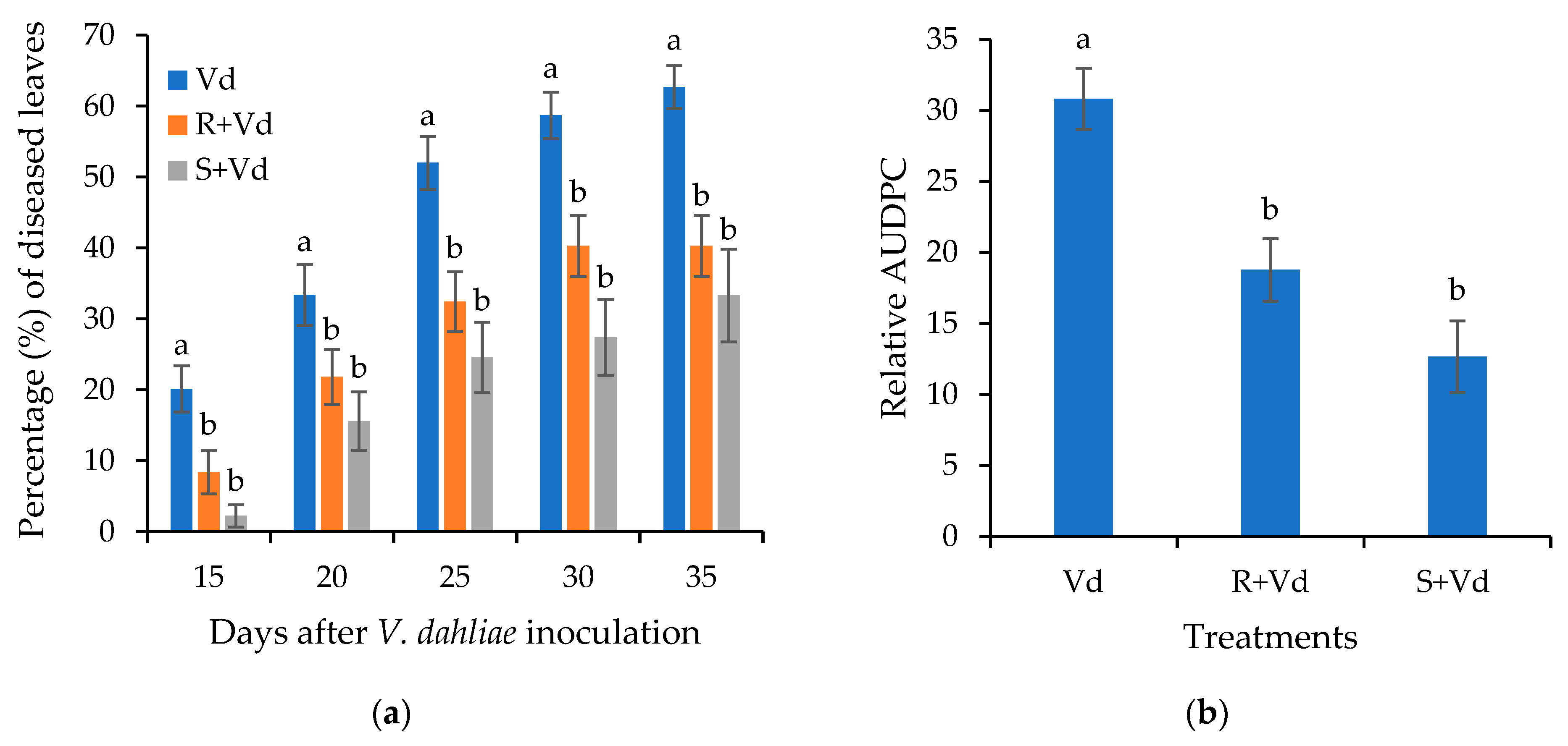
2.1. The Autoclaved Spores of V. dahliae Protect A. thaliana and Eggplants against V. dahliae

The addition of autoclaved spores of V. dahliae reduced Verticillium wilt symptoms in A. thaliana Col-0. The Verticillium symptoms appeared mainly in the form of wilting. The disease severity of Verticillium was recorded from 10 days post-inoculation (dpi) to 25 dpi (Figure 1).
The A. thaliana plants treated with the autoclaved spores of V. dahliae showed a significantly lower amount of disease during the pathogenicity experiment. At the last recording (25 dpi), the percentage of diseased leaves was ca. 14% and 22% in the leaf-injected and root-treated plants with the autoclaved spores of V. dahliae, respectively, while in the controls, the percentage of diseased leaves was 46%. The analysis of the relative area under the disease progress curve (AUDPC) values verified that the overall amount of the disease severity in the plants treated with the autoclaved spores of the pathogen was lower than the control.
The disease severity results were confirmed by qPCR analysis of the endophytic DNA levels of the pathogen in the different treatments. It was shown that the A. thaliana plants that have been treated with the autoclaved spores of V. dahliae harbored significantly lower relative V. dahliae DNA levels than the controls (Figure 2). Additionally, a statistically significant difference between the plant protective treatments was not recorded. Therefore, treating plants with necrotized propagules of the pathogen leads to plant protection.
The plant protective activity of the autoclaved spores of V. dahliae was also examined in eggplants. As in A. thaliana, the initial disease symptoms appeared in the form of wilting (Supplementary Figure S1). Symptom recording started at 15 dpi and lasted until 35 dpi (Figure 3). The eggplants treated with autoclaved spores of V. dahliae had less Verticillium wilt symptoms than the controls during the experiment. At the last recording (35 dpi), the percentage of diseased leaves was ca. 33% and 40% in the leaf-injected and root-treated plants with the autoclaved spores of V. dahliae, respectively, while in the controls the percentage of diseased leaves was 63%. The analysis of the relative AUDPC values verified that the overall level of disease severity in the plants treated with the autoclaved spores of the pathogen was lower than the control.

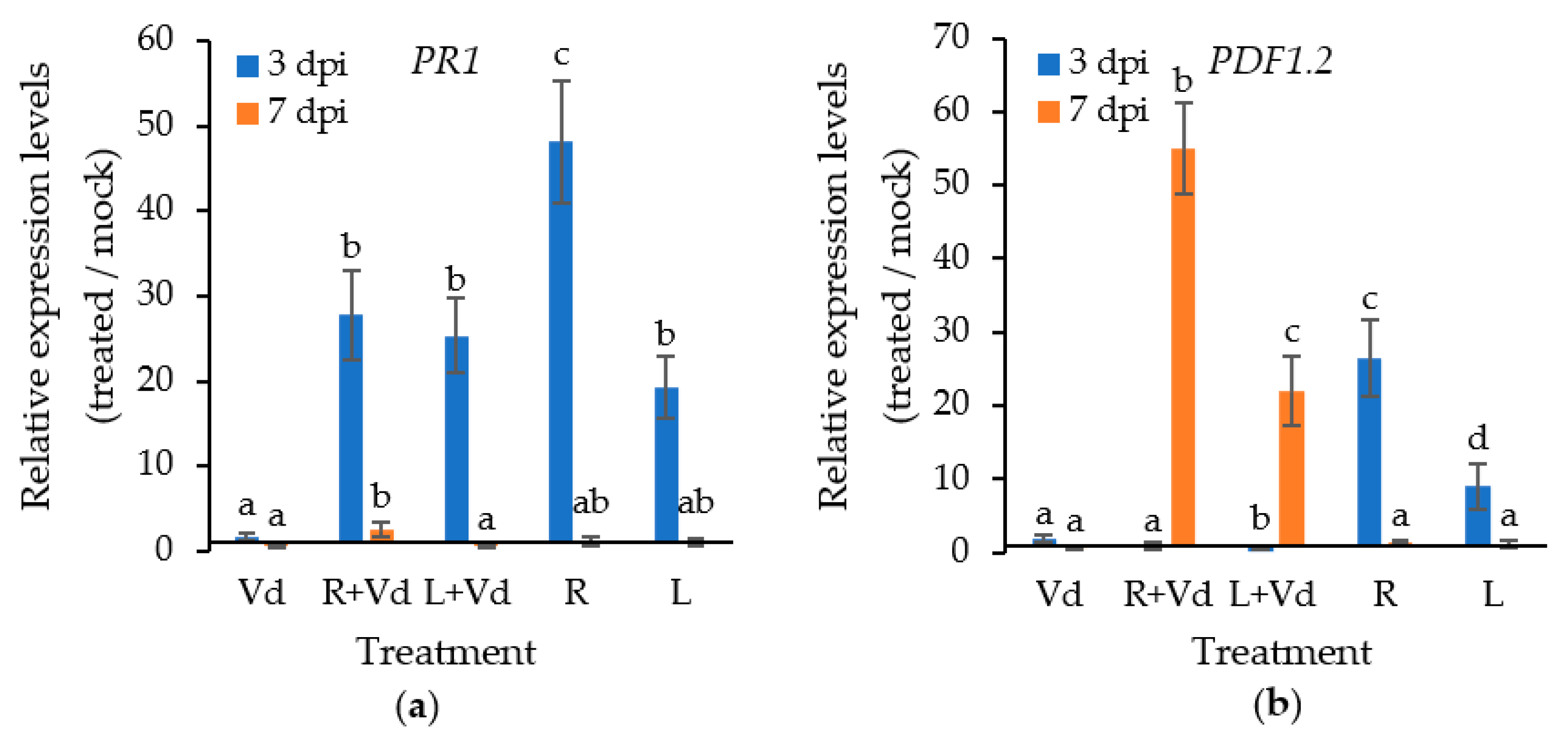
2.2. The Autoclaved Spores of V. dahliae Trigger the Plant Defence Mechanisms against V. dahliae

The plant pathogenicity experiments revealed that treating A. thaliana and eggplants with autoclaved spores of V. dahliae leads to a significant reduction in disease severity when compared to the control. Therefore, we wanted to investigate whether certain defense-related genes were upregulated in the plant protective treatments. For this purpose, we investigated the expression levels of the plant defense genes PR1 and PDF1.2, marker genes of the SA and ET/JA dependent defenses, at 3 and 7 dpi.
The application of autoclaved spores of V. dahliae upregulated the expression of PR1 and PDF1.2 at 3 dpi; however, this was a transient phenomenon, since the expression of both genes became similar to the mock-inoculated samples at 7 dpi (Figure 4). Upon pathogen inoculation, the expression of PR1 and PDF1.2 followed opposite patterns, since the expression of PR1 was higher at 3 dpi than 7 dpi, while the expression of PDF1.2 was higher at 7 dpi than at 3 dpi in the plants treated with the autoclaved spores of V. dahliae, irrespective of the application method. The expression of PR1 was upregulated by 22 folds in the plants treated with the autoclaved V. dahliae spores compared to mocks at 3 dpi, while PDF1.2 was upregulated by 22- and 55-fold in the leaf-injected and root-drenched plants with the autoclaved spores of V. dahlia at 7 dpi, respectively. In the control treatment (Vd), the expression of PR1 and PDF1.2 was upregulated at 3 dpi by 1.7- and 2-fold when compared to the mock-inoculated plants, respectively, and downregulated at 7 dpi.

2.3. The Plant Protective Activity of the Autoclaved V. dahliae Spores Depends on CERK1

Having observed the plant protective activity of the autoclaved spores of V. dahliae and the upregulation of defense-related genes in these treatments upon V. dahliae infection, we investigated whether the chitin receptor CERK1 is involved in the suggested PAMP-triggered immunity against V. dahliae. For this purpose, we investigated the CERK1 levels in wild-type A. thaliana treated with autoclaved spores of V. dahliae upon V. dahliae infection at 3 and 7 dpi and the plant protective activity of the autoclaved spores of V. dahliae in the A. thaliana mutants cerk1.
Indeed, in the case of the root-drenched autoclaved spores of V. dahliae, the expression of CERK1 was upregulated by 15- and 6-fold when compared to the mock treatment at 3 and 7 dpi, respectively (Figure 5). The application of the autoclaved spores of V. dahliae in the leaves resulted in the marginal upregulation of CERK1 upon V. dahliae infection, similar to the control treatment (V. dahliae) at 3 dpi. On the contrary, the expression levels of CERK1 were not upregulated in the absence of infection. The importance of CERK1 in the plant protective activity of the autoclaved spores of V. dahliae was further examined in cerk1.
Such as in the previous pathogenicity experiments, the initial Verticillium symptoms in cerk1 were wilting, followed by leaf yellowing and necrosis. Symptom recording started at 10 days post inoculation (dpi) and lasted until 25 dpi (Figure 6). In contrast to the wild-type A. thaliana, the application of the autoclaved spores of V. dahliae in the cerk1 plants did not result in reduced diseased severity in comparison to the control. In agreement with the pathogenicity experiments, the qPCR analysis revealed that the endophytic V. dahliae DNA levels were similar across the different treatments (Figure 6).

3. Discussion

Verticillium wilt, mainly caused by V. dahliae, is a devastating disease throughout the world, causing significant losses in plant yield every year. The lack of chemical control and the scarcity of plant resistance genes has boosted research efforts to identify novel disease management strategies [14]. Our results showed that the application of autoclaved spores of V. dahliae in A. thaliana and eggplants resulted in a significant reduction of Verticillium symptoms when compared to controls. According to our best knowledge, this is the first report showing the plant protective activity of necrotized spores of a pathogen against its active live form. This disease control approach resembles the vaccination effects in mammals, where inactive forms of a pathogen are used to promote immunity.
As a concept, plant vaccination is gaining increasing grounds over the last decades, mainly using natural or synthetic molecules that stimulate plant immunity [17]. These molecules are recognized by plants as either damage-associated molecular patterns (DAMPs) or PAMPs [18]. In our case, the molecules of the autoclaved V. dahliae spores fell into the second category, PAMPs, with the most prominent and well-studied molecule to be chitin. Our results suggest that eggplants and A. thaliana perceived the V. dahliae autoclaved spores as PAMPs and elicited their immune responses as a priming effect. Primed plants display timely and robust triggering of their defense responses upon biotic or abiotic stress [7,8,9,10]. Indeed, the treatment of A. thaliana plants with the autoclaved V. dahliae spores resulted in the upregulation of the examined defense genes PR1 and PDF1.2 upon pathogen infection.
The plant defense mechanism comprises of two main branches, depending on SA and ET/JA. The SA dependent defense mechanism is active against biotrophic pathogens, while the ET/JA defense branch is triggered against necrotrophs [19]. V. dahliae is considered as a hemibiotroph, having a biotrophic life style at the early infection stages, so as to establish itself in the xylem vessels, then switches to necrotrophism, producing various compounds toxic to the plant, leading to plant death [20,21]. This dual life style was anticipated in the expression of the defense genes PR1 and PDF1.2 in the pathogen inoculated A. thaliana that have been treated with the V. dahliae autoclaved spores, since PR1 expression was higher at 3 dpi than 7 dpi while the expression of PDF1.2 was higher at 7 dpi than 3 dpi. The opposite expression patterns between PR1 and PDF1.2 also anticipates the existing antagonism between the SA and the ET/JA defense mechanisms [19]. This antagonism protects plants from a fitness cost caused by the allocation of metabolic resources toward defense that would otherwise have been allocated to growth and reproduction [19]. Therefore, the early upregulation of PR1 may be crucial for plant defense against the biotrophic phase of the pathogen, while the later upregulation of PDF1.2 may target the necrotrophic phase. In the past, Thaler et al. (2004) [20] suggested a critical role of the JA-dependent defenses against V. dahliae, since the jasmonate-deficient tomato mutant plants, def1, were more susceptible than the wild type. Furthermore, exogenous application of SA and ectopic activation of the JA pathway provided am increased resistance to A. thaliana against the hemibiotrophic vascular pathogen Fusarium oxysporum, which has a similar disease pattern to V. dahliae [22].
A main component of the fungal cell wall is chitin. The plants, having evolutionarily anticipated the presence of chitin in their potential fungal enemies, were equipped with the plasma membrane CERK1 receptor, which binds chitin in association with LYK5 in A. thaliana [4,5,6]. In our experiments, the plant protective activity of the V. dahliae autoclaved spores was lost in the cerk1 mutants, suggesting the importance of the CERK1 receptor in the interaction of plants with the V. dahliae autoclaved spores and the concomitant triggering of plant defenses. In other studies, the treatment of plants or their cell cultures with chitin oligomers elicited various defense responses, such as synthesis of PRs and phytoalexins [23,24]. Interestingly, after being identified as a chitin receptor, CERK1 was also identified as a receptor for linear 1,3-b-D-glucans, a major component of the fungal and oomycete cell wall. CERK1 was also identified as having a role in the perception of bacterial peptidoglycans in Arabidopsis [25]. Since chitin represents a smaller proportion of the fungal cell walls than glucans [25], glucans may have played a stronger role than chitin in the observed plant protection against V. dahliae. Overall, the potential PAMPs sensed by eggplants and A. thaliana are chitin and glucans. An interesting decoupling was observed between the two application methods of the autoclaved V. dahliae spores, since the expression of CERK1 was upregulated in the plants that have been root drenched by the V. dahliae autoclaved spores upon pathogen infection, while the leaf infiltration application did not result in the upregulation of CERK1. A possible explanation is the occurrence of higher ligand availability in the case of root drenching when compared to leaf infiltration, since 10 milliliters of V. dahliae autoclaved spores were applied per plant in the root-drenching application, compared to 20 microliters in the case of leaf infiltration. Nevertheless, the plant protection levels were statistically identical between the two treatments. Therefore, the expression levels of CERK1 did not influence the observed plant protection, suggesting the need for a functional CERK1 rather than a multicopy CERK1 in the triggering of immune responses against V. dahliae.
In conclusion, our results show the potential of necrotized spores of a pathogen to protect plants against its active form. It remains an open question for further studies whether the use of pure chitin, glucan, or other PAMPs could offer a higher level of protection against V. dahliae when compared to the autoclaved spores. However, we can suggest that the use of the V. dahliae propagules may offer an array of PAMPs that trigger and more effectively shape the defense mechanisms in a larger group of plant species than a single PAMP. Since it is known that some PAMPs are perceived by a limited number of plant species while others activate defense responses in a broad range of species [26], the use of autoclaved spores may offer protection against a large group of plant species. Overall, this study constitutes the first described attempt in the scientific literature to control V. dahliae through plant vaccination using its own propagating structures. The results suggest the involvement of the CERK1 receptor and the concomitant triggering of the plant defense genes PR1 and PDF1.2 in a manner that resembles the biotrophic and necrotrophic disease pattern of V. dahliae.

4. Materials and Methods

4.1. Verticillium dahliae Culture

A V. dahliae isolate, originated either from infected Raphanus sativus L. or Solanum melongena L. were used in the A. thaliana and eggplant bioassays, respectively [27,28]. A suspension of 107 V. dahliae conidia/mL of distilled sterile water was prepared from a culture grown for 5 days at 24 °C in a sucrose sodium nitrate liquid medium [29].

4.2. Seeds Origin and Plant Growth Conditions

Wild-type A. thaliana Col-0 and the cerk1-2 mutant were provided by J. P. Métraux (University of Fribourg, Switzerland). For the experiments, A. thaliana and eggplant were grown separately in pots (9 × 9 × 10 cm) containing pasteurized soil. The plant growth conditions were: 25 °C, 16 h photoperiod, and 60–70% relative humidity.

4.3. Pathogenicity Experiments

Twenty-day-old wild-type A. thaliana, Col-0 plants were either root drenched with 10 mL or leaf injected with 10 μL of 107 autoclaved (121 °C, 1.2 atm, 15 min) V. dahliae conidia mL−1 sterile distilled water per plant or sterile distilled water (mock treatments). After 3 days, the plants were root drenched with 10 mL of 107 V. dahliae conidia mL−1 sterile distilled water per plant or sterile distilled water (mocks, negative control) [11]. The experiment was repeated three times with 10 plants per treatment per replication (a total of 30 plants per treatment). Disease severity was calculated as a percentage of the leaves showing Verticillium symptoms over the total number of leaves of each plant. Subsequently, the relative AUDPC value, the percentage of the AUDPC value over the maximum possible AUDPDC for the experimental time period, was calculated for each plant [30]. The same methodology was followed for the pathogenicity experiments using the A. thaliana, Col-0, cerk1-2 mutants.
The evaluation of the plant protective activity of V. dahliae necrotized spores against V. dahliae on eggplants was performed by root drenching 10 mL or stem injecting 10 μL of 107 autoclaved (121 °C, 1.2 atm, 15 min) V. dahliae spores mL−1 sterile distilled water per plant and following the same methodology as it is described above.

4.4. DNA extraction and qPCR V. dahliae quantification

In the A. thaliana bioassays (wt and cerk1-2), the relative V. dahliae DNA endophytic levels of the different treatments were quantified at 25 dpi. Total DNA was isolated from the aerial plant parts of 10 plants per treatment, pooled to one sample and ground to fine powder using liquid nitrogen. The extraction of DNA was performed according to the methodology of Dellaporta et al. (1983) [31]. qPCRs were performed in an Applied Biosystems StepOnePlus thermocycler and for the amplification reactions, KAPA SYBR® FAST (Merck SA) was used. The primers used for real-time PCR were ITS1-F 5–AAAGTTTTAATGGTTCGCTAAGA–3 and ST-VE1-R 5–CTTGGTCATTTAGAGGAAGTAA–3 for the ITS1 and ITS2 regions of the 5.8S ribosomal RNA gene (Z29511) of V. dahliae. [11] and 5-GAGCTGAAGTGGCTTCCATGAC-3 and 5-GGTCCGACATACCCATGATCC-3 for the reference Arabidopsis gene At4g26410 [32]. PCR efficiency for each amplicon was calculated by employing the linear regression method on log (fluorescence) per cycle number data, using the Lin-Reg PCR software [33]. For each treatment, three biological repeats and three technical repeats per biological repeat were conducted. The absence of nonspecific products and primer dimers was confirmed by analysis of the melting curves. The relative V. dahliae DNA quantity, calculated by using the formula 2−∆Ct, was expressed as a percentage compared to the wild type; while the value (2−∆Ct) of the wild type was set to 100% [11].

4.5. Determination of PR1, PDF1.2 and CERK1 transcript levels

For RNA analysis, the aerial parts of five A. thaliana wt plants from each treatment and experimental replication (a total of 3 replications) were collected and pooled to one sample at 3 and 7 dpi. The pooled tissues were ground with liquid nitrogen and total RNA was extracted by using TRIzol® Reagent (Invitrogen, Paisley, Renfrewshire, UK), according to the manufacturer’s instructions. The RNA samples were treated with DNase I (Macherey-Nagel) to eliminate traces of contaminating genomic DNA. First-strand cDNA was synthesized using PrimeScript One Step RT-PCR Kit (Takara) following the manufacturer’s procedure. qPCRs were performed in an Applied Biosystems StepOnePlus thermocycler and for the amplification reactions, KAPA SYBR® FAST (Merck SA) was used. The primers used for real-time PCR were CERK1 (AT3G21630; 5-CAAATCAAGAGATGGTGTTGGTGC-3 and 5- CACCACCCAAACCTCCACT-3) [34], PR1 (AT2G14610; 5-GTGCCAAAGTGAGGTGTAACAA-3 and 5-CGTGTGTATGCATGATCACATC-3) and PDF1.2 (AT5G44420; 5-TTTGCTGCTTTCGACGCAC-3 and 5-CGCAAACCCCTGACCATG-3) [35]. PCR efficiency for each amplicon was calculated by employing the linear regression method on log (fluorescence) per cycle number data, using the Lin-Reg PCR software [33]. For each treatment, three biological repeats and three technical repeats per biological repeat were conducted per sampling time point (3 and 7 dpi). The absence of nonspecific products and primer dimers was confirmed by analysis of the melting curves. The relative PR1, PDF1.2, and CERK1 expression levels (2−∆Ct) in the treated plants were expressed relative to the normalized transcript levels of PR1, PDF1.2 and CERK1 in the mock-inoculated plants (negative control).

4.6. Statistics

Data on disease severity, relative AUDPC, relative V. dahliae DNA levels, and gene expression were subjected to analyses of variance. When a significant (p < 0.05) F-test was obtained for the treatments, data were analyzed by LSD multiple range test (p < 0.05).

Supplementary Materials

The following supporting information can be downloaded at: https://www.mdpi.com/article/10.3390/plants11131691/s1, Figure S1: Verticillium wilt symptoms caused on stem-injected (a) or root-drenched (b) eggplants with autoclaved spores of V. dahliae, controls (V. dahliae inoculated) (c) and mock-inoculated plants (d). The photograph was taken at 25 dpi.

Author Contributions

Conceptualization, E.G.P. and S.E.T.; data curation, E.G.P. and S.E.T.; formal analysis, E.G.P.; investigation, E.G.P., M.F.T. and M.M.; methodology, E.G.P., M.F.T. and M.M.; project administration, S.E.T.; resources, S.E.T.; supervision, S.E.T.; validation, E.G.P.; visualization, E.G.P. and S.E.T.; writing—original draft, S.E.T.; writing—review and editing, S.E.T. All authors have read and agreed to the published version of the manuscript.

Funding

This research received no external funding.

Institutional Review Board Statement

Not applicable.

Informed Consent Statement

Not applicable.

Data Availability Statement

The data presented in this study are available on request from the corresponding author.

Conflicts of Interest

The authors declare no conflict of interest.

References

  1. Jones, J.D.; Dangl, J.L. The plant immune system. Nature 2006, 444, 323–329. [Google Scholar] [CrossRef] [Green Version]
  2. Sánchez-Vallet, A.; Mesters, J.R.; Thomma, B.P. The battle for chitin recognition in plant-microbe interactions. FEMS Microbiol. Rev. 2015, 39, 171–183. [Google Scholar] [CrossRef] [Green Version]
  3. Pusztahelyi, T. Chitin and chitin-related compounds in plant-fungal interactions. Mycology 2018, 9, 189–201. [Google Scholar] [CrossRef]
  4. Petutschnig, E.K.; Jones, A.M.; Serazetdinova, L.; Lipka, U.; Lipka, V. The lysin motif receptor-like kinase (LysM-RLK) CERK1 is a major chitin-binding protein in Arabidopsis thaliana and subject to chitin-induced phosphorylation. J. Biol. Chem. 2010, 285, 28902–28911. [Google Scholar] [CrossRef] [Green Version]
  5. Miya, A.; Albert, P.; Shinya, T.; Desaki, Y.; Ichimura, K.; Shirasu, K.; Narusaka, Y.; Kawakami, N.; Kaku, H.; Shibuya, N. CERK1, a LysM receptor kinase, is essential for chitin elicitor signaling in Arabidopsis. Proc. Natl. Acad. Sci. USA 2007, 104, 19613–19618. [Google Scholar] [CrossRef] [Green Version]
  6. Liu, T.; Liu, Z.; Song, C.; Hu, Y.; Han, Z.; She, J.; Fan, F.; Wang, J.; Jin, C.; Chang, J.; et al. Chitin-induced dimerization activates a plant immune receptor. Science 2012, 336, 1160–1164. [Google Scholar] [CrossRef] [Green Version]
  7. Conrath, U.; Beckers, G.J.M.; Flors, V.; García-Agustín, P.; Jakab, G.; Mauch, F.; Newman, M.-A.; Pieterse, C.M.J.; Poinssot, B.; Pozo, M.J.; et al. Priming: Getting ready for battle. Mol. Plant Microbe Interact. 2006, 19, 1062–1071. [Google Scholar] [CrossRef] [Green Version]
  8. Hilker, M.; Schwachtje, J.; Baier, M.; Balazadeh, S.; Bäurle, I.; Geiselhardt, S.; Hincha, D.K.; Kunze, R.; Mueller-Roeber, B.; Rillig, M.C.; et al. Priming and memory of stress responses in organisms lacking a nervous system. Biol. Rev. 2016, 91, 1118–1133. [Google Scholar] [CrossRef]
  9. Conrath, U.; Beckers, G.J.; Langenbach, C.J.; Jaskiewicz, M.R. Priming for enhanced defense. Annu. Rev. Phytopathol. 2015, 53, 97–119. [Google Scholar] [CrossRef]
  10. Martinez-Medina, A.; Flors, V.; Heil, M.; Mauch-Mani, B.; Pieterse, C.M.J.; Pozo, M.J.; Ton, J.; van Dam, N.M.; Conrath, U. Recognizing plant defense priming. Trends Plant Sci. 2016, 21, 818–822. [Google Scholar] [CrossRef] [Green Version]
  11. Gkizi, D.; Lehmann, S.; L’Haridon, F.; Serrano, M.; Paplomatas, E.J.; Métraux, J.P.; Tjamos, S.E. The innate immune signaling system as a regulator of disease resistance and ISR activity against Verticillium dahliae. Mol. Plant Microbe Interact. 2016, 29, 313–323. [Google Scholar] [CrossRef] [PubMed] [Green Version]
  12. Pegg, G.F.; Brady, B.L. Verticillium Wilts; CABI Publishing: New York, NY, USA, 2002. [Google Scholar]
  13. Schnathorst, W.C. Life cycle and epidemiology of Verticillium. In Fungal Wilt Diseases of Plants; Mace, M.A., Bell, A.A., Beckman, C.H., Eds.; Academic Press: New York, NY, USA, 1981; pp. 81–111. [Google Scholar]
  14. Klosterman, S.J.; Atallah, Z.K.; Vallad, G.E.; Subbarao, K.V. Diversity, pathogenicity, and management of Verticillium species. Annu. Rev. Phytopathol. 2009, 47, 39–62. [Google Scholar] [CrossRef] [PubMed] [Green Version]
  15. Spoel, S.H.; Johnson, J.S.; Dong, X. Regulation of tradeoffs between plant defenses against pathogens with different lifestyles. Proc. Natl. Acad. Sci. USA 2007, 104, 18842–18847. [Google Scholar] [CrossRef] [PubMed] [Green Version]
  16. Gimenez-Ibanez, S.; Hann, D.R.; Ntoukakis, V.; Petutschnig, E.; Lipka, V.; Rathjen, J.P. AvrPtoB targets the LysM receptor kinase CERK1 to promote bacterial virulence on plants. Curr. Biol. 2009, 19, 423–429. [Google Scholar] [CrossRef] [Green Version]
  17. Bektas, Y.; Eulgem, T. Synthetic plant defense elicitors. Front. Plant Sci. 2015, 5, 804. [Google Scholar] [CrossRef]
  18. Quintana-Rodriguez, E.; Duran-Flores, D.; Heil, M.; Camacho-Coronel, X. Damage-associated molecular patterns (DAMPs) as future plant vaccines that protect crops from pests. Sci. Hortic. 2018, 237, 207–220. [Google Scholar] [CrossRef]
  19. Glazebrook, J. Contrasting mechanisms of defense against biotrophic and necrotrophic pathogens. Annu. Rev. Phytopathol. 2005, 43, 205–227. [Google Scholar] [CrossRef]
  20. Thaler, J.S.; Owen, B.; Higgins, V.J. The role of the JA response in plant susceptibility to diverse pathogens with a range of lifestyles. Plant Physiol. 2004, 135, 530–538. [Google Scholar] [CrossRef] [Green Version]
  21. Veronese, P.; Narasimhan, M.L.; Stevenson, R.A.; Zhu, J.K.; Weller, S.C. Identification of a locus controlling Verticillium disease symptom response in Arabidopsis thaliana. Plant J. 2003, 35, 574–587. [Google Scholar] [CrossRef]
  22. Edgar, C.I.; McGrath, K.C.; Dombrecht, B.; Manners, J.M.; Maclean, D.C.; Schenk, P.M.; Kazan, K. Salicylic acid mediates resistance to the vascular wilt pathogen Fusarium oxysporum in the model host Arabidopsis thaliana. Australas. Plant Pathol. 2006, 35, 581–591. [Google Scholar] [CrossRef]
  23. Shibuya, N.; Minami, E. Oligosaccharide signaling for defence responses in plant. Physiol. Mol. Plant Pathol. 2001, 59, 223–233. [Google Scholar] [CrossRef] [Green Version]
  24. Stacey, G.; Shibuya, N. Chitin recognition in rice and legumes. Plant Soil 1997, 194, 161–169. [Google Scholar] [CrossRef]
  25. Willmann, R.; Lajunen, H.M.; Erbs, G.; Newman, M.A.; Kolb, D.; Tsuda, K.; Katagiri, F.; Fliegmann, J.; Bono, J.J.; Cullimore, J.V.; et al. Arabidopsis lysin-motif proteins LYM1 LYM3 CERK1 mediate bacterial peptidoglycan sensing and immunity to bacterial infection. Proc. Natl. Acad. Sci. USA 2011, 108, 19824–19829. [Google Scholar] [CrossRef] [Green Version]
  26. Zipfel, C.; Kunze, G.; Chinchilla, D.; Caniard, A.; Jones, J.D.G.; Boller, T.; Felix, G. Perception of the Bacterial PAMP EF-Tu by the receptor EFR restricts Agrobacterium-mediated transformation. Cell 2006, 125, 749–760. [Google Scholar] [CrossRef]
  27. Tjamos, S.E.; Flemetakis, E.; Paplomatas, E.J.; Katinakis, P. Induction of resistance to Verticillium dahliae in Arabidopsis thaliana by the biocontrol agent K-165 and pathogenesis-related proteins gene expression. Mol. Plant Microbe Interact. 2005, 18, 555–561. [Google Scholar] [CrossRef] [PubMed] [Green Version]
  28. Tjamos, E.C.; Tsitsigiannis, D.I.; Tjamos, S.E.; Antoniou, P.P.; Katinakis, P. Selection and screening of endorhizosphere bacteria from solarised soils as biocontrol agents against Verticillium dahliae of solanaceous hosts. Eur. J. Plant Pathol. 2004, 110, 35–44. [Google Scholar] [CrossRef]
  29. Sinha, A.K.; Wood, R.K.S. Studies on the nature of resistance in tomato plants to Verticillium albo-atrum. Ann. Appl. Biol. 1968, 62, 319–327. [Google Scholar] [CrossRef]
  30. Campbell, C.L.; Madden, L.V. Introduction to Plant Disease Epidemiology; Wiley: New York, NY, USA, 1990. [Google Scholar]
  31. Dellaporta, S.L.; Wood, J.; Hicks, J.B. A plant DNA minipreparation, version II. Plant Mol. Biol. Rep. 1983, 1, 19–21. [Google Scholar] [CrossRef]
  32. Czechowski, T.; Stitt, M.; Altmann, T.; Udvardi, M.K.; Scheible, W.R. Genome wide identification and testing of superior reference genes for transcript normalization in Arabidopsis. Plant Physiol. 2005, 139, 5–17. [Google Scholar] [CrossRef] [Green Version]
  33. Ramakers, C.; Ruijter, J.M.; Deprez, R.H.; Moorman, A.F. Assumption free analysis of quantitative real-time polymerase chain reaction (PCR) data. Neurosci. Lett. 2003, 339, 62–66. [Google Scholar] [CrossRef]
  34. Huang, C.; Yan, Y.; Zhao, H.; Ye, Y.; Cao, Y. Arabidopsis CPK5 Phosphorylates the Chitin Receptor LYK5 to Regulate Plant Innate Immunity. Front. Plant Sci. 2020, 11, 702. [Google Scholar] [CrossRef] [PubMed]
  35. Pantelides, I.S.; Tjamos, S.E.; Paplomatas, E.J. Ethylene perception via ETR1 is required in Arabidopsis infection by Verticillium dahliae. Mol. Plant Pathol. 2010, 11, 191–202. [Google Scholar] [CrossRef] [PubMed]
Figure 1. Disease severity (a) and relative AUDPC (b) caused by artificial inoculation of V. dahliae (Vd), on Arabidopsis thaliana leaf injected (L + Vd) or root drenched (R + Vd) with autoclaved spores of V. dahliae or mock inoculated (positive control, Vd). Columns represent means of three replications per treatment (n = 30 plants, ± SE). Different letters denote statistically significant differences between treatments (p < 0.05) according to LSD test. For disease severity, statistics were performed at each time point.
Plants 11 01691 g001
Figure 2. Relative quantification of the V. dahliae DNA levels in wild-type A. thaliana leaf injected (L + Vd) or root drenched (R + Vd) with autoclaved spores of V. dahliae or mock inoculated (positive control, Vd) and inoculated after 3 days with V. dahliae. The relative V. dahliae DNA levels were estimated by qPCR using total DNA isolated from the aerial parts of plants at 25 days post-inoculation. Columns represent means of three biological and technical replications per treatment (n = 9, ± SE) at 25 dpi (end of pathogenicity experiment). Different letters denote statistically significant differences between treatments (p < 0.05) according to LSD test.
Plants 11 01691 g002
Figure 3. Verticillium disease severity (a) and relative AUDPC (b) caused by artificial inoculation of V. dahliae (Vd), on eggplants stem injected (S + Vd) or root drenched (R + Vd) with autoclaved spores of V. dahliae or mock inoculated (positive control, Vd). Columns represent means of three replications per treatment (n = 30 plants, ± SE). Different letters denote statistically significant differences between treatments (p < 0.05) according to LSD test. For disease severity, statistics were performed at each time point.
Plants 11 01691 g003
Figure 4. Relative transcript levels of PR1 (a) and PDF1.2 (b) in the aerial plant parts of A. thaliana leaf injected (L + Vd) or root drenched (R + Vd) with autoclaved spores of V. dahliae or mock inoculated (positive control, Vd) in response to infection with V. dahliae at 3 and 7 dpi. The samples for RNA isolation were collected at 3 and 7 days post inoculation (dpi) with the pathogen. The expression levels of PR1 and PDF1.2 were normalized to the expression of the reference gene At4g26410 measured in the same samples and they are presented in comparison to the normalized expression level of the respective gene in the mock treatment. Columns represent means of three biological and technical replications per treatment (n = 9, ± SE). At each day, different letters denote statistically significant differences between treatments (p < 0.05) according to LSD test.
Plants 11 01691 g004
Figure 5. Relative transcript levels of CERK1 in the aerial plant parts of A. thaliana leaf injected (L + Vd) or root drenched (R + Vd) with autoclaved spores of V. dahliae or mock inoculated (positive control, Vd) in response to infection with V. dahliae. The samples for RNA isolation were collected at 3 and 7 days post inoculation (dpi) with the pathogen. The expression levels of CERK1 were normalized to the expression of the reference gene At4g26410 measured in the same samples and they are presented in comparison with the normalized expression level of the respective gene in the mock treatment. Columns represent means of three biological and technical replications per treatment (n = 9, ± SE). At each day, different letters denote statistically significant differences between treatments (p < 0.05) according to LSD test.
Plants 11 01691 g005
Figure 6. Disease severity and relative quantification of the V. dahliae DNA levels in A. thaliana, cerk1, leaf injected (L + Vd) or root drenched (R + Vd) with autoclaved spores of V. dahliae or mock inoculated (positive control, Vd) and inoculated after 3 days with V. dahliae. Fungal DNA levels were estimated at 25 dpi. In (a), columns represent means of three replications per treatment (n = 30 plants, ± SE) and in (b) they represent means of three biological and technical replications per treatment (n = 9, ± SE) at 25 dpi (end of pathogenicity experiment). In (b), the control treatment (Vd) is set to 100%. Different letters denote statistically significant differences between treatments (p < 0.05) according to LSD test. For disease severity, statistics were performed at each time point.
Plants 11 01691 g006
Publisher’s Note: MDPI stays neutral with regard to jurisdictional claims in published maps and institutional affiliations.

