Article

Anther Culture-Derived Haploids of Citrus aurantium L. (Sour Orange) and Genetic Verification of Haploid-Derived Regenerated Plants

Citrus Research Institute, National Institute of Horticultural & Herbal Science, RDA, Jeju 63613, Korea
*
Author to whom correspondence should be addressed.
Plants 2022, 11(22), 3022; https://doi.org/10.3390/plants11223022
Submission received: 4 October 2022 / Revised: 31 October 2022 / Accepted: 6 November 2022 / Published: 9 November 2022
(This article belongs to the Section Plant Development and Morphogenesis)

Abstract

:
Citrus plants are important fruit tree species; however, the breeding of high-quality varieties of citrus species is a time-consuming process. Using haploid-derived plants from anther culture may reduce the time required for obtaining purebred lines. This study aimed to genetically verify whether anther culture-derived sour orange (Citrus aurantium L.) plants developed from somatic embryos or haploid tissues. Sour orange anthers were cultured in N6 and MS media to induce calli and somatic embryos. N6 liquid medium supplemented with 1 mg·L−1 gibberellic acid and 200 µM spermidine resulted in a 10% increase in callus and embryo induction rates. Regenerated plants were validated using simple sequence repeat markers. Out of the 109 regenerated plants, ploidy analysis identified 99 diploids, two haploids, and eight putative aneuploids; out of the 99 diploid plants, 33 were haploid-derived homozygous diploids. The chromosomal analysis confirmed most plants as diploids, whereas some were identified as aneuploids (19–21 chromosomes). Furthermore, phylogenetic analysis confirmed that the resultant homozygous or heterozygous plants were haploid-derived. This is the first report of haploid-derived homozygous diploid and aneuploid sour orange plants obtained through anther culture. Moreover, the anther cultivation technique described herein can be applied to other citrus varieties.

1. Introduction

Citrus plants belong to the family Rutaceae, which includes some of the most economically important and globally widespread fruit tree crops [1,2,3,4]. In terms of the cultivated area and production of domestic fruit trees, citrus fruits rank third (after apples and pears) and are in high supply and demand [5]. Although sour orange (Citrus aurantium L.) propagates through seeds, it is mainly cultivated using rootstocks [6,7]. Recent studies have shown that this species has various pharmacological properties, including anti-cancer, anti-anxiety, anti-obesity, antibacterial, antioxidant, insecticidal, and antidiabetic effects [8,9]. However, sour orange is highly susceptible to infection by citrus tristeza virus [6,10]. Furthermore, sour oranges produce numerous seeds, which is a major disadvantage in terms of its market value [11]. Despite these drawbacks, it has high utility as it can be effectively used as a tissue culture model [12].
Generating superior lines of citrus cultivars using conventional breeding methods, such as crossing, is time-consuming and labor-intensive owing to their high heterozygosity, long juvenility, self-incompatibility, and polyploidy [1,13,14,15,16,17]. Furthermore, breeding and selection of purebred lines are time-consuming processes [18]. However, compared to conventional breeding, anther culture facilitates the generation of haploid plants in a shorter period of time. This allows the production of completely homozygous lines from heterozygous parents in a single generation [15,19]. This technique also permits the immediate confirmation of recessive traits and phenotypes (for example, morphological features such as dwarfism) since haploid plants are generally smaller and exhibit significantly less vigor than their parents [18,20,21].
Haploid plants cannot undergo normal meiosis, which hinders generational progress [18,19]. In contrast, double haploids (homozygous diploids) obtained through natural or artificial treatments undergo normal meiosis, thus allowing generational progress [18]. Consequently, homozygous individuals obtained from haploids have important applications in crop breeding, such as mutagenesis, transformation, genetic analysis, and gene sequencing [15,22,23]. Therefore, anther culture has a high potential for plant breeding and crop improvement [24,25], and has been studied in a wide range of plant species [26,27]. To date, citrus plants obtained using anther culture include Poncirus trifoliata (L.) Raf. (trifoliate orange), Citrus clementina Hort. ex Tan. (clementine), Citrus sinensis (L.) Osbeck (sweet orange), Citrus sinensis (L.) Osbeck (Valencia sweet orange), Citrus limon (L.) Burm. f. (lemon), and Clausena excavata [1,28,29,30,31,32]. However, systematic breeding of citrus varieties using a combination of different pure lineages is insufficient for obtaining more pure-lineage varieties. Therefore, it is necessary to develop a suitable anther culture technique that can be used to obtain more pure-lineage varieties in large numbers.
Aneuploidy is a type of chromosomal anomaly that involves an increase or decrease in the number of chromosomes [33] and is caused by the non-separation of chromosomes during meiosis and mitosis [34,35,36]. While aneuploidy causes serious disorders in animals [35,37], in plants, the known consequences include myriad phenotypic and structural changes, such as growth retardation [38,39,40,41]. Although aneuploidy occurs spontaneously in some plants [42], it has also been reported to occur through crossbreeding in many crops, such as tobacco, corn, guava, pear, apple, and citrus [43,44,45,46]. Moreover, anther culture has been used to produce aneuploid tobacco plants [47]. Among haploid-derived plants obtained from some maize lines and anthurium plants, the incidence of aneuploidy is reportedly up to 15% [48,49]. Aneuploid plants have also been generated in citrus cultivars through anther culture [30,34,50,51]. The frequent occurrence of aneuploid individuals—including haploids and diploids—in plants obtained through anther culture [49] requires further investigation.
In this study, an optimum medium for anther culture of sour orange was developed. This medium was used to culture citrus embryos to determine the efficiency of anther culture of sour orange plants. Furthermore, the ploidy status of haploid-derived plants was verified and the associated genetic changes were investigated. Additionally, chromosomal analysis was used to verify the presence of homodiploid and aneuploid plants, and phylogenetic analysis was used to confirm whether homozygous or heterozygous plants were haploid-derived.

2. Results

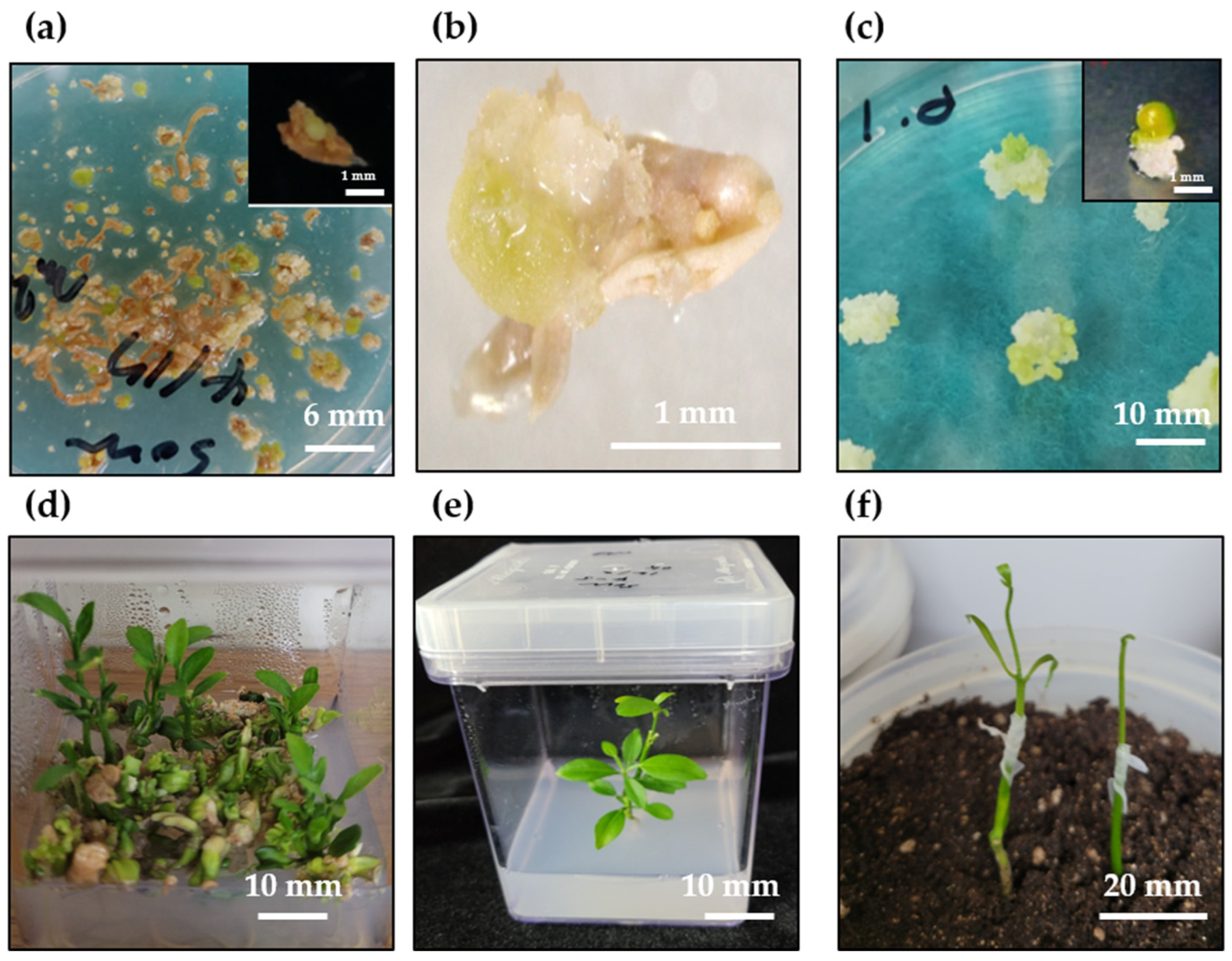
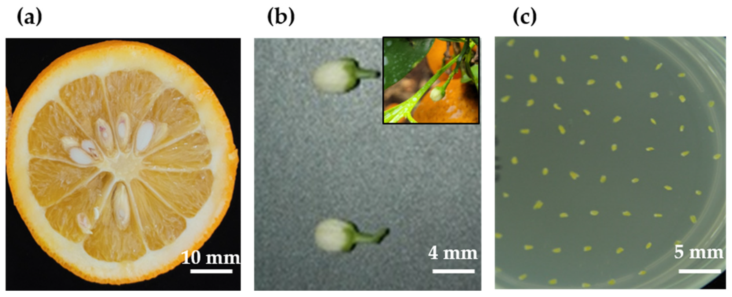
2.1. Plant Regeneration System

The anthers swelled within 1–2 weeks; a few callus cells and embryos formed in 3–4 weeks (Figure 1a,b). When N6 liquid medium was supplemented with 1 mg·L−1 GA3 and 200 µM spermidine was added to the anther culture medium, the rates of callus induction and embryo formation increased by approximately 10%; the original callus induction rate was 25% and that of embryo formation was approximately 2% without the addition of the liquid medium (Table S1). The obtained callus cells were transferred to a medium for somatic embryo induction to induce embryo formation (Figure 1c). After 6 weeks of culture, the induced somatic embryos were transferred to a plant regeneration medium, and normal plants were obtained at a rate of approximately 50–60% after 4–6 weeks of culture (Figure 1d). Normal plants with roots and shoots were transferred to Murashige and Skoog (MS) medium without hormones. The remaining abnormal embryos were transferred to MS medium containing 500 mg·L−1 malt extract, 50 g·L−1 sucrose, and 0.5 mg·L−1 GA3, which induced continuous plant growth from non-regenerated calli and embryos at a rate of approximately 70–80%. Normal plants were rapidly induced to proliferate and grow through in vitro and in vivo grafting (Figure 1e,f). Ultimately, more than 1000 regenerated plants were obtained using the anther culture method.

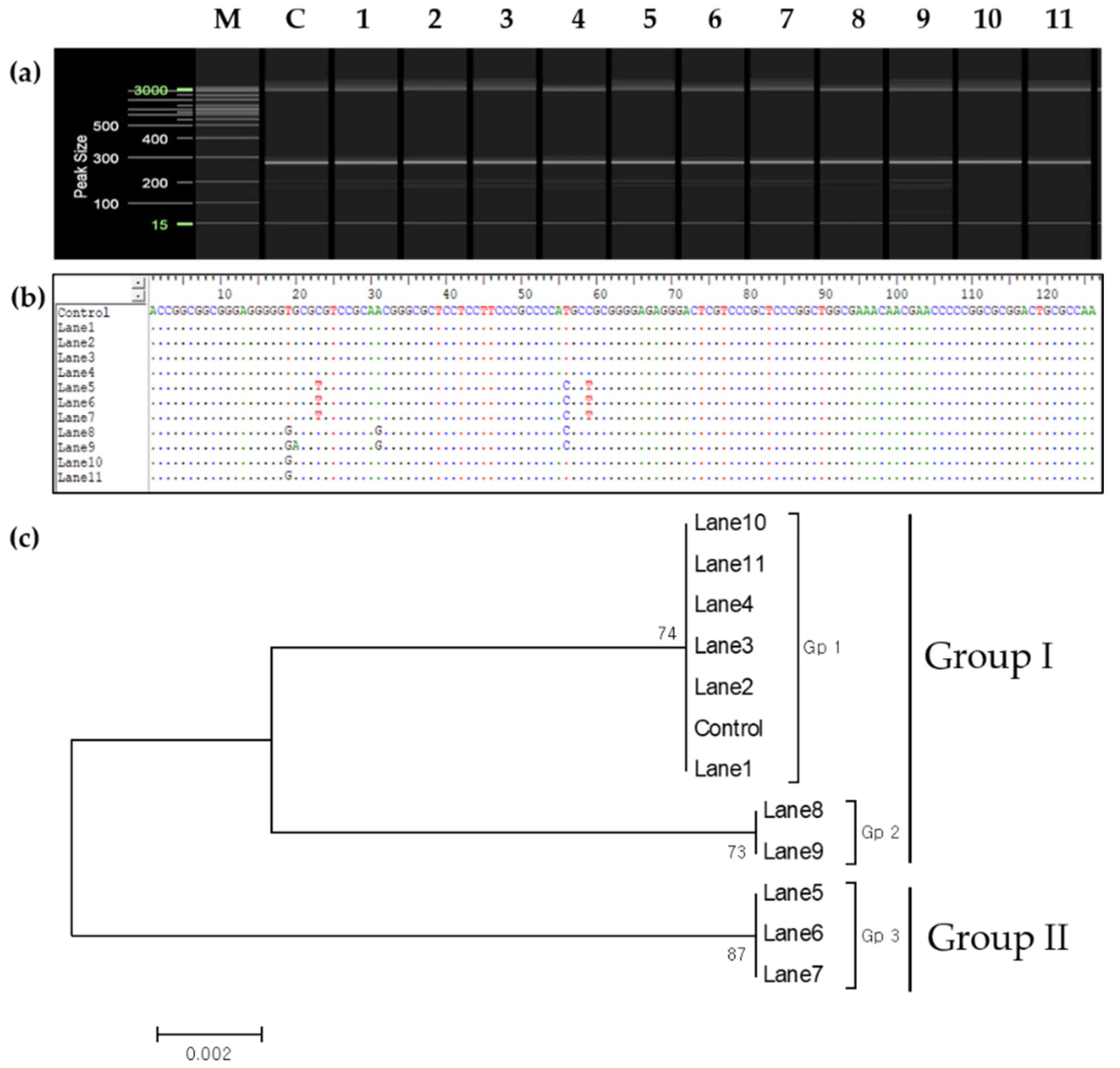
2.2. Genetic Verification

To confirm whether the plants obtained through anther culture were haploid-derived, eight simple sequence repeat (SSR) markers (CiSSR-P1, -P2, -43, -226, -246, -253, -254, and -260) out of eight combinations of heterozygous SSR markers were used. Out of >1000 plants obtained through anther culture, 271 were randomly selected for analysis using P1 and P2 markers (Table S2). Of the 271 plants, 11 were randomly selected for analysis using all eight SSR markers. Amplification using the SSR marker CiSSR-P1 showed four plants (lanes 1–4; Figure 2a) with similar amplification patterns (double band) as that in the control (donor plant). In contrast to the control, three of the seven putative haploid-derived plants (lanes 5–7) showed a single amplification product. The remaining four plants (lanes 8–11) did not show a single amplification product, but their amplification patterns differed from those of the control.
The SSR marker CiSSR-43 showed two amplification bands, similar to that shown by CiSSR-P1 in the control (Figure 2b). Putative heterozygous plants (lanes 1–4) also showed the same amplification pattern of bands as the control plants (Figure 2c). However, three of the seven putative haploid-derived plants (lanes 5–7) exhibited the same pattern as the upper amplification band of the control. Four other plants (lanes 8–11) had the same pattern as the lower amplification band of the control (Figure 2b). In four plants (lanes 1–4; Figure 2c,d) regenerated from anther culture, the SSR markers CiSSR-226 and CiSSR-246 showed the same amplification pattern (double band as in the control. Seven regenerated plants (lanes 5–11) that were putatively haploid-derived showed a single amplification product (Figure 2c,d). In the case of the CiSSR-246 marker, putative haploid plants (lanes 5–7) showed a pattern of double amplification bands, which differed from those of the control.
The SSR markers CiSSR-254 and CiSSR-260 showed two amplification bands, which was similar to the pattern exhibited by the CiSSR-P1 marker in the control (Figure 2e,f). Three of the seven putative haploid-derived plants (lanes 5–7) showed a single band pattern. However, the other four plants (lanes 8–11) had a different amplification pattern (double amplification bands) from that of the control (Figure 2e,f).
Unlike CiSSR-P1, the SSR markers CiSSR-P2 (Figure 3a) and CiSSR-253 (Figure 3b) showed three amplification bands in the control (Figure 3). Putative heterozygous plants (lanes 1–4) showed the same amplification band pattern as those of the control (Figure 3). However, putative haploid-derived plants (lanes 5–11) showed a single band pattern (Figure 3). The abovementioned results (Table S2) indicate that approximately 84% (228 plants among a total of 271) were heterozygous plants derived from the anther wall, and approximately 13% (35 plants among a total of 271) were haploid-derived homozygous; a few plants (approximately 3%; eight plants out of 271) were presumed as aneuploid plants (Table S2).

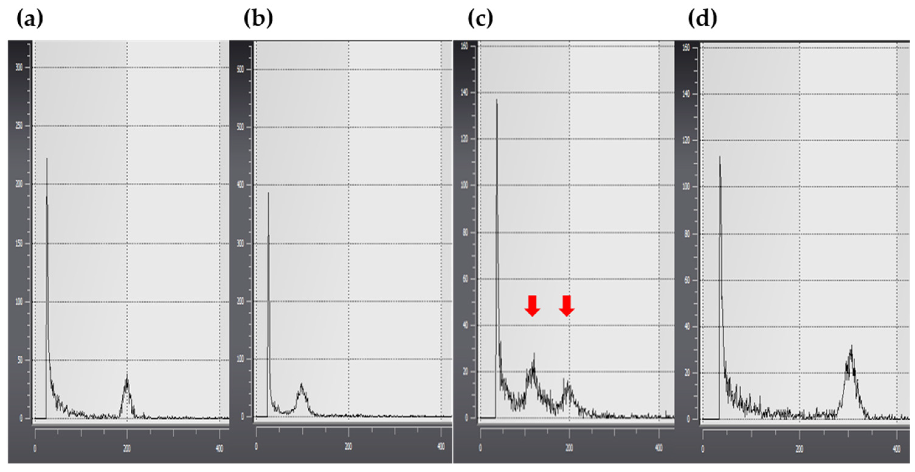
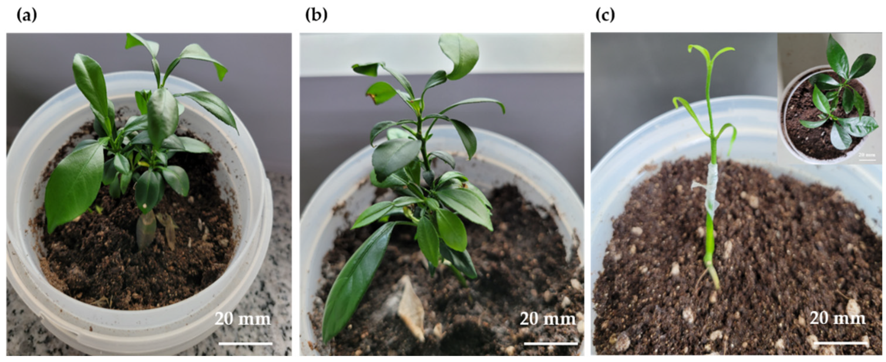
2.3. Polyploidy Verification and Morphological Selection

Polyploidy was investigated in 109 plants obtained through anther culture. These included 41 out of 43 (two specimens were lost due to contamination) genetically selected plants and 68 genetically validated plants (Figure 4 and Table S3).
Verification of ploidy in the 109 plants identified about 91% of plants as diploids (66 of the 109 plants were heterozygous diploids; 33 were haploid-derived homozygous diploids; Figure 4a); about 2% of the plants were identified as haploids (two plants; Figure 4b,c), and 7% were identified as putative aneuploids (eight plants; Figure 4d and Table S3).
Although most of the haploid-derived diploid plants (Figure 5b) were morphologically similar to the control plants (Figure 5a), morphological differences were observed in some of the plants (Figure 5c) in terms of leaf width, size, and growth rate. Moreover, the roots of putative haploid-derived plants appeared as thin threads; either the shoots failed to form, or the roots grew without shoot elongation (sample lost due to contamination).

2.4. Chromosomal Analysis

The number of chromosomes was analyzed in the root tips of 30 haploid-derived plants identified using SSR markers (Table S2). Most plants (22 of 30 plants) were identified as diploids with 18 chromosomes (Figure 6a and Table S4). In contrast, some plants (8 of 30 plants) that appeared to be either diploids or triploids were identified as putative aneuploids with 19–21 chromosomes (Figure 6b–d and Table S4).

2.5. Phylogenetic Analysis

The internal transcribed spacer (ITS) chromosomal regions were analyzed to determine the genetic relationship between the donor and regenerated plantlets and to establish whether the anther culture-derived plants were somatically mutated or haploid-derived. The results indicated a genetic difference in the chromosomal ITS region between plants identified using SSR markers and the control plants (Figure 7). Subsequently, genetic relationships between these plants were analyzed using nucleotide sequences in the ITS1 region to determine the zygosity of cultivars identified using SSR markers (Figure 8a,b). Phylogenetic analysis allowed the clustering of cultivars into two large groups and three small groups: Group 1 included Gp1 (Figure 8c: control, lanes 1–4, and lanes 10 and 11) and Gp2 (Figure 8c: lanes 8 and 9), and Group 2 included Gp3 (Figure 8c: lanes 5–7).

3. Discussion

This study investigated embryo formation and callus induction in sour oranges using anther culture to obtain haploid and double-haploid plants. The production of haploid- derived plants through anther culture is more challenging in citrus cultivars than in herbaceous species [1] because Citrus plants are characterized by long juvenility [14]. Successful production of haploid plants using anther culture has primarily been reported in grain species (especially Cruciferae) and some Solanaceae species [52,53,54,55]. However, recent studies have reported successful embryo induction and plant regeneration through anther culture in Citrus species [1,16,17,56]. In addition, the generation of haploid (homozygous diploid) and triploid plants has also been reported [16,17,34,57]. Among Citrus species, sour orange varieties are widely used as rootstocks for industrial use and tissue culture. The rootstocks also serve as useful models for anther culture [6,27]. However, there are no previous reports on the generation of putative haploid or aneuploid plants, as most anther-derived plants are reportedly diploids [12].
Previous studies on anther culture in sour orange cultivars [1,12,27] have shown that N6 medium is more effective than MS medium (both supplemented with growth hormones) for embryo and callus formation, although no significant differences have been reported between the two media. Both gibberellic acid (GA3) and spermidine are indicated in callus induction or stimulate somatic embryogenesis [57,58]. In this study, the N6 liquid medium supplemented with spermidine and GA3, when added to the solid medium during culturing of sour orange anthers, caused swelling of the anthers and exerted a greater effect on embryo and callus formation (Table S1). A previous study also showed that the addition of liquid medium to a solid medium [59] proved effective for anther culture in several plant species, including barley [60], wheat [61], tobacco [62], and black pepper [63]; similar results were obtained in this study (Figure 1 and Table S1). However, the composition of plant hormones used for anther culture in this study differed from those used in previous studies [1,12,27,31]. Satisfactory results were obtained when a combination of plant growth regulators similar to that described by Chiancone et al. [57] was used (Figure 1 and Table S1).
Genetic verification of plants obtained through anther culture was performed for 271 plants using eight combinations of heterozygote-specific SSR markers (Figure 2 and Figure 3). Therefore, eight SSR markers (CiSSR-P1, -P2, -43, -226, -246, -253, -254, and -260) were confirmed to indicate whether haploid plants were present. The results confirmed the existence of haploid-derived and heterozygous plants. PCR amplification products that were not found in the control were also identified. Through the verification of ploidy, most plants were confirmed as diploids. Some plants were also determined to be putative diploids or triploids; however, based on chromosomal analysis, it was confirmed that an aneuploid plant had 19–21 chromosomes instead of 27. Previous studies have reported that approximately 80% of anther culture-derived clementine plants were triploid when the anther culture was subjected to spermidine treatment [30,57]. However, this pattern was not observed in the present study, which could be attributed to differences between citrus varieties.
Aneuploid plants were obtained with two types of leaf morphologies; leaves of aneuploid plants were either narrower or wider than those of their diploid counterparts. This may be attributed to spermidine treatment, which stimulates embryogenesis during anther culture [57]. To confirm whether these individuals were formed via somatic mutations or from the anther walls [64], the chromosomes were analyzed by investigating the ITS region, which is related to parental inheritance [65,66,67]. The two morphological types differed from the control group and could be considered the same plant type based on their ITS regions (Figure 5 and Figure 7). Hence, these two plant types were not generated from somatic cell mutations or the anther wall. Based on the phylogenetic analysis, four (Figure 2 and Figure 3, lanes 8–11) out of seven putative haploid-derived plants (Figure 2 and Figure 3, lanes 5–11) were identified as anther-derived aneuploids (Figure 8c, Group I). The remaining three individuals (Figure 2 and Figure 3, lanes 5–7) were estimated to be diploids derived from a completely homozygous line (Figure 8c, Group II). Therefore, the aneuploid plants obtained in this study presumably underwent a mutation that occurred during meiosis.
In this study, the majority of the regenerated plants derived from anther culture were identified as heterozygous plants (228 out of 271), 33 of the remaining plants were haploid-derived homozygous diploids, two plants were haploids, and eight were identified as putative aneuploids (Figure 5 and Tables S2 and S3). Hidaka et al. [12] reported that haploid-derived diploid plants could not be obtained in sour orange. In addition, there are no reports of anther culture-derived aneuploid plants. Most anther culture-derived clementine plants were triploids, and few, or none, were haploids [30,57]. In this study, two haploid plants were obtained (although these samples were lost due to contamination), which is consistent with the results of Germanà et al. [30]. The stems of these two plants did not grow, and only small shoots proliferated as if they were stacked in layers. In some cases, the roots grew extremely thin compared with those of other plants. These results were similar to those of Chiancone et al. [57], who identified similar plant types as triploids. However, chromosomal analysis revealed that these were aneuploid plants. Therefore, estimating polyploidy based on morphological differences may cause errors. Further studies are needed to confirm whether the plants obtained in this study were triploids or aneuploids. Nevertheless, the proposed microspore culture method [68] can help obtain triploid plants more efficiently.

4. Materials and Methods

4.1. Plant Materials and Pretreatment

Approximately 100 sour orange (seed-forming variety) flower buds were randomly sampled from approximately 15-year-old C. aurantium L. trees obtained from the greenhouse at Citrus Research Center, RDA, Korea, in April 2020 (Figure 9a,b). The selected flower buds (3–5 mm in diameter) were stored in the dark at 4 °C for 24 h and sterilized by immersion in 70% ethanol for 30–60 s. The flower buds were then dried on sterilized filter paper for approximately 2–3 h. The petals were aseptically removed using a small pair of forceps and a scalpel, and the anthers were collected in a Petri dish (60 mm in diameter) containing a solid medium (refer to Section 4.2 for medium composition). Based on the efficiency of anther culture, N6 liquid medium supplemented with GA3 (1 mg·L−1) and spermidine (200 µM) was added—except in the control treatment—to two types of culture media [59]. The culture media were (1) N6 and (2) MS basic media supplemented with plant growth hormones (Figure 9c).

4.2. Composition of Anther Culture Medium and Culture Conditions

Callus cells and embryos were induced in an anther culture medium comprising N6 medium [69] supplemented with Nitsch and Nitsch vitamins [70], and MS medium [71] supplemented with MS vitamins, sucrose (50 g·L−1) and malt extract (500 mg·L−1). The following growth regulators were added to the two media: 0.2 mg·L−1 2,4-dichlorophenoxy acetic acid, 0.2 mg·L−1 α-naphthalene acetic acid, 1.0 mg·L−1 kinetin, 0.8 mg·L−1 6-benzyladenine, 0.43 mg·L−1 zeatin, and 0.44 mg·L−1 thidiazuron. The pH of the media was adjusted to 5.8 using 1 N KOH and 0.1 N HCl; 0.8% (w/v) agar was used for solid media. Callus and embryo induction rates were compared between anther cultures with and without N6 liquid medium supplemented with GA3 (1 mg·L−1) and spermidine (200 µM). The anthers were cultured in the dark at 4 °C for the first 14 d and then cultured under a 16 h light-dark photoperiod at 25 ± 2 °C.

4.3. Plant Regeneration

Callus cells induced in the anther culture medium were transferred to a somatic embryo induction medium (MS medium supplemented with 500 mg·L−1 malt extract and 164 mM lactose) according to the method described by Jin et al. [72] and cultured for 6 weeks. Somatic embryos were then transferred to MS medium supplemented with thidiazuron (0.5 mg·L−1), GA3 (1 mg·L−1), malt extract (500 mg·L−1), sucrose (50 g·L−1), and Gelrite® (0.2%) to induce germination. The embryos showing shoot development were transferred and cultured in a Magenta box (PlantMediaTM, Dublin, OH, USA) containing MS medium with GA3 (0.5 mg·L−1), malt extract (500 mg·L−1), sucrose (50 g·L−1), and agar (8 g·L−1). The roots were induced following a 6-week culture period. Each of the newly formed shoots was grafted onto the cultivated citron root. Subsequently, these plants were transferred outdoors from the greenhouse for acclimation to field conditions.

4.4. Genetic Analysis of Regenerants

To verify whether the plants obtained from the anther culture were haploid-derived, two SSR marker primers (S. Biomedics Co., Ltd., Seoul, Korea) specific to the heterozygote were used (Table 1). Primers specific to citrus plants were used to screen 53 microsatellite markers [73]; six SSR markers specific to sour orange plants were used (Table 1). Using the donor plant as a control, total genomic DNA was extracted from approximately 0.2 g of plant material (leaves) using an automated DNA extraction system (MX 16; Promega, Madison, WI, USA). The extracted DNA was stored at −20 °C until further use. The PCR reaction consisted of genomic DNA and AccuPower® Multiplex PCR PreMix (Bioneer, Corp., Daejeon, Korea), comprising 250 μM dNTPs, 1.5 mM MgCl2, 1 U Taq DNA polymerase, 10 mM Tris-HCl (pH 9.0), and 40 mM KCl. To this mixture, 20 ng of 0.5 μM primer was added, and the reaction volume was adjusted to 20 µL. The PCR cycle conditions were as follows: initial denaturation at 94 °C for 5 min, followed by 35 cycles of denaturation at 94 °C for 30 s, annealing at 58 °C for 30 s, and elongation at 72 °C for 40 s, followed by final extension at 72 °C for 5 min. The PCR products were then analyzed electrophoretically using the QiAxcel Advanced System (Qiagen, Hilden, Germany).

4.5. Analysis of Polyploidy

To verify the ploidy of the plants derived from anther culture, leaf samples were collected from each plant obtained through in vitro grafting. The samples were prepared according to Chiancone et al. [57]. The ploidy of the prepared samples was determined using flow cytometry (CyStain UV Precise PAm Flugplatz 13. 02828; Sysmex Partec GmbH, Görlitz, Germany). Approximately 0.1 g of leaf sample from heterozygous donor plants were chopped using a sharp razor blade and added to a plastic Petri dish containing 0.5 mL of nuclei extraction buffer and 2 mL of staining buffer. The samples were passed through a 30-µm Partec CellTrics® filter (Sysmex Partec GmbH) directly into the sample test tube. This test tube was loaded into the machine for analysis. A total of 109 regenerant lines were analyzed.

4.6. Chromosomal Analysis

To confirm ploidy, the samples were pretreated and prepared according to the method described by Ha et al. [74]. Young root tips (0.5–1.0 cm) were collected from plants obtained through anther culture. The root tips were pretreated in 2 mM 8-hydroxyquinoline for 5 h at 25 °C, followed by treatment with a fixative solution (ethyl alcohol: acetic acid, 3:1). The fixed root tips were washed three times with distilled water and then treated with an enzyme mixture (0.3% cellulase, 0.3% cytohelicase, and 0.3% pectolyase) at 37 °C for 60 min. The enzyme-treated roots were then transferred to a 1.5-mL tube containing the fixative solution and homogenized by vortexing for 20 s. The homogenate was placed on ice for 5 min and then centrifuged at 13,000 rpm. The supernatant was discarded, and the pelleted material was immediately resuspended in an acetic acid-ethanol (9:1) solution. To prevent changes in chromosome morphology during chromosome preparation, the suspension was spread onto pre-warmed (80 °C) glass slides [75], which were then placed in a humid chamber and air-dried at room temperature. The slides were then counterstained with 1 µg·mL−1 4,6-diamidino-2-phenylindole (Roche, Indianapolis, IN, USA), mounted in VECTASHIELD® (H-1000; Vector Laboratories, Newark, CA, USA), and examined under an Olympus BX53 fluorescence microscope (100× magnification).

4.7. Phylogenetic Analysis

The genetic relationship between the donor plant (control plants) and anther culture-derived regenerated plants was analyzed as described previously [65,66]. The entire ITS region (ITS15.8S-ITS2) in the nuclear ribosomal DNA was analyzed by PCR using the primers ITS1F1 (5′-GAAGGATCATTGTCGACCTGCCAGCAGACG-3′) and ITS2R2 (5′-GACCTGGGGTCGCAATGCGAGCGCCGCTT-3′) [65]. The amplified PCR products were electrophoresed at 100 V for 30 min in a 1.2% agarose gel. Amplified products con-firmed on the agarose gel were extracted using the GeneAll® Gel purification kit (GeneAll Biotechnology, Co., Seoul, Korea). The purified amplification products were cloned using the pLUG-Prime® TA-Cloning Vector kit (iNtRON, Seongnam-si, Korea) [66]. The cloned PCR products were then sent to SolGent (SolGent Co., Daejeon, Korea) for nucleotide sequence analysis [66]; the sequences were edited using BioEdit [76] and used for phylogenetic relationship analysis via MEGA (Molecular Evolutionary Genetics Analysis) software ver. 5.2.

5. Conclusions

In this study, anther culture of sour orange plants was performed and haploid-derived homozygous diploid and aneuploid plants were obtained. The findings of this study can be effectively applied to mutant breeding of citrus cultivars through anther culture. More importantly, the results would be helpful in the study of anther cultures of other citrus varieties since haploid-derived homozygous diploid plants were obtained using anther culture in this study. Future research should focus on chromosomal analysis of pollination-derived individuals and incorporate microspore culture for the development of marketable, seedless varieties.

Supplementary Materials

The following supporting information can be downloaded at: https://www.mdpi.com/article/10.3390/plants11223022/s1, Table S1: Effect of media composition on callus and somatic embryo formation during anther culture of Citrus aurantium (sour orange); Table S2: Occurrence rate of haploid-derived plants obtained from anther culture of Citrus aurantium (sour orange); Table S3. Ploidy analysis of regenerated plants identified through genetic analysis; Table S4: Chromosome number of regenerated plants.

Author Contributions

Methodology, genetic analysis, and establishment of the anther culture system, S.B.J. and M.J.K.; Investigation, S.B.J., M.J.K., C.W.C., S.M.P. and S.H.Y.; Writing—original draft preparation, S.B.J. All authors have read and agreed to the published version of the manuscript.

Funding

This study was supported by the Rural Development Administration, grant number PJ01446603.

Institutional Review Board Statement

The anthers of Citrus aurantium L. (sour orange) plants were used in this study. This cultivar has been cultivated in the Genetic Resources greenhouse of the Citrus Research Institute (National Institute of Horticultural & Herbal Science, RDA, Korea) for approximately 15 years.

Data Availability Statement

The datasets generated during and/or analyzed during the current study are available from the corresponding author on reasonable request.

Acknowledgments

Chromosomal analysis was performed with the assistance of Y.J.H. of Sahmyook University, Seoul, Korea.

Conflicts of Interest

The authors declare no conflict of interest.

References

  1. Cardoso, J.C.; Martinelli, A.P.; Germanà, M.A.; Latado, R.R. In vitro anther culture of sweet orange (Citrus sinensis L. Osbeck) genotypes and of a C. clementina × C. sinensis ‘Hamlin’ hybrid. Plant Cell Tissue Organ Cult. 2014, 117, 455–464. [Google Scholar] [CrossRef]
  2. Ezrari, S.; Radouane, N.; Tahiri, A.; El Housni, Z.; Mokrini, F.; Özer, G.; Lazraq, A.; Belabess, Z.; Amiri, S.; Lahlali, R. Dry root rot disease, an emerging threat to citrus industry worldwide under climate change: A review. Physiol. Mol. Plant Pathol. 2022, 117, 101753. [Google Scholar] [CrossRef]
  3. Huh, M.K.; Yoon, H.J.; Choi, J.S. Phylogenic study of genus citrus and two relative genera in Korea by trnL-trnF sequence. J. Life Sci. 2011, 21, 1452–1459. [Google Scholar] [CrossRef] [Green Version]
  4. Jung, Y.H.; Kwon, H.M.; Kang, S.H.; Kang, J.H.; Kim, S.C. Investigation of the phylogenetic relationships within the genus Citrus (Rutaceae) and related species in Korea using plastid trnL-trnF sequences. Sci. Hortic. 2005, 104, 179–188. [Google Scholar] [CrossRef]
  5. Chung, W.-H. An analysis of policy effect of the orchard scale improvement project. J. Agric. Life Sci. 2022, 56, 127–135. [Google Scholar] [CrossRef]
  6. Abbate, L.; Panno, S.; Mercati, F.; Davino, S.; Del Bosco, S.F. Citrus rootstock breeding: Response of four allotetraploid somatic hybrids to Citrus tristeza virus induced infections. Eur. J. Plant Pathol. 2019, 153, 837–847. [Google Scholar] [CrossRef]
  7. Mekki, M.H.; Elamin, O.M.; Elkashif, M.E. Vegetative growth of some grapefruit cultivars, grafted on sour orange and grown in the heavy clay soils of Gezira State, Sudan. SUJ 2017, 5. Available online: http://repo.uofg.edu.sd/handle/123456789/2066 (accessed on 3 October 2022).
  8. Nagappan, A.; Park, H.-S.; Hong, G.-E.; Yumnam, S.; Lee, H.-J.; Kim, D.-H.; Kim, E.-H.; Kim, G.-S. Anti-cancer and anti-inflammatory properties of Korean citrus fruits (Citrus aurantium L.). J. Korean Clin. Health Sci. 2014, 2, 73–78. [Google Scholar] [CrossRef] [Green Version]
  9. Suntar, I.; Khan, H.; Patel, S.; Celano, R.; Rastrelli, L. An overview on Citrus aurantium L.: Its functions as food ingredient and therapeutic agent. Oxid. Med. Cell Longev. 2018, 2018, 7864269. [Google Scholar] [CrossRef] [Green Version]
  10. Davies, F.S.; Albrigo, L.G. Citrus; CAB International: Wallingford, UK, 1998; pp. 154–196. [Google Scholar]
  11. Bowman, K.D.; Faulkner, L.; Kesinger, M. New citrus rootstocks released by USDA 2001–2010: Field performance and nursery characteristics. HortScience 2016, 51, 1208–1214. [Google Scholar] [CrossRef] [Green Version]
  12. Hidaka, T.; Yamada, Y.; Shichijo, T. Plantlet formation by anther culture of Citrus aurantium L. Jpn. J. Breed. 1982, 32, 247–252. [Google Scholar] [CrossRef]
  13. Benelli, C.; Germanà, M.; Ganino, T.; Beghè, D.; Fabbri, A. Morphological and anatomical observations of abnormal somatic embryos from anther cultures of Citrus reticulata. Biol. Plant. 2010, 54, 224–230. [Google Scholar] [CrossRef]
  14. Chiancone, B.; Karasawa, M.M.G.; Gianguzzi, V.; Abdelgalel, A.M.; Bárány, I.; Testillano, P.S.; Marinoni, D.T.; Botta, R.; Germanà, M.A. Early embryo achievement through isolated microspore culture in Citrus clementina Hort. ex Tan., cvs. ‘Monreal Rosso’ and ‘Nules’. Front. Plant Sci. 2015, 6, 413. [Google Scholar] [CrossRef] [PubMed] [Green Version]
  15. Chiancone, B.; Germanà, M.A. Microspore embryogenesis through anther culture in Citrus clementina Hort. ex Tan. In In Vitro Embryogenesis in Higher Plants; Germana, M.A., Lambardi, M., Eds.; Humana Press: New York, NY, USA, 2016; pp. 475–487. [Google Scholar]
  16. Germanà, M.A. Haploidy. In Citrus Genetics, Breeding and Biotechnology; Khan, I., Ed.; CABI Publishing: Wallingford, UK, 2007; pp. 167–196. [Google Scholar]
  17. Germanà, M.A. Haploids and doubled haploids in fruit trees. In Advances in Haploid Production in Higher Plants; Touraev, A., Forster, B.P., Jain, S.M., Eds.; Springer: Dordrecht, The Netherlands, 2009; pp. 241–263. [Google Scholar]
  18. Lee, S.; Kim, J.S.; Kang, S.-H.; Sohn, S.-H.; Won, S.Y. Rediscovery of haploid breeding in the genomics era. J. Plant Biotechnol. 2016, 43, 12–20. [Google Scholar] [CrossRef] [Green Version]
  19. Karjee, S.; Mahapatra, S.; Singh, D.; Saha, K.; Viswakarma, P.K. Production of double haploids in ornamental crops. J. Pharmacogn. Phytochem. 2020, 9, 555–565. [Google Scholar]
  20. Burbulis, N.; Blinstrubienė, A.; Sliesaravičius, A.; Venskutonienė, E. Influence of genotype, growth regulators, sucrose level and preconditioning of donor plants on flax (Linum usitatissimum L.) anther culture. Acta Biol. Hung. 2005, 56, 323–331. [Google Scholar] [CrossRef]
  21. Niroula, R.K.; Bimb, H.P. Overview of wheat X maize system of crosses for dihaploid induction in wheat. World Appl. Sci. J. 2009, 7, 1037–1045. [Google Scholar]
  22. Cimò, G.; Casamento, D.; Marinoni, D.T.; Botta, R.; Germanà, M.A. Gametic embryogenesis through isolated microspore culture in mandarin (Citrus reticulata Blanco), Mandarino Tardivo Di Ciaculli: Effect of meta-Topolin and temperature treatments. Citrus Res. Technol. 2016, 37, 113–122. [Google Scholar] [CrossRef]
  23. Germanà, M.A.; Aleza, P.; Carrera, E.; Chen, C.; Chiancone, B.; Costantino, G.; Dambier, D.; Deng, X.; Federici, C.T.; Froelicher, Y.; et al. Cytological and molecular characterization of three gametoclones of Citrus clementina. BMC Plant Biol. 2013, 13, 129. [Google Scholar] [CrossRef]
  24. Das, A.; Kumar, K.; Tribhuvan, K.U.; Das, S.S.; Mishra, M. Development of haploid and double haploid in fruit crops—A review. Int. J. Curr. Microbiol. Appl. Sci. 2018, 7, 2119–2132. [Google Scholar] [CrossRef]
  25. Mishra, V.K.; Goswami, R. Haploid production in higher plant. Int. J. Chem. Biol. Sci. 2014, 1, 26–45. [Google Scholar]
  26. Aboshama, H.M. Anther culture and plant regeneration in Citrus (Citrus volkameriana). J. Plant Prod. 2011, 2, 1717–1729. [Google Scholar] [CrossRef]
  27. Hidaka, T. Effects of sucrose concentration, pH of media, and culture temperature on anther culture of Citrus. Jpn. J. Breed. 1984, 34, 416–422. [Google Scholar] [CrossRef] [Green Version]
  28. Cao, H.; Biswas, M.K.; Lü, Y.; Amar, M.H.; Tong, Z.; Xu, Q.; Xu, J.; Guo, W.; Deng, X. Doubled haploid callus lines of Valencia sweet orange recovered from anther culture. Plant Cell Tissue Organ Cult. 2011, 104, 415–423. [Google Scholar] [CrossRef]
  29. Froelicher, Y.; Ollitrault, P. Effects of the hormonal balance on Clausena excavata androgenesis. Acta Hortic. 2000, 535, 139–146. [Google Scholar] [CrossRef]
  30. Germanà, M.A.; Chiancone, B.; Lain, O.; Testolin, R. Anther culture in Citrus clementina: A way to regenerate tri-haploids. Aust. J. Agric. Res. 2005, 56, 839–845. [Google Scholar] [CrossRef]
  31. Germanà, M.A.; Chiancone, B. Improvement of Citrus clementina Hort. ex Tan. microspore-derived embryoid induction and regeneration. Plant Cell Rep. 2003, 22, 181–187. [Google Scholar] [CrossRef]
  32. Wang, S.-M.; Lan, H.; Cao, H.-B.; Xu, Q.; Chen, C.-L.; Deng, X.-X.; Guo, W.-W. Recovery and characterization of homozygous lines from two sweet orange cultivars via anther culture. Plant Cell Tissue Organ Cult. 2015, 123, 633–644. [Google Scholar] [CrossRef]
  33. Zhang, C.-H.; Park, S.-M. Growth and fruit characteristics of aneuploid apple obtained from crosses between diploid and triploid. J. Bio-Environ. Control 2009, 18, 481–491. [Google Scholar]
  34. Germanà, M.A. Protocol of somatic embryogenesis from Citrus spp. anther culture. In Protocol for Somatic Embryogenesis in Woody Plants; Jain, S.M., Gupta, P.K., Eds.; Springer: Dordrecht, The Netherlands, 2005; pp. 191–207. [Google Scholar]
  35. Makarevitch, I.; Harris, C. Aneuploidy causes tissue-specific qualitative changes in global gene expression patterns in maize. Plant Physiol. 2010, 152, 927–938. [Google Scholar] [CrossRef] [Green Version]
  36. Zhu, J.; Tsai, H.J.; Gordon, M.R.; Li, R. Cellular stress associated with aneuploidy. Dev. Cell 2018, 44, 420–431. [Google Scholar] [CrossRef] [PubMed] [Green Version]
  37. Epstein, C.J. Down syndrome (trisomy 21). In The Metabolic and Molecular Bases of Inherited Disease, 8th ed.; Scriver, C.R., Beaudet, A.L., Sly, W.S., Valle, D., Eds.; McGraw-Hill: New York, NY, USA, 2001; pp. 1123–1256. [Google Scholar]
  38. Birchler, J.A.; Bhadra, U.; Bhadra, M.P.; Auger, D.L. Dosage-dependent gene regulation in multicellular eukaryotes: Implications for dosage compensation, aneuploid syndromes, and quantitative traits. Dev. Biol. 2001, 234, 275–288. [Google Scholar] [CrossRef] [Green Version]
  39. Birchler, J.A.; Veitia, R.A. The gene balance hypothesis: From classical genetics to modern genomics. Plant Cell 2007, 19, 395–402. [Google Scholar] [CrossRef] [Green Version]
  40. Dang, J.; Wu, T.; Liang, G.; Wu, D.; He, Q.; Guo, Q. Identification and characterization of a loquat aneuploid with novel leaf phenotypes. HortScience 2019, 54, 804–808. [Google Scholar] [CrossRef] [Green Version]
  41. Makarevitch, I.; Phillips, R.L.; Springer, N.M. Profiling expression changes caused by a segmental aneuploid in maize. BMC Genom. 2008, 9, 7. [Google Scholar] [CrossRef] [PubMed]
  42. Zhuang, D.-H.; Qu, Y.; Xu, D.-X.; Li, J.; Chen, Z.-L. Analysis on chromosome number and morphology of varieties in Phalaenopsis. Acta Hortic. Sin. 2007, 34, 1257–1262. [Google Scholar]
  43. Chagné, D.; Kirk, C.; Whitworth, C.; Erasmuson, S.; Bicknell, R.; Sargent, D.J.; Kumar, S.; Troggio, M. Polyploid and aneuploid detection in apple using a single nucleotide polymorphism array. Tree Genet. Genomes 2015, 11, 94. [Google Scholar] [CrossRef]
  44. Park, S.M.; Kim, J.H. Development of endosperm and embryo of aneuploid seeds obtained from 3x × 2x and 3x × 4x crosses and its rescue in grapes. J. Korean Soc. Hortic. Sci. 2001, 42, 315. [Google Scholar]
  45. Simard, M.H.; Guisnel, R.; Daguin, F.; Demilly, D.; Billy, B.; Honoré, D. Is dwarfing in pear rootstocks due to aneuploid genetic structures? Acta Hortic. 2011, 909, 59–66. [Google Scholar] [CrossRef]
  46. Yasuda, K.; Yahata, M.; Komatsu, H.; Kurogi, Y.; Kunitake, H. Triploid and aneuploid hybrids from diploid-diploid intergeneric crosses between citrus cultivar ‘Kiyomi’ tangor and Meiwa kumquat (Fortunella crassifolia Swingle) for seedless breeding of kumquats. J. Jpn. Soc. Hortic. Sci. 2010, 79, 16–22. [Google Scholar] [CrossRef] [Green Version]
  47. Niizeki, M.; Kita, F. Production of aneuploid plants by anther culture in Nicotiana tabacum L. Jpn. J. Breed. 1975, 25, 52–59. [Google Scholar] [CrossRef] [Green Version]
  48. Li, L.; Xu, X.; Jin, W.; Chen, S. Morphological and molecular evidences for DNA introgression in haploid induction via a high oil inducer CAUHOI in maize. Planta 2009, 230, 367–376. [Google Scholar] [CrossRef]
  49. Winarto, B.; Rachmawati, F.; Pramanik, D.; Teixeira da Silva, J.A. Morphological and cytological diversity of regenerants derived from half-anther cultures of anthurium. Plant Cell Tissue Organ Cult. 2011, 105, 363–374. [Google Scholar] [CrossRef]
  50. Chiancone, B.; Germanà, M.A. Micropropagation of Citrus spp. by organogenesis and somatic embryogenesis. In Protocols for Micropropagation of Selected Economically-Important Horticultural Plants; Lambardi, M., Ozudogru, E.A., Jain, S.M., Eds.; Humana Press: Totowa, NJ, USA, 2012; pp. 99–118. [Google Scholar]
  51. Antonietta, G.M.; Ahmad, H.I.; Maurizio, M.; Alvaro, S. Preliminary research on conversion of encapsulated somatic embryos of Citrus reticulata Blanco, cv. Mandarino Tardivo di Ciaculli. Plant Cell Tissue Organ Cult. 2007, 88, 117–120. [Google Scholar] [CrossRef]
  52. Parra-Vega, V.; Renau-Morata, B.; Sifres, A.; Seguí-Simarro, J.M. Stress treatments and in vitro culture conditions influence microspore embryogenesis and growth of callus from anther walls of sweet pepper (Capsicum annuum L.). Plant Cell Tissue Organ Cult. 2013, 112, 353–360. [Google Scholar] [CrossRef]
  53. Prem, D.; Solís, M.T.; Bárány, I.; Rodríguez-Sanz, H.; Risueño, M.C.; Testillano, P.S. A new microspore embryogenesis system under low temperature which mimics zygotic embryogenesis initials, expresses auxin and efficiently regenerates doubled-haploid plants in Brassica napus. BMC Plant Biol. 2012, 12, 127. [Google Scholar] [CrossRef] [PubMed] [Green Version]
  54. Seguí-Simarro, J.M.; Corral-Martínez, P.; Parra-Vega, V.; González-García, B. Androgenesis in recalcitrant solanaceous crops. Plant Cell Rep. 2011, 30, 765–778. [Google Scholar] [CrossRef] [PubMed]
  55. Wedzony, M.; Forster, B.P.; Żur, I.; Golemiec, E.; Szechyńska-Hebda, M.; Dubas, E.; Gotębiowska, G.; Wędzony, M. Progress in doubled haploid technology in higher plants. In Advances in Haploid Production in Higher Plants; Touraev, A., Forster, B.P., Jain, S.M., Eds.; Springer: Dordrecht, The Netherlands, 2009; pp. 1–33. [Google Scholar]
  56. Germanà, M.A. Doubled haploid production in fruit crops. Plant Cell Tissue Organ Cult. 2006, 86, 131–146. [Google Scholar] [CrossRef] [Green Version]
  57. Chiancone, B.; Tassoni, A.; Bagni, N.; Germanà, M.A. Effect of polyamines on in vitro anther culture of Citrus clementina Hort. ex Tan. Plant Cell Tissue Organ Cult. 2006, 87, 145–153. [Google Scholar] [CrossRef]
  58. Jin, S.B.; Park, J.H.; Park, S.M.; Lee, D.H.; Yun, S.H. Production of citrus plants from ovule cell culture and verification of CTV-free plants. Korean J. Hortic. Sci. Technol. 2017, 35, 121–130. [Google Scholar] [CrossRef]
  59. Dunwell, J.M. Haploids in flowering plants: Origins and exploitation. Plant Biotechnol. J. 2010, 8, 377–424. [Google Scholar] [CrossRef] [PubMed]
  60. Ziauddin, A.; Simion, E.; Kasha, K.J. Improved plant regeneration from shed microspore culture in barley (Hordeum vulgare L.) cv. igri. Plant Cell Rep. 1990, 9, 69–72. [Google Scholar] [CrossRef] [PubMed]
  61. Touraev, A.; Indrianto, A.; Wratschko, I.; Vicente, O.; Heberle-Bors, E. Efficient microspore embryogenesis in wheat (Triticum aestivum L.) induced by starvation at high temperature. Sex. Plant Reprod. 1996, 9, 209–215. [Google Scholar] [CrossRef]
  62. Dunwell, J.M.; Thurling, N. Role of sucrose in microspore embryo production in Brassica napus ssp. oleifera. J. Exp. Bot. 1985, 36, 1478–1491. [Google Scholar] [CrossRef]
  63. Supena, E.D.J.; Suharsono, S.; Jacobsen, E.; Custers, J.B.M. Successful development of a shed-microspore culture protocol for doubled haploid production in Indonesian hot pepper (Capsicum annuum L.). Plant Cell Rep. 2006, 25, 1–10. [Google Scholar] [CrossRef]
  64. Germanà, M.A. Anther culture for haploid and doubled haploid production. Plant Cell Tissue Organ Cult. 2011, 104, 283–300. [Google Scholar] [CrossRef]
  65. Jin, S.B.; Park, J.H.; Park, S.M.; Lee, D.H.; Yun, S.H. Genetic phylogenetic relationship of the Jeju native citrus ‘Byungkyool’ (Citrus platymamma Hort. ex Tanaka) using ITS (internal transcribed spacer) region of nuclear ribosomal DNA. Korean J. Breed. Sci. 2016, 48, 241–253. [Google Scholar] [CrossRef]
  66. Jin, S.B.; Park, J.H.; Park, S.M.; Lee, D.H.; Lee, W.J.; Yun, S.H. A phylogenic analysis of citrus cultivars native to Jeju using chloroplast DNA trnL-trnF and internal transcribed spacer region sequences. Korean J. Hortic. Sci. Technol. 2018, 36, 585–597. [Google Scholar] [CrossRef] [Green Version]
  67. Kim, M.J.; Kim, M.S.; Shin, K.; Park, S.; Choi, C.; Yun, S.H.; Jin, S.B. Comparative genetic analysis between the Jeju ‘Inchangkyool’ and Chinese ‘Ichangensis’ (Citrus ichangensis) using internal chloroplast trnL-trnF intergenic spacers and transcribed spacer sequence regions. Korean J. Breed. Sci. 2021, 53, 16–31. [Google Scholar] [CrossRef]
  68. Li, J.R.; Zhuang, F.Y.; Ou, C.G.; Hu, H.; Zhao, Z.W.; Mao, J.H. Microspore embryogenesis and production of haploid and doubled haploid plants in carrot (Daucus carota L.). Plant Cell Tissue Organ Cult. 2013, 112, 275–287. [Google Scholar] [CrossRef] [Green Version]
  69. Chu, C.C. The N6 medium and its applications to anther culture of cereal crops. In Proceedings of the Symposium on Plant Tissue Culture, Beijing, China, 25–30 May 1978; Peking Science Press: Beijing, China, 1978; pp. 43–50. [Google Scholar]
  70. Nitsch, J.P.; Nitsch, C. Haploid plants from pollen grains. Science 1969, 163, 85–87. [Google Scholar] [CrossRef] [PubMed]
  71. Murashige, T.; Skoog, F. A revised medium for rapid growth and bio assays with tobacco tissue cultures. Physiol. Plant 1962, 15, 473–497. [Google Scholar] [CrossRef]
  72. Jin, S.B.; Song, K.J.; Riu, K.Z. Several factors affecting embryogenic culture maintenance and shoot regeneration in ‘Miyagawa Wase’ Satsuma Mandarin (Citrus unshiu). Hortic. Environ. Biotechnol. 2007, 48, 165–170. [Google Scholar]
  73. Woo, J.K.; Yun, S.H.; Yi, K.U.; Park, Y.C.; Lee, H.Y.; Kim, M.; Lee, Y.; Song, K.J.; Kim, H.B. Identification of Citrus varieties bred in Korea using microsatellite markers. Korean J. Hortic. Sci. Technol. 2020, 38, 374–384. [Google Scholar] [CrossRef]
  74. Ha, M.K.T.T.; Sevilleno, S.S.; Cabahug, R.A.M.; Yi, J.H.; Oh, H.J.; Kwon, H.H.; Kim, S.Y.; Hwang, Y.J. Cytogenetic analysis and genome size estimation of Korean native Veronica taxa. Flower Res. J. 2022, 30, 41–52. [Google Scholar] [CrossRef]
  75. Kirov, I.; Divashuk, M.; Laere, K.V.; Soloviev, A.; Khrustaleva, L. An easy “SteamDrop” method for high quality plant chromosome preparation. Mol. Cytogenet. 2014, 7, 21. [Google Scholar] [CrossRef]
  76. Hall, T.A. BioEdit: A user-friendly biological sequence alignment editor and analysis program for Windows 95/98/NT. Nucl. Acids Symp. Ser. 1999, 41, 95–98. [Google Scholar]
Figure 1. Plant regeneration from anther culture of Citrus aurantium L. (sour orange). (a) Callus and embryo induction on N6 liquid medium supplemented with GA3 (1 mg·L−1) and spermidine; (b) formation of callus and direct somatic embryo caused by bursting anther; (c) proliferation of induced callus cells and induction of somatic embryo; (d) plant regeneration; (e) in vitro grafting; (f) in vivo grafting.
Plants 11 03022 g001
Figure 2. Molecular analysis of simple sequence repeat (SSR) marker in control and regenerated plants obtained from the anther culture. Polymerase chain reaction was performed using the following primers: (a) BM-CiSSR-P1; (b) BM-CiSSR-43; (c) BM-CiSSR-226; (d) BM-CiSSR-246; (e) BM-CiSSR-254; and (f) BM-CiSSR-260. M, molecular marker (20 and 100 bp DNA ladder); C, control plant; lanes 2–11, regenerated plants obtained using anther culture.
Plants 11 03022 g002
Figure 3. Simple sequence repeat (SSR) molecular analysis of the control and regenerated plants obtained using anther culture. Polymerase chain reaction analysis using the primers (a) BM-CiSSR-P2; (b) BM-CiSSR-253. M, molecular marker (20 and 100 bp DNA ladder); C, control plant; lanes 2–11, regenerated plants obtained through anther culture.
Plants 11 03022 g003
Figure 4. Ploidy analysis using flow cytometry. (a) Donor plant used as the control; (b) Haploid plants obtained through anther culture; (c) Mixed haploids and diploids obtained through anther culture; (d) Putative aneuploid plants obtained through anther culture. The red arrows indicate patterns associated with polyploidy.
Plants 11 03022 g004
Figure 5. Morphological characteristics of plants produced using anther culture. Plants obtained through anther culture after approximately 2 months of in vivo grafting. (a), hetero-diploid plant (control); (b,c), putative haploid-derived regenerated plants.
Plants 11 03022 g005
Figure 6. Chromosomal analysis of plants obtained through sour orange anther culture. (a) Diploid plants (2n:18); (b) Aneuploid plants (2n:19); (c) Aneuploid plants (2n:20); (d) Aneuploid plants (2n:21). Numbers in green indicate chromosome numbers; scale bar = 5 µm.
Plants 11 03022 g006
Figure 7. Maximum likelihood phylogenetic tree of anther culture-derived regenerated plants based on nucleotide sequences in the internal transcribed spacer (ITS; ITS1 + 5.8S rDNA + ITS2) region. A and B: regenerated plants obtained through anther culture; control: donor and heterozygous plants; Ku535472: accession number of “sour orange” registered in the GenBank.
Plants 11 03022 g007
Figure 8. Maximum likelihood phylogenetic tree of regenerated anther-derived plants based on sequences in the internal transcribed spacer (ITS) 1 region. (a) Amplification patterns of the ITS1 region. (b) Alignment of the nucleotide sequence in the ITS1 region. Lanes 1–11: plants obtained through anther culture; control: leaf from the donor plant. (c) Phylogenetic analysis. M, molecular marker (20 and 100 bp DNA ladder); C, control plant; GP, subgroup.
Plants 11 03022 g008
Figure 9. Citrus aurantium L. (sour orange) anther culture. (a) Transverse plane of a sour orange fruit; (b) sour orange flower bud; (c) anther culture in a Petri dish.
Plants 11 03022 g009
Table 1. Nucleotide sequence and repeat motifs of the simple sequence repeat markers used in the study.
No.Product NamePrimer Sequence (5′-3′)Repeat MotifHomozygous Cultivars [References]
ForwardReverse
1BM-CiSSR-P1CCCCCTCTTCTTTCACACAAGGTGAGCAGCCATCTTCTTC(TA)6C. clementina ‘Fina Sodea’, C. erythrosa ‘Dingjeongkyul’
2BM-CiSSR-P2GAATTGGGAGGACGAACTGACGAGCCCTAGACAGAGATGG(AGA)7C. pseudogulgul, Citrus hybrid ‘Haruka’
3BM-CiSSR-043ATTAGTGCGGGTAAGATGAAAAGGATTTGGTGTAGGAAGTAA(AAAAT)3Woo et al. [73]
4BM-CiSSR-226ATTAAGGCTGGAAATGCCACATTCTGCTGACGCTTCAATG(ATT)9Woo et al. [73]
5BM-CiSSR-246CCCTAGGGAAATTTGGGAATGCACTCGAGAGTTCTCGTTAAG(CAT)11Woo et al. [73]
6BM-CiSSR-253AATTTCCTGCTCCAAACCAGTCCAACAACTTGAACACGGT(TAA)14Woo et al. [73]
7BM-CiSSR-254TAAAATCCCTCGGAAACAGGCTTTGCATGTTCAACGTTCC(ATC)6Woo et al. [73]
8BM-CiSSR-260TCATCTGAACGGACCACAATTAACTGCACTTGCTTCCCTG(TTC)6Woo et al. [73]
Publisher’s Note: MDPI stays neutral with regard to jurisdictional claims in published maps and institutional affiliations.

