Review

Roles of Glutamate Receptor-Like Channels (GLRs) in Plant Growth and Response to Environmental Stimuli

Key Laboratory of Biorheological Science and Technology of Ministry of Education, Bioengineering College, Chongqing University, Chongqing 400044, China
*
Author to whom correspondence should be addressed.
Plants 2022, 11(24), 3450; https://doi.org/10.3390/plants11243450
Submission received: 3 November 2022 / Revised: 6 December 2022 / Accepted: 6 December 2022 / Published: 9 December 2022

Abstract

:
Plant glutamate receptor-like channels (GLRs) are the homologues of ionotropic glutamate receptors (iGluRs) that mediate neurotransmission in mammals, and they play important roles in various plant-specific physiological processes, such as pollen tube growth, sexual reproduction, root meristem proliferation, internode cell elongation, stomata aperture regulation, and innate immune and wound responses. Notably, these biological functions of GLRs have been mostly linked to the Ca2+-permeable channel activity as GLRs can directly channel the transmembrane flux of Ca2+, which acts as a key second messenger in plant cell responses to both endogenous and exogenous stimuli. Thus, it was hypothesized that GLRs are mainly involved in Ca2+ signaling processes in plant cells. Recently, great progress has been made in GLRs for their roles in long-distance signal transduction pathways mediated by electrical activity and Ca2+ signaling. Here, we review the recent progress on plant GLRs, and special attention is paid to recent insights into the roles of GLRs in response to environmental stimuli via Ca2+ signaling, electrical activity, ROS, as well as hormone signaling networks. Understanding the roles of GLRs in integrating internal and external signaling for plant developmental adaptations to a changing environment will definitely help to enhance abiotic stress tolerance.

1. Introduction

Plant glutamate-like channels (GLRs) are homologues of ionotropic glutamate receptors (iGluRs), which are nonselective ligand-gated cation channels in the nervous system of mammals and mediate the most excited synaptic signals between neurons [1]. The mammal iGluR family consists of complex allosteric proteins that change conformation by binding to the neurotransmitter glutamate, leading to the opening of transmembrane pores through which ions can flux [2]. Generally, the iGluR family includes three major subtypes: α-amino-3-hydroxy-5-methyl-4-isoxazole propionic (AMPA), kainite (KA), and N-methyl-D-aspartate (NMDA) receptors [1], which share common structural features but have diverse kinetic and pharmacological properties and different functions in synaptic transmission, learning, memory formation, and brain development [3]. Plant GLR homologues were first reported in Arabidopsis (Arabidopsis thaliana), and twenty GLR members were grouped into three clades [4,5]. Evolutionary insight into plant GLRs over the entire plant timescale showed that tandem duplications occupied the largest proportion of the GLR gene family expansion and also identified unique targets for manipulation of the woody-growth behaviors of GLRs [6,7]. As a highly conserved family of ligand-gated ion channels, plant GLRs have the same conserved primary domain as Escherichia coli glutamine permease (GlnH) and animal iGluRs, including the ‘three-plus-one’ transmembrane domains (M1 to M4) and the putative ligand-binding domains (GlnH1 and GlnH2) [4], and these findings lay a solid foundation for studying and elucidating the functions of GLRs in plants [8]. Plant GLRs are similar to iGluRs in channel properties but have a distinct symmetry, inter-domain interfaces, ligand specificity, and a non-swapped domain arrangement [8,9,10,11]. Notably, plant GLRs have been shown to be involved in various Ca2+-mediated developmental processes and physiological responses, including the response to light [4], the spontaneously and chemically-elicited electrical activity of the root apex [12], pollen tube growth [13], microtubule-mediated aluminum-sensitivity [14], and wound response [15].
Despite recent progress in the roles of the plant GLR family, limited knowledge concerning the biochemical properties of GLRs is understood, and the complex physical interaction and coordination between GLRs and other proteins located on the plasma membrane still need to be further explored. This review summarizes recent progress in the roles of GLRs in plant growth and development and responses to environmental stimuli, which will surely fuel future research on the many unanswered questions about GLRs that plant biologists have been interested in for decades.

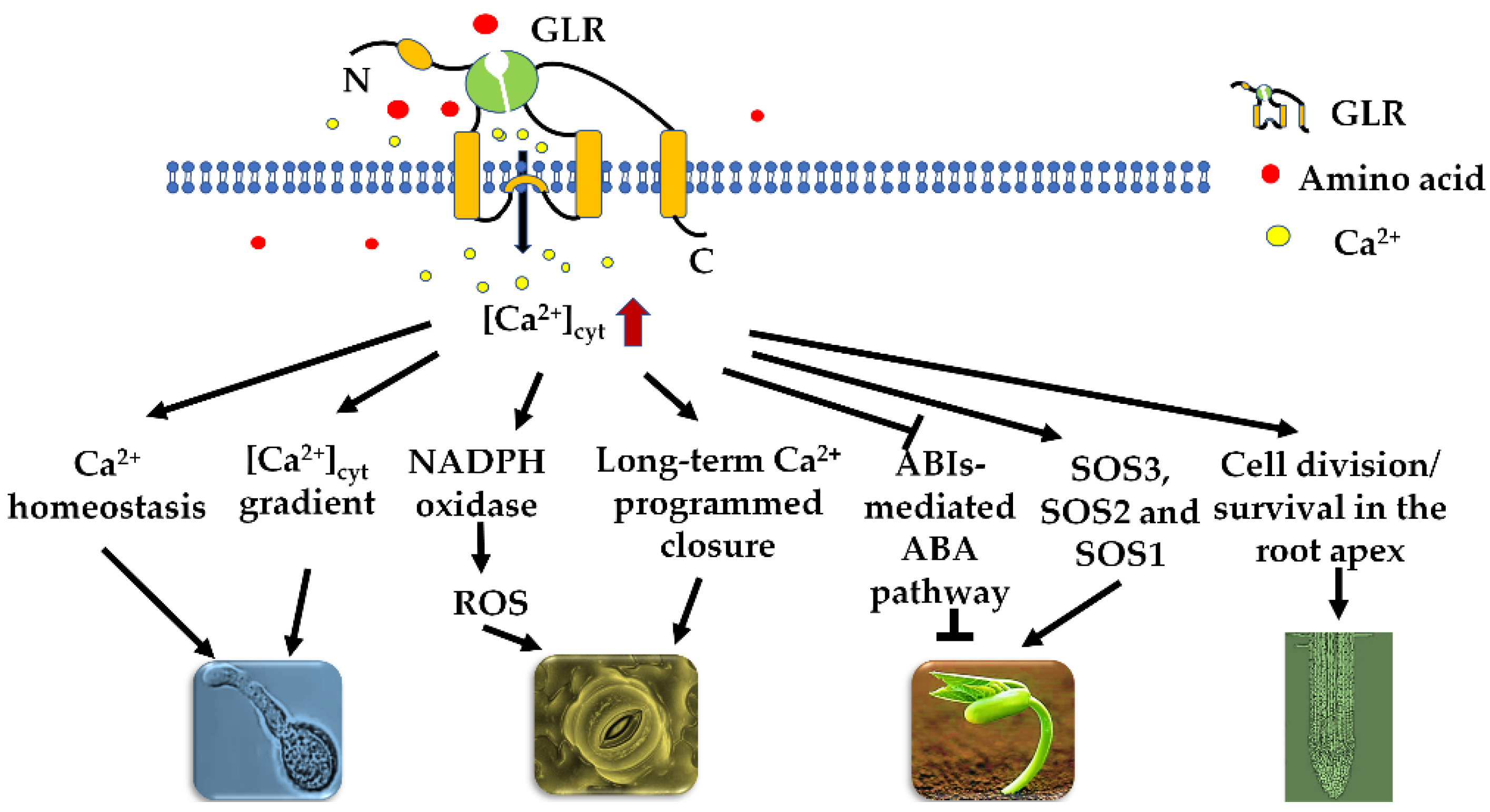
2. GLR-Mediated Ca2+ Signaling Regulates Various Plant Physiological Processes

As extracellular amino acid sensors, plant GLRs play fundamental roles in regulating various physiological processes, which are closely associated with Ca2+ signaling [16,17,18,19]. With effective amino acids, plant GLRs channel many kinds of cation fluxes across the membrane into the cytoplasm, in particular Ca2+, which acts as a main signal messenger [20,21]. Arabidopsis GLR3.1 (AtGLR3.1) is preferentially expressed in stomatal guard cells, and the overexpression of AtGLR3.1 impairs stomatal closure, suggesting that AtGLR3.1 is closely correlated with the cytosolic Ca2+ concentration ([Ca2+]cyt) that regulates stomatal movement [22]. As a fact, convincing evidence has been presented to confirm the Ca2+ permeability of GLRs, which have a broad agonist profile. Studies of Arabidopsis hypocotyl cells indicated that six effective amino acids trigger transient Ca2+ influx and membrane depolarization by a mechanism that depends on AtGLR3.3 [23], and rice GLR3.4 (OsGLR3.4) has a broad agonist profile with eleven amino acids that induce transient Ca2+ influx in an OsGLR3.4-dependent manner in coleoptile epidermal cells [19]. AtGLR3.4-mediated Ca2+ signaling was involved in the regulation of seed germination under salt conditions through the SOS pathway, and the atglr3.4 mutant showed impaired [Ca2+]cyt induction as well as a reduced expression of ABSCISIC ACID-INSENSITIVE (ABI) genes, AtABI3 and AtABI4, in response to salt stress [20,24]. Similarly, AtGLR3.5-mediated [Ca2+]cyt enhancement promotes seed germination by counteracting the inhibitory effects of ABA through the repression of ABI4 expression [25]. Consistently, applying exogenous glutathione to Arabidopsis leaves triggered a transient rise in [Ca2+]cyt, but this response was impaired in the atglr3.3 mutant [13]. In another report, AtGLR1.2 channeled Ca2+ influx into the pollen tube cells when they extended in the pistil, but Ca2+ signaling was largely impaired in atglr1.2 pollen tubes [26]. In brief, these plant-specific physiological processes are regulated by GLR-mediated Ca2+ signaling (Figure 1). Understanding of the roles of GLRs in plant growth and abiotic stress will help with the engineering of perfect-fitness and stress-tolerant crops.

3. Roles of GLRs in Plant Growth and Development

3.1. Roles in Seed Germination

Seed germination is a major step in plant growth, and it is strictly controlled by endogenous and environmental signals such as phytohormones and environmental factors including water, temperature, and light [27]. ABA is known to regulate the sophisticated process of seed maturation and germination [28]. Recent studies indicated that GLRs are involved in the seed germination by modulating the levels of ABA and ethylene in Arabidopsis [29,30]. Carbon (C) and nitrogen (N) are the two most critical elements that are required for normal plant development and metabolism [31]. Plants need to coordinate C and N metabolism to control their growth and development during different periods, and they therefore have evolved a sophisticated regulatory system to continuously monitor the levels of different carbon–nitrogen ratio ‘‘check point’’ molecules, including sucrose (Suc), glucose (Glc), 2-oxoglutarate, glutamine (Gln), glutamate (Glu), NO3, and NH4+ [29,32]. AtGLR1.1 is involved in C and N metabolism and controlling seed germination by affecting ABA levels [29]. During seed germination, the addition of NO3 can stimulate the formation of Glu and Gln, which activate the expression of AtGLR1.1 and ultimately inhibit ABA synthesis; alternatively, the transcription of AtGLR1.1 can be repressed by Suc, which induces the expression of ABA biosynthetic genes and thus leads to ABA-mediated seed germination inhibition [29]. Despite the implications that AtGLR1.1 might function as a C/N receptor or sensor, the true ligand of AtGLR3.1 is yet unclear, and this needs to be experimentally investigated. In other reports, transcription factors associated with the ABA signaling pathway, AtABI3, AtABI4, and AtABI5, were found to have important regulatory effects on seed maturation and germination, among which ABI3 was essential for seed maturation and desiccation tolerance, while ABI4 and ABI5 played a crucial role in ABA inhibition during seed germination [33,34,35,36]. It has been shown that there is a large amount of Ca2+ in the seed coat, cell wall, and apoplast [37,38]. Extracellular Ca2+ influx can promote seed germination, but the process can be remarkably inhibited when this influx is disturbed, and the exogenous application of Ca2+ can attenuate ABA inhibition during seed germination [25,39]. AtGLR3.5 is mainly expressed in germinated seeds, and the AtGLR3.5-mediated [Ca2+]cyt elevation can alleviate the inhibition of ABA during seed germination, demonstrating the essential roles of AtGLR3.5 in seed germination [25]. Compared to the control, the transcript abundance of AtABI4 was significantly increased in germinating AtGLR3.5-RNAi seeds, whereas it was reduced in AtGLR3.5-overexpression seeds, and the changes in germination were consistent with the alteration of [Ca2+]cyt [25]. This observation demonstrates that the AtGLR3.5-mediated increase in [Ca2+]cyt promotes seed germination due in large part to the decrease in the ABI4 expression. The application of Glu can reduce the inhibiting effect of salt stress on Arabidopsis seed germination [40], suggesting that the GLR-mediated [Ca2+]cyt elevation may alleviate the inhibition caused by environmental stress during seed germination. Presently, seeds face challenges in viability. Seed dormancy is a key characteristic which inhibits seed germination during the harsh and tough growing season. Due to the crucial roles of ABA in dormancy induction and maintenance in the adverse environment, GLRs have the potential to improve seed vigor by restricting ABA signaling and thus are expected to be used for genetic modifications in crops. Therefore, the identification of GLRs as potential players in seed germination facilitates their practical use in breeding to attain a high capacity of seed germination and seedling establishment, especially under salinity stress.

3.2. Roles in Root Development

Plant root system (including primary roots and lateral roots) development is a highly ordered process [41,42], which is critical for efficient nutrient uptake, drought resilience, and crop yield [43]. During root development, the maintenance of cell division and individual cell survival in the root apical meristem are correlated with spatiotemporal characteristics of the electrical network activity of the root apex [12,42]. The Arabidopsis atglr3.6-1 mutant has a reduced mitotic activity and root meristem size in the root tip, which results in shorter primary roots and fewer lateral roots, while AtGLR3.6 overexpression stimulates both primary and lateral root development [44]. The cyclin-dependent kinase (CDK) inhibitor Kip-related protein 4 (KRP4) is an inhibitor of the cell cycle [45,46]. Accordingly, the transcript abundance of AtKRP4 in atglr3.6-1 roots was significantly enhanced, while reduced in transgenic lines overexpressing AtGLR3.6 [44], suggesting that AtGLR3.6 plays a positive role in maintaining root meristem by repressing the expression of AtKRP4. Consistently, [Ca2+]cyt was greatly reduced in the root meristem of atglr3.6-1, and the addition of calcium reduced the expression of AtKRP4 and promoted root growth [44]. Members of the PIN (PIN-FORMED) family are responsible for polar auxin transport in plants [47]. In atglr3.6-1 roots, AtPIN1 mRNA was less abundant, and auxin levels were lower, indicating that the downregulation of the AtPIN1 expression could be the cause of the reduced auxin level [44]. The reduction in auxin levels in atglr3.6-1 roots resulted in a reduced mitotic activity in the root tips [44], which implies that AtGLR3.6 is involved in PIN-mediated auxin polar transport by mediating Ca2+ influx. In addition, AtGLR3.2 interacts with AtGLR3.4 to form the heteromeric AtGLR3.2/AtGLR3.4 channel, and the single mutants atglr3.2 and atglr3.4 as well as the double mutant atglr3.2atglr3.4 display an equally severe phenotype, a large overproduction, and aberrant placement of lateral root primordial [48], suggesting they play essential roles in lateral root development via Ca2+ signaling. In addition to cell division and differentiation, the fundamental development process of root development is also accompanied by programmed cell death, including aerenchyma formation, root cap cell production, and shedding [49,50], suggesting that root development is a well-coordinated program regulated by multiple factors. Research on the nervous system in mammals has shown that iGluRs are vital in the fate determination of nerve cells at the early stage of development [51]. In the rice Osglr3.1 mutant, cell division and differentiation in the root apex were seriously affected, and the programmed cell death of root meristem was increased, resulting in the inhibition of primary root elongation [42], suggesting that plant GLRs might have similar roles to those in their animal homologues. It is recognized that treating Arabidopsis roots with Glu elicits rapid changes in the membrane potential and increases Ca2+ flux, and specific members of the GLR family are required for this response [23,52]. Similarly, our recent study showed that the application of Glycin (Gly, one agonist of OsGLR3.4) inhibited primary root growth in rice, while Osglr3.4 mutants exhibited hyposensitivity to the exogenous Gly treatment. The mechanistic study showed that Gly treatment triggered OsGLR3.4-mediated [Ca2+]cyt elevation, which subsequently induced membrane depolarization and ROS production in the root tips [53]. Despite the genetic evidence about the roles of GLRs in root growth, we still largely misunderstand how GLRs act in the coordination of primary root growth and lateral root proliferation. Improving crop root architecture and function may be challenging, but a series of cases have emerged showing that genetic modifications of roots, when the modified gene is specifically expressed in the root, result in enhanced plant performance, increased yield, and elevated stress tolerance. Therefore, the prominent roles of GLRs in root development make them potential candidates for increasing crop production. Additionally, more work is needed to explore the role of different GLR isoforms in root growth.

3.3. Roles in Other Developmental Programs

Pollen tubes are a model system for studying tip growth, which involves many parameters including vesicle trafficking, cell wall precursor exocytosis, actin microfilament polymerization, apical ion flux, and [Ca2+]cyt oscillation [54]. Arabidopsis atglr1.2 and atglr3.7 knockout plants displayed male reproductive phenotypes, and further mechanistic investigation demonstrated that AtGLR1.2 and AtGLR3.7 could facilitate Ca2+ influx across the plasma membrane, modulate the apical [Ca2+]cyt gradient, and consequently affect pollen tube growth and morphogenesis [26]. As a fact, some GLRs are able to function in a series of programs related to plant growth and development. For instance, in addition to the modulation of primary root growth, new roles for OsGLR3.4 were found in the development of rice plant architecture. Osglr3.4 mutants showed a semi-dwarf phenotype with erect leaves, and OsGLR3.4 was shown to be involved in brassinosteroid (BRs) signaling to modulate cell elongation of the internode and lamina joint [19]. As a Ca2+-permeable channel activated by multiple amino acids, OsGLR3.4 modulates actin filament organization and vesicle trafficking by mediating the [Ca2+]cyt rise, which is required for cell elongation [19]. More recently, we found that OsGLR3.4 modulated root tropism growth towards amino acids via plasma membrane depolarization and ROS generation [53]. Excitingly, OsGLR3.4 was demonstrated to promote nitrate uptake by modulating the Gly-induced expression of nitrate transporter genes [53]. Therefore, OsGLR3.4 is a potential candidate for plant adaption to an uneven nutrient distribution as well as to promote nitrate uptake, which will facilitate the practical use of OsGLR3.4 in rice molecular breeding for a high crop yield.

4. Roles of GLRs in Plant Response to Environmental Stress

Plants are subjected to a series of environmental stresses during their growth and development, and they often display considerable plasticity in their developmental and physiological behaviors by responding to a variety of environmental signals, including mechanical wounding, unexpected herbivory attack, water deficit, and soil salinity [15,55,56]. It has been recognized that GLRs play a vital role in environmental signal reception and transmission and plant adaption to environmental stress, including biotic and abiotic stresses [20,57,58,59,60]. Recent progress regarding the functions of GLRs in plant response to environmental stress is discussed in detail below.

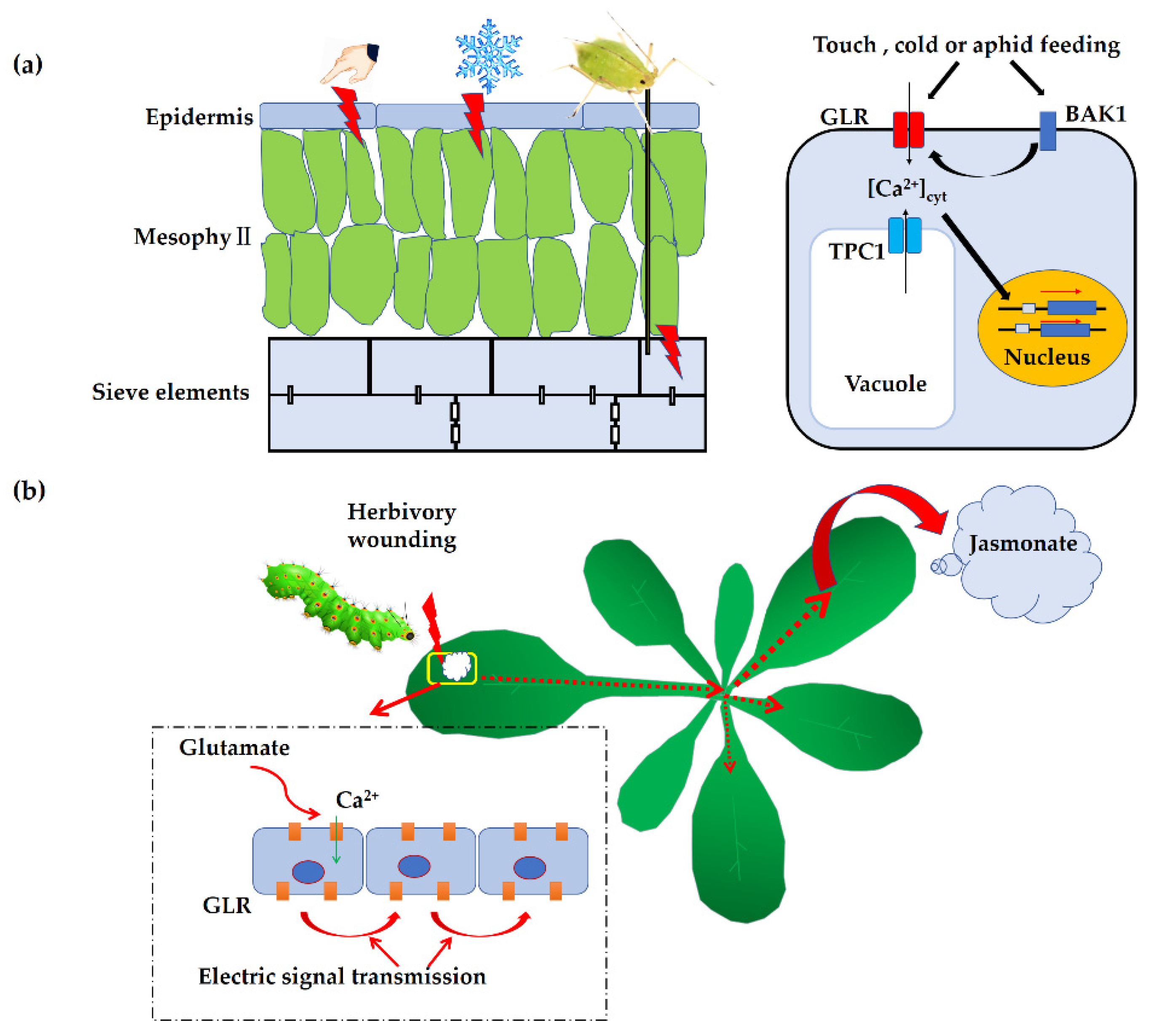
4.1. Roles in Mechanical Wounding or Herbivory Attack

Accumulating evidence has demonstrated that GLR-mediated Ca2+ signaling is rapidly activated in response to touch or mechanical wounding in plants [61] (Figure 2). Studies have shown that when Arabidopsis is exposed to touch or cold, the transcripts of AtGLR3.4 increase significantly, suggesting that AtGLR3.4 is involved in the response to these environmental stimuli [61]. In addition, GLRs were shown to play essential roles when plants were subjected to herbivory attack. Unlike animals [62], plants do not have specialized nerve cells with axons, and they cannot rely on a rapid nervous system to avoid dangerous threats in the environment [15]. However, similar to animals, they do operate long-distance electrical signals [63]. By using noninvasive electrodes to detect changes in the membrane potential in Arabidopsis leaves, researchers found that membrane potential depolarization is correlated with the distal production of jasmonic acid (JA) in undamaged leaves [15]. When attacked by herbivores, plants suffer from mechanical wounding or invasion of exogenous chemicals, and they therefore generate electrical signals by activating GLRs [64,65]. These signals are then transferred to adjacent tissues where the biosynthesis of JA is induced, which in turn triggers the JA resistance pathway [66,67,68]. As expected, when mutations in GLR genes of clade 3 (AtGLR3.2/3.3/3.6) happen, the electrical signals transmitted to the adjacent leaves are attenuated in these mutants [15]. Further study indicated that wounding initiates the AtGLR3.3- or AtGLR3.6-dependent propagation of membrane depolarization in the form of short wave potentials (SWPs) that leads to defense gene activation, and atglr3.3 and atglr3.6 mutants were found to be compromised in their defense against herbivores [69,70]. It is notable that the signal transmission between leaves of the double mutant atglr3.3atgrl3.6 decreased significantly, and the expression of JA-responsive genes was also reduced significantly in the leaves adjacent to the injured leaves [15]. The long-distance wounding response is also conserved in monocotyledon plant rice. Root injury triggering SWPs as well as the JA response in leaves are impaired in Osglr3.4 mutants, indicating that OsGLR3.4 is required for root-to-shoot systemic wound signaling in rice [19]. Tomato SlGLR3.3- and SlGLR3.5-mediated leaflet-to-leaflet electrical signal transduction and herbivory-induced JA accumulation and Helicoverpa armigera resistance were reduced in slglr3.3 and slglr3.5 mutants [71], revealing the key roles of SlGLR3.3 and SlGLR3.5 in electrical signal transduction and JA signal activation. These studies provide a genetic basis for further study on the transmission mechanism of plant electrical signals between organs and also reveal some similarities in the transmission modes of electrical signals between plants and animals. Despite the genetic evidence about the role of GLRs in the local and long-distance transmission of electrical signaling, knowledge about how GLRs are activated in vivo in response to wounding is still limited, and more work is needed to elucidate the sophisticated mechanism.
Research on plant resistance to herbivory attacks found that GLRs have a critical role in plant immunity against aphids in leaves [72,73] (Figure 2). By using a fluorescent Ca2+ biosensor (GCaMP3) to monitor the real-time [Ca2+]cyt dynamics in Arabidopsis leaves during a green peach aphid feeding, a strong fluctuation in [Ca2+]cyt was detected when aphids’ probes penetrated into the epidermal and mesophyll cells in the leaves [73]. Further study showed that an unknown receptor cooperates with BRASSINOSTEROID INSENSITIVE-ASSOCIATED KINASE1 (AtBAK1) [74,75], which then activates the expression of AtGLR3.3 and AtGLR3.6 to transform signals during aphid feeding [73]. More recent research indicated that AtGLR3.3 and AtGLR3.6 are involved in the regulation of various metabolites locally and systemically, including amino acids, carbohydrates, and organic acids [76], which provides new insight into the function of AtGLR3.3 and AtGLR3.6 in mediating metabolites in local and systemic leaves under insect attacks and highlights their roles in regulating insect resistance in systemic leaves. As a fact, the triad of GLR-mediated Ca2+, ROS, and electrical activity has been implicated in the response to wounding in plants [77,78]. For example, upon wounding, systemic changes in the membrane potential, Ca2+, and ROS are coordinated by AtGLR3.3 and AtGLR3.6 [79]. Although there is a knowledge gap about plant GLRs that needs to be filled, a deeply conserved function for GLRs that links wounding perception to distal protective responses makes them promising candidates for genetic modification in engineering pest-resistance in crops. Nowadays, crop yield is under enormous pressure, which is caused by biotic stresses including pests. Therefore, genetic modifications by which the GLR genes are expected to be expressed at the appropriate time only when plants are attacked by pests would be effective for pest control but do not interfere with or negatively affect their physiological processes under normal growth conditions.

4.2. Roles in Drought Response

Exposure of plants to a water-limiting environment during the developmental stages appears to activate cascades of physiological and developmental changes [80]. Plant leaves can control the rate of water evaporation by adjusting the opening and closing of stomata, which is precisely regulated by ABA signaling [81,82,83]. When plants are exposed to drought stress, the increased ABA levels in the guard cells induce the accumulation of reactive oxygen species (ROS) produced by RbohD and RbohF NADPH oxidases [84,85]. The enhanced ROS activate the Ca2+ channels on the cell membrane, causing [Ca2+]cyt elevation [86,87]. The increase in [Ca2+]cyt eventually causes stomatal closure by activating S-type anion channels or inhibiting inward-rectifying K+ channels and H+-ATPases [88,89]. Recently, a new mechanism that differs from ABA-induced ROS production to stimulate stomatal closure was found [17]. L-methionine (L-Met) at the physiological concentration can increase [Ca2+]cyt by activating AtGLR3.1 and AtGLR3.5, which eventually leads to stomatal closure [17]. The basal levels of [Ca2+]cyt were significantly reduced in the single mutants atglr3.1 and atglr3.5 as well as the double mutant atglr3.1atglr3.5, and consequently, ABA-induced stomatal closure was also remarkably repressed in these mutants [17], suggesting that AtGLR3.1 and AtGLR3.5 might be involved in ROS-mediated stomatal closure and drought tolerance. It is notable that AtGLR3.5 was found to be the first cation channel located on the mitochondrial membrane. AtGLR3.5 has two different splicing variants, and one of the variants targets the inner-mitochondrial membrane, while the other variant localizes to chloroplasts [90], suggesting an intricate mechanism that AtGLR3.5 is involved in to modulate stomatal movement. The ability of mitochondrial Ca2+ absorption was found to slightly decrease and the leaf showed obvious accelerated senescence in the atglr3.5 defect mutant [90]. A study of Medicago truncatula showed that MtGLRs are required for adaptive responses under short-term water deficit stress during Medicago seedling establishment by mediating NO production [91]. Due to climatic variability, the incidence of drought stress at various crop growth stages is becoming a major hindering factor to yield improvement. Undoubtedly, knowledge about GLRs in plant drought response has important theoretical and practical significance for cultivating new drought-resistant crop varieties to enhance yield. Therefore, tight regulation and fine-tuning of GLR genes during plant stress responses will contribute to the establishment of complex signaling networks, and the important roles of GLRs in plant abiotic stress responses make them potential candidates for conferring stress tolerance.

4.3. Roles in Salinity Response

Soil salinization is an increasingly serious problem in global agriculture because excessive Na+ and Cl uptake by plant roots can disrupt metabolic processes and reduce photosynthetic efficiency and thus severely affect plant growth and development [92,93]. Generally, plants respond to osmotic stress by reducing water evaporation and maximizing water absorption [94]. In addition, plants can reduce the harmful effects of ion Na+ stress by excluding Na+ from leaf tissues and dividing Na+ into vacuoles [93,95]. Studies have shown that the sensitivity of the Arabidopsis atglr3.4 mutant to NaCl was much higher than that of the wild-type plants [58], which implies that GLRs are required in the adaption to salinity stress. NaCl can significantly raise [Ca2+]cyt in wild-type plants, and this response can be prevented by GLR antagonists; however, the increase in [Ca2+]cyt induced by NaCl was significantly repressed in the atglr3.4 mutant, and the expression of AtSOS1, AtSOS2, and AtSOS3 in the mutant was also reduced [58]. Compared with that in the wild type, the Na+ content in the atglr3.4 mutant was increased significantly [58], suggesting that AtGLR3.4-mediated Ca2+ signaling may regulate Na+ absorption through ROS signaling and may participate in the response to NaCl. Recently, AtGLR3.7 was reported to interact with 14-3-3 omega to modulate the salt stress response [96]. The mutant atglr3.7-2 was more sensitive to salt stress, while AtGLR3.7 overexpression lines exhibited the opposite trend [96].
When plants are subjected to salt stress, they are physiologically exposed to osmotic fluctuations, the toxicity of Na+ and K+, excessive ROS production, and unbalanced cytosolic K+ homeostasis [97]. Maintaining high K+ levels in plant cells has a positive effect on their response to salt stress, which allows vacuolar H+-PPase to maintain high activity and Na+ to be sequestered [98]. Plant root tips are more sensitive to salt stress than mature tissues, which is mainly attributed to the higher H+-ATPase activity in cells in mature tissues than in meristem [97], which might be associated with the fact that multiple GLRs are expressed in Arabidopsis roots [99,100]. As an activator of GLRs, the Glu gradient in the root tips is increased by about five times under salt stress [97]. It is estimated that Glu produced in the root tips under salt stress activates GLRs, which further activate the plasma membrane NADPH oxidase to produce excessive hydrogen peroxide (H2O2) [101], leading to the activation of outward-rectified K+ channels that cause programmed cell death [97]. On the contrary, the mature zones in roots do not produce excessive Glu under salt stress, and thus, the activation of GLRs is inhibited [97]. Ca2+ also acts as nutrition in plant growth and development, and a lack of Ca2+ leads to a series of physiological reactions in plants, including browning and death of the shoot apex, necrosis of leaf tips, and deformation of leaves [60]. The reduced growth resulting from calcium nutrition can also lead to enhanced sensitivity to salt stress in plants. The overexpression of AtGluR2 (a homolog of the mammal ionotropic glutamate receptor gene) does not affect Ca2+ uptake but reduces its utilization, thus resulting in the phenotype of Ca2+ deficiency as well as hypersensitivity to Na+ and K+ stress [60]. AtGluR2 is mainly expressed in vascular tissues, particularly in cells adjacent to conducting vessels, where Ca2+ in xylem sap is absorbed and distributed, and it is supposed to encode a functional channel that unloads Ca2+ from the xylem vessels into the cell membrane [60]. Thus, the appropriate expression of AtGluR2 is required in Ca2+ nutrition by controlling the ion allocation among different Ca2+ sinks, either in normal development or adaptation to ionic stresses.
Plant defense against biotic and abiotic stresses generally comes at the expense of growth, and thereby, the tradeoff between defense and growth is a major constraint on plant evolution. Amino acids and their derivatives play vital roles in mediating defense priming and growth tradeoff [102]. Gated by amino acids and related molecules to induce Ca2+ signaling, GLRs are proposed to be potential molecules that induce defense priming and the equilibrium between growth and defense against stress including salinity, which is expected to help maintain relative plant fitness under unpredictable conditions and maximize reproductive success. Therefore, GLRs are promising candidates for genetic modification to improve plant tolerance to various stresses including salt stress and also provide an outlook on the prospects of engineering the tradeoff between defense and growth in plants.

4.4. Other Environmental Stimuli

In addition to herbivory attacks and osmotic stress, chilling is another major environmental stress that not only affects plant growth, but also minimizes the productivity and quality of crops [102,103]. SlGLR3.3 and SlGLR3.5 mediate the cold acclimation-induced chilling tolerance by regulating apoplastic H2O2 production and redox homeostasis [104]. Cold induces the expression of SlGLR3.3 and SlGLR3.5 in tomatoes coupled with an increased tolerance against subsequent chilling, while the silencing of SlGLR3.3 or/and SlGLR3.5 or the application of the antagonist of the ionotropic glutamate receptor compromises the cold-induced increase in the transcripts of RBOH1 [104]. Aluminum is abundant in nature but harmful to plants, which largely limits the productivity of crops [14]. It was shown that the addition of aluminum could cause abnormal shapes of organs by depolymerizing root cell microtubules as well as depolarizing the plasma membrane, but the Ca2+ channel blockade can prevent these changes [14,105,106]. Plants have evolved sophisticated mechanisms to minimize or avoid aluminum toxicity, such as isolating aluminum in a stable form within cells or chelating them and making them harmless by excreting organic anions [107,108]. Root secretions are rich in glutamate and other amino acids, and the distribution of the amino acids secreted varies with the environment and development of the plants [109]. When plants are exposed to aluminum stress, the roots are stimulated to secrete organic acids, which are believed to be used for the activation of the plasma membrane anion channels including GLRs [14]. In support of this, a recent study on Arabidopsis showed that the Ca2+-dependent calmodulin-like protein CML24 interacts with CALMODULIN BINDING TRANSPORTER ACTIVATOR 2 (CAMTA2) and WRKY46 to regulate the ALUMINIUM-ACTIVATED MALATE TRANSPORTER 1 (ALMT1)-mediated secretion of malate from roots and consequently achieves aluminum tolerance [110].
Nitric oxide (NO) has been proven to be a key signal molecule in various physiological processes [111], including programmed cell death, growth, and the development of organs, seed germination, flowering, and response to biotic or abiotic stresses [112,113,114,115]. NO can be produced as the response of plants to different pathogen attacks and can promote the expression of defense-related genes, induce the formation of defense-related hormones, and ultimately is involved in the hypersensitivity reaction mechanism [116]. Studies have shown that, on one hand, NO affects the expression of genes encoding the CaM or Ca2+ channels, and on the other hand, directly or indirectly activates Ca2+ channels, the Ca2+/CaM-dependent protein kinase, and/or other Ca2+ sensors [59,117,118]. Other studies showed that, when plants are attacked by pathogens, the released Glu activates the GLR channel and causes cell membrane depolarization, allowing the signal to be transmitted to surrounding tissues [18,57]. The increased levels of GLRs promote the production of cryptogein (elicitor of the defense response) and NO [59]. Collectively, GLRs play an important role in the plant defense response by inducing NO production, although the mechanism still needs to be further explored. Based on the performance of GLRs during environmental stimuli, they are therefore considered to be promising candidates for crop breeding through genetic manipulations to increase the tolerance to biotic or abiotic stresses.

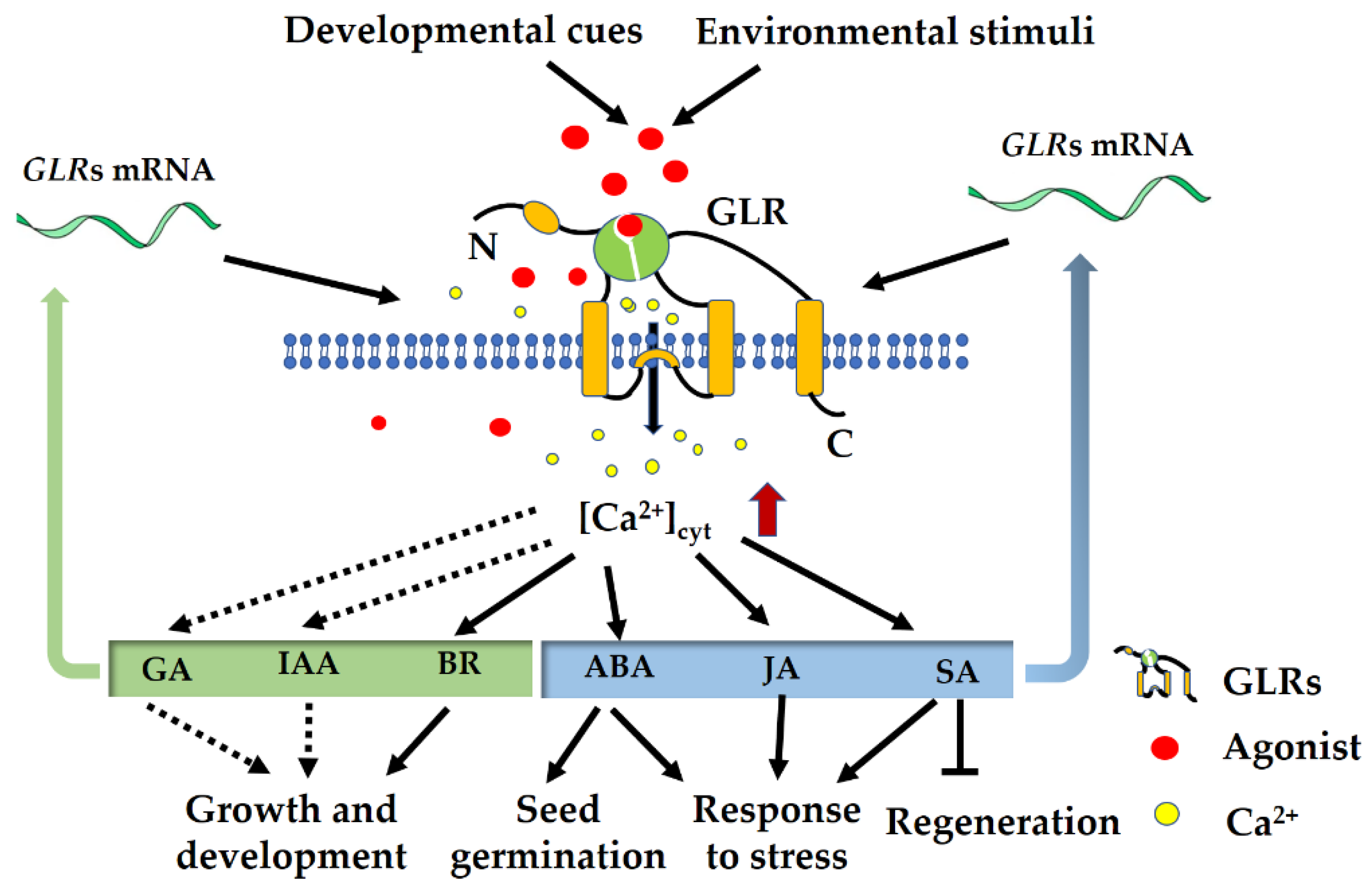
5. Interaction of GLRs with Hormone Signaling

For plants, both the growth and environmental response require rapid activation of Ca2+ signaling as well as hormone signaling (Figure 3). The tradeoff between growth and defense incorporates the balance between the plant ‘‘defense’’-related hormones, ABA, JA, and salicylic acid (SA), and those involved in growth, including BR, auxin, and gibberellic acid (GA) [119]. Accumulating evidence has revealed the central role of plant GLRs in the tradeoff between growth and defense. Our recent investigation showed OsGLR3.4, functioning as a target gene of transcription factor OsBZR1, is involved in BR-mediated plant height and architecture in rice [19]. Although the AtGLR3.6 mutation leads to impaired [Ca2+]cyt elevation coupled with a reduced root auxin level [44], which is tempting to speculate that the AtGLR3.6 interacts with auxin signaling, further work is needed to address the role of GLRs in auxin signaling. Research has shown that Ca2+ signaling integrates with GA signaling to contribute to plant growth and development, but there is little direct evidence that GLRs interact with GA signaling [120,121]. As for defense-related hormones, the interaction of GLR signaling with ABA and JA has been well discussed above, and here, GLRs interplaying with SA is focused upon. A recent study showed that plant GLRs work through SA signaling in their effects on tissue regeneration, and mutants of the SA receptor NPR1 are hyper-regenerative and partially resistant to GLR perturbation, indicating that SA acts downstream of GLR signaling [122]. Meanwhile, the transcription of SA-responsive marker genes increases as cells mature, suggesting that SA mediates GLR signaling in older tissues and thus inhibits regeneration. Therefore, present evidence reveals the central role of GLRs in the tradeoff between wounding-triggered regeneration and defense, which offers new strategies to improve plant regeneration. It is important to note that several questions remain as to the molecular mechanisms underlying the modulation of GLRs on the tradeoff, and more work is required to elucidate how GLRs interact with hormone signaling to regulate the balance between plant growth and environmental response. Future studies dissecting the exact mechanisms underlying the dichotomous function of GLRs will undoubtedly provide even more tractable ways to increase plant growth as well as adaption to adverse environments.

6. Conclusions and Future Perspectives

Plant GLRs are a highly conserved membrane protein family, and they have overlapping expression patterns and biological functions. Present knowledge about the roles of GLRs in plant growth and response to environmental stress is mainly from the research on dicotyledon model Arabidopsis, whereas functions of most of the GLR members in monocotyledon plants have yet to be identified. Therefore, substantial experimental work is required to determine the specific biological function of GLRs from cereal crops, which will remain a substantial challenge in the coming years. Meanwhile, considering the large number of networks regulated by GLR-mediated Ca2+ signaling, it is believed that multiple unknown functions of GLRs remain to be explored. Recently, great progress on plant GLRs has been achieved through molecular genetics and electrophysiological methods. Despite the genetic evidence about the role of GLRs in the local and long-distance transmission of electrical signaling, the knowledge about the roles of different GLR isoforms in electrical signal transmission is still limited, and some open questions remain unanswered. For example, it is still unclear how the system’s electrical activity is propagated from the damaged tissues to the distal undamaged organs. Although long-distance electrical signals have been implicated to travel through vascular tissues, direct evidence is needed to answer this question, and future research will be directed toward addressing the question of plant long-distance signaling.
To achieve a better understanding of their roles during plant growth and environmental responses, it is essential to identify the interacting partners of GLRs that cooperate in regulating these physiological processes. It is also crucial to elucidate the effects of the interaction of GLRs with partners on their activation and reveal the possible molecular mechanism. Recent advances in elucidating the functions of Arabidopsis GLRs have revealed a mechanism for sorting and activation of Arabidopsis GLRs by CORNICHON HOMOLOG (CNIH) proteins [13]. CNIH proteins play an essential role in sorting, trafficking, and localizing GLRs, and more importantly, CNIHs can activate GLRs via the physical interaction between them [13]. Additionally, to gain a thorough understanding of how GLRs are activated in vivo, further studies on the mechanism are required, such as the possible role of CNIH in nonpollen tissues, the precise role of amino acid binding via the ligand-binding domain (LBD), and the significance of C-terminus phosphorylation. It is important to note that the interactions between GLRs and different partners may play different roles under different environmental stresses, such as drought, salt, cold stress, insect attacks, and pathogen infections. At present, only a few known interaction proteins of GLRs are characterized. The identification of interaction partners of GLRs would be helpful for understanding the detailed networks where GLRs function. Certainly, further molecular studies of GLRs will clarify the fine-tuned mechanisms that control Ca2+ signaling in plants, and it is likely to be crucial in helping to guide future breeding plans and consequently be beneficial for crop production.

Author Contributions

J.H. developed the review concept. All authors prepared various parts of the review. B.Y. and N.L. wrote the draft. S.T. and T.Q. added the drawings. J.H. revised and edited the manuscript. All authors have read and agreed to the published version of the manuscript.

Funding

This work was supported by National Natural Science Foundation of China (32071985, 31771747), the Natural Science Foundation of Chongqing, China (cstc2020jcyj-msxmX0656), and the Fundamental Research Funds for the Central Universities, China (2020CDJ-LHZZ-034).

Conflicts of Interest

The authors declare that they have no conflict of interest.

References

  1. Armstrong, N.; Sun, Y.; Chen, G.-Q.; Gouaux, E. Structure of a glutamate-receptor ligand-binding core in complex with kainate. Nature 1998, 395, 913. [Google Scholar] [CrossRef] [PubMed]
  2. Zheng, Y.; Mellem, J.E.; Brockie, P.J.; Madsen, D.M.; Maricq, A.V. SOL-1 is a CUB-domain protein required for GLR-1 glutamate receptor function in C. elegans. Nature 2004, 427, 451. [Google Scholar] [CrossRef] [PubMed]
  3. Mayer, M.L. Glutamate receptor ion channels: Where do all the calories go? Nat. Struct. Mol. Biol. 2011, 18, 253. [Google Scholar] [CrossRef] [PubMed]
  4. Lam, H.-M.; Chiu, J.; Hsieh, M.-H.; Meisel, L.; Oliveira, I.C.; Shin, M.; Coruzzi, G. Glutamate-receptor genes in plants. Nature 1998, 396, 125–126. [Google Scholar] [CrossRef] [PubMed]
  5. Chiu, J.; DeSalle, R.; Lam, H.M.; Meisel, L.; Coruzzi, G. Molecular evolution of glutamate receptors: A primitive signaling mechanism that existed before plants and animals diverged. Mol. Biol. Evol. 1999, 16, 826–838. [Google Scholar] [CrossRef]
  6. Wudick, M.M.; Michard, E.; Oliveira, N.C.; Feijó, J.A. Comparing plant and animal glutamate receptors: Common traits but different fates? J. Exp. Bot. 2018, 69, 22–957. [Google Scholar] [CrossRef]
  7. De Bortoli, S.; Teardo, E.; Szabo, I.; Morosinotto, T.; Alboresi, A. Evolutionary insight into the ionotropic glutamate receptor superfamily of photosynthetic organisms. Biophys. Chem. 2016, 218, 14–26. [Google Scholar] [CrossRef]
  8. Forde, B.G. Glutamate signalling in roots. J. Exp. Bot. 2014, 65, 779–787. [Google Scholar] [CrossRef] [Green Version]
  9. Grenzi, M.; Bonza, M.C.; Alfieri, A.; Costa, A. Structural insights into long-distance signal transduction pathways mediated by plant glutamate receptor-like channels. New Phytol. 2020, 229, 1261–1267. [Google Scholar] [CrossRef]
  10. Gangwar, S.P.; Green, M.N.; Michard, E.; Simon, A.A.; Feijo, J.A.; Sobolevsky, A.I. Structure of the Arabidopsis glutamate receptor-like channel GLR3.2 ligand-binding domain. Structure 2021, 29, 161–169. [Google Scholar] [CrossRef]
  11. Green, M.N.; Gangwar, S.P.; Michard, E.; Simon, A.A.; Portes, M.T.; Barbosa-Caro, J.; Wudick, M.M.; Lizzio, M.A.; Klykov, O.; Yelshanskaya, M.V.; et al. Structure of the Arabidopsis thaliana glutamate receptor-like channel GLR3.4. Mol. Cell 2021, 81, 3216–3226. [Google Scholar] [CrossRef] [PubMed]
  12. Masi, E.; Ciszak, M.; Stefano, G.; Renna, L.; Azzarello, E.; Pandolfi, C.; Mugnai, S.; Baluska, F.; Arecchi, F.T.; Mancuso, S. Spatiotemporal dynamics of the electrical network activity in the root apex. Proc. Natl. Acad. Sci. USA 2009, 106, 4048–4053. [Google Scholar] [CrossRef] [PubMed] [Green Version]
  13. Wudick, M.M.; Portes, M.T.; Michard, E.; Rosas-Santiago, P.; Lizzio, M.A.; Nunes, C.O.; Campos, C.; Damineli, D.S.C.; Carvalho, J.C.; Lima, P.T.; et al. CORNICHON sorting and regulation of GLR channels underlie pollen tube Ca2+ homeostasis. Science 2018, 360, 533–536. [Google Scholar] [CrossRef] [PubMed] [Green Version]
  14. Sivaguru, M.; Pike, S.; Gassmann, W.; Baskin, T.I. Aluminum rapidly depolymerizes cortical microtubules and depolarizes the plasma membrane: Evidence that these responses are mediated by a glutamate receptor. Plant Cell Physiol. 2003, 44, 667–675. [Google Scholar] [CrossRef] [PubMed] [Green Version]
  15. Mousavi, S.A.R.; Chauvin, A.; Pascaud, F.; Kellenberger, S.; Farmer, E.E. Glutamate receptor-like genes mediate leaf-to-leaf wound signalling. Nature 2013, 500, 422. [Google Scholar] [CrossRef] [PubMed] [Green Version]
  16. Ni, J.; Yu, Z.; Du, G.; Zhang, Y.; Taylor, J.L.; Shen, C.; Xu, J.; Liu, X.; Wang, Y.; Wu, Y. Heterologous expression and functional analysis of rice GLUTAMATE RECEPTOR-LIKE family indicates its role in glutamate triggered calcium flux in rice roots. Rice 2016, 9, 9. [Google Scholar] [CrossRef] [PubMed] [Green Version]
  17. Kong, D.; Hu, H.-C.; Okuma, E.; Lee, Y.; Lee, H.S.; Munemasa, S.; Munemasa, S.; Cho, D.; Ju, C.; Pedoeim, L.; et al. L-met activates Arabidopsis GLR Ca2+ channels upstream of ROS production and regulates stomatal movement. Cell Rep. 2016, 17, 2553–2561. [Google Scholar] [CrossRef]
  18. Li, F.; Wang, J.; Ma, C.; Zhao, Y.; Wang, Y.; Hasi, A.; Qi, Z. Glutamate receptor-like channel3.3 is involved in mediating glutathione-triggered cytosolic calcium transients, transcriptional changes, and innate immunity responses in Arabidopsis. Plant Physiol. 2013, 162, 1497–1509. [Google Scholar] [CrossRef] [Green Version]
  19. Yu, B.; Wu, Q.; Li, X.; Zeng, R.; Min, Q.; Huang, J. GLUTAMATE RECEPTOR-like gene OsGLR3.4 is required for plant growth and systemic wound signaling in rice (Oryza sativa). New Phytol. 2022, 233, 1238–1256. [Google Scholar] [CrossRef]
  20. Cheng, Y.; Tian, Q.; Zhang, W.-H. Glutamate receptors are involved in mitigating effects of amino acids on seed germination of Arabidopsis thaliana under salt stress. Environ. Exp. Bot. 2016, 130, 68–78. [Google Scholar] [CrossRef]
  21. Grenzi, M.; Bonza, M.C.; Costa, A. Signaling by plant glutamate receptor-like channels: What else! Curr. Opin. Plant Biol. 2022, 68, 102253. [Google Scholar] [CrossRef] [PubMed]
  22. Cho, D.; Kim, S.A.; Murata, Y.; Lee, S.; Jae, S.-K.; Nam, H.G.; Kwak, J.M. De-regulated expression of the plant glutamate receptor homolog AtGLR3.1 impairs long-term Ca2+-programmed stomatal closure. Plant J. 2009, 58, 437–449. [Google Scholar] [CrossRef] [PubMed]
  23. Stephens, N.R.; Qi, Z.; Spalding, E.P. Glutamate receptor subtypes evidenced by differences in desensitization and dependence on the GLR3.3 and GLR3.4 genes. Plant Physiol. 2008, 146, 529–538. [Google Scholar] [CrossRef] [Green Version]
  24. Vincill, E.D.; Bieck, A.M.; Spalding, E.P. Ca2+ conduction by an amino acid-gated ion channel related to glutamate receptors. Plant Physiol. 2012, 159, 40–46. [Google Scholar] [CrossRef] [PubMed] [Green Version]
  25. Kong, D.; Ju, C.; Parihar, A.; Kim, S.; Cho, D.; Kwak, J.M. Arabidopsis glutamate receptor homolog3.5 modulates cytosolic Ca2+ level to counteract effect of abscisic acid in seed germination. Plant Physiol. 2015, 167, 1630–1642. [Google Scholar] [CrossRef] [PubMed] [Green Version]
  26. Michard, E.; Lima, P.T.; Borges, F.; Silva, A.C.; Portes, M.T.; Carvalho, J.E.; Gilliham, M.; Liu, L.-H.; Obermeyer, G.; Feijo, J.A. Glutamate receptor-like genes form Ca2+ channels in pollen tubes and are regulated by pistil D-serine. Science 2011, 332, 434–437. [Google Scholar] [CrossRef] [PubMed] [Green Version]
  27. Rajjou, L.; Duval, M.; Gallardo, K.; Catusse, J.; Bally, J.; Job, C.; Job, D. Seed germination and vigor. Annu. Rev. Plant Biol. 2012, 63, 507–533. [Google Scholar] [CrossRef] [Green Version]
  28. Finkelstein, R.R.; Gampala, S.S.L.; Rock, C.D. Abscisic acid signaling in seeds and seedlings. Plant Cell 2002, 14, S15–45. [Google Scholar] [CrossRef] [Green Version]
  29. Kang, J.; Turano, F.J. The putative glutamate receptor 1.1 (AtGLR1.1) functions as a regulator of carbon and nitrogen metabolism in Arabidopsis thaliana. Proc. Natl. Acad. Sci. USA 2003, 100, 6872–6877. [Google Scholar] [CrossRef] [Green Version]
  30. Chang, C.; Wang, B.; Shi, L.; Li, Y.; Duo, L.; Zhang, W. Alleviation of salt stress-induced inhibition of seed germination in cucumber (Cucumis sativus L.) by ethylene and glutamate. J. Plant Physiol. 2010, 167, 1152–1156. [Google Scholar] [CrossRef]
  31. Coruzzi, G.; Bush, D.R. Nitrogen and carbon nutrient and metabolite signaling in plants. Plant Physiol. 2001, 125, 61–64. [Google Scholar] [CrossRef] [PubMed] [Green Version]
  32. Jang, J.C.; León, P.; Zhou, L.; Sheen, J. Hexokinase as a sugar sensor in higher plants. Plant Cell 1997, 9, 5–19. [Google Scholar] [CrossRef] [PubMed] [Green Version]
  33. Penfield, S.; Li, Y.; Gilday, A.D.; Graham, S.; Graham, I.A. Arabidopsis ABA INSENSITIVE4 regulates lipid mobilization in the embryo and reveals repression of seed germination by the endosperm. Plant Cell 2006, 18, 1887–1899. [Google Scholar] [CrossRef] [PubMed] [Green Version]
  34. Lopez-Molina, L.; Mongrand, S.; Chua, N.H. A postgermination developmental arrest checkpoint is mediated by abscisic acid and requires the ABI5 transcription factor in Arabidopsis. Proc. Natl. Acad. Sci. USA 2001, 98, 4782–4787. [Google Scholar] [CrossRef] [Green Version]
  35. Nambara, E.; Keith, K.; McCourt, P.; Naito, S. A Regulatory Role for the Abi3 Gene in the Establishment of Embryo Maturation in Arabidopsis-Thaliana. Development 1995, 121, 629–636. [Google Scholar] [CrossRef]
  36. Finkelstein, R.R.; Lynch, T.J. The Arabidopsis abscisic acid response gene ABI5 encodes a basic leucine zipper transcription factor. Plant Cell 2000, 12, 599–609. [Google Scholar] [CrossRef] [Green Version]
  37. Clarkson, D.T. Calcium transport between tissues and its distribution in the plant. Plant Cell Environ. 1984, 7, 449–456. [Google Scholar] [CrossRef]
  38. Punshon, T.; Hirschi, K.; Yang, J.; Lanzirotti, A.; Lai, B.; Guerinot, M.L. The role of CAX1 and CAX3 in elemental distribution and abundance in Arabidopsis seed. Plant Physiol. 2012, 158, 352–362. [Google Scholar] [CrossRef] [Green Version]
  39. Knight, H.; Trewavas, A.J.; Knight, M.R. Cold calcium signaling in Arabidopsis involves two cellular pools and a change in calcium signature after acclimation. Plant Cell 1996, 8, 489–503. [Google Scholar] [CrossRef] [Green Version]
  40. Dubois, M.; Van den Broeck, L.; Inzé, D. The pivotal role of ethylene in plant growth. Trends Plant Sci. 2018, 23, 311–323. [Google Scholar] [CrossRef]
  41. Barlow, P.W. The root cap: Cell dynamics, cell differentiation and cap function. J. Plant Growth Regul. 2002, 21, 261–286. [Google Scholar] [CrossRef]
  42. Li, J.; Zhu, S.; Song, X.; Shen, Y.; Chen, H.; Yu, J.; Yi, K.; Liu, Y.; Karplus, V.J.; Wu, P.; et al. A rice glutamate receptor-like gene is critical for the division and survival of individual cells in the root apical meristem. Plant Cell 2006, 18, 340–349. [Google Scholar] [CrossRef] [PubMed] [Green Version]
  43. Korver, R.A.; Koevoets, I.T.; Testerink, C. Out of shape during stress: A key role for auxin. Trends Plant Sci. 2018, 23, 783–793. [Google Scholar] [CrossRef] [PubMed]
  44. Singh, S.K.; Chien, C.-T.; Chang, I.-F. The Arabidopsis glutamate receptor-like gene GLR3.6 controls root development by repressing the Kip-related protein gene KRP4. J. Exp. Bot. 2016, 67, 1853–1869. [Google Scholar] [CrossRef] [PubMed] [Green Version]
  45. De Veylder, L.; Beeckman, T.; Beemster, G.T.; Krols, L.; Terras, F.; Landrieu, I.; Van der Schueren, E.; Maes, S.; Naudts, M.; Inze, D. Functional analysis of cyclin-dependent kinase inhibitors of Arabidopsis. Plant Cell 2001, 13, 1653–1668. [Google Scholar] [CrossRef] [Green Version]
  46. Wang, H.; Zhou, Y.; Gilmer, S.; Whitwill, S.; Fowke, L.C. Expression of the plant cyclin-dependent kinase inhibitor ICK1 affects cell division, plant growth and morphology. Plant J. 2000, 24, 613–623. [Google Scholar] [CrossRef]
  47. Weller, B.; Zourelidou, M.; Frank, L.; Barbosa, I.C.R.; Fastner, A.; Richter, S.; Jurgens, G.; Hammes, U.Z.; Schwechheimer, C. Dynamic PIN-FORMED auxin efflux carrier phosphorylation at the plasma membrane controls auxin efflux-dependent growth. Proc. Natl. Acad. Sci. USA 2017, 114, E887–E896. [Google Scholar] [CrossRef] [Green Version]
  48. Vincill, E.D.; Clarin, A.E.; Molenda, J.N.; Spalding, E.P. Interacting glutamate receptor-like proteins in phloem regulate lateral root initiation in Arabidopsis. Plant Cell 2013, 25, 1304–1313. [Google Scholar] [CrossRef] [Green Version]
  49. Benfey, P.N.; Scheres, B. Root development. Curr. Biol. 2000, 10, R813–815. [Google Scholar] [CrossRef] [Green Version]
  50. Samuilov, V.D.; Oleskin, A.V.; Lagunova, E.M. Programmed cell death. Biochemistry 2000, 65, 873–887. [Google Scholar] [PubMed]
  51. Ikonomidou, C.; Bosch, F.; Miksa, M.; Bittigau, P.; Vöckler, J.; Dikranian, K.; Tenkova, T.I.; Stefovska, V.; Turski, L.; Olney, J.W. Blockade of NMDA receptors and apoptotic neurodegeneration in the developing brain. Science 1999, 283, 70–74. [Google Scholar] [CrossRef] [PubMed]
  52. Qi, Z.; Stephens, N.R.; Spalding, E.P. Calcium entry mediated by GLR3.3, an Arabidopsis glutamate receptor with a broad agonist profile. Plant Physiol. 2006, 142, 963–971. [Google Scholar] [CrossRef] [PubMed] [Green Version]
  53. Yu, B.; Sun, Y.; Jin, X.; Xie, Z.; Li, X.; Huang, J. Rice glutamate receptor-like channel OsGLR3.4 modulates the root tropism growth towards amino acids via plasma membrane depolarization and ROS generation. Environ. Exp. Bot. 2023, 205, 105146. [Google Scholar] [CrossRef]
  54. Iwano, M.; Entani, T.; Shiba, H.; Kakita, M.; Nagai, T.; Mizuno, H.; Miyawaki, A.; Shoji, T.; Kubo, K.; Isogai, A.; et al. Fine-tuning of the cytoplasmic Ca2+ concentration is essential for pollen tube growth. Plant Physiol. 2009, 150, 1322–1334. [Google Scholar] [CrossRef] [PubMed] [Green Version]
  55. Brenner, E.D.; Stahlberg, R.; Mancuso, S.; Vivanco, J.; Baluska, F.; Van Volkenburgh, E. Plant neurobiology: An integrated view of plant signaling. Trends Plant Sci. 2006, 11, 413–419. [Google Scholar] [CrossRef] [PubMed]
  56. Raghavendra, A.S.; Gonugunta, V.K.; Christmann, A.; Grill, E. ABA perception and signalling. Trends Plant Sci. 2010, 15, 395–401. [Google Scholar] [CrossRef]
  57. Manzoor, H.; Kelloniemi, J.; Chiltz, A.; Wendehenne, D.; Pugin, A.; Poinssot, B.; Garcia-Brugger, A. Involvement of the glutamate receptor AtGLR3.3 in plant defense signaling and resistance to Hyaloperonospora arabidopsidis. Plant J. 2013, 76, 466–480. [Google Scholar] [CrossRef]
  58. Cheng, Y.; Zhang, X.; Sun, T.; Tian, Q.; Zhang, W.-H. Glutamate receptor homolog3.4 is involved in regulation of seed germination under salt stress in Arabidopsis. Plant Cell Physiol. 2018, 59, 978–988. [Google Scholar] [CrossRef] [Green Version]
  59. Vatsa, P.; Chiltz, A.; Bourque, S.; Wendehenne, D.; Garcia-Brugger, A.; Pugin, A. Involvement of putative glutamate receptors in plant defence signaling and NO production. Biochimie 2011, 93, 2095–2101. [Google Scholar] [CrossRef]
  60. Kim, S.A.; Kwak, J.M.; Jae, S.K.; Wang, M.H.; Nam, H.G. Overexpression of the AtGluR2 gene encoding an Arabidopsis homolog of mammalian glutamate receptors impairs calcium utilization and sensitivity to ionic stress in transgenic plants. Plant Cell Physiol. 2001, 42, 74–84. [Google Scholar] [CrossRef]
  61. Meyerhoff, O.; Müller, K.; Roelfsema, M.R.G.; Latz, A.; Lacombe, B.; Hedrich, R.; Dietrich, P.; Becker, D. AtGLR3.4, a glutamate receptor channel-like gene is sensitive to touch and cold. Planta 2005, 222, 418–427. [Google Scholar] [CrossRef] [PubMed]
  62. Card, G.; Dickinson, M.H. Visually mediated motor planning in the escape response of Drosophila. Curr. Biol. 2008, 18, 1300–1307. [Google Scholar] [CrossRef] [PubMed] [Green Version]
  63. Christmann, A.; Grill, E. Electric defence. Nature 2013, 500, 404. [Google Scholar] [CrossRef] [PubMed]
  64. Hedrich, R.; Salvador-Recatalà, V.; Dreyer, I. Electrical wiring and long-distance plant communication. Trends Plant Sci. 2016, 21, 376–387. [Google Scholar] [CrossRef]
  65. Chen, J.; Jing, Y.; Zhang, X.; Li, L.; Wang, P.; Zhang, S.; Zhou, H.; Wu, J. Evolutionary and expression analysis provides evidence for the plant glutamate-like receptors family is involved in woody growth-related function. Sci. Rep. 2016, 6, 32013. [Google Scholar] [CrossRef] [Green Version]
  66. Glauser, G.; Dubugnon, L.; Mousavi, S.A.R.; Rudaz, S.; Wolfender, J.-L.; Farmer, E.E. Velocity estimates for signal propagation leading to systemic jasmonic acid accumulation in wounded Arabidopsis. J. Biol. Chem. 2009, 284, 34506–34513. [Google Scholar] [CrossRef] [PubMed] [Green Version]
  67. Koo, A.J.K.; Gao, X.; Jones, A.D.; Howe, G.A. A rapid wound signal activates the systemic synthesis of bioactive jasmonates in Arabidopsis. Plant J. 2009, 59, 974–986. [Google Scholar] [CrossRef]
  68. Browse, J. Jasmonate passes muster: A receptor and targets for the defense hormone. Annu. Rev. Plant Biol. 2009, 60, 183–205. [Google Scholar] [CrossRef]
  69. Chi Tam, N.; Kurenda, A.; Stolz, S.; Chetelat, A.; Farmer, E.E. Identification of cell populations necessary for leaf-to-leaf electrical signaling in a wounded plant. Proc. Natl. Acad. Sci. USA 2018, 115, 10178–10183. [Google Scholar] [CrossRef] [Green Version]
  70. Shao, Q.; Gao, Q.; Lhamo, D.; Zhang, H.; Luan, S. Two glutamate- and pH-regulated Ca2+ channels are required for systemic wound signaling in Arabidopsis. Sci. Signal. 2020, 13, eaba1453. [Google Scholar] [CrossRef]
  71. Hu, C.; Duan, S.; Zhou, J.; Yu, J. Characteristics of herbivory/wound-elicited electrical signal transduction in tomato. Front. Agric. Sci. Eng. 2021, 8, 292–301. [Google Scholar] [CrossRef]
  72. Salvador-Recatalà, V.; Tjallingii, W.F.; Farmer, E.E. Real-time, in vivo intracellular recordings of caterpillar-induced depolarization waves in sieve elements using aphid electrodes. New Phytol. 2014, 203, 674–684. [Google Scholar] [CrossRef] [PubMed]
  73. Vincent, T.R.; Avramova, M.; Canham, J.; Higgins, P.; Bilkey, N.; Mugford, S.T.; Pitino, M.; Toyota, M.; Gilroy, S.; Miller, A.J.; et al. Interplay of plasma membrane and vacuolar ion channels, together with BAK1, elicits rapid cytosolic calcium elevations in Arabidopsis during aphid feeding. Plant Cell 2017, 29, 1460–1479. [Google Scholar] [CrossRef] [PubMed] [Green Version]
  74. Chinchilla, D.; Zipfel, C.; Robatzek, S.; Kemmerling, B.; Nürnberger, T.; Jones, J.D.G.; Felix, G.; Boller, T. A flagellin-induced complex of the receptor FLS2 and BAK1 initiates plant defence. Nature 2007, 448, 497–500. [Google Scholar] [CrossRef] [Green Version]
  75. Heese, A.; Hann, D.R.; Gimenez-Ibanez, S.; Jones, A.M.E.; He, K.; Li, J.; Schroeder, J.I.; Peck, S.C.; Rathjen, J.P. The receptor-like kinase SERK3/BAK1 is a central regulator of innate immunity in plants. Proc. Natl. Acad. Sci. USA 2007, 104, 12217–12222. [Google Scholar] [CrossRef] [Green Version]
  76. Xue, N.; Zhan, C.; Song, J.; Li, Y.; Zhang, J.; Qi, J.; Wu, J. The glutamate receptor-like 3.3 and 3.6 mediate systemic resistance to insect herbivores in Arabidopsis. J. Exp. Bot. 2022. [Google Scholar] [CrossRef]
  77. Wasternack, C. New light on local and systemic wound signaling. Trends Plant Sci. 2019, 24, 102–105. [Google Scholar] [CrossRef]
  78. Suda, H.; Toyota, M. Integration of long-range signals in plants: A model for wound-induced Ca2+, electrical, ROS, and glutamate waves. Curr. Opin. Plant Biol. 2022, 69, 102270. [Google Scholar] [CrossRef]
  79. Fichman, Y.; Mittler, R. Integration of electric, calcium, reactive oxygen species and hydraulic signals during rapid systemic signaling in plants. Plant J. 2021, 107, 7–20. [Google Scholar] [CrossRef]
  80. Wahab, A.; Abdi, G.; Saleem, M.H.; Ali, B.; Ullah, S.; Shah, W.D.; Mumtaz, S.; Yasin, G.; Muresan, C.C.; Marc, R.A. Plants’ physio-Biochemical and phyto-hormonal responses to alleviate the adverse effects of drought stress: A comprehensive review. Plants 2022, 11, 1620. [Google Scholar] [CrossRef]
  81. Casson, S.A.; Hetherington, A.M. Environmental regulation of stomatal development. Curr. Opin. Plant Biol. 2010, 13, 90–95. [Google Scholar] [CrossRef] [PubMed]
  82. McAinsh, M.R.; Gray, J.E.; Hetherington, A.M.; Leckie, C.P.; Ng, C. Ca2+ signalling in stomatal guard cells. Biochem. Soc. Trans. 2000, 28, 476–481. [Google Scholar] [CrossRef] [PubMed]
  83. Hedrich, R. Ion channels in plants. Physiol. Rev. 2012, 92, 1777–17811. [Google Scholar] [CrossRef]
  84. Kwak, J.M.; Mori, I.C.; Pei, Z.-M.; Leonhardt, N.; Torres, M.A.; Dangl, J.L.; Bloom, R.E.; Bodde, S.; Jones, J.D.G.; Schroeder, J.I. NADPH oxidase AtrbohD and AtrbohF genes function in ROS-dependent ABA signaling in Arabidopsis. EMBO J. 2003, 22, 2623–2633. [Google Scholar] [CrossRef] [PubMed]
  85. Sirichandra, C.; Gu, D.; Hu, H.-C.; Davanture, M.; Lee, S.; Djaoui, M.; Valot, B.; Zivy, M.; Leung, J.; Merlot, S.; et al. Phosphorylation of the Arabidopsis AtrbohF NADPH oxidase by OST1 protein kinase. FEBS Lett. 2009, 583, 2982–2986. [Google Scholar] [CrossRef] [PubMed] [Green Version]
  86. Pei, Z.M.; Murata, Y.; Benning, G.; Thomine, S.; Klüsener, B.; Allen, G.J.; Grill, E.; Schroeder, J.I. Calcium channels activated by hydrogen peroxide mediate abscisic acid signalling in guard cells. Nature 2000, 406, 731–734. [Google Scholar] [CrossRef]
  87. Mittler, R.; Blumwald, E. The roles of ROS and ABA in systemic acquired acclimation. Plant Cell 2015, 27, 64–70. [Google Scholar] [CrossRef] [Green Version]
  88. Vahisalu, T.; Kollist, H.; Wang, Y.-F.; Nishimura, N.; Chan, W.-Y.; Valerio, G.; Lamminmaki, A.; Brosche, M.; Moldau, H.; Desikan, R.; et al. SLAC1 is required for plant guard cell S-type anion channel function in stomatal signalling. Nature 2008, 452, 487–491. [Google Scholar] [CrossRef] [Green Version]
  89. Zhang, A.; Ren, H.-M.; Tan, Y.-Q.; Qi, G.-N.; Yao, F.-Y.; Wu, G.-L.; Yang, L.-W.; Hussain, J.; Sun, S.-J.; Wang, Y.-F. S-type anion channels SLAC1 and SLAH3 function as essential negative regulators of inward K+ channels and stomatal opening in Arabidopsis. Plant Cell 2016, 28, 949–965. [Google Scholar] [CrossRef] [PubMed]
  90. Teardo, E.; Carraretto, L.; De Bortoli, S.; Costa, A.; Behera, S.; Wagner, R.; Lo Schiavo, F.; Formentin, E.; Szabo, I. Alternative splicing-mediated targeting of the Arabidopsis GLUTAMATE RECEPTOR3.5 to mitochondria affects organelle morphology. Plant Physiol. 2015, 167, 216–227. [Google Scholar] [CrossRef]
  91. Philippe, F.; Verdu, I.; Morere-Le Paven, M.-C.; Limami, A.M.; Planchet, E. Involvement of Medicago truncatula glutamate receptor-like channels in nitric oxide production under short-term water deficit stress. J. Plant Physiol. 2019, 236, 1–6. [Google Scholar] [CrossRef] [PubMed]
  92. Qadir, M.; Quillérou, E.; Nangia, V.; Murtaza, G.; Singh, M.; Thomas, R.; Drechsel, P.; Noble, A. Economics of salt-induced land degradation and restoration. Nat. Resour. Forum 2014, 38, 282–295. [Google Scholar] [CrossRef]
  93. Blumwald, E. Sodium transport and salt tolerance in plants. Curr. Opin. Cell Biol. 2000, 12, 431–434. [Google Scholar] [CrossRef] [PubMed]
  94. Deinlein, U.; Stephan, A.B.; Horie, T.; Luo, W.; Xu, G.; Schroeder, J.I. Plant salt-tolerance mechanisms. Trends Plant Sci. 2014, 19, 371–379. [Google Scholar] [CrossRef] [Green Version]
  95. Munns, R.; Tester, M. Mechanisms of salinity tolerance. Annu. Rev. Plant Biol. 2008, 59, 651–681. [Google Scholar] [CrossRef] [Green Version]
  96. Wang, P.-H.; Lee, C.-E.; Lin, Y.-S.; Lee, M.-H.; Chen, P.-Y.; Chang, H.-C.; Chang, I.-F. The glutamate receptor-like protein GLR3.7 interacts with 14-3-3 omega and participates in salt stress response in Arabidopsis thaliana. Front. Plant Sci. 2019, 10, 1169. [Google Scholar] [CrossRef]
  97. Shabala, L.; Zhang, J.; Pottosin, I.; Bose, J.; Zhu, M.; Fuglsang, A.T.; Velarde-Buendia, A.; Massart, A.; Hill, C.B.; Roessner, U.; et al. Cell-Type-Specific H+-ATPase Activity in Root Tissues Enables K+ Retention and Mediates Acclimation of Barley (Hordeum vulgare) to Salinity Stress. Plant Physiol. 2016, 172, 2445–4258. [Google Scholar] [CrossRef] [Green Version]
  98. Shabala, S. Learning from halophytes: Physiological basis and strategies to improve abiotic stress tolerance in crops. Ann. Bot. 2013, 112, 1209–1221. [Google Scholar] [CrossRef]
  99. Chiu, J.C.; Brenner, E.D.; DeSalle, R.; Nitabach, M.N.; Holmes, T.C.; Coruzzi, G.M. Phylogenetic and expression analysis of the glutamate-receptor-like gene family in Arabidopsis thaliana. Mol. Biol. Evol. 2002, 19, 1066–1082. [Google Scholar] [CrossRef] [Green Version]
  100. Roy, S.J.; Gilliham, M.; Berger, B.; Essah, P.A.; Cheffings, C.; Miller, A.J.; Davenport, R.J.; Liu, L.-H.; Skynner, M.J.; Davies, J.M.; et al. Investigating glutamate receptor-like gene co-expression in Arabidopsis thaliana. Plant Cell Environ. 2008, 31, 861–871. [Google Scholar] [CrossRef]
  101. Lecourieux, D.; Mazars, C.; Pauly, N.; Ranjeva, R.; Pugin, A. Analysis and effects of cytosolic free calcium increases in response to elicitors in Nicotiana plumbaginifolia cells. Plant Cell 2002, 14, 2627–2641. [Google Scholar] [CrossRef] [PubMed] [Green Version]
  102. Cai, J.; Aharoni, A. Amino acids and their derivatives mediating defense priming and growth tradeoff. Curr. Opin. Plant Biol. 2022, 69, 102288. [Google Scholar] [CrossRef]
  103. Nievola, C.C.; Carvalho, C.P.; Carvalho, V.; Rodrigues, E. Rapid responses of plants to temperature changes. Temperature 2017, 4, 371–405. [Google Scholar] [CrossRef] [PubMed]
  104. Li, H.; Jiang, X.; Lv, X.; Ahammed, G.J.; Guo, Z.; Qi, Z.; Yu, J.; Zhou, Y. Tomato GLR3.3 and GLR3.5 mediate cold acclimation-induced chilling tolerance by regulating apoplastic H2O2 production and redox homeostasis. Plant Cell Environ. 2019, 42, 3326–3339. [Google Scholar] [CrossRef] [PubMed]
  105. Blancaflor, E.B.; Jones, D.L.; Gilroy, S. Alterations in the cytoskeleton accompany aluminum-induced growth inhibition and morphological changes in primary roots of maize. Plant Physiol. 1998, 118, 159–172. [Google Scholar] [CrossRef] [Green Version]
  106. Sivaguru, M.; Yamamoto, Y.; Matsumoto, H. Differential impacts of aluminium on microtubule organisation depends on growth phase in suspension-cultured tobacco cells. Physiol. Plant. 1999, 107, 110–119. [Google Scholar] [CrossRef]
  107. Matsumoto, H. Cell biology of aluminum toxicity and tolerance in higher plants. Int. Rev. Cytol. 2000, 200, 1–46. [Google Scholar] [CrossRef]
  108. Ma, J.F.; Ryan, P.R.; Delhaize, E. Aluminium tolerance in plants and the complexing role of organic acids. Trends Plant Sci. 2001, 6, 273–278. [Google Scholar] [CrossRef]
  109. Kollmeier, M.; Dietrich, P.; Bauer, C.S.; Horst, W.J.; Hedrich, R. Aluminum activates a citrate-permeable anion channel in the aluminum-sensitive zone of the maize root apex. A comparison between an aluminum- sensitive and an aluminum-resistant cultivar. Plant Physiol. 2001, 126, 397–410. [Google Scholar] [CrossRef] [Green Version]
  110. Zhu, X.; Wang, P.; Bai, Z.; Herde, M.; Ma, Y.; Li, N.; Liu, S.; Huang, C.-F.; Cui, R.; Ma, H.; et al. Calmodulin-like protein CML24 interacts with CAMTA2 and WRKY46 to regulate ALMT1-dependent Al resistance in Arabidopsis thaliana. New Phytol. 2022, 233, 2471–2487. [Google Scholar] [CrossRef]
  111. Jeandroz, S.; Lamotte, O.; Astier, J.; Rasul, S.; Trapet, P.; Besson-Bard, A.; Bourque, S.; Nicolas-Frances, V.; Ma, W.; Berkowitz, G.A.; et al. There’s More to the Picture Than Meets the Eye: Nitric Oxide Cross Talk with Ca2+ Signaling. Plant Physiol. 2013, 163, 459–470. [Google Scholar] [CrossRef] [PubMed] [Green Version]
  112. Chamizo-Ampudia, A.; Sanz-Luque, E.; Llamas, A.; Galvan, A.; Fernandez, E. Nitrate reductase regulates plant nitric oxide homeostasis. Trends Plant Sci. 2017, 22, 163–174. [Google Scholar] [CrossRef] [PubMed]
  113. Wendehenne, D.; Hancock, J.T. New frontiers in nitric oxide biology in plant. Plant Sci. 2011, 181, 507–508. [Google Scholar] [CrossRef]
  114. Farnese, F.S.; Menezes-Silva, P.E.; Gusman, G.S.; Oliveira, J.A. When bad guys become good ones: The key role of reactive oxygen species and nitric oxide in the plant responses to abiotic stress. Front. Plant Sci. 2016, 7, 471. [Google Scholar] [CrossRef] [PubMed] [Green Version]
  115. Sanz-Luque, E.; Chamizo-Ampudia, A.; Llamas, A.; Galvan, A.; Fernandez, E. Understanding nitrate assimilation and its regulation in microalgae. Front. Plant Sci. 2015, 6, 899. [Google Scholar] [CrossRef] [PubMed] [Green Version]
  116. Moreau, M.; Lindermayr, C.; Durner, J.; Klessig, D.F. NO synthesis and signaling in plants—Where do we stand? Physiol. Plant. 2010, 138, 372–383. [Google Scholar] [CrossRef] [PubMed]
  117. Stamler, J.S.; Lamas, S.; Fang, F.C. Nitrosylation. the prototypic redox-based signaling mechanism. Cell 2001, 106, 675–683. [Google Scholar] [CrossRef] [Green Version]
  118. Takata, T.; Kimura, J.; Tsuchiya, Y.; Naito, Y.; Watanabe, Y. Calcium/calmodulin-dependent protein kinases as potential targets of nitric oxide. Nitric Oxide-Biol. Chem. 2011, 25, 145–152. [Google Scholar] [CrossRef]
  119. Huot, B.; Yao, J.; Montgomery, B.L.; He, S.Y. Growth-defense tradeoffs in plants: A balancing act to optimize fitness. Mol. Plant 2014, 7, 1267–1287. [Google Scholar] [CrossRef] [Green Version]
  120. Ito, T.; Nakata, M.; Fukazawa, J.; Ishida, S.; Takahashi, Y. Scaffold function of Ca2+-dependent protein kinase: Tobacco Ca2+-Dependent Protein Kinase1 transfers 14-3-3 to the substrate Repression Of Shoot Growth after phosphorylation. Plant Physiol. 2014, 165, 1737–1750. [Google Scholar] [CrossRef]
  121. Okada, K.; Ito, T.; Fukazawa, J.; Takahashi, Y. Gibberellin induces an increase in cytosolic Ca2+ via a DELLA-independent signaling pathway. Plant Physiol. 2007, 175, 1536–1542. [Google Scholar] [CrossRef] [PubMed] [Green Version]
  122. Hernandez-Coronado, M.; Araujo, P.C.D.; Ip, P.L.; Nunes, C.O.; Rahni, R.; Wudick, M.M.; Lizzio, M.A.; Feijo, J.A.; Birnbaum, K.D. Plant glutamate receptors mediate a bet-hedging strategy between regeneration and defense. Dev. Cell. 2022, 57, 451–465. [Google Scholar] [CrossRef] [PubMed]
Figure 1. Summary of the main roles played by glutamate receptor-like channels (GLRs) in various physiological processes in plants. ABI, ABSCISIC ACID INSENSITIVE.
Plants 11 03450 g001
Figure 2. Plant glutamate receptor-like channels (GLRs) are involved in the response to environmental stimuli. (a) Touch, cold, or aphid feeding induces GLR-mediated signal transduction. (b) The role of GLRs in plant defense against herbivory wounding. TCP1, two-pore channel 1. BAK1, BRASSINOSTEROID INSENSITIVE-ASSOCIATED KINASE1.
Plants 11 03450 g002
Figure 3. Glutamate receptor-like channel (GLR)-mediated Ca2+ signaling regulates hormone signaling to regulate plant growth and stress response. GA, gibberellic acid. IAA, indole-3-acetic acid. BR, brassinosteroid. ABA, abscisic acid. JA, jasmonic acid. SA, salicylic acid.
Plants 11 03450 g003
Publisher’s Note: MDPI stays neutral with regard to jurisdictional claims in published maps and institutional affiliations.

