Article

Comparative Study of the Antioxidant Constituents, Activities and the GC-MS Quantification and Identification of Fatty Acids of Four Selected Helichrysum Species

by
Kolajo Adedamola Akinyede
1,2,*,
Gail Denise Hughes
1,
Okobi Eko Ekpo
1,3 and
Oluwafemi Omoniyi Oguntibeju
4,*
1
Department of Medical Bioscience, University of the Western Cape, Bellville, Cape Town 7530, South Africa
2
Department of Science Technology, Biochemistry Unit, The Federal Polytechnic P.M.B.5351, Ado Ekiti 360231, Nigeria
3
Department of Anatomy and Cellular Biology, College of Medicine and Health Sciences, Khalifa University, Abu Dhabi P.O. Box 127788, United Arab Emirates
4
Phytomedicine and Phytochemistry Group, Department of Biomedical Sciences, Faculty of Health and Wellness Sciences, Cape Peninsula University of Technology, P.O. Box 1906, Bellville, Cape Town 7535, South Africa
*
Authors to whom correspondence should be addressed.
Plants 2022, 11(8), 998; https://doi.org/10.3390/plants11080998
Submission received: 18 September 2021 / Revised: 4 February 2022 / Accepted: 6 February 2022 / Published: 7 April 2022

Abstract

:
Helichrysum Mill. (Asteraceae) is a plant genus comprising distinctively of aromatic plants of about 500–600 species. Since most of these plants have not been previously studied, extensive profiling helps to validate their folkloric uses and determine their potential value as sources of plant-derived drug candidates. This study, therefore, aims to investigate the antioxidant activity (DPPH, NO, FRAP); total antioxidant capacity, total phenolic, total flavonoid, and fatty acid compositions of the aqueous acetone extracts from four Helichrysum plants namely, Helichrysum pandurifolium, Helichrysum foetidum, Helichrysum petiolare, and Helichrysum cymocum. The results obtained showed that the H. cymocum extract had the best DPPH radical scavenging activity (IC50 = 11.85 ± 3.20 µg/mL) and H. petiolare extract had the best nitric oxide scavenging activity (IC50 = 20.81 ± 3.73 µg/mL), while H. pandurifolium Schrank extract (0.636 ± 0.005 µg/mL) demonstrated the best ferrous reducing power, all of which are comparable with results from ascorbic acid used as the standard. The IC50 values of the radical scavenging activity ranged from 11.85–41.13 µg/mL (DPPH), 20.81–36.19 µg/mL (NO), and 0.505–0.636 µg/mL (FRAP), for all the plants studied. The H. petiolare has the highest total antioxidant capacity (48.50 ± 1.55 mg/g), highest total phenolic content (54.69 ± 0.23 mg/g), and highest total flavonoid content (56.19 ± 1.01 mg/g) compared with other species. The fatty acid methyl esters were analysed using gas chromatography-mass spectrometry (GC-MS). The results obtained showed variations in the fatty acid composition of the plant extracts, with H. petiolare having the highest saturated fatty acid (SFA) content (7184 µg/g) and polyunsaturated fatty acid (PUFA) content (7005.5 µg/g). In addition, H. foetidum had the highest monounsaturated fatty acid (MUFA) content (1150.3 µg/g), while H. cymocum had the highest PUFA:SFA ratio of 1.202. In conclusion, the findings from this study revealed that H. pandurifolium Schrank, H. foetidum, H. petiolare, and H. cymocum are repositories of natural bioactive compounds with potential health-promoting benefits that need to be investigated, for both their antioxidant activity in a number of disease conditions and for further exploration in drug discovery and development projects.

1. Introduction

The discovery of natural, safe, and very effective antioxidants has highlighted the need to address health-related problems in recent years. The effectiveness and safety of antioxidant use and the integrity of the body’s antioxidant system are linked to healthy living and the prevention of both life and non-life-threatening diseases. Exogenous or dietary antioxidants work in tandem with the body’s antioxidant system to protect against or combat reactive oxygen species (ROS), also known as free radicals, which cause oxidative stress [1,2]. The overproduction of ROS, such as superoxide anion (O2), per hydroxy radical (HOO.), hydroxyl radical (HO.) singlet oxygen (1O2), and hydrogen peroxide (H2O2), involve consistent or persistent electron reductive pathways to molecular oxygen [3,4]. As a result, chain reactions or processes are created, which cause lipid peroxidation, leading to damage to cell membrane phospholipids, DNA, and protein molecules which are often implicated as oxidative stress inducers in cancer, diabetes mellitus, inflammation, stroke, immunosuppression, anaemia, and neurodegenerative diseases [5].
Antioxidants are chemicals that counteract the imbalances caused by oxidative processes, triggering a defence mechanism against the overproduction of free radicals. Natural, safe, and potent antioxidants that provide defence against the harmful effects or actions of free radicals are gaining immense interest in medical research, as they offer protection against free radicals. Synthetic antioxidants, such as butylated hydroxyanisole (BHA), butylated hydroxytoluene (BHT), and tert-butyl hydroquinone (TBHQ), are linked to high levels of toxicity in humans and are generally expensive [6,7,8,9]. For instance, BHA, a synthetic phenolic antioxidant widely utilised in various sectors, affects endocrine functions, causing significant alterations in oestrogen secretion and steroid hormone homeostasis [10,11,12]. Hence, antioxidants in plant or other natural sources, with few or no adverse effects, are preferred alternatives to synthetic antioxidants, especially because they are affordable and readily available. Fatty acids (FAs) are essential chemical constituents in the cells, which serve as fuel for many biological and metabolic activities, including muscular contraction, and have both nutritional and medicinal values. Medicinal plants are excellent sources of fatty acids in nature and occur in different forms like saturated fatty acids (SFA), monounsaturated fatty acids (MUFA), and polyunsaturated fatty acids (PUFA) [13]. Most of the components of medicinal plants, including fatty acids, terpenes, alkaloids, tannins, terpenoids, saponins, have been shown in various studies to prevent and treat many oxidative stress-related disease conditions [14,15].
The genus Helichrysum Mill is a distinctively aromatic medicinal plant of the family Asteraceae, well-distributed in many countries worldwide, including South Africa. Helichrysum Mill consists of approximately 600 plant species with at least one-third (245 species) available in South Africa. Many of these species differ in morphology and are therefore classified into 30 different groups. Plants in this genus have been traditionally used for the treatment of such human ailments as cold, cough, skin infections, inflammation, insomnia, cystitis, jaundice, stomach pain, menstrual pain, asthma, arthritis disorders, diabetes mellitus wound healing, etc. The in vitro antioxidant, antifungal, anti-inflammatory, anti-bacterial, hepatoprotective, anti-proliferative, and anti-diabetic properties of some species in this genus have been previously studied [16,17], however the therapeutic potential of Helichrysum petiolare & B. L Burtt, Helichrysum cymocum (L) D. Don, Helichrysum foetidum (L). Moench, Helichrysum pandurifolium Schrank as sources of antioxidants, fatty acids, and other constituents has not been fully explored [18]. Only the antioxidant activities of the acetone and methanol extracts of H. petiolare, the essential oils from the leaves of H. cymocum, and the methanolic extract of H. foetidum have been reported in the literature [19,20,21]. Therefore, in this study, GC-MS analysis of the extracts of four selected Helichrysum species was done to identify and quantify total phenolic content (TPC), total flavonoids content (TFC), antioxidant activity as well as total fatty acids and lipids.

2. Methods

2.1. Collection of Plant Material

Four Helichrysum species were collected from the Western Cape in the environment of the Cape Peninsula University of Technology (CPUT), Bellville, in October 2020, and samples were identified by Prof. Christopher N. Cupido of the Department of Botany, University of Fort Hare, Alice, South Africa. The accession numbers of the Helichrysum species in this study were: Helichrysum petiolare-UFH-2020-10-01, Helichrysum cymosum- UFH 2020-10-02, Helichrysum foetidum-UFH 2020-10-03, and Helichrysum pandurifolium- UFH 2020-10-04.

2.2. Plant Extraction

The leaves of the plants were cleaned and air-dried to a constant weight and the dried samples were pulverised using an electronic blender, grounded, and weighed. The powdered plant materials in conical flasks were soaked and subjected to intermittent stirring in 90% aqueous acetone and warmed in the water bath at 60 °C for 2 h with slight modification [22]. The mixture was filtered with Whatman cellulose filter paper under pressure using a pump and the plant material was subjected to a second extraction after soaking overnight and the filtrate pooled before rotary evaporation. The final residue or extract obtained was allowed to dry in the fume cupboard and stored at −20 °C until required for use. (1 mg extract dissolved in 1 ml acetone is used in subsequent analysis).

2.3. Chemicals and Reagents

Acetone and other solvents used in this work were purchased from Merck (Darmstadt, Germany), while 1,1-diphenyl-2-picrylhydrazyl (DPPH), ascorbic acid, gallic acid, quercetin, FeCl3, AlCl3, and Folin- Ciocalteu reagent were purchased from Sigma chemical Co. (St. Louis, MO, USA). All chemicals used including solvents were of analytical grade.

2.4. In Vitro Evaluation

DPPH Radical Scavenging Activity Assay

The DPPH radical scavenging activity assay was performed as previously described [23]. Briefly, a 2000 µL stock concentration of DPPH (0.004 g in 100 mL methanol) was added to aliquots of 500 µL plant extracts at different concentrations (10–250 µg/mL) and the reaction mixture was shaken in the dark for 30 min at room temperature. The controls contained the DPPH solution without the plant extract, while methanol was used as the blank. A decrease in absorbance of the test mixture read at 517 nm will result from quenching of DPPH free radicals after the exposure time interval. The following formula is used to determine the scavenging effects of the plant extracts:
% inhibition = [A0 − A1] × 100/[A0]
where A0 is the absorbance of the blank and A1 is the absorbance of the extract.

2.5. Nitric Oxide Scavenging Activity Assay

Sodium nitroprusside generates Nitric oxide (NO) in aqueous physiological pH, measured in the Greiss reaction that produces nitrite ions as previously described [24]. Briefly, 4000 µL of the plant extract or standard solution at different concentrations (10–250 µg/mL) was added to 1000 µL of Sodium nitroprusside solution and 2000 µL of the mixture was added to 1200 µL of the Griess reagent containing 1% sulphanilamide, 0.1% naphthyl ethylenediamine dihydrochloride and 2% H3PO4. The absorbance of the chromophore formed during diazotisation of nitrite with sulphanilamide, and its subsequent coupling with naphthyl ethylenediamine dihydrochloride, was measured at 550 nm. The percentage (%) inhibition activity was calculated from the following equation, with ascorbic acid as the standard:
% inhibition = [A0 − A1] × 100/[A0]
where A0 is the absorbance of the control while A1 is the absorbance of the extract or standard.

2.6. Reducing Power Assay

Different concentrations of plant extracts (10–250 µg/mL) and corresponding concentrations of standard ascorbic acid were added to 2500 µL and 2500 µL of phosphate buffer (pH6.6) and 1% potassium ferricyanide, respectively. Incubation of the mixture was done at 50 °C for 20 min after which, 2500 µL of 10% trichloroacetic acid was added to the mixture and centrifuged at 3000 rpm for 10 min. Thereafter, 2500 µL of the supernatant was added to 2500 µL distilled water and 500 µL of freshly prepared 0.1% ferric chloride solution [25], and absorbance was read at 700 nm. Ascorbic acid was used as the standard at the various concentrations.

2.7. Estimation of Total Phenolic Compounds

The Folin–Ciocalteu reagent method was used to determine the phenolic content as previously described [26]. Briefly, 500 µL of the plant extracts and 100 µL of Folin–Ciocalteu reagent (0.5 N) were added and incubated for 15 min at room temperature after which 2500 µL of sodium carbonate (7.5% w/v) was added to the mixture (plant extract + Folin–Ciocalteu) and incubated for 30 min at room temperature, and absorbance read at 760 nm. Phenolic concentration was expressed as gallic acid equivalent (GAE) (mg/g of dry mass) as the reference value.

2.8. Total Flavonoid Content Estimation

Aluminium chloride solution was used to determine the flavonoid content as previously described, with quercetin as the standard [27]. Briefly, 1000 µL of a 100 μg/mL extract stock solution was added to 3000 µL of methanol and mixed with 200 µL of 10% aluminium chloride, 200 µL of 1 M potassium acetate, and 5600 µL of distilled water. The mixture was incubated at room temperature for 30 min, and absorbance read at 415 nm. The calibration curve was prepared from quercetin solutions in methanol, at the various concentrations.

2.9. Determination of Total Antioxidant Capacity

The plant extract (3000 µL) was added to 3000 µL of the reagent solution containing 0.6 M sulphuric acid, 28 mM sodium phosphate, and 4 mM ammonium molybdate as previously described [28]. The tubes containing the mixture were capped and incubated in the water bath at 95 °C for 90 min and allowed to cool at room temperature, followed by an absorbance reading at 695 nm against the blank.

2.10. GC-MS/MS Quantification

2.10.1. Analysis of Phenolic Acids and Phenolic Aldehydes in H. pandurifolium, H. foetidum, H. petiolare, and H. cymocum Aqueous Acetone ExtractsSample Preparation

The extraction of 100 mg plant extracts was done for 3 h at 60 °C using 1 ml of 70% methanol, and 130 µL of the extract was freeze-dried and derivatised using 30 µL N, O-Bis (trimethylsilyl)trifluoroacetamide (BSTFA), and 100 µL acetonitrile at 60 °C for 30 min. The sample was then transferred into a 2 mL GC vial, and 1 µL was injected onto the GC-MS/MS in splitless mode.

Chromatographic Separation

Helium gas at a flow rate of 1 mL/min, injector temperature maintained at 250 °C, and separation of the analytes was performed on a non-polar Rxi-5Sil MS (30 m, 0.25 mm ID, 0.25 µm film thickness) (instrument type, Trace 1300, Thermo Scientific, Waltham, MA, USA) coupled to triple quadrupole mass spectrometer (TSQ 8000, Thermo Scientific). The oven temperature was programmed as follows: 100 °C for 4 min, then ramped to 180 °C at 10 °C/min rate and held for 2 min before finally ramped at 20 °C/min until 320 °C and held for 5 min. The mass spectrometer detector (MSD) operated in tandem mass spectrometry (MS/MS) mode, the source, and quad temperature were maintained at 250 °C and 150 °C, respectively. The transfer temperature was maintained at 250 °C.

2.10.2. Analysis of Fatty Acid Methyl Esters (FAMEs) in H. pandurifolium, H. foetidum, H. petiolare, and H. cymocum Aqueous Acetone Extracts

Sample Preparation

Briefly, 100 mg of the plant extract was vortexed and sonicated at room temperature for 30 min in a mixture of 1 m of chloroform and 1 mL of methanol. This was centrifuged at 3000 rpm for 1 min after which 500 µL of the chloroform fraction (bottom layer) was completely dried with a gentle stream of nitrogen, reconstituted and vortexed with 500 µL of methyl tert-butyl ether (MTBE), and 100 µL was derivatised with 30 µL of trimethyl sulfonium hydroxide (TMSH). Thereafter, 1 µL of the derivatised sample was injected into the GC-MS, in a 5:1 split ratio.

Chromatographic Separation

Helium gas at a flow rate of 1.2 mL/min, injector temperature maintained at 240 °C and separation of the FAMEs was performed on a polar RT- 2560 (100 m, 0.25 mm ID, 0.20 µm film thickness) capillary column (instrument type, 6890 N, Agilent technologies network) coupled to Agilent technologies inert XL EI/CI Mass Selective Detector (MSD) (5975, Agilent Technologies Inc., Palo Alto, CA, USA). The oven temperature was programmed as follows: 100 °C for 4 min, then ramped to 240 °C at 3 °C/min rate and held for 10 min. The mass spectrometer detector (MSD) operated in scan mode, and the source and quad temperature were maintained at 250 °C and 150 °C, respectively. The transfer temperature was maintained at 250 °C. The mass spectrometer was operated under electron impact (EI) mode at ionisation energy of 70 Ev, scanning from 40 to 650 m/z.

2.11. Statistical Analysis

The mean ± SD from three experimental observations in triplicates of data were used for statistical analysis. The in vitro antioxidant assays were analysed using the ANOVA test, followed by Tukey’s test, with statistical significance at (p < 0.05).

3. Results

3.1. In Vitro Antioxidant Capacities and Profiles of the Four Helichrysum Species

The antioxidant capacity of the aqueous acetone extracts of four Helichrysum species, namely H. pandurifolium, H. foetidum H. petiolare, and H. cymocum, were investigated.

3.2. DPPH Scavenging Activity

The radical inhibitory or scavenging activity of the selected aqueous acetone extracts of the Helichrysum species (10–250 µg/mL) was concentration-dependent. Results showed that the extracts caused increased activity with increasing concentrations as shown in Table 1, which is in tandem with results from previous studies [6,29]. Figure 1 shows IC50 values of 14.17 ± 1.77 µg/mL (H. pandurifolium), 41.13 ± 3.62 µg/mL (H. foetidum), 23.57 ± 2.59 µg/mL (H. petiolare), and 11.85 ± 3.20 µg/mL (H. cymocum) respectively. Hence, the ranking order for the scavenging free radical activity could be represented as H. cymocum ˃ H. pandurifolium ˃ H. petiolare ˃ H. foetidum for these extracts, with ascorbic acid used as the standard, showing the best radical scavenging activity and an IC50 value of 2.66 µg/mL compared with all four selected Helichrysum species.

3.3. Nitric Oxide Scavenging Activity

The scavenging effects of the four Helichrysum species on nitric oxide were concentration-dependent (Table 2) and Figure 2 shows IC50 values for the nitric oxide scavenging activity for H. pandurifolium (36.19 ± 2.08 µg/mL), H. foetidum (24.31 ± 3.67 µg/mL), H. petiolare (20.81 ± 3.73 µg/mL), and H. cymocum (24.68 ± 4.78 µg/mL), respectively. Thus, the ranking order for the scavenging free radical activity could be represented as H. petiolare > H. foetidum > H. cymocum ˃ H. pandurifolium in scavenging nitric oxide, whereas the ascorbic showed excellent potency of inhibiting nitric oxide with IC50 of 0.86 µg/mL.

3.4. Reducing Power Activity

The reducing power dose-response curves of all extracts (10–250 µg/mL) of the selected Helichrysum species are concentration-dependent, as shown in Table 3. The ranking order for the reducing power at the highest concentration of 250 µg/mL indicates that H. cymocum ˃ H. foetidum ˃ H. pandurifolium ˃ H. petiolare of 0.636 µg/mL, 0.619 µg/mL, 0.602 µg/mL and 0.505 µg/mL, respectively (Figure 3). The ascorbic acid has the highest value of 0.853 µg/mL at the highest concentration of 250 µg/mL.

3.5. Total Antioxidant Capacity (TAC), Total Flavonoid (TF), and Total Phenolic (TP) Content

Figure 4 shows total antioxidant capacity (TAC) of H. pandurifolium (26.11 ± 3.38), H. foetidum (47.44 ± 0.41 mg/g), H. petiolare (48.50 ± 1.55 mg/g), and H. cymocum (30.82 ± 4.44 mg/g) extracts, respectively, while the flavonoid content was 51.65 ± 0.40 mg/g for H. pandurifolium, 46.59 ± 0.75 mg/g for H. foetidum, 56.19 ± 1.01 mg/g for H. petiolare, and 49.65 ± 0.74 mg/g for H. cymocum. On the other hand, the phenolic content was 53.11 ± 0.47 mg/g for H. pandurifolium, 42.14 ± 0. 50 mg/g for H. foetidum, 54.69 ± 0.23 mg/g for H. petiolare, and 47.93 ± 0.57 mg/g for H. cymocum acetone extracts, respectively. Overall, H. petiolare has the best antioxidant capacity, total flavonoids, and total phenolics compared with other species (Figure 4). Previous studies have shown that many factors, including genetic diversity, biological, environmental, seasonal variations as well as the harvesting period, may account for any differences or similarities seen in the results of TF, TP, and TAC of plant extracts for the same plant species [30,31,32].

3.6. Total Phenolic Acid and Phenolic Aldehyde Composition

In Table 4, the composition of the phenolics and their aldehyde content in the four different species vary. The vanillin (78.5 µg/g), protocatechuic acid (297.5 µg/g), coniferaldhyde (13.5 µg/g), and caffeic acid (1424.3 µg/g) were more in the aqueous acetone extract of H. petiolare in comparison with that of H. pandurifolium, H. foetidum, and H. cymocum. It was also observed that syringaldehyde (23.1 µg/g), m-coumaric acid (0.392 µg/g), and ferulic acid (144.3 µg/g) were higher in H. pandurifolium compared with that of H. petiolare, H. foetidum, and H. cymocum. Additionally, vanillic acid (43.5 µg/g) and p-coumaric acid (0.122 µg/g) showed a higher concentration in H. foetidum compared with the other species, meanwhile, syringic acid (13.7 µg/g) and gallic acid (685.7 µg/g) were higher in the aqueous acetone extract of H. cymocum in comparison with H. pandurifolium, H. petiolare, and H. foetidum as showed in Table 4. Hence, it is important to know that each component was determined using an external standard calibration by GC-MS/MS.

3.7. Composition of Saturated Fatty Acids

Table 5 shows the amount (µg/g) of the individual fatty acid and classes of fatty acids of the aqueous acetone extract of H. pandurifolium, H. foetidum, H. petiolare, and H. cymocum. Eleven saturated fatty acids (C12–C24), two monounsaturated fatty acids (C16:1, C18:1 n 9 (cis)), and two polyunsaturated fatty acids (C18: 2 n 6 (cis), C18:3n3) were identified in the extracts of the four plants. The amount of the fatty acids varied widely, viz, 3.1 to 728.3 µg/g (for saturated fatty acids), 76.7 to 1057.9 µg/g (for monounsaturated fatty acids), and 624.9 to 4688.6 µg/g (for polyunsaturated fatty acids), respectively. The PUFA:SFA ratios are 0.604 (H. pandurifolium), 0.726 (H. foetidum), 0.975 (H. petiolare), and 1.202 (H. cymocum) with attributable health benefits. Table 6 depicts the GC-MS chromatograms of the fatty acids of aqueous acetone extract of H.pandurifolium, H.foetidum H.petiolare, and H.cymocum while Figure 5 shows the area of the peaks, ratio area, and the retention time (R, time) of aqueous acetone extract of H. pandurifolium, H. foetidum H. petiolare, and H. cymocum.

4. Discussion

Antioxidants, fatty acids, and other constituents of medicinal plants have been reported to be beneficial for preventing, alleviating, or treating, oxidative stress-induced diseases [33,34]. Antioxidants are known to be involved in halting redox imbalances by activating the antioxidant defence system to scavenge free radicals through a number of mechanisms, including increased chain-breaking antioxidant activity (synergistic effect), conversion of unstable hydroperoxides in a non–radical pathway to stable components (reducing effect), singlet oxygen (quencher), conversion of pro-oxidant metal derivatives to stable products (metallic chelation), inactivation of pro-oxidant enzymes, decreased activity of free radical oxidation reactions, and inactivation of autoxidation of chain reactions [35,36].
Antioxidants have great therapeutic value as anti-viral, anti-fungal, anti-bacterial, anti-tumoural, anti-cancer, anti-angiogenic, anti-inflammatory, anti-allergic, anti-diabetic, and neuroprotective actions [37,38,39,40]. Flavonoids and phenolics are the main phytocompounds present in most medicinal plants, with more than 4500 flavonoid compounds having been identified [41] and over 8000 phenolic compounds reported [42,43]. Flavonoids and phenolics with potent antioxidant activities have been shown to effectively modulate oxidative stress-related diseases through clearly-defined mechanisms of action [6]. Apart from their medicinal use, antioxidants are also used in the food industry to preserve and improve the shelf-lives of most foods [44].
The determination of the antioxidant potential of plant extracts is dependent on the different methods used and their underlying mechanisms, which explains the multiplicity of techniques in most related studies [45,46]. Therefore, DPPH scavenging activity, nitric oxide scavenging activity, and reducing power activity were used in this study to investigate the antioxidant activities of the aqueous acetone extracts of H. pandurifolium, H. foetidum H. petiolare, and H. cymocum. The phenolic antioxidants have been shown to disrupt the formation of ROS and other free radicals by the transfer of hydrogen atoms from its hydroxyl group [47] while the antioxidant flavonoids are known to stabilise ROS via their scavenging actions through the oxidation of free radicals into more stable but less active or reactive radicals [47]. In this study, the aqueous acetone extracts of 
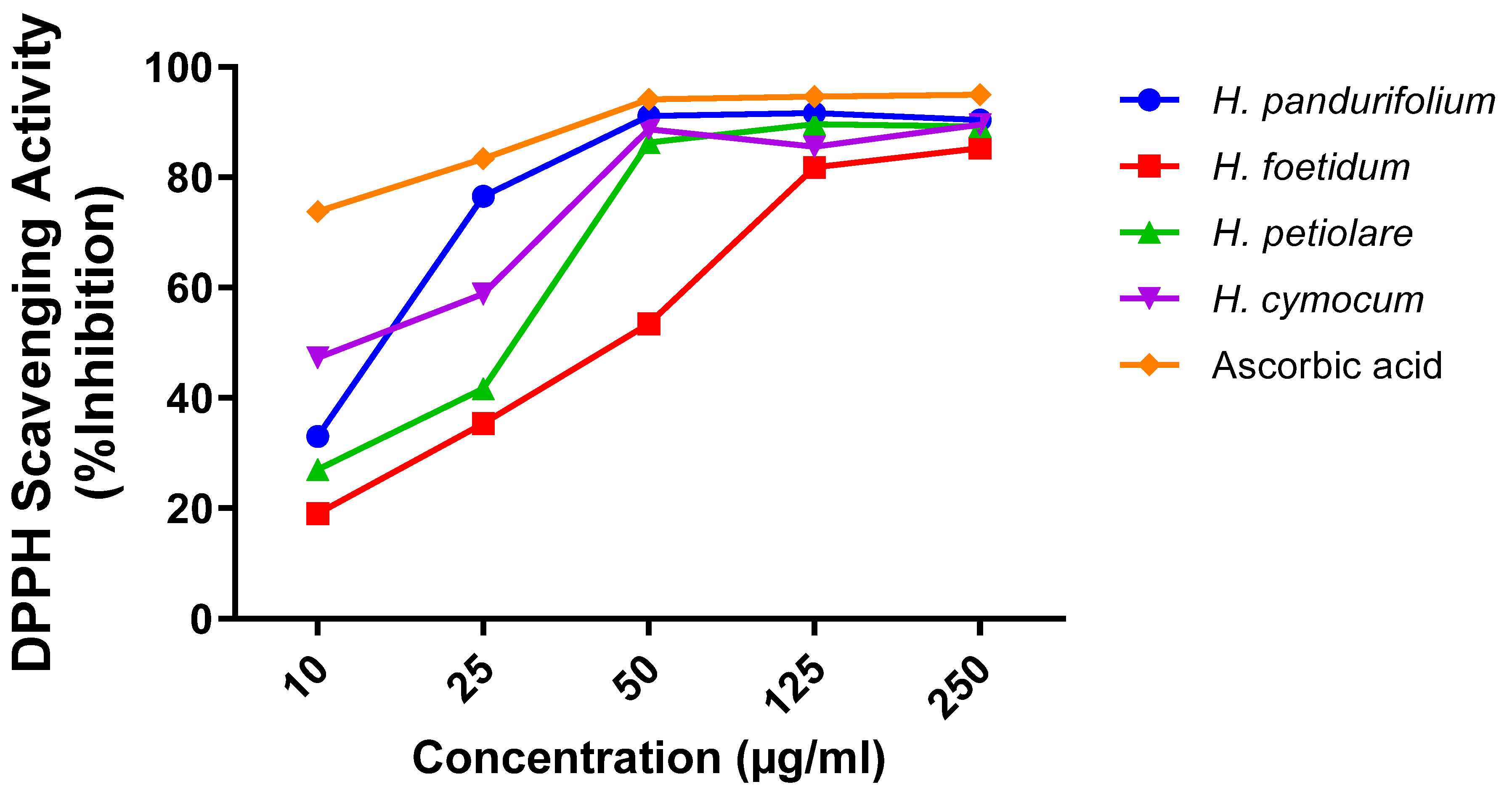
H. pandurifolium, H. foetidum, H. petiolare, and H. cymocum produced radical decolourisation of the DPPH solution because of the high free radical scavenging activity of the plant extracts [5,48].
The results reveal that the extracts tested have a dose-dependent activity. In fact, at the concentration of 250 μg/mL, the aqueous acetone extracts tested reduce the DPPH radical with an excellent percentage of 90.36 ± 1.00%, 85.28 ± 1.34%, 89.18 ± 0.59%, 89.55 ± 1.22%, 89.55 ± 1.22% for aqueous acetone extracts of H. pandurifolium, H. foetidum H. petiolare, and H. cymocum, respectively. Additionally, the IC50 is inversely proportional to the antioxidant capacity of a compound. However, the lowest value of IC50 indicates a strong antioxidant capacity of a compound. H. Cymocum showed the lowest IC50 values of 11.85 ± 3.20 µg/mL which had better antioxidant activity compared with H. pandurifolium, H. foetidum, and H. petiolare, (Figure 1). The antioxidant power of the aqueous acetone extracts could be explained by the presence of phenolic compounds including flavonoids present in the species of Helichrysum studied and which are known as antioxidant substances with the ability to trap radical species and reactive forms of oxygen. (Figure 1).
The results of the IC50 DPPH assay of the methanolic extracts of similar species namely H. dasyanthum, H. excisum, and H. felinum were 12.33, 13.67, and 20.71 μg/mL, respectively, which were within the range of the IC50 obtained in our study, as reported by Lourens et al. [21]. However, only the H. pandurufolium of IC50 (41.13 ± 3.62 µg/mL) is similar and in agreement with those reported [49] with the species name, H. chionophilum, H. plicatum subsp. plicatum, and H. arenarium subsp. Aucheri having IC50 of 40.5, 48.0, and 47.6 μg/mL, respectively. From literature, the flavonoids are the main compounds in the helichrysum genus with remarkable antioxidant activity, as reported [49,50].
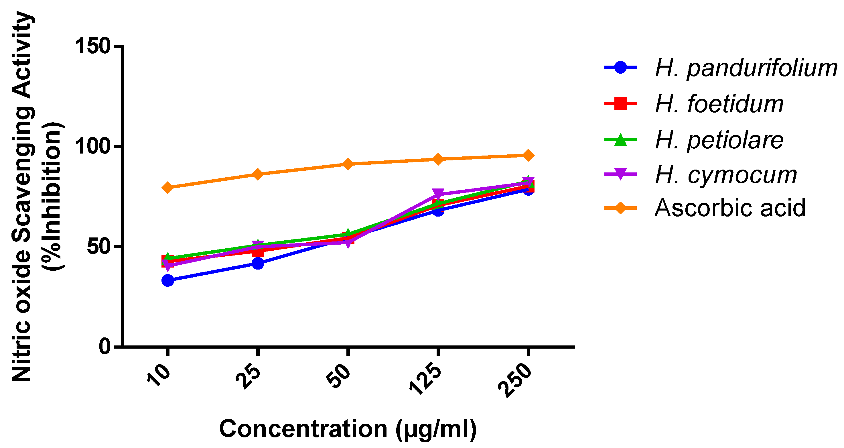
The reaction of sodium nitroprusside with oxygen produces nitric oxide and nitrite that scavenge free radicals via diazotisation with a sulphanilamide acid coupled reaction, producing a pink colour [51]. The antioxidant activities in NO assay involve the donation of protons to the nitrite radicals that show decreased absorbance. In line with the antioxidant activity, the nitric oxide scavenging revealed dose-dependent activity. It is worth mentioning here that all the doses are highly significant among the groups. Although H. petiolare (20.81 ± 3.73 µg/mL) with the lowest IC50 indicates the best nitric oxide scavenging effect and good antioxidant compared with the IC50 of H. pandurifolium (36.19 ± 2.08 µg/mL), H. foetidum (24.31 ± 3.67 µg/mL), and H. cymocum (24.68 ± 4.78 µg/mL). At the concentration of 250 μg/mL, the aqueous acetone extracts tested have NO scavenging activity with an excellent percentage of 78.64 ± 0.38%, 80.34 ± 0.38%, 82.67 ± 0.58%, 81.98 ± 0.48%, and 89.55 ± 1.22% for aqueous acetone extracts of H. pandurifolium, H. foetidum H. petiolare, and H. cymocum, respectively. The difference in the antioxidant of aqueous acetone extracts of H. pandurifolium, H. foetidum H. petiolare, and H. cymocum could be attributed to the variation in the chemical composition. Indeed, several types of bioactive compounds known for their antioxidant activity [52,53] are identified in H. petiolare with high levels of some compounds, including phenolic (caffeic acid, coniferaldehyde, protocatechuic acid, vanillin) compared to the other species (
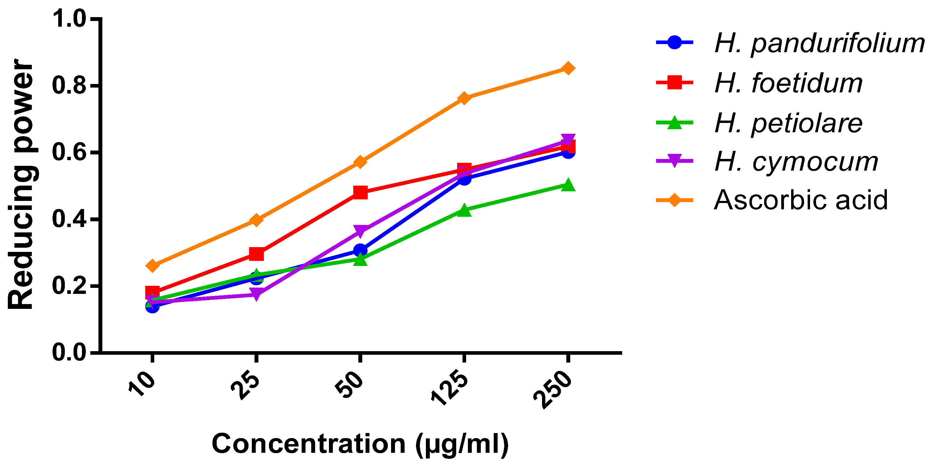
Table 4). The reducing power of natural products or plant extracts indicates their potential to transfer electrons from Fe3+ to Fe2+, which is synonymous with the antioxidant activity and is linked to reductones that donate a hydrogen atom to break the free radical chain, thus preventing peroxide formation [54]. The colour change from yellow to various shades of green and blue following treatment is dependent on the reducing power of the plant extract, with the blue colour indicating the highest reducing power. Thus, with increasing concentration of the aqueous acetone extract of H. pandurifolium, H. foetidum, H. petiolare, and H. cymocum, the observed blue colour indicates greater reducing power, which is similar to results in previous studies [55,56].
Consequently, the decrease in absorbance observed is an indication of the extent of nitrite radical scavenging potentials [57] and this could be attributed to components such as flavonoids, as reported in previous studies [58,59]. Similarly, the aqueous acetone extracts of H. pandurifolium, H. foetidum H. petiolare, and H. cymocum can act as natural antioxidants with relative activities scavenging free radical species. The reducing ability or potential is synonymous with the free radical scavenging activity of the plant extracts which is attributable to different amounts of the plant’s phytochemicals constituents [
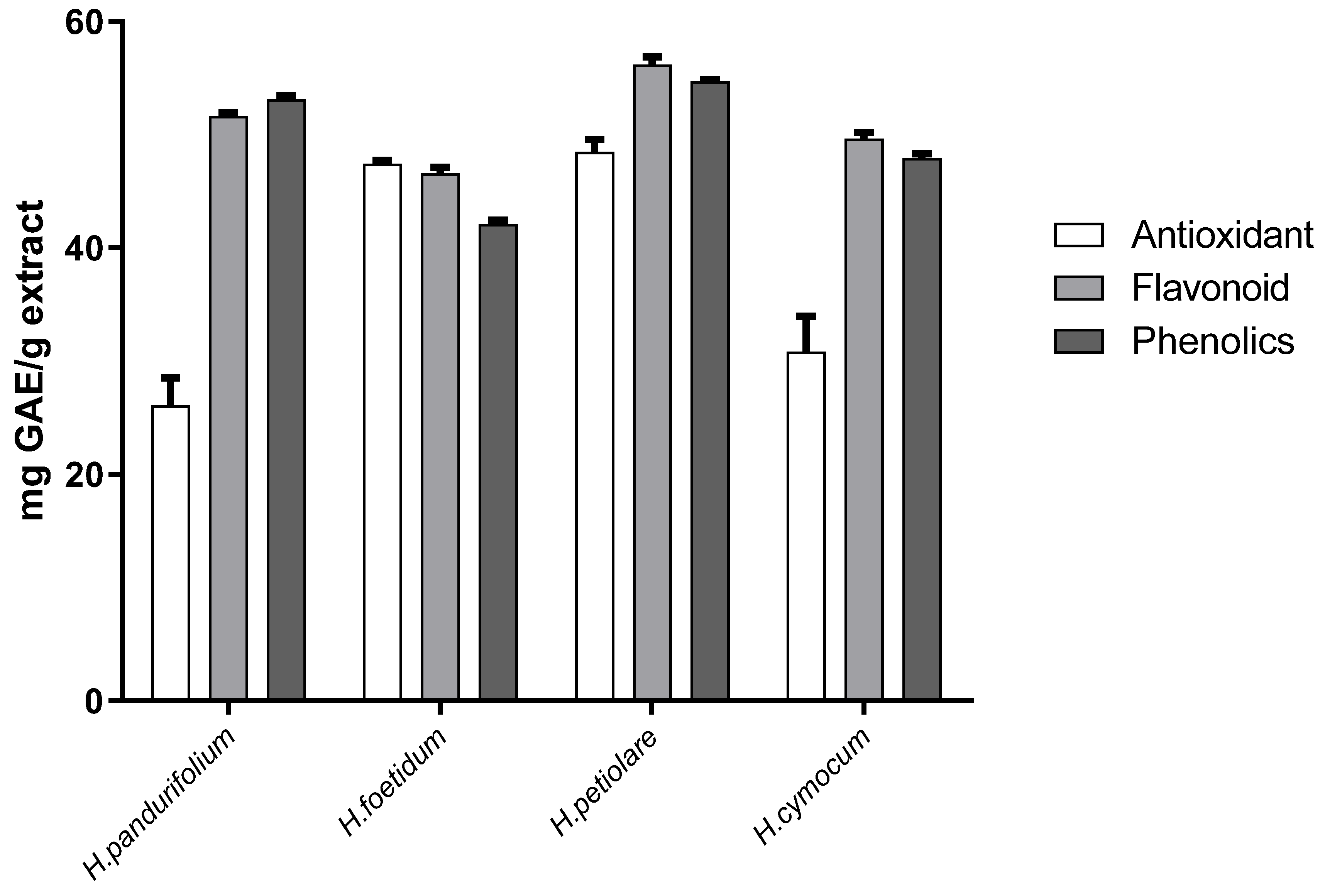
6] Overall, the antioxidant activities of these plant extracts are attributed to the constituents of total phenolic, total flavonoid, and total antioxidant capacity.
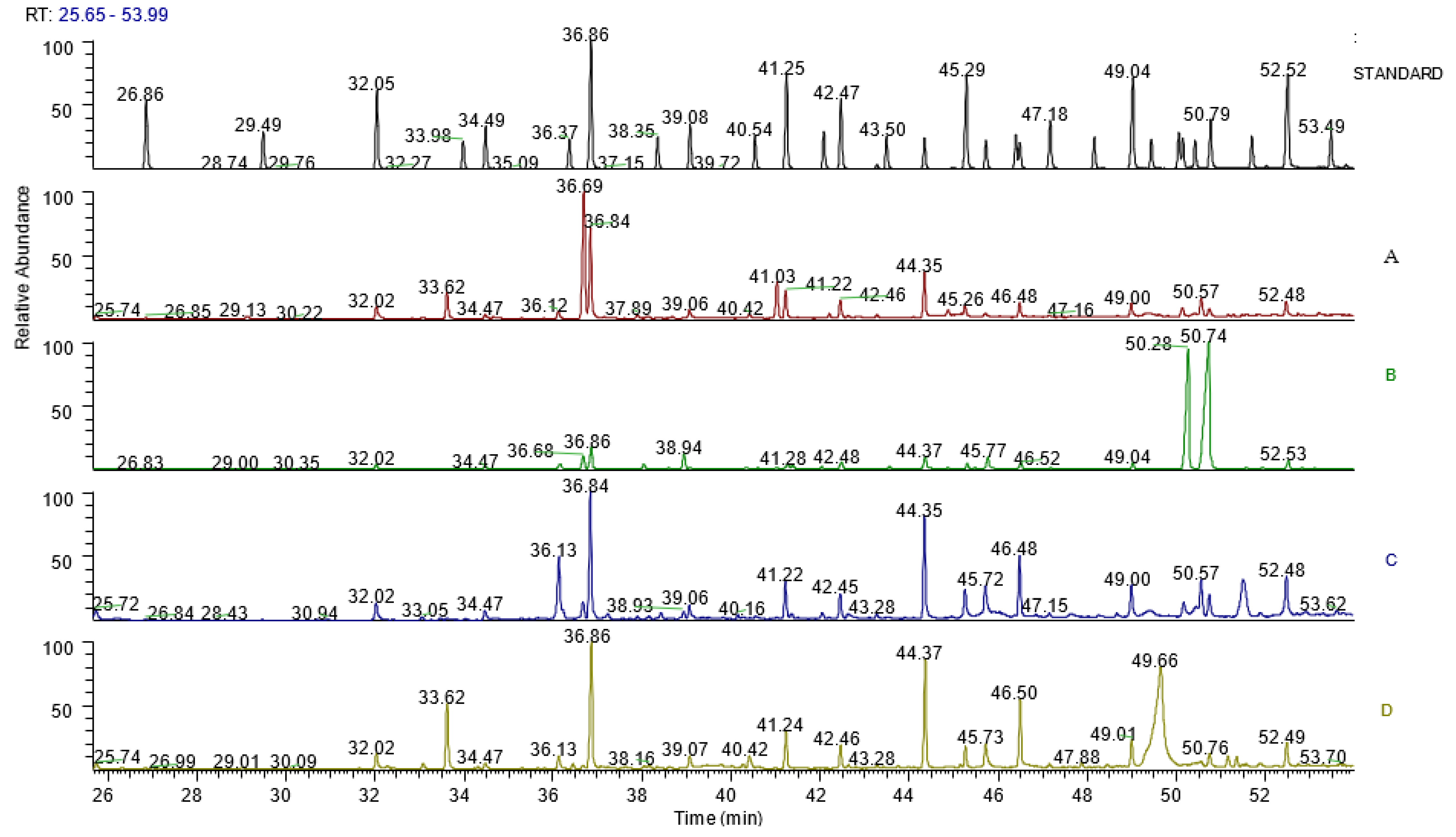
The fatty acid and lipid composition of the aqueous acetone extracts of H. pandurifolium, H. foetidum, H. petiolare, and H. cymocum were determined by fatty acid methyl esters (FAMEs) analysis involving the derivatisation, which was analysed by gas chromatography [60]. Previous studies have shown that geographical location, plant species, and seasonal changes could influence the fatty acid content of plants [61,62]. Unsaturated (monounsaturated and polyunsaturated) fatty acids have been reported to ameliorate cardiovascular diseases, modulate inflammation and support the immune system against cancer, diabetes mellitus, neurodegenerative diseases, etc. [63,64]. This study has shown that the aqueous acetone extracts of H. pandurifolium, H. foetidum H. petiolare, and H. cymocum contain various amounts of fatty acids with different compositions, as previously reported [61,65]. Our results showed two monounsaturated (MUFA) and two polyunsaturated fatty acids (PUFA), most of which cannot be synthesised by the human body and are only available in dietary sources, making them of great nutritional health benefit [65,66]. Stearic acid (C18:0), oleic acid (C18:1n9 (cis)), and linoleic acid (C18:2n9 (cis)), with known health benefits, were high in the aqueous acetone extracts of H. pandurifolium, H. foetidum H. petiolare, and H. cymocum as revealed in Table 5, which is similar to findings from previous studies that involved different extractants, different parts, and different Helichrysum species, e.g., H. chionophilum and H. plicatum subsp. [65]. The high dietary fatty acid ratio of PUFA:SFA are implicated in oxidative stress and are prone to lipid peroxidation because PUFA is highly susceptible, however, raising the PUFA/SFA ratio in the body helps to prevent cardiovascular disease (CVD) and conditions [67]. The PUFA/SFA varied considerably in the aqueous acetone extracts of H. pandurifolium, H. foetidum H. petiolare, and H. cymocum having 0.604, 0.726, 0.975, and 1.202 (for PUFA/SFA), respectively in our study, and these were seen to be comparable with the values in some seaweed plants considered to be of great health benefits in literature [13]. To the best of our knowledge, no study has reported the comparative study of antioxidant activities, constituents, and fatty acid compositions of four selected aqueous acetone extracts of the Helichrysum species. However, few studies have investigated the antioxidant activity of one species of this plant [18]. The many folkloric benefits of the plants in the Helichrysum species are under-explored in scientific investigations [18]. Natural, plant-based fatty acids are considered to be the best sources of dietary fatty acids because it has been recommended to prevent cardiovascular (CVD) and other disease conditions [67]. Thus, they could serve as potential sources of effective nutraceutical compounds for the prevention of various disease conditions.
In conclusion, our work provides relevant information on the phenolic, flavonoid, antioxidant capacity, and fatty acid profiles of the aqueous acetone extracts of H. pandurifolium, H. foetidum H. petiolare, and H. cymocum which demonstrate significant antioxidant activities. Since these constituents have been reported in previous studies to be effective in the prevention and treatment of various diseases, further research leading to possible drug discovery and development from these four Helichrysum species, especially for diabetes and its related cognitive decline conditions, is encouraged.

Author Contributions

Conceptualisation, K.A.A., O.E.E., O.O.O. and G.D.H.; writing-original draft, K.A.A.; editing: K.A.A., O.E.E., O.O.O. and G.D.H.; supervision, O.E.E., O.O.O. and G.D.H. All authors have read and agreed to the published version of the manuscript.

Funding

The financial assistance from the Cape Peninsula University of Technology, RJ23, granted to Professor Oluwafemi Omoniyi Oguntibeju is acknowledged in this research.

Institutional Review Board Statement

Not applicable.

Informed Consent Statement

Not applicable.

Data Availability Statement

The data used to support the finding of this study is included in the article.

Acknowledgments

The authors thank Prof Cupido at the University Fort Hare (UFH), South Africa, for identifying the plants, and also appreciate the Central Research Laboratory in Stellenbosch University, South Africa, for the GC-MS analysis. The support of the following institutions towards the completion of this research article is appreciated: University of the Western Cape (UWC), Cape Town, South Africa; Cape Peninsula University of Technology (CPUT), Cape Town, South Africa; The Federal Polytechnic, Ado Ekiti, Nigeria; Khalifa University, Abu Dhabi, UAE.

Conflicts of Interest

The authors declare that they have no competing interests.

References

  1. Sies, H. Oxidative stress: Oxidants and antioxidants. Exp. Physiol. Transl. Integr. 1997, 82, 291–295. [Google Scholar] [CrossRef] [PubMed]
  2. Halliwell, B. Free radicals and antioxidants–quo vadis? Trends Pharmacol. Sci. 2011, 32, 125–130. [Google Scholar] [CrossRef] [PubMed]
  3. Pryor, W.A. Free radical reactions and their importance in biochemical systems. In Federation Proceedings; Federation of American Societies For Experimental Biology: Bethesda, MD, USA, 1973; pp. 1862–1869. [Google Scholar]
  4. Mittler, R. Oxidative stress, antioxidants and stress tolerance. Trends Plant Sci. 2002, 7, 405–410. [Google Scholar] [CrossRef]
  5. Saeed, N.; Khan, M.R.; Shabbir, M. Antioxidant activity, total phenolic and total flavonoid contents of whole plant extracts Torilis leptophylla L. BMC Comple. Altern. Med. 2012, 12, 1–12. [Google Scholar] [CrossRef] [PubMed] [Green Version]
  6. Olajuyigbe, O.O.; Afolayan, A.J. Phenolic content and antioxidant property of the bark extracts of Ziziphus mucronata Willd. subsp. mucronata Willd. BMC Complement. Altern. Med. 2011, 11, 1–8. [Google Scholar] [CrossRef] [Green Version]
  7. Zhao, Y.; Zhao, Q.; Lu, Q. Purification, structural analysis, and stability of antioxidant peptides from purple wheat bran. BMC Chem. 2020, 14, 1–12. [Google Scholar] [CrossRef]
  8. Ng, K.L.; Tan, G.H.; Khor, S.M. Graphite nanocomposites sensor for multiplex detection of antioxidants in food. Food Chem. 2017, 237, 912–920. [Google Scholar] [CrossRef]
  9. Li, Z.-j.; Yang, F.-j.; Yang, L.; Zu, Y.-g. Comparison of the antioxidant effects of carnosic acid and synthetic antioxidants on tara seed oil. Chem. Cent. J. 2018, 12, 1–6. [Google Scholar] [CrossRef] [Green Version]
  10. Zhao, H.-J.; Xu, J.-K.; Yan, Z.-H.; Ren, H.-Q.; Zhang, Y. Microplastics enhance the developmental toxicity of synthetic phenolic antioxidants by disturbing the thyroid function and metabolism in developing zebrafish. Environ. Int. 2020, 140, 105750. [Google Scholar] [CrossRef]
  11. Yang, X.; Sun, Z.; Wang, W.; Zhou, Q.; Shi, G.; Wei, F.; Jiang, G. Developmental toxicity of synthetic phenolic antioxidants to the early life stage of zebrafish. Sci. Total Environ. 2018, 643, 559–568. [Google Scholar] [CrossRef]
  12. Ham, J.; Lim, W.; You, S.; Song, G. Butylated hydroxyanisole induces testicular dysfunction in mouse testis cells by dysregulating calcium homeostasis and stimulating endoplasmic reticulum stress. Sci. Total Environ. 2020, 702, 134775. [Google Scholar] [CrossRef] [PubMed]
  13. Chen, J.; Liu, H. Nutritional indices for assessing fatty acids: A mini-review. Int. J. Mol. Sci. 2020, 21, 5695. [Google Scholar] [CrossRef] [PubMed]
  14. Nwakiban, A.P.A.; Cicolari, S.; Piazza, S.; Gelmini, F.; Sangiovanni, E.; Martinelli, G.; Bossi, L.; Carpentier-Maguire, E.; Tchamgoue, A.D.; Agbor, G. Oxidative stress modulation by cameroonian spice extracts in hepg2 cells: Involvement of nrf2 and improvement of glucose uptake. Metabolites 2020, 10, 182. [Google Scholar] [CrossRef]
  15. Khan, M.S.A.; Ahmad, I. Herbal medicine: Current trends and future prospects. In New Look to Phytomedicine; Elsevier: Amsterdam, The Netherlands, 2019; pp. 3–13. [Google Scholar]
  16. Reidel, R.V.B.; Cioni, P.L.; Ruffoni, B.; Cervelli, C.; Pistelli, L. Aroma profile and essential oil composition of Helichrysum species. Nat. Prod. Commun. 2017, 12, 1934578. [Google Scholar]
  17. Aslan, M.; Orhan, D.D.; Orhan, N.; Sezik, E.; Yeşilada, E. A study of antidiabetic and antioxidant effects of Helichrysum graveolens capitulums in streptozotocin-induced diabetic rats. J. Med. Food 2007, 10, 396–400. [Google Scholar] [CrossRef] [PubMed]
  18. Akinyede, K.A.; Cupido, C.N.; Hughes, G.D.; Oguntibeju, O.O.; Ekpo, O.E. Medicinal Properties and In Vitro Biological Activities of Selected Helichrysum Species from South Africa: A Review. Plants 2021, 10, 1566. [Google Scholar] [CrossRef]
  19. Tirillini, B.; Menghini, L.; Leporini, L.; Scanu, N.; Marino, S.; Pintore, G. Antioxidant activity of methanol extract of Helichrysum foetidum Moench. Nat. Prod. Res. 2013, 27, 1484–1487. [Google Scholar] [CrossRef]
  20. François, T.; Lambert, S.M.; Dongmo, J.; Michel, P.; Gaby, N.; Fabrice, F.; Zache, N.; Henri, A.; Chantal, M. Composition, radical scavenging and antifungal activities of essential oils from 3 Helichrysum species growing in Cameroon against Penicillium oxalicum a yam rot fungi. Afr. J. Agric. Res. 2010, 5, 121–127. [Google Scholar]
  21. Lourens, A.; Reddy, D.; Başer, K.; Viljoen, A.; Van Vuuren, S. In vitro biological activity and essential oil composition of four indigenous South African Helichrysum species. J. Ethnopharmacol. 2004, 95, 253–258. [Google Scholar] [CrossRef]
  22. Nasr, A.; Zhou, X.; Liu, T.; Yang, J.; Zhu, G.-P. Acetone-water mixture is a competent solvent to extract phenolics and antioxidants from four organs of Eucalyptus camaldulensis. Turk. J. Biochem. 2019, 44, 231–239. [Google Scholar] [CrossRef]
  23. Chen, Y.-X.; Liu, X.-Y.; Xiao, Z.; Huang, Y.-F.; Liu, B. Antioxidant activities of polysaccharides obtained from Chlorella pyrenoidosa via different ethanol concentrations. Int. J. Biol. Macromol. 2016, 91, 505–509. [Google Scholar] [CrossRef]
  24. Silva, I.K.; Soysa, P. Evaluation of phytochemical composition and antioxidant capacity of a decoction containing Adenanthera pavonina L. and Thespesia populnea L. Pharmacogn. Mag. 2011, 7, 193. [Google Scholar]
  25. Oraiza, M. Studies on product of browning reaction prepared from glucosamine. Jpn. J. Nutr. 1986, 44, 307–315. [Google Scholar]
  26. McDonald, S.; Prenzler, P.D.; Antolovich, M.; Robards, K. Phenolic content and antioxidant activity of olive extracts. Food Chem. 2001, 73, 73–84. [Google Scholar] [CrossRef]
  27. Chang, C.-C.; Yang, M.-H.; Wen, H.-M.; Chern, J.-C. Estimation of total flavonoid content in propolis by two complementary colorimetric methods. J. Food Drug Anal. 2002, 10, 155. [Google Scholar]
  28. Prieto, P.; Pineda, M.; Aguilar, M. Spectrophotometric quantitation of antioxidant capacity through the formation of a phosphomolybdenum complex: Specific application to the determination of vitamin E. Anal. Biochem. 1999, 269, 337–341. [Google Scholar] [CrossRef] [PubMed]
  29. Motalleb, G.; Hanachi, P.; Kua, S. Evaluation of phenolic content and total antioxidant activity in Berberis vulgaris fruit extract. J. Biol. Sci. 2005, 5, 648–653. [Google Scholar]
  30. Kumar, V.; Roy, B.K. Population authentication of the traditional medicinal plant Cassia tora L. based on ISSR markers and FTIR analysis. Sci. Rep. 2018, 8, 1–11. [Google Scholar] [CrossRef]
  31. Aryal, S.; Baniya, M.K.; Danekhu, K.; Kunwar, P.; Gurung, R.; Koirala, N. Total phenolic content, flavonoid content and antioxidant potential of wild vegetables from Western Nepal. Plants 2019, 8, 96. [Google Scholar] [CrossRef] [Green Version]
  32. Doshi, P.; Adsule, P.; Banerjee, K. Phenolic composition and antioxidant activity in grapevine parts and berries (Vitis vinifera L.) cv. Kishmish Chornyi (Sharad Seedless) during maturation. Int. J. Food Sci. Technol. 2006, 41, 1–9. [Google Scholar] [CrossRef]
  33. Bland, J.S. Oxidants and antioxidants in clinical medicine: Past, present and future potential. J. Nutr. Environ. Med. 1995, 5, 255–280. [Google Scholar] [CrossRef]
  34. Thomas, M.J. The role of free radicals and antioxidants: How do we know that they are working? Crit. Rev. Food Sci. Nutr. 1995, 35, 21–39. [Google Scholar] [CrossRef] [PubMed]
  35. Pokorný, J. Are natural antioxidants better–and safer–than synthetic antioxidants? Eur. J. Lipid Sci. Technol. 2007, 109, 629–642. [Google Scholar] [CrossRef]
  36. Riedl, K.M.; Lee, J.H.; Renita, M.; St Martin, S.K.; Schwartz, S.J.; Vodovotz, Y. Isoflavone profiles, phenol content, and antioxidant activity of soybean seeds as influenced by cultivar and growing location in Ohio. J. Sci. Food Agric. 2007, 87, 1197–1206. [Google Scholar] [CrossRef]
  37. Kotha, R.R.; Luthria, D.L. Curcumin: Biological, pharmaceutical, nutraceutical, and analytical aspects. Molecules 2019, 24, 2930. [Google Scholar] [CrossRef] [Green Version]
  38. Gunathilake, K.; Ranaweera, K.; Rupasinghe, H. Change of phenolics, carotenoids, and antioxidant capacity following simulated gastrointestinal digestion and dialysis of selected edible green leaves. Food Chem. 2018, 245, 371–379. [Google Scholar] [CrossRef]
  39. Wang, T.-y.; Li, Q.; Bi, K.-S. Bioactive flavonoids in medicinal plants: Structure, activity and biological fate. Asian J. Pharm. Sci. 2018, 13, 12–23. [Google Scholar] [CrossRef]
  40. Tapiero, H.; Tew, K.; Ba, G.N.; Mathe, G. Polyphenols: Do they play a role in the prevention of human pathologies? Biomed. Pharmacother. 2002, 56, 200–207. [Google Scholar] [CrossRef]
  41. Croteau, R.; Kutchan, T.M.; Lewis, N.G. Natural products (secondary metabolites). Biochem. Mol. Biol. Plants 2000, 24, 1250–1319. [Google Scholar]
  42. Tungmunnithum, D.; Thongboonyou, A.; Pholboon, A.; Yangsabai, A. Flavonoids and other phenolic compounds from medicinal plants for pharmaceutical and medical aspects: An overview. Medicines 2018, 5, 93. [Google Scholar] [CrossRef]
  43. Kumar, S.; Pandey, A.K. Chemistry and biological activities of flavonoids: An overview. Sci. World J. 2013, 2013, 3506. [Google Scholar] [CrossRef] [PubMed] [Green Version]
  44. Leyva-Porras, C.; Román-Aguirre, M.; Cruz-Alcantar, P.; Pérez-Urizar, J.T.; Saavedra-Leos, M.Z. Application of Antioxidants as an Alternative Improving of Shelf Life in Foods. Polysaccharides 2021, 2, 594–607. [Google Scholar] [CrossRef]
  45. Frankel, E.N.; Meyer, A.S. The problems of using one-dimensional methods to evaluate multifunctional food and biological antioxidants. J. Sci. Food Agric. 2000, 80, 1925–1941. [Google Scholar] [CrossRef]
  46. Nunes, X.P.; Silva, F.S.; Almeida, J.R.G.d.S.; Barbosa Filho, J.M.; de Lima, J.T.; de Araújo Ribeiro, L.A.; Júnior, L.J.Q. Biological Oxidations and Antioxidant Activity of Natural Products; Intech Open Access Publisher: New York, NY, USA, 2012. [Google Scholar]
  47. Kaurinovic, B.; Vastag, D. Flavonoids and phenolic acids as potential natural antioxidants. In Antioxidants; IntechOpen: London, UK, 2019; pp. 1–20. [Google Scholar]
  48. Krishnaiah, D.; Sarbatly, R.; Nithyanandam, R. A review of the antioxidant potential of medicinal plant species. Food Bioprod. Processing 2011, 89, 217–233. [Google Scholar] [CrossRef]
  49. Tepe, B.; Sokmen, M.; Akpulat, H.A.; Sokmen, A. In vitro antioxidant activities of the methanol extracts of four Helichrysum species from Turkey. Food Chem. 2005, 90, 685–689. [Google Scholar] [CrossRef]
  50. Czinner, E.; Hagymasi, K.; Blazovics, A.; Kery, A.; Szőke, É.; Lemberkovics, E. The in vitro effect of Helichrysi flos on microsomal lipid peroxidation. J. Ethnopharmacol. 2001, 77, 31–35. [Google Scholar] [CrossRef]
  51. Balakrishnan, N.; Panda, A.; Raj, N.; Shrivastava, A.; Prathani, R. The evaluation of nitric oxide scavenging activity of Acalypha indica Linn root. Asian J. Res. Chem. 2009, 2, 148–150. [Google Scholar]
  52. Sayah, K.; El Omari, N.; Kharbach, M.; Bouyahya, A.; Kamal, R.; Marmouzi, I.; Cherrah, Y.; Faouzi, M.E.A. Comparative Study of Leaf and Rootstock Aqueous Extracts of Foeniculum vulgare on Chemical Profile and In Vitro Antioxidant and Antihyperglycemic Activities. Adv. Pharmacol. Pharm. Sci. 2020, 2020, 122. [Google Scholar] [CrossRef]
  53. Zengin, G.; Sinan, K.I.; Ak, G.; Angeloni, S.; Maggi, F.; Caprioli, G.; Kaplan, A.; Çakılcıoğlu, U.; Akan, H.; Jugreet, S. Preliminary investigation on chemical composition and bioactivity of differently obtained extracts from Symphytum aintabicum Hub.-Mor. &Wickens. Biochem. Syst. Ecol. 2021, 94, 104203. [Google Scholar]
  54. Govindan, P.; Muthukrishnan, S. Evaluation of total phenolic content and free radical scavenging activity of Boerhavia erecta. J. Acute Med. 2013, 3, 103–109. [Google Scholar] [CrossRef] [Green Version]
  55. Hazra, B.; Biswas, S.; Mandal, N. Antioxidant and free radical scavenging activity of Spondias pinnata. BMC Complementary Altern. Med. 2008, 8, 1–10. [Google Scholar] [CrossRef] [PubMed] [Green Version]
  56. Sharma, S.; Vig, A.P. Preliminary phytochemical screening and in vitro antioxidant activities of Parkinsonia aculeata Linn. BioMed Res. Int. 2014, 2014, 26. [Google Scholar] [CrossRef] [PubMed] [Green Version]
  57. Turkoglu, A.; Duru, M.E.; Mercan, N.; Kivrak, I.; Gezer, K. Antioxidant and antimicrobial activities of Laetiporus sulphureus (Bull.) Murrill. Food Chem. 2007, 101, 267–273. [Google Scholar] [CrossRef]
  58. Lakhanpal, P.; Rai, D.K. Quercetin: A versatile flavonoid. Internet J. Med. Update 2007, 2, 22–37. [Google Scholar] [CrossRef] [Green Version]
  59. Boora, F.; Chirisa, E.; Mukanganyama, S. Evaluation of nitrite radical scavenging properties of selected Zimbabwean plant extracts and their phytoconstituents. J. Food Processing 2014, 2014, 149. [Google Scholar] [CrossRef] [Green Version]
  60. Eder, K. Gas chromatographic analysis of fatty acid methyl esters. J. Chromatogr. B: Biomed. Sci. Appl. 1995, 671, 113–131. [Google Scholar] [CrossRef]
  61. Foseid, L.; Devle, H.; Stenstrøm, Y.; Naess-Andresen, C.F.; Ekeberg, D. Fatty acid profiles of stipe and blade from the Norwegian brown macroalgae Laminaria hyperborea with special reference to acyl glycerides, polar lipids, and free fatty acids. J. Lipids 2017, 2017, 257. [Google Scholar] [CrossRef] [Green Version]
  62. Schmid, M.; Stengel, D.B. Intra-thallus differentiation of fatty acid and pigment profiles in some temperate Fucales and Laminariales. J. Phycol. 2015, 51, 25–36. [Google Scholar] [CrossRef]
  63. Ahmad, S.; Ahmad, S.; Bibi, A.; Ishaq, M.S.; Afridi, M.S.; Kanwal, F.; Zakir, M.; Fatima, F. Phytochemical analysis, antioxidant activity, fatty acids composition, and functional group analysis of Heliotropium bacciferum. Sci. World J. 2014, 2014, 29. [Google Scholar] [CrossRef]
  64. Calder, P. Dietary fatty acids and the immune system. Lipids 1999, 34, S137–S140. [Google Scholar] [CrossRef]
  65. Acet, T.; Ozcan, K.; Zengin, G. An assessment of phenolic profiles, fatty acid compositions, and biological activities of two Helichrysum species: H. plicatum and H. chionophilum. J. Food Biochem. 2020, 44, e13128. [Google Scholar] [CrossRef] [PubMed]
  66. Sabudak, T.; Ozturk, M.; Goren, A.C.; Kolak, U.; Topcu, G. Fatty acids and other lipid composition of five Trifolium species with antioxidant activity. Pharm. Biol. 2009, 47, 137–141. [Google Scholar] [CrossRef] [Green Version]
  67. Kang, M.J.; Shin, M.S.; Park, J.N.; Lee, S.S. The effects of polyunsaturated: Saturated fatty acids ratios and peroxidisability index values of dietary fats on serum lipid profiles and hepatic enzyme activities in rats. Br. J. Nutr. 2005, 94, 526–532. [Google Scholar] [CrossRef] [PubMed] [Green Version]
Figure 1. Percentage (%) inhibition of aqueous acetone extract of H. pandurifolium, H. foetidum H. petiolare, and H. cymocum.
Plants 11 00998 g001
Figure 2. Percentage (%) inhibition of aqueous acetone extract of H. pandurifolium, H. foetidum H. petiolare, and H. cymocum.
Plants 11 00998 g002
Figure 3. Reducing power activity of aqueous acetone extract of H. pandurifolium, H. foetidum H. petiolare, and H. cymocum.
Plants 11 00998 g003
Figure 4. Antioxidant profiles of the aqueous acetone extracts of H. pandurfolium, H. foetidum, H. petiolare, and H. cymocum. Data is illustrated as mean ± SD (n = 3).
Plants 11 00998 g004
Figure 5. Shows the GC-MS chromatogram of the fatty acids of aqueous acetone extract of H.pandurifolium, H.foetidum H.petiolare, and H.cymocum with regularly labelled signals detected by the GC-MS detector. LEGEND: (A)—FAMEs chromatogram of H. pandurifolium; (B)—FAMEs chromatogram of H. foetidum; (C)—FAMEs chromatogram of H. petiolare; (D)—FAMEs chromatogram of H. cymocum.
Plants 11 00998 g005
Table 1. DPPH scavenging activity of aqueous acetone extracts of H. pandurifolium, H. foetidum H. petiolare, and H. cymocum.
Extract Concentration (µg/mL)H. pandurifoliumH. foetidumH. petiolareH. cymocumAscorbic Acid
1033.04 ± 1.78 bde19.06 ± 3.22 acde27.08 ± 5.33 bde47.31 ± 1.41 bce73.77 ± 5.10 abcd
2576.53 ± 2.65 bcd35.39 ± 2.34 ade41.72 ± 8.80 ade58.86 ± 6.3 abce83.38 ± 0.79 bcd
5091.10 ± 0.67 b53.50 ± 3.52 acde86.31 ± 1.81 be88.67 ± 0.46 b94.11 ± 0.55 bc
12591.61 ± 1.11 b81.83 ± 0.67 ace89.62 ± 0.45 b85. 58 ± 2.67 e94.63 ± 0.67 bd
250 90.36 ± 1.0085.28 ± 1.34 e89.18 ± 0.5989.55 ± 1.2294.99 ± 0.55 b
Data is presented as a mean ± SD value (n = 3); (a–e) represents significance (p < 0.05) when compared among the groups.
Table 2. Nitric oxide scavenging activity of H. pandurifolium, H. foetidum H. petiolare, and H. cymocum aqueous acetone extracts.
Extract Concentration (µg/mL)H. pandurifoliumH. foetidumH. petiolareH. cymocumAscorbic Acid
1033.27 ± 0.50 bcde42.85 ± 0.29 ade44.24 ± 0.50 ade40.52 ± 0.58 abce79.58 ± 0.87 abcd
2541.78 ± 2.05 bcde47.95 ± 0.66 acde50.79 ± 1.47 abe50.09 ±0.38 abe86.14 ± 0.48 abcd
5054.51 ± 0.85 cde54.44 ± 0.38 cde56.27 ± 0.66 bde52.17 ± 1.14 abce91.37 ± 0.61 abcd
12568.24 ± 0.87 bcde70.63 ± 0.58 ade71.52 ± 0.61 ade76.05 ± 0.29 abce93.63 ± 0.40 abcd
250 78.64 ± 0.38 cde80.34 ± 0.38 ce82.67 ± 0.58 abe81.98 ± 0.48 ae95.71 ± 0.40 abcd
Data is presented as a mean ± SD value (n = 3); (a–e) represents significance (p < 0.05) when compared among the groups.
Table 3. Reducing power activity of aqueous acetone extracts of H. pandurifolium, H. foetidum H. petiolare, and H. cymocum.
Extract Concentration (µg/mL)H. pandurifoliumH. foetidumH. petiolareH. cymocumAscorbic Acid
100.140 ± 0.002 bcde0.181 ± 0.003 acde0.158 ± 0.003 abe0.152 ± 0.003 abe0.262 ± 0.005 abcd
250.224 ± 0.003 bd0.296 ± 0.006 acde0.234 ± 0.007 bde0.174 ± 0.003 bce0.398 ± 0.005 abcd
500.307 ± 0.004 bcde0.480 ± 0.004 acde0.282 ± 0.003 abde0.363 ± 0.004 abce0.572 ± 0.005 abcd
1250.522 ± 0.003 bcde0.549 ± 0.002 acde0.429 ± 0.005 abde0.537 ± 0.004 abce0.763 ± 0.008 abcd
250 0.602 ± 0.007 bcde0.619 ± 0.005 acde0.505 ± 0.010 abde0.636 ± 0.005 bce0.853 ± 0.008 abcd
Each value represents mean ± SD value (n = 3); (a–e) indicates significance at (p < 0.05) among groups.
Table 4. Phenolic acid and phenolic aldehyde composition (µg/g) of the acetone extracts of H. pandurifolium, H. foetidum, H. petiolare, and H. cymocum.
TMS Derivative Mass SpectrumPlant Extracts (Concentration, µg/g)
Pseudomolecular IonsFragment IonsH. pandurifoliumH. foetidumH. petiolareH. cymocum
Vanillin22420970.141.978.527.5
trans-cinnamic acid2202052.222.1229.646.4
Syringaldehyde25422423.18.2314.18.11
Vanillic acid31228210.643.514.813.6
Protocatechuic acid35531194.684.5297.5115.9
Coniferaldehyde2502353.71.1913.52.19
m-coumaric acid 2932490.3920.2590.1920.237
p-coumaric acid 3082030.0640.1220.0460.022
Syringic_Acid3423122.024.512.9513.7
Gallic_acid45828131.513.7180.6685.7
Sinapinaldehyde28026511.274.318.216.9
Ferulic_Acid338308144.361.298.453.7
Caffeic_Acid3962191328.2928.61424.31363.2
Legend: the acetone extracts of H. pandurifolium, H. foetidum, H. petiolare, and H. cymocum were identified by authentic certified reference materials (CRMs) and compared with calibration standards.
Table 5. Composition (µg/g) of saturated fatty acids, monounsaturated fatty acids, polyunsaturated fatty acids, and total fatty acids of the acetone extracts of H. pandurifolium, H. foetidum H. petiolare, and H. cymocum.
SFAMUFAPUFA
Concentration (µg/g)C12:0C13:0C14:0C15:0C16:0C17:0C18:0C20:0C21:0C22:0C24:0C16:1C18:1n9 (cis)C18:2n6 (cis)C18:3n3SFAMUFAPUFAPUFA:SFAn − 6n − 3(n − 6)/(n − 3)TFA
H. pandurifolium18.612.8451.8126.32446.8223.1572.1240.239.0286.9166.476.7595.92144.1624.94584.0672.72768.90.6042144.1624.93.438025.6
H. foetidum62.325.3493.2349.42128.7160.3644.7466.983.5497.6467.892.41057.92759.61147.95379.81150.33907.50.7262759.61147.92.4010437.5
H. petiolare22.17.9621.6318.83064.0261.6728.3695.8100.0705.7656.2286.4740.34688.62316.87182.01026.77005.50.9754688.62316.82.0215214.2
H. cymocum17.33.1372.0135.42487.8218.4563.7329.354.1439.4238.090.1516.73796.92041.34858.5606.85838.21.2023796.92041.31.8611303.5
SFA—Saturated fatty acids; MUFA—Monounsaturated fatty acids; PUFA—Polyunsaturated fatty acids; TFA—Total fatty acids.
Table 6. Shows fatty acids, area of the peaks, ratio area, and the retention time (R, time) obtained from gas chromatography-mass spectrometry (GC- MS) analysis of the aqueous acetone extract of H. pandurifolium, H. foetidum H. petiolare, and H. cymocum.
H. pandurifoliumH. foetidumH. petiolareH. cymocum
S/NNameR. TimeAreaArea RatioR. TimeAreaArea RatioR. TimeAreaArea RatioR. TimeAreaArea Ratio
1C12:026.831293900.32926.836694691.04326.841201390.33626.841966810.379
2C13:029.48842010.21429.472975640.46429.47554480.15529.49672030.129
3C14:032.0219080024.85432.0250514437.86932.0220779925.81732.0228438725.478
4C15:034.474920911.25234.4735647165.55334.4710368562.90334.479859861.899
5C16:036.841105745628.12836.862359276736.75236.841101404430.83436.862044557139.383
6C16:138.352060580.52438.344627680.89138.426708901.87838.344627680.891
7C1739.0612693553.22939.0721901593.41239.0611753303.2939.0720979684.041
8C18:041.2231207787.93941.28805815612.55341.2231074968.69941.24535263210.311
9C18:1n9c42.4621068535.35942.48895910513.95642.4520751415.80942.4632979516.353
10C19:1STD43.28393119N/A43.33641949N/A43.28357208N/A42.28519143N/A
11C182n6c44.35531428013.51844.371667959425.98344.35919907225.75344.371699573032.738
12C20:045.2613374183.40245.3159434549.25845.2628913268.09445.2731709626.108
13C18:3n346.4817002594.32546.52754751911.75746.48493891113.82646.5991961019.108
14C21:047.161878980.47847.1910356811.61347.153986861.11647.164903020.944
15C22:04916738194.25849.04657338510.244930581608.56149.0143542878.387
16C2452.4817971824.57252.531019243515.87752.48470944513.18452.4941364167.968
The peaks, ratio area, and the retention time obtained from gas chromatography-mass spectrometry (GC- MS) are described by the National Institute of Standards and Technology (NIST) library to that of a known compound. This is depicted in the different compositions of fatty acids in the above table.
Publisher’s Note: MDPI stays neutral with regard to jurisdictional claims in published maps and institutional affiliations.

