
Wild-Grown Romanian Helleborus purpurascens Approach to Novel Chitosan Phyto-Nanocarriers—Metabolite Profile and Antioxidant Properties

 ,
,  , ,
, , 
Abstract
:1. Introduction
2. Results and Discussion
2.1. Screening and Classification of the Differential Metabolites
2.2. Phyto-Nanocarriers
2.3. FT-IR Spectroscopy
2.4. X-ray Diffraction Spectroscopy
2.5. Scanning Electron Microscopy (SEM)
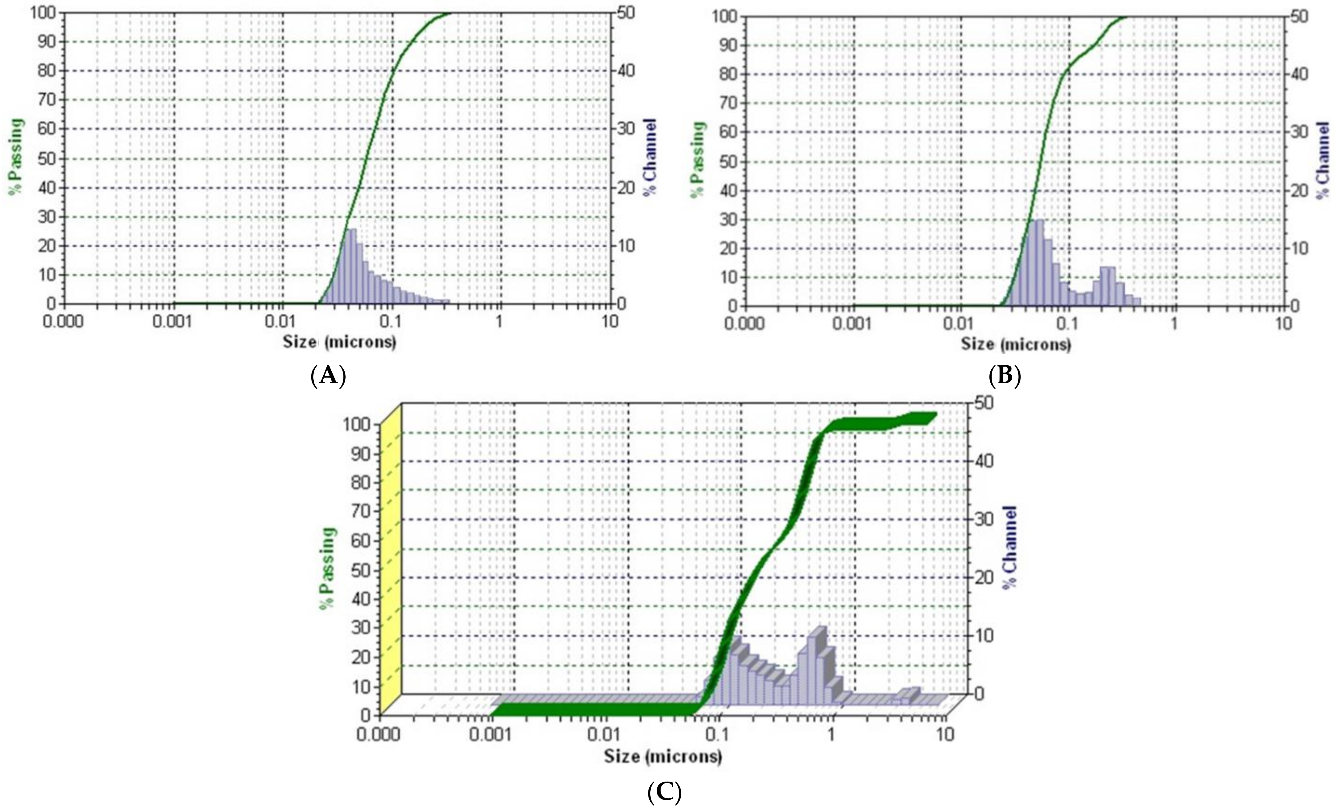
2.6. Dynamic Light Scattering
2.7. Thermal Behavior Study
2.8. Screening of the Antioxidant Activity
2.9. DPPH (1,1-Diphenyl-2-Picrylhydrazyl) Free Radical Scavenging Assay
2.10. Total Polyphenolic Contents (TPCs) Assay—Folin–Ciocalteu
2.11. Phosphomolybdate Assay (Total Antioxidant Capacity)
2.12. Iron(III)-Phenanthroline Antioxidant Assay
2.13. Encapsulation Efficiency, Loading Capacity and Encapsulation Yield (EY%)
3. Materials and Methods
3.1. Phyto-Carrier-System Components’ Preparation
Plant Samples’ Preparation for Chemical Screening
3.2. GC-MS Analysis
GC–MS Separation Conditions
3.3. Mass Spectrometry
3.3.1. Phyto-Carrier System Preparation (Hellebore-AgNPs System)
3.3.2. Chitosan Phyto-Nanocarrier Preparation (Encapsulation Procedure)
3.4. Characterization of Nanocarriers
3.4.1. Fourier Transform Infrared (FT-IR) Spectroscopy
3.4.2. XRD Spectroscopy
3.4.3. Scanning Electron Microscopy (SEM)
3.4.4. Dynamic Light Scattering (DLS) Particle Size Distribution Analysis
3.4.5. Thermal Analysis
3.4.6. Antioxidant Activity
3.5. Total Polyphenol Content Using the Folin–Ciocalteu Method
3.6. DPPH Radical Scavenging Assay
- A0 = vehicle control absorbance;
- Al = sample absorbance.
3.7. Phosphomolybdate Assay (Total Antioxidant Capacity)
3.8. Iron(III)-Phenanthroline Antioxidant Assay (OPM)
3.9. Encapsulation Yield, Encapsulation Efficiency and Loading Capacity
3.10. Encapsulation Efficiency (EE%)
3.11. Preparation of the Curves of the Concentrations of the Loaded Materials (Hellebore and Hellebore-AgNPs System)
3.12. Statistical Analysis
4. Conclusions
Supplementary Materials
Author Contributions
Funding
Data Availability Statement
Conflicts of Interest
References
- Balázs, V.L.; Filep, R.; Ambrus, T.; Kocsis, M.; Farkas, Á.; Stranczinger, S.; Papp, N. Ethnobotanical, historical and histological evaluation of Helleborus L. genetic resources used in veterinary and human ethnomedicine. Genet. Resour. Crop. Evol. 2020, 67, 781–797. [Google Scholar] [CrossRef]
- Maior, M.C.; Dobrotă, C. Natural compounds with important medical potential found in Helleborus sp. Cent. Eur. J. Biol. 2013, 8, 272–285. [Google Scholar]
- Borozan, A.B.; Popescu, S.; Moldovan, C.; Sărac, I.; Bordean, D. Helleborus—Phytochemistry and antimicrobial properties. A review. J. Hortic. For. Biotechnol. 2022, 26, 69–75. [Google Scholar]
- Segneanu, A.-E.; Damian, D.; Hulka, I.; Grozescu, I.; Salifoglou, A. A simple and rapid method for calixarene-based selective extraction of bioactive molecules from natural products. Amino Acids 2016, 48, 849–858. [Google Scholar] [CrossRef]
- Stroescu, V. Pharmacological Basis of Medical Practice; Medical Printing House: Bucharest, Romania, 1998; pp. 236–241. [Google Scholar]
- Neacsu, C.; Ciobanu, C.; Barbu, I.; Toader, O.; Szegli, G.; Kerek, F.; Babes, A. Substance MCS-18 isolated from Helleborus purpurascens is a potent antagonist of the capsaicin receptor, TRPV1, in rat cultured sensory neurons. Physiol. Res. 2010, 59, 289–298. [Google Scholar] [CrossRef] [PubMed]
- Kerek, F. Boicil, a new and very efficient antialgic spasmolytic and blood vessel regulating drug obtained from the plant Helleborus. Int. Conf. Chem. Biotechnol. Biol. Nat. Prod. 1981, 2, 22–37. [Google Scholar]
- Paun, G.; Neagu, E.; Radu, G.L.; Rotinberg, P.; Mihai, C. Evaluation of cytostatic potential of Helleborus purpurascens extracts concentrated by membrane techniques. Planta Med. 2011, 77, PF78. [Google Scholar] [CrossRef]
- Grigore, A.; Bubueanu, C.; Pirvu, L.; Neagu, G.; Bejanaru, I.; Vulturescu, V.; Panteli, M.; Rasit, I. Immunomodulatory effect of Helleborus purpurascens Waldst & Kit. Plants 2021, 10, 1990. [Google Scholar]
- Pilut, C.N.; Manea, A.; Macasoi, I.; Dobrescu, A.; Georgescu, D.; Buzatu, R.; Faur, A.; Dinu, S.; Chioran, D.; Pinzaru, I.; et al. Comparative evaluation of the potential antitumor of Helleborus purpurascens in skin and breast cancer. Plants 2022, 1, 194. [Google Scholar] [CrossRef]
- Segneanu, A.E.; Grozescu, I.; Cziple, F.; Berki, D.; Damian, D.; Niculite, C.M.; Florea, A.; Leabu, M. Helleborus purpurascens—Amino acid and peptide analysis linked to the chemical and antiproliferative properties of the extracted compounds. Molecules 2015, 20, 22170–22187. [Google Scholar] [CrossRef]
- Xie, P.; Wang, Y.; Wei, D.; Zhang, L.; Zhang, B.; Xiao, H.; Song, H.; Mao, X. Nanoparticle-based drug delivery systems with platinum drugs for overcoming cancer drug resistance. J. Mater. Chem. B 2021, 9, 5173–5194. [Google Scholar] [CrossRef] [PubMed]
- Available online: https://www.who.int/initiatives/who-global-centre-for-traditional-medicine (accessed on 18 June 2023).
- Atanasov, A.G.; Zotchev, S.B.; Dirsch, V.M.; Supuran, C.T. Natural products in drug discovery: Advances and opportunities. Nat. Rev. Drug Discov. 2021, 20, 200–216. [Google Scholar] [PubMed]
- Singh, I.P.; Ahmad, F.; Chatterjee, D.; Bajpai, R.; Sengar, N. Natural products: Drug discovery and development. In Drug Discovery and Development from Targets and Molecules to Medicines; Poduri, R., Ed.; Springer: Singapore, 2021; ISBN 978-981-15-5533-6. [Google Scholar]
- Nath, R.; Roy, R.; Barai, G.; Bairagi, S.; Manna, S.; Chakraborty, R. Modern developments of nano based grug delivery system by combined with phytochemicals-presenting new aspects. Int. J. Sci. Res. Sci. Technol. 2021, 8, 107–129. [Google Scholar]
- Patra, J.K.; Das, G.; Fraceto, L.F.; Campos, E.V.R.; del Pilar Rodriguez-Torres, M.; Acosta-Torres, L.S.; Diaz-Torres, L.A.; Grillo, R.; Swamy, M.K.; Sharma, S.; et al. Nano based drug delivery systems: Recent developments and future prospects. J. Nanobiotechnol. 2018, 16, 71. [Google Scholar]
- Rahman, H.S.; Othman, H.H.; Hammadi, N.I.; Yeap, S.K.; Amin, K.M.; Abdul Samad, N.; Alitheen, N.B. Novel drug delivery systems for loading of natural plant extracts and their biomedical applications. Int. J. Nanomed. 2020, 15, 2439–2483. [Google Scholar]
- Cele, T. Preparation of nanoparticles. In Engineered Nanomaterials-Health and Safety; IntechOpen: London, UK, 2020. [Google Scholar]
- Baig, N.; Kammakakam, I.; Falath, W. Nanomaterials: A review of synthesis methods, properties, recent progress, and challenges. Mater. Adv. 2021, 2, 1821–1871. [Google Scholar]
- Rane, A.V.; Kanny, K.; Abitha, V.K.; Thomas, S. Chapter 5—Methods for Synthesis of Nanoparticles and Fabrication of Nanocomposites. In Synthesis of Inorganic Nanomaterials; Woodhead Publishing: Cambridge, UK, 2018; pp. 121–139. [Google Scholar] [CrossRef]
- Chopra, H.; Bibi, S.; Singh, I.; Hasan, M.M.; Khan, M.S.; Yousafi, Q.; Baig, A.A.; Rahman, M.M.; Islam, F.; Emran, T.B.; et al. Green metallic nanoparticles: Biosynthesis to applications. Front. Bioeng. Biotechnol. 2022, 10, 874742. [Google Scholar]
- Hussein, H.A.; Abdullah, M.A. Novel drug delivery systems based on silver nanoparticles, hyaluronic acid, lipid nanoparticles and liposomes for cancer treatment. Appl. Nanosci. 2022, 12, 3071–3096. [Google Scholar] [CrossRef]
- Gomes, H.I.O.; Martins, C.S.M.; Prior, J.A.V. Silver nanoparticles as carriers of anticancer drugs for efficient target treatment of cancer cells. Nanomaterials 2021, 11, 964. [Google Scholar] [CrossRef]
- Galatage, S.T.; Hebalkar, S.A.; Dhobale, S.V.; Mali, O.R.; Kumbhar, P.S.; Nikade, S.V.; Killedar, S.G. Silver nanoparticles: Properties, synthesis, characterization, applications and future trends. In Silver Micro-Nanoparticles—Properties, Synthesis, Characterization, and Applications; Kumar, S., Kumar, P., Pathak, C.S., Eds.; IntechOpen: London, UK, 2021; ISBN 978-1-83968-660-3. [Google Scholar]
- Riva, R.; Ragelle, H.; des Rieux, A.; Duhem, N.; Jérôme, C.; Préat, V. Chitosan and chitosan derivatives in drug delivery and tissue engineering. Chitosan Biomater. 2011, II, 19–44. [Google Scholar]
- Domingues, J.M.; Miranda, C.S.; Silva, A.F.G.; Homem, N.C.; Amorim, M.T.P.; Felgueiras, H.P. Bioactivity of chitosan-based particles loaded with plant-derived extracts for biomedical applications: Emphasis on antimicrobial fiber- based systems. Mar. Drugs 2021, 19, 359. [Google Scholar]
- Ibrahim, H.M.; El-Zairy, E.M.R. Chitosan as a biomaterial—Structure, Properties and Electrospun Nanofibers. Concepts Compd. Altern. Antibact. 2015, 1, 81–101. [Google Scholar]
- Fuchs-Tarlovsky, V. Role of antioxidants in cancer therapy. Nutrition 2013, 29, 15–21. [Google Scholar] [CrossRef] [PubMed]
- Sardas, S. The Role of Antioxidants in Cancer Prevention and Treatment. Indoor Built Environ. 2003, 12, 401–404. [Google Scholar] [CrossRef]
- Luo, M.; Zhou, L.; Huang, Z.; Li, B.; Nice, E.C.; Xu, J.; Huang, C. Antioxidant Therapy in Cancer: Rationale and Progress. Antioxidants 2022, 11, 1128. [Google Scholar] [CrossRef]
- Akanji, M.A.; Fatinukun, H.D.; Rotimi, E.D.; Afolabi, B.L.; Adeyemi, O.S. The two sides of dietary antioxidants in cancer therapy. In Antioxidants-Benefits, Sources, Mechanisms of Action; Waisundara, V., Ed.; IntechOpen: London, UK, 2021; ISBN 978-1-83968-865-2. [Google Scholar]
- Yuan, H.; Ma, Q.; Ye, L.; Piao, G. The traditional medicine and modern medicine from natural products. Molecules 2016, 21, 559. [Google Scholar] [CrossRef]
- Yarnell, E. Synergy in herbal medicines: Part 1. J. Restor. Med. 2015, 4, 60–73. [Google Scholar] [CrossRef]
- Flieger, J.; Flieger, W.; Baj, J.; Maciejewski, R. Antioxidants: Classification, natural sources, activity/capacity measurements, and usefulness for the synthesis of nanoparticles. Materials 2021, 14, 4135. [Google Scholar] [CrossRef]
- Amorati, R.; Valgimigli, L. Methods to measure the antioxidant activity of phytochemicals and plant extracts. J. Agric. Food Chem. 2018, 66, 3324–3329. [Google Scholar] [CrossRef]
- Dinkova-Kostova, A.T.; Talalay, P. Direct and indirect antioxidant properties of inducers of cytoprotective proteins. Mol. Nutr. Food Res. 2008, 52, S128–S138. [Google Scholar] [CrossRef]
- Losada-Barreiro, S.; Sezgin-Bayindir, Z.; Paiva-Martins, F.; Bravo-Díaz, C. Biochemistry of antioxidants: Mechanisms and pharmaceutical applications. Biomedicines 2022, 10, 3051. [Google Scholar] [CrossRef] [PubMed]
- Shahidi, F.; Zhong, Y. Measurement of antioxidant activity. J. Funct. Foods 2015, 18, 757–781. [Google Scholar] [CrossRef]
- Wawrosch, C.; Zotchev, S.B. Production of bioactive plant secondary metabolites through in vitro technologies—Status and outlook. Appl. Microbiol. Biotechnol. 2021, 105, 6649–6668. [Google Scholar] [CrossRef]
- Ashraf, M.A.; Iqbal, M.; Rasheed, R.; Hussain, I.; Riaz, M.; Arif, M.S. Environmental stress and secondary metabolites in plants. In Plant Metabolites and Regulation under Environmental Stress; Academic Press: Cambridge, MA, USA, 2018; pp. 153–167. [Google Scholar]
- Segneanu, A.E.; Grozescu, I.; Sfirloaga, P. The influence of extraction process parameters of some biomaterials precursors from Helianthus annuus. Dig. J. Nanomater. Biostruct. 2013, 8, 1423–1433. [Google Scholar]
- Isah, T. Stress and defense responses in plant secondary metabolites production. Biol. Res. 2019, 52, 39. [Google Scholar] [CrossRef] [PubMed]
- Yang, L.; Wen, K.-S.; Ruan, X.; Zhao, Y.-X.; Wei, F.; Wang, Q. Response of plant secondary metabolites to environmental factors. Molecules 2018, 23, 762. [Google Scholar] [CrossRef]
- Ahmad, R.; Srivastava, S.; Ghosh, S.; Khare, S.K. Phytochemical delivery through nanocarriers: A review. Colloids Surf. B Biointerfaces 2021, 197, 111389. [Google Scholar] [CrossRef]
- Hill, C.B.; Roessner, U. Metabolic profiling of plants by GC–MS. In The Handbook of Plant Metabolomics, 1st ed.; Weckwerth, W., Kahl, G., Eds.; Wiley-VCH Verlag GmbH: Weinheim, Germany, 2013. [Google Scholar]
- Rossellia, S.; Maggio, A.; Formisano, C.; Napolitano, F.; Senatore, F.; Spadaro, V.; Bruno, M. Chemical composition and antibacterial activity of extracts of Helleborus bocconei Ten. subsp. intermedius. Nat. Prod. Commun. 2007, 2, 675–679. [Google Scholar] [CrossRef]
- Kusmenoglu, S.; Bingol, F.; Sener, B. Gas-chromatographic determination of the seed oil of Helleborus orientalis Lam. J. Fac. Pharm. Gazi 1995, 12, 23–27. [Google Scholar]
- Aziz, M.; Ahmad, S.; Khurshid, U.; Pervaiz, I.; Lodhi, A.H.; Jan, N.; Khurshid, S.; Arshad, M.A.; Ibrahim, M.M.; Mersal, G.A.M.; et al. Comprehensive biological potential, phytochemical profiling using GC-MS and LC-ESI-MS, and in-silico assessment of Strobilanthes glutinosus Nees: An important medicinal plant. Molecules 2022, 27, 6885. [Google Scholar] [CrossRef]
- Wu, L.; Zhao, J.; Wu, L.; Zhang, Y.; Li, J. Simultaneous determination of squalene, tocopherols and phytosterols in edible vegetable oil by SPE combined with saponification and GC-MS. LWT 2022, 169, 114026. [Google Scholar] [CrossRef]
- Hao, D.C. Mining Chemodiversity from biodiversity: Pharmacophylogeny of Ranunculaceae medicinal plants. In Ranunculales Medicinal Plants; Academic Press: Cambridge, MA, USA, 2019; pp. 35–71. [Google Scholar] [CrossRef]
- Vitalini, S.; Braca, A.; Fico, G. Study on secondary metabolite content of Helleborus niger L. leaves. Fitoterapia 2011, 82, 152–154. [Google Scholar] [CrossRef] [PubMed]
- Duckstein, S.M.; Stintzing, F.C. Comprehensive study of the phenolics and saponins from Helleborus niger L. leaves and stems by liquid chromatography/tandem mass spectrometry. Chem. Biodivers. 2014, 11, 276–298. [Google Scholar] [CrossRef] [PubMed]
- Deore, S.; Khadabadi, S. Isolation and characterization of phytoconstituents from Chlorophytum Borivilianum. Pharmacogn. Res. 2010, 2, 343. [Google Scholar] [CrossRef] [PubMed]
- Tsiftsoglou, O.S.; Stefanakis, M.K.; Lazari, D.M. Chemical constituents isolated from the rhizomes of Helleborus odorus subsp. cyclophyllus (Ranunculaceae). Biochem. Syst. Ecol. 2018, 79, 8–11. [Google Scholar] [CrossRef]
- Beridze, M.; Kalandia, A.; Japaridze, I.; Vanidze, M.; Varshanidze, N.; Turmanidze, N.; Dolidze, K.; Diasamidze, I.; Jakeli, E. Phytochemical study of endemic species Helleborus caucasicus, Helleborus abchasicus and Ficaria popovii spread in Southern Colchis. Proc. CBU Med. Pharm. 2020, 1, 1–7. [Google Scholar] [CrossRef]
- Jensen, U. Secondary compounds of the Ranunculiflorae. In Systematics and Evolution of the Ranunculiflorae; Plant Systematics and Evolution Supplement; Jensen, U., Kadereit, J.W., Eds.; Springer: Vienna, Austria, 1995; Volume 9. [Google Scholar]
- Cuny, E.; Klingler, D. Efficient isolation and structure analysis of (+)-ranuncoside, a unique tricyclic spiroacetal glycoside, from Christmas rose (Helleborus niger L.). Nat. Prod. Commun. 2022, 17, 1934578X2110694. [Google Scholar] [CrossRef]
- Schmitzer, V.; Mikulic-Petkovsek, M.; Stampar, F. Sepal phenolic profile during Helleborus niger flower development. J. Plant Physiol. 2013, 170, 1407–1415. [Google Scholar] [CrossRef]
- Li, Y.; Zhang, H.; Liang, X.; Song, B.; Zheng, X.; Wang, R.; Liu, L.; Song, X.; Liu, J. New cytotoxic bufadienolides from the roots and rhizomes of Helleborus thibetanus Franch. Nat. Prod. Res. 2018, 34, 950–957. [Google Scholar] [CrossRef]
- Brillatz, T.; Jacmin, M.; Vougogiannopoulou, K.; Petrakis, E.A.; Kalpoutzakis, E.; Houriet, J.; Pellissier, L.; Rutz, A.; Marcourt, L.; Queiroz, E.F.; et al. Antiseizure potential of the ancient Greek medicinal plant Helleborus odorus subsp. cyclophyllus and identification of its main active principles. J. Ethnopharmacol. 2020, 259, 112954. [Google Scholar] [CrossRef] [PubMed]
- Cheng, W.; Tan, Y.F.; Tian, H.Y.; Gong, X.W.; Chen, K.L.; Jiang, R.W. Two new bufadienolides from the rhizomes of Helleborus thibetanus with inhibitory activities against prostate cancer cells. Nat. Prod. Res. 2014, 28, 901–908. [Google Scholar] [CrossRef]
- Yokosuka, A.; Inomata, M.; Yoshizawa, Y.; Iguchi, T.; Mimaki, Y. Bufadienolides and ecdysteroids from the whole plants of Helleborus niger and their cytotoxicity. J. Nat. Med. 2021, 75, 393–402. [Google Scholar] [CrossRef] [PubMed]
- Colombo, M.L.; Tome, F.; Servettaz, O.; Bugatti, C. Phytochemical evaluation of Helleborus species growing in northern Italy. Int. J. Crude Drug Res. 1990, 28, 219–223. [Google Scholar] [CrossRef]
- Gu, C.; Mao, X.; Chen, D.; Yu, B.; Yang, Q. Isoleucine plays an iImportant role for maintaining immune function. Curr. Protein Pept. Sci. 2019, 20, 644–651. [Google Scholar] [CrossRef] [PubMed]
- Dhama, K.; Karthik, K.; Khandia, R.; Munjal, A.; Tiwari, R.; Rana, R.; Khurana, S.K.; Ullah, S.; Khan, R.U.; Alagawany, M.; et al. Medicinal and therapeutic potential of herbs and plant metabolites/Extracts countering viral pathogens-current knowledge and future prospects. Curr. Drug Metab. 2018, 19, 236–263. [Google Scholar] [CrossRef] [PubMed]
- Kim, S.H.; Roszik, J.; Grimm, E.A.; Ekmekcioglu, S. Impact of L-arginine metabolism on immune response and anticancer immunotherapy. Front. Oncol. 2018, 8, 67. [Google Scholar] [CrossRef]
- Chiangjong, W.; Chutipongtanate, S.; Hongeng, S. Anticancer peptide: Physicochemical property, functional aspect and trend in clinical application (Review). Int. J. Oncol. 2020, 57, 678–696. [Google Scholar] [CrossRef]
- Lieu, E.L.; Nguyen, T.; Rhyne, S.; Kim, J. Amino acids in cancer. Exp. Mol. Med. 2020, 52, 15–30. [Google Scholar] [CrossRef]
- Albaugh, V.L.; Pinzon-Guzman, C.; Barbul, A. Arginine metabolism and cancer. Surg. Oncol. March 2017, 115, 273–280. [Google Scholar] [CrossRef]
- Wei, Z.; Liu, X.; Cheng, C.; Yu, W.; Yi, P. Metabolism of amino acids in cancer. Front. Cell Dev. Biol. 2021, 8, 603837. [Google Scholar] [CrossRef]
- Azmi, S.; Hussain, M.K. Analysis of structures, functions, and transgenicity of phytopeptides defensin and thionin: A review. Beni-Suef Univ. J. Basic Appl. Sci. 2021, 10, 1–11. [Google Scholar] [CrossRef]
- Mani, S.; Bhatt, S.B.; Vasudevan, V.; Prabhu, D.; Rajamanikandan, S.; Velusamy, P.; Ramasamy, P.; Raman, P. The updated review on plant peptides and their applications in human health. Int. J. Pept. Res. Ther. 2022, 28, 135. [Google Scholar] [CrossRef]
- Slezina, M.P.; Odintsova, T.I. Plant antimicrobial peptides: Insights into structure-function relationships for practical applications. Curr. Issues Mol. Biol. 2023, 45, 3674–3704. [Google Scholar] [CrossRef]
- Liao, Y.; Wang, M.; Jiang, X. Sulfur-containing peptides: Synthesis and application in the discovery of potential drug candidates. Curr. Opin. Chem. Biol. 2023, 75, 102336. [Google Scholar] [CrossRef]
- Pirintsos, S.; Panagiotopoulos, A.; Bariotakis, M.; Daskalakis, V.; Lionis, C.; Sourvinos, G.; Karakasiliotis, I.; Kampa, M.; Castanas, E. From traditional ethnopharmacology to modern natural drug discovery: A methodology discussion and specific examples. Molecules 2022, 27, 4060. [Google Scholar] [CrossRef] [PubMed]
- Pang, Z.; Chen, J.; Wang, T.; Gao, C.; Li, Z.; Guo, L.; Xu, J.; Cheng, Y. Linking plant secondary metabolites and plant microbiomes: A Review. Front. Plant. Sci. 2021, 12, 621276. [Google Scholar] [CrossRef] [PubMed]
- Chen, G.; Wang, S.; Huang, X.; Hong, J.; Du, L.; Zhang, L.; Ye, L. Environmental factors affecting growth and development of banlangen (Radix Isatidis) in China. Afr. J. Plant. Sci. 2015, 9, 421–426. [Google Scholar]
- Kamboj, A.; Rathour, A.; Kaur, M. Bufadienolides and their medicinal utility: A review. Int. J. Pharm. Pharm. Sci. 2013, 5, 20–27. [Google Scholar]
- Li, F.; Weng, Y.; Wang, L.; He, H.; Yang, J.; Tang, X. The efficacy and safety of bufadienolides-loaded nanostructured lipid carriers. Int. J. Pharm. 2010, 393, 204–212. [Google Scholar] [CrossRef]
- Novotny, L.; Abbas Ghuloom, H.A.; Al-Hasawi, N.A. Structural features and biological activities of bufadienolides. J. Pharm. Biol. Chem. Sci. Res. 2019, 10, 1147–1157. [Google Scholar]
- Jiang, L.; Chi, C.; Yuan, F.; Lu, M.; Hu, D.; Wang, L.; Liu, X. Anti-inflammatory effects of anemonin on acute ulcerative colitis via targeted regulation of protein kinase C-θ. Chin. Med. 2022, 17, 1–12. [Google Scholar] [CrossRef] [PubMed]
- Husain, A.; Alam, M.M.; Shaharyar, M.; Lal, S. Antimicrobial activities of some synthetic butenolides and their pyrrolone derivatives. J. Enzym. Inhib. Med. Chem. 2009, 25, 54–61. [Google Scholar] [CrossRef] [PubMed]
- Das, N.; Mishra, S.K.; Bishayee, A.; Ali, E.S.; Bishayee, A. The phytochemical, biological, and medicinal attributes of phytoecdysteroids: An updated review. Acta Pharm. Sin. B 2021, 11, 1740–1766. [Google Scholar] [CrossRef]
- Cox-Georgian, D.; Ramadoss, N.; Dona, C.; Basu, C. Therapeutic and medicinal uses of terpenes. In Medicinal Plants; Joshee, N., Dhekney, S., Parajuli, P., Eds.; Springer: Cham, Switzerland, 2019; pp. 333–359. [Google Scholar]
- Fongang Fotsing, Y.S.; Bankeu Kezetas, J.J. Terpenoids as important bioactive constituents of essential oils. In Essential oils—Bioactive Compounds, New Perspectives and Application; Santana de Oliveira, M., Almeida da Costa, W., Gomes Silva, S., Eds.; Intechopen: London, UK, 2020; ISBN 978–1-83962–698–2. [Google Scholar]
- Islam, M.T.; Ali, E.S.; Uddin, S.J.; Shaw, S.; Islam, A.; Ahmed, I.; Shill, M.C.; Karmakar, U.K.; Yarla, N.S.; Khan, I.N.; et al. Phytol: A review of biomedical activities. Food Chem Toxicol. 2018, 121, 82–94. [Google Scholar] [CrossRef]
- Jahangeer, M.; Fatima, R.; Ashiq, M.; Basharat, A.; Qamar, S.A.; Bilal, M.; Iqbal, H.M. Therapeutic and biomedical potentialities of terpenoids–A review. J. Pure Appl. Microbiol. 2021, 15, 471–483. [Google Scholar] [CrossRef]
- Das, K.; Gezici, S. Secondary plant metabolites, their separation and identification, and role in human disease prevention. Ann. Phytomed. Int. J. 2018, 7, 13–24. [Google Scholar] [CrossRef]
- Martin, M.L.; Ortíz de Urbina, A.V.; Montero, M.J.; Carrón, R.; San Román, L. Pharmacologic effects of lactones isolated from Pulsatilla alpina subsp. apiifolia. J. Ethnopharmacol. 1988, 24, 185–191. [Google Scholar] [CrossRef]
- Mares, D. Antimicrobial activity of protoanemonin, a lactone from Ranunculaceous plants. Mycopathologia 1987, 98, 133–140. [Google Scholar] [CrossRef]
- Nagy, K.; Tiuca, I.D. Importance of fatty acids in physiopathology of human body. In Fatty Acids; Catala, A., Ed.; IntechOpen: London, UK, 2017. [Google Scholar]
- Kozłowska, A.; Szostak-Węgierek, D. Flavonoids–food sources, health benefits, and mechanisms involved. In Bioactive Molecules in Food; Reference Series in Phytochemistry; Mérillon, J.M., Ramawat, K., Eds.; Springer: Cham, Switzerland, 2018. [Google Scholar]
- Kozłowska, A.; Szostak-Wegierek, D. Flavonoids-food sources and health benefits. Rocz. Panstw. Zakl. Hig. 2014, 65, 79–85. [Google Scholar] [PubMed]
- Panche, A.N.; Diwan, A.D.; Chandra, S.R. Flavonoids: An overview. J. Nutr. Sci. 2016, 5, e47. [Google Scholar] [CrossRef]
- Rasouli, H.; Farzaei, M.H.; Khodarahmi, R. Polyphenols and their benefits: A review. Int. J. Food Prop. 2017, 20, 1700–1741. [Google Scholar] [CrossRef]
- Kumar, N.; Goel, N. Phenolic acids: Natural versatile molecules with promising therapeutic applications. Biotechnol. Rep. Amst. 2019, 24, e00370. [Google Scholar] [CrossRef] [PubMed]
- Minatel, I.O.; Vanz Borges, C.; Ferreira, M.I.; Gomez, H.A.G.; Chen, C.Y.O.; Pace Pereira Lima, G. Phenolic compounds: Functional properties, impact of processing and bioavailability. In Phenolic Compounds-Biological Activity; Soto-Hernandez, M., Palma-Tenango, M., del Rosario Garcia-Mateos, M., Eds.; IntechOpen: Rijeka, Croatia, 2017. [Google Scholar]
- Salehi, B.; Quispe, C.; Sharifi-Rad, J.; Cruz-Martins, N.; Nigam, M.; Mishra, A.P.; Konovalov, D.A.; Orobinskaya, V.; Abu-Reidah, I.M.; Zam, W.; et al. Phytosterols: From preclinical evidence to potential clinical applications. Front. Pharmacol. 2021, 11, 599959. [Google Scholar] [CrossRef] [PubMed]
- Kilcoyne, M.; Joshi, L. Carbohydrates in therapeutics. Cardiovasc. Hematol. Agents Med. Chem. 2007, 5, 186–197. [Google Scholar] [CrossRef] [PubMed]
- Singh, D.; Rajput, A.; Bhatia, A.; Kumar, A.; Kaur, H.; Sharma, P.; Kaur, P.; Singh, S.; Attri, S.; Buttar, H.; et al. Plant-based polysaccharides and their health functions. Funct. Foods Health Dis. 2021, 11, 179–200. [Google Scholar]
- Khan, H.; Saeedi, M.; Nabavi, S.; Mubarak, M.; Bishayee, A. Glycosides from medicinal plants as potential anticancer agents: Emerging trends towards future drugs. Curr. Med. Chem. 2018, 26, 2389–2406. [Google Scholar] [CrossRef]
- Okon, E.; Kukula-Koch, W.; Halasa, M.; Jarzab, A.; Baran, M.; Dmoszynska-Graniczka, M.; Angelis, A.; Kalpoutzakis, E.; Guz, M.; Stepulak, A.; et al. Magnoflorine—Isolation and the anticancer potential against NCI-H1299 lung, MDA-MB-468 breast, T98G glioma, and TE671 rhabdomyosarcoma cancer cells. Biomolecules 2020, 10, 1532. [Google Scholar] [CrossRef]
- Baumel, B.S.; Doraiswamy, P.M.; Sabbagh, M.; Wurtman, R. Potential neuroregenerative and neuroprotective effects of uridine/choline-enriched multinutrient dietary intervention for mild cognitive impairment: A narrative review. Neurol. Ther. 2021, 10, 43–60. [Google Scholar] [CrossRef]
- Meng, X.; Zhou, J.; Zhao, N.; Gan, Y.; Li, B. Health benefits and molecular mechanisms of resveratrol: A narrative review. Foods 2020, 9, 340. [Google Scholar] [CrossRef]
- Afzal, O.; Altamimi, A.S.A.; Nadeem, M.S.; Alzarea, S.I.; Almalki, W.H.; Tariq, A.; Mubeen, B.; Murtaza, B.N.; Iftikhar, S.; Riaz, N.; et al. Nanoparticles in drug delivery: From history to therapeutic applications. Nanomaterials 2022, 12, 4494. [Google Scholar] [CrossRef] [PubMed]
- Alhadrami, H.A.; Orfali, R.; Hamed, A.A.; Ghoneim, M.M.; Hassan, H.M.; Hassane, A.S.I.; Rateb, M.E.; Sayed, A.M.; Gamaleldin, N.M. Flavonoid-coated gold nanoparticles as efficient antibiotics against gram-negative bacteria-evidence from in silico-supported in vitro studies. Antibiotics 2021, 10, 968. [Google Scholar] [CrossRef] [PubMed]
- Emran, T.B.; Shahriar, A.; Mahmud, A.R.; Rahman, T.; Abir, M.H.; Siddiquee, M.F.; Ahmed, H.; Rahman, N.; Nainu, F.; Wahyudin, E.; et al. Multidrug resistance in cancer: Understanding molecular mechanisms, immunoprevention and therapeutic approaches. Front. Oncol. 2022, 12, 2581. [Google Scholar] [CrossRef] [PubMed]
- Stuart, B.H. Infrared Spectroscopy: Fundamentals and Applications; John Wiley & Sons, Ltd: Chichester, UK, 2004; ISBN 9780470854273. [Google Scholar]
- Pecio, Ł.; Kowalczyk, M.; Stochmal, A. New bufadienolides isolated from the roots of Kalanchoe daigremontiana (Crassulaceae). Molecules 2016, 21, 243. [Google Scholar]
- Knickelbein, R.G. Pharmacological Effects of a Bufadienolide Isolated from the Toad Bufo Boreas Halophilus. Master’s Thesis, University of the Pacific, Stockton, CA, USA, 1972. Available online: https://scholarlycommons.pacific.edu/uop_etds/1766 (accessed on 18 June 2023).
- Guo, F.; Li, Z.; Xu, X.; Wang, K.; Shao, M.; Zhao, F.; Wang, H.; Hua, H.; Pei, Y.; Bai, J. Butenolide derivatives from the plant endophytic fungus Aspergillus Terreus. Fitoterapia 2016, 113, 44–50. [Google Scholar] [CrossRef]
- Zaldo Castro, A.; Tacoronte, J.E.; Coll Mancha, F.; Aguilera de la Paz, L.; Cabrera, M.T. Ecdysteroid analogs based on steroidal sapogenins I. Synthesis of bromo-derivatives from diosgenin. Preliminary study of their biological activity. Rev. CENIC. Cienc. Químicas 2002, 33, 19–24. [Google Scholar]
- Sreejit, C.M.; Bose, C.; Banerji, A.; Thomas, M.P. Isolation, quantification and chemical characterisation of ecdysterone from medicinal plants of Kerala, Western Ghats. J. Pharmacogn. Phytochem. 2019, 8, 254–257. [Google Scholar]
- Bakshi, K.; Liyanage, M.R.; Volkin, D.B.; Middaugh, C.R. Fourier Transform Infrared spectroscopy of peptides. Ther. Pept. 2013, 1088, 255–269. [Google Scholar]
- Fabian, H.; Schultz, C.P. Fourier Transform Infrared Spectroscopy in Peptide and Protein Analysis. Encycl. Anal. Chem. 2006. [Google Scholar] [CrossRef]
- Segneanu, A.E.; Marin, C.N.; Herea, D.D.; Stanusoiu, I.; Muntean, C.; Grozescu, I. Romanian Viscum album L.—Untargeted low-molecular metabolomic approach to engineered Viscum–AuNPs carrier assembly. Plants 2022, 11, 820. [Google Scholar] [CrossRef]
- Scarsini, M.; Thurotte, A.; Veidl, B.; Amiard, F.; Niepceron, F.; Badawi, M.; Lagarde, F.; Schoefs, B.; Marchand, J. Metabolite quantification by Fourier transform infrared spectroscopy in diatoms: Proof of concept on Phaeodactylum tricornutum. Front. Plant Sci. 2021, 12, 756421. [Google Scholar] [CrossRef] [PubMed]
- Topala, C.M.; Tatarua, L.D.; Ducu, C. ATR-FTIR spectra fingerprinting of medicinal herbs extracts prepared using microwave extraction. Arab. J. Med. Aromat. Plants 2017, 3, 1–9. [Google Scholar]
- Heneczkowski, M.; Kopacz, M.; Nowak, D.; Kuźniar, A. Infrared spectrum analysis of some flavonoids. Acta Pol. Pharm. Drug Res. 2001, 58, 415–420. [Google Scholar]
- Noh, C.H.C.; Azmin, N.F.M.; Amid, A. Principal component analysis application on flavonoids characterization. Adv. Sci. Technol. Eng. Syst. J. 2017, 2, 435–440. [Google Scholar] [CrossRef]
- Pang, M.; Jiang, S.; Cao, L.; Pan, L. Novel synthesis of steryl esters from phytosterols and amino acid. J. Agric. Food Chem. 2011, 59, 10732–10736. [Google Scholar] [CrossRef] [PubMed]
- Wiercigroch, E.; Szafraniec, E.; Czamara, K.; Pacia, M.Z.; Majzner, K.; Kochan, K.; Kaczor, A.; Baranska, M.; Malek, K. Raman and infrared spectroscopy of carbohydrates: A review. Spectrochim. Acta Part A Mol. Biomol. Spectrosc. 2017, 185, 317–335. [Google Scholar] [CrossRef]
- Segneanu, A.; Velciov, S.M.; Olariu, S.; Cziple, F.; Damian, D.; Grozescu, I. Bioactive molecules profile from natural compounds. In Amino Acid—New Insights and Roles in Plant and Animal; Asao, T., Asaduzzaman, M., Eds.; IntechOpen: Rijeka, Croatia, 2017. [Google Scholar]
- Li, B.; Han, L.; Cao, B.; Yang, X.; Zhu, X.; Yang, B.; Zhao, H.; Qiao, W. Use of magnoflorine-phospholipid complex to permeate blood-brain barrier and treat depression in the CUMS animal model. Drug Deliv. 2019, 26, 566–574. [Google Scholar] [CrossRef]
- Olejniczak, A.B.; Sut, A.; Wróblewski, A.E.; Leśnikowski, Z.J. Infrared spectroscopy of nucleoside and DNA-oligonucleotide conjugates labeled with carborane or metallacarborane cage. Vib. Spectrosc. 2005, 39, 177–185. [Google Scholar] [CrossRef]
- Trung, B.V.; Thi Thao, D.; Anh, D.H.; Van Kiem, P.; Viet, P.H. Antioxidant and hepatoprotective activity of phenyl glycosides isolated from Heliciopsis lobata. Nat. Prod. Commun. 2020, 15, 1934578X20946255. [Google Scholar] [CrossRef]
- Billes, F.; Mohammed-Ziegler, I.; Mikosch, H.; Tyihák, E. Vibrational spectroscopy of resveratrol. Spectrochim. Acta Part A Mol. Biomol. Spectrosc. 2007, 68, 669–679. [Google Scholar] [CrossRef]
- Kumpugdee-Vollrath, M.; Ibold, Y.; Sriamornsak, P. Solid state characterization of trans resveratrol complexes with different cyclodextrins. JAASP 2012, 1, 125–136. [Google Scholar]
- Segneanu, A.E.; Vlase, G.; Lukinich-Gruia, A.T.; Herea, D.D.; Grozescu, I. Untargeted metabolomic approach of Curcuma longa to neurodegenerative phytocarrier system based on silver nanoparticles. Antioxidants 2022, 11, 2261. [Google Scholar] [CrossRef]
- Hong, T.; Yin, J.Y.; Nie, S.P.; Xie, M.Y. Applications of infrared spectroscopy in polysaccharide structral analysis: Progress, challenge and perspective. Food Chem. X 2021, 12, 100168. [Google Scholar] [CrossRef] [PubMed]
- Fernandes Queiroz, M.; Melo, K.R.; Sabry, D.A.; Sassaki, G.L.; Rocha, H.A. Does the use of chitosan contribute to oxalate kidney stone formation? Mar. Drugs 2014, 3, 141–158. [Google Scholar] [CrossRef] [PubMed]
- Suédina, M.L.S.; Braga, C.R.C.; Fook, M.V.L.; Raposo, C.M.O.; Carvalho, L.H.; Canedo, E.L. Application of Infrared Spectroscopy to Analysis of Chitosan/Clay Nanocomposites, Infrared Spectroscopy—Materials Science, Engineering and Technology; Theophile, T., Ed.; InTech: London, UK, 2012; ISBN 978-953-51-0537-4. [Google Scholar]
- Kavaz, D.; Kirac, F.; Kirac, M.; Vaseashta, A. Low releasing mitomycin C molecule encapsulated with chitosan nanoparticles for intravesical installation. J. Biomater. Nanobiotechnol. 2017, 8, 203–219. [Google Scholar] [CrossRef]
- Ainali, N.M.; Xanthopoulou, E.; Michailidou, G.; Zamboulis, A.; Bikiaris, D.N. Microencapsulation of fluticasone propionate and salmeterol xinafoate in modified chitosan microparticles for release optimization. Molecules 2020, 25, 3888. [Google Scholar] [CrossRef]
- Danish, M.S.S.; Estrella-Pajulas, L.L.; Alemaida, I.M.; Grilli, M.L.; Mikhaylov, A.; Senjyu, T. Green synthesis of silver oxide nanoparticles for photocatalytic environmental remediation and biomedical Applications. Metals 2022, 12, 769. [Google Scholar] [CrossRef]
- Morsy, M.; Khaled, M.; Amyn, H.; El Ebissy, A.; Salah, A.; Youssef, M. Synthesis and characterization of freeze dryer chitosan nano particles as multi functional eco-friendly finish for fabricating easy care and antibacterial cotton textiles. Egypt. J. Chem. 2019, 62, 1277–1293. [Google Scholar] [CrossRef]
- Soleymanfallah, S.; Khoshkhoo, Z.; Hosseini, S.E.; Azizi, M.H. Preparation, physical properties, and evaluation of antioxidant capacity of aqueous grape extract loaded in chitosan-TPP nanoparticles. Food Sci. Nutr. 2022, 10, 3272–3281. [Google Scholar] [CrossRef]
- Cui, S.F.; Wang, J.W.; Li, H.F.; Fang, R.; Yu, X.; Lu, Y.J. Microencapsulation of capsaicin in chitosan microcapsules: Characterization, release behavior, and pesticidal properties against Tribolium castaneum (Herbst). Insects 2023, 14, 27. [Google Scholar] [CrossRef]
- Vanaja, M.; Annadurai, G. Coleus aromaticus leaf extract mediated synthesis of silver nanoparticles and its bactericidal activity. Appl. Nanosci. 2012, 3, 217–223. [Google Scholar] [CrossRef]
- Jaya, S.; Durance, T.D.; Wang, R. Physical characterization of drug loaded microcapsules and controlled in vitro release study. Open Biomater. J. 2010, 2, 9–17. [Google Scholar]
- Chen, C.; Li, Z.; Wang, C.; Liu, S.; Wang, Y.; Zhang, M.; Tian, Y.; Lv, J.; Xu, H.; Xia, G. Stability and antioxidant activity of chitosan/β-lactoglobulin on anthocyanins from Aronia melanocarpa. LWT Food Sci. Technol. 2023, 173, 114335. [Google Scholar] [CrossRef]
- Da Silva, N.C.; Garrido Assis, O.B.; de Oliveira Sartori, A.G.; de Alencar, S.M.; Martelli-Tosi, M. Chitosan suspension as extractor and encapsulating agent of phenolics from acerola by-product. Food Res. Int. 2022, 161, 111855. [Google Scholar] [CrossRef] [PubMed]
- Georgieva, V.; Zvezdova, D.; Vlaev, L. Non-isothermal kinetics of thermal degradation of chitosan. Chem. Cent. J. 2012, 6, 81–91. [Google Scholar] [CrossRef]
- Ochiuz, L.; Popa, G.; Stoleriu, I.; Tomoiagă, A.M.; Popa, M. Microencapsulation of metoprolol tartrate into chitosan for improved oral administration and patient compliance. Ind. Eng. Chem. Res. 2013, 52, 17432–17441. [Google Scholar] [CrossRef]
- Negi, A.; Kesari, K.K. Chitosan nanoparticle encapsulation of antibacterial essential oils. Micromachines 2022, 13, 1265. [Google Scholar] [CrossRef]
- Alehosseini, E.; Shahiri Tabarestani, H.; Kharazmi, M.S.; Jafari, S.M. Physicochemical, thermal, and morphological properties of chitosan nanoparticles produced by ionic gelation. Foods 2022, 11, 3841. [Google Scholar] [CrossRef]
- Eulalio, H.Y.C.; Rodrigues, J.F.B.; Santos, K.O.; Peniche, C.; LiaFook, M.V. Characterization and thermal properties of chitosan films prepared with different acid solvents. Rev. Cuba Química 2019, 31, 309–323. [Google Scholar]
- Othayoth, R.; Khatri, K.; Gadicherla, R.; Kodandapani, S.; Botlagunta, M. Multivitamin–cisplatin encapsulated chitosan nanoparticles modulate DDX3X expression in cancer cell lines. Nano Biomed. Eng. 2023, 15, 74–85. [Google Scholar] [CrossRef]
- Chanaj-Kaczmarek, J.; Rosiak, N.; Szymanowska, D.; Rajewski, M.; Wender-Ozegowska, E.; Cielecka-Piontek, J. The chitosan-based system with Scutellariae baicalensis radix extract for the local treatment of vaginal infections. Pharmaceutics 2022, 14, 740. [Google Scholar] [CrossRef] [PubMed]
- Moreno-Vásquez, M.J.; Valenzuela-Buitimea, E.L.; Plascencia-Jatomea, M.; Encinas-Encinas, J.C.; Rodríguez-Félix, F.; Sánchez-Valdes, S.; Rosas-Burgos, E.C.; Ocaño-Higuera, V.M.; Graciano-Verdugo, A.Z. Functionalization of chitosan by a free radical reaction: Characterization, antioxidant and antibacterial potential. Carbohydr. Polym. 2017, 155, 117–127. [Google Scholar] [CrossRef] [PubMed]
- Gulcin, İ. Antioxidants and antioxidant methods: An updated overview. Arch. Toxicol. 2020, 94, 651–715. [Google Scholar] [CrossRef] [PubMed]
- Dontha, S. A review on antioxidant methods. Asian J. Pharm. Clin. Res. 2016, 9, 14–32. [Google Scholar]
- Kedare, S.B.; Singh, R.P. Genesis and development of DPPH method of antioxidant assay. J. Food Sci. Technol. 2011, 48, 412–422. [Google Scholar] [CrossRef] [PubMed]
- Haida, Z.; Hakiman, M. A comprehensive review on the determination of enzymatic assay and nonenzymatic antioxidant activities. Food Sci. Nutr. 2019, 7, 1555–1563. [Google Scholar] [CrossRef] [PubMed]
- Rahman, M.M.; Islam, M.B.; Biswas, M.; Alam, A.H.M.K. In vitro antioxidant and free radical scavenging activity of different parts of Tabebuia pallida growing in Bangladesh. BMC Res. Notes 2015, 8, 621. [Google Scholar] [CrossRef] [PubMed]
- Soltanzadeh, M.; Peighambardoust, S.H.; Ghanbarzadeh, B.; Mohammadi, M.; Lorenzo, J.M. Chitosan nanoparticles as a promising nanomaterial for encapsulation of pomegranate (Punica granatum L.) peel extract as a natural source of antioxidants. Nanomaterials 2021, 11, 1439. [Google Scholar] [CrossRef]
- Purgiyanti, K.; Kumoro, A.C.; Legowo, A.M. The antioxidant and antibacterial activities of chitosan extract from white shrimp shell (Penaeus indicus) in the waters north of Brebes, Indonesia. Biodiversitas 2022, 23, 1267–1272. [Google Scholar]
- Li, Q.; Wei, L.; Zhang, J.; Gu, G.; Guo, Z. Significantly enhanced antioxidant activity of chitosan through chemical modification with coumarins. Polym. Chem. 2019, 10, 1480–1488. [Google Scholar] [CrossRef]
- Lamuela-Raventós, R.M. Folin-Ciocalteu method for the measurement of total phenolic content and antioxidant capacity. In Measurement of Antioxidant Activity & Capacity; Wiley Online Library: New York, NY, USA, 2017; pp. 107–115. [Google Scholar]
- Carmona-Hernandez, J.C.; Taborda-Ocampo, G.; González-Correa, C.H. Folin-Ciocalteu reaction alternatives for higher polyphenol quantitation in Colombian passion fruits. Int. J. Food Sci. 2021, 2021, 8871301. [Google Scholar] [CrossRef]
- Casettari, L.; Gennari, L.; Angelino, D.; Ninfali, P.; Castagnino, E. ORAC of chitosan and its derivatives. Food Hydrocoll. 2012, 28, 243–247. [Google Scholar] [CrossRef]
- Sadeer, N.B.; Montesano, D.; Albrizio, S.; Zengin, G.; Mahomoodally, M.F. The versatility of antioxidant assays in food science and safety—Chemistry, applications, strengths, and limitations. Antioxidants 2020, 9, 709. [Google Scholar] [CrossRef] [PubMed]
- Zhao, X.; Zhou, L.; Riaz Rajoka, M.S.; Yan, L.; Jiang, C.; Shao, D.; Zhu, J.; Shi, J.; Huang, Q.; Yang, H.; et al. Fungal silver nanoparticles: Synthesis, application and challenges. Crit. Rev. Biotechnol. 2017, 38, 1–19. [Google Scholar] [CrossRef] [PubMed]
- Bedlovičová, Z.; Strapáč, I.; Baláž, M.; Salayová, A. A brief overview on antioxidant activity determination of silver nanoparticles. Molecules 2020, 25, 3191. [Google Scholar] [CrossRef]
- George, S.; Abrahamse, H. Redox potential of antioxidants in cancer progression and prevention. Antioxidants 2020, 9, 1156. [Google Scholar] [CrossRef]
- Shivakumar, A.; Yogendra Kumar, M.S. Critical review on the analytical mechanistic steps in the evaluation of antioxidant activity. Crit. Rev. Anal. Chem. 2018, 48, 214–236. [Google Scholar] [CrossRef]
- Özyürek, M.; Çelik, S.E.; Berker, K.I.; Güçlü, K.; Tor, İ.; Apak, R. Sensitivity enhancement of CUPRAC and iron(III)-phenanthroline antioxidant assays by preconcentration of colored reaction products on a weakly acidic cation exchanger. React. Funct. Polym. 2007, 67, 1478–1486. [Google Scholar] [CrossRef]
- Mashentseva, A.; Dehaen, W.; Seitembetov, T.; Seitembetova, A. Comparison of the antioxidant activity of the different Betula pendula Roth. extracts from Northern Kazakhstan. J. Phytol. 2011, 3, 18–25. [Google Scholar]
- Jing, Y.; Zhang, S.; Li, M.; Zhang, R.; Zhang, H.; Zheng, Y.; Zhang, D.; Wu, L. Structural characterization and biological activities of polysaccharide iron complex synthesized by plant polysaccharides: A review. Front. Nutr. 2022, 9, 1013067. [Google Scholar] [CrossRef]
- Miranda, R.R.; Sampaio, I.; Zucolotto, V. Exploring silver nanoparticles for cancer therapy and diagnosis. Colloids Surf. B Biointerfaces 2021, 210, 112254. [Google Scholar] [CrossRef] [PubMed]
- Gibis, M.; Rahn, N.; Weiss, J. Physical and oxidative stability of uncoated and chitosan-coated liposomes containing grape seed extract. Pharmaceutics 2013, 5, 421–433. [Google Scholar] [CrossRef]
- Morales-Olán, G.; Luna-Suárez, S.; De Dios Figueroa-Cárdenas, J.; Corea, M.; Rojas-López, M. Synthesis and characterization of chitosan particles loaded with antioxidants extracted from chia (Salvia hispanica L.) seeds. Int. J. Anal. Chem. 2021, 2012, 5540543. [Google Scholar] [CrossRef] [PubMed]
- Khorshidian, N.; Mahboubi, A.; Kalantari, N.; Hosseini, H.; Yousefi, M.; Arab, M.; Gomez da Cruzf, A.; Mortazavian, A.M.; Sadat Mahdavi, F. Chitosan-coated alginate microcapsules loaded with herbal galactagogue extract: Formulation optimization and characterization. Iran. J. Pharm. Res. 2019, 18, 1180–1195. [Google Scholar] [PubMed]
- Ribeiro, E.F.; Polachini, T.C.; Alvim, I.D.; Quiles, A.; Hernando, I.; Nicoletti, V.R. Microencapsulation of roasted coffee oil pickering emulsions using spray- and freeze-drying: Physical, structural and in vitro bioaccessibility studies. Int. J. Food Sci. Technol. 2022, 57, 145–153. [Google Scholar] [CrossRef]
- Maghraby, Y.R.; Farag, M.A.; Kontominas, M.G.; Shakour, Z.T.; Ramadan, A.R. Nanoencapsulated extract of a red seaweed (Rhodophyta) species as a promising source of natural antioxidants. ACS Omega 2022, 7, 6539–6548. [Google Scholar] [CrossRef] [PubMed]
- Kulikouskaya, V.; Hileuskaya, K.; Kraskouski, A.; Kozerozhets, I.; Stepanova, E.; Kuzminski, I.; You, L.; Agabekov, V. Chitosan-capped silver nanoparticles: A comprehensive study of polymer molecular weight effect on the reaction kinetic, physicochemical properties, and synergetic antibacterial potential. SPE Polym. 2022, 3, 77–90. [Google Scholar] [CrossRef]
- Adams, R.P. Identification of Essential Oil Components by Gas Chromatography/Mass Spectrometry; Allured Publishing Corporation: Carol Stream, IL, USA, 2007; Volume 456. [Google Scholar]
- Öztürk, M.; Bulduk, İ.; Korcan, S.; Liman, R.; Karabag, F.; Kargıoğlu, M.; Konuk, M. Total phenolics, flavonoids contents, antioxidant activity and DNA protective effect of lenten rose (Helleborus orientalis). Asian J. Biochem. Genet. Mol. Biol. 2019, 1, 1–12. [Google Scholar] [CrossRef]
- Baliyan, S.; Mukherjee, R.; Priyadarshini, A.; Vibhuti, A.; Gupta, A.; Pandey, R.P.; Chang, C.M. determination of antioxidants by DPPH radical scavenging activity and quantitative phytochemical analysis of Ficus religiosa. Molecules 2022, 27, 1326. [Google Scholar] [CrossRef]
- Christodoulou, M.C.; Orellana Palacios, J.C.; Hesami, G.; Jafarzadeh, S.; Lorenzo, J.M.; Domínguez, R.; Moreno, A.; Hadidi, M. Spectrophotometric methods for measurement of antioxidant activity in food and pharmaceuticals. Antioxidants 2022, 11, 2213. [Google Scholar] [CrossRef]
- Khatoon, M.; Islam, E.; Islam, R.; Rahman, A.A.; Alam, A.K.; Khondkar, P.; Rashid, M.; Parvin, S. Estimation of total phenol and in vitro antioxidant activity of Albizia procera leaves. BMC Res. Notes 2013, 6, 121. [Google Scholar] [CrossRef] [PubMed]
- Yefrida, H.; Suyani, A.; Efdi, M.; Hermansyah, A. Modification of phenanthroline method to determine antioxidant content in tropical fruits methanolic extract. Res. J. Chem. Environ. 2018, 22, 28–35. [Google Scholar]
- Garcia, L.G.S.; da Rocha, M.G.; Lima, L.R.; Cunha, A.P.; de Oliveira, J.S.; de Andrade, A.R.C.; Ricardo, N.M.P.S.; Pereira-Neto, W.A.; Sidrim, J.J.C.; Rocha, M.F.G.; et al. Essential oils encapsulated in chitosan microparticles against Candida albicans biofilms. Int. J. Biol. Macromol. 2020, 166, 621–632. [Google Scholar] [CrossRef] [PubMed]
- Villate, A.; San Nicolas, M.; Olivares, M.; Aizpurua-Olaizola, O.; Usobiaga, A. Chitosan-coated alginate microcapsules of a full-spectrum Cannabis extract: Characterization, long-term stability and in vitro bioaccessibility. Pharmaceutics 2023, 15, 859. [Google Scholar] [CrossRef] [PubMed]


| No. | Retention Time (RT) (min) | Retention Index (RI) Determined | Adams Index (AI) | Area (%) | Compound Name | Ref. |
|---|---|---|---|---|---|---|
| 1 | 11.43 | 1972 | 1983 | 2.18 | palmitic acid | [47,48,49] |
| 2 | 11.98 | 1598 | 1602 | 5.32 | hexadecane | [47] |
| 3 | 14.61 | 3160 | 3168 | 6.76 | stigmasterol | [47,50] |
| 4 | 15.87 | 3195 | 3213 | 9.85 | sitosterol | [47,50] |
| 5 | 17.22 | 2543 | 2634 | 8.53 | stearic acid | [47,48,49] |
| 6 | 25.96 | 2078 | 2123 | 21.84 | phytol | [47,50] |
| 7 | 26.88 | 2848 | 2861 | 35.69 | squalene | [47,50] |
| 8 | 28.45 | 2688 | 2694 | 6.58 | heptacosane | [47,48] |
| No. | m/z Detected | Theoretic m/z | Formula | Tentative of Identification | Category | Ref. |
|---|---|---|---|---|---|---|
| 1 | 76.07 | 75.07 | C2H5NO2 | glycine | amino acids | [3,4,11] |
| 2 | 89.88 | 89.09 | C3H7NO2 | alanine | amino acids | [4,11,13] |
| 3 | 96.09 | 96.08 | C5H4O2 | protoanemonin | lactones | [2,6,11] |
| 4 | 116.15 | 116.16 | C6H12O2 | caproic acid | fatty acids | [47] |
| 5 | 119.11 | 119.12 | C4H9NO3 | threonine | amino acids | [3,4,11] |
| 6 | 131.17 | 131.17 | C6H13NO2 | isoleucine | amino acids | [3,4,11] |
| 7 | 132.15 | 132.16 | C5H12N2O2 | ornithine | amino acids | [3,4,11] |
| 8 | 147.13 | 146.14 | C5H10N2O3 | alanylglycine | peptides | [4,11] |
| 9 | 148.12 | 147.13 | C5H9NO4 | glutamic acid | amino acids | [3,4,11] |
| 10 | 151.14 | 150.13 | C5H10O5 | arabinose | carbohydrates | [51,52] |
| 11 | 156.16 | 155.15 | C6H9N3O2 | histidine | amino acids | [3,4,11] |
| 12 | 155.15 | 164.16 | C9H8O3 | p-coumaric acid | phenolic acids | [3,10,53] |
| 13 | 166.18 | 165.19 | C9H11NO2 | phenylalanine | amino acids | [3,4,11] |
| 14 | 167.16 | 166.17 | C9H10O3 | 3-phenyllactic acid | organic acid | [52,53] |
| 15 | 171.11 | 170.12 | C7H6O5 | gallic acid | phenolic acids | [3,10,53] |
| 16 | 175.19 | 174.20 | C6H14N4O2 | arginine | amino acids | [4,11] |
| 17 | 177.18 | 176.17 | C6H12N2O4 | glycylthreonine | peptides | [4,11] |
| 18 | 181.17 | 180.16 | C9H8O4 | caffeic acid | phenolic acids | [3,10,53] |
| 19 | 193.16 | 192.17 | C10H8O4 | anemonin | butenolides | [2,3,51] |
| 20 | 195.18 | 194.18 | C10H10O4 | ferulic acid | phenolic acids | [3,10,52] |
| 21 | 201.33 | 200.32 | C12H24O2 | lauric acid | fatty acids | [47] |
| 22 | 203.26 | 202.25 | C9H18N2O3 | alanyl-isoleucine | peptides | [4,11] |
| 23 | 205.21 | 204.22 | C11H12N2O2 | tryptophan | amino acids | [3,4,11] |
| 24 | 213.22 | 212.21 | C8H12N4O3 | glycyl-histidine | peptides | [4,11] |
| 25 | 218.26 | 217.27 | C9H19N3O3 | alanyl-lysine | peptides | [4,11] |
| 26 | 220.23 | 219.24 | C8H17N3O4 | ornithinoalanine | peptides | [4,11] |
| 27 | 221.29 | 220.29 | C8H16N2O3S | alanylmethionine | peptides | [4,11] |
| 28 | 225.29 | 224.30 | C6H12N2O3S2 | cysteylcysteine | peptides | [4,11] |
| 29 | 227.45 | 226.44 | C16H34 | hexadecane | hydrocarbons | [54] |
| 30 | 229.23 | 228.24 | C14H12O3 | resveratrol | stilbenoids | [3,10] |
| 31 | 241.29 | 240.30 | C6H12N2O4S2 | cystine | amino acids | [3,4,11] |
| 32 | 244.31 | 243.30 | C11H21N3O3 | lysylproline | peptides | [4,11] |
| 33 | 245.19 | 244.20 | C9H12N2O6 | uridine | nucleosides | [55] |
| 34 | 249.35 | 248.34 | C10H20N2O3S | valyl-methionine | peptides | [4,11] |
| 35 | 253.39 | 252.40 | C8H16N2O3S2 | cysteylmethionine | peptides | [4,11] |
| 36 | 255.28 | 254.29 | C11H18N4O3 | histidylvaline | peptides | [4,11] |
| 37 | 257.24 | 256.25 | C12H16O6 | phenylglucoside | glycosides | [55] |
| 38 | 257.43 | 256.42 | C16H32O2 | palmitic acid | fatty acids | [47] |
| 39 | 260.35 | 259.35 | C12H25N3O3 | leucyllysine | peptides | [4,11] |
| 40 | 262.27 | 261.28 | C13H15N3O3 | tryptophylglycine | peptides | [4,11] |
| 41 | 269.33 | 268.33 | C12H16N2O3S | phenylalanylcysteine | peptides | [4,11] |
| 42 | 270.25 | 269.26 | C10H15N5O4 | asparaginyl-Histidine | peptides | [4,11] |
| 43 | 271.25 | 270.24 | C15H10O5 | apigenin | flavonoids | [55] |
| 44 | 271.49 | 270.50 | C17H34O2 | margaric acid | fatty acids | [47] |
| 45 | 276.31 | 275.30 | C14H17N3O3 | tryptophylalanine | peptides | [4,11] |
| 46 | 277.25 | 276.24 | C11H16O8 | ranunculin | glycosides | [51,56,57] |
| 47 | 278.35 | 277.35 | C9H19N5O3S | cysteinyl-arginine | peptides | [4,11] |
| 48 | 279.34 | 278.35 | C15H22N2O3 | phenylalanylleucine | peptides | [4,11] |
| 49 | 284.28 | 283.28 | C11H17N5O4 | alanyl-glycyl-histidine | peptides | [4,11] |
| 50 | 284.49 | 284.50 | C18H36O2 | stearic acid | fatty acids | [46] |
| 51 | 287.23 | 286.24 | C15H10O6 | kaempferol | flavonoids | [3,10,58,59] |
| 52 | 287.34 | 286.35 | C11H18N4O3S | histidylmethionine | peptides | [4,11] |
| 53 | 288.37 | 287.36 | C12H25N5O3 | leucylarginine | peptides | [4,11] |
| 54 | 291.26 | 290.27 | C15H14O6 | epicatechin | flavonoids | [3,10] |
| 55 | 294.31 | 293.32 | C14H19N3O4 | phenylalanylglutamine | peptides | [4,11] |
| 56 | 295.29 | 294.30 | C14H18N2O5 | glutamyl-phenylalanine | peptides | [4,11] |
| 57 | 296.28 | 295.29 | C13H17N3O5 | tyrosylglycylglycine | peptides | [4,11] |
| 58 | 297.49 | 296.50 | C20H40O | phytol | terpenoids | [57] |
| 59 | 303.24 | 302.23 | C15H10O7 | quercetin | flavonoids | [3,59] |
| 60 | 306.39 | 305.40 | C12H23N3O4S | arginylmethionine | peptides | [4,11] |
| 61 | 308.36 | 307.37 | C11H21N3O5S | glycyl-threonyl-methionine | peptides | [4,11] |
| 62 | 309.51 | 308.50 | C20H36O2 | eicosadienoic acid | fatty acids | [54] |
| 63 | 312.33 | 311.34 | C12H21N7O3 | histidinyl-arginine | peptides | [4,11] |
| 64 | 320.41 | 319.42 | C13H25N3O4S | leucyl-methionyl-glycine | peptides | [4,11] |
| 65 | 323.36 | 321.37 | C15H23N5O3 | arginylphenylalanine | peptides | [4,11] |
| 66 | 333.34 | 332.35 | C16H20N4O4 | glycyl-tryptophanyl-alanine | peptides | [4,11] |
| 67 | 335.41 | 334.40 | C11H22N6O4S | glycyl-arginyl-cysteine | peptides | [4,11] |
| 68 | 338.37 | 337.37 | C16H23N3O5 | threonyl-phenylalanyl-alanine | peptides | [4,11] |
| 69 | 341.59 | 340.60 | C22H44O2 | behenic acid | fatty acids | [47] |
| 70 | 343.39 | 342.40 | C20H24NO4+ | magnoflorine | alkaloids | [51] |
| 71 | 349.41 | 348.42 | C12H24N6O4S | arginyl-alanyl-cysteine | peptides | [4,11] |
| 72 | 355.49 | 354.50 | C24H34O2 | bufadienolide | terpenoids | [60] |
| 73 | 357.34 | 356.33 | C13H20N6O6 | asparaginyl-seryl-histidine | peptides | [4,11] |
| 74 | 361.29 | 360.30 | C18H16O8 | rosmarinic acid | phenolic acids | [10] |
| 75 | 365.41 | 364.40 | C18H28N4O4 | lysyl-phenylalanyl-alanine | peptides | [4,11] |
| 76 | 368.51 | 367.50 | C17H25N3O4S | alanyl-methionyl-phenylalanine | peptides | [4,11] |
| 77 | 370.41 | 369.40 | C16H23N3O5S | methionyl-tyrosyl-glycine | peptides | [4,11] |
| 78 | 374.41 | 373.40 | C18H23N5O4 | phenylalanyl-histidyl-alanine | peptides | [4,11] |
| 79 | 381.69 | 380.70 | C27H56 | heptacosane | hydrocarbons | [47] |
| 80 | 383.43 | 382.42 | C16H26N6O5 | leucyl-asparaginyl-histidine | peptides | [4,11] |
| 81 | 384.37 | 383.36 | C14H21N7O6 | histidyl-asparaginyl-asparagine | peptides | [4,11] |
| 82 | 367.71 | 386.70 | C27H46O | furostan | terpenoids | [2,3] |
| 83 | 393.51 | 392.50 | C18H24N4O4S | tryptophanyl-methionyl-glycine | peptides | [4,11] |
| 84 | 398.49 | 397.50 | C18H27N3O5S | threonyl-phenylalanyl-methionine | peptides | [4,11] |
| 85 | 401.59 | 400.60 | C27H44O2 | spirostan | saponins | [2,3] |
| 86 | 406.51 | 405.50 | C15H31N7O4S | lysyl-arginyl-cysteine | peptides | [4,11] |
| 87 | 408.50 | 407.50 | C20H29N3O6 | glutamyl-isoleucyl-phenylalanine | peptides | [4,11] |
| 88 | 411.69 | 410.70 | C30H50 | squalene | terpenoids | [47] |
| 89 | 413.70 | 412.70 | C29H48O | stigmasterol | sterols | [47] |
| 90 | 415.69 | 414.70 | C29H50O | sitosterol | sterols | [47] |
| 91 | 417.51 | 416.50 | C24H32O6 | hellebrigenin | terpenoids | [2,3,61] |
| 92 | 434.49 | 433.50 | C15H31N9O4S | arginyl-arginyl-cysteine | peptides | [4,11] |
| 93 | 450.51 | 449.50 | C15H27N7O7S | arginyl-glycyl-aspartyl-cysteine | peptides | [4,11] |
| 94 | 453.49 | 452.50 | C21H36N6O5 | alanyl-leucyl-leucyl-histidine | peptides | [4,11] |
| 95 | 463.61 | 462.60 | C27H42O6 | shidasterone | phytoecdysteroids | [62,63] |
| 96 | 465.39 | 464.40 | C21H20O12 | hyperoside | flavonoids | [9] |
| 97 | 469.51 | 468.50 | C24H32N6O4 | phenylalanyl-arginyl-phenylalanine | peptides | [4,11] |
| 98 | 481.59 | 480.60 | C27H44O7 | ecdysterone | terpenoids | [2,3,9,64] |
| 99 | 482.61 | 481.60 | C16H31N7O6S2 | arginyl-threonyl-cysteinyl-cysteine | peptides | [4,11] |
| 100 | 494.51 | 493.50 | C21H31N7O7 | arginyl-glycyl-aspartyl-phenylalanine | peptides | [4,11] |
| 101 | 521.59 | 520.60 | C24H40N8O5 | H-Lys-Ala-Phe-Arg-OH | peptides | [4,11] |
| 102 | 563.59 | 562.60 | C30H42O10 | degluco-hellebrin | terpenoids | [60,64] |
| 103 | 572.69 | 571.70 | C27H37N7O5S | arginyl-phenylalanyl-phenylalanyl-cysteine | peptides | [4,11] |
| 104 | 611.49 | 610.50 | C27H30O16 | rutin | flavonoids | [3,9] |
| 105 | 725.81 | 724.80 | C36H52O15 | hellebrin | terpenoids | [2,3] |
| Secondary Metabolites | Wavenumber (cm−1) | Ref. |
|---|---|---|
| bufadienolides | 3560, 3035, 1740–1760, 1718–1736 | [110,111] |
| butenolide | 1770, 1605, 830 | [83,112] |
| phytoecdysteroids | 3393, 3446, 1652 | [113,114] |
| peptides | 3300, 3100, 1695–1610, 1575–1480,1320–1220, 800–640, 765–625, 605–535 | [115,116] |
| terpenoids | 2939, 1740, 1651, 810 | [117] |
| fatty acids | 3020–3010, 2924–2915, 2855–2847, 2800–2900, 1746, 1710, 1250, 720 | [118,119] |
| flavonoids | 4000–3125, 3140–3000, 1670–1620, 1650–1600, 1600–1500, 1450–1490 | [120,121] |
| phenolic acids | 1800–1650, 1734, 1720, 1627, 1522, 1440, 1410, 1420–1300, 1367, 1315, 1255, 1170–1100 | [117] |
| phytosterols | 3427, 2937, 1466, 1383, 1192, 1063, 740.5 | [122] |
| carbohydrates | 3524, 3324, 2943, 2917, 2874, 1129, 1089, 1048, 1016, 991, 782, 671, 602 | [123] |
| amino acids | 3400; 3330–3130; 2530–2760; 2130; 1724–1754, 1687, 1675, 1663, 1652, 1644, 1632, 1621, 1611, 1500–1600 | [124] |
| alkaloids | 3362, 1645.9, 1515.7, 1283, 1250.5 | [125] |
| nucleoside | 3351, 3104, 2925, 2800, 1670, 1470, 1396, 1269, 1209, 1137. 1095, 1053, 980, 906, 830, 766, 572, 451 | [126] |
| glycosides | 3401, 1711, 1450, 1073, 991, 914, 863.5; 769, 743, 684, 630, 432 | [127] |
| stilbenoids | 1605, 1583, 1380, 960 | [128,129] |
| Sample Name | IC50 (mg/mL) | Total Phenolic Content (mg GAE/mL) | mg AAE/mL | mmol Fe/g |
|---|---|---|---|---|
| hellebore | 3.86 ± 0.16 | 5.16 ± 0.18 | 91.41 ± 0.012 | 34.91 ± 0.024 |
| chitosan | 17.21± 0.09 | 1.82 ± −1.24 | 15.16 ± 0.014 | 9.16 ± 0.021 |
| HC phyto-nanocarrier | 4.17 ± 0.12 | 4.85 ± 0.14 | 100.48 ± 0.017 | 27.75 ± 0.013 |
| hellebore-AgNPs system | 2.35 ± 0.15 | 9.41 ± 0.17 | 156.87 ± 0.019 | 48.08 ± 0.027 |
| HAgC phyto-nanocarrier | 2.56 ± 0.14 | 8.93 ± 0.15 | 186.98 ± 0.021 | 41.34 ± 0.018 |
| Sample Name | EE (%) | EC (%) | EY (%) |
|---|---|---|---|
| CN | - | - | 70.34 ± 0.23 |
| HC | 82.45 ± 0. 21 | 81.65 ± 0.35 | 87.45 ± 0.35 |
| HAgC | 91.05 ± 0.18 | 92.18 ± 0.28 | 95.34 ± 0.48 |
Disclaimer/Publisher’s Note: The statements, opinions and data contained in all publications are solely those of the individual author(s) and contributor(s) and not of MDPI and/or the editor(s). MDPI and/or the editor(s) disclaim responsibility for any injury to people or property resulting from any ideas, methods, instructions or products referred to in the content. |
© 2023 by the authors. Licensee MDPI, Basel, Switzerland. This article is an open access article distributed under the terms and conditions of the Creative Commons Attribution (CC BY) license (https://creativecommons.org/licenses/by/4.0/).
Share and Cite
Segneanu, A.-E.; Vlase, G.; Vlase, T.; Sicoe, C.A.; Ciocalteu, M.V.; Herea, D.D.; Ghirlea, O.-F.; Grozescu, I.; Nanescu, V. Wild-Grown Romanian Helleborus purpurascens Approach to Novel Chitosan Phyto-Nanocarriers—Metabolite Profile and Antioxidant Properties. Plants 2023, 12, 3479. https://doi.org/10.3390/plants12193479
Segneanu A-E, Vlase G, Vlase T, Sicoe CA, Ciocalteu MV, Herea DD, Ghirlea O-F, Grozescu I, Nanescu V. Wild-Grown Romanian Helleborus purpurascens Approach to Novel Chitosan Phyto-Nanocarriers—Metabolite Profile and Antioxidant Properties. Plants. 2023; 12(19):3479. https://doi.org/10.3390/plants12193479
Chicago/Turabian StyleSegneanu, Adina-Elena, Gabriela Vlase, Titus Vlase, Crina Andreea Sicoe, Maria Viorica Ciocalteu, Dumitru Daniel Herea, Ovidiu-Florin Ghirlea, Ioan Grozescu, and Valentin Nanescu. 2023. "Wild-Grown Romanian Helleborus purpurascens Approach to Novel Chitosan Phyto-Nanocarriers—Metabolite Profile and Antioxidant Properties" Plants 12, no. 19: 3479. https://doi.org/10.3390/plants12193479
APA StyleSegneanu, A.-E., Vlase, G., Vlase, T., Sicoe, C. A., Ciocalteu, M. V., Herea, D. D., Ghirlea, O.-F., Grozescu, I., & Nanescu, V. (2023). Wild-Grown Romanian Helleborus purpurascens Approach to Novel Chitosan Phyto-Nanocarriers—Metabolite Profile and Antioxidant Properties. Plants, 12(19), 3479. https://doi.org/10.3390/plants12193479