

The Role of MbEGS1 and MbEGS2 in Methyleugenol Biosynthesis by Melaleuca bracteata
Abstract
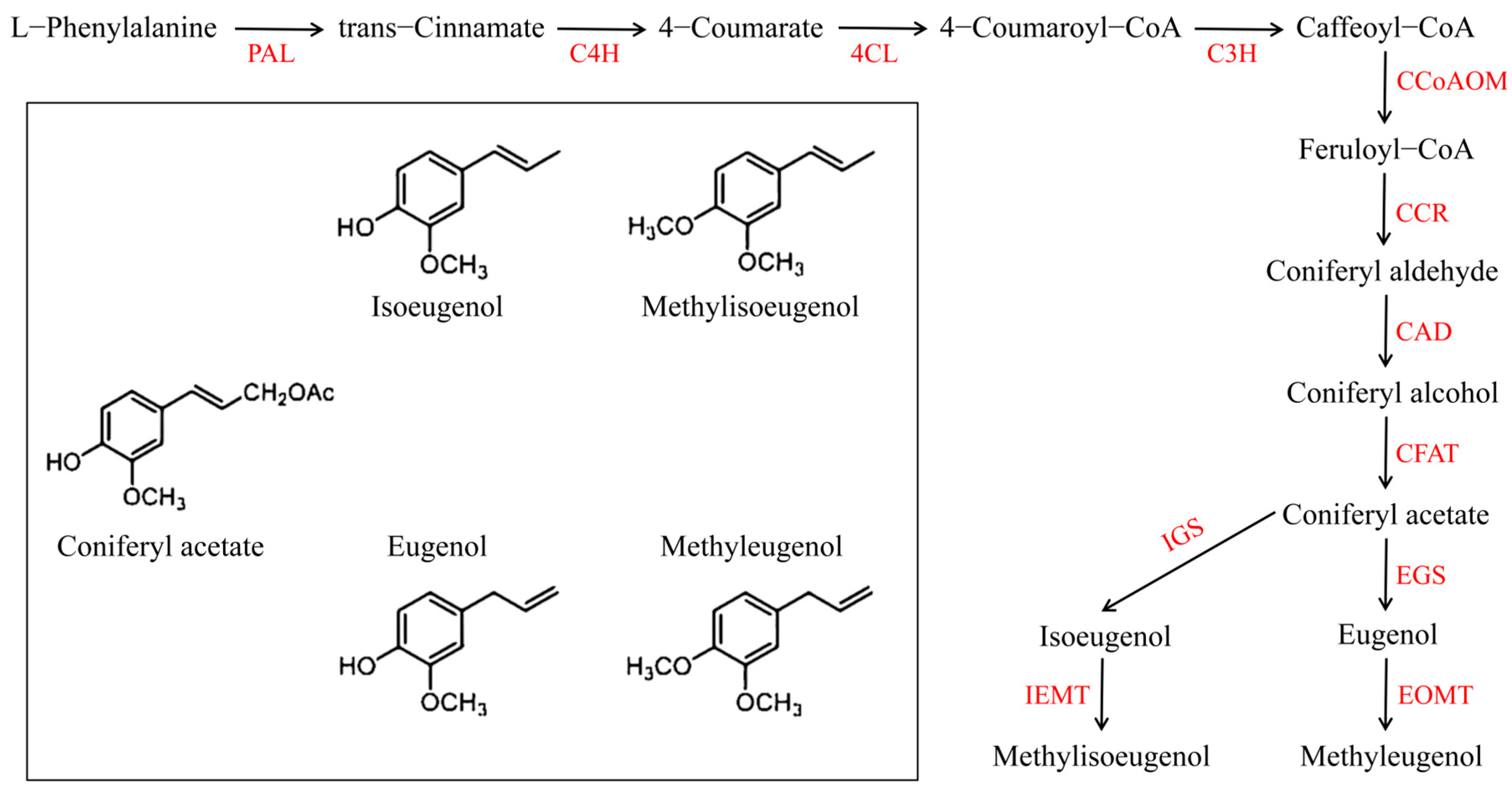
:1. Introduction
2. Results
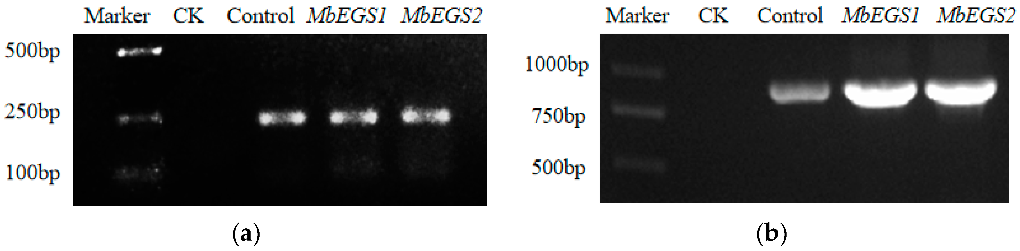
2.1. Verification of Recombinant M. bracteata
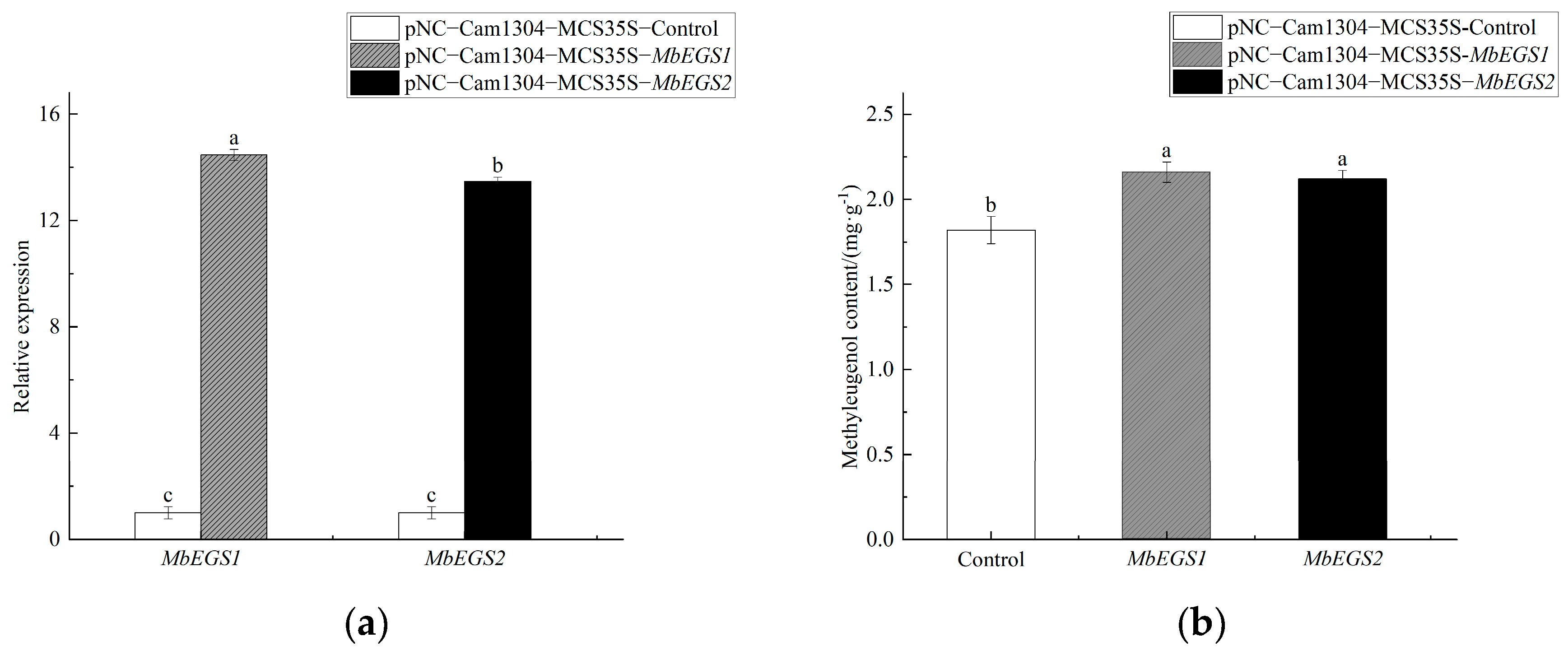
2.2. Overexpression of MbEGSs in M. bracteata
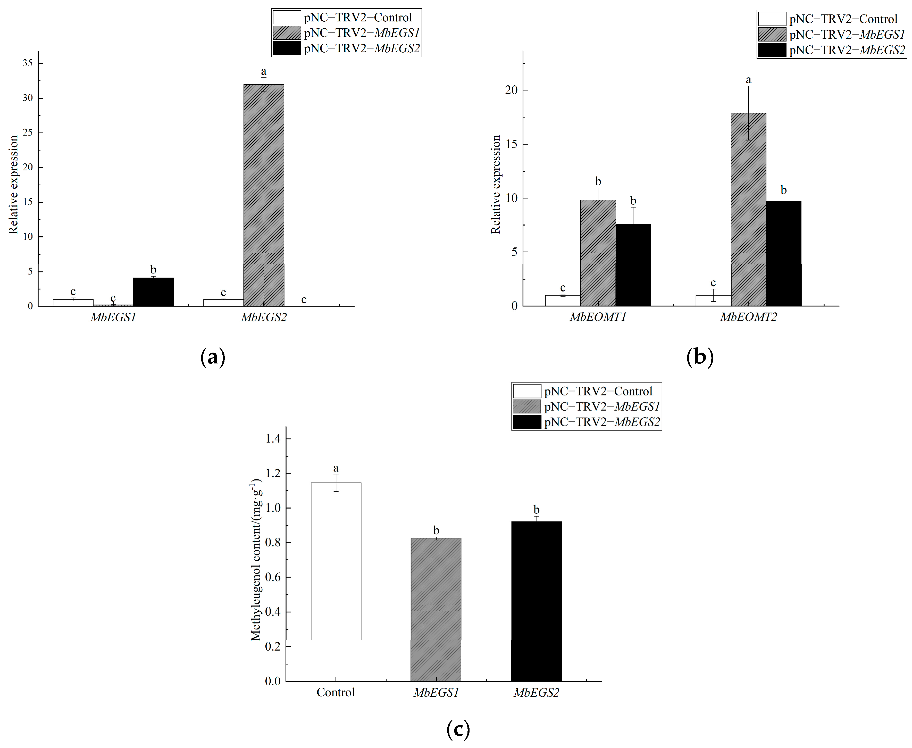
2.3. VIGS of MbEGSs in M. bracteata
3. Discussion
4. Materials and Methods
4.1. Plant Materials
4.2. Total RNA Isolation and cDNA Synthesis
4.3. Target Fragment Amplification
4.4. Vector Construction
4.4.1. Construction of Overexpression Vectors
4.4.2. Construction of VIGS Vectors
4.5. Infection Preparation and Vacuum Infiltration
4.6. Detection of the GUS Reporter Gene and TRV Virus
4.7. Quantitative RT-PCR Analysis
4.8. Determination of Eugenol and Methyleugenol Content
5. Conclusions
Supplementary Materials
Author Contributions
Funding
Data Availability Statement
Conflicts of Interest
References
- Gang, D.R.; Wang, J.; Dudareva, N.; Nam, K.H.; Simon, J.E.; Lewinsohn, E.; Pichersky, E. An Investigation of the Storage and Biosynthesis of Phenylpropenes in Sweet Basil. Plant Physiol. 2001, 125, 539–555. [Google Scholar] [CrossRef] [PubMed] [Green Version]
- Yauk, Y.K.; Chagné, D.; Tomes, S.; Matich, A.J.; Atkinson, R.G. The O-methyltransferase gene MdoOMT1 is required for biosynthesis of methylated phenylpropenes in ripe apple fruit. Plant J. 2015, 82, 937–950. [Google Scholar] [CrossRef] [PubMed]
- Pichersky, E.; Noel, P.J.; Dudareva, N. Biosynthesis of plant volatiles: Nature’s diversity and ingenuity. Science 2006, 311, 808–811. [Google Scholar] [CrossRef] [PubMed] [Green Version]
- Tan, K.H.; Nishida, R. Methyl eugenol: Its occurrence, distribution, and role in nature, especially in relation to insect behavior and pollination. J. Insect Sci. 2012, 12, 56. [Google Scholar] [CrossRef] [PubMed] [Green Version]
- Raguso, R.A.; Pichersky, E. Floral volatiles fromClarkia breweri andC. concinna (Onagraceae): Recent evolution of floral scent and moth pollination. Plant Syst. Evol. 1995, 194, 55–67. [Google Scholar] [CrossRef] [Green Version]
- Koeduka, T.; Louie, G.V.; Orlova, I.; Kish, C.M.; Ibdah, M.; Wilkerson, C.G.; Bowman, M.E.; Baiga, T.J.; Noel, J.P.; Dudareva, N.; et al. The multiple phenylpropene synthases in both Clarkia breweri and Petunia hybrida represent two distinct protein lineages. Plant J. Cell Mol. Biol. 2008, 54, 362–374. [Google Scholar] [CrossRef] [Green Version]
- Verdonk, J.; De Vos, C.R.; Verhoeven, H.A.; Haring, M.A.; Van Tunen, A.J.; Schuurink, R.C. Regulation of floral scent production in petunia revealed by targeted metabolomics. Phytochemistry 2003, 62, 997–1008. [Google Scholar] [CrossRef]
- Grossman, J.M. Botanical pesticides in Africa. Ipm Pract. 1993, 15, 1–9. [Google Scholar]
- Sisk, C.B.; Shorey, H.H.; Gerber, R.G.; Gaston, L.K. Semiochemicals that Disrupt Foraging by the Argentine Ant (Hymenoptera: Formicidae): Laboratory Bioassays. J. Econ. Entomol. 1996, 89, 385–391. [Google Scholar] [CrossRef]
- Koeduka, T.; Fridman, E.; Gang, D.R.; Vasso, D.G.; Pichersky, E. Eugenol and isoeugenol, characteristic aromatic constituents of spices, are biosynthesized via reduction of a coniferyl alcohol ester. Proc. Natl. Acad. Sci. USA 2006, 103, 10128–10133. [Google Scholar] [CrossRef] [Green Version]
- Gang, D.R.; Lavid, N.; Zubieta, C.; Chen, F.; Beuerle, T.; Lewinsohn, E.; Noel, J.P. Characterization of Phenylpropene O-Methyltransferases from Sweet Basil: Facile Change of Substrate Specificity and Convergent Evolution within a Plant O-Methyltransferase Family. Plant Cell 2002, 14, 505–519. [Google Scholar] [CrossRef] [PubMed] [Green Version]
- Muhlemann, J.K.; Woodworth, B.D.; Morgan, J.A.; Dudareva, N. The monolignol pathway contributes to the biosynthesis of volatile phenylpropenes in flowers. New Phytol. 2014, 204, 661–670. [Google Scholar] [CrossRef] [PubMed]
- Anand, A.; Jayaramaiah, R.H.; Beedkar, S.D.; Singh, P.A.; Joshi, R.S.; Mulani, F.A.; Dholakia, B.B.; Punekar, S.A.; Gade, W.N.; Thulasiram, H.V.; et al. Comparative functional characterization of eugenol synthase from four different Ocimum species: Implications on eugenol accumulation. BBA Proteins Proteom. 2016, 1864, 1539–1547. [Google Scholar] [CrossRef] [PubMed]
- Schuurink, R.C.; Haring, M.A.; Clark, D.G. Regulation of volatile benzenoid biosynthesis in petunia flowers. Trends Plant Sci. 2006, 11, 20–25. [Google Scholar] [CrossRef]
- Cheng, S.; Fu, X.; Mei, X.; Zhou, B.; Naoharu, W.; Yang, Z. Regulation of biosynthesis and emission of volatile phenylpropanoids/benzenoids in petuniax hybrida flowers by multi-factors of circadian clock, light, and temperature. Plant Physiol. Biochem. 2016, 107, 1–8. [Google Scholar] [CrossRef] [PubMed]
- Sun, T.; Xu, Y.; Zhang, D.; Zhuang, H.; Wu, J.; Sun, G. An acyltransferase gene that putatively functions in anthocyanin modification was horizontally transferred from Fabaceae into the genus Cuscuta. Plant Divers. 2016, 38, 149–155. [Google Scholar] [CrossRef] [Green Version]
- Rodrigues, J.; Perrier, V.; Lecomte, J.; Dubreucq, E.; Ferreira-Dias, S. Biodiesel production from crude jatropha oil catalyzed by immobilized lipase/acyltransferase from Candida parapsilosis in aqueous medium. Bioresour. Technol. 2016, 218, 1224–1229. [Google Scholar] [CrossRef]
- Min, T.; Kasahara, H.; Bedgar, D.L.; Youn, B.; Lawrence, P.K.; Gang, D.R.; Halls, S.C.; Park, H.; Hilsenbeck, J.L.; Davin, L.B. Crystal structures of pinoresinol-lariciresinol and phenylcoumaran benzylic ether reductases and their relationship to isoflavone reductases. J. Biol. Chem. 2003, 278, 50714–50723. [Google Scholar] [CrossRef] [Green Version]
- Dal Cin, V.; Tieman, D.M.; Tohge, T.; McQuinn, R.; de Vos, R.C.; Osorio, S.; Schmelz, E.A.; Taylor, M.G.; Smits-Kroon, M.T.; Schuurink, R.C.; et al. Identification of Genes in the Phenylalanine Metabolic Pathway by Ectopic Expression of a MYB Transcription Factor in Tomato Fruit. Plant Cell 2011, 23, 2738–2753. [Google Scholar] [CrossRef]
- Kyo, W.; Atsushi, I. Metabolic engineering of the tryptophan and phenylalanine biosynthetic pathways in rice. Plant Biotechnol. 2009, 26, 523–533. [Google Scholar]
- Hatzimanikatis, V.; Li, C.; Ionita, J.A.; Henry, C.S.; Jankowski, M.D.; Broadbelt, L.J. Exploring the diversity of complex metabolic networks. Bioinformatics 2004, 21, 1603–1609. [Google Scholar] [CrossRef] [PubMed] [Green Version]
- Pascual, M.B.; El-Azaz, J.; de la Torre, F.N.; Cañas, R.A.; Avila, C.; Cánovas, F.M. Biosynthesis and Metabolic Fate of Phenylalanine in Conifers. Front. Plant Sci. 2016, 7, 1030. [Google Scholar] [CrossRef] [PubMed] [Green Version]
- Brophy, J.J.; Goldsack, R.J.; Doran, J.C.; Craven, L.A.; Lepschi, B.J. A Comparison of the Leaf Oils of Melaleuca squamophloia with Those of Its Close Relatives, M. styphelioides and M. bracteata. J. Essent. Oil Res. 1999, 11, 327–332. [Google Scholar] [CrossRef]
- Carson, C.F.; Hammer, K.A.; Riley, T.V. Melaleuca alternifolia (Tea Tree) oil: A review of antimicrobial and other medicinal properties. Clin. Microbiol. Rev. 2006, 19, 50–62. [Google Scholar] [CrossRef] [Green Version]
- Adesanwo, J.K.; Shode, F.O.; Aiyelaagbe, O.O.; Rabiu, O.O.; Oyede, R.T. Antisecretory and antiulcerogenic activities of the stem bark extract of Melaleuca bracteata and isolation of principles. J. Med. Plants Res. 2009, 3, 822–824. [Google Scholar]
- Kardinan, A.K.; Hidayat, P. Potency of Melaleuca bracteata and Ocimum sp. Leaf Extractsas Fruit Fly (Bactrocera dorsalis complex) Attractants in Guava and Star Fruit Orchards in Bogor, West Java, Indonesia. J. Dev. Sustain. Agric. 2013, 8, 79–84. [Google Scholar]
- Wang, W.; Huang, X.; Yang, H.; Niu, X.; Li, Y. Antibacterial Activity and Anti-Quorum Sensing Mediated Phenotype in Response to Essential Oil from Melaleuca bracteata Leaves. Int. J. Mol. Sci. 2019, 20, 5696. [Google Scholar] [CrossRef] [Green Version]
- Wang, W.; Lin, X.; Yang, H.; Huang, X.; Pan, L.; Wu, S.; Yang, C.; Zhang, L.; Li, Y. Anti-quorum sensing evaluation of methyleugenol, the principal bioactive component, from the Melaleuca bracteata leaf oil. Front. Microbiol. 2022, 13, 970520. [Google Scholar] [CrossRef]
- Goswami, P.; Verma, S.K.; Chauhan, A.; Venkatesha, K.T.; Verma, R.S.; Singh, V.R.; Darokar, M.P.; Chanotiya, C.S.; Padalia, R.C. Chemical Composition and Antibacterial Activity of Melaleuca bracteata Essential Oil from India: A Natural Source of Methyl Eugenol. Nat. Prod. Commun. 2017, 12, 965–968. [Google Scholar] [CrossRef] [Green Version]
- Li, Y.; Ye, Z.; Wang, W.; Yang, C.; Liu, J.; Zhou, L.; Shen, Y.; Wang, Z.; Chen, J.; Wu, S.; et al. Composition Analysis of Essential Oil from Melaleuca bracteata Leaves Using Ultrasound-assisted Extraction and its Antioxidative and Antimicrobial Activities. BioResources 2018, 13, 8488–8504. [Google Scholar] [CrossRef]
- Yasin, M.; Younis, A.; Ramzan, F.; Javed, T.; Shabbir, R.; Noushahi, H.A.; Skalicky, M.; Ondrisik, P.; Brestic, M.; Hassan, S. Extraction of Essential Oil from River Tea Tree (Melaleuca bracteata F. Muell.): Antioxidant and Antimicrobial Properties. Sustainability 2021, 13, 4827. [Google Scholar] [CrossRef]
- Wang, X.; He, X.; Lin, J.; Shao, H.; Chang, Z.; Dixon, R.A. Crystal Structure of Isoflavone Reductase from Alfalfa (Medicago sativa L.). J. Mol. Biol. 2006, 358, 1341–1352. [Google Scholar] [CrossRef] [PubMed]
- Gang, D.R.; Kasahara, H.; Xia, Z.Q.; Vander Mijnsbrugge, K.; Bauw, G.; Boerjan, W.; Van Montagu, M.; Davin, L.B.; Lewis, N.G. Evolution of plant defense mechanisms. Relationships of phenylcoumaran benzylic ether reductases to pinoresinol-lariciresinol and isoflavone reductases. J. Biol. Chem. 1999, 274, 7516–7527. [Google Scholar] [CrossRef] [PubMed] [Green Version]
- Akashi, T.; Koshimizu, S.; Aoki, T.; Ayabe, S.I. Identification of cDNAs encoding pterocarpan reductase involved in isoflavan phytoalexin biosynthesis in Lotus japonicus by EST mining. FEBS Lett. 2006, 580, 5666–5670. [Google Scholar] [CrossRef] [Green Version]
- Qiu, Z.; Liu, L.; Lin, Y.; Lin, X.; Li, Y.; Wu, S.; Yang, C. Cloning and Functional Analysis of the MbEGS Gene from Melaleuca bracteata. Acta Hortic. Sin. 2022, 49, 1747. [Google Scholar]
- El Asbahani, A.; Miladi, K.; Badri, W.; Sala, M.; Addi, E.A.; Casabianca, H.; El Mousadik, A.; Hartmann, D.; Jilale, A.; Renaud, F.N.R.; et al. Essential oils: From extraction to encapsulation. Int. J. Pharm. 2015, 483, 220–243. [Google Scholar] [CrossRef]
- Porto, C.D.; Decorti, D. Ultrasound-assisted extraction coupled with under vacuum distillation of flavour compounds from spearmint (carvone-rich) plants: Comparison with conventional hydrodistillation. Ultrason. Sonochem. 2009, 16, 795–799. [Google Scholar] [CrossRef]
- Bakkali, F.; Averbeck, S.; Averbeck, D.; Idaomar, M. Biological effects of essencial oils-a review. Food Chem. Toxicol. 2008, 46, 446–475. [Google Scholar] [CrossRef]
- Siddique, S.; Parveen, Z.; Mazhar, S. Chemical composition, antibacterial and antioxidant activities of essential oils from leaves of three Melaleuca species of Pakistani flora. Arab. J. Chem. 2020, 13, 67–74. [Google Scholar] [CrossRef]
- Aragüez, I.; Osorio, S.; Hoffmann, T.; Rambla, J.L.; Medina-Escobar, N.; Granell, A.; Botella, M.Á.; Schwab, W.; Valpuesta, V. Eugenol production in achenes and receptacles of strawberry fruits is catalyzed by synthases exhibiting distinct kinetics. Plant Physiol. 2013, 163, 946–958. [Google Scholar] [CrossRef] [Green Version]
- Gupta, A.K.; Schauvinhold, I.; Pichersky, E.; Schiestl, F.P. Eugenol synthase genes in floral scent variation in Gymnadenia species. Funct. Integr. Genom. 2014, 14, 779–788. [Google Scholar] [CrossRef] [PubMed]
- Yahyaa, M.; Berim, A.; Nawade, B.; Ibdah, M.; Dudareva, N.; Ibdah, M. Biosynthesis of methyleugenol and methylisoeugenol in Daucus carota leaves: Characterization of eugenol/isoeugenol synthase and O-Methyltransferase. Phytochemistry 2019, 159, 179–189. [Google Scholar] [CrossRef] [PubMed]
- Koeduka, T.; Orlova, I.; Baiga, T.J.; Noel, J.P.; Dudareva, N.; Pichersky, E. The lack of floral synthesis and emission of isoeugenol in Petunia axillaris subsp. parodii is due to a mutation in the isoeugenol synthase gene. Howard Hughes Med. Inst. 2009, 58, 961–969. [Google Scholar]
- Wang, J.; Dudareva, N.; Bhakta, S.; Raguso, R.A.; Pichersky, E. Floral scent production in Clarkia breweri (Onagraceae). II. Localization and developmental modulation of the enzyme S-adenosyl-L-methionine:(iso)eugenol O-methyltransferase and phenylpropanoid emission. Plant Physiol. 1997, 114, 213–221. [Google Scholar] [CrossRef] [PubMed] [Green Version]
- Yan, H.; Baudino, S.; Caissard, J.C.; Nicolè, F.; Zhang, H.; Tang, K.; Li, S.; Lu, S. Functional characterization of the eugenol synthase gene (RcEGS1) in rose. Plant Physiol. Biochem. 2018, 129, 21–26. [Google Scholar] [CrossRef]
- Gong, B.; Yi, J.; Wu, J.; Sui, J.; Khan, M.A.; Wu, Z.; Zhong, X.; Seng, S.; He, J.; Yi, M. LlHSFA1, a novel heat stress transcription factor in lily (Lilium longiflorum), can interact with LlHSFA2 and enhance the thermotolerance of transgenic Arabidopsis thaliana. Plant Cell Rep. 2014, 33, 1519–1533. [Google Scholar] [CrossRef]

| Primer Name | Forward Primer Sequence (5′→3′) | Reverse Primer Sequence (5′→3′) |
|---|---|---|
| MbEGS1 | ATGGCAGGAGAGGCCGAGAAAA | TCATTCGAAAGCAGCCCTAGCTG |
| MbEGS2 | ATGACGATCATAAGCAGTAGTTGCA | TCATTCCAATACAGCGCTTGC |
| NC-MbEGS1 | agtggtctctgtccagtcctATGGCAGGAGAGGCCGAGAAAA | ggtctcagcagaccacaagtTCATTCGAAAGCAGCCCTAGCTG |
| NC-MbEGS2 | agtggtctctgtccagtcctATGACGATCATAAGCAGTAGTTGCA | ggtctcagcagaccacaagtTCATTCCAA TACAGCGCTTGC |
| VIGS-MbEGS1 | GCAGGCATTCCTTACACC | CGGGCACTTGGACTTTC |
| VIGS-MbEGS2 | GCACTCCCACCATTCG | AGTCGTTCGCCACTTTT |
| NC-VIGS-MbEGS1 | agtggtctctgtccagtcctGCAGGCATTCCTTACACC | ggtctcagcagaccacaagtCGGGCACTTGGACTTTC |
| NC-VIGS-MbEGS2 | agtggtctctgtccagtcctGCACTCCCACCATTCG | ggtctcagcagaccacaagtAGTCGTTCGCCACTTTT |
| GUS | ACGGGGAAACTCAGCAAGC | ATGTAATGTTCTGCGACGCTCA |
| TRV2 | GGCGGTTCTTGTGTGTCAAC | GGCGGTTCTTGTGTGTCAAC |
| qMbEGS1 | TTCGGGAGTGAGGAGGATAA | TGGGGATGAAGGAGGTTGTT |
| qMbEGS2 | CCCCACCTTTGTCTTCGCTC | AACCTCTTTATGTTGCCCGC |
| qMbEOMT1 | CACCAACATACGACGGAATCACTCA | TCATCGAACATGCCATCGCCTTC |
| qMbEOMT2 | CCTTGCTCCGATGGTGCTGATG | ATGCGATGCCACCGTTCTTGAC |
| qMbCYP | CGCCGTGAAGGGATGTTTGT | ACGGGAAGCATTTCATCCTCTG |
Disclaimer/Publisher’s Note: The statements, opinions and data contained in all publications are solely those of the individual author(s) and contributor(s) and not of MDPI and/or the editor(s). MDPI and/or the editor(s) disclaim responsibility for any injury to people or property resulting from any ideas, methods, instructions or products referred to in the content. |
© 2023 by the authors. Licensee MDPI, Basel, Switzerland. This article is an open access article distributed under the terms and conditions of the Creative Commons Attribution (CC BY) license (https://creativecommons.org/licenses/by/4.0/).
Share and Cite
Lin, Y.; Qiu, Z.; Lin, X.; Wu, Y.; Niu, X.; Yin, G.; Shao, D.; Xiang, X.; Li, Y.; Yang, C. The Role of MbEGS1 and MbEGS2 in Methyleugenol Biosynthesis by Melaleuca bracteata. Plants 2023, 12, 1026. https://doi.org/10.3390/plants12051026
Lin Y, Qiu Z, Lin X, Wu Y, Niu X, Yin G, Shao D, Xiang X, Li Y, Yang C. The Role of MbEGS1 and MbEGS2 in Methyleugenol Biosynthesis by Melaleuca bracteata. Plants. 2023; 12(5):1026. https://doi.org/10.3390/plants12051026
Chicago/Turabian StyleLin, Yongsheng, Ziwen Qiu, Xiaojie Lin, Yingxiang Wu, Xianqian Niu, Guanwen Yin, Dandan Shao, Xuwen Xiang, Yongyu Li, and Chao Yang. 2023. "The Role of MbEGS1 and MbEGS2 in Methyleugenol Biosynthesis by Melaleuca bracteata" Plants 12, no. 5: 1026. https://doi.org/10.3390/plants12051026