Article

MtWOX2 and MtWOX9-1 Effects on the Embryogenic Callus Transcriptome in Medicago truncatula

by
Elizaveta Y. Krasnoperova
1,
Varvara E. Tvorogova
1,2,3,*,
Kirill V. Smirnov
4,
Elena P. Efremova
1,
Elina A. Potsenkovskaia
1,2,3,
Anastasia M. Artemiuk
1,
Zakhar S. Konstantinov
2,
Veronika Y. Simonova
2,
Anna V. Brynchikova
2,
Daria V. Yakovleva
1,
Daria B. Pavlova
1 and
Ludmila A. Lutova
1,2
1
Department of Genetics and Biotechnology, Saint Petersburg State University, 7/9 Universitetskaya Emb, 199034 St. Petersburg, Russia
2
Plant Biology and Biotechnology Department, Sirius University of Science and Technology, 1 Olympic Avenue, 354340 Sochi, Russia
3
Center for Genetic Technologies, N. I. Vavilov All-Russian Institute of Plant Genetic Resources (VIR), 42 Bolshaya Morskaya Street, 190000 St. Petersburg, Russia
4
All-Russia Research Institute for Agricultural Microbiology, Podbelsky Chausse 3, Pushkin, 196608 St. Petersburg, Russia
*
Author to whom correspondence should be addressed.
Plants 2024, 13(1), 102; https://doi.org/10.3390/plants13010102
Submission received: 1 December 2023 / Revised: 19 December 2023 / Accepted: 27 December 2023 / Published: 28 December 2023
(This article belongs to the Special Issue Plant Meristems:The Cradle of Life)

Abstract

:
WOX family transcription factors are well-known regulators of plant development, controlling cell proliferation and differentiation in diverse organs and tissues. Several WOX genes have been shown to participate in regeneration processes which take place in plant cell cultures in vitro, but the effects of most of them on tissue culture development have not been discovered yet. In this study, we evaluated the effects of MtWOX2 gene overexpression on the embryogenic callus development and transcriptomic state in Medicago truncatula. According to our results, overexpression of MtWOX2 leads to an increase in callus weight. Furthermore, transcriptomic changes in MtWOX2 overexpressing calli are, to a large extent, opposite to the changes caused by overexpression of MtWOX9-1, a somatic embryogenesis stimulator. These results add new information about the mechanisms of interaction between different WOX genes and can be useful for the search of new regeneration regulators.

1. Introduction

Plant in vitro tissue cultures have a wide range of applications in biotechnology, including the obtaining of transgenic or edited plants and plant reproduction, as well as the synthesis of different biologically active compounds [1]. Despite numerous studies exploring the genetic regulation of in vitro tissue culture development, this variant of plant existence has many unknown aspects. This is probably due to the high diversity of possible cultivation conditions, multiplied by the high diversity of plant genotypes. The main processes occurring in vitro in the isolated plant tissue or organ are callus formation, i.e., unorganized cell division, and different types of regeneration, including rhizogenesis (root regeneration), caulogenesis (shoot regeneration) and somatic embryogenesis (SE), the formation of embryo-like structures which are able to develop into a new plant [2]. All these regeneration variants can be direct, when new differentiated structures are formed from the explant tissue itself, or indirect, when these structures are developed from a callus [2]. Indirect SE serves as a valuable model for plant development studies, as in this case both unorganized cell division in a callus and highly organized divisions in somatic embryos occur side by side.
WUSCHEL-related homeobox (WOX) genes, well-known regulators of cell division in plants, have demonstrated their involvement in callus formation and embryogenesis. Among these, WOX2, WOX8, and WOX9 genes in Arabidopsis thaliana and their orthologs in other species play specific roles in the zygotic embryogenesis [3].
WOX2 and WOX8 expression begins in the zygote, while WOX9 expression is initiated during the two-cell stage [4,5]. WOX8 and WOX9, being close relatives, play crucial roles in maintaining embryo patterning. Loss of their function leads to patterning defects in the suspensor, resulting in death [4,6]. Furthermore, expression of WOX9 homologs is associated with SE in Gossypium hirsutum [7], Phoebe bournei [8], Hybrid Sweetgum (Liquidambar styraciflua × Liquidambar formosana) [9], Dimocarpus longan [10], and Medicago truncatula [11].
WOX2 is specifically expressed in the apical domain of developing zygotic embryos, and loss of its function leads to the impaired control of cell division in the apical domain and increased frequency of cotyledon development disturbance. According to the genetic analysis, this function is partly carried by other WOX genes, including WOX1, 3, and 5, but WOX2 plays a key role in it [6,12].
WOX2 orthologs are also specifically expressed in somatic embryos in many plant species, serving as markers of the SE process. For instance, a high level of expression of PpWOX2 in Pinus pinaster is detected in early SE. Later on, its expression decreases during maturation and development of somatic embryos [13]. High levels of expression of WOX2 or its orthologs are associated with the early stages of somatic embryo development in other species, including Picea abies [14], Dimocarpus longan [10], Hybrid sweetgum (Liquidambar styraciflua × L. formosana) [15], Cunninghamia lanceolata [16], and A. thaliana [17,18].
WOX2 and WOX8/9 functions in zygotic or somatic embryogenesis have been shown to be interconnected in many studies. For example, WOX8 or 9 activity is necessary for maintaining proper WOX2 expression patterns in the apical part of the zygotic embryo [6]. A recent study also showed that both WOX2 and 8 expression patterns are disturbed in the embryos with mutations in the YODA gene, encoding embryo-specific protein kinase, and/or HEAT SHOCK PROTEIN90 genes encoding YODA partners, which again confirms the connection between the WOX8 and 2 genes [19]. In tobacco cell cultures, ectopic expression of A. thaliana WOX2 and WOX8 or WOX2 and WOX9 genes stimulated regeneration, whereas neither of these genes could induce this process on their own [20].
At the same time, the WOX2 and WOX8 genes may be considered as antagonists from some perspectives. Indeed, they exhibit complementary expression patterns after the first zygote division when WOX2 is expressed in the apical cell and its descendants, whereas WOX8 is expressed in the basal domain of the embryo [4]. Although either WOX8 or 9 induces WOX2 expression in the apical domain [6], WOX2 is suggested to repress WOX8 expression [21], providing a negative regulatory loop for proper embryo domain specification.
According to our previous research, MtWOX9-1, a close WOX9 relative, stimulates SE in Medicago truncatula [22]. The MtWUSCHEL gene and STENOFOLIA (STF, MtWOX1 gene), members of the modern clade of the WOX family, can also stimulate SE in this species [23,24]. However, the effects of the M. truncatula WOX2 ortholog—assumed to be embryo-specific—on the in vitro regeneration and SE, have not been studied yet. To investigate the contribution of MtWOX2 on SE in M. truncatula, we evaluated the effect of its overexpression on the development and transcriptome state of the embryogenic callus.

2. Results

2.1. MtWOX2 Expression Pattern during Somatic Embryogenesis

To find out whether changes occur in the MtWOX2 gene expression level during SE, we analyzed its expression dynamics in the calli of the embryogenic line 2HA [25] and the non-embryogenic line A17 at different time points after the start of cultivation (at 0, 1, 2, 3, 4, 5 and 6 weeks) using qPCR. Contrary to our expectations, no specific expression of MtWOX2 in the calli of the embryogenic or non-embryogenic line at any analyzed stage was detected (Figure S1).

2.2. MtWOX2 Overexpression Increases Callus Weight in M. truncatula

To analyze the role of MtWOX2 in SE, we examined the SE capacity of calli overexpressing this gene. For that purpose, we transformed R108 plants with constructs for overexpression of MtWOX2 or the beta-glucuronidase (GUS) gene (as a control). We cultivated transformed leaf explants on a callus induction medium containing auxin and cytokinin, and then on a hormone-free medium for regeneration. After 43 days of cultivation (including 33 days of cultivation on the callus-inducing medium and 10 days of cultivation on a hormone-free medium), samples were taken from several calli for MtWOX2 expression analysis. According to the qPCR, calli transformed with the construct for MtWOX2 overexpression demonstrated increased levels of MtWOX2 transcripts (Figure S2A).
On the 40th day of cultivation on the medium for regeneration, the calli were collected, and their weight and the number of embryos per callus were evaluated. Calli overexpressing MtWOX2 (MtWOX2oe calli) did not demonstrate an increased number of somatic embryos per callus (Figure 1A). Their median weight was greater in comparison with the control calli; however, the differences in values were not statistically significant, according to the Mann–Whitney test (Figure 1B). At the same time, we observed several individual calli of a very large weight (more than 1 g), which occurred only in the MtWOX2oe genotype (Figure S3). When the calli were divided into two categories (weight greater than 1 g and less than 1 g), Fisher’s exact test confirmed that MtWOX2 overexpression is a significant factor influencing the frequency of occurrence of calli greater than 1 g (p-value = 0.007389).
To check our results, we obtained two independent lines containing the construct for MtWOX2 overexpression. T1 plants from these lines and R108 plants were used as a source of leaf explants, which were cultivated on callus formation and regeneration media, similarly to the previous experiment, but without antibiotics in the media. As a result, calli containing constructs for MtWOX2 overexpression indeed have a significantly larger weight than R108 calli (p-value = 0.0061, Mann–Whitney test), whereas no significant change in the embryo number per callus was observed (Figure 1C,D). Unlike T0 calli generation, T1 calli, obtained from transgenic MtWOX2oe T1 plants, did not demonstrate increased expression of MtWOX2 (Figure S2B). This absence of increased MtWOX2 expression is noteworthy, considering the significant differences in weight observed between T1 MtWOX2oe calli and control R108 calli.

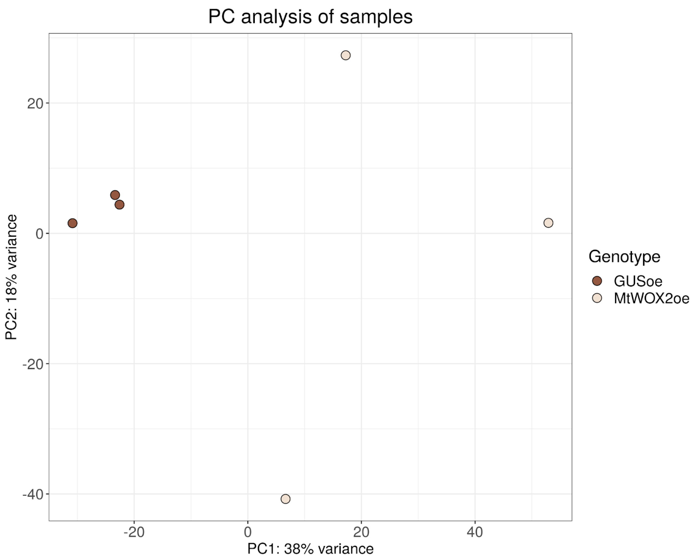
2.3. Transcriptomic Analysis of MtWOX2 Overexpressing Calli

To find out what transcriptomic changes occur in MtWOX2 overexpressing calli, we performed transcriptomic analysis of calli obtained after transformation of leaf explants with constructs for MtWOX2 overexpression or GUS overexpression (as a control).
From these calli, after 43 days of cultivation (including 33 days of cultivation on the callus-inducing medium and 10 days of cultivation on a hormone-free medium), RNA was isolated, and cDNA was obtained from it and sequenced (3 biological replicates for each genotype). Differential gene expression analysis was performed using the DeSeq2 package. Principal component analysis showed that MtWOX2oe samples were more similar to each other than to control samples (Figure 2). It is worth noting that MtWOX2oe samples apparently have more differences from each other in comparison with control GUSoe group, according to the PCA. Such a high level of dispersion suggests a potential connection with the more intensive callus growth in MtWOX2oe. This phenomenon may be attributed to the typical cell structure of the callus, which has been reported to display significant heterogeneity [26].
To identify differentially expressed genes (DEGs), p-value and log2 fold change thresholds of 0.01 and 1.0, respectively, were adopted. For 1670 genes out of 50,803 analyzed, differential expression was detected, including 803 genes with increased expression and 894 genes with decreased expression in MtWOX2oe calli (Table S1).
We compared the MtWOX2oe DEGs with DEGs in calli with MtWOX9-1 overexpression (in comparison with R108 calli) [27]. Unlike MtWOX2, which stimulated callus development according to our data, MtWOX9-1 can stimulate development of somatic embryos. MtWOX2 and MtWOX9-1 belong to the modern and intermediate clades of the WOX family, respectively. We checked if MtWOX2 and MtWOX9-1 overexpressing calli have some common DEGs. Indeed, 451 genes were found to show differential expression in both experiments. If the total number of genes is taken to be all genes with non-zero expression level in at least one of the analyzed samples (amounting to 34,135 genes), this value significantly deviates from the theoretically expected value (p-value = 4.439458 × 10−136, hypergeometric test). This suggests the influence of MtWOX9-1 and MtWOX2 transcription factors on significantly overlapping sets of genes (Figure 3A). Of these, 118 genes showed the same directions of change (a decrease or increase in the expression level) in the case of overexpression of MtWOX2 and MtWOX9-1, and for 333 genes the directions of regulation were opposite. This distribution significantly deviates from the expected result (equal number of similarly and oppositely regulated genes) (p-value = 2.822 × 10⁻¹³, Chi-square). Therefore, we can suppose that MtWOX9-1 and MtWOX2 have at least a partly opposite impact on the callus transcriptome.
Most of the common DEGs that were oppositely regulated in MtWOX2 and MtWOX9-1 overexpressing calli were activated by MtWOX9-1 and repressed by MtWOX2 (216 genes out of 333). We performed GO enrichment analysis, which has shown that among these 216 genes 22 pathways are overrepresented, most of which are related to somatic and zygotic embryogenesis, morphogenesis, and seed development (Figure 3B, Table S2). Interestingly, other groups of common DEGs, including genes activated by MtWOX2 overexpression and repressed by MtWOX9-1 overexpression as well as genes which are similarly regulated by MtWOX2 and MtWOX9-1 overexpression, had much fewer overrepresented GO pathways (8 and 1, respectively) (Figure S4).

3. Discussion

The search for genes which can regulate the development of tissue culture in vitro represents an important goal for biotechnology as well as for the investigation of plant development [28]. In this study, we evaluated expression patterns and possible effects on in vitro cell culture for MtWOX2, a transcription factor from the WOX family that is orthologous to the well-known embryogenesis and cell division regulator WOX2.
We did not detect specific expression of the MtWOX2 gene during any stage of SE. These results are unexpected, because in many species WOX2 orthologs are specifically expressed during the early stages of SE. MtWOX2 is the only ortholog of the WOX2 gene in the M. truncatula genome. Interestingly, it also does not show specific expression in the generative structures according to the MtExpress V3 dataset [29]. According to our previous studies, STF (MtWOX1), another member of the WOX family modern clade, has a specific expression increase during SE and it is expressed in ovules [11]. Given the redundancy between WOX2 and WOX1 in the regulation of zygotic embryo development [6], it is plausible to suggest that some WOX2 functions during SE are carried out by the STF gene in M. truncatula.
According to our data, overexpression of MtWOX2 led to increased callus weight, but did not have any effect on SE capacity. These data are consistent with the study, showing that ectopic WOX2 expression is not sufficient to induce SE in A. thaliana [17]. Analysis of M. truncatula plants with MtWOX2 loss of function will help to learn if this gene is necessary for SE.
The increased weight of calli transformed with the construct for MtWOX2 overexpression can be caused by different factors. There is a possibility that this weight increase is related to increasing cell size or changes in the biochemical composition of cells. However, previous reports on the WOX2 gene and its orthologs as cell division regulators suggest that such calli exhibit more intensive proliferation of non-differentiated callus cells.
Interestingly, we did not detect an increased level of MtWOX2 expression in calli obtained from T1 plants containing constructs for MtWOX2 overexpression, yet these calli demonstrated significantly increased weight compared to control. This may suggest some epigenetic inherited changes caused by MtWOX2 overexpression in T0 calli, but, at the same time, allows for consideration of the negative influence of MtWOX2 overexpression on plant development from which only transgenic plants with silenced transgene overexpression can regenerate or survive. That hypothesis is supported by the transcriptomic analysis of MtWOX2oe calli showing the repressive effect of MtWOX2 on genes related to development and morphogenesis.
Transcriptomic analysis of calli with MtWOX2 overexpression revealed that a significant portion of DEGs in such calli also are affected by MtWOX9-1 overexpression. Furthermore, most of those genes were activated by MtWOX9-1 and repressed by MtWOX2. This supports the data on WOX2 and WOX9 tobacco orthologs, which demonstrated their repressing and activating activities in a luciferase assay [30]. Interestingly, the category of genes activated by MtWOX9-1 and repressed by MtWOX2 was enriched with multiple GO terms related to embryogenesis and development in general, whereas other categories (genes activated by MtWOX2 and repressed by MtWOX9-1, as well as genes similarly regulated by overexpression of the MtWOX9-1 and MtWOX2 genes) exhibited much fewer overrepresented GO pathways. That allows for the suggestion that MtWOX2 acts antagonistically to MtWOX9-1 in embryogenic calli and these two TFs promote two alternative pathways of cell development during in vitro cultivation: callus formation or somatic embryogenesis.
MtWOX9-1 has specific expression during SE and in ovules [11]. Although there are three other genes orthologous to WOX9 in the M. truncatula genome [31], none of them demonstrate significant expression levels in any M. truncatula tissue, according to the MtExpress V3 dataset [29]. At the same time, the M. truncatula genome lacks the WOX8 ortholog [32]. It should be noted that in Nicotiana tabacum, which also lacks WOX8 orthologs, NtWOX9 together with NtWOX2 are co-expressed in the zygote [30], suggesting that WOX9 and WOX8 orthologs can perform each other’s functions in different species. These data suggest that MtWOX9-1 carries out both WOX8 and WOX9 functions in M. truncatula development. MtWOX2 transcription is stimulated by MtWOX9-1 overexpression, according to the transcriptome analysis [27]. Our data on the antagonistic effects of MtWOX2 and MtWOX9-1 in the embryogenic calli suggest the existence of a negative feedback loop between these two genes, reminiscent of WOX2 and WOX8 relations in the zygotic embryos [21].
Together, our results add new information on the relationships between different WOX genes and can be used in biotechnology for species with poor callus formation.

4. Materials and Methods

4.1. Plant Material and Bacterial Strains

Plants of Medicago truncatula A17 and 2HA lines derived from the Jemalong cultivar and the R108 line derived from ecotype 108-1 were used in the study. Seeds of the A17 line were provided by colleagues from Wageningen University (The Netherlands). Seeds of the 2HA line were provided by Dr Mireille Chabaud (French National Institute for Agriculture, Food, and Environment)). Seeds of the R108 line were provided by colleagues from Samuel Roberts Institute (USA).
Rhizobium radiobacter (Agrobacterium tumefaciens) AGL1 strain and Escherichia coli TOP10 strain were used for plant transformation and cloning, respectively.

4.2. Plant Cultivation Conditions

For germination, M. truncatula seeds were treated with concentrated sulfuric acid (95–97%) for 10 min, then washed 10 times with sterile distilled water. Sterilized seeds were germinated on the 1% agar at 4 °C in the dark. Plants were grown in soil and under in vitro conditions at 21–24 °C, photoperiod 16 (light)/8 (dark). Terra Vita soil (Nord Pulp, St. Petersburg, Russia) mixed with vermiculite (3:1) was used for growth in growth chambers. Modified Fahraeus medium [22,33] was used for growth in sterile conditions.
Plant transformation and the obtaining of R108 line T0 calli were performed as described in [22]. For callus induction, modified PCI-4 medium [34] was used with 4 mg/L (18 μM) 2,4-D, 0.5 mg/L (2.22 μM) BAP, 250 mg/L cefotaxime and 25 mg/L hygromycin. The medium for the SE induction has the same composition as callus induction medium, but it did not contain hormones and had 12.5 mg/L hygromycin instead of 25 mg/L. The SE capacity and weight of T0 calli were assessed on day 74 after transformation (34 days of culturing on callus induction medium and 40 days of culturing on hormone-free medium).
Transgenic T0 plants from transformed calli were obtained by further cultivation of calli on the medium for the SE induction. After the formation of 1–2 true leaves, the regenerants were transferred to the germination medium-modified PCI-4 medium [34] without any hormones and antibiotics, and then, after 10–14 days, to the rooting medium (half-strength modified PCI-4 medium [34] without hormones and antibiotics). After the explants formed roots, they were transferred to the modified Fahraeus medium [22,33] for 10–14 days and then to the soil to obtain seeds.
In vitro cultivation of leaf explants from R108 and MtWOX2oe T1 transgenic plants was performed as described in [22]. Callus-inducing and SE-inducing media had the same composition as the respective media for obtaining T0 calli, but they did not contain cefotaxime and hygromycin. The SE capacity and weight of T1 calli were assessed on day 75 after transformation (40 days of culturing on callus induction medium and 35 days of culturing on hormone-free SE induction medium).
In vitro cultivation of leaf explants from A17 and 2HA lines was performed as described in [35].

4.3. Microorganism Cultivation Conditions

E. coli bacteria were grown in solid or liquid LB medium in standard cultivation conditions [36]. The transformation of E. coli was performed according to [37]. A. tumefaciens bacteria were grown in solid or liquid YEP medium (per 1 L of distilled water: 5 g NaCl, 10 g tryptone, 10 g yeast extract, 15 g agar (in case of solid medium)). Transformation of A. tumefaciens was performed using the freeze-thaw method [38].

4.4. Molecular Cloning and qPCR Analysis

For molecular cloning, the Gateway method [39] was used. The MtWOX2 coding sequence was cloned into the pDONR207 vector which was used as a donor plasmid. For overexpression, the pMDC32 destination plasmid was used [40]. Plasmids were isolated from bacteria night cultures using the Plasmid MiniPrep kit (Evrogen, Moscow, Russia). The cds fragments were isolated from the agarose gel using the Cleanup Mini kit (Evrogen).
For qPCR analysis, total RNA was isolated from plant tissues using TRIzol reagent (ThermoFisher Scientific, Waltham, MA, USA) according to the manufacturer’s instructions. DNA was purified using the RapidOut DNA Removal Kit (ThermoFisher Scientific). cDNA was synthesized from 100–500 ng of RNA. Reverse transcription was performed with RevertAid reverse transcriptase, RiboLock RNA inhibitor (ThermoFisher Scientific), and oligo-dT18 primer in a volume of 20 μL according to the manufacturer’s instructions. The cDNA samples were diluted with deionized water up to 100 µL. A kit with Eva Green dye (R-441, Syntol, Moscow, Russia) was used for qPCR. qPCR was performed in the CFX96 Real-Time PCR Detection System (Bio-Rad, Hercules, CA, USA). The values of threshold cycles were estimated using CFX-Manager software (Bio-Rad). The MtH3L gene was used as a reference. The delta-delta Ct method was used for qPCR data processing [41]. Primers used in the study are listed in the Table S3. Primers for qPCR for the MtH3L reference gene were taken from [42].
Ugene (version 37.1) [43], SnapGene Viewer (version 5.2.1, from GSL Biotech; available at snapgene.com), Primer3 [44], 2012), and ApE (version 2.0.7) (M. Wayne Davis) were used for sequence analysis and primer design. Statistical analysis and diagram drawing were performed using RStudio (RStudio Team, 2020).

4.5. Transcriptome Analysis

Library preparation and sequencing were carried out using Illumina technology by the resource center of St. Petersburg State University “Development of molecular and cellular technologies”. The library was prepared using the NEBNext® Ultra II Directional RNA Library Prep Kit for Illumina (New England Biolabs, Ipswich, MA, USA). Sequencing was performed on a HiSeq4000 instrument in PE151 sequencing mode.
Raw sequence files were screened using FastQC [45]. Trimming of low-quality sequences was performed with the BBDuk package belonging to the BBMap software (version 39.01) [46]. Alignment on reference genome (MtrunA17r5.0-ANR, [32]) was performed by the HISAT2 program [47], and reads were counted with the StringTie software package (version 2.2.0) [48] using the reference genome mentioned above and without de novo assembled transcripts. Differential gene expression analysis was performed using the DeSeq2 package (version 1.40.2) [49]. To identify DEGs, p-value and log2 fold change thresholds of 0.01 and 1.0, respectively, were adopted. The enriched Gene Ontology (‘Biological process’) groups were analyzed using the GSEAbase package (version 1.64.0).

5. Conclusions

The potential role of MtWOX2 as a stimulator of callus development has been demonstrated. Transcriptomic analysis reveals predominantly contrasting effects of MtWOX2 and MtWOX9-1 transcription factors on the development of the embryogenic callus.

Supplementary Materials

The following supporting information can be downloaded at: https://www.mdpi.com/article/10.3390/plants13010102/s1, Figure S1. MtWOX2 gene expression dynamics during in vitro cultivation of explants of embryogenic 2HA (pink) and non-embryogenic A17 (dark-red) lines. Error bars represent the standard error. Data were obtained from three biological replicates. To assess the statistical significance of the observed differences, one-way analysis of variance (one-way ANOVA) with Tukey’s post hoc test was used, with confidence level 0.95. Different lower case letters represent expression levels with statistically significant differences (p-value < 0.05). Figure S2. (A) Expression levels of MtWOX2 in T0 calli from explants transformed with constructs for GUS (GUSoe) and MtWOX2 (MtWOX2oe) overexpression. (B) Expression levels of MtWOX2 in calli obtained from control R108 plants and from T1 plants containing constructs for MtWOX2 overexpression. Error bars represent the standard error. Data were obtained from 4-7 biological repeats per genotype. To assess the statistical significance of the observed differences between different genotypes of calli, the Wilcoxon signed-rank test was used. Figure S3. (A,B) Calli obtained from explants transformed with constructs for GUS (A) and MtWOX2 (B) overexpression. Scale bars are 1 cm. (C) Mosaic plot showing number of calli with weight exceeding 1 g (yellow) or not exceeding 1 g (brown). (D) Bar plot representing frequencies of T0 GUSoe and MtWOX2oe calli with different weights. Figure S4. (A) Overrepresented “Biological process” GO pathways in genes downregulated in MtWOX9-1oe calli and upregulated in MtWOX2oe calli. (B) Overrepresented “Biological process” GO pathways in DEGs with similar sign of expression change in MtWOX9-1oe calli and MtWOX2oe calli. Table S1: List of DEGs in calli with MtWOX2 overexpression in comparison with calli with GUS overexpression. Table S2. DEGs corresponding to the enriched GO terms (“Biological Process”) in genes activated by MtWOX9-1 overexpression and repressed by MtWOX2 overexpression. Table S3: Primers used in the study.

Author Contributions

Conceptualization, V.E.T. and L.A.L.; methodology, V.E.T., E.A.P. and E.Y.K.; validation, K.V.S.; formal analysis, E.Y.K.; investigation, E.Y.K., V.E.T., K.V.S., E.P.E. and A.M.A.; resources, L.A.L.; data curation, V.E.T. and K.V.S.; writing—original draft preparation, E.Y.K., V.E.T., K.V.S., E.A.P., Z.S.K., V.Y.S., A.V.B., D.V.Y. and D.B.P.; writing—review and editing, V.E.T. and K.V.S.; visualization, V.E.T.; supervision, L.A.L.; funding acquisition, L.A.L. All authors have read and agreed to the published version of the manuscript.

Funding

This work was supported by the Ministry of Science and Higher Education of the Russian Federation in accordance with contract No. 075-15-2022-322 (date: 22 April 2022), which agreed to provide a grant in the form of subsidies from the Federal Budget of the Russian Federation. The grant was provided for the creation and development of a world-class scientific center, “Agrotechnologies for the Future”.

Data Availability Statement

The data presented in this study are available in Tables S1 and S2.

Acknowledgments

The authors thank the Research Resource Center for Molecular and Cell Technologies of Saint-Petersburg State University for the sequencing of DNA samples. V.E.T. thanks Maria Gancheva for her help with manuscript submission. This paper is dedicated to the 300th anniversary of St. Petersburg State University.

Conflicts of Interest

The authors declare no conflict of interest.

References

  1. Loyola-Vargas, V.M.; Ochoa-Alejo, N. An Introduction to Plant Tissue Culture: Advances and Perspectives. In Plant Cell Culture Protocols; Loyola-Vargas, V.M., Ochoa-Alejo, N., Eds.; Methods in Molecular Biology; Springer: New York, NY, USA, 2018; pp. 3–13. ISBN 978-1-4939-8594-4. [Google Scholar]
  2. Long, Y.; Yang, Y.; Pan, G.; Shen, Y. New Insights into Tissue Culture Plant-Regeneration Mechanisms. Front. Plant Sci. 2022, 13, 926752. [Google Scholar] [CrossRef] [PubMed]
  3. Tvorogova, V.E.; Krasnoperova, E.Y.; Potsenkovskaia, E.A.; Kudriashov, A.A.; Dodueva, I.E.; Lutova, L.A. What Does the WOX Say? Review of Regulators, Targets, Partners. Mol. Biol. 2021, 55, 311–337. [Google Scholar] [CrossRef]
  4. Haecker, A.; Gross-Hardt, R.; Geiges, B.; Sarkar, A.; Breuninger, H.; Herrmann, M.; Laux, T. Expression Dynamics of WOX Genes Mark Cell Fate Decisions during Early Embryonic Patterning in Arabidopsis Thaliana. Development 2004, 131, 657–668. [Google Scholar] [CrossRef] [PubMed]
  5. Ueda, M.; Zhang, Z.; Laux, T. Transcriptional Activation of Arabidopsis Axis Patterning Genes WOX8/9 Links Zygote Polarity to Embryo Development. Dev. Cell 2011, 20, 264–270. [Google Scholar] [CrossRef] [PubMed]
  6. Breuninger, H.; Rikirsch, E.; Hermann, M.; Ueda, M.; Laux, T. Differential Expression of WOX Genes Mediates Apical-Basal Axis Formation in the Arabidopsis Embryo. Dev. Cell 2008, 14, 867–876. [Google Scholar] [CrossRef] [PubMed]
  7. Sun, R.; Zhang, X.; Ma, D.; Liu, C. Identification and Evolutionary Analysis of Cotton (Gossypium hirsutum) WOX Family Genes and Their Potential Function in Somatic Embryogenesis. Int. J. Mol. Sci. 2023, 24, 11077. [Google Scholar] [CrossRef]
  8. Zhang, M.; Chen, X.; Lou, X.; Zhang, Y.; Han, X.; Yang, Q.; Tong, Z.; Zhang, J.; Zhang, M.; Chen, X.; et al. Identification of WUSCHEL-Related Homeobox (WOX) Gene Family Members and Determination of Their Expression Profiles during Somatic Embryogenesis in Phoebe bournei. For. Res. 2023, 3, 5. [Google Scholar] [CrossRef]
  9. Qi, S.; Zhao, R.; Yan, J.; Fan, Y.; Huang, C.; Li, H.; Chen, S.; Zhang, T.; Kong, L.; Zhao, J.; et al. Global Transcriptome and Coexpression Network Analyses Reveal New Insights into Somatic Embryogenesis in Hybrid Sweetgum (Liquidambar styraciflua × Liquidambar formosana). Front. Plant Sci. 2021, 12, 751866. [Google Scholar] [CrossRef]
  10. Chen, Y.; Xu, X.; Liu, Z.; Zhang, Z.; XuHan, X.; Lin, Y.; Lai, Z. Global Scale Transcriptome Analysis Reveals Differentially Expressed Genes Involve in Early Somatic Embryogenesis in Dimocarpus longan Lour. BMC Genom. 2020, 21, 4. [Google Scholar] [CrossRef]
  11. Tvorogova, V.E.; Lebedeva, M.A.; Lutova, L.A. Expression of WOX and PIN Genes during Somatic and Zygotic Embryogenesis in Medicago truncatula. Russ. J. Genet. 2015, 51, 1189–1198. [Google Scholar] [CrossRef]
  12. Wu, X.; Chory, J.; Weigel, D. Combinations of WOX Activities Regulate Tissue Proliferation during Arabidopsis Embryonic Development. Dev. Biol. 2007, 309, 306–316. [Google Scholar] [CrossRef] [PubMed]
  13. Hassani, S.B.; Trontin, J.-F.; Raschke, J.; Zoglauer, K.; Rupps, A. Constitutive Overexpression of a Conifer WOX2 Homolog Affects Somatic Embryo Development in Pinus Pinaster and Promotes Somatic Embryogenesis and Organogenesis in Arabidopsis Seedlings. Front. Plant Sci. 2022, 13, 838421. [Google Scholar] [CrossRef] [PubMed]
  14. Ranade, S.S.; Egertsdotter, U. In Silico Characterization of Putative Gene Homologues Involved in Somatic Embryogenesis Suggests That Some Conifer Species May Lack LEC2, One of the Key Regulators of Initiation of the Process. BMC Genom. 2021, 22, 392. [Google Scholar] [CrossRef] [PubMed]
  15. Zhao, R.; Qi, S.; Cui, Y.; Gao, Y.; Jiang, S.; Zhao, J.; Zhang, J.; Kong, L. Transcriptomic and Physiological Analysis Identifies a Gene Network Module Highly Associated with Brassinosteroid Regulation in Hybrid Sweetgum Tissues Differing in the Capability of Somatic Embryogenesis. Hortic. Res. 2022, 9, uhab047. [Google Scholar] [CrossRef] [PubMed]
  16. Hao, Z.; Wu, H.; Zheng, R.; Li, R.; Zhu, Z.; Chen, Y.; Lu, Y.; Cheng, T.; Shi, J.; Chen, J. The Plant Peptide Hormone Phytosulfokine Promotes Somatic Embryogenesis by Maintaining Redox Homeostasis in Cunninghamia lanceolata. Plant J. 2023, 113, 716–733. [Google Scholar] [CrossRef] [PubMed]
  17. Wang, F.-X.; Shang, G.-D.; Wu, L.-Y.; Xu, Z.-G.; Zhao, X.-Y.; Wang, J.-W. Chromatin Accessibility Dynamics and a Hierarchical Transcriptional Regulatory Network Structure for Plant Somatic Embryogenesis. Dev. Cell 2020, 54, 742–757.e8. [Google Scholar] [CrossRef] [PubMed]
  18. Godel-Jedrychowska, K.; Kulinska-Lukaszek, K.; Horstman, A.; Soriano, M.; Li, M.; Malota, K.; Boutilier, K.; Kurczynska, E.U. Symplasmic Isolation Marks Cell Fate Changes during Somatic Embryogenesis. J. Exp. Bot. 2020, 71, 2612–2628. [Google Scholar] [CrossRef] [PubMed]
  19. Samakovli, D.; Tichá, T.; Vavrdová, T.; Závorková, N.; Pecinka, A.; Ovečka, M.; Šamaj, J. HEAT SHOCK PROTEIN 90 Proteins and YODA Regulate Main Body Axis Formation during Early Embryogenesis. Plant Physiol. 2021, 186, 1526–1544. [Google Scholar] [CrossRef]
  20. Kyo, M.; Maida, K.; Nishioka, Y.; Matsui, K. Coexpression of WUSCHEL Related Homeobox (WOX) 2 with WOX8 or WOX9 Promotes Regeneration from Leaf Segments and Free Cells in Nicotiana tabacum L. Plant Biotechnol. 2018, 35, 23–30. [Google Scholar] [CrossRef]
  21. Lie, C.; Kelsom, C.; Wu, X. WOX2 and STIMPY-LIKE/WOX8 Promote Cotyledon Boundary Formation in Arabidopsis. Plant J. 2012, 72, 674–682. [Google Scholar] [CrossRef]
  22. Tvorogova, V.E.; Fedorova, Y.A.; Potsenkovskaya, E.A.; Kudriashov, A.A.; Efremova, E.P.; Kvitkovskaya, V.A.; Wolabu, T.W.; Zhang, F.; Tadege, M.; Lutova, L.A. The WUSCHEL-Related Homeobox Transcription Factor MtWOX9-1 Stimulates Somatic Embryogenesis in Medicago truncatula. Plant Cell Tiss. Organ. Cult. 2019, 138, 517–527. [Google Scholar] [CrossRef]
  23. Tvorogova, V.E.; Fedorova, Y.A.; Zhang, F.; Lutova, L.A. STENOFOLIA Gene and Regulation of Somatic Embryogenesis in Medicago truncatula. Russ. J. Plant Physiol. 2016, 63, 811–821. [Google Scholar] [CrossRef]
  24. Kadri, A.; Grenier De March, G.; Guerineau, F.; Cosson, V.; Ratet, P. WUSCHEL Overexpression Promotes Callogenesis and Somatic Embryogenesis in Medicago truncatula Gaertn. Plants 2021, 10, 715. [Google Scholar] [CrossRef] [PubMed]
  25. Rose, R.J.; Nolan, K.E.; Bicego, L. The Development of the Highly Regenerable Seed Line Jemalong 2HA for Transformation of Medicago truncatula—Implications for Regenerability via Somatic Embryogenesis. J. Plant Physiol. 1999, 155, 788–791. [Google Scholar] [CrossRef]
  26. Zhai, N.; Xu, L. Pluripotency Acquisition in the Middle Cell Layer of Callus Is Required for Organ Regeneration. Nat. Plants 2021, 7, 1453–1460. [Google Scholar] [CrossRef] [PubMed]
  27. Tvorogova, V.E.; Krasnoperova, E.Y.; Kudriashov, A.A.; Kuznetsova, K.A.; Potsenkovskaya, E.A.; Fedorova, Y.A. Ludmila Lutova Transcriptomic Analysis of Medicago truncatula Calli with MtWOX9-1 Overexpression. Vavilov J. Genet. Breed. 2019, 23, 691–699. [Google Scholar] [CrossRef]
  28. Nalapalli, S.; Tunc-Ozdemir, M.; Sun, Y.; Elumalai, S.; Que, Q. Morphogenic Regulators and Their Application in Improving Plant Transformation. Methods Mol. Biol. 2021, 2238, 37–61. [Google Scholar] [CrossRef]
  29. Carrere, S.; Verdier, J.; Gamas, P. MtExpress, a Comprehensive and Curated RNAseq-Based Gene Expression Atlas for the Model Legume Medicago truncatula. Plant Cell Physiol. 2021, 62, 1494–1500. [Google Scholar] [CrossRef]
  30. Zhou, X.; Guo, Y.; Zhao, P.; Sun, M.-X. Comparative Analysis of WUSCHEL-Related Homeobox Genes Revealed Their Parent-of-Origin and Cell Type-Specific Expression Pattern During Early Embryogenesis in Tobacco. Front. Plant Sci. 2018, 9, 311. [Google Scholar] [CrossRef]
  31. Kudriashov, A.A.; Zlydneva, N.S.; Efremova, E.P.; Tvorogova, V.E.; Lutova, L.A. MtCLE08, MtCLE16, and MtCLE18 Transcription Patterns and Their Possible Functions in the Embryogenic Calli of Medicago truncatula. Plants 2023, 12, 435. [Google Scholar] [CrossRef]
  32. Pecrix, Y.; Staton, S.E.; Sallet, E.; Lelandais-Brière, C.; Moreau, S.; Carrère, S.; Blein, T.; Jardinaud, M.-F.; Latrasse, D.; Zouine, M.; et al. Whole-Genome Landscape of Medicago truncatula Symbiotic Genes. Nat. Plants 2018, 4, 1017. [Google Scholar] [CrossRef]
  33. Fåhraeus, G. The Infection of Clover Root Hairs by Nodule Bacteria Studied by a Simple Glass Slide Technique. J. Gen. Microbiol. 1957, 16, 374–381. [Google Scholar] [CrossRef] [PubMed]
  34. Hoffmann, B.; Trinh, T.H.; Leung, J.; Kondorosi, A.; Kondorosi, E. A New Medicago truncatula Line with Superior in Vitro Regeneration, Transformation, and Symbiotic Properties Isolated Through Cell Culture Selection. Mol. Plant-Microbe Interact. 1997, 10, 307–315. [Google Scholar] [CrossRef]
  35. Potsenkovskaia, E.; Tvorogova, V.; Yakovleva, D.; Zlydneva, N.; Lutova, L. Novel NF-Y Genes Expressed during Somatic Embryogenesis in Medicago truncatula. Plant Gene 2022, 31, 100364. [Google Scholar] [CrossRef]
  36. Green, M.; Sambrook, J. Molecular Cloning: Laboratory Manual, 4th ed.; Cold Spring Harbor Laboratory Press: Long Island, NY, USA, 2012. [Google Scholar]
  37. Sambrook, J.; Russell, D.W. Preparation and Transformation of Competent E. coli Using Calcium Chloride. Cold Spring Harb. Protoc. 2006, 2006, pdb.prot3932. [Google Scholar] [CrossRef]
  38. Jyothishwaran, G.; Kotresha, D.; Selvaraj, T.; Srideshikan, S.H.; Rajvanshi, P.K.; Jayabaskaran, C. A Modified Freeze–Thaw Method for Efficient Transformation of Agrobacterium Tumefaciens. Curr. Sci. 2007, 93, 770–772. [Google Scholar]
  39. Hartley, J.L.; Temple, G.F.; Brasch, M.A. DNA Cloning Using in Vitro Site-Specific Recombination. Genome Res. 2000, 10, 1788–1795. [Google Scholar] [CrossRef] [PubMed]
  40. Curtis, M.D.; Grossniklaus, U. A Gateway Cloning Vector Set for High-Throughput Functional Analysis of Genes in Planta. Plant Physiol. 2003, 133, 462–469. [Google Scholar] [CrossRef]
  41. Livak, K.J.; Schmittgen, T.D. Analysis of Relative Gene Expression Data Using Real-Time Quantitative PCR and the 2(-Delta Delta C(T)) Method. Methods 2001, 25, 402–408. [Google Scholar] [CrossRef]
  42. Ariel, F.; Diet, A.; Verdenaud, M.; Gruber, V.; Frugier, F.; Chan, R.; Crespi, M. Environmental Regulation of Lateral Root Emergence in Medicago truncatula Requires the HD-Zip I Transcription Factor HB1. Plant Cell 2010, 22, 2171–2183. [Google Scholar] [CrossRef]
  43. Okonechnikov, K.; Golosova, O.; Fursov, M. UGENE team Unipro UGENE: A Unified Bioinformatics Toolkit. Bioinformatics 2012, 28, 1166–1167. [Google Scholar] [CrossRef] [PubMed]
  44. Untergasser, A.; Cutcutache, I.; Koressaar, T.; Ye, J.; Faircloth, B.C.; Remm, M.; Rozen, S.G. Primer3—New Capabilities and Interfaces. Nucleic Acids Res. 2012, 40, e115. [Google Scholar] [CrossRef] [PubMed]
  45. Andrews, S. FastQC A Quality Control Tool for High Throughput Sequence Data. 2014. Available online: http://www.bioinformatics.babraham.ac.uk/projects/fastqc/ (accessed on 1 October 2023).
  46. Bushnell, B. BBMap: A Fast, Accurate, Splice-Aware Aligner; Lawrence Berkeley National Laboratory: Berkeley, CA, USA, 2014. [Google Scholar]
  47. Kim, D.; Langmead, B.; Salzberg, S.L. HISAT: A Fast Spliced Aligner with Low Memory Requirements. Nat. Methods 2015, 12, 357–360. [Google Scholar] [CrossRef] [PubMed]
  48. Pertea, M.; Pertea, G.M.; Antonescu, C.M.; Chang, T.-C.; Mendell, J.T.; Salzberg, S.L. StringTie Enables Improved Reconstruction of a Transcriptome from RNA-Seq Reads. Nat. Biotechnol. 2015, 33, 290–295. [Google Scholar] [CrossRef]
  49. Love, M.I.; Huber, W.; Anders, S. Moderated Estimation of Fold Change and Dispersion for RNA-Seq Data with DESeq2. Genome Biol. 2014, 15, 550. [Google Scholar] [CrossRef]
Figure 1. Effect of transformation with construct for MtWOX2 overexpression on callus growth. (A,B). Boxplots representing the number of somatic embryos per callus (A) or weight (B) of transgenic T0 calli with GUS and MtWOX2 overexpression. Data were obtained from 20–22 calli for different genotypes. To assess the statistical significance of the observed differences, the Wilcoxon signed-rank test was used. (C,D). Boxplots representing the number of somatic embryos per callus (C) or weight (D) of calli obtained from R108 plants and plants containing construct for MtWOX2 overexpression. Data were obtained from 4–7 calli for different genotypes.
Plants 13 00102 g001
Figure 2. Results of principal component analysis of samples taken from GUS and MtWOX2 overexpressing calli. Analysis was performed with the DeSeq2 package.
Plants 13 00102 g002
Figure 3. (A) Venn diagram representing an overlap between DEGs in MtWOX2oe calli in comparison with GUSoe calli and DEGs in MtWOX9-1oe calli in comparison with R108 calli [27]. (B) Overrepresented “Biological process” GO pathways in DEGs upregulated in the MtWOX9-1oe calli and downregulated in the MtWOX2oe calli.
Plants 13 00102 g003
Disclaimer/Publisher’s Note: The statements, opinions and data contained in all publications are solely those of the individual author(s) and contributor(s) and not of MDPI and/or the editor(s). MDPI and/or the editor(s) disclaim responsibility for any injury to people or property resulting from any ideas, methods, instructions or products referred to in the content.

