Article

The Micromorphology and Its Taxonomic Value of the Genus Sanicula L. in China (Apiaceae)

1
Key Laboratory of Bio-Resources and Eco-Environment of Ministry of Education, College of Life Sciences, Sichuan University, Chengdu 610065, China
2
School of Life Science and Engineering, Lanzhou University of Technology, Lanzhou 730050, China
3
College of Resources Environment and Chemistry, Chuxiong Normal University, Chuxiong 675000, China
*
Author to whom correspondence should be addressed.
These authors contributed equally to this work.
Plants 2024, 13(12), 1635; https://doi.org/10.3390/plants13121635
Submission received: 7 May 2024 / Revised: 10 June 2024 / Accepted: 11 June 2024 / Published: 13 June 2024
(This article belongs to the Special Issue Systematics, Taxonomy and Floristics of Angiosperms)

Abstract

:
The genus Sanicula L. possesses many medically important plants, belonging to the family Apiaceae. It is one of the most taxonomically difficult taxa, largely due to the great variability in habit, foliage, flowers and fruits. Previous studies have mainly focused on the molecular studies of this genus, and the morphological research for this genus was limited, especially in the micromorphological research. In the current study, we newly obtained leaf materials from twenty-two Sanicula members, fruit and pollen materials from twenty Sanicula members and performed comprehensively micromorphological analyses for this complicated genus. The results of the leaf epidermis showed that the upper and lower epidermis were smooth and glabrous, and the cell shape was polygonal or irregular. The patterns of anticlinal wall were shallowly undulating, deeply undulating, subflat or flat. The cuticular membrane ornamentations were diverse, and some species had epidermal appendage. All Sanicula species observed the stomata in the lower epidermis, and only five species (S. rugulosa, S. elongata, S. hacquetioides, S. tienmuensis and S. elata) observed stomata in the upper epidermis, which can easily identify them from other Sanicula members. In addition, we found that the fruits scarcely compressed, and some fruits had their distinctive shape, such as the fruit shape of S. tienmuensis was subglobose, S. subgiraldii was broadly ovate and S. pengshuiensis was ellipsoid. All Sanicula taxa fruits surfaces were covered with prickles, bristles, protuberance, or tubercles, prickles were either long or short, uncinate or straight, rarely scale-like, ribs inconspicuous or slightly prominent, but the prickles/bristles/tubercles were different in shape, sparseness and arrangement. The vittae were distinct in S. rubriflora, S. chinensis, S. caerulescens, S. pengshuiensis, S. pauciflora, S. lamelligera, S. oviformis, S. flavovirens and S. elata, and the remaining taxa were obscure. These findings indicated that the fruits can clearly distinguish these Sanicula members. Furthermore, the micromorphological characteristics of pollen showed that the equatorial view included four shapes: ellipsoid, subrectangular, equatorially constricted and super-rectangular-equatorially constricted; and the polar view possessed four shapes: triangular, triangular–circular, suborbicular and trilobate circular. The germ furrow and the outer wall ornamentation of all Sanicula taxa were quite similar, indicating that the genus was a natural unit. In summary, our study promoted the improvement of a taxonomic system for the genus and also provided additional evidence for future taxonomic study of the genus Sanicula.

1. Introduction

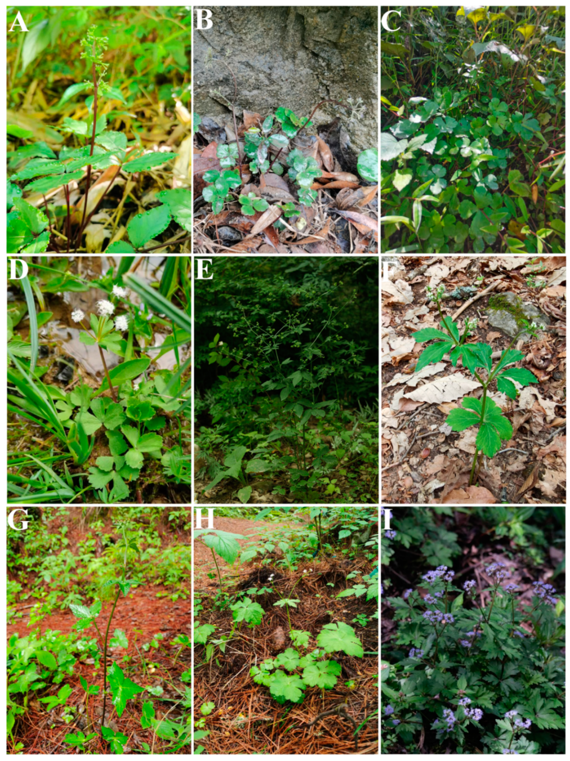
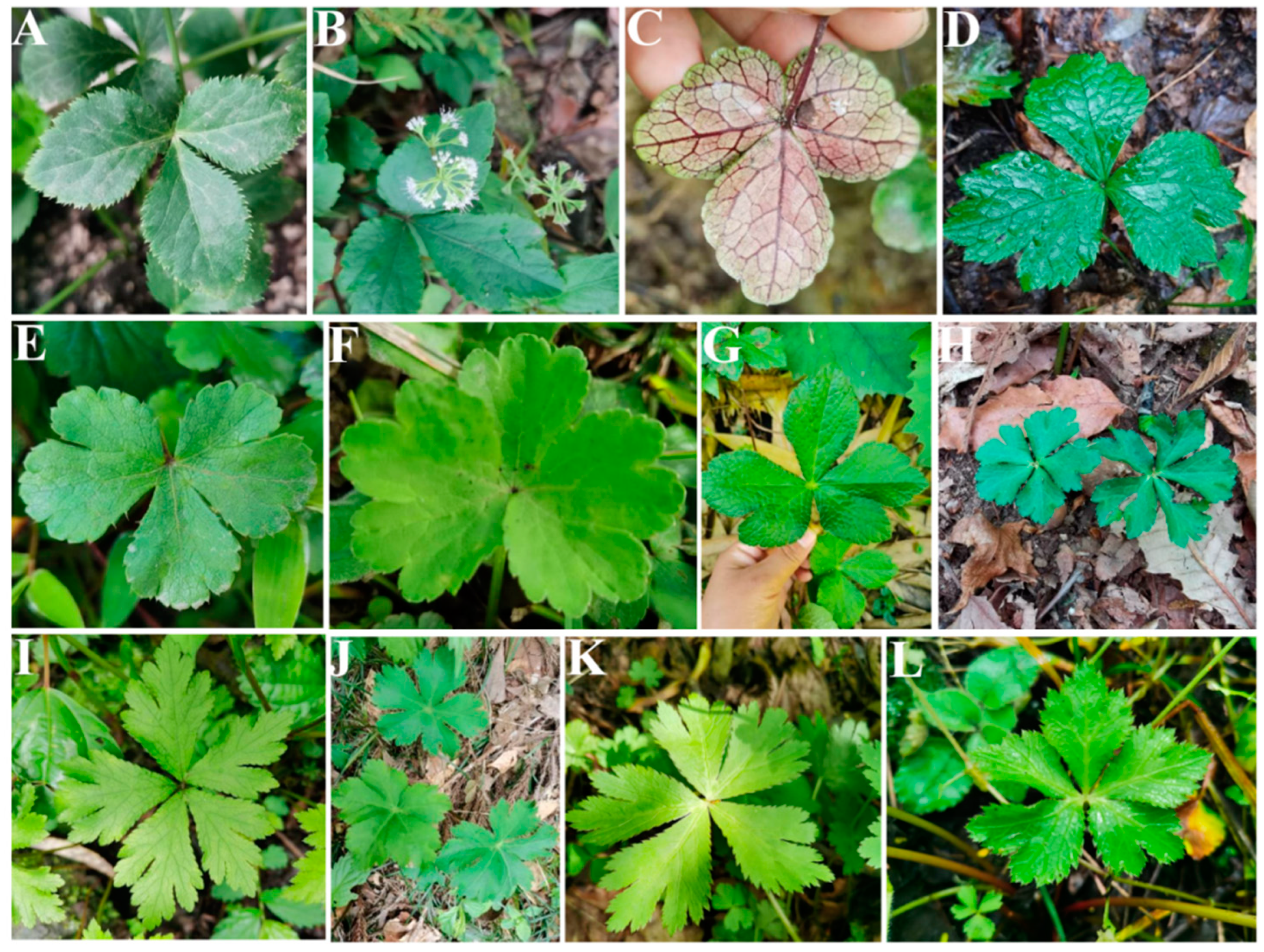
Sanicula L. (Apiaceae-Saniculoideae) contains about 45 species worldwide that are widely distributed from East Asia to North America [1,2], with China and North America as two distribution centers [3]. Of those, nineteen species and five varieties are distributed in China, with eleven species and five varieties of them endemic [4,5,6,7]. The genus was considered as a taxonomically difficult group within Apiaceae and exhibited diversity in habits, leaves, inflorescences, fruits and rhizomes [2,3]. The plants of the genus mainly grow in grassy places on stream banks, rocky walls, alpine scrub, shady wet places, mixed forests on mountain slopes, bamboo forests, wet shady valleys and other habits [5,6] (Figure 1). The members of the genus are biennial or perennial herbs, and the stems are erect, ascending or rarely decumbent. Interestingly, the stems of Chinese Sanicula species are glabrous [5,6] (Figure 2). Their leaves are varied, mainly including blade orbicular, round–cordate or cordate–pentagonal, membranous, papery, or subleathery, trifoliolate, palmately 3-parted or 3–5-parted and often lobed, margin serrate or doubly setose serrate. For example, the basal leaves of S. caerulescens Franch. are trifoliolate; S. rubriflora F. Schmidt, S. rugulosa Diels and S. hacquetioides Franchet. are palmately 3-parted; S. giraldii H. Wolff and S. elongata K. T. Fu are palmately 3–5-parted [5,6,8,9] (Figure 3). The genus has its distinctive inflorescences in that their umbels are simple or in irregularly elongated compound umbels, peduncles racemous, cymous or corymbose-branched. For example, S. caerulescens Franch. has inflorescence subracemose, sometimes several umbels in fascicles; S. orthacantha S. Moore has inflorescence 2–3-branched, umbels 3–8 and S. hacquetioides Franchet. has inflorescence terminal [5,6,8,9] (Figure 4). The fruit of the genus also has its unique characteristic features (conspicuously prominent and persistent calyx, two persistent styles and fruit covered with scales, bristles or hooked prickles). All these distinctive characteristics can easily distinguish the genus Sanicula from other genera of the Apiaceae [10,11].
Since 1975, Behnke proposed the concept of “ultrastructural taxonomy” [12], opening up a new research field for the search for extensive evidence in taxonomy. Micromorphology has become an important tool for studying the evolutionary relationships and the taxonomy of plant systems, especially for those taxonomically notorious groups, such as Impatiens L. [13], Camellia L. [14], Yulania Spach [15], Pugionium Gaertn. [16], as well as the genera of Apiaceae [17,18,19,20,21,22]. In recent years, some scholars have also performed micromorphological studies on the genus Sanicula [23,24,25,26,27,28,29,30,31,32]. For example, Ma et al. observed the leaf epidermis of six Sanicula species under a light microscope and found that these species had the same type of epidermal structure [23]. Chen et al. observed the fruit surface micromorphology of fifteen Sanicula plants (only including eight Chines taxa) and detected that the fruit size, degree of curvature of prickles, and the wax pattern of the fruit stalks of plants in this genus were rich and diverse [29]. Yang et al. observed the pollen micromorphology of fifteen Sanicula species and concluded that the pollen of the genus Sanicula had moderately evolved and relatively evolved morphological features [32]. Therefore, it can be seen that micromorphological characteristics (for example: leaf epidermis, fruit and pollen) of species are useful for distinguishing morphologically similar or taxonomically difficult species, as well as providing more additional evidence for these species. Although previous micromorphological studies have greatly filled the gaps and provided valuable references for the genus Sanicula, the relationships of species with taxonomic difficulties within the genus have not been satisfactorily resolved because these studies involved fewer species (especially the lack of Chinese endemic species) and did not conduct a comprehensive study. Therefore, it is urgent to further explore the micromorphological features of this genus based on expanding sampling.
In this study, we obtained leaf materials from twenty-two species, fruit and pollen materials from twenty species in which these taxa have similar morphology and species identification is difficult, and we performed comprehensively micromorphological analyses of this taxonomically difficult genus. Our major aims were to: (1) reveal the micromorphology of leaf epidermis, fruit and pollen of the genus; (2) evaluate the potential of micromorphology for resolving the taxonomy; (3) provide more additional evidence for exploring the interspecific relationship of the genus.

2. Material and Method

2.1. The Micromorphological Study on the Leaf Epidermis of the Genus Sanicula L.

The leaves materials used in this study were collected in the wild. To avoid errors in experimental materials, we selected clean and fresh basal leaves from each species and then immediately dried the leaves with silica gel and brought the dried leaves back to the laboratory for experiments. Details of the material sources and voucher specimens were shown in Table S1, and the voucher specimens were deposited in the Sichuan University Herbarium (SZ). We used a light microscope and scanning electron microscope to observe the leaf epidermal micromorphological features, and the terminology related to leaf epidermal features referred to the studies of Baranova [33], Wilkinson [34], Zhang et al. [19] and Xu et al. [35].
(1)
Light microscope observation: Silica gel-dried basal leaves were placed in 30% sodium hypochlorite solution (NaClO3) in the dark treatment for 1–3 h. When the leaves faded to white and were nearly transparent, we removed the leaves and immersed them in distilled water for 5–10 min. Then, the leaves were removed and placed on a glass plate, and we gently tore the thin and transparent upper and lower epidermis with tweezers and placed them on a slide with a drop of distilled water to unfold, covered with a coverslip, gently pressed to squeeze out the excess water and air under the coverslip, and the excess water was carefully sucked off with absorbent paper to make a temporary slide. Finally, under 10 × 40 magnification, we respectively selected six fields of view with high clarity of the upper and lower epidermis, used a light microscope (Olympus-BX51) to observe the cells and stomata of the leaf epidermis, and photographed and preserved them. We randomly selected thirty stomata in the lower epidermis of each taxon, and due to the small number of stomata in the upper epidermis of some species, all stomata were selected. Then, we observed the cell shape, the type of stomatal apparatus of both the upper and lower epidermis, counted the number of cells, measured the size of the stomatal apparatus, and calculated the stomatal density and stomatal index. Stomatal density: number of stomata per unit area/unit area. Stomatal index: number of stomata per unit area/(number of stomata per unit area + number of epidermal cells in the same unit area) × 100%. The above measurement processes were carried out in MATO v4.2 software [36].
(2)
Scanning electron microscope observation: The silica gel-dried basal leaves were washed with distilled water and then treated with different concentrations of ethanol for gradient dehydration (15%, 30%, 50%, 70%, 85%, 95% Ⅰ, 95% Ⅱ, 100% Ⅰ, 100% Ⅱ), with each concentration being processed for 1 min. Then, we took out the leaves and dried them naturally. Subsequently, the upper and lower epidermis of each species were cut out and were pasted on double-sided conductive adhesive and sprayed with gold coating, respectively. Finally, we observed and photographed the micromorphological features of the upper and lower epidermis using a JSM-7500F scanning electron microscope.

2.2. The Micromorphological and Anatomical Study on the Fruits of the Genus Sanicula L.

The fruit materials used in this study were collected in the wild. First, we collected the mature and intact fruits and then immersed them in formaldehyde–acetic acid–alcohol (FAA) fixation fluid (38% formaldehyde: glacial acetic acid: 70% ethanol: glycerol = 1:1:18:1). Fruit materials were stored in the Laboratory of Plant Taxonomy and Systematic Evolution, School of Life Sciences, Sichuan University, and the information of fruit material sources and voucher specimens were shown in Table S1. The voucher specimens were deposited in the Sichuan University Herbarium (SZ). The terminology for the description of micromorphological and anatomical characteristics of fruits referred to the studies of Kljuykov [37] and Ostroumova et al. [38].
(1)
Observation of external micromorphological characteristics of fruits. We removed the fruits from the FAA fixation fluid and placed them on absorbent paper to allow them to dry naturally. Then, ten morphologically intact fruits of each species were randomly selected for observation under a stereomicroscope (SMZ25, Nikon Corp., Tokyo, Japan), and then the dorsal and commissure views of fruits of each species were observed, photographed and preserved. Finally, the length and width values of ten fruits of each species were measured using MATO v4.2 software [36], and we then calculated the mean values.
(2)
Observation of anatomical characteristics of fruits.
a:
Freehand slicing method. We observed and randomly selected ten morphologically intact fruits of each species using a stereomicroscope (SMZ25, Nikon Corp., Tokyo, Japan). Then, we used double-layer blades to cut the fruits in the vertical horizontal plane at the middle position of the fruits. Finally, we observed and photographed the transverse section of the fruits with a stereomicroscope (SMZ25, Nikon Corp., Tokyo, Japan).
b:
Paraffin sectioning method. We observed and randomly selected one or two morphologically intact fruits of each species and placed them in FAA fixation fluid for 2–24 h. After the fixation process was completed, we removed them and washed them with distilled water 2–3 times. Then, the fruits were subjected to gradient dehydration treatment with ethanol (30%, 50%, 70%, 80%, 90%, 100%) for 1 h at each concentration. After the gradient dehydration process was completed, we used xylene for deethanolization for 30 min. Finally, we permeabilized, embedded, sectioned, glued, dewaxed the treated fruit materials and stained them using toluidine blue solution (Toluidine Blue solution), and meanwhile, the slices were sealed with neutral gum to create permanent mounts. The anatomical features of the fruits (such as the vittae, endosperm, exocarp and mesocarp) were observed and photographed using a stereomicroscope (SMZ25, Nikon Corp., Tokyo, Japan).
(3)
Observation of micromorphological characteristics of fruits: We randomly selected one or two morphologically intact fruits of each species from the FAA fixation fluid. The remaining steps referred to the scanning electron microscopy method for leaf epidermis (Section 2.1 (2)).

2.3. The Morphological Study on the Pollen of the Genus Sanicula L.

We collected the mature, dry and full pollen materials used in this study in the wild, which were naturally dried and used for subsequent experiments. Details of pollen material sources and voucher specimens were shown in Table S1, and the voucher specimens were deposited in the Sichuan University Herbarium (SZ). The pollen morphological features were observed using a light microscope and scanning electron microscope, and the terminology related to pollen morphological features referred to the studies of Wang et al. [39], She and Shu [40,41] and Yang et al. [32].
(1)
Light microscope observation: The G. Erdtman acetic anhydride decomposition method [42] was used to treat pollen, and the specific steps were as follows:
a:
Take an appropriate amount of mature, dry and full pollen of each species in a 5 mL cryostat tube and submerge the pollen with 1–2 mL of glacial acetic acid for 24 h;
b:
Use a glass rod to thoroughly crush the pollen in the cryostat tube, filter impurities with a 100-mesh copper mesh, wash with glacial acetic acid and collect 5 mL of filtrate into a new cryostat tube;
c:
Put the new cryostat tube in a centrifuge at 5000 r/min for 10 min and then repeat again;
d:
Add a mixture of acetic anhydride and concentrate sulfuric acid (volume ratio = 9:1, currently used and prepared) into the cryostat tube containing discarded supernatant and add dropwise to 5 mL;
e:
Place the frozen tube in a water bath at 80 °C for 4 min, then remove and centrifuge at 5000 r/min for 10 min;
f:
Discard the supernatant from the frozen tube, add 4 mL of distilled water and centrifuge at 5000 r/min for 10 min;
g:
Discard the supernatant from the freezing tube, then add 2–3 drops of glycerol to the freezing tube and mix well;
h:
Pipette 1–2 drops of pollen suspension onto a glass slide and create a temporary slide.
Firstly, we searched for the pollen with intact morphology under low magnification. Then, we randomly selected twenty pollen of each species to observe and photograph their shape and the equatorial and polar views under a light microscope (Olympus-BX51) at 10 × 100 magnification. Finally, MATO v4.2 software [36] was used to measure the pollen size, including the polar axis length (P) and the equatorial axis length (E) of pollen. In addition, we also calculated the ratio of polar axis length and equatorial axis length (P/E).
(2)
Scanning electron microscope observation: Firstly, we observed and randomly selected ten dry, mature and full anthers of each species under a dissecting microscope. Then, the selected anthers were pasted on the double-sided conductive adhesive, and the pollen capsule was gently poked with a clean dissecting needle to release the pollen grains, so that they were evenly spread in all areas of the conductive adhesive, and the shell of the pollen was clamped away with tweezers. Finally, after gold spray coating, we used a JSM-7500F scanning electron microscope to observe and photograph the overall view of pollen, the polar view, the equatorial view, germ furrow and the exine ornamentation.

3. Results

3.1. The Morphological Characteristics of the Leaf Epidermis

3.1.1. Leaf Epidermal Characteristics by Light Microscope

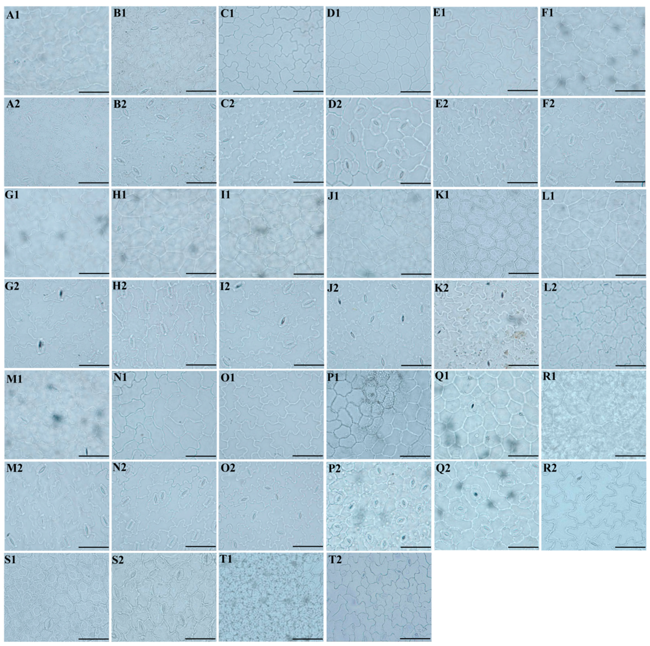
We used a light microscope to observe the micromorphological characteristics of the leaf epidermis of twenty Sanicula plants, of which the eleven taxa were reported for the first time (Figure 5).
Our study observed that the upper and lower epidermis were smooth and glabrous. For the upper epidermis, the cell shape of these twenty Sanicula taxa can be divided into two types: polygonal and irregular. Among them, the cell shape in the upper epidermis of S. chinensis, S. elongata, S. brevispina, S. subgiraldii, S. pengshuiensis, S. pauciflora, S. oviformis, S. tienmuensis, S. flavovirens and S. elata were polygonal, and the cell shape of the remaining taxa were irregular. The pattern of anticlinal wall had three types: shallowly undulating, deeply undulating and subflat or flat. Of which, the pattern of the anticlinal wall of S. brevispina and S. orthacantha was shallowly undulating; S. elongata, S. subgiraldii, S. pengshuiensis, S. pauciflora, S. oviformis, S. tienmuensis, S. flavovirens, S. elata and S. nanchuanensis were subflat or flat, and the remaining taxa were deeply undulating. The stomata were only observed in S. rugulosa, S. elongata, S. hacquetioides, S. tienmuensis and S. elata, and the stomatal shape was ellipsoid in S. rugulosa, S. hacquetioides, S. tienmuensis, S. elata and suborbicular in S. elongata. As for stomatal size (length × width), the largest was 32.43 μm × 19.73 μm in S. rugulosa, and the smallest was 22.81 μm × 22.49 μm in S. elongata. The largest stomatal ratio was 2.31 for S. elata, and the smallest was 1.01 for S. elongata. The largest stomatal density and stomatal index were observed for S. rugulosa (82.54/mm2, 15.52%), and the smallest was for S. elata (16.18/mm2, 1.30%) (Figure 5(A1–T1), Table S2-1).
For the lower epidermis, the cell shape of these twenty Sanicula taxa were irregular. The pattern of the anticlinal wall had two types: shallowly undulating and deeply undulating. For instance, S. chinensis, S. elongata, S. subgiraldii, S. pauciflora, S. oviformis, S. tienmuensis, S. elata and S. nanchuanensis were shallowly undulating, and the remaining taxa were deeply undulating. We observed that the stomata existed in these twenty Sanicula taxa. The stomatal shapes were ellipsoid, long-ellipsoid, ovoid and suborbicular. Among them, S. tienmuensis had the suborbicular stomata; S. serrata, S. pauciflora, S. hacquetioides, S. orthacantha and S. flavovirens had the ellipsoid stomata; S. chinensis, S. subgiraldii, S. oviformis, S. elata and S. nanchuanensis had the ovoid stomata; and the remaining taxa had the ellipsoid stomata. The stomatal size (length × width) was 27.20 × 18.49 μm–20.03 × 17.90 μm, and the largest stomata was observed in S. serrata, and the smallest stomata was for S. nanchuanensis. The stomatal ratio ranged from 1.12 (S. nanchuanensis) to 1.56 (S. orthacantha). The stomatal density of S. oviformis was the largest (681.16), and that of S. pengshuiensis was the smallest (81.09). The stomatal index ranged from 8.46 (S. pengshuiensis) to 29.41 (S. rugulosa) (Figure 5(A2–T2), Table S2-2).

3.1.2. Leaf Epidermal Characteristics by Scanning Electron Microscope

We used a scanning electron microscope to observe the micromorphological characteristics of the leaf epidermis of twenty-two Sanicula plants, of which the leaf epidermis of sixteen members were reported for the first time (Figure 6 and Figure 7).
In the upper epidermis of these twenty-two Sanicula plants, we observed that the cuticular membrane ornamentation can be divided into four types: sparsely striated and clustered into protuberances, densely striated and clustered into protuberances, sparsely striated and striated protuberance, densely striated and striated protuberance. The cell contour was visible in eight taxa (S. giraldii, S. caerulescens, S. serrata, S. brevispina, S. subgiraldii, S. oviformis, S. nanchuanensis, S. petagnioides), and the cell contour was invisible in the other fourteen taxa. Of these, five species (S. caerulescens, S. serrata, S. elongata, S. hacquetioides and S. pauciflora) had no epidermal appendage, and the remaining seventeen species had epidermal appendages. The stomata were only observed in S. rugulosa, S. elongata, S. hacquetioides, S. tienmuensis and S. elata (Figure 6, Table S3-1).
In addition, in the lower epidermis of these twenty-two Sanicula plants, the cuticular membrane ornamentation features were smooth, nearly smooth, sparsely striated and striated protuberance, sparsely striated and clustered into protuberances. The cell contour was invisible in all these Sanicula taxa. The epidermal appendage was absent in five species (S. caerulescens, S. serrata, S. hacquetioides, S. orthacantha, S. brevispina), and the remaining seventeen species possessed epidermal appendage. All members had stoma, and the stoma included two types: protuberant and sunken. Among them, the stoma of ten species (S. rugulosa, S. giraldii, S. caerulescens, S. serrata, S. hacquetioides, S. orthacantha, S. oviformis, S. tienmuensis, S. flavovirens, S. nanchuanensis) was sunken, and the stoma was protuberant in the remaining twelve species (Figure 7, Table S3-2).

3.2. The Micromorphological and Anatomical Characteristics of the Fruits

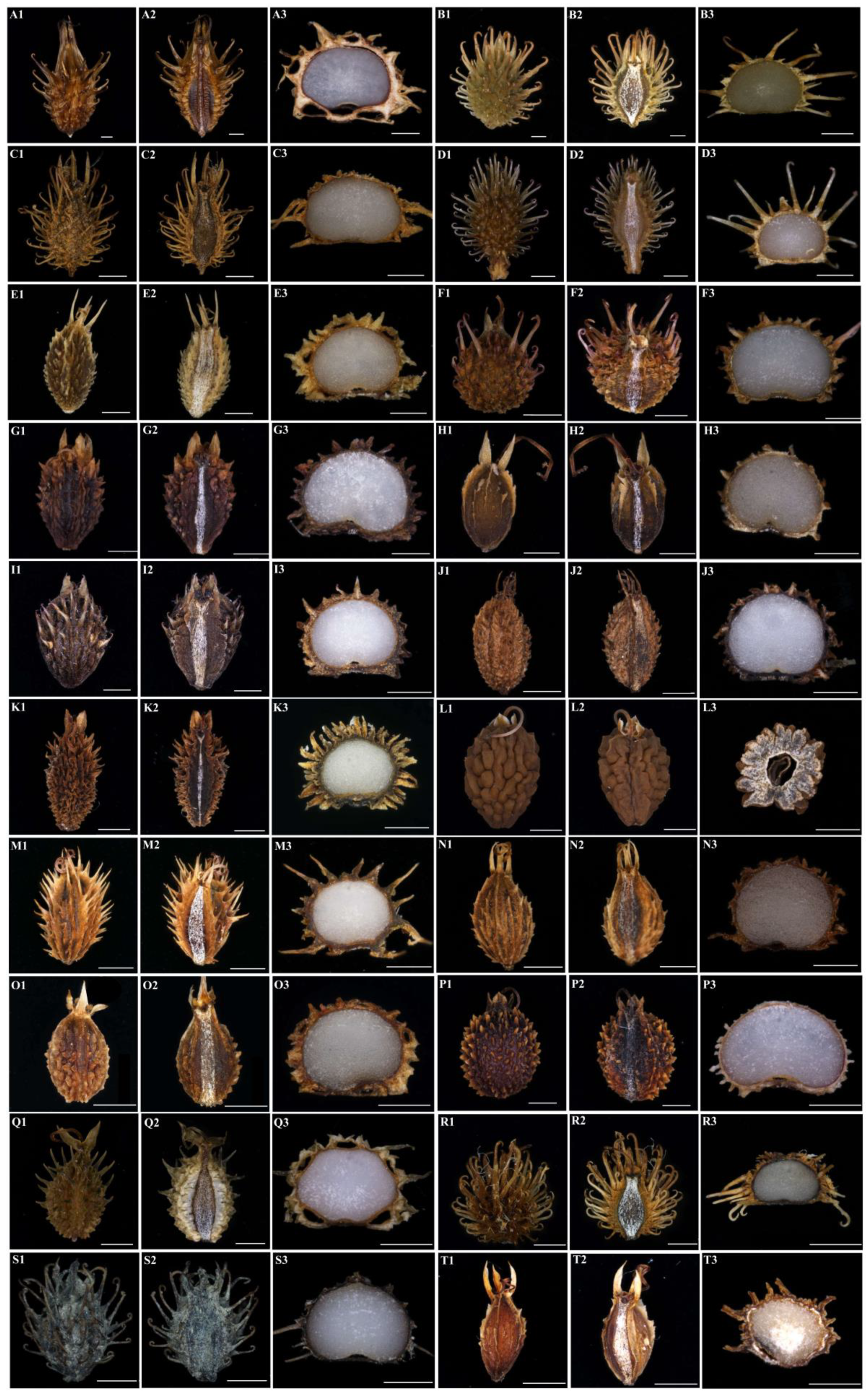
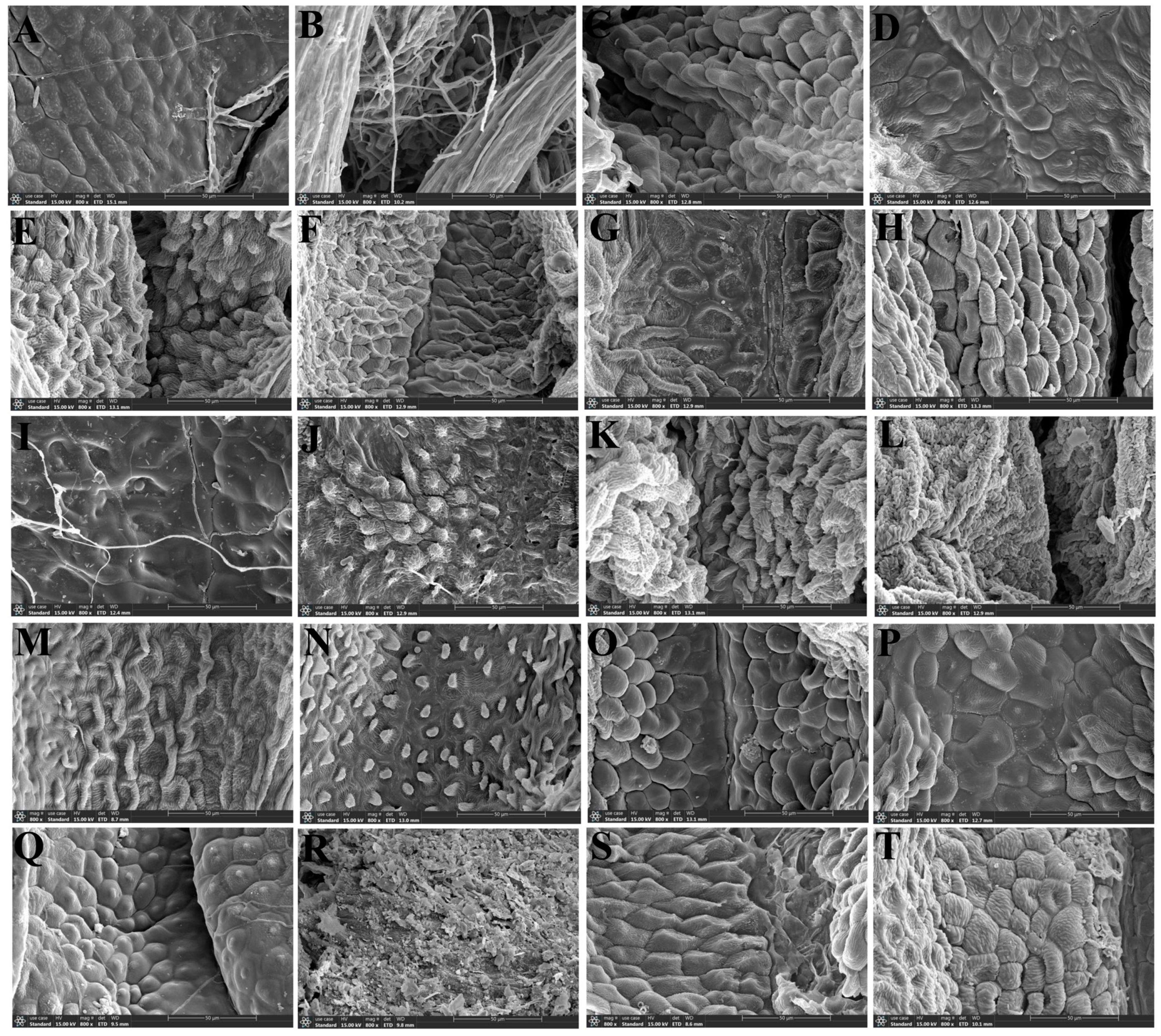
We observed twenty Sanicula fruits, and the fruits micromorphological characters of thirteen taxa were reported for the first time (Figure 8, Figure 9 and Figure 10). We found that the fruit colors were brownish-yellow, brownish-brown or purplish-red. The fruits scarcely compressed, and the fruit shapes were diverse, such as ovoid or ovoid–globose, globose or ellipsoid, broadly ovate and subglobose, of which some Sanicula members fruits had their distinctive shape, such as the fruit shape of S. tienmuensis was subglobose; S. subgiraldii was broadly ovate; S. pengshuiensis, S. rugulosa and S. nanchuanensis were ellipsoid, S. elongata, S. oviformis and S. brevispina were ovoid; S. rubriflora, S. chinensis, S. serrata, S. hacquetioides, S. flavovirens and S. elata were ovoid–globose. The fruit size varied from 1.89 × 0.90 mm (S. astrantiifolia) to 6.97 × 2.68 mm (S. rubriflora). The fruit surface was covered with prickles or protuberances, or tubercles, prickles were either long or short, straight or hooked, rarely scale-like. Among them, S. astrantiifolia, S. chinensis, S. giraldii and S. rugulosa had the extremely well-developed uncinate prickles; S. flavovirens, S. pauciflora and S. orthacantha had the straight bristles; S. hacquetioides, S. oviformis and S. tienmuensis were covered with scales and tubercles, but never spinulose; S. caerulescens, S. elongata, S. pengshuiensis and S. lamelligera were covered with short and straight spinous-bristles, usually fused at the base forming a thin tier; S. nanchuanensis were proximal end with degenerated-to-disappeared prickles, nearly smooth, distal end with prickles and formed a thin layer; S. rubriflora, S. serrata and S. subgiraldii had their proximal part covered with scales, distal part covered with slightly uncinate bristles. The calyx teeth were prominent in all Sanicula taxa, and the shape was lanceolate, ovate and linear. Of them, S. giraldii, S. serrata and S. tienmuensis had ovate calyx teeth; S. chinensis, S. pengshuiensis and S. lamelligera had linear calyx teeth; and the remaining taxa had the varied shape of these three shapes (Figure 8, Table 1).
In addition, we also detected the micromorphological characteristics of fruits surface, and we found that the cell shapes of fruit furrows were diverse, including circular shape, polygon shape and invisible. For example, S. rubriflora, S. giraldii, S. flavovirens, S. tienmuensis and S. nanchuanensis had the polygon cell shape; S. elongata, S. brevispina and S. oviformis had the circular cell shape. The surfaces of fruit furrows had various types, such as high convex, immersed, concave–convex and smooth (Figure 9).
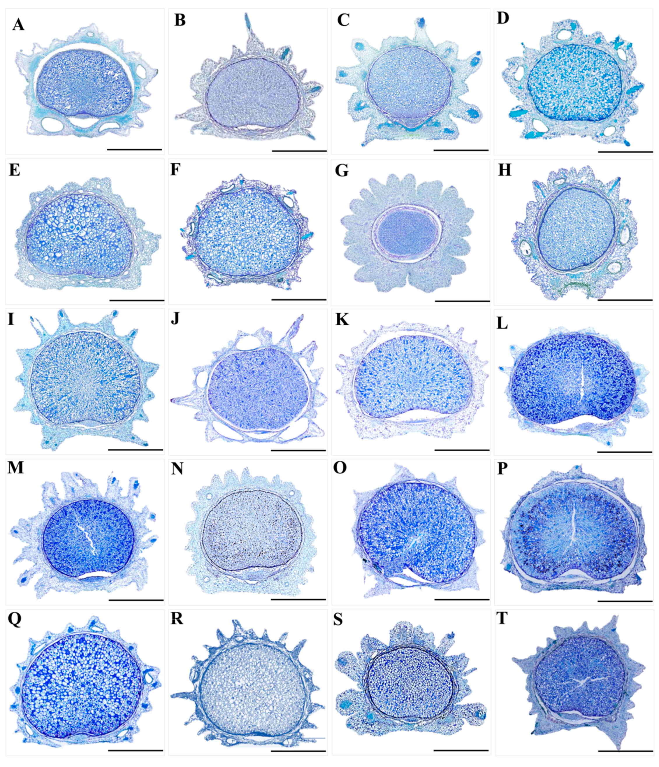
Furthermore, exocarp and mesocarp were protruding as prickles or protuberances. The mericarp were suborbicular in the transverse section, sparsely ellipsoid–orbicular or subovoid. The endosperm on the commissural side was slightly concave or flat, which ten species (S. rubriflora, S. caerulescens, S. serrata, S. elongata, S. brevispina, S. lamelligera, S. oviformis, S. tienmuensis, S. rugulosa and S. subgiraldii) had the slightly concave endosperm on the commissural side, and the other ten species had the flat endosperm on the commissural side (Table 1). The exocarp consisted of 1 layer of slightly compressed or irregularly rounded thin-walled cells, the outer wall often with papillate projections. The prickles often contained lignified longitudinal elongated thick-walled tissue. The vittae were distinct in S. rubriflora, S. caerulescens, S. pengshuiensis, S. pauciflora, S. lamelligera, S. oviformis and S. flavovirens, and the vittae of the remaining taxa were obscure (Figure 8 and Figure 10).

3.3. The Micromorphological Characteristics of Pollen

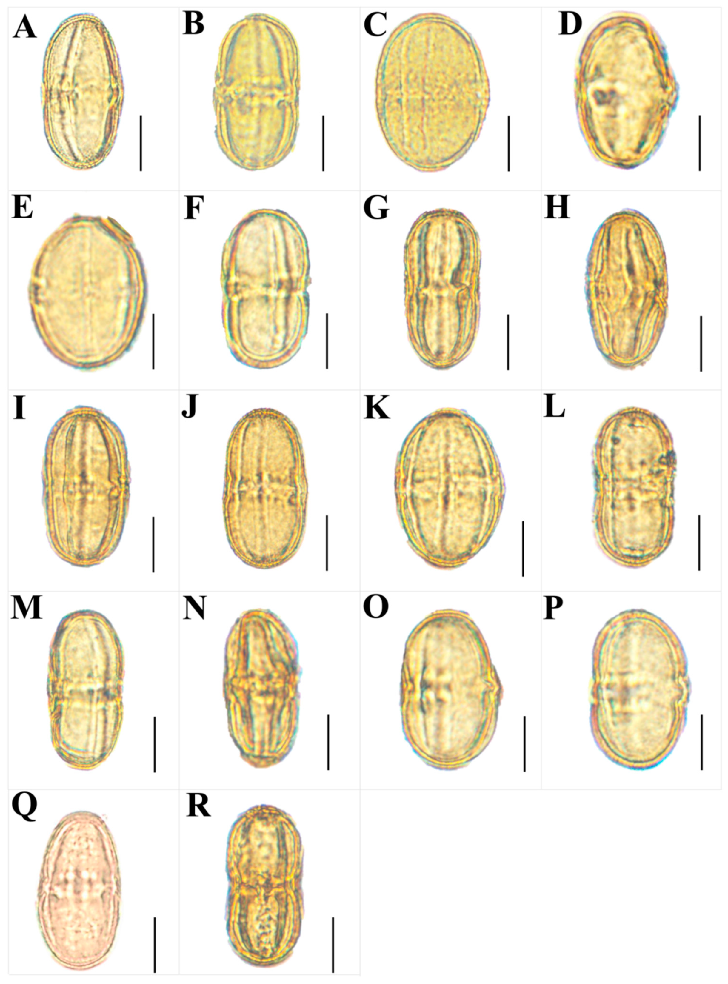
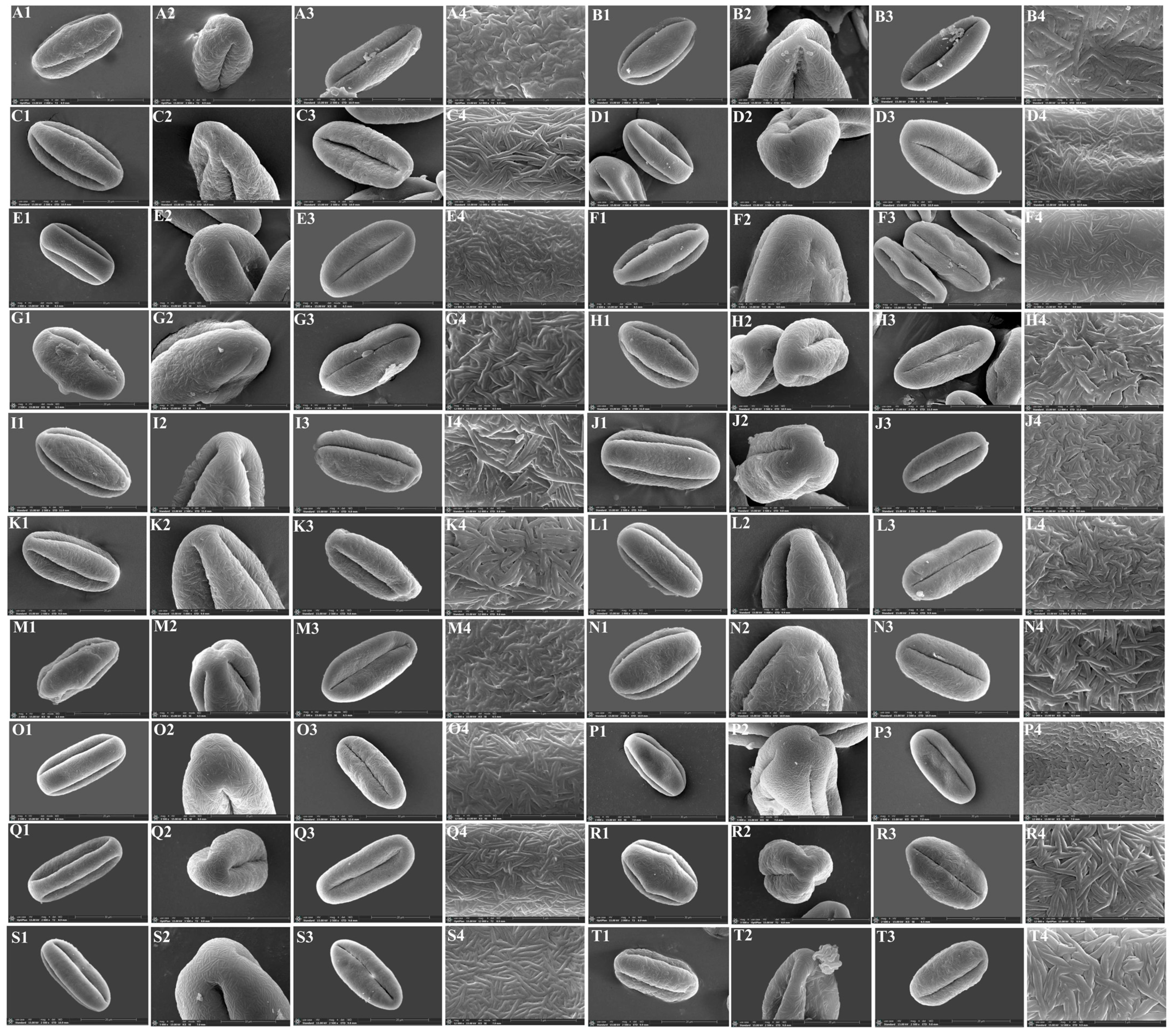
We observed micromorphological characteristics of pollen from twenty Sanicula taxa, and the pollen micromorphological characteristics of seven taxa were reported for the first time (Figure 11 and Figure 12). The pollen size (polar axis × equatorial axis) ranged from 30.03 × 16.95 μm to 57.18 × 23.59 μm, with the smallest pollen in S. chinensis and the largest in S. flavovirens. The ratio (P/E) of the polar axis (P) to the equatorial axis (E) ranged from 1.58 to 2.65, with the minimum value occurring in S. oviformis and the maximum value occurring in S. pauciflora. The equatorial view of the pollen had four shapes: ellipsoid, subrectangular, equatorially constricted, super-rectangular-equatorially constricted. Of these, the equatorial views of S. giraldii, S. elata and S. lamelligera were subrectangular; S. caerulescens, S. nanchuanensis and S. pauciflora were equatorially constricted; S. flavovirens was superrectangular-equatorially constricted, and the remaining taxa had the elliptical equatorial view. The polar view of the pollen had four shapes: triangular–circular, triangular, suborbicular and trilobate circular. Among them, S. giraldii, S. caerulescens and S. orthacantha had suborbicular polar view; S. rugulosa and S. astrantiifolia had triangular polar view; S. subgiraldii, S. pengshuiensis, S. lamelligera, S. elata, S. flavovirens and S. nanchuanensis had trilobate circular polar view; and the remaining species had triangular–circular polar view. Except for the exine ornamentation of the equatorial view in S. rubriflora was brain-striped, and the remaining species had striped exine ornamentation in both their equatorial view and polar view. All Sanicula members had a side germinal aperture, and the germinal furrows were all long (Figure 11 and Figure 12, Table S4).

4. Discussion

4.1. The Micromorphological Characteristics and Its Taxonomic Value of Leaf Epidermis in the Genus Sanicula L.

The micromorphology of plant leaf epidermis can reflect species interspecific relationships and affinities of species to some extent, which can provide a valuable reference for taxonomic issues, such as species identification and delimitation [43,44]. Therefore, leaf epidermal micromorphological features have become one of the most important evidence for plant taxonomic studies [45,46]. In recent years, the micromorphological characteristics of plant leaf epidermis have been widely used in the classification and systematic evolution of Apiaceae plants [5,6,7,8,9,10,47,48,49,50,51]. For example, Zhou et al. used light microscopy and scanning electron microscopy to study the leaf epidermis morphology of 21 Peucedanum L. plants, revealing that the leaf epidermis morphology of 21 Peucedanum L. plants was stable at the species level, and it can be used for species identification; however, there was almost no consistency at the group level, suggesting that the genus Peucedanum L. may not be a natural group [47]. Xu et al. observed the leaf epidermis micromorphology of seven Phytophermopsis H. Wollf plants using scanning electron microscopy and found that the contours of leaf epidermal cells, cell shape, ornamentation of cuticle and the stoma were different among these seven species. For example, the leaf epidermal cells of P. muliensis were regular polygonal, and the ornamentation of cuticle had irregular stripes. The stoma of P. delavayi, P. shaniana, P. rubrinervis and P. obtusiuscula were only distributed in the lower leaf epidermalon. The shape of the stoma was elliptical or ovate. Thus, these seven Physospermopsis species can be distinguished based on their micromorphological characteristics of leaf epidermis [35]. Ren et al. observed the leaf epidermal micromorphology of eleven Pleurospermum Hoffm. plants and found that the distribution of stoma was a valuable taxonomic evidence for these Pleurospermum taxa, and they also speculated that P. franchetianum and P. davidii had a close relationship [50].
Our study found that the micromorphological characteristics of leaf epidermis have high consistency within the genus Sanicula. For instance, the cell shape in the lower epidermis was irregular in all taxa, and the corresponding pattern of the anticlinal wall was shallowly or deeply undulating. The stomata was presented in the lower epidermis of all species. These findings suggested that the genus Sanicula was a natural taxon, which was consistent with the previous studies [13,14].
Based on the cell shape in the upper epidermis, we can easily distinguish eleven species (S. chinensis, S. elongata, S. brevispina, S. subgiraldii, S. pengshuiensis, S. pauciflora, S. oviformis, S. tienmuensis, S. flavovirens, S. nanchuanensis and S. elata) from other species because their cell shape was polygonal, while the cell shape of the remaining taxa was irregular. We can also identify five species (S. rugulosa, S. elongata, S. hacquetioides, S. tienmuensis and S. elata) from other species by the stomata because the stomata were only observed in these five members, and the other members had no stomata. Li et al. had treated S. pengshuiensis as a synonymy of S. lamelligera [52]. However, we did not support this view based on the study of leaf epidermal micromorphology. The cell shape of the upper epidermal in S. pengshuiensis was polygonal, while S. lamelligera was irregular, whereby they can be easily distinguished. In addition, the stomata in the lower epidermis of S. pengshuiensis was ovoid, while that in S. lamelligera was elliptical, so we considered them as two independent species. The previous molecular research also justified our micromorphological study [3]. Song et al. performed phylogenetic analyses based on plastome data and found that S. tienmuensis and S. astrantiifolia had close relationship with weak supports and low resolutions, which made them difficult to distinguish [3]. However, our study found that these two species had their own distinctive micromorphological features. For example, the upper epidermis of S. astrantiifolia had no stomata, the cell shape was irregular, and the cuticular membrane ornamentation was densely striated and striated protuberance, whereas the upper epidermis of S. tienmuensis had the ellipsoid stomata, the cell shape was irregular polygonal, and the cuticular membrane ornamentation was sparsely striated and clustered into protuberances. In addition, the stomata shape in the lower epidermis was also different, with ellipsoid stomata existing in S. astrantiifolia, whereas S. tienmuensis had a suborbicular stomata. From the above, it was noticed that S. tienmuensis and S. astrantiifolia had differences in the micromorphological characteristics of leaf epidermis, which can clearly distinguish them. We also found that three varieties (S. subgiraldii, S. brevispina, S. pauciflora) had distant affinities with their type varieties. In detail, S. giraldii and S. orthacantha had an irregular cell shape, while S. subgiraldii and S. brevispina had a polygonal cell shape in the upper epidermis. S. tienmuensis had an ellipsoid stomata, whereas S. pauciflora had no stomata in the upper epidermis. These findings implied that these three varieties should be regarded as three independent species. Furthermore, the stomatal size, stomatal ratio, stomatal index and stomatal density of epidermis had differences among these Sanicula taxa involved in this study, which can also serve as additional evidence to distinguish them. Therefore, it can be seen that the micromorphological characteristics of leaf epidermis can serve as a supplement to molecular data, providing new evidence for interspecific classification research of the genus Sanicula.

4.2. The Micromorphological Characteristics and Its Taxonomic Value of Fruit in the Genus Sanicula L.

The micromorphological characteristics of fruits play a vital role in the classification and systematic evolution of angiosperms at different levels of family, genus and species [53]. Especially, the characteristics of fruits in the family Apiaceae are very distinctive and stable, which have been widely used in the taxonomic research of this family [54,55,56,57,58,59]. For example, Liu et al. observed the fruit micromorphology of eighteen representative genera in Apiales and found that the anatomical characteristics of fruits were closely related to the systematic position of these genera, indicating that the anatomical characteristics of fruits were of high value in solving the phylogenetic and taxonomic problems of Apiales [58]. Kljuykov et al. enumerated fifteen features of the anatomical characteristics in the family Apiaceae and concluded that the anatomical characteristics of the fruit were usually constant in the genus, which can be used to describe the genus or closely related genera or groups [59]. Zhang et al. used a freehand sectioning method to observe and compare the external morphology and anatomical features of the fruits of 23 species in 17 genera of the family Apiaceae. They concluded that the characteristics of the fruit surface with hook spines or bristles and the fruit ribs showed significant differences among genera but showed consistency within genera, which can be used as the reference for the classification of the Apiaceae between genera. They also found that the shape of the fruit transverse section, the concavity and convexity of the endosperm surface, and the morphological characteristics of the calyx teeth were important in research studies on intragenus and interspecies classification. Based on the morphological characters of the fruit, a taxonomic search table for 23 plant species belonging to 17 genera was compiled [30].
We also observed the micromorphological and anatomical characteristics of the genus Sanicula. The most distinctive characteristic features of the genus Sanicula were the fruits covered with scales, bristles, or hooked prickles, a rather prominent and persistent calyx, and two persistent styles that can easily distinguish it from other genera of Apiaceae, which were in accordance with the previous studies [5,6,10]. In this study, twenty Sanicula species showed some differentiation in fruit micromorphology, and their characters reflected the similarities and differences among different species. Some Sanicula members fruits had their distinctive shape, prickles shape and calyx teeth. In addition, the cell shapes of the fruits surface were diverse, and the surface of fruit furrows had various types, which can also serve as important identified features among these Sanicula members. Therefore, the micromorphological characteristics of the Sanicula fruit had a certain taxonomic value and can be used as the basis for taxonomic identification. For example, the fruit size of S. rubriflora was significantly larger than other Sanicula species and can be easily distinguished from other members. The fruit shape of S. tienmuensis was subglobose, which was clearly different from other Sanicula species fruit. S. hacquetioides was easily distinguishable from other species because of its fruit covered with scales and tubercles, but never spinulose. S. orthacantha can be distinguished by its developed straight bristles, while S. astrantiifolia, S. chinensis, S. giraldii and S. rugulosa had the extremely well-developed uncinate prickles. S. nanchuanensis were proximal end with degenerated-to-disappeared prickles, nearly smooth, distal end with prickles and formed a thin layer. In addition, we also found that three varieties had distant affinities with their type varieties from fruits. For example, S. giraldii had fruit densely covered with developed yellow or purplish-red uncinate bristles, long and hard, calyx teeth ovate and small, tip mucronate, while S. subgiraldii had the unique fruits rarely covered with purplish-red short bristles, proximal end with tubercles, obscure, distal end with uncinate bristles or straight, calyx teeth broadly ovate and large. S. tienmuensis had fruits that were subglobose, covered with short and obtuse prickles, slight formed scales and tubercles, calyx teeth broadly ovate, vittae obscure, whereas S. pauciflora had fruits that were long ellipsoid, densely covered with sharp prickles, calyx teeth long-lanceolate, vittae 2 in the commissural side. S. orthacantha had narrowly ovoid fruit, densely covered with short, straight and sharp spines, and sometimes the base formed a thin layer, fruit ribs and furrows spinulose, narrowly lanceolate calyx teeth, while S. brevispina had the oblong ovoid to ovoid fruit with proximal end with degenerated-to-disappeared prickles, nearly smooth and distal end with prickles and formed a thin layer, usually with erose-spinulose ribs and furrows smooth or barely spinulose, linear to lanceolate calyx teeth, tip sharp. It can be seen that fruit micromorphology also supported that these three varieties should be considered as three independent species. The vittae was also one of the most important distinguishing features, such as S. rubriflora, S. chinensis, S. caerulescens, S. pengshuiensis, S. pauciflora, S. lamelligera, S. oviformis, S. flavovirens and S. elata, which possessed vittae, while other species had no vittae. The cell shapes of fruit furrows in S. rubriflora, S. giraldii, S. flavovirens, S. tienmuensis and S. nanchuanensis were polygonal, whereas S. elongata, S. brevispina and S. oviformis were circular. All these distinctive features can easily distinguish Sanicula members. Thus, it was noticed that prickles, fruit size, shape and vittae were the most distinctive features that can distinguish morphologically similar species of the genus Sanicula.

4.3. The Micromorphological Characteristics and Its Taxonomic Value of Pollen in the Genus Sanicula L.

The pollen is an important reproductive organ of plants and the pollen morphology, including pollen size, outer wall ornamentation, and germinal pore characteristics, which can reflect plant affinities to a certain extent, which has long been widely used in plant classification and phylogenetic studies [60,61]. In taxonomic studies of the family Apiaceae, the rich and diverse pollen micromorphological characteristics can also provide a valuable reference for exploring the phylogenetic relationships and evolutionary status between species [62,63,64,65,66,67]. For example, Xu et al. observed the pollen of seven Physospermopsis species and illustrated that P. obtusiuscula and P. kingdon-wardii can be distinguished based on their pollen shape because P. obtusiuscula was a relatively primitive rhombic-shaped pollen, while P. kingdon-wardii was a relatively evolved elliptical pollen [35]. Zhang et al. used light microscopy and scanning electron microscopy to observe the pollen morphology of Ostericum Hoffm. (Apiaceae) of eight species from seventeen populations. Their study showed that pollen morphology of Ostericum exhibited certain intraspecific stability in pollen shapes and exine ornamentations, while interspecies diversities in aperture types, central protrudents and length, density and appendages of exine ornamentation. In addition, they also found that the volume index, germinal furrows, and the outer wall decoration and wax stripes of O. atropurpureum and O. citriodorum were very similar, which can clearly distinguish them from other Ostericum members [65].
In this study, we observed that the location of germination pores and exine ornamentation of these twenty taxa were quite similar, indicating that the genus Sanicula was a natural unit, which was consistent with the pollen studies of Yang et al. [22]. However, the pollen shape and size of twenty Sanicula taxa were different. For example, S. caerulescens, S. pauciflora and S. nanchuanensis had equatorially constricted pollen, and thus they can be easily identified from others by the pollen shape. S. flavovirens had the largest pollen size (57.18 × 23.59 μm), while S. caerulescens had the smallest pollen size (32.83 × 14.09 μm), based on which they can be identified from each other by the pollen size. S. pengshuiensis had the largest P/E ratio of 2.35, whereas S. oviformis had the smallest P/E ratio of 1.58, which can identify them from other Sanicula members by the pollen P/E ratio. We also found that three varieties had distant affinities with their type varieties from pollen. For example, S. giraldii had a subrectangular equatorial view and a suborbicular polar view, while an ellipsoid equatorial view and trilobate circular polar view were observed in S. subgiraldii. S. tienmuensis had an ellipsoid equatorial view, whereas S. pauciflora had an equatorially constricted equatorial view. S. orthacantha had a suborbicular polar view, whereas S. brevispina had a triangular–circular polar view. Thus, pollen micromorphological also robustly supported that these three varieties should be treated as three independent species. Overall, the pollen micromorphological characteristics (such as pollen shape, size, P/E ratio, equatorial view and polar view) can also be used as an additional evidence to discriminate the complicated species of this genus.

5. Conclusions

In this study, we newly obtained leaf materials from twenty-two Sanicula members, fruit and pollen materials from twenty Sanicula members, covering about 90% of the Chinese samples, and performed comprehensively micromorphological analyses of this taxonomically difficult genus. Our study revealed the micromorphology of leaf epidermis, fruit and pollen of the genus in detail. In addition, we also evaluated the potential of micromorphology to distinguish species with taxonomic difficulties within the genus. Furthermore, we also provided more additional evidence for exploring the interspecific relationship of the genus. In summary, our study will be beneficial to future taxonomic studies of the genus Sanicula.

Supplementary Materials

The following supporting information can be downloaded at: https://www.mdpi.com/article/10.3390/plants13121635/s1, Table S1. The collection information of the genus Sanicula plants. Table S2-1. The features of upper leaf epidermis for Sanicula plants observed with light microscope. Table S2-2. The features of lower leaf epidermis for Sanicula plants observed with light microscope. Table S3-1. The features of upper leaf epidermis for Sanicula plants observed with scanning electron microscope. Table S3-2. The features of lower leaf epidermis for Sanicula plants observed with scanning electron microscope. Table S4. The pollen morphology of Sanicula plants observed with scanning electron microscope.

Author Contributions

X.H. and S.Z. designed the research. B.S., Y.W. (Yunyi Wang), L.Y., Y.W. (Yuan Wang) and L.C. collected the data. B.S. and F.Y. performed the experiments, B.S., F.Y. and C.L. conducted data analysis. B.S. and C.L. prepared the manuscript. All authors have read and agreed to the published version of the manuscript.

Funding

This research was funded by the National Natural Science Foundation of China (Grant Nos. 32170209, 32070221) and the Survey on the Background Resources of Chengdu Area of Giant Panda National Park (Project No.: 510101202200376).

Data Availability Statement

Data are contained within the article and Supplementary Materials.

Acknowledgments

We are grateful to Deng-Feng Xie and Ting Ren for their valuable discussions.

Conflicts of Interest

The authors declare no conflicts of interest.

References

  1. Van Wyk, B.E.; Tilney, P.M.; Magee, A.R. African Apiaceae: A Synopsis of the Apiaceae/Umbelliferae of Sub-Saharan Africa and Madagascar/Ben-Erik Van Wyk, Patricia M Tilney & Anthony R Magee; Briza Academic Books: Pretoria, South Africa, 2013. [Google Scholar]
  2. Li, H.M.; Wu, M.S.; Lai, Q.; Zhou, W.; Song, C.F. Complete chloroplast of four Sanicula taxa (Apiaceae) endemic to China: Lights into genome structure, comparative analysis, and phylogenetic relationships. BMC Plant Biol. 2023, 23, 444. [Google Scholar] [CrossRef] [PubMed]
  3. Song, B.N.; Liu, C.K.; Zhao, A.Q.; Tian, R.M.; Xie, D.F.; Xiao, Y.L.; Chen, H.; Zhou, S.D.; He, X.J. Phylogeny and diversification of genus Sanicula L. (Apiaceae): Novel insights from plastid phylogenomic analyses. BMC Plant Biol. 2024, 24, 70. [Google Scholar] [CrossRef] [PubMed]
  4. Pimenov, M.G.; Leonov, M.V. The Genera of the Umbelliferae: A Nomenclator; Royal Botanic Gardens: Kew, Richmond, UK, 1993. [Google Scholar] [CrossRef]
  5. Shan, R.H.; Constance, L. The Genus Sanicula (Umbelliferae) in the Old World and the New; University of California Press: California, UK, 1951; Volume 25, pp. 1–7. [Google Scholar]
  6. Sheh, M.L.; Phillippe, L.R. Sanicula L. In Flora of China; Wu, Z.Y., Raven, P.H., Hong, D.Y., Eds.; Beijing Press: Beijing, China; Missouri Botanical Garden Press: St. Louis, MO, USA; Science Press: Beijing, China, 2005; Volume 14, pp. 19–24. [Google Scholar]
  7. Xie, W.Y.; Ma, D.D.; Chen, F.; Wang, P.; Chen, J.F.; Chen, Z.H. Sanicula favovirens—A new species of the genus Sanicula (Umbelliferae) in Zhejiang. J. Hangzhou Norm. Univ. Nat. Sci. Ed. 2019, 18, 9–12. [Google Scholar]
  8. Liou, S.L. Sanicula L. In Flora Reipublicae Popularis Sinicae; Shan, R.H., Sheh, M.L., Eds.; Science Press: Beijing, China, 1979; Volume 55, pp. 35–63. [Google Scholar]
  9. Wolf, H. Umbelliferae-Saniculoideae. In Das Pfanzenreich; Engler, A., Ed.; Wilhelm Engelmann: Leipzig/Berlin, Germany, 1913; Volume IV (228), pp. 1–305. [Google Scholar]
  10. Pryer, K.M.; Phillippe, L.R. A synopsis of the genus Sanicula (Apiaceae) in eastern Canada. Can. J. Bot. 1989, 67, 694–707. [Google Scholar] [CrossRef]
  11. Calviño, C.I.; Downie, S.R. Circumscription and phylogeny of apiaceae subfamily saniculoideae based on chloroplast DNA sequences. Mol. Phylogenetics Evol. 2007, 44, 175–191. [Google Scholar] [CrossRef] [PubMed]
  12. Behnke, H.D. The bases of angiosperm phylogeny: Ultrastructure. Ann. Mo. Bot. Gard. 1975, 62, 647–663. [Google Scholar] [CrossRef]
  13. Liang, X.L.; Shu, H.J.; Wang, T.; Cai, X.Z.; Cong, Y.Y.; Kuang, R.P. Research on karyotype analysis and leaf epidermal micromorphology of related species of Impatiens. Guihaia 2022, 42, 152–160. [Google Scholar]
  14. Yan, C.; Xiao, X.; Ran, Z.H.; Li, Z. Pollen morphology and leaf epidermal micromorphology of ten species (Camellia, sect. Tuberculata). Guihaia 2024, 1–14. [Google Scholar] [CrossRef]
  15. Wang, X.B.; Cao, Y.; Guo, W.; Ma, L.; Liu, X.L. Morphological characteristics of pollen from 44 species of Subgen. Yulania 2023, 50, 2417–2434. [Google Scholar] [CrossRef]
  16. Qin, X.Y.; Chang, Z.L.; Huang, X.M.; Hao, L.Z.; Fu, N.N.; Zhang, F.L. Study on the pollen and fruit morphology of Pugionium Gaertn. J. North. Agric. 2022, 50, 109–115. [Google Scholar]
  17. Yang, M.L.; Liao, C.Y. Palynological research on the Angelica species (Apiaceae) from North America. Acta Bot. Boreali-Occident. Sin. 2023, 43, 1236–1250. [Google Scholar]
  18. Zhou, X.T.; Yang, G.; Zhou, L.F.; Shao, A.J.; Wei, Y.; Chi, X.L.; Li, X.L. Study on the characteristics and microscopic identification of fruits of Bupleurum chinense and Bupleurum scorzonerifolium. Mod. Chin. Med. 2018, 20, 1251–1254. [Google Scholar] [CrossRef]
  19. Zhang, X.M.; He, X.J. Leaf epidermal characters of six species in Peucedanum and its taxonomic significance. Chin. Agric. Sci. Bull. 2009, 25, 177–180. [Google Scholar]
  20. Li, G.Q.; Hua, J.X. A comparison study on pollen morphology between Ferula and Heracleum of Umbeliferae. Chin. Agric. Sci. Bull. 2006, 22, 114–118. [Google Scholar]
  21. Liu, J. Systematics of Heracleum L. in Southwest China; Sichuan University: Chengdu, China, 2008. [Google Scholar]
  22. Li, L.B.; Yuan, C.Q.; Ting, C.; Cheo, T. A taxonomic study on the genus Cnidium in China. Acta Bot. Boreali-Occident. Sin. 1993, 13, 63–69+89–90. [Google Scholar]
  23. Ma, Y.H.; Zhang, X.M.; Zeng, C.T.; Zhao, H. Micro-morphology of leaf epidermis and its taxonomical significance of Sanicula L. Bull. Batanical Res. 2010, 30, 12–17. [Google Scholar]
  24. Yao, X.; Chen, Z.X.; Wang, Q.Z. Leaf epidermal micromorphological characters of 11 species of Sanicula L. Bull. Batanical Res. 2019, 39, 683–691. [Google Scholar]
  25. Guo, S.Q.; Wang, C.Q.; Liu, M.; Ru, J.; Wang, J.R.; Wang, X.W. Stomatal structures of some taxa in Apiaceae and their taxonomic values. Acta Bot. Boreali-Occident. Sin. 2016, 36, 1787–1793. [Google Scholar]
  26. Liu, Q.X.; Hui, H.; Li, B.Y.; Pan, Z.H. Comparative anatomical studies on the fruits of Saniculoideae in China (Umbelliferae) and its systematic significance. J. Plant Resour. Environ. 2002, 11, 1–8. [Google Scholar]
  27. Phillippe, L. A Biosystematic Study of Sanicula Section Sanicla; University of Tennessee: Knoxville, TN, USA, 1978. [Google Scholar]
  28. Ostroumova, T.A.; Kljuykov, E.V. Stomatal types in Chinese and Himalayan Umbelliferae. Feddes Repert. 2007, 118, 84–102. [Google Scholar] [CrossRef]
  29. Chen, Z.X.; Yao, X.Y.; Downie, S.R.; Wang, Q.Z. Fruit features of 15 species of Sanicula (Apiaceae) and their taxonomic significance. Plant Sci. J. 2019, 37, 1–9. [Google Scholar]
  30. Zhang, L.; Li, M.; Zhao, J.C. Fruit morphology of 23 species in Umbelliferae and its taxonomic significance. Acta Bot. Boreali-Occident. Sin. 2016, 36, 1787–1793. [Google Scholar]
  31. Chen, Z.X. Taxonomic Study on the Genus Sanicula (Apiaceae) from China. Master’s Thesis, Huaqiao University: Major of Biology, Quanzhou, China, 2019. [Google Scholar]
  32. Yang, C.; Chen, Z.X.; Yao, X.Y.; Wang, H.S.; Wang, Q.Z. Pollen morphology and systematic analysis of fifteen Sanicula species from China. Bull. Batanical Res. 2020, 40, 805–812. [Google Scholar]
  33. Baranova, M. Principles of comparative stomatographic studies of flowering plants. Bot. Rev. 1992, 58, 49–99. [Google Scholar] [CrossRef]
  34. Wilkinson, H.P. The Plant Surface (Mainly Leaf). Anatomy of the Dicotyledons, 2nd ed.; Clarendon Press: Oxford, UK, 1979; pp. 97–167. [Google Scholar] [CrossRef]
  35. Xu, X.R.; Zhou, S.D.; He, X.J. Pollen morphology and leaf epidermis micromorphological characters of genus Physospermopsis in China. Acta Bot. Boreali-Occident. Sin. 2021, 41, 766–774. [Google Scholar]
  36. Liu, L.J.; Wang, Q.; Zhang, Z.; Yu, Y. MATO: An updated tool for capturing and analyzing cytotaxonomic and morphological data. Innov. Life 2023, 1, 100010. [Google Scholar] [CrossRef]
  37. Kljuykov, E.V.; Liu, M.; Ostroumova, T.A.; Pimenov, M.G.; Tilney, P.M.; van Wyk, B.E.; van Stadenet, J. Towards a standardised terminology for taxonomically important morphological characters in the Umbelliferae. S. Afr. J. Bot. 2004, 70, 488–496. [Google Scholar] [CrossRef]
  38. Ostroumova, T.A. Fruit micromorphology of Siberian Apiaceae and its value for taxonomy of the family. Turczaninowia 2021, 24, 120–143. [Google Scholar] [CrossRef]
  39. Wang, K.F.; Wang, X.Z. Introduction to Pollen; Peking University Press: Beijing, China, 1983. [Google Scholar]
  40. Sheh, M.L.; Su, P. Pollen morphology of Hydrocotyloideae and Saniculoideae (Apiaceae) in China. J. Univ. Chin. Acad. Sci. 1992, 30, 126–136. [Google Scholar]
  41. Su, P.; Sheh, M.L. Pollen Atlas of Chinese Umbelliferae; Shanghai Science and Technology Press: Shanghai, China, 2001. [Google Scholar]
  42. Erdtman, G. Handbook of Spore Pollen. Spore Pollen Group of the Palaeobotanical Research Laboratory, Institute of Botany, Chinese Academy of Sciences, Translation; Science Press: Beijing, China, 1978. [Google Scholar]
  43. Stace, C.A. The significance of the leaf epidermis in the taxonomy of the Combretaceae: Conclusions. Bot. J. Linn. Soc. 1980, 81, 327–339. [Google Scholar] [CrossRef]
  44. Khan, R.; Abidin, S.Z.U.; Ahmad, M.; Zafar, M.; Liu, J.; Lubna; Jamshed, S.; Kiliç, Ö. Taxonomic importance of SEM and LM foliar epidermal micro-morphology: A tool for robust identification of gymnosperms. Flora 2019, 255, 42–68. [Google Scholar] [CrossRef]
  45. Zhang, H.; Zhuang, X.Y. Study on leaf epidermis of some plants of Theaceae. J. S. Chin. Agric. Univ. 2004, 25, 87–93. [Google Scholar]
  46. Ao, C.Q.; Chen, G.X.; Zhang, H.D. Leaf epidermis morphology of Camellia and its taxonomic significance. Acta Bot. Yunnan 2002, 24, 68–74. [Google Scholar] [CrossRef]
  47. Zhou, J.; Wang, W.C.; Gong, X.; Liu, Z.W. Leaf epidermal morphology in Peucedanum L. (Umbelliferae) from China. Acta Bot. Gall. 2014, 161, 21–31. [Google Scholar] [CrossRef]
  48. Li, M.L.; Liao, C.Y.; Ye, C.; Zhang, J.Z.; Feng, T.Y.; Zhou, B. Analysis of leaf epidermal micromorphological characters of Ostericum Hoffm. (Apiaceae). Acta Bot. Boreali-Occident. Sin. 2017, 37, 1540–1549. [Google Scholar]
  49. Guo, X.L.; Yu, H.X.; Bai, J.; He, X.J. Morphological features of leaf epidermis of Chinese Chamaesium species (Apiaceae). Acta Bot. Boreali-Occident. Sin. 2017, 37, 486–494. [Google Scholar]
  50. Ren, H.Y.; Pang, Y.L.; He, X.J.; Deng, X.L.; Zhang, Y.C. Characters of leaf epidermis and systematic research in Pleurospermum from China. Acta Bot. Boreali-Occident. Sin. 2009, 29, 49–60. [Google Scholar]
  51. Zhou, J.; Liu, Z. Comparative morphology of the leaf epidermis in Ligusticum (Apiaceae) from China. Am. J. Plant Sci. 2018, 9, 1105–1123. [Google Scholar] [CrossRef]
  52. Li, H.M.; Song, C.F. Taxonomic studies on the genus Sanicula (Apiaceae) from China (I): The identity of S. orthacantha var. pumila and S. Pengshuiensis. Phytotaxa 2022, 532, 114–138. [Google Scholar] [CrossRef]
  53. Barthlott, W. Epidermal and seed surface characters of plants: Systematic applicability and some evolutionary aspects. Nord. J. Bot. 1981, 1, 345–355. [Google Scholar] [CrossRef]
  54. Ostroumova, T.A.; Pimenov, M.G. Carpological diversity of African Peucedanum s.l. (Umbelliferae). I. The species of southern Africa. Feddes Repert. 1997, 108, 299–318. [Google Scholar] [CrossRef]
  55. Chang, X.Q.; Liu, M.; Cheng, X.Y.; Wang, Y.T.; Wei, X.Y.; Wang, X.W. The phylogenetic significance of fruit structures in Bupleurum of the family Apiaceae. Acta Prataculturae Sin. 2015, 24, 108–119. [Google Scholar]
  56. Safina, L.K.; Pimenov, M.G. Carpology of the species of type subgenus of the genus Ferula and some problems of their systematics. Feddes Repert. 1990, 101, 135–151. [Google Scholar] [CrossRef]
  57. Cai, J.; Qin, H.H.; Lei, J.Q.; Liu, C.K.; He, X.J.; Zhou, S.D. The phylogeny of Seseli (Apiaceae, Apioideae): Insights from molecular and morphological data. BMC Plant Biol. 2022, 22, 534. [Google Scholar] [CrossRef]
  58. Liu, M.; Plunkett, G.M.; Lowry, P.P.; Van Wyk, B.E.; Tilney, P.M. The taxonomic value of fruit wing types in the order Apiales. Am. J. Bot. 2006, 93, 1357–1368. [Google Scholar] [CrossRef] [PubMed]
  59. Kljuykov, E.V.; Zakharova, E.A.; Ostroumova, T.A.; Tilney, P.M. Most important carpological anatomical characters in the taxonomy of Apiaceae. Bot. J. Linn. Soc. 2021, 195, 532–544. [Google Scholar] [CrossRef]
  60. Tellería, M.C.; Daners, G. Pollen types in Southern New World Convolvulaceae and their taxonomic significance. Plant Syst. Evol. 2003, 243, 99–118. [Google Scholar] [CrossRef]
  61. Arora, A.; Modi, A. An acetolysis technique for pollen slide preparation. Indian J. Aerobiol. 2008, 21, 90–91. [Google Scholar]
  62. Qin, X.M.; Shen, G.M. Taxonomic study on the genus Ferula and its similar genera in Xinjiang. Arid Zone Res. 1990, 4, 23–33. [Google Scholar] [CrossRef]
  63. Meng, D.Y.; Zhou, S.D.; He, X.J. Pollen morphology of the genus Peucedanum from Sichuan and its systematic significance. Acta Bot. Boreali-Occident. Sin. 2004, 24, 2341–2345. [Google Scholar]
  64. Chen, W.W.; He, X.J. Zhang, X.M.; Pu, J.X. Pollen morphology of the genus Angelica from Southwest China and its systematic evolution analysis. Acta Bot. Boreali-Occident. Sin. 2007, 27, 1364–1372. [Google Scholar]
  65. Zhang, S.Y.; Liao, C.Y.; Li, M.L.; Chen, Y.; Zhou, B. Research on pollen morphologies of Ostericum Hoffm. (Apiaceae) of eight species from seventeen populations. Acta Bot. Boreali-Occident. Sin. 2018, 38, 2224–2235. [Google Scholar]
  66. Wang, Z.X.; Tan, J.B.; Peng, L.; He, X.J. Pollen morphology of sixteen Pimpinella species (Apiaceae) and the systematic analysis. Acta Bot. Boreali-Occident. Sin. 2012, 32, 1592–1598. [Google Scholar]
  67. Jardine, P.E.; Palazzesi, L.; Tellería, M.C.; Barreda, V.D. Why does pollen morphology vary? Evolutionary dynamics and morphospace occupation in the largest angiosperm order (Asterales). New Phytol. 2022, 234, 1075–1087. [Google Scholar] [CrossRef] [PubMed]
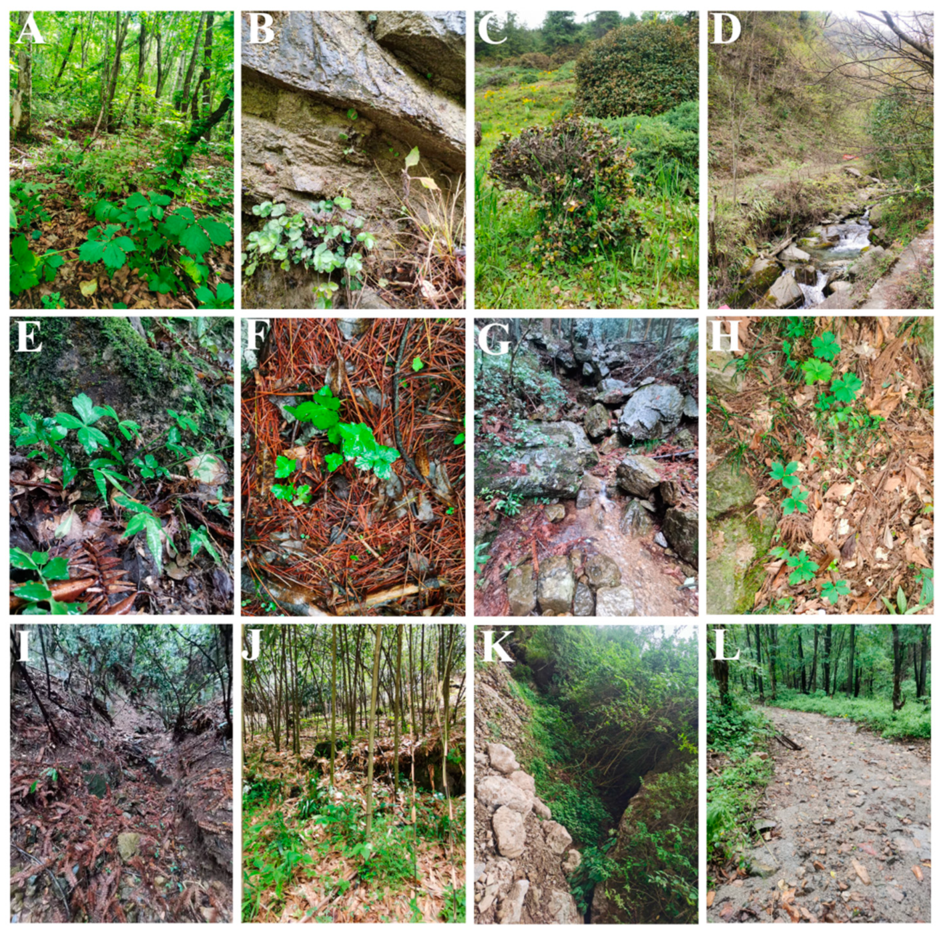
Figure 1. The habit of the Sanicula plants. (A): mixed forests on mountain slopes; (B): rocky walls; (C): alpine scrub; (D): grassy places on stream banks; (E): rock crevices; (F): inside the humus; (G): beside the stream; (H): crack of a stone slab; (I): wet valleys; (J): bamboo forests; (K): shady slope; (L): shady wet places.
Plants 13 01635 g001
Figure 2. The morphology of the Sanicula plants. (A): S. caerulescens; (B): S. oviformis; (C): S. rugulosa; (D): S. hacquetioides; (E): S. chinensis; (F): S. giraldii; (G): S. astrantiifolia; (H): S. serrata; (I): S. orthacantha.
Plants 13 01635 g002
Figure 3. The basal leaves of Sanicula plants. (AC): trifoliolate; (DG): palmately 3-parted; (HL): palmately 3-5-parted.
Plants 13 01635 g003
Figure 4. The inflorescence of Sanicula plants. (A): S. caerulescens; (B): S. giraldii; (C): S. astrantiifolia; (D): S. serrata; (E): S. brevispina; (F): S. subgiraldii; (G): S. hacquetioides; (H): S. orthacantha; (I): S. oviformis; (J): S. elongata; (K): S. flavovirens; (L): S. pengshuiensis.
Plants 13 01635 g004
Figure 5. Leaf epidermal characteristics of Sanicula taxa observed with a light microscope. (A): S. rubriflora; (B): S. rugulosa; (C): S. astrantiifolia; (D): S. chinensis; (E): S. giraldii; (F): S. caerulescens; (G): S. serrata; (H): S. elongata; (I): S. brevispina; (J): S. subgiraldii; (K): S. pengshuiensis; (L): S. pauciflora; (M): S. hacquetioides; (N): S. orthacantha; (O): S. lamelligera; (P): S. oviformis; (Q): S. tienmuensis; (R): S. flavovirens; (S): S. elata; (T): S. nanchuanensis. 1: upper epidermis; 2: lower epidermis. Scale bar = 50 μm.
Plants 13 01635 g005
Figure 6. The micromorphological characteristics of the upper leaf epidermis observed with scanning electron microscope. (A): S. rubriflora; (B): S. rugulosa; (C): S. astrantiifolia; (D): S. chinensis; (E): S. giraldii; (F): S. caerulescens; (G): S. serrata; (H): S. elongata; (I): S. brevispina; (J): S. subgiraldii; (K): S. pengshuiensis; (L): S. pauciflora; (M): S. hacquetioides; (N): S. orthacantha; (O): S. lamelligera; (P): S. oviformis; (Q): S. tienmuensis; (R): S. flavovirens; (S): S. elata; (T): S. nanchuanensis; (U): S. petagnioides; (V): S. tuberculata. 1: 200×; 2: 1000×; 3: 5000×.
Plants 13 01635 g006
Figure 7. The micromorphological characteristics of the lower leaf epidermis observed with scanning electron microscope. (A): S. rubriflora; (B): S. rugulosa; (C): S. astrantiifolia; (D): S. chinensis; (E): S. giraldii; (F): S. caerulescens; (G): S. serrata; (H): S. elongata; (I): S. brevispina; (J): S. subgiraldii; (K): S. pengshuiensis; (L): S. pauciflora; (M): S. hacquetioides; (N): S. orthacantha; (O): S. lamelligera; (P): S. oviformis; (Q): S. tienmuensis; (R): S. flavovirens; (S): S. elata; (T): S. nanchuanensis; (U): S. petagnioides; (V): S. tuberculata. 4: 200×; 5: 1000×; 6: 5000×; 7: stoma.
Plants 13 01635 g007
Figure 8. The morphological characteristics of the Sanicula fruit. (A): S. rubriflora; (B): S. astrantiifolia; (C): S. chinensis; (D): S. giraldii; (E): S. caerulescens; (F): S. serrata; (G): S. elongata; (H): S. brevispina; (I): S. subgiraldii; (J): S. pengshuiensis; (K): S. pauciflora; (L): S. hacquetioides; (M): S. orthacantha; (N): S. lamelligera; (O): S. oviformis; (P): S. tienmuensis; (Q): S. flavovirens; (R): S. elata; (S): S. rugulosa; (T): S. nanchuanensis. 1: dorsal side views of fruits; 2: commissural side views of fruits; 3: transverse section. Scale bar = 0.5 mm.
Plants 13 01635 g008
Figure 9. The micromorphological features of the Sanicula fruit observed with scanning electron microscope. (A): S. rubriflora; (B): S. astrantiifolia; (C): S. chinensis; (D): S. giraldii; (E): S. caerulescens; (F): S. serrata; (G): S. elongata; (H): S. brevispina; (I): S. subgiraldii; (J): S. pengshuiensis; (K): S. pauciflora; (L): S. hacquetioides; (M): S. orthacantha; (N): S. lamelligera; (O): S. oviformis; (P): S. tienmuensis; (Q): S. flavovirens; (R): S. rugulosa; (S): S. elata; (T): S. nanchuanensis.
Plants 13 01635 g009
Figure 10. The micromorphological features of paraffin slices of Sanicula fruits. (A): S. rubriflora; (B): S. astrantiifolia; (C): S. giraldii; (D): S. caerulescens; (E): S. elongata; (F): S. pengshuiensis; (G): S. hacquetioides; (H): S. oviformis; (I): S. orthacantha; (J): S. flavovirens; (K): S. tienmuensis; (L): S. serrata; (M): S. subgiraldii; (N): S. pauciflora; (O): S. brevispina; (P): S. rugulosa; (Q): S. lamelligera; (R): S. chinensis; (S): S. elata; (T): S. nanchuanensis. Scale bar = 0.5 mm.
Plants 13 01635 g010
Figure 11. The micromorphological characteristics of pollen for Sanicula plants observed with a light microscope. (A): S. rubriflora; (B): S. rugulosa; (C): S. astrantiifolia; (D): S. chinensis; (E): S. giraldii; (F): S. caerulescens; (G): S. serrata; (H): S. elongata; (I): S. brevispina; (J): S. subgiraldii; (K): S. pengshuiensis; (L): S. pauciflora; (M): S. hacquetioides; (N): S. orthacantha; (O): S. lamelligera; (P): S. tienmuensis; (Q): S. elata; (R): S. nanchuanensis. Scale bar = 10 μm.
Plants 13 01635 g011
Figure 12. The micromorphological characteristics of pollen for Sanicula plants observed with a scanning electron microscope. (A): S. rubriflora; (B): S. rugulosa; (C): S. astrantiifolia; (D): S. chinensis; (E): S. giraldii; (F): S. caerulescens; (G): S. serrata; (H): S. elongata; (I): S. brevispina; (J): S. subgiraldii; (K): S. pengshuiensis; (L): S. pauciflora; (M): S. hacquetioides; (N): S. orthacantha; (O): S. lamelligera; (P): S. tienmuensis; (Q): S. elata; (R): S. oviformis; (S): S. flavovirens; (T): S. nanchuanensis. 1: equatorial view; 2: polar view; 3: germinal furrow; 4: exine ornamentation of equatorial view.
Plants 13 01635 g012
Table 1. The morphological characteristics of fruits for Sanicula plants.
TaxonShapeSize (mm)Fruit SurfaceCalyx TeethEndosperm on Commissural SideVittae
S. rubrifloraOvoid or ovoid–globose6.97 × 2.68proximal part covered with scales, distal part covered with yellow uncinate bristlesovate–lanceolateslightly concave5
S. astrantiifoliaObovate or subglobose1.89 × 0.90proximal end with short bristles, distal end with uncinate bristles, bristles yellow or purple-redlinear–lanceolateflatObscure
S. chinensisOvoid–globose3.28 × 1.37bristles uncinate above, dilated at baselinearflat5
S. giraldiiNarrowly
ovoid
3.86 × 1.74densely covered with developed yellow or purplish red uncinate
bristles, long and hard
ovate and small, tip mucronateflatObscure
S. caerulescensGlobose or ellipsoid2.35 × 0.68short and straight spinous-bristles usually fused at the base forming a thin tierlinear–lanceolateslightly concave5
S. serrataOvoid or ovoid–globose2.08 × 1.60proximal part covered with scales, distal part covered with slightly uncinate bristles, bristles pale yellow or purplish redovateslightly concaveObscure
S. elongataOvoid2.39 × 1.11densely covered with pale yellow scalesnarrow–ovateslightly concaveObscure
S. brevispinaOblong
ovoid
to ovoid
1.95 × 1.25proximal end with degenerated to disappeared the prickles, nearly smooth, distal end with prickles and formed a thin layer; usually with erose-spinulose ribs and furrows smooth or barely spinuloselinear to lanceolateslightly concaveObscure
S. subgiraldiiBroadly ovate4.01 × 2.51rarely covered with purplish-red short bristles, proximal end with tubercles, obscure, distal end with uncinate bristles or straightbroadly ovate
and large
slightly concaveObscure
S. pengshuiensisEllipsoid2.38 × 0.77bristles in regular rows in furrows, ribs glabrous, stout and prominentlinearflat5
S. paucifloraLong ellipsoid2.72 × 0.83densely covered with sharp prickleslong–lanceolateflatvittae 2 in
commissural side
S. hacquetioidesOvoid–globose2.81 × 2.08covered with scales and tubercles, but never spinulosebroadly ovate or obovateflatObscure
S. orthacanthaNarrowly ovoid2.02 × 0.86short and straight spinesnarrowly lanceolateflatObscure
S. lamelligeraLong-ovoid1.97 × 0.72short and straight spines, never uncinate, fused at the base forming a thin tierlinearslightly concave5
S. oviformisOvoid2.13 × 1.44densely short and straight-spinulose; ribs prominentlinear–lanceolateslightly concave5
S. tienmuensisSubglobose2.52 × 1.82covered with short and obtuse prickles, slight formed scales and tuberclesbroadly ovateslightly concaveObscure
S. flavovirensOvoid–globose3.26 × 1.79proximal part covered with tubercles, distal part covered with yellow straight pricklestriangular–lanceolate, tip mucronateflat5
S. elataOvoid–globose2.27 × 1.35densely covered with yellow uncinate bristlesnarrowly lanceolateflat5
S. rugulosaEllipsoid2.41 × 1.45densely covered with uncinate bristlesnarrowly lanceolateslightly concaveObscure
S. nanchuanensisEllipsoid2.74 × 1.65proximal part covered with scales, rarely formed bristlestriangular–lanceolateflatObscure
Disclaimer/Publisher’s Note: The statements, opinions and data contained in all publications are solely those of the individual author(s) and contributor(s) and not of MDPI and/or the editor(s). MDPI and/or the editor(s) disclaim responsibility for any injury to people or property resulting from any ideas, methods, instructions or products referred to in the content.