Article

Impact of Methyl Jasmonate on Terpenoid Biosynthesis and Functional Analysis of Sesquiterpene Synthesis Genes in Schizonepeta tenuifolia

1
Jiangsu Collaborative Innovation Center of Chinese Medicinal Resources Industrialization, Nanjing University of Chinese Medicine, Nanjing 210023, China
2
State Key Laboratory on Technologies for Chinese Medicine Pharmaceutical Process Control and Intelligent Manufacture, Nanjing University of Chinese Medicine, Nanjing 210023, China
3
School of Pharmacy, Nanjing University of Chinese Medicine, Nanjing 210023, China
*
Authors to whom correspondence should be addressed.
Plants 2024, 13(14), 1920; https://doi.org/10.3390/plants13141920 (registering DOI)
Submission received: 15 June 2024 / Revised: 5 July 2024 / Accepted: 9 July 2024 / Published: 12 July 2024
(This article belongs to the Special Issue Chemical Characteristics and Bioactivity of Plant Natural Products)

Abstract

:
This study investigates the impact of methyl jasmonate (MeJA) on the volatile oil composition of Schizonepeta tenuifolia and elucidates the function of the StTPS45 gene, a key player in terpenoid biosynthesis. The effect of different concentrations of MeJA (0, 50, 100, 200, and 300 μmol/L) on the growth of S. tenuifolia adventitious bud clusters was analyzed over a 20 d period. Using gas chromatography–mass spectrometry (GC-MS), 17 compounds were identified from the adventitious bud clusters of S. tenuifolia. Significant changes in the levels of major monoterpenes, including increased contents of (+)-limonene and (+)-menthone, were observed, particularly at higher concentrations of MeJA. Analysis of transcriptome data from three groups treated with 0, 100, and 300 μmol/L MeJA revealed significant changes in the gene expression profiles following MeJA treatment. At 100 μmol/L MeJA, most terpene synthase (TPS) genes were overexpressed. Additionally, gene expression and functional predictions suggested that StTPS45 acts as germacrene D synthase. Therefore, StTPS45 was cloned and expressed in Escherichia coli, and enzyme activity assays confirmed its function as a germacrene D synthase. Molecular docking and structural prediction of StTPS45 further suggested specific interactions with farnesyl diphosphate (FPP), aligning with its role in the terpenoid synthesis pathway. These findings provide valuable insights into the modulation of secondary metabolite pathways by jasmonate signaling and underscore the potential of genetic engineering approaches to enhance the production of specific terpenoids in medicinal plants.

1. Introduction

Schizonepeta tenuifolia, commonly known as Japanese catnip, is a valuable medicinal plant in the Lamiaceae family, renowned for its antipyretic, anti-inflammatory, and analgesic properties in traditional Chinese medicine [1]. The therapeutic effects of S. tenuifolia are largely attributed to its rich volatile oil content, which includes significant terpenoids such as (−)-pulegone, (+)-limonene, (−)-isopulegone and (+)-menthone [2]. These bioactive compounds exhibit diverse pharmacological activities: (−)-pulegone possesses anti-hyperalgesic properties [3]; (+)-limonene is known for its anti-anxiety and anticancer effects [4,5]; (−)-isopulegone has antimicrobial activity [6]; and (+)-menthone is valued for its anti-allergic and antitumor properties [7,8]. These attributes make them the focus of extensive research into their biosynthesis and regulation.
The synthesis pathway of monoterpenes in S. tenuifolia begins with the MEP and MVA pathways, which together produce isopentenyl pyrophosphate (IPP) and dimethylallyl pyrophosphate (DMAPP) [9,10]. These compounds then undergo a series of reactions catalyzed by terpene synthases (TPS), including those catalyzed by limonene synthase (LS), limonene-3-hydroxylase (L3OH), isopiperitenol dehydrogenase (ISPD), isopiperitenone reductase (IPR), pulegone reductase (PR), and menthone reductase (MR), ultimately resulting in the synthesis of (+)-menthol (Figure 1). The genes encoding these enzymes form unique bidirectional gene clusters in the S. tenuifolia genome, indicating that these genes are grouped together through inverted duplication, and work synergistically to accomplish the biosynthesis of monoterpenoids [11]. Additionally, analyses of the TPS gene family members in S. tenuifolia, including their physicochemical properties, phylogenetic relationships, conserved motifs, and gene structures, have also been reported [12]. However, there has been no progress in the research on the sesquiterpene synthesis pathway in S. tenuifolia.
Previous studies have shown that terpenoid compounds are influenced by various factors, including light [13], salt stress [14], temperature [15], and endogenous plant hormones [16,17]. Methyl jasmonate (MeJA), an endogenous plant hormone, plays a crucial role in regulating the biosynthesis of secondary metabolites, including terpenoids [18]. Studies have demonstrated that 0.1 mM MeJA can effectively promote the biosynthesis of terpenoids in Oenanthe javanica [19]. MeJA can induce the production of terpenoids by activating specific genes involved in their biosynthetic pathways [20,21]. However, the precise mechanisms by which MeJA influences terpenoid biosynthesis in S. tenuifolia have not been fully elucidated.
This study used clonal adventitious bud clusters for MeJA induction experiments because they offer greater uniformity and controllability in genetic background, physiological state, and experimental conditions, thereby enhancing the precision and reproducibility of the experiments. Different concentrations of MeJA were applied to the adventitious bud clusters of S. tenuifolia, and changes in growth, volatile oil composition and content, and gene expression levels were recorded to elucidate the regulatory role of jasmonate signaling in terpenoid biosynthesis. Additionally, the germacrene D synthase gene StTPS45 was cloned, expressed, and functionally characterized to confirm its role. Enzyme activity assays and molecular docking studies were conducted to further understand the biochemical properties and interaction mechanisms between StTPS45 and its substrate, farnesyl diphosphate (FPP). These findings will provide a foundation for optimizing the production of valuable medicinal compounds in S. tenuifolia and other related medicinal plants.

2. Methods and Materials

2.1. Plant Material

This study utilized plant materials from a laboratory setting, which were identified by professor Qinan Wu from the school of pharmacy at Nanjing University of Chinese Medicine as Schizonepeta tenuifolia, a species within the Lamiaceae family. S. tenuifolia seeds were disinfected with 75% ethanol for 2 min, followed by 20% hydrogen peroxide for 6 to 8 min, then rinsed 6 to 8 times with sterile water, and sown on MS culture medium. The seeds were germinated under aseptic conditions in an incubator maintained at 25 °C with a 16 h photoperiod. After approximately 5 days (d), the germinated seeds were cultivated for over 30 d to obtain healthy, sterile seedlings of S. tenuifolia. Sterile S. tenuifolia seedlings grown for 60 d were removed from the culture bottles, and stem segments with leaf axils were cut and inoculated onto MS differentiation medium. Under conditions of 25 °C and a light intensity of 2200 lux with a 16 h photoperiod, the seedlings were cultured for 30 d to induce bud generation through methyl jasmonate (MeJA) treatment. Fresh leaves were first immersed in pure water for cleaning, then dried with filter paper to prepare for total RNA extraction.

2.2. Effects of MeJA Treatment on Volatile Components of Schizonepeta tenuifolia

2.2.1. MeJA Induces the Formation of Schizonepeta tenuifolia

Healthy, uniformly grown, and non-browned S. tenuifolia adventitious bud clusters, pre-cultivated for 30 d, were inoculated onto MS culture medium containing 0 (CK), 50 (TM), 100 (LM), 200 (MM) and 300 (HM) μmol/L MeJA. Three bottles were prepared for each concentration, with each bottle containing five samples. The experiment was replicated three times. These groups were cultivated at 25 °C with 70–80% humidity and sampled at 0, 5, 10, 15, and 20 d. Samples were preserved at −80 °C for subsequent uniform extraction and analysis of relevant indicators.

2.2.2. Effect of MeJA Treatment on the Growth of Schizonepeta tenuifolia

The proliferation rate of S. tenuifolia adventitious bud clusters was calculated using the following formula: Proliferation Rate (%) = [(Weight at Harvest − Weight at Inoculation)/Weight at Inoculation] × 100%. Observations and recordings of the glandular scale phenotype on the leaves of the adventitious buds were conducted using a Zeiss stereomicroscope. For each concentration, three plants were randomly selected for observation. The diameter of six glandular scales was measured using the length measurement tool in the microscopy photography software (ZEN Blue Edition v3.8). Additionally, the number of glandular scales was counted within three known area regions using the area measurement tool, and their density (number/mm2) was calculated. The average of these measurements was then determined.

2.2.3. Effect of MeJA Treatment on Volatile Components of Schizonepeta tenuifolia

Volatile oils were extracted from MeJA-induced S. tenuifolia sterile adventitious bud clusters using simultaneous distillation extraction. The extraction involved 8 mL of n-hexane for 4 g of fresh bud tissue over a period of 1 h. The extracted samples were then analyzed for content using gas chromatography–mass spectrometry (GC-MS). The GC-MS was equipped with an Agilent 19091S-433 HP-5ms column (30 m × 250 μm × 0.25 μm), and helium was used as the carrier gas. The injector temperature was set at 250 °C. The temperature program started at 50 °C, held for 32 min, then increased at 10 °C/min to 90 °C and held for 15 min, followed by an increase at 5 °C/min to 200 °C and held for 5 min. The split ratio was 20:1, and the injection volume was 1 μL. Mass spectrometry conditions included an electron ionization (EI) source at 70 eV, source temperature of 230 °C, quadrupole temperature of 150 °C, and a mass scan range of 30~500 amu, with a solvent delay of 3 min. Component identification was conducted by comparing retention times and mass spectral data with standards and using the NIST database. Total ion chromatograms were obtained and further analyzed against the NTST.14 database (https://www.nist.gov/) to identify the chemical components in the volatile oil of S. tenuifolia.

2.3. RNA Extraction, cDNA Library Construction and Data Assembly

Total RNA was extracted from S. tenuifolia leaves treated with CK, LM, and HM using TRIzol reagent (Sigma, St. Louis, MI, USA). The mRNA containing poly-A tails was enriched using oligo-dT magnetic beads. A DNA probe hybridized with rRNA, and RNase H selectively digested the DNA/RNA hybrid strand. DNase I was then used to remove the DNA probe, yielding the purified RNA. RNA was fragmented with a breakage buffer and reverse-transcribed using random N6 primers to synthesize double-stranded cDNA. The synthesized double-stranded DNA was end-repaired and 5′-phosphorylated to produce 3′-overhanging “A” sticky ends. A bubble adapter with a 3′-overhanging “T” was then ligated to the DNA. The ligated DNA was PCR-amplified using specific primers, heat-denatured to form single-stranded DNA, and circularized with a bridge primer to create a single-stranded circular DNA library. The library was then sequenced. Data were processed using SOAPnuke (v1.4.0) [22] and Trimmomatic (v0.36) [23] to filter out low-quality reads, adapter contamination, and reads with a high N base content. The clean reads were aligned to the reference genome using HISAT (v2.1.0) [24], and gene expression levels were calculated with StringTie (v2.2.3) [25], using fragments per kilobase of transcript per million mapped reads (FPKM) as the standard measure. PCA and Pearson’s correlation analysis of gene expression values (FPKM) were conducted to evaluate the differences between groups. Differentially expressed genes were identified with DESeq2 [26], using |log2(FoldChange)| > 1 and FDR < 0.05 as selection criteria. Volcano plots were used to visualize gene expression differences. Functional and pathway analyses were then conducted using GO [27] and KEGG [28] to reveal the functions and signaling pathways involved.

2.4. Optimization and Prokaryotic Expression of Candidate Gene of Germacrene D Synthase

To enhance the expression of TPS45 protein in E. coli, a 1695 bp DNA sequence was optimized (Figure S1). The optimized sequence was synthesized by Sangon Biotech (Shanghai) Co., Ltd. (Shanghai, China), and ligated into the pET32a vector to construct the recombinant plasmid pET32a-TPS45. This plasmid was transformed into BL21 (DE3) competent cells and inoculated using a positive single colony into 5 mL of LB medium containing 100 ng/mL ampicillin. The culture was then scaled up by inoculating 1 mL of the recombinant culture into 50 mL of ampicillin-containing LB medium and incubated at 37 °C with shaking at 180 rpm until the OD600 reached 0.6–0.8. Isopropyl β-D-1-thiogalactopyranoside (IPTG) was added to a final concentration of 1 mmol/L to induce protein expression. The induced culture was expressed at 16 °C and 180 rpm for 18 h, with a control culture set up simultaneously. Post-induction, the culture was transferred to 50 mL sterile centrifuge tubes and centrifuged at 5000 rpm for 15 min at 4 °C to precipitate the bacteria. The supernatant was discarded, and the precipitate was resuspended in 1 mL of lysis buffer. The resuspended culture was sonicated for 5 min in an ice bath at 4 °C and centrifuged under the same conditions. The supernatant was transferred to a new 2 mL centrifuge tube, and the precipitate was resuspended in 1 mL of lysis buffer and stored at −80 °C in another 2 mL centrifuge tube.

2.5. Functional Validation of the Protein Encoded by Candidate Gene

To verify the function of the TPS45 target protein, substrate feeding was employed [19]. All procedures were performed on ice. A total volume of 500 μL of reagents was sequentially added to 2 mL sample vials (Table S2), with protein expressed from an empty vector serving as the negative control.
After adding the reagents, 200 μL of n-hexane was immediately added to seal the liquids. The vial cap was tightened and sealed with film. The reaction was conducted at 31 °C for 16 h. Afterward, the reaction mixture was stored at −80 °C for 1 h to freeze. Then, using a sampler, the organic layer was carefully extracted and transferred to a sample vial lined with an inner tube, where it was temporarily stored at −80 °C.

2.6. Analysis of the Enzyme Products of the Candidate Gene by GC-MS

The analysis of relevant components was conducted using a GC-MS system (Agilent 7890B-7000D) (Agilent Technologies, Santa Clara, CA, USA) equipped with an HP-5ms capillary column (30 m × 250 μm × 0.25 μm, Agilent 19091S-433) (Agilent Technologies, Santa Clara, CA, USA). The temperature program for TPS45 enzyme activity detection was as follows: the initial temperature was held at 50 °C for 3 min, then increased at a rate of 3 °C/min to 90 °C, followed by 5 °C/min to 150 °C, and finally at 10 °C/min to 220 °C, where it was held for 5 min. Mass spectrometry conditions were as follows: EI mode at 70 eV, ion source temperature of 230 °C, and a mass range of 50–500 amu with an acquisition frequency of 50 Hz. Data collection and analysis were performed using ChemStation software vC.01.05 (Agilent Technologies, Santa Clara, CA, USA), and compounds were identified by comparing mass spectral data against the NIST14.L database and further verified using standards.

2.7. Structural Prediction and Molecular Docking of the Protein Encoded by the Candidate Gene

This study conducted a physicochemical analysis of the functionally validated TPS45 protein and predicted its secondary and tertiary structures. Additionally, molecular docking was performed to predict interactions between the TPS45 protein and the FPP small molecule ligand, aiming to identify active sites and infer mechanisms of action. Initially, physicochemical properties were analyzed using various tools: signal peptide prediction with Signal P 4.1 [29] (https://services.healthtech.dtu.dk/service.php?SignalP-4.1, accessed on 10 July 2023), transmembrane regions predicted by TMHMM2.0 (https://services.healthtech.dtu.dk/services/TMHMM-2.0/, accessed on 10 July 2023), glycosylation sites using NetNGlyc 1.0 Server [30] (https://services.healthtech.dtu.dk/service.php?NetNGlyc-1.0, accessed on 10 July 2023), and kinase phosphorylation sites with NetPhos 3.1 Server [31] (https://services.healthtech.dtu.dk/service.php?NetPhos-3.1, accessed on 15 July 2023). Secondary structure was analyzed using SOPMA (https://npsa-prabi.ibcp.fr/cgi-bin/npsa_automat.pl?page=npsa_sopma.html, accessed on 15 July 2023), while three-dimensional modeling was performed via SWISS-MODEL [32] (https://swissmodel.expasy.org/interactive, accessed on 15 July 2023), and the model’s validity assessed with a Ramachandran Plot. Additionally, receptor preparation steps included dehydration and complete hydrogenation. FPP’s chemical structure was prepared using MGLTools (v1.5.6) (https://ccsb.scripps.edu/mgltools, accessed on 22 July 2023) for hydrogen addition and charge calculations. The format was converted from PDBQT to PDB using OpenBabel (v3.0.1) (http://openbabel.org/wiki/Category:Installation, accessed on 23 July 2023), followed by molecular docking with AutoDock [33] (https://autodock.scripps.edu, accessed on 25 July 2023). Interaction analysis between the protein and ligand was visualized and assessed using Discovery Studio 2022 Client (www.imatsoft.com). Finally, amino acid sequence alignments were performed using Clustal Omega [34] (https://www.ebi.ac.uk/Tools/msa/clustalo, accessed on 26 July 2023), and a phylogenetic tree was constructed using the neighbor-joining method in MEGA11 v11.0.10 [35] software.

3. Result

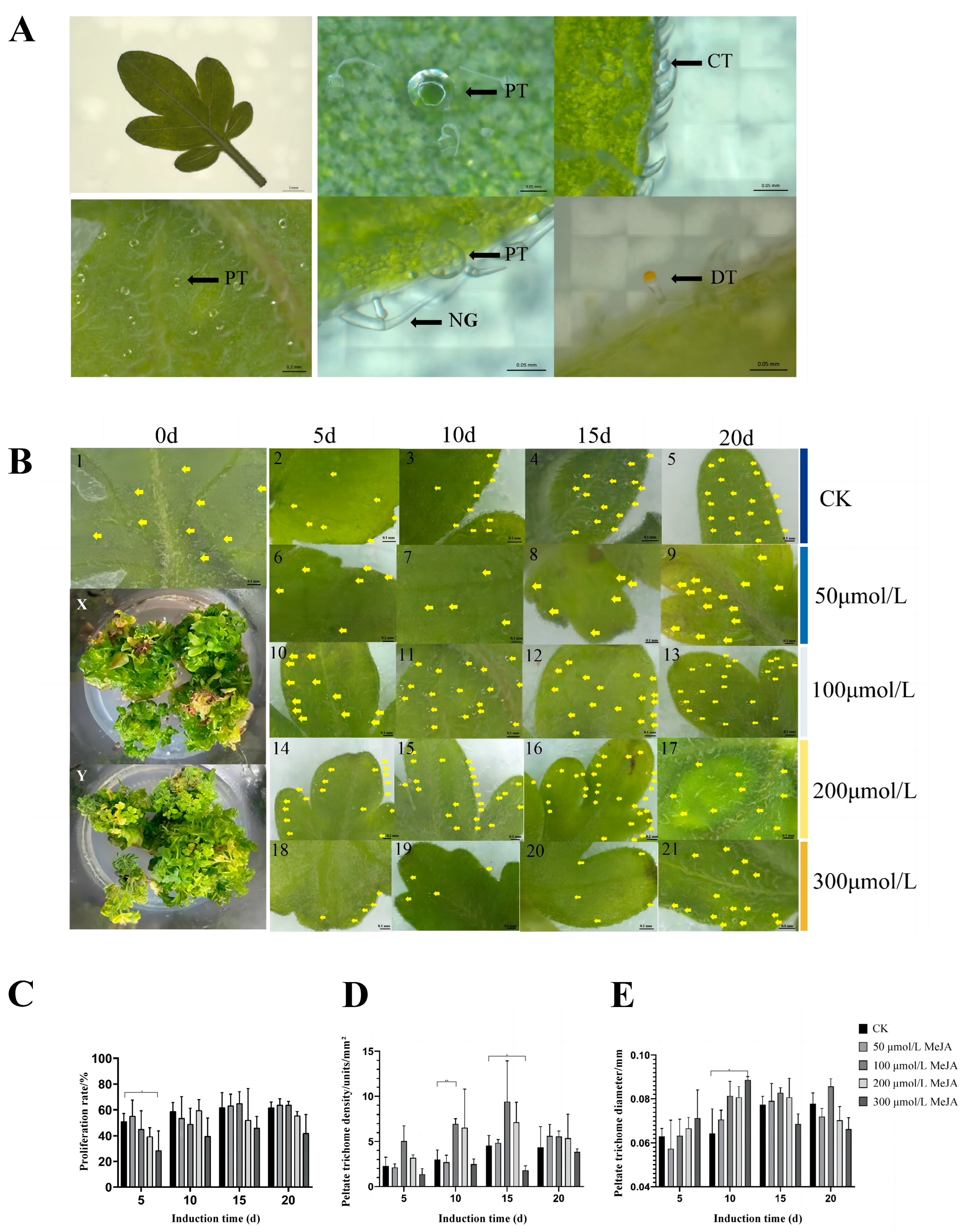
3.1. Effect of MeJA Treatment on the Phenotype of Schizonepeta tenuifolia

The stereomicroscope revealed three types of glandular hairs on the leaves and stems of sterile S. tenuifolia seedlings: glandular scales, digitiform trichomes, and capitate trichomes (Figure 2A). The morphology and structure of glandular hairs in the leaves of sterile tissue-cultured S. tenuifolia seedlings were normal, resembling those of known cultivated S. tenuifolia. Stem segments with leaf axils from tissue-cultured seedlings at around 30 d, during their vigorous growth stage, were used as explant samples for subsequent MeJA induction experiments.
Treatment of S. tenuifolia adventitious bud clusters with different concentrations of MeJA for various durations showed a slight impact on proliferation (Figure 2C). Phenotypic observations revealed that higher concentrations of MeJA resulted in more severe browning at the base of the buds, leading to some loss of plant samples. Specifically, a significant reduction in proliferation rate was observed in the HM group for 5 d (0.01 < p < 0.05). However, the LM and MM groups for 15–20 d showed a trend of increased proliferation compared to the CK group, suggesting that moderate concentrations of MeJA might promote the growth and development of S. tenuifolia adventitious buds. In contrast, high concentrations of MeJA may adversely affect the normal development of the buds, leading to decreased proliferation rates.
Different concentrations of MeJA variably affect the distribution, diameter, and density of glandular scales on the abaxial side of S. tenuifolia leaves (Figure 2B,D,E). In the CK, glandular scales were initially sparsely distributed, but their number and density increased over time, predominantly at the distal edges of the leaves, with fewer glandular scales near the proximal ends. In the TM group, the glandular scale distribution and density resembled that of the CK group, indicating that this lower concentration had no significant effect. However, the LM and MM groups resulted in a marked increase in glandular scale density compared to CK, with glandular scales more widely distributed across the entire leaf surface and particularly denser at the distal edges. Additionally, the diameter of glandular scales also increased to varying degrees. In the HM group, there was a significant decline in glandular scale density from 5 to 15 d, with reductions observed at both leaf ends and almost no glandular scales at the proximal ends. After 20 d at this concentration, glandular scale density slightly increased, returning to a distribution pattern similar to normal, mainly at the distal ends. This recovery could be due to a decrease in MeJA concentration within the plant tissues over time.

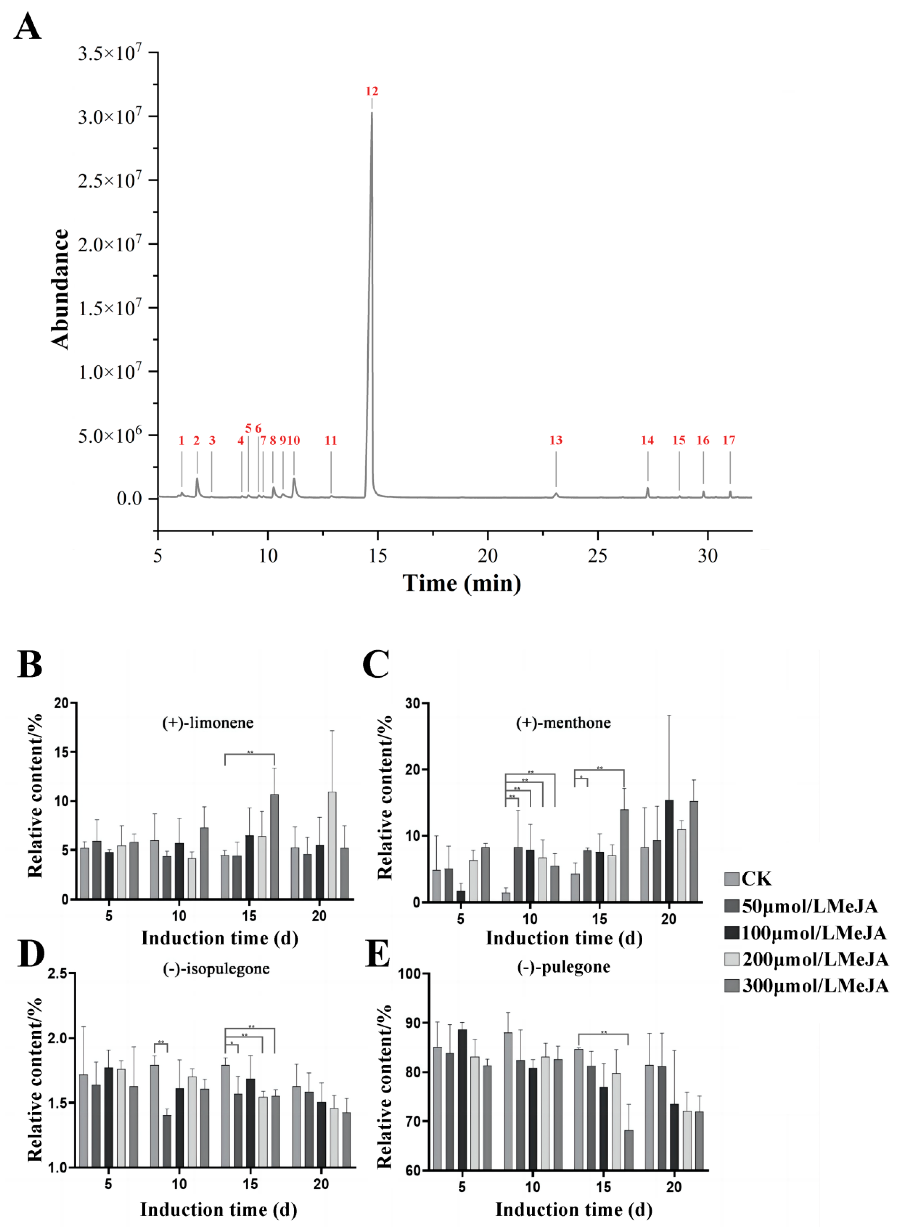
3.2. Effects of MeJA Induction on Main Terpenoids in Volatile Oil from Schizonepeta tenuifolia

The CK group (untreated with MeJA) samples of tissue-cultured S. tenuifolia clustered adventitious buds were extracted using simultaneous distillation extraction and analyzed via GC-MS to obtain a total ion chromatogram (Figure 3A). The results were compared with the NIST.14 library and existing standards, summarizing compounds with a similarity score greater than 90% (Table 1). Seventeen distinct compounds were identified, and the compound composition aligned with prior studies [2]. This indicates that the primary monoterpene components in tissue-cultured S. tenuifolia essential oil are similar to those in the wild type. The main components remain (+)-limonene, (+)-menthone, (−)-isopulegone, and (−)-pulegone, which together account for about 90% of the total volatile oil in tissue-cultured S. tenuifolia clustered adventitious buds.
Adventitious bud clusters of S. tenuifolia were induced with 0, 50, 100, 200, and 300 μmol/L concentrations of MeJA for 5, 10, 15, and 20 d. Following induction, volatile oils were extracted using simultaneous distillation extraction and analyzed by GC-MS. The relative concentrations of chemical components were quantified using peak area normalization. The accumulation patterns of the main monoterpenes, particularly menthane derivatives in S. tenuifolia, were observed at different exogenous MeJA concentrations and durations (Figure 3B–E). The content of (+)-limonene did not change significantly during the induction process. However, after 15 to 20 d of induction at LM and MM groups, an upward trend in (+)-limonene content was noted. At HM group, a significant increase in (+)-limonene was observed from 5 to 15 d, which then stabilized by 20 d, suggesting that high concentrations of MeJA might positively regulate the synthesis and accumulation of (+)-limonene. In contrast, the content of (+)-menthone increased significantly with both the concentration and duration of induction compared to the CK, with noticeable increases across all concentrations at 20 d. Similarly, the accumulation patterns of (−)-isopulegone and (−)-pulegone showed a gradual decrease with increasing concentrations and duration of induction, with a more pronounced reduction in (−)-pulegone content, indicating that exogenous MeJA might negatively regulate the accumulation of these components, possibly by promoting their conversion into downstream (+)-menthone.

3.3. Transcriptome Analysis and Screening of Candidate Genes for Germacrene D Synthase

Transcriptome sequencing of leaves treated with MeJA for 20 d (CK, LM, and HM) produced an average of 6.38 Gb of data per sample, with an average total clean read count of 42.56 M. The average alignment rate of each sample to the genome was 94.45% (Table S1), indicating high sequencing quality. The first principal component (PC1) and the second principal component (PC2) accounted for 87.27% and 5.07% of the variance, respectively, explaining a cumulative total of 92.34% (Figure 4A). This indicates that the data were well explained. The LM and HM group were more dispersed compared to the CK group. This means that MeJA had a notable impact, leading to a broader range of responses. PCA and Pearson’s correlation heatmaps showed that the similarity between HM and CK was lower than that between LM and CK, indicating that high-dose MeJA has a greater impact on gene expression (Figure 4A,B). Therefore, HM and CK were selected for differential analysis, which identified 383 significantly upregulated genes and 506 downregulated genes in the HM group (Figure 4C). GO and KEGG enrichment analyses revealed that differentially expressed genes were mainly enriched in cellular process, catalytic activity, and biological regulation functions, as well as the translation, carbohydrate metabolism, and signal transduction pathways (Figure 4D,F). The top 20 functions and pathways based on q-value are also presented. Overall, differentially expressed genes were primarily enriched in biological process functions and metabolism pathways (Figure 4E,G).
Previous studies identified 57 TPS genes in S. tenuifolia [12]. The higher overall expression levels of the TPS family genes in the LM group compared to the CK and HM groups suggest that moderate concentrations of MeJA may specifically enhance the expression of genes involved in terpene biosynthesis (Table S3). This observation aligns with previous research findings that inducing S. tenuifolia with the LM group leads to an increase in the density of glandular scales, indicating a direct relationship between MeJA concentration and glandular scale production. The biosynthetic pathway for (+)-menthol in S. tenuifolia was documented. This study examined changes in the expression levels of enzymes involved in this pathway following induction with MeJA (Figure 1). The HM group revealed significant intergroup variations in the expression levels of enzymes associated with the (+)-menthol synthesis pathway, suggesting potential instability in reactions induced by high concentrations of MeJA. Notably, the expression levels of the downstream genes StIPR and StPR in the HM group exceeded those in the CK and LM groups, indicating that high concentrations of MeJA might promote a greater shift toward downstream synthetic processes.
Genome annotation of S. tenuifolia indicated that StTPS5, StTPS18, StTPS32, and StTPS45 are annotated as germacrene D synthase. Notably, the expression level of StTPS45 was higher (Table S3). Sequence comparison results revealed that StTPS45 has high similarity to the sesquiterpene synthase of Lavandula angustifolia. Thus, StTPS45 was selected for further gene cloning and heterologous expression.
The sequences of transcription factors (AtMYC2, AaWRKY1, AaWRKY9, AaERF1, AaERF2) from Arabidopsis thaliana and Artemisia annua were obtained from NCBI and compared with the S. tenuifolia genome using local BLAST analysis to examine their gene expression levels (Table 2). The results indicated that the gene expression level of Sch000013364 was consistent with the JA-responsive MYC2. Alignment with AaWRKY1 revealed that Sch000023850, Sch000000909, and Sch000024381 had multiple matched CDS regions, with Sch000023850 and Sch000000909 showing expression levels consistent with JA-responsive WRKY1. Other transcription factors did not exhibit a clear pattern. Therefore, it can be inferred that under the LM group, the expression level of Sch000013364 (partially functioning as MYC2) increased, enhancing glandular trichome development and the expression of TPS family-related genes in S. tenuifolia. Additionally, the increased expression of Sch000023850 and Sch000000909 (partially functioning as WRKY1) under MeJA induction likely further elevated the expression levels of downstream genes IPR and PR in the (+)-menthol synthesis pathway, thereby increasing the content of these compounds.

3.4. Overexpression and Enzyme Assay of the Protein Encoded by the Candidate Gene

In the early stage, StTPS45 was cloned and prokaryotic expression vector was constructed [12]. The successfully constructed pET32a-TPS45 recombinant plasmid was introduced into BL21 (DE3) cells for the inducible expression of the target protein. The cells were lysed, and the expressed crude protein was subjected to SDS-PAGE gel electrophoresis. The results (Figure S2) indicated that the cells containing the recombinant plasmid expressed a protein of approximately 72 kDa. This size is consistent with the expected 64.67 kDa of the candidate gene, confirming the successful expression of the target protein. However, compared to the precipitate, the corresponding band in the supernatant was less distinct, suggesting that the TPS45 protein predominantly formed inclusion bodies found in the precipitate.
To validate the function of the candidate gene, the crude protein obtained from its prokaryotic expression was reacted with the substrate FPP, and the enzymatic products were analyzed using GC-MS. The experimental results showed a distinct peak at a retention time of 27.789 min for the TPS45 group, in contrast to the negative control with the empty vector, where no corresponding peak was detected (Figure 5A). Additionally, a standard of germacrene D was subjected to GC-MS analysis, revealing a peak at 27.697 min, closely matching the retention time of the TPS45 enzymatic product. Furthermore, comparisons of the mass spectra showed that the ion fragments of the TPS45 enzymatic product were consistent with those of the germacrene D standard (Figure 5B). These results preliminarily confirm that the TPS45 protein can catalyze the formation of germacrene D from FPP, verifying that TPS45 is a germacrene D synthase (Figure 5C).

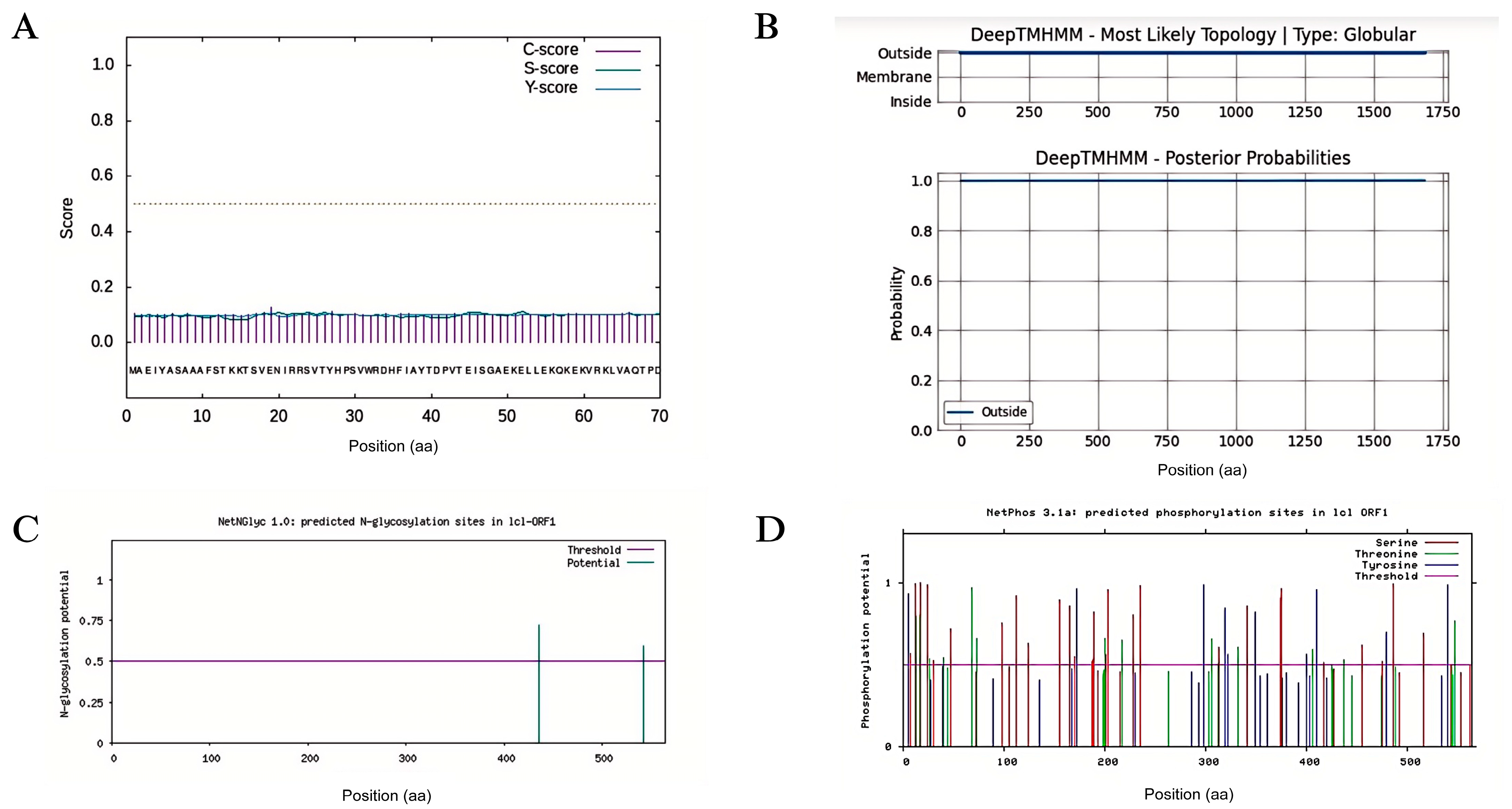
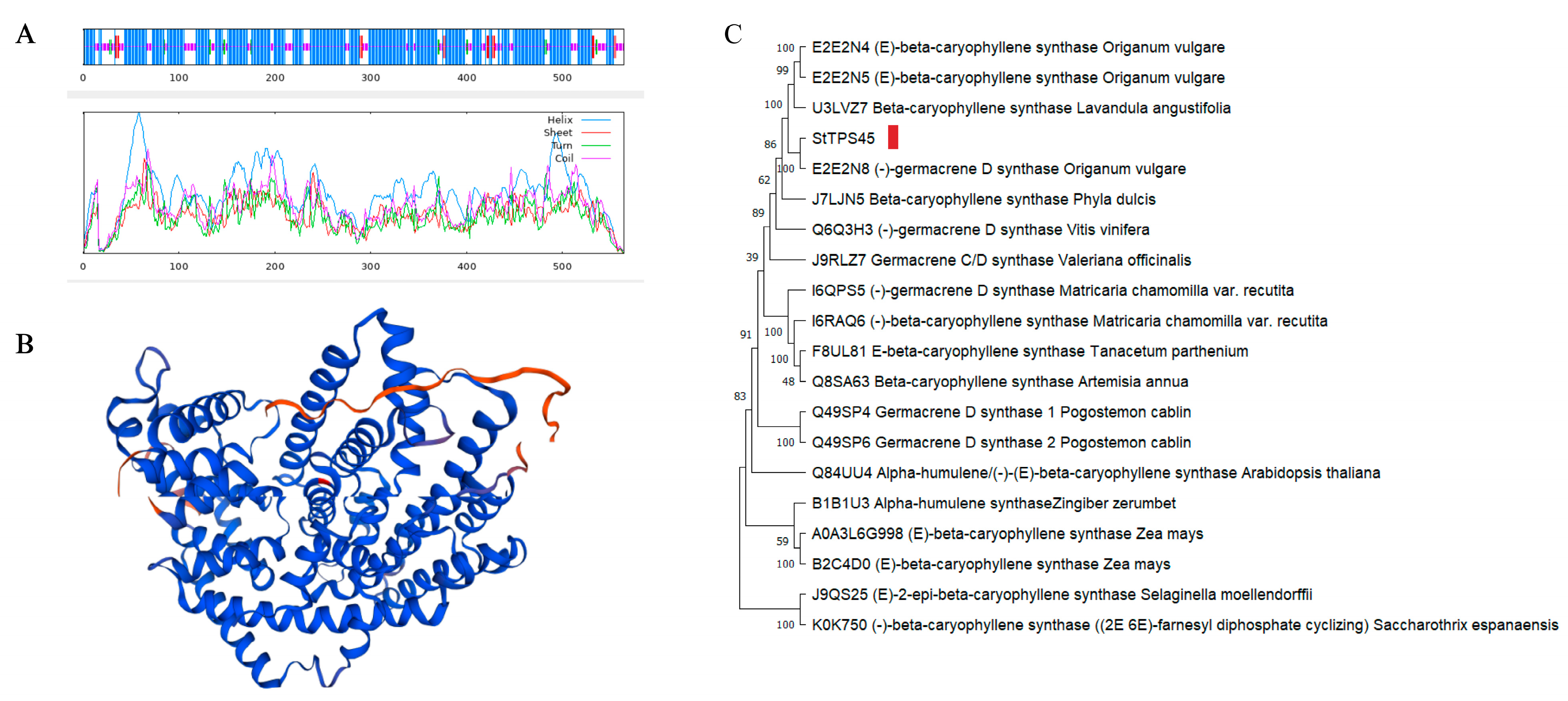
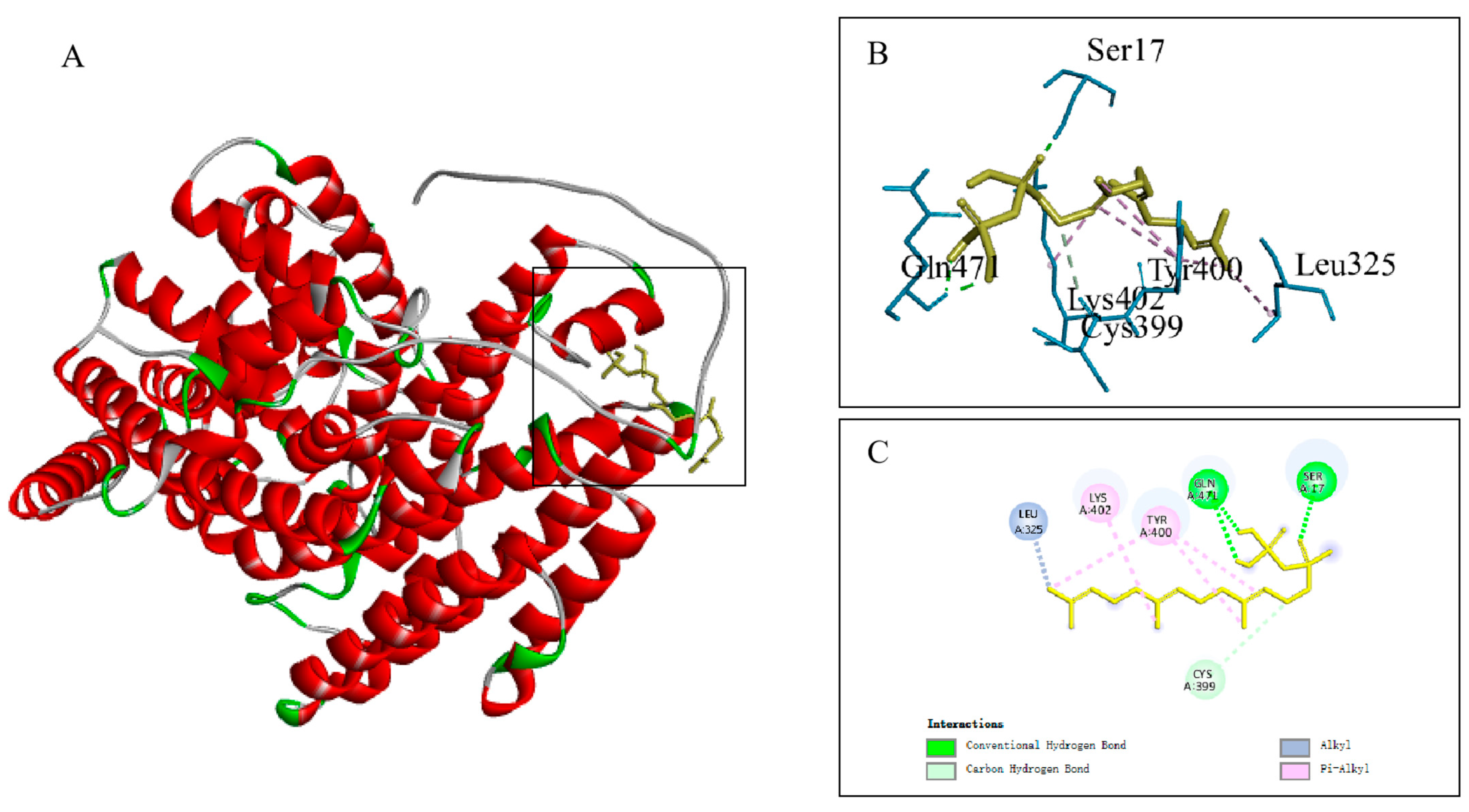
3.5. Physicochemical Property Analysis and Molecular Docking of the Protein Encoded by the Candidate Gene

Analysis of the TPS45 protein using the Signal P 4.1 Server indicated that the cleavage sites did not exceed the threshold, suggesting that TPS45 does not possess a signal peptide and is a non-secretory protein (Figure 6A). The TMHMM2.0 online tool revealed no transmembrane regions in the protein (Figure 6B). Glycosylation sites predicted by the NetNGlyc 1.0 Server identified potential glycosylation at positions 435 and 542 (Figure 6C). However, as TPS45 lacks a signal peptide, it is unlikely to be glycosylated in vivo, given its probable inaccessibility to N-glycosylation mechanisms. Phosphorylation sites, predicted using NetPhos 3.1 Server, showed that StTPS45 has 53 potential phosphorylation sites, including 29 serines, 14 threonines, and 10 tyrosines, with a phosphorylation potential greater than 0.5. These sites include binding sites for specific protein kinases such as PKC, PKA, CKII, p38 MAPK, and unsp (Figure 6D).
Secondary structure analysis of TPS45 protein using SOPMA indicated that α-helices are the predominant structure (70.57%), followed by random coils (23.05%), extended strands (3.55%), and β-turns (2.84%) (Figure 7A). To enhance the credibility of the SWISS-MODEL 3D structural model, a Ramachandran Plot assessed the predicted protein structure, with 93.9% of amino acid residues located in favored regions, validating the TPS45 protein model’s rationality (Figure 7B). The phylogenetic tree shows that the TPS45 sequence is on the same branch as the germacrene D synthase sequence of Origanum vulgare, indicating the closest relationship. This further verifies the enzymatic activity of TPS45 (Figure 7C).
Molecular docking predictions performed between TPS45 protein and the FPP ligand showed a binding energy of −3.51 kcal/mol, indicating binding activity as the binding energy is less than 0 kcal/mol. Additionally, interaction analyses revealed that six amino acids in TPS45 interact with FPP, forming hydrogen bonds at SER-17 and GLN-471 (Figure 8).

4. Discussion

This study reveals the impact of MeJA on the volatile oil composition and biosynthetic gene expression in S. tenuifolia, highlighting the crucial role of the StTPS45 gene in terpenoid biosynthesis. To ensure consistency in genetic background, physiological state, and experimental conditions, clonal adventitious shoots were used, confirming that this method had no significant effect on the volatile components of S. tenuifolia. Treatment with different concentrations of MeJA resulted in significant changes in the content of major monoterpenes. Transcriptome analysis showed significant changes in gene expression profiles following MeJA treatment, with most TPS genes upregulated, particularly under the LM group. Based on gene annotation and expression levels, TPS45 was identified as a functional germacrene D synthase. The StTPS45 gene was cloned and expressed in Escherichia coli, and its function was validated through enzyme activity assays. Molecular docking and structural prediction further revealed specific interactions between TPS45 and its substrate, FPP, consistent with its role in the terpenoid synthesis pathway.
Jasmonate compounds (JAs) are important signaling molecules in plants, playing key roles in various stress responses and developmental processes. Studies have shown that MeJA primarily regulates the biosynthesis, accumulation, and biotechnological applications of secondary metabolites. Current research focuses on gene and promoter cloning, transcription factor regulation, and cell- and tissue-specific analyses [36]. Plant trichomes are often rich in secondary metabolites, and regulating trichome development can significantly increase the content of these compounds, achieving the desired product levels or improved plant phenotypes. MeJA has a significant effect on the development of glandular trichomes and the accumulation of their contents. For example, in mint, MeJA treatment significantly increases glandular trichome density [37]; in thyme, glandular trichomes are known to be the structures for thymol and carvacrol biosynthesis and accumulation. MeJA promotes the high expression of late biosynthetic genes such as TvTPS1, CYP71D178, and CYP71D180 in glandular trichomes, resulting in increased thymol production [38]. These phenomena are similar to the increased expression of downstream synthesis genes IPR and PR in S. tenuifolia observed in this study following MeJA treatment.
The enhancement in terpenoid production in S. tenuifolia by MeJA warrants further investigation. KEGG analysis revealed significant enrichment in signaling pathways, suggesting that MeJA may regulate key transcription factors (such as MYC2, WRKY, AP2/ERF, and bHLH families) within these pathways, thereby influencing terpenoid biosynthesis. MYC2 is a central regulator in the MeJA signaling pathway, controlling the expression of many secondary-metabolism-related genes, including TPS genes. MeJA signaling leads to the degradation of JAZ repressor proteins, releasing MYC2 to activate downstream gene expression [19]. The WRKY transcription factor family plays a critical role in plant stress responses and secondary metabolism. Studies have shown that WRKY transcription factors AaWRKY1 and AaWRKY9 are regulated by JA, and their overexpression significantly increases artemisinin and dihydroartemisinic acid levels, acting as positive regulators in the artemisinin biosynthesis pathway [39,40]. Additionally, numerous other WRKY family transcription factors have been identified as being associated with the synthesis of terpenoids such as artemisinin [41,42]. AP2/ERF transcription factors interact with ethylene and jasmonate signaling pathways. Specific AP2/ERF transcription factors are activated by JA signaling, regulating secondary metabolism genes and influencing terpenoid synthesis. The bHLH family member AabHLH113 participates in JA signaling by directly binding to the promoters of artemisinin biosynthesis genes DBR2 and ALDH1, positively regulating artemisinin production. The AP2/ERF (AaERF1 and AaERF2) and bHLH (AabHLH113) family transcription factors also play important roles in JA signal regulation [43,44]. According to the results, it can be inferred that under the LM group, the expression level of Sch000013364 (partially functioning as MYC2) increased, enhancing glandular trichome development and the expression of TPS family-related genes in S. tenuifolia. Additionally, the increased expression of Sch000023850 and Sch000000909 (partially functioning as WRKY1) under MeJA induction likely further elevated the expression levels of downstream genes IPR and PR in the (+)-menthol synthesis pathway, thereby increasing the content of these compounds.
The selected TPS45 gene from S. tenuifolia was cloned and sequenced, and then ligated into different pET series vectors (such as pET-21a and pET-32a) to construct prokaryotic expression vectors. However, when these vectors were introduced into the BL21 (DE3) expression strain and induced with IPTG, the target TPS45 protein was not significantly expressed. Despite optimizing induction temperature, time, and IPTG concentration, the expression was still unsuccessful. It was hypothesized that the TPS45 protein might contain rare codons [45]. Therefore, the Rosetta (DE3) strain, which carries the pRARE plasmid encoding tRNAs for rare codons (including those for arginine, isoleucine, leucine, proline, and glycine), was used to promote high-frequency expression of genes with rare codons [46]. However, this also did not yield satisfactory results. This suggests a significant codon usage bias between the TPS45 gene from S. tenuifolia and the optimal codon usage in E. coli. Consequently, codon optimization of StTPS45 was performed to improve translation efficiency and enhance TPS45 protein expression in E. coli c [47,48,49]. Additionally, using FPP as a substrate in reactions with the TPS45 protein did not produce new products, indicating that no side reactions occurred.
The study of the physicochemical properties and structure of the TPS45 protein in S. tenuifolia is crucial for understanding its mechanism of action. The TPS45 protein is a non-secretory protein without transmembrane domains and is primarily composed of α-helices. Its sequence is most closely related to germacrene D synthase in oregano. Molecular docking predictions indicated that the TPS45 protein forms hydrogen bonds with the FPP ligand at SER17 and GLN471, providing direction for further research. Currently, site-directed mutagenesis and fragment swapping techniques are widely used in the study of sesquiterpene synthases, particularly regarding the conserved motifs DDXXD and NSE/DTE [50], and the active site amino acids [51,52,53,54]. The specific roles of active site amino acids in different sesquiterpene synthases vary during the catalytic process. Compared to wild-type sesquiterpene synthases, mutant enzymes may affect product specificity and diversity and alter catalytic efficiency [55]. Investigating site-directed mutations at SER17 and GLN471 in the TPS45 protein and validating their functions can further explore product specificity and catalytic mechanisms.

5. Conclusions

This study systematically revealed the impact of MeJA on the volatile oil composition and biosynthetic gene expression in S. tenuifolia, highlighting the crucial role of the TPS45 gene in terpenoid biosynthesis. These findings provide a theoretical basis and potential genetic engineering pathways for optimizing the production of valuable terpenoid compounds in S. tenuifolia and other related medicinal plants. Future research could further verify the roles of relevant transcription factors in the MeJA signaling pathway and elucidate their mechanisms in regulating secondary metabolic pathways.

Supplementary Materials

The following supporting information can be downloaded at: https://www.mdpi.com/article/10.3390/plants13141920/s1, Figure S1: Gene optimization of the TPS45 DNA sequence; Figure S2: SDS-PAGE gel electrophoresis image of TPS45 protein expressed in prokaryotic cells; Table S1: Reference genome comparison results; Table S2: Reagent composition and setup for Schizonepeta tenuifolia TPS45 substrate feeding assay; Table S3: The expression levels of genes in the Schizonepeta tenuifolia TPS family.

Author Contributions

Conceptualization, D.L., C.J. and G.L.; methodology, C.J. and G.L.; software, D.L.; validation, D.L. and C.J.; formal analysis, D.L.; investigation, J.D.; resources, Q.W. and C.L.; data curation, D.L.; writing—original draft preparation, D.L.; writing—review and editing, D.L. and C.L.; visualization, Q.W.; supervision, Q.W. and C.L.; project administration, C.L.; funding acquisition, Q.W. and C.L. All authors have read and agreed to the published version of the manuscript.

Funding

This work was supported by the National Natural Science Foundation for Young Scientists of China (grant number 81903756), and the National Natural Science Foundation of China (grant numbers 81973435 and 82373978).

Data Availability Statement

Data are contained within this article.

Conflicts of Interest

The authors declare no conflicts of interest.

References

  1. Zhao, X.; Zhou, M. Review on Chemical Constituents of Schizonepeta tenuifolia Briq. and Their Pharmacological Effects. Molecules 2022, 27, 5249. [Google Scholar] [CrossRef] [PubMed]
  2. Bai, X.; Liu, L.; Zhang, J.; Chen, L.; Wu, T.; Aisa, H.A.; Maiwulanjiang, M. Spectrum-effect relationship between GC-QTOF-MS fingerprint and antioxidant, anti-inflammatory activities of Schizonepeta tenuifolia essential oil. Biomed. Chromatogr. 2021, 35, e5106. [Google Scholar] [CrossRef]
  3. Hilfiger, L.; Triaux, Z.; Marcic, C.; Héberlé, E.; Emhemmed, F.; Darbon, P.; Marchioni, E.; Petitjean, H.; Charlet, A. Anti-Hyperalgesic Properties of Menthol and Pulegone. Front. Pharmacol. 2021, 12, 753873. [Google Scholar] [CrossRef] [PubMed]
  4. Song, Y.; Seo, S.; Lamichhane, S.; Seo, J.; Hong, J.T.; Cha, H.J.; Yun, J. Limonene has anti-anxiety activity via adenosine A2A receptor-mediated regulation of dopaminergic and GABAergic neuronal function in the striatum. Phytomedicine 2021, 83, 153474. [Google Scholar] [CrossRef]
  5. Araújo-Filho, H.G.D.; Dos Santos, J.F.; Carvalho, M.T.; Picot, L.; Fruitier-Arnaudin, I.; Groult, H.; Quintans-Júnior, L.J.; Quintans, J.S. Anticancer activity of limonene: A systematic review of target signaling pathways. Phytother. Res. 2021, 35, 4957–4970. [Google Scholar] [CrossRef] [PubMed]
  6. Chessa, M.; Sias, A.; Piana, A.; Mangano, G.S.; Petretto, G.L.; Masia, M.D.; Tirillini, B.; Pintore, G. Chemical composition and antibacterial activity of the essential oil from Mentha requienii Bentham. Nat. Prod. Res. 2013, 27, 93–99. [Google Scholar] [CrossRef] [PubMed]
  7. Su, Y.H.; Lin, J.Y. Menthone Inhalation Alleviates Local and Systemic Allergic Inflammation in Ovalbumin-Sensitized and Challenged Asthmatic Mice. Int. J. Mol. Sci. 2022, 23, 4011. [Google Scholar] [CrossRef]
  8. Huang, M.; Duan, W.; Chen, N.; Lin, G.; Wang, X. Synthesis and Antitumor Evaluation of Menthone-Derived Pyrimidine-Urea Compounds as Potential PI3K/Akt/mTOR Signaling Pathway Inhibitor. Front. Chem. 2021, 9, 815531. [Google Scholar] [CrossRef]
  9. Banerjee, A.; Sharkey, T.D. Methylerythritol 4-phosphate (MEP) pathway metabolic regulation. Nat. Prod. Rep. 2014, 31, 1043–1055. [Google Scholar] [CrossRef]
  10. Frank, A.; Groll, M. The Methylerythritol Phosphate Pathway to Isoprenoids. Chem. Rev. 2017, 117, 5675–5703. [Google Scholar] [CrossRef]
  11. Liu, C.; Smit, S.J.; Dang, J.; Zhou, P.; Godden, G.T.; Jiang, Z.; Liu, W.; Liu, L.; Lin, W.; Duan, J.; et al. A chromosome-level genome assembly reveals that a bipartite gene cluster formed via an inverted duplication controls monoterpenoid biosynthesis in Schizonepeta tenuifolia. Mol. Plant 2023, 16, 533–548. [Google Scholar] [CrossRef]
  12. Jia, C.-L.; Shu, J.; Dang, J.-J.; Wang, X.; Wu, Q.-N.; Liu, C.-C. Identification and analysis of terpene synthase (TPS) gene family in Schizonepeta tenuifolia. Zhongguo Zhong Yao Za Zhi 2023, 48, 6039–6050. [Google Scholar] [PubMed]
  13. Goswami, A.; Mitra, A. Light spectra manipulation stimulates growth, specialized metabolites and nutritional quality in Anethum graveolens. J. Photochem. Photobiol. B 2023, 249, 112812. [Google Scholar] [CrossRef] [PubMed]
  14. Ciriello, M.; Cirillo, V.; Formisano, L.; De Pascale, S.; Romano, R.; Fusco, G.M.; Nicastro, R.; Carillo, P.; Kyriacou, M.C.; Soteriou, G.A.; et al. Salt-Induced Stress Impacts the Phytochemical Composition and Aromatic Profile of Three Types of Basil in a Genotype-Dependent Mode. Plants 2023, 12, 2167. [Google Scholar] [CrossRef]
  15. Wei, J.; Yang, Y.; Peng, Y.; Wang, S.; Zhang, J.; Liu, X.; Liu, J.; Wen, B.; Li, M. Biosynthesis and the Transcriptional Regulation of Terpenoids in Tea Plants (Camellia sinensis). Int. J. Mol. Sci. 2023, 24, 6937. [Google Scholar] [CrossRef]
  16. Kianersi, F.; Pour-Aboughadareh, A.; Majdi, M.; Poczai, P. Effect of Methyl Jasmonate on Thymol, Carvacrol, Phytochemical Accumulation, and Expression of Key Genes Involved in Thymol/Carvacrol Biosynthetic Pathway in Some Iranian Thyme Species. Int. J. Mol. Sci. 2021, 22, 11124. [Google Scholar] [CrossRef] [PubMed]
  17. Arimura, G.-I.; Garms, S.; Maffei, M.; Bossi, S.; Schulze, B.; Leitner, M.; Mithöfer, A.; Boland, W. Herbivore-induced terpenoid emission in Medicago truncatula: Concerted action of jasmonate, ethylene and calcium signaling. Planta 2008, 227, 453–464. [Google Scholar] [CrossRef]
  18. Ghorbel, M.; Brini, F.; Sharma, A.; Landi, M. Role of jasmonic acid in plants: The molecular point of view. Plant Cell Rep. 2021, 40, 1471–1494. [Google Scholar] [CrossRef]
  19. Feng, K.; Yan, Y.-J.; Sun, N.; Yang, Z.-Y.; Zhao, S.-P.; Wu, P.; Li, L.-J. Exogenous methyl jasmonate treatment induced the transcriptional responses and accumulation of volatile terpenoids in Oenanthe javanica (Blume) DC. Int. J. Biol. Macromol. 2024, 265, 131017. [Google Scholar] [CrossRef]
  20. Shi, J.; Wang, J.; Lv, H.; Peng, Q.; Schreiner, M.; Baldermann, S.; Lin, Z. Integrated proteomic and metabolomic analyses reveal the importance of aroma precursor accumulation and storage in methyl jasmonate-primed tea leaves. Hortic. Res. 2021, 8, 95. [Google Scholar] [CrossRef]
  21. D’Onofrio, C.; Matarese, F.; Cuzzola, A. Effect of methyl jasmonate on the aroma of Sangiovese grapes and wines. Food Chem. 2018, 242, 352–361. [Google Scholar] [CrossRef] [PubMed]
  22. Chen, Y.; Chen, Y.; Shi, C.; Huang, Z.; Zhang, Y.; Li, S.; Li, Y.; Ye, J.; Yu, C.; Li, Z.; et al. SOAPnuke: A MapReduce acceleration-supported software for integrated quality control and preprocessing of high-throughput sequencing data. Gigascience 2018, 7, gix120. [Google Scholar] [CrossRef] [PubMed]
  23. Bolger, A.M.; Lohse, M.; Usadel, B. Trimmomatic: A flexible trimmer for Illumina sequence data. Bioinformatics 2014, 30, 2114–2120. [Google Scholar] [CrossRef] [PubMed]
  24. Kim, D.; Langmead, B.; Salzberg, S.L. HISAT: A fast spliced aligner with low memory requirements. Nat. Methods 2015, 12, 357–360. [Google Scholar] [CrossRef] [PubMed]
  25. Pertea, M.; Pertea, G.M.; Antonescu, C.M.; Chang, T.-C.; Mendell, J.T.; Salzberg, S.L. StringTie enables improved reconstruction of a transcriptome from RNA-seq reads. Nat. Biotechnol. 2015, 33, 290–295. [Google Scholar] [CrossRef] [PubMed]
  26. Love, M.I.; Huber, W.; Anders, S. Moderated estimation of fold change and dispersion for RNA-seq data with DESeq2. Genome Biol. 2014, 15, 550. [Google Scholar] [CrossRef] [PubMed]
  27. Ashburner, M.; Ball, C.A.; Blake, J.A.; Botstein, D.; Butler, H.; Cherry, J.M.; Davis, A.P.; Dolinski, K.; Dwight, S.S.; Eppig, J.T.; et al. Gene ontology: Tool for the unification of biology. The Gene Ontology Consortium. Nat. Genet. 2000, 25, 25–29. [Google Scholar] [CrossRef] [PubMed]
  28. Kanehisa, M.; Goto, S. KEGG: Kyoto encyclopedia of genes and genomes. Nucleic Acids Res. 2000, 28, 27–30. [Google Scholar] [CrossRef] [PubMed]
  29. Nielsen, H. Predicting Secretory Proteins with SignalP. Methods Mol. Biol. 2017, 1611, 59–73. [Google Scholar]
  30. Gupta, R.; Brunak, S. Prediction of glycosylation across the human proteome and the correlation to protein function. Pac. Symp. Biocomput. 2002, 7, 310–322. [Google Scholar]
  31. Blom, N.; Gammeltoft, S.; Brunak, S. Sequence and structure-based prediction of eukaryotic protein phosphorylation sites. J. Mol. Biol. 1999, 294, 1351–1362. [Google Scholar] [CrossRef]
  32. Waterhouse, A.; Bertoni, M.; Bienert, S.; Studer, G.; Tauriello, G.; Gumienny, R.; Heer, F.T.; De Beer, T.A.P.; Rempfer, C.; Bordoli, L.; et al. SWISS-MODEL: Homology modelling of protein structures and complexes. Nucleic Acids Res. 2018, 46, w296–w303. [Google Scholar] [CrossRef] [PubMed]
  33. Forli, S.; Huey, R.; E Pique, M.; Sanner, M.F.; Goodsell, D.S.; Olson, A.J. Computational protein-ligand docking and virtual drug screening with the AutoDock suite. Nat. Protoc. 2016, 11, 905–919. [Google Scholar] [CrossRef] [PubMed]
  34. Sievers, F.; Wilm, A.; Dineen, D.; Gibson, T.J.; Karplus, K.; Li, W.; Lopez, R.; McWilliam, H.; Remmert, M.; Söding, J.; et al. Fast, scalable generation of high-quality protein multiple sequence alignments using Clustal Omega. Mol. Syst. Biol. 2011, 7, 539. [Google Scholar] [CrossRef] [PubMed]
  35. Kumar, S.; Stecher, G.; Li, M.; Knyaz, C.; Tamura, K. MEGA X: Molecular Evolutionary Genetics Analysis across Computing Platforms. Mol. Biol. Evol. 2018, 35, 1547–1549. [Google Scholar] [CrossRef] [PubMed]
  36. Wasternack, C.; Strnad, M. Jasmonates are signals in the biosynthesis of secondary metabolites—Pathways, transcription factors and applied aspects—A brief review. New Biotechnol. 2019, 48, 1–11. [Google Scholar] [CrossRef] [PubMed]
  37. Cappellari, L.D.R.; Santoro, M.V.; Schmidt, A.; Gershenzon, J.; Banchio, E. Induction of essential oil production in Mentha x piperita by plant growth promoting bacteria was correlated with an increase in jasmonate and salicylate levels and a higher density of glandular trichomes. Plant Physiol. Biochem. 2019, 141, 142–153. [Google Scholar] [CrossRef] [PubMed]
  38. Majdi, M.; Malekzadeh-Mashhady, A.; Maroufi, A.; Crocoll, C. Tissue-specific gene-expression patterns of genes associated with thymol/carvacrol biosynthesis in thyme (Thymus vulgaris L.) and their differential changes upon treatment with abiotic elicitors. Plant Physiol. Biochem. 2017, 115, 152–162. [Google Scholar] [CrossRef] [PubMed]
  39. Chen, M.; Yan, T.; Shen, Q.; Lu, X.; Pan, Q.; Huang, Y.; Tang, Y.; Fu, X.; Liu, M.; Jiang, W.; et al. GLANDULAR TRICHOME-SPECIFIC WRKY 1 promotes artemisinin biosynthesis in Artemisia annua. New Phytol. 2017, 214, 304–316. [Google Scholar] [CrossRef]
  40. Fu, X.; Peng, B.; Hassani, D.; Xie, L.; Liu, H.; Li, Y.; Chen, T.; Liu, P.; Tang, Y.; Li, L.; et al. AaWRKY9 contributes to light- and jasmonate-mediated to regulate the biosynthesis of artemisinin in Artemisia annua. New Phytol. 2021, 231, 1858–1874. [Google Scholar] [CrossRef]
  41. Chen, T.; Li, Y.; Xie, L.; Hao, X.; Liu, H.; Qin, W.; Wang, C.; Yan, X.; Wu-Zhang, K.; Yao, X.; et al. AaWRKY17, a positive regulator of artemisinin biosynthesis, is involved in resistance to Pseudomonas syringae in Artemisia annua. Hortic. Res. 2021, 8, 217. [Google Scholar] [CrossRef]
  42. Xie, L.; Yan, T.; Li, L.; Chen, M.; Ma, Y.; Hao, X.; Fu, X.; Shen, Q.; Huang, Y.; Qin, W.; et al. The WRKY transcription factor AaGSW2 promotes glandular trichome initiation in Artemisia annua. J. Exp. Bot. 2021, 72, 1691–1701. [Google Scholar] [CrossRef]
  43. Yuan, M.; Shu, G.; Zhou, J.; He, P.; Xiang, L.; Yang, C.; Chen, M.; Liao, Z.; Zhang, F. AabHLH113 integrates jasmonic acid and abscisic acid signaling to positively regulate artemisinin biosynthesis in Artemisia annua. New Phytol. 2023, 237, 885–899. [Google Scholar] [CrossRef] [PubMed]
  44. Yu, Z.X.; Li, J.X.; Yang, C.Q.; Hu, W.L.; Wang, L.J.; Chen, X.Y. The jasmonate-responsive AP2/ERF transcription factors AaERF1 and AaERF2 positively regulate artemisinin biosynthesis in Artemisia annua L. Mol. Plant 2012, 5, 353–365. [Google Scholar] [CrossRef]
  45. Paremskaia, A.I.; Kogan, A.A.; Murashkina, A.; Naumova, D.A.; Satish, A.; Abramov, I.S.; Feoktistova, S.G.; Mityaeva, O.N.; Deviatkin, A.A.; Volchkov, P.Y. Codon-optimization in gene therapy: Promises, prospects and challenges. Front. Bioeng. Biotechnol. 2024, 12, 1371596. [Google Scholar] [CrossRef] [PubMed]
  46. Loyevsky, M.; Mompoint, F.; Yikilmaz, E.; Altschul, S.F.; Madden, T.; Wootton, J.C.; Kurantsin-Mills, J.; Kassim, O.O.; Gordeuk, V.R.; Rouault, T.A. Expression of a recombinant IRP-like Plasmodium falciparum protein that specifically binds putative plasmodial IREs. Mol. Biochem. Parasitol. 2003, 126, 231–238. [Google Scholar] [CrossRef] [PubMed]
  47. Burgess-Brown, N.A.; Sharma, S.; Sobott, F.; Loenarz, C.; Oppermann, U.; Gileadi, O. Codon optimization can improve expression of human genes in Escherichia coli: A multi-gene study. Protein Expr. Purif. 2008, 59, 94–102. [Google Scholar] [CrossRef]
  48. Simmons, L.C.; Yansura, D.G. Translational level is a critical factor for the secretion of heterologous proteins in Escherichia coli. Nat. Biotechnol. 1996, 14, 629–634. [Google Scholar] [CrossRef]
  49. Wu, X.; Jörnvall, H.; Berndt, K.D.; Oppermann, U. Codon optimization reveals critical factors for high level expression of two rare codon genes in Escherichia coli: RNA stability and secondary structure but not tRNA abundance. Biochem. Biophys. Res. Commun. 2004, 313, 89–96. [Google Scholar] [CrossRef]
  50. Grundy, D.J.; Chen, M.; González, V.; Leoni, S.; Miller, D.J.; Christianson, D.W.; Allemann, R.K. Mechanism of Germacradien-4-ol Synthase-Controlled Water Capture. Biochemistry 2016, 55, 2112–2121. [Google Scholar] [CrossRef]
  51. Hong, Y.J.; Irmisch, S.; Wang, S.C.; Garms, S.; Gershenzon, J.; Zu, L.; Köllner, T.G.; Tantillo, D.J. Theoretical and experimental analysis of the reaction mechanism of MrTPS2, a triquinane-forming sesquiterpene synthase from chamomile. Chemistry 2013, 19, 13590–13600. [Google Scholar] [CrossRef] [PubMed]
  52. Garms, S.; Köllner, T.G.; Boland, W. A multiproduct terpene synthase from Medicago truncatula generates cadalane sesquiterpenes via two different mechanisms. J. Org. Chem. 2010, 75, 5590–5600. [Google Scholar] [CrossRef] [PubMed]
  53. Li, R.; Chou, W.K.W.; Himmelberger, J.A.; Litwin, K.M.; Harris, G.G.; Cane, D.E.; Christianson, D.W. Reprogramming the chemodiversity of terpenoid cyclization by remolding the active site contour of epi-isozizaene synthase. Biochemistry 2014, 53, 1155–1168. [Google Scholar] [CrossRef] [PubMed]
  54. Garms, S.; Chen, F.; Boland, W.; Gershenzon, J.; Köllner, T.G. A single amino acid determines the site of deprotonation in the active center of sesquiterpene synthases SbTPS1 and SbTPS2 from Sorghum bicolor. Phytochemistry 2012, 75, 6–13. [Google Scholar] [CrossRef]
  55. Li, J.-X.; Fang, X.; Zhao, Q.; Ruan, J.-X.; Yang, C.-Q.; Wang, L.-J.; Miller, D.J.; Faraldos, J.A.; Allemann, R.K.; Chen, X.-Y.; et al. Rational engineering of plasticity residues of sesquiterpene synthases from Artemisia annua: Product specificity and catalytic efficiency. Biochem. J. 2013, 451, 417–426. [Google Scholar] [CrossRef]
Figure 1. The synthesis pathway of (+)-menthol in S. tenuifolia and the gene expression levels of key enzymes involved in this pathway.
Plants 13 01920 g001
Figure 2. Morphological and physiological responses of S. tenuifolia to MeJA treatment. (A) Microscopic characteristics of sterile seedlings in tissue culture of S. tenuifolia. PT: glandular scale; CT: capitate trichome; DT: digitiform trichome; NG: non-glandular hairs. (B) The distribution of glandular scales on the leaves of adventitious buds treated with different concentrations of MeJA. In the X group, clustered adventitious buds were treated with 100 μmol/L MeJA for 15 d, while the Y group served as the CK and was cultured for 15 d. Leaf samples 1 to 5 represent the CK cultured for different time periods. Samples 6 to 9 were treated with 50 μmol/L MeJA for varying durations, 10 to 13 were treated with 100 μmol/L MeJA, 14 to 17 with 200 μmol/L MeJA, and 18 to 21 with 300 μmol/L MeJA, each for different lengths of time. The scale bar is uniformly 0.2 mm, and all observations were made on the abaxial surface of the leaves. Yellow arrows indicate glandular scales. (C) The changes in the proliferation rate of S. tenuifolia clustered adventitious buds under MeJA treatment. (D) The changes in the glandular scale density of S. tenuifolia under MeJA treatment. (E) The changes in the diameter of S. tenuifolia glandular scale under MeJA treatment. ** p < 0.01, * p < 0.05.
Plants 13 01920 g002
Figure 3. Effects of MeJA on monoterpene components in tissue-cultured S. tenuifolia and chemical characterization. (A) The GC-MS total ion chromatogram of tissue-cultured S. tenuifolia leaves. Effects of different concentrations and induction durations of MeJA on (+)-limonene (B), (+)-menthone (C), (−)-isopulegone (D), and (−)-pulegone (E). ** p < 0.01, * p < 0.05.
Plants 13 01920 g003
Figure 4. Comprehensive analysis of gene expression in response to MeJA treatment. (A) PCA analysis of different treatment groups. (B) Pearson’s correlation heatmap of treatment groups. (C) Volcano plot of differentially expressed genes in HM compared to CK. (D) GO enrichment analysis of differentially expressed genes in HM and CK groups. (E) Analysis of top 20 differential GO terms between CK and HM based on q-values. (F) KEGG enrichment analysis of differentially expressed genes in HM and CK groups. (G) Analysis of top 20 differential KEGG pathway between CK and HM based on q-values.
Plants 13 01920 g004
Figure 5. Enzymatic activity and mass spectral analysis of TPS45 in the synthesis of germacrene D. (A) The enzyme activity product of TPS45 with FPP. (B) Mass spectra of the enzymatic product of TPS45 compared with the reference mass spectrum of germacrene D. (C) FPP is synthesized into germacrene D under the action of TPS45.
Plants 13 01920 g005
Figure 6. The prediction of TPS45 protein signal peptide (A), protein transmembrane domains (B), protein glycosylation sites (C), and protein phosphorylation sites (D).
Plants 13 01920 g006
Figure 7. The prediction of TPS45 protein secondary structure (A), tertiary structure (B), and phylogenetic analysis (the red rectangle marks the location information of StTPS45) (C).
Plants 13 01920 g007
Figure 8. Docking diagram of TPS45 protein with the FPP molecule. The 3D structure of the TPS45 encoded protein and its interaction with FPP (A), details of the interaction between the TPS45 encoded protein and FPP (B), interaction model of the TPS45 encoded protein with FPP (C).
Plants 13 01920 g008
Table 1. Identification of the main components of volatile oils in tissue-cultured adventitious bud clusters of Schizonepeta tenuifolia.
No.CompoundChemical FormulaCASRT/minRelative Content/%
1β-MyrceneC10H16123-35-36.0900.691
2d-LimoneneC10H1689-27-56.7844.327
3γ-TerpineneC10H1699-85-47.4610.184
41-octen-3-yl-acetateC10H18O22442-10-68.8690.313
5Trans-p-mentha-2,8-dien-1-olC10H16O52154-82-29.1900.303
6Cis-p-mentha-2,8-dien-1-olC10H16O22771-44-49.6780.213
7(−)-CarveolC10H16O99-48-99.8540.183
8(+)-MenthoneC10H18O3391-87-510.2652.268
9(+)-MenthofuranC10H14O17957-94-710.6850.842
10(−)-IsopulegoneC10H16O29606-79-911.1903.737
11(−)-VerbenoneC10H14O1196-01-612.9560.231
12(−)-PulegoneC10H16O89-82-714.72484.215
13PiperitenoneC10H14O491-09-823.1080.557
14β-CaryophylleneC15H2487-44-527.2641.152
15Germacrene DC15H2423986-74-528.8020.161
16β-CopaeneC15H243856-25-529.8060.611
17Cycloheptasiloxane, tetradecamethyl-C14H42O7Si7107-50-631.0240.241
Table 2. The identification of transcription factors related to the JA pathway in Schizonepeta tenuifolia was achieved by performing a local BLAST search against the transcription factors from Arabidopsis thaliana and Artemisia annua.
Transcription FactorNameE ValueGradeHit StartHit EndGeneCK (FPKM)LM (FPKM)HM (FPKM)
AtMYC2HiC_scaffold_64.04 × 10−3841.6%3,066,9893,067,216Sch00002714415.22 ± 3.7611.95 ± 2.4613.31 ± 6.17
HiC_scaffold_68.90 × 10−3441.2%3,066,6503,066,855Sch00002714415.22 ± 3.7611.95 ± 2.4613.31 ± 6.17
HiC_scaffold_32.55 × 10−3441.0%14,757,04114,757,259Sch00001507940.05 ± 4.3139.08 ± 9.4730.44 ± 3.88
HiC_scaffold_31.24 × 10−2540.5%55,950,07755,949,895Sch00001392911.11 ± 3.877.92 ± 1.86.31 ± 0.21
HiC_scaffold_32.09 × 10−3540.1%14,756,59114,756,862Sch00001507940.05 ± 4.3139.08 ± 9.4730.44 ± 3.88
HiC_scaffold_35.26 × 10−2439.9%78,128,35678,128,177Sch00001336414.16 ± 1.5817.29 ± 3.0213.94 ± 0.43
AaWRKY1HiC_scaffold_5081.17 × 10−3555.6%15,78115,953Sch00002385014.11 ± 6.5414.77 ± 5.8120.81 ± 5.47
HiC_scaffold_11.17 × 10−3555.6%6,624,0006,623,828Sch00000090914.03 ± 6.3514.63 ± 5.6620.52 ± 5.31
HiC_scaffold_45.67 × 10−2752.5%94,706,06494,706,195Sch00001692311.59 ± 6.8310.23 ± 11.144.59 ± 4.15
HiC_scaffold_61.25 × 10−2252.3%77,347,19077,347,360Sch0000243812.31 ± 0.810.25 ± 0.212.53 ± 2.6
HiC_scaffold_5088.42 × 10−2552.0%14,40814,534Sch00002385014.11 ± 6.5414.77 ± 5.8120.81 ± 5.47
HiC_scaffold_18.42 × 10−2552.0%6,625,3736,625,247Sch00000090914.03 ± 6.3514.63 ± 5.6620.52 ± 5.31
HiC_scaffold_61.25 × 10−2251.5%77,346,49377,346,632Sch0000243812.31 ± 0.810.25 ± 0.212.53 ± 2.6
AaWRKY9HiC_scaffold_63.59 × 10−2243.4%86,739,99486,740,109Sch0000275903.02 ± 2.441.72 ± 1.122.36 ± 1.39
AaERF1HiC_scaffold_67.96 × 10−2751.8%9,096,2809,096,039Sch00002766412.24 ± 13.7415.62 ± 9.399.69 ± 6.7
AaERF2HiC_scaffold_43.29 × 10−2452.1%41,015,06741,015,255Sch000018564234.55 ± 190.45148.59 ± 49.3195.03 ± 9.47
Disclaimer/Publisher’s Note: The statements, opinions and data contained in all publications are solely those of the individual author(s) and contributor(s) and not of MDPI and/or the editor(s). MDPI and/or the editor(s) disclaim responsibility for any injury to people or property resulting from any ideas, methods, instructions or products referred to in the content.

