Article

Sugar Transporter HmSWEET8 Cooperates with HmSTP1 to Enhance Powdery Mildew Susceptibility in Heracleum moellendorffii Hance

1
College of Horticulture and Landscape Architecture, Northeast Agricultural University, Harbin 150030, China
2
Key Laboratory of Biology and Genetic Improvement of Horticulture Crops (Northeast Region), Ministry of Agriculture and Rural Afairs, Northeast Agricultural University, Harbin 150030, China
3
College of Agriculture, Northeast Agricultural University, Harbin 150030, China
*
Authors to whom correspondence should be addressed.
These authors contributed equally to this work.
Plants 2024, 13(16), 2302; https://doi.org/10.3390/plants13162302
Submission received: 28 July 2024 / Revised: 14 August 2024 / Accepted: 14 August 2024 / Published: 19 August 2024
(This article belongs to the Section Plant Protection and Biotic Interactions)

Abstract

:
The powdery mildew caused by Eeysiphe heraclei is a serious concern in Heracleum moellendorffii Hance. Therefore, exploring the mechanisms underlying sugar efflux from host cells to the fungus during the plant–fungus interaction showed great significance. The study successfully cloned HmSWEET8 and HmSTP1 genes based on RNA-seq technology. The complementation assays in yeast EBY.VW4000 found HmSWEET8 and HmSTP1 transporting hexose. Over-expressing or silencing HmSWEET8 in H. moellendorffii leaves increased or decreased powdery mildew susceptibility by changing glucose concentration in infective sites. Meanwhile, over-expressing HmSTP1 in H. moellendorffii leaves also increased powdery mildew susceptibility by elevating the glucose content of infective areas. Additionally, HmSTP1 expression was up-regulated obviously in HmSWEET8 over-expressed plants and inhibited significantly in HmSWEET8 silenced plants. Co-expressing HmSWEET8 and HmSTP1 genes significantly increased powdery mildew susceptibility compared with over-expressed HmSWEET8 or HmSTP1 plants alone. The results demonstrated that HmSTP1 may assist with HmSWEET8 to promote E. heraclei infection. Consequently, the infection caused by E. heraclei resulted in the activation of HmSWEET8, leading to an increased transfer of glucose to the apoplasmic spaces at the sites of infection, then, HmSTP1 facilitated the transport of glucose into host cells, promoting powdery mildew infection.

1. Introduction

Heracleum moellendorffii Hance is classified under the order Apiaceae and is renowned not only for its palatable flavor but also for its abundance of flavonoids and coumarins, which exhibit significant anti-cancer properties [1]. The infestation of powdery mildew, induced by Erysiphe heraclei, poses a substantial threat to the developmental progression of H. moellendorffii, as it infiltrates diverse plant components such as seeds, flowers, and leaves, leading to compromised plant quality and yield reductions ranging from 10% to 40% [2], and restricting the large-scale planting. Currently, the ability of resistance (R) genes to resist pathogens is usually limited by the capacity of the pathogen to neutralize the plant’s immune response. Numerous researchers have thus examined and utilized the susceptibility genes (S-genes) that enhance the powdery mildew infection by increasing the pathogenic attack [3].
Microbial pathogens strategically exploit carbohydrates from the host to promote infection and proliferation in host cells [4,5]. Sugar transporters play a vital role in facilitating access to diverse sugar-dependent tissues and cells within the plant [6,7]. SWEETs (sugar will eventually be exported transporters) play a critical role as sugar transporters in plants, facilitating sugar transport and the regulation of key physiological processes throughout plant development, particularly in the context of plant–pathogen interactions [8]. The “pathogen starvation” and “sugar signaling” two concepts were proposed to explain sugar-mediated pathogen resistance. The “pathogen starvation” hypothesis indicates that fungal and bacterial pathogens can hijack sugars to promote infection and colonization by inducing SWEET over-expression, enhancing host susceptibility [5]. In many instances, SWEET sugar transporters are exploited by pathogens for the transport of sugars, without the transporter’s voluntary involvement [9]. For instance, bacterial blight in rice and cotton caused by Xanthomonas can utilize the TAL effectors to significantly induce SWEET gene expression, which then transports large amounts of sugars to extracellular spaces for pathogen proliferation [10,11,12,13]. In addition to bacteria, necrotizing fungi often induce SWEET gene upregulation accompanied by increased sugar accumulation in the fungal infection site and facilitate hyphal expansion and conidiophore germination [14]. Furthermore, the SWEET genes also participate in the “sugar signaling” pathway [15,16]. For instance, sugar accumulation in leaves of Arabidopsis thaliana sweet mutant activated salicylic-acid mediating defense response to inhibit fungal hemibiotroph Colletotrichum higginsianum (Ch) infection [17]. These findings therefore demonstrate that SWEETs may mediate different interaction mechanisms between pathogens and plants.
Powdery mildew and stipe rust are examples of fungal biotrophs that produce a haustorium structure to absorb sugars from the plant–haustorium interface [18,19]. Sugar must be transported from the host cytoplasm across the extra haustorial membrane (EHM), that is the host–pathogen interface, into the EHMx before it can be taken by haustoria [20]. Studies have shown that the sugar transporter protein (STP) is capable of transferring sugars across the EHMx from host cells to biotrophic fungus [21,22,23]. For example, in Arabidopsis, ER-localized sugar transporter AtSTP8 can be recruited to the extra haustorial membrane (EHM) where it may be involved in sugar acquisition by haustoria of powdery mildew [20]. The infective regions of powdery mildew are recognized as sink organs, requiring ample sugar for their expansion [24]. Therefore, the pathogen has to manipulate another sugar transporter which can mediate long-distance transport of sugar from the source into the infective areas [25]. In the process, SWEET transporters can facilitate sugar transport from high to low concentration over long distances [26], assuming that it may be recruited to supply sugars for powdery mildew [27]. Arbuscular mycorrhizal (AM) fungi utilize MtSWEET1b to transport glucose into the peri-arbuscular spaces, where it can be taken up by the AM fungus via monosaccharide transporter-like MST2. These fungi develop haustoria in host cells, exhibiting a structural arrangement similar to powdery mildew [28].
At present, several SWEET genes are induced dramatically by biotrophic fungi [27], however, the underlying molecular mechanisms are not clear. Therefore, we hypothesize that powdery mildew and AM fungus may share a molecular mechanism by which the former may control SWEET transporters to transfer sugars into the powdery mildew’s infectious site and collaborate with another sugar transporter to facilitate infection. In the current study, according to previously reported RNA-seq data in H. moellendorffii following E. heraclei infection [1], we cloned HmSWEET8 and HmSTP1 genes successfully in H. moellendorffii and investigated the sugar-transporting functions of HmSWEET8 and HmSTP1 in yeast. Additionally, the results of transient expression and silencing assays showed that HmSTP1 may assist with HmSWEET8 to promote E. heraclei infection by increasing the glucose content of infective sites. These findings provide new insights into the process of nutritional competition between E. heraclei and its host, H. moellendorffii, and provide innovative strategies for managing the infection.

2. Results

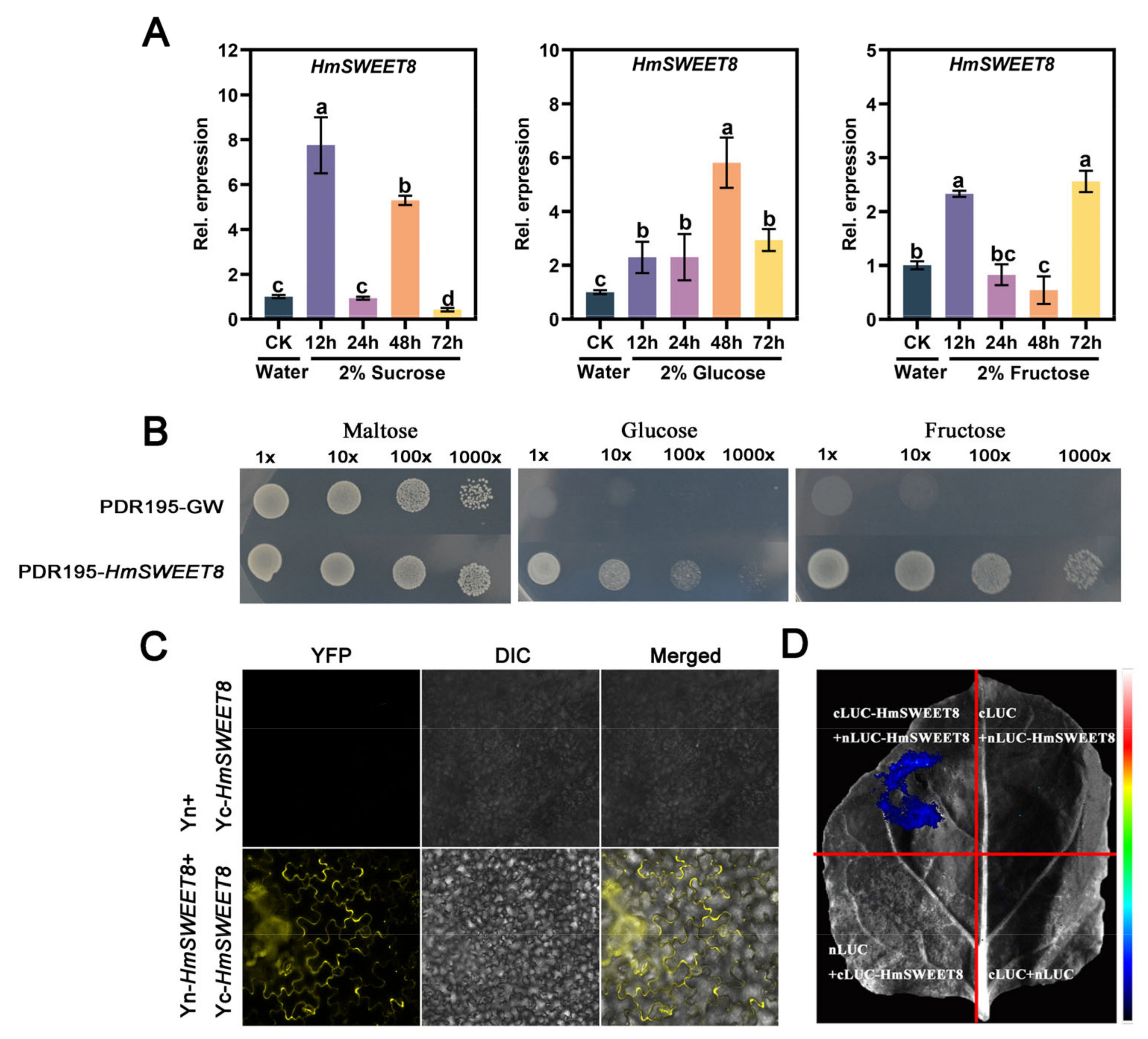
2.1. Cloning of HmSWEET8 Gene in H. moellendorffii

Based on the previously published RNA-seq data in H. moellendorffii following E. heraclei infection, a SWEET sugar transporter HmSWEET8 was induced dramatically after E. heraclei infection (Figure 1A), and artificial inoculation of E. heraclei could induce the expression of HmSWEET8 obviously (Figure 1B), indicating that HmSWEET8 gene may involve in the interaction of H. moellendorffiiE. heraclei. Therefore, we cloned the HmSWEET8 gene successfully by designing a specific primer. The HmSWEET8 protein encoded seven complete transmembrane structures (Figure 1C), and phylogenetic analysis showed that HmSWEET8 was grouped with SWEET proteins obtained from A. thaliana in Clade II (Figure 1D). Subcellular location found HmSWEET8 encoding a plasma membrane protein (Figure 1E).

2.2. HmSWEET8 Could Transport Hexose

The sugar-transporting ability of HmSWEET8 was explored in the next experiment. The 2% sucrose, fructose, and glucose treatments could induce HmSWEET8 expression obviously compared with water treatment (Figure 2A). The complementation assays in the hexose transport-deficient yeast strain EBY.VW4000 found that pDR195-HmSWEET8 expression restored EBY.VW4000 growth on 2% glucose and fructose (Figure 2B), suggesting that HmSWEET8 could transport glucose and fructose. Bimolecular fluorescence complementation (BiFC) found that the YFP signal was observed in Nicotiana benthamiana epidermal cells when co-expressed Yn-HmSWEET8 and Yc-HmSWEET8 (Figure 2C). Luciferase Complementation Assay (LCA) experiments found that LCA activity was observed when co-expressed nLUC-HmSWEET8 and cLUC-HmSWEET8 in N. benthamiana leaves (Figure 2D). These results demonstrated that HmSWEET8 could form the homodimer to perform transport functions.

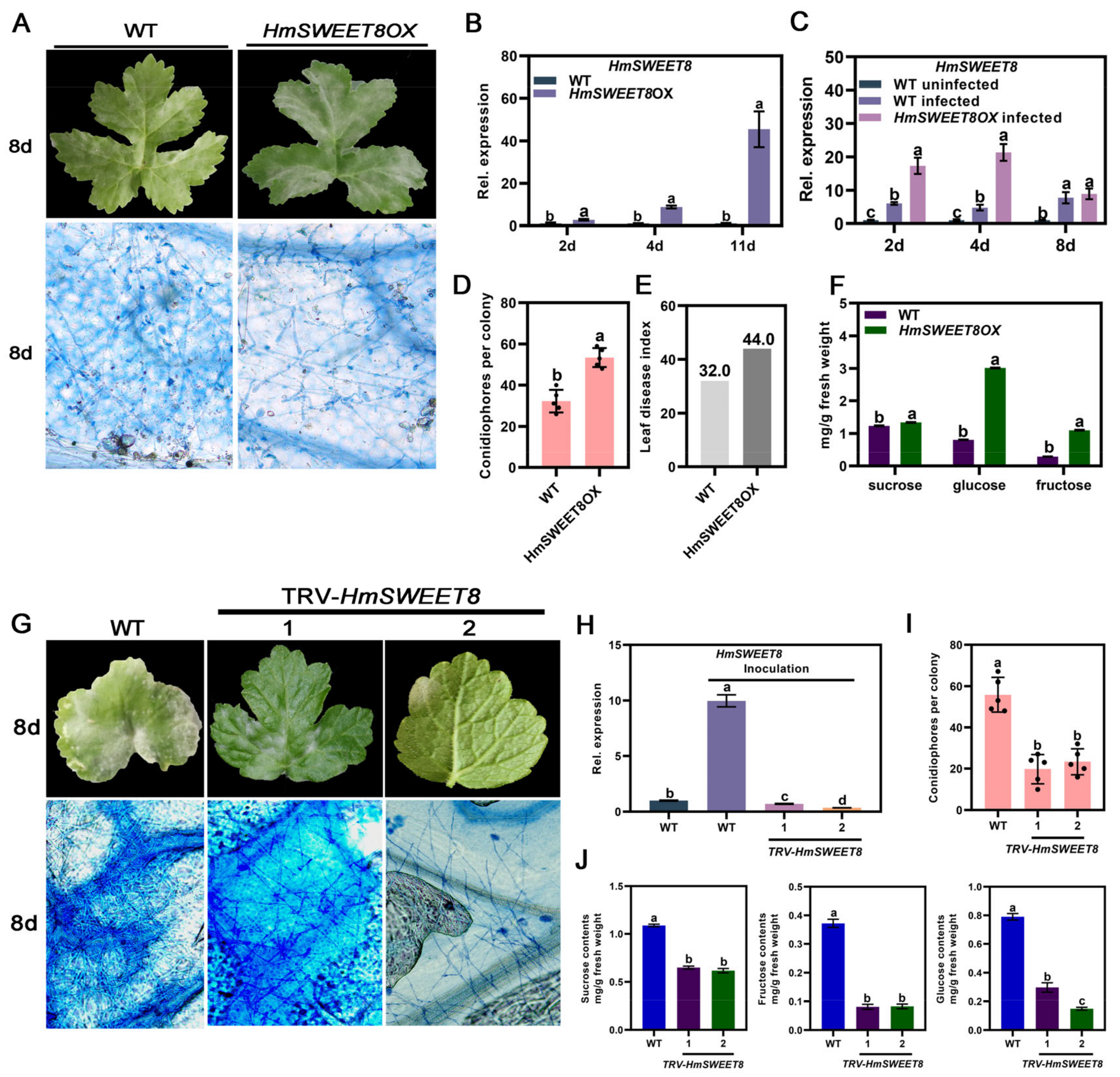
2.3. HmSWEET8 Promoted E. heraclei Infection in H. moellendorffii

To verify the performed function mediated by HmSWEET8 in the interaction of H. moellendorffiiE. heraclei, the HmSWEET8 gene was transiently over-expressing in H. moellendorffii leaves. HmSWEET8 expression was elevated significantly in the over-expression of plants (HmSWEET8OX) compared with WT plants (Figure 3B). After the artificial inoculation of E. heraclei, HmSWEET8 expression in HmSWEET8OX plants was induced dramatically (Figure 3C). The powdery mildew occurred seriously in HmSWEET8OX plants, and higher leaf disease index and conidiophores per colony were observed compared with WT plants at 8d (Figure 3A,D,E). These results demonstrated that HmSWEET8 was a susceptible gene to promote E. heraclei infection. The soluble sugar content of H. moellendorffii infected leaves was measured. Compared with WT plants, the contents of soluble sugar, especially glucose, were obviously accumulated in HmSWEET8OX plants (Figure 3F). The TRV silenced system was used to silence HmSWEET8 expression in H. moellendorffii leaves. HmSWEET8 expression in TRV-HmSWEET8 plants was inhibited significantly, and a decreased number of mycelia and conidiophores was observed compared with WT plants (Figure 3G–I). The sugar concentration, especially hexose, of infected leaves decreased significantly in TRV-HmSWEET8 plants (Figure 3J). These results indicated that HmSWEET8 over-expression contributed to E. heraclei infection by elevating the glucose content in infective sites.

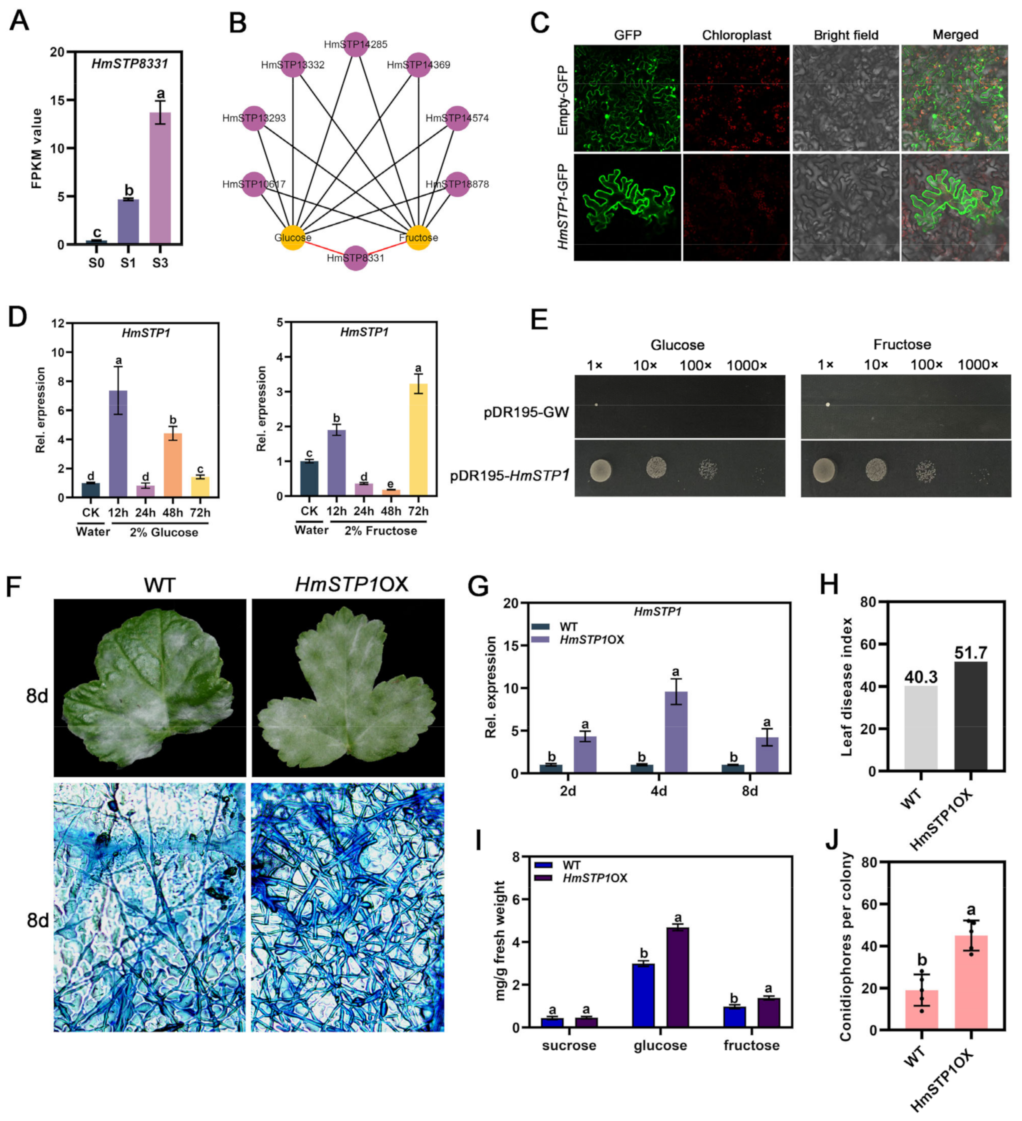
2.4. HmSTP1 Promoted E. heraclei Infection

Prior research has shown that powdery mildew exploits STP sugar transporters to facilitate the movement of sugars from host cells into the powdery mildew across the EHMx. Therefore, we assumed that HmSTPs may be involved in the powdery mildew susceptibility mediated by HmSWEET8 protein in H. moellendorffii. Interestingly, eight HmSTP genes were all induced in HmSWEET8OX plants compared with WT plants (Figure 4). The results indicated that HmSWEET8 and HmSTPs may have synergistic effects in transporting sugars in H. moellendorffii.
HmSTP proteins were identified based on the RNA-seq analysis (Table S1). Among them, HmSTP8331 expression was induced dramatically in S1 and S3 infected leaves, and positively correlated with the accumulation of hexose (Figure 5A,B, Table S2). Therefore, HmSTP1 (HmSTP8331) was selected as the target gene for further research. Subcellular location experiment found HmSTP1 encoding a plasma membrane protein (Figure 5C). HmSTP1 expression could be induced by 2% glucose and fructose treatments (Figure 5D), and the complementation assays in the yeast strain EBY.VW4000 found that HmSTP1 could transport glucose and fructose (Figure 5E). With over-expressing HmSTP1 in H. moellendorffii leaves, the powdery mildew severity increased in HmSTP1OX plants with more visible mycelia and conidiophores at 8 d, compared to WT (Figure 5F–H,J). Furthermore, the hexose level, especially glucose, of infected leaves significantly increased in HmSTP1OX plants (Figure 5I), suggesting that HmSTP1 as the susceptible gene promoted E. heraclei infection by elevating the glucose content in infective sites.

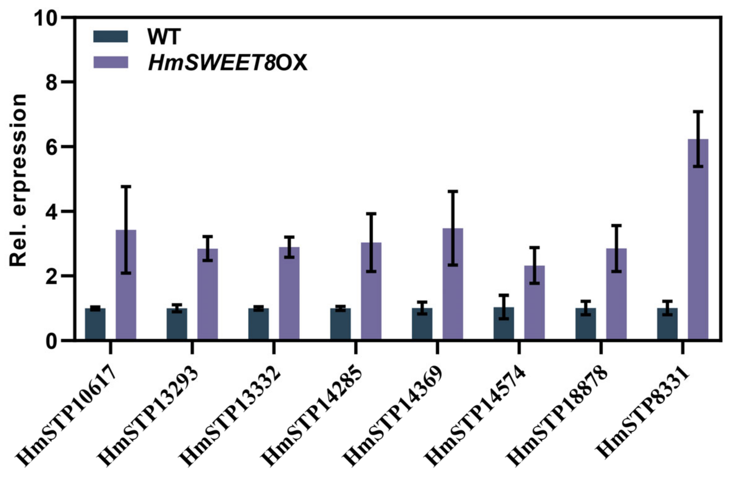
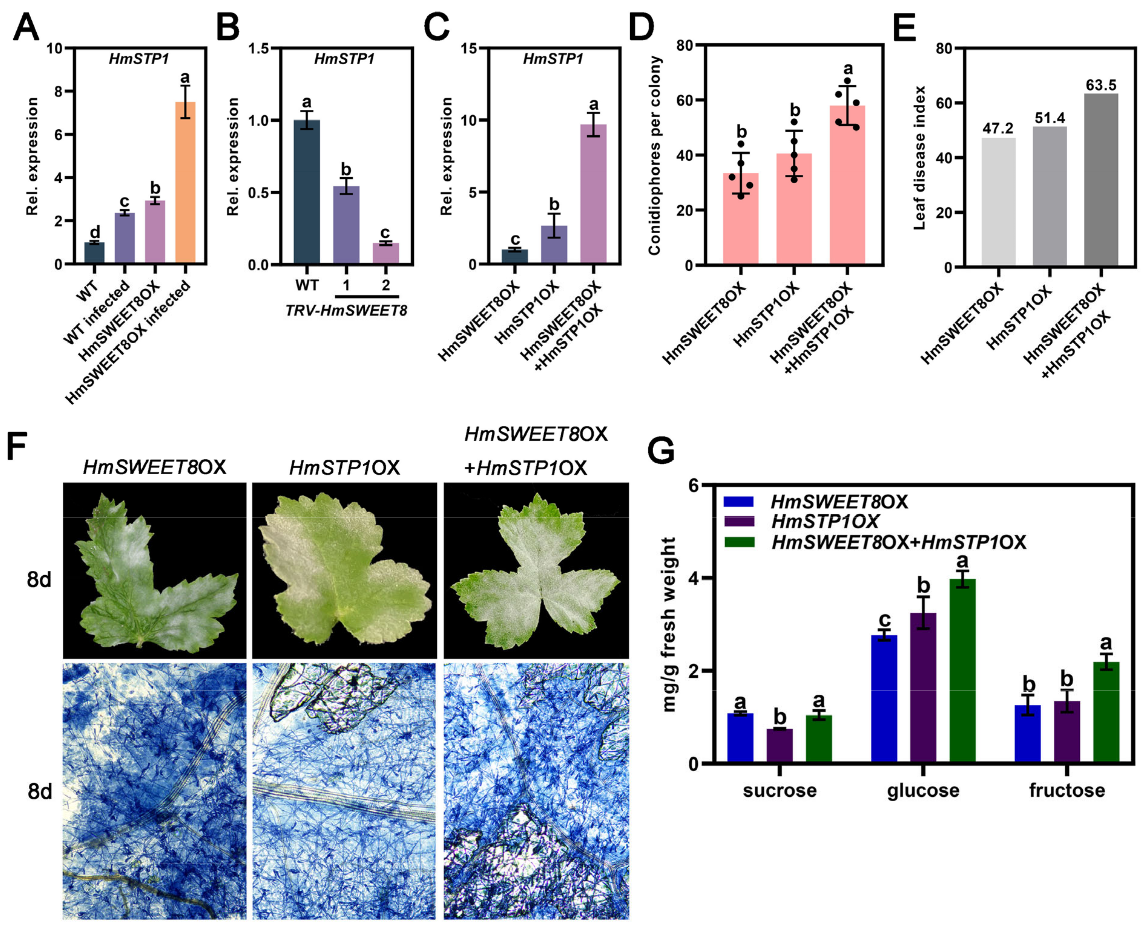
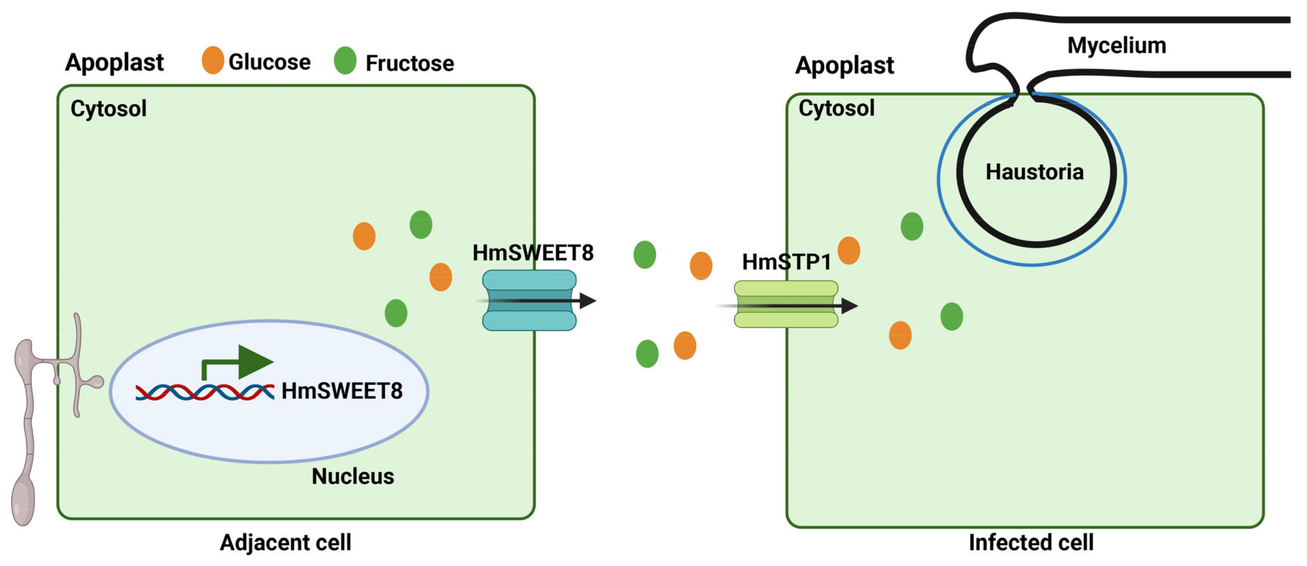
2.5. HmSTP1 May Assist with HmSWEET8 to Promote E. heraclei Infection

Further, we explored the interaction between HmSWEET8 and HmSTP1 in powdery mildew infection. The transiently over-expressing HmSWEET8 gene in H. moellendorffii induced HmSTP1 up-regulation and gene expression was significantly enhanced by powdery mildew infection (Figure 6A). HmSWEET8 gene silencing in H. moellendorffii suppressed HmTP1 expression (Figure 6B). Consequently, there may exist synergistic effects between HmSWEET8 and HmSTP1 in transporting sugars into the infective site of powdery mildew. Co-expressing HmSWEET8 and HmSTP1 genes elevated HmSTP1 expression dramatically compared with over-expressing HmSWEET8 or HmSTP1 alone (Figure 6C). The powdery mildew severity increased in co-expressed HmSWEET8OX and HmSTP1OX plants with more visible conidiophores and higher leaf disease index at 8 d, compared to HmSWEET8OX or HmSTP1OX plants (Figure 6D–F). Furthermore, the hexose level, especially glucose, of infected leaves significantly increased in co-expressed HmSWEET8OX and HmSTP1OX plants (Figure 6G). Consequently, we thought that HmSWEET8 and HmSTP1 may have synergistic effects in transporting sugars to promote powdery mildew infection. After the E. heraclei infection, the HmSWEET8 protein transported more glucose into apoplasmic spaces of infective areas, and the glucose was then transported to the interface of infection areas for the powdery mildew infection via HmSTP1 protein.

3. Discussion

Powdery mildew is an example of a fungal biotroph that forms a haustorium structure to absorb sugars from the plant–haustorium interface [20]. Sugar transporters play a crucial role in facilitating this process by enabling the transfer of sugars to different sugar-dependent tissues and cells [7]. In the study, the HmSWEET8 gene significantly responded to E. heraclei infection, indicating that HmSWEET8 may play important roles in the E. heraclei and H. moellendorffii interaction (Figure 1A,B). SWEET proteins are present at both the source and sink terminals of the phloem pathway, facilitating the transport of sugars from the phloem parenchyma cells to the apoplasmic spaces [26,29]. In the study, the over-expression of HmSWEET8 promoted E. heraclei infection, and the glucose of infected leaves significantly increased in HmSWEET8OX plants (Figure 3A–F), and silenced mutants showed opposite results (Figure 3G–J). The findings indicated that the gene HmSWEET8 was susceptible to powdery mildew infection and may carry more glucose into the infectious region [30]. Similar to AM fungus, E. heraclei infection induced HmSWEET8 gene expression and may accelerate more glucose efflux into the apoplasmic spaces of infective areas of powdery mildew [28,31]. The study demonstrated that certain SWEET family members in H. moellendorffii may be recruited in the phloem loading process by E. heraclei, and E. heraclei retools a critical physiological function to gain sugars from the host at the site of infection.
The sugar efflux rate mediated by SWEET proteins may be enhanced by hexose transporters’ withdrawal from the host apoplasm [5]. In wheat and Arabidopsis leaves, the glucose uptake could be correlated with a change in hexose transporter gene expression [32,33]. In Medicago truncatula, MtSWEET1b exported glucose from the plant cortical cells into the peri-arbuscular space, where it can be taken up by the AM fungus via monosaccharide transporters like MST2 [34]. Over-expressing the HmSWEET8 gene promoted HmSTP gene expression dramatically (Figure 4), and HmSTP1 expression was significantly enhanced by powdery mildew infection (Figure 6A). Therefore, similar to previously reported research, there may exist synergistic effects between HmSWEET8 and HmSTP1 in transporting sugars into the site of powdery mildew infection. HmSTP1 was identified as the plasma membrane protein that transported the hexose (Figure 5C–E). Over-expression of HmSTP1 elevated the hexose contents of infective sites, promoting powdery mildew infection (Figure 5F–J), which was consistent with previous studies [20,35]. Co-expressing HmSWEET8 and HmSTP1 genes increased the powdery mildew severity, and the glucose content was elevated significantly. The results seemingly indicated that HmSTP1 may assist with HmSWEET8 to promote E. heraclei infection. In the process, HmSWEET8 transported more glucose from adjacent cells into the apoplasmic spaces, and HmSTP1 may import the glucose into the haustorium or import the glucose into the cell that feeds the haustorium [5].
This result was not consistent with the “sugar starvation” hypothesis. In which, the microbial pathogen controls the secretion of hexose by SWEETs into the area between cell walls, where the microorganism utilizes hexoses as a source of nourishment and for reproduction. The STPs prevent the build-up of hexose in the cell wall by using secondary active transport to remove them [5]. The variability in outcomes may be attributed to how infections obtain nutrients [36]. It is crucial to recognize that the process of obtaining nutrients by symbiotic bacteria and necrotrophic and biotrophic pathogens may vary significantly [5]. Bacterial pathogens and necrotrophic fungi efficiently manipulate SWEET proteins during the pre-necrotic stage to transport a higher amount of sugars into the apoplasmic regions, providing nourishment for the pathogens [11,37]. STPs take up hexoses from apoplasmic space, depriving the fungus by changing sugar fluxes toward host cells and enhancing resistance to Botrytis cinerea [38], supporting the “sugar starvation” hypothesis. Different from bacterial pathogens, the powdery mildew relies on haustorial invasion turns the invaded host cell into a net sink for photoassimilates, and STPs absorb sugars from the apoplasmic space into the EHMs for infection [18,20,39], and the process needs SWEETs to generate a larger apoplasmic hexose pool for retrieval by STPs supporting the infection process [28,31]. Therefore, in the study, the sugar accumulation in the apoplasmic space, facilitated by HmSWEETs, established a consistent supply of hexose for the retrieval of STP into the cells during E. heraclei infection. Thus, it is possible that the sugars carried by HmSWEETs did not directly contribute to E. heraclei infection and instead relied on the aid of HmSTP.

4. Materials and Methods

4.1. Plant Material

H. moellendorffii susceptible plants were selected from the germplasm garden located in Haerbin City, China. The susceptible plants were cultivated (planting date was 8 May 2022) in the greenhouse of the Gardening Test Station with a 16/8 h (light/dark) photoperiod and a temperature range of 25/18 °C (day/night). The powdery mildew isolate, identified as E. heraclei [2], was separated and cultivated from infected H. moellendorffii leaves.

4.2. Isolation of HmSWEET8 and HmSTP1

The 783 bp and 1572 bp cDNA sequences of HmSWEET8 and HmSTP1 were amplified by primers, respectively. The design of primers was based on the sequence template of H. moellendorffii using Single Molecule Real-Time (SMRT) (BioProject accession: PRJNA1032719). The cDNA fragments were inserted into PMD19-T vector and sequenced (Takara, Shanghai, China).

4.3. Subcellular Localization

The pCAMBIA1300-HmSWEET8-sGFP and pCAMBIA1300-HmSTP1-sGFP vectors were constructed and transiently expressed in Nicotiana benthamiana via Agrobacterium-mediated transformation using the GV3101 strain (Angyu, Shanghai, China) as previously described [40]. The resultant GFP fusion proteins were then visualized using a laser-scanning confocal microscope (FV3000, Olympus, Tokyo, Japan).

4.4. Complementation Assays in Yeast

The PDR195-HmSWEET8 and PDR195-HmSTP1 recombinant plasmids were constructed and transformed into the hexose transport-deficient yeast strain EBY.VW4000. After the transformation, yeast cells underwent cultivation on SC-ura solid medium supplemented with 2% maltose at 30 °C for a duration of 3 days. Subsequently, for complementation growth assays, the cells were grown overnight in SC-ura liquid medium with a 2% maltose supplement, reaching an optical density of 1.0 at 600 nm (OD600). One milliliter of cells was suspended and adjusted with sterile water. Subsequently, serial dilutions (1×, 10×, 100×, and 1000×) were plated on SC-ura solid medium containing either 2% maltose, 2% glucose, and 2% fructose. After incubating for 3 days at 30 °C, the colony growth on the plates was evaluated.

4.5. Bimolecular Fluorescence Complementation (BiFC) Assay

The pCAMBIA1300-HmSWEET8-YFPN and pCAMBIA1300-HmSWEET8-YFPC vectors were constructed and co-expressed in N. benthamiana leaves using the GV3101 Agrobacterium strain. After 2–3 days of cultivation, the YFP signals were then visualized using a laser-scanning confocal microscope (FV3000, Olympus, Tokyo, Japan).

4.6. Luciferase Complementation Assay (LCA)

The pCAMBIA1300-HmSWEET8-nLUC and pCAMBIA1300-HmSWEET8-cLUC vectors were constructed and co-expressed in N. benthamiana leaves with the GV3101 Agrobacterium strain. Following a cultivation period of 2–3 days, the outcomes of LUA studies were captured utilizing an automated chemiluminescence image analysis system (Tanon 5200, Shanghai, China).

4.7. Transient Transformation of H. moellendorffii

The recombinant plasmids pCAMBIA1300-HmSWEET8-sGFP, pCAMBIA1300-HmSTP1-sGFP were transformed using the GV3101 Agrobacterium strain as previously reported in H. moellendorffii [1]. The cells were initially suspended at OD600 0.8 using MMA, followed by a 3 h dark incubation, before being transformed into H. moellendorffii leaves via the vacuum infiltration (SHZ-D, Shanghai, China). Subsequently, the H. moellendorffii leaves were cultured for 2 days at 22 °C in a light incubator (MGC-350HP-2, Shanghai, China), after which they were inoculated with E. heraclei spores at a concentration of 1 × 10⁶. Post-inoculation, samples were collected for gene expression analysis and quantification of soluble sugar content. Finally, a visual assessment of the disease condition caused by E. heraclei was performed.

4.8. Virus-Induced Gene Silencing (VIGS) in H. moellendorffii

A 300 bp fragment of HmSWEET8 was ligated into TRV2 vectors. The empty TRV1 vector and recombinant plasmids were transformed into the GV3101 Agrobacterium strain. The cells were collected and suspended at an optical density of 600 nm (OD600) of 0.8 using MMA. Leaves of H. moellendorffii were excised, retaining only the roots with bud apices [41]. Subsequently, the suspended cell mixture was introduced into H. moellendorffii roots containing buds via vacuum infiltration (SHZ-D, Shanghai, China) [42]. Incisions were made in the roots using a knife before the vacuum step to facilitate the entry of suspended cells. During vacuum infiltration, the roots were immersed in the cell suspension, subjected to a 0.1 MPa vacuum for 10 min, and then swiftly depressurized to promote rapid inoculum penetration. This process was repeated once. The transformed roots were planted in a soil matrix and cultivated at 22 °C for 3 days under the dark condition and then shifted to normal conditions at 22 °C with the 16/10 h light/dark cycle. After a 15-day period, the emerging leaves were inoculated with a suspension of E. heracleid spores at a concentration of 1 × 10⁶, grown in a light incubator, and cultivated at a photoperiod of 16/8 h (light/dark) and a temperature of 25/18 °C (day/night) in the light incubator (MGC-350HP-2, Shanghai, China). Samples were collected for gene expression and soluble sugar analysis. Subsequently, observations were made to assess the disease status induced by E. heraclei.

4.9. E. heraclei Infection in H. moellendorffii

The disease index calculation, trypan blue staining observing the powdery mildew infection, and conidiophores per colony calculation were conducted as previously reported in H. moellendorffii [1].

4.10. Soluble Sugar Content Measurement in H. moellendorffii

The soluble sugar of fresh leaves in H. moellendorffii was extracted and measured contents as previously described [30]. The reference sugars were purchased from the HerbSubstance company (HerbSubstance, Chengdu, China).

4.11. Quantitative Real-Time PCR Analysis

The EasyPure Plant RNA Kit (TransGen Biotech, Beijing, China) was used to extract Plant RNA. The EasyScript One-Step gDNA Removal and cDNA Synthesis SuperMix (TransGen Biotech, Beijing, China) was used to synthesize cDNA Synthesis SuperMix. The selection of reference gene HmActin, qRT-PCR assays, and the calculation of relative gene expression were performed as previously described in H. moellendorffii [1].

4.12. Statistical Analysis

The Excel 2010 software was used to perform data statistics, and the GraphPad Prism 8.0.1 software was used to plot bar graphs. Differentiating letters were used to denote statistical significance with Student’s t-test or one-way analysis of variance (ANOVA) assessed at a significance level of p < 0.05. All primers used in this study are presented in Table S3.

5. Conclusions

The objective of the research was to clarify the molecular processes by which sugar transporters and sugars react to the powdery mildew infection. qRT-PCR analysis found that HmSWEET8 and HmSTP1 were involved in E. heraclei infection. Complementation of yeast EBY.VW4000 and subcellular localization experiments demonstrated that HmSWEET8 and HmSTP1 localized the plasma membrane with hexose transporting activity. HmSWEET8 and HmSTP1 were determined to be susceptible genes by transporting more hexose to the infection areas of E. heraclei. After E. heraclei infection, HmSWEET8 transported more glucose into apoplasmic spaces of infective areas, and then transported to the interface of the infection areas for the powdery mildew infection via HmSTP1 protein. In the process, the glucose efflux mediated by HmSWEET8 may be enhanced by HmSTP1 (Figure 7).

Supplementary Materials

The following supporting information can be downloaded at: https://www.mdpi.com/article/10.3390/plants13162302/s1, Table S1: STP sugar transporters were identified using RNA-seq analysis; Table S2: The Pearson’s correlation analysis between sugar transporters and soluble sugars; Table S3: Primer design of qRT-PCR and vector construction.

Author Contributions

Conceptualization, X.Y.; formal analysis, H.L. and J.L.; investigation, H.L.; methodology, X.Y.; resources, X.J.; supervision, Y.C.; visualization, L.Z. and X.T.; data curation, H.L., J.L., S.Z. and X.S.; writing—original draft, H.L.; writing—review and editing, X.J. and Y.C. All authors have read and agreed to the published version of the manuscript.

Funding

This research was funded by the Basic Research Support Program for Outstanding Young Teachers in Heilongjiang Province [grant number YQJH2023208], the Advantageous and Characteristic Disciplines of Biogenetics of Traditional Chinese Medicine in the Construction of Double First-class in Heilongjiang Province.

Data Availability Statement

The data presented in this study are available upon request from the corresponding author.

Acknowledgments

We thank Jing Jiang for kindly providing the yeast strain EBY.VW4000. Figure 7 (license number: KE2743YE4D) was created with BioRender.com.

Conflicts of Interest

The authors declare no conflicts of interest.

References

  1. Liu, H.; Wang, Y.; Chang, Q.; Li, Q.; Fang, J.; Cao, N.; Tong, X.; Jiang, X.; Yu, X.; Cheng, Y. Combined Metabolome and Transcriptome Reveal HmF6’H1 Regulating Simple Coumarin Accumulation against Powdery Mildew Infection in Heracleum Moellendorffii Hance. BMC Plant Biol. 2024, 24, 507. [Google Scholar] [CrossRef] [PubMed]
  2. Liu, H.; Jiang, X.; Cheng, Y.; Yu, X. First Report of Powdery Mildew Caused by Erysiphe Heraclei on Heracleum Moellendorffii Hance in China. Plant Dis. 2019, 103, 218–232. [Google Scholar] [CrossRef]
  3. Zhang, T.; Cui, H.; Luan, F.; Liu, H.; Ding, Z.; Amanullah, S.; Zhang, M.; Ma, T.; Gao, P. A Recessive Gene Cmpmr2F Confers Powdery Mildew Resistance in Melon (Cucumis melo L.). Theor. Appl. Genet. 2023, 136, 4. [Google Scholar] [CrossRef] [PubMed]
  4. Yamada, K.; Saijo, Y.; Nakagami, H.; Takano, Y. Regulation of Sugar Transporter Activity for Antibacterial Defense in Arabidopsis. Science 2016, 354, 1427–1430. [Google Scholar] [CrossRef] [PubMed]
  5. Bezrutczyk, M.; Yang, J.; Eom, J.-S.; Prior, M.; Sosso, D.; Hartwig, T.; Szurek, B.; Oliva, R.; Vera-Cruz, C.; White, F.F.; et al. Sugar Flux and Signaling in Plant-Microbe Interactions. Plant J. 2018, 93, 675–685. [Google Scholar] [CrossRef] [PubMed]
  6. Chen, L.-Q.; Qu, X.-Q.; Hou, B.-H.; Sosso, D.; Osorio, S.; Fernie, A.R.; Frommer, W.B. Sucrose Efflux Mediated by SWEET Proteins as a Key Step for Phloem Transport. Science 2012, 335, 207–211. [Google Scholar] [CrossRef] [PubMed]
  7. Chen, Y.; Miller, A.J.; Qiu, B.; Huang, Y.; Zhang, K.; Fan, G.; Liu, X. The Role of Sugar Transporters in the Battle for Carbon between Plants and Pathogens. Plant Biotechnol. J. 2024. [Google Scholar] [CrossRef] [PubMed]
  8. Breia, R.; Conde, A.; Badim, H.; Fortes, A.M.; Gerós, H.; Granell, A. Plant SWEETs: From Sugar Transport to Plant-Pathogen Interaction and More Unexpected Physiological Roles. Plant Physiol. 2021, 186, 836–852. [Google Scholar] [CrossRef]
  9. Hu, Y.; Zhang, J.; Jia, H.; Sosso, D.; Li, T.; Frommer, W.B.; Yang, B.; White, F.F.; Wang, N.; Jones, J.B. Lateral Organ Boundaries 1 Is a Disease Susceptibility Gene for Citrus Bacterial Canker Disease. Proc. Natl. Acad. Sci. USA 2014, 111, E521–E529. [Google Scholar] [CrossRef]
  10. Blanvillain-Baufumé, S.; Reschke, M.; Solé, M.; Auguy, F.; Doucoure, H.; Szurek, B.; Meynard, D.; Portefaix, M.; Cunnac, S.; Guiderdoni, E.; et al. Targeted Promoter Editing for Rice Resistance to Xanthomonas oryzae Pv. oryzae reveals Differential Activities for SWEET14-Inducing TAL Effectors. Plant Biotechnol. J. 2017, 15, 306–317. [Google Scholar] [CrossRef]
  11. Cox, K.L.; Meng, F.; Wilkins, K.E.; Li, F.; Wang, P.; Booher, N.J.; Carpenter, S.C.D.; Chen, L.-Q.; Zheng, H.; Gao, X.; et al. TAL Effector Driven Induction of a SWEET Gene Confers Susceptibility to Bacterial Blight of Cotton. Nat. Commun. 2017, 8, 15588. [Google Scholar] [CrossRef]
  12. Hutin, M.; Sabot, F.; Ghesquière, A.; Koebnik, R.; Szurek, B. A Knowledge-Based Molecular Screen Uncovers a Broad-Spectrum OsSWEET14 Resistance Allele to Bacterial Blight from Wild Rice. Plant J. 2015, 84, 694–703. [Google Scholar] [CrossRef]
  13. Zhou, J.; Peng, Z.; Long, J.; Sosso, D.; Liu, B.; Eom, J.-S.; Huang, S.; Liu, S.; Vera Cruz, C.; Frommer, W.B.; et al. Gene Targeting by the TAL Effector PthXo2 Reveals Cryptic Resistance Gene for Bacterial Blight of Rice. Plant J. 2015, 82, 632–643. [Google Scholar] [CrossRef]
  14. Yang, S.; Fu, Y.; Zhang, Y.; Peng Yuan, D.; Li, S.; Kumar, V.; Mei, Q.; Hu Xuan, Y. Rhizoctonia Solani Transcriptional Activator Interacts with Rice WRKY53 and Grassy Tiller 1 to Activate SWEET Transporters for Nutrition. J. Adv. Res. 2023, 50, 1–12. [Google Scholar] [CrossRef]
  15. Fatima, U.; Senthil-Kumar, M. Sweet Revenge: AtSWEET12 in Plant Defense against Bacterial Pathogens by Apoplastic Sucrose Limitation. Cold Spring Harb. Lab. 2021, 10, 1–49. [Google Scholar] [CrossRef]
  16. Herbers, K.; Sonnewald, U. Altered Gene Expression Brought about by Inter- and Intracellularly Formed Hexoses and Its Possible Implications for Plant-Pathogen Interactions. J. Plant Res. 1998, 111, 323–328. [Google Scholar] [CrossRef]
  17. Gebauer, P.; Korn, M.; Engelsdorf, T.; Sonnewald, U.; Koch, C.; Voll, L.M. Sugar Accumulation in Leaves of Arabidopsis Sweet11/Sweet12 Double Mutants Enhances Priming of the Salicylic Acid-Mediated Defense Response. Front. Plant Sci. 2017, 8, 1378. [Google Scholar] [CrossRef]
  18. Hall, J.L.; Williams, L.E. Assimilate Transport and Partitioning in Fungal Biotrophic Interactions. Funct. Plant Biol. 2000, 27, 549. [Google Scholar] [CrossRef]
  19. Staples, R.C. Research on the Rust Fungi During the Twentieth Century. Annu. Rev. Phytopathol. 2000, 38, 49–69. [Google Scholar] [CrossRef] [PubMed]
  20. Liu, J.; Liu, M.; Tan, L.; Huai, B.; Ma, X.; Pan, Q.; Zheng, P.; Wen, Y.; Zhang, Q.; Zhao, Q.; et al. AtSTP8, an Endoplasmic Reticulum-Localised Monosaccharide Transporter from Arabidopsis, Is Recruited to the Extrahaustorial Membrane during Powdery Mildew Infection. New Phytol. 2021, 230, 2404–2419. [Google Scholar] [CrossRef]
  21. Huai, B.; Yuan, P.; Ma, X.; Zhang, X.; Jiang, L.; Zheng, P.; Yao, M.; Chen, Z.; Chen, L.; Shen, Q.; et al. Sugar Transporter TaSTP3 Activation by TaWRKY19/61/82 Enhances Stripe Rust Susceptibility in Wheat. New Phytol. 2022, 236, 266–282. [Google Scholar] [CrossRef]
  22. Huai, B.; Yang, Q.; Qian, Y.; Qian, W.; Kang, Z.; Liu, J. ABA-Induced Sugar Transporter TaSTP6 Promotes Wheat Susceptibility to Stripe Rust. Plant Physiol. 2019, 181, 1328–1343. [Google Scholar] [CrossRef] [PubMed]
  23. Voegele, R.T.; Mendgen, K. Rust Haustoria: Nutrient Uptake and Beyond. New Phytol. 2003, 159, 93–100. [Google Scholar] [CrossRef]
  24. Saja, D.; Janeczko, A.; Barna, B.; Skoczowski, A.; Dziurka, M.; Kornaś, A.; Gullner, G. Powdery Mildew-Induced Hormonal and Photosynthetic Changes in Barley Near Isogenic Lines Carrying Various Resistant Genes. Int. J. Mol. Sci. 2020, 21, 4536. [Google Scholar] [CrossRef]
  25. Doidy, J.; Grace, E.; Kühn, C.; Simon-Plas, F.; Casieri, L.; Wipf, D. Sugar Transporters in Plants and in Their Interactions with Fungi. Trends Plant Sci. 2012, 17, 413–422. [Google Scholar] [CrossRef] [PubMed]
  26. Singh, J.; Das, S.; Jagadis Gupta, K.; Ranjan, A.; Foyer, C.H.; Thakur, J.K. Physiological Implications of SWEETs in Plants and Their Potential Applications in Improving Source-Sink Relationships for Enhanced Yield. Plant Biotechnol. J. 2023, 21, 1528–1541. [Google Scholar] [CrossRef]
  27. Chen, L.-Q.; Hou, B.-H.; Lalonde, S.; Takanaga, H.; Hartung, M.L.; Qu, X.-Q.; Guo, W.-J.; Kim, J.-G.; Underwood, W.; Chaudhuri, B.; et al. Sugar Transporters for Intercellular Exchange and Nutrition of Pathogens. Nature 2010, 468, 527–532. [Google Scholar] [CrossRef]
  28. An, J.; Zeng, T.; Ji, C.; de Graaf, S.; Zheng, Z.; Xiao, T.T.; Deng, X.; Xiao, S.; Bisseling, T.; Limpens, E.; et al. A Medicago Truncatula SWEET Transporter Implicated in Arbuscule Maintenance during Arbuscular Mycorrhizal Symbiosis. New Phytol. 2019, 224, 396–408. [Google Scholar] [CrossRef] [PubMed]
  29. Bezrutczyk, M.; Hartwig, T.; Horschman, M.; Char, S.N.; Yang, J.; Yang, B.; Frommer, W.B.; Sosso, D. Impaired Phloem Loading in Zmsweet13a,b,c Sucrose Transporter Triple Knock-out Mutants in Zea mays. New Phytol. 2018, 218, 594–603. [Google Scholar] [CrossRef]
  30. Chang, Q.; Liu, J.; Wang, Q.; Han, L.; Liu, J.; Li, M.; Huang, L.; Yang, J.; Kang, Z. The Effect of Puccinia striiformis f. Sp Tritici on the Levels of Water-Soluble Carbohydrates and the Photosynthetic Rate in Wheat Leaves. Physiol. Mol. Plant Pathol. 2013, 84, 131–137. [Google Scholar] [CrossRef]
  31. Manck-Götzenberger, J.; Requena, N. Arbuscular Mycorrhiza Symbiosis Induces a Major Transcriptional Reprogramming of the Potato SWEET Sugar Transporter Family. Front. Plant Sci. 2016, 7, 487. [Google Scholar] [CrossRef] [PubMed]
  32. Fotopoulos, V.; Gilbert, M.J.; Pittman, J.K.; Marvier, A.C.; Buchanan, A.J.; Sauer, N.; Hall, J.L.; Williams, L.E. The Monosaccharide Transporter Gene, AtSTP4, and the Cell-Wall Invertase, Atbetafruct1, Are Induced in Arabidopsis during Infection with the Fungal Biotroph Erysiphe cichoracearum. Plant Physiol. 2003, 132, 821–829. [Google Scholar] [CrossRef] [PubMed]
  33. Sutton, P.N.; Gilbert, M.J.; Williams, L.E.; Hall, J.L. Powdery Mildew Infection of Wheat Leaves Changes Host Solute Transport and Invertase Activity. Physiol. Plant. 2007, 129, 787–795. [Google Scholar] [CrossRef]
  34. Helber, N.; Wippel, K.; Sauer, N.; Schaarschmidt, S.; Hause, B.; Requena, N. A Versatile Monosaccharide Transporter That Operates in the Arbuscular Mycorrhizal Fungus Glomus Sp Is Crucial for the Symbiotic Relationship with Plants. Plant Cell 2011, 23, 3812–3823. [Google Scholar] [CrossRef] [PubMed]
  35. Moore, J.W.; Herrera-Foessel, S.; Lan, C.; Schnippenkoetter, W.; Ayliffe, M.; Huerta-Espino, J.; Lillemo, M.; Viccars, L.; Milne, R.; Periyannan, S.; et al. A Recently Evolved Hexose Transporter Variant Confers Resistance to Multiple Pathogens in Wheat. Nat. Genet. 2015, 47, 1494–1498. [Google Scholar] [CrossRef] [PubMed]
  36. Liu, Y.-H.; Song, Y.-H.; Ruan, Y.-L. Sugar Conundrum in Plant-Pathogen Interactions: Roles of Invertase and Sugar Transporters Depend on Pathosystems. J. Exp. Bot. 2022, 73, 1910–1925. [Google Scholar] [CrossRef] [PubMed]
  37. Asai, Y.; Kobayashi, Y. Increased Expression of the Tomato SISWEET15 Gene during Grey Mold Infection and the Possible Involvement of the Sugar Efflux to Apoplasm in the Disease Susceptibility. J. Plant Pathol. Microbiol. 2016, 7, 1. [Google Scholar] [CrossRef]
  38. Lemonnier, P.; Gaillard, C.; Veillet, F.; Verbeke, J.; Lemoine, R.; Coutos-Thévenot, P.; La Camera, S. Expression of Arabidopsis Sugar Transport Protein STP13 Differentially Affects Glucose Transport Activity and Basal Resistance to Botrytis cinerea. Plant Mol. Biol. 2014, 85, 473–484. [Google Scholar] [CrossRef]
  39. Harrison, M.J. Biotrophic Interfaces and Nutrient Transport in Plant/Fungal Symbioses. J. Exp. Bot. 1999, 50, 1013–1022. [Google Scholar] [CrossRef]
  40. Bracha-Drori, K.; Shichrur, K.; Katz, A.; Oliva, M.; Angelovici, R.; Yalovsky, S.; Ohad, N. Detection of Protein-Protein Interactions in Plants Using Bimolecular Fluorescence Complementation. Plant J. 2004, 40, 419–427. [Google Scholar] [CrossRef]
  41. Spitzer, B.; Zvi, M.M.B.; Ovadis, M.; Marhevka, E.; Barkai, O.; Edelbaum, O.; Marton, I.; Masci, T.; Alon, M.; Morin, S.; et al. Reverse Genetics of Floral Scent: Application of Tobacco Rattle Virus-Based Gene Silencing in Petunia. Plant Physiol. 2007, 145, 1241–1250. [Google Scholar] [CrossRef] [PubMed]
  42. Ye, J.; Qu, J.; Bui, H.T.N.; Chua, N.-H. Rapid Analysis of Jatropha Curcas Gene Functions by Virus-Induced Gene Silencing. Plant Biotechnol. J. 2009, 7, 964–976. [Google Scholar] [CrossRef] [PubMed]
Figure 1. Cloning of HmSWEET8 gene in Heracleum moellendorffii Hance: (A) The FPKM value of HmSWEET8 in S0, S1, and S3 infected leaves. Leaves infected with Eeysiphe heraclei exhibiting varying degrees of disease. S1: The area of occurring colonies accounted for less than 10% of the whole leaf area; S3: the area of occurring colonies accounts for 30–50% of the whole leaf area; S0: the leaves with the water treatment were collected as the control. Statistical significance was indicated by different letters, as determined by one-way ANOVA measured at p < 0.05. (B) The expression level of HmSWEET8 after E. heraclei infection. Statistical significance was indicated by different letters, as determined by Student’s t-test measured at p < 0.05. (C) Multiple sequences alignment of HmSWEET8 with A. thaliana and Oryza sativa Japonica Group. SWEET protein sequences. HmSWEET8 (accession number: OM515217) was obtained from H. moellendorffii. AtSWEET1, AtSWEET4, and AtSWEET5 (accession number: Q8L9J7, Q944M5, and Q9FM10, respectively), were obtained from A. thaliana. OsSWEET2a, OsSWEET2b, and OsSWEET12 (accession number: Q5JJY5, Q5N8J1, and Q10LI8, respectively), were obtained from Oryza sativa Japonica Group. (D) The phylogenetic tree was constructed using HmSWEET8 and A. thaliana SWEET protein sequences. (E) Subcellular localization of HmSWEET8 in Nicotiana benthamiana epidermal cells. Scale bar: 20 μm.
Plants 13 02302 g001
Figure 2. The confirmation of sugar transporting ability mediated by HmSWEET8: (A) The expression of HmSWEE8 with different 2% sugar treatments. Statistical significance was indicated by different letters, as determined by one-way ANOVA measured at p < 0.05. (B) The complementation assays in the hexose transport-deficient yeast strain EBY.VW4000. (C) Bimolecular fluorescence complementation (BiFC) was used for detection of HmSWEET8 homodimer. Scale bar: 20 μm. (D) Luciferase Complementation Assay (LCA) was used for detection of HmSWEET8 homodimer.
Plants 13 02302 g002
Figure 3. HmSWEET8 gene was transiently over-expressed in H. moellendorffii leaves: (A) The typical genotypes of H. moellendorffii leaves were observed after E. heraclei infection at 8 d. Scale bar: 400 μm. (B) The expression of HmSWEET8 without E. heraclei infection. Statistical significance was indicated by different letters, as determined by Student’s t-test measured at p < 0.05. (C) The expression of HmSWEET8 after artificial inoculation E. heraclei. Statistical significance was indicated by different letters, as determined by one-way ANOVA measured at p < 0.05. (D,E) The calculation of conidiophores per colony (D) and leaf disease index (E). Statistical significance was indicated by different letters, as determined by Student’s t-test measured at p < 0.05. (F) The soluble sugar content of infected leaves was measured. Statistical significance was indicated by different letters, as determined by Student’s t-test measured at p < 0.05. (G) The typical genotypes of H. moellendorffii leaves were observed in HmSWET8 (1#, 2#) silenced seedlings after E. heraclei infection. Scale bar: 400 μm. (H) The expression of HmSWEET8 was measured. Statistical significance was indicated by different letters, as determined by one-way ANOVA measured at p < 0.05. (I) The calculation of conidiophores per colony in infected leaves. Statistical significance was indicated by different letters, as determined by one-way ANOVA measured at p < 0.05. (J) The soluble sugar content of infected leaves was measured. Statistical significance was indicated by different letters, as determined by one-way ANOVA measured at p < 0.05.
Plants 13 02302 g003
Figure 4. HmSTP genes were all induced significantly in HmSWEET8OX plants.
Plants 13 02302 g004
Figure 5. HmSTP1 protein responding to E. heraclei infection in H. moellendorffii: (A) The FPKM value of HmSTP8331 in S0, S1, and S3 infected leaves. Statistical significance was indicated by different letters, as determined by one-way ANOVA measured at p < 0.05. (B) The correlation network analysis between HmSTP8331 expression with hexose contents, the red line: r2 > 0.90. (C) Subcellular localization of HmSTP1 in N. benthamiana epidermal cells. Scale bar: 20 μm. (D) The expression of HmSTP1 with different 2% sugar treatments. Statistical significance was indicated by different letters, as determined by one-way ANOVA measured at p < 0.05. (E) Complementation of yeast EBY.VW4000 with HmSTP1. (F) The typical genotypes of H. moellendorffii leaves were observed after E. heraclei infection at 8 d. Scale bar: 50 μm. (G) The expression of HmSTP1 after artificial inoculation E. heraclei. Statistical significance was indicated by different letters, as determined by Student’s t-test measured at p < 0.05. (H) The calculation of leaf disease index in infected leaves. (I) The soluble sugar content of infected leaves was measured. Statistical significance was indicated by different letters, as determined by Student’s t-test measured at p < 0.05. (J) The calculation of conidiophores per colony in infected leaves. Statistical significance was indicated by different letters, as determined by Student’s t-test measured at p < 0.05.
Plants 13 02302 g005
Figure 6. HmSWEET8 and HmSTP1 genes were transiently co-expressed in H. moellendorffii leaves: (A) The expression of HmSTP1 in HmSWEET8 over-expressed plants. Statistical significance was indicated by different letters, as determined by one-way ANOVA measured at p < 0.05. (B) The expression of HmSTP1 in HmSWEET8 silenced plants. Statistical significance was indicated by different letters, as determined by one-way ANOVA measured at p < 0.05. (C) The expression of HmSTP1 in co-expressed HmSWEET8 and HmSTP1 plants. Statistical significance was indicated by different letters, as determined by one-way ANOVA measured at p < 0.05. (D,E) The calculation of conidiophores per colony and leaf disease index (E) in infected leaves. Statistical significance was indicated by different letters, as determined by one-way ANOVA measured at p < 0.05. (F) The typical genotypes of H. moellendorffii leaves were observed after E. heraclei infection at 8 d. Scale bar: 400 μm. (G) The soluble sugar content of infected leaves was measured. Statistical significance was indicated by different letters, as determined by one-way ANOVA measured at p < 0.05.
Plants 13 02302 g006
Figure 7. A working model portraying the underlying mechanisms by which E. heraclei manipulates host sugar transporters to acquire nutrients and promote fungal infection. E. heraclei infection activates the expression of HmSWEET8 and HmSTP1, then, HmSWEEμmT8 protein transports more glucose into apoplasmic spaces of infective areas. HmSWEET8 generates a larger apoplasmic hexose pool and promotes HmSTP1 expression. The glucose then is transported into host cells via HmSTP1 sugar transporter for promoting E. heraclei infection.
Plants 13 02302 g007
Disclaimer/Publisher’s Note: The statements, opinions and data contained in all publications are solely those of the individual author(s) and contributor(s) and not of MDPI and/or the editor(s). MDPI and/or the editor(s) disclaim responsibility for any injury to people or property resulting from any ideas, methods, instructions or products referred to in the content.

