Communication

A Fast, Efficient, and Tissue-Culture-Independent Genetic Transformation Method for Panax notoginseng and Lilium regale

1
Faculty of Life Science and Technology, Kunming University of Science and Technology, Kunming 650500, China
2
State Key Laboratory of Conservation and Utilization of Bio-Resources in Yunnan, The Key Laboratory of Medicinal Plant Biology of Yunnan Province, National & Local Joint Engineering Research Center on Germplasms Innovation & Utilization of Chinese Medicinal Materials in Southwest China, Yunnan Agricultural University, Kunming 650201, China
3
Yunnan Seed Laboratory, Yunnan Agricultural University, Kunming 650201, China
*
Authors to whom correspondence should be addressed.
Plants 2024, 13(17), 2509; https://doi.org/10.3390/plants13172509
Submission received: 22 July 2024 / Revised: 4 September 2024 / Accepted: 4 September 2024 / Published: 6 September 2024
(This article belongs to the Section Plant Development and Morphogenesis)

Abstract

:
The Agrobacterium-based transgenic technique is commonly used for gene function validation and molecular breeding. However, it is not suitable for plants with a low regeneration capacity or a low transformation rate, such as Panax notoginseng (Burk) F.H. Chen and Lilium regale Wilson. In this study, a novel Agrobacterium transformation method based on injection in the meristems was developed using P. notoginseng and L. regale as experimental models. PCR analysis confirmed the successful integration of the reporter gene DsRed2 (Discosoma striata red fluorescence protein 2) into the genome of two experimental models. QRT-PCR and Western blot analysis demonstrated the transcriptional and translational expression of DsRed2. Additionally, laser confocal microscopy confirmed the significant accumulation of the red fluorescent protein in the leaves, stems, and roots of transformed P. notoginseng and L. regale. Most importantly, in the second year after injection, the specific bright orange fluorescence from DsRed2 expression was observed in the transgenic P. notoginseng and L. regale plants. This study establishes a fast, efficient, and tissue-culture-independent transgenic technique suitable for plants with a low regeneration capacity or a low transformation rate. This technique may improve the functional genomics of important medicinal and ornamental plants such as P. notoginseng and L. regale, as well as their molecular breeding.

Graphical Abstract

1. Introduction

The Agrobacterium-based transgenic technique has been extensively used for gene function validation and genetic modification due to its practicality, cost-effectiveness, and high efficiency [1]. The key steps of this technique include explant preparation, explant infection, co-cultivation with Agrobacterium, explant regeneration, and the screening of transgenic lines [2], but tissue culture is a limiting step for some species that exhibit a low regenerative capacity and low rate of genetic transformation [3]. Therefore, the Agrobacterium-based transformation method is particularly difficult to apply to plants such as Panax spp. and Lilium spp. [4]. For example, Panax notoginseng (Burk) F.H. Chen and Lilium regale Wilson are both perennial plants with important economic value, but these two species have a low tissue regeneration ability and transformation rate.
The plants of Panax spp. are well-known medicinal herbs with valuable medicinal properties, primarily attributed to their saponin content [5]. However, the lack of a well-established genetic transformation system has hindered further exploration of the saponin biosynthesis pathways and the development of elite P. notoginseng cultivars with high and consistent yields and quality [6]. Adventitious roots were regenerated from Panax ginseng roots through tissue culture and transgenic P. ginseng roots were obtained via Agrobacterium-based transformation [7]. RNA interference of PgRg1-3 in the P. ginseng roots was mediated by Agrobacterium rhizogenes C58C1 [8]. PgERF120 was overexpressed in the hairy roots of P. ginseng roots based on Agrobacterium tumefaciens A4 transformation [9]. P. notoginseng cell lines overexpressing Panax japonicus βAS (β-amyrin synthase) were obtained via Agrobacterium-mediated genetic transformation in order to reveal the influence of PjβAS on saponin biosynthesis in P. notoginseng [10]. The current transformation method involves traditional Agrobacterium-based techniques applied to callus tissue induced from P. notoginseng explants, but successful generation of transgenic P. notoginseng plants has not been achieved using this approach [11]. At the same time, aseptic operation makes the acquisition of genetically modified materials more time-consuming and more difficult.
Similarly, Lilium spp., known for their wide range of flower shapes, fragrances, and colors, hold significant ornamental value and rank as the sixth-largest fresh cut flower globally [12]. However, the traditional Agrobacterium-mediated transformation method, characterized by its low efficiency, lengthy cycle, and demanding sterile operation, is not widely applicable in molecular breeding efforts for lilies [13]. Although the transformation rate of Lilium ‘Sorbonne’ was improved by adjusting the composition of the culture medium; only 14 positive transgenic Lilium ‘Sorbonne’ lines out of 173 lines were successfully identified [14]. Fu and co-authors [15] found that the transformation rate of Lilium brownie embryogenic callus was 65.56%. The β-glucuronidase (GUS) gene and green fluorescent protein (GFP) gene were successfully expressed in L. regale via pollen magnetofection, but a complete transgenic plant was not produced [16]. Therefore, the transformation rate of lilies is still low, and the protocols remain difficult and complex. L. regale, a wild lily species with high resistance to various stresses, remains underexplored due to the lack of a mature transformation system, hindering the elucidation of its resistance mechanisms and the utilization of its resistance-related genes to improve lily disease resistance. Establishing an efficient genetic transformation system is crucial for enhancing the ornamental and disease-resistant traits of lilies to improve the economic value of Lilium spp.
Establishing a fast, efficient, and tissue-culture-independent genetic transformation method is important for the fundamental research and molecular breeding of Panax spp. and Lilium spp. However, the transitional Agrobacterium-based transformation method is not suitable for some perennial plants. To address these challenges, we utilized P. notoginseng and L. regale as two experimental models to establish a rapid, efficient, and tissue-culture-independent genetic transformation system. Every spring, the aboveground part of P. notoginseng and L. regale grows up depending on the meristems located in the rhizome and bulb, respectively. Therefore, the meristems in the P. notoginseng rhizome and L. regale bulb could be excellent targets for genetic transformation. Additionally, DsRed2 (Discosoma striata red fluorescence protein 2) is a red fluorescent protein isolated from Discosoma sp. that was found to be a better reporter gene than GUS and GFP because its excitation light was higher than that of GUS and GFP; thus, it can avoid plant autofluorescence [17]. DsRed2 has been used to screen transgenic cotton (Gossypium herbaceum) and transgenic peanut (Arachis hypogaea) via fluorescence microscopy and handheld fluorescence observation equipment through the detection of red and bright orange fluorescence, respectively [17,18]. Therefore, Agrobacterium tumefaciens of the LBA4404 strain containing the vector pCAMBIA2300::35S::DsRed2 were injected into the meristems of P. notoginseng rhizome and L. regale bulb. Throughout the 40–60 days of cultivation after Agrobacterium injection, a series of experiments were conducted to evaluate the expression levels and activity of the reporter gene in transgenic plants.

2. Results

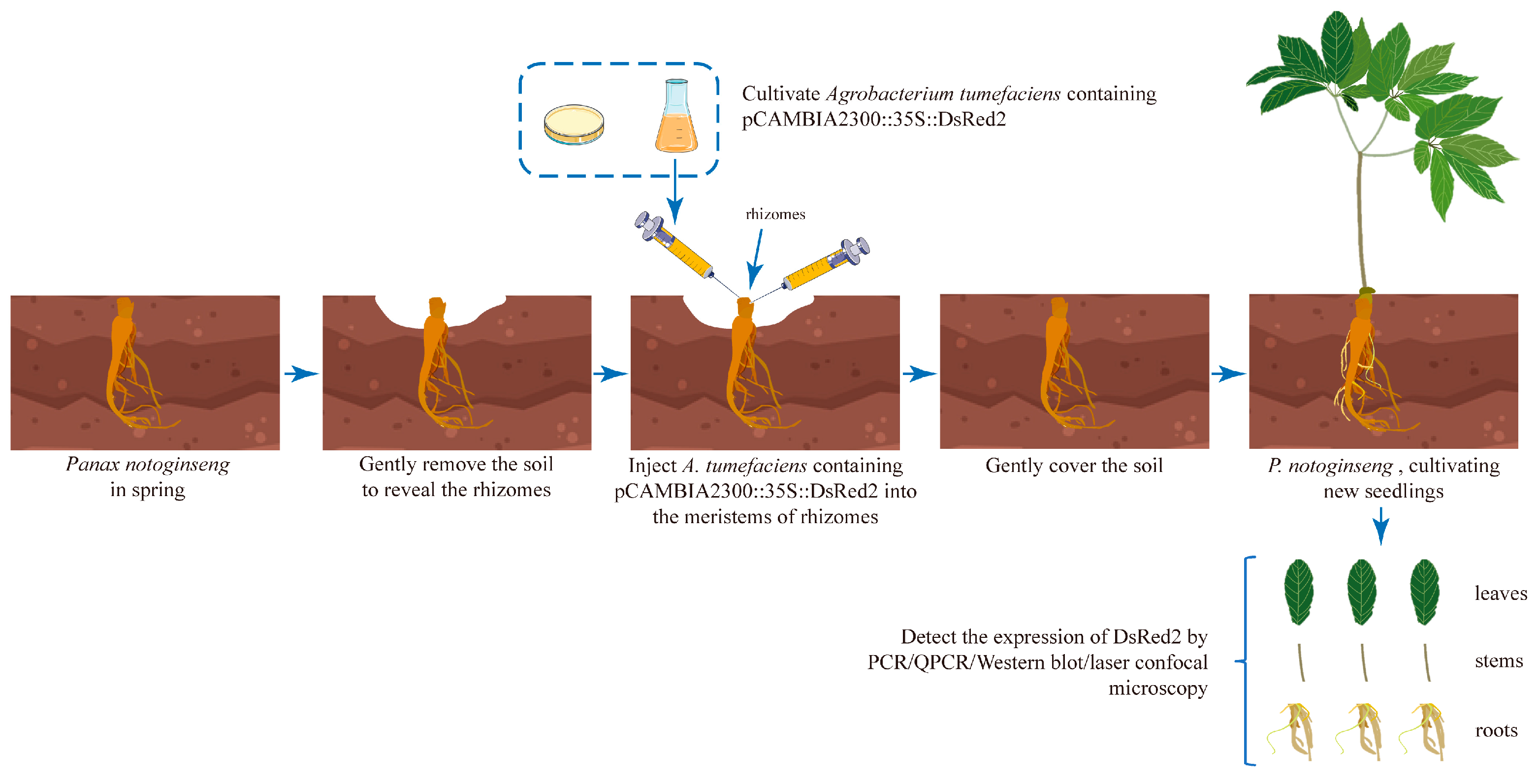
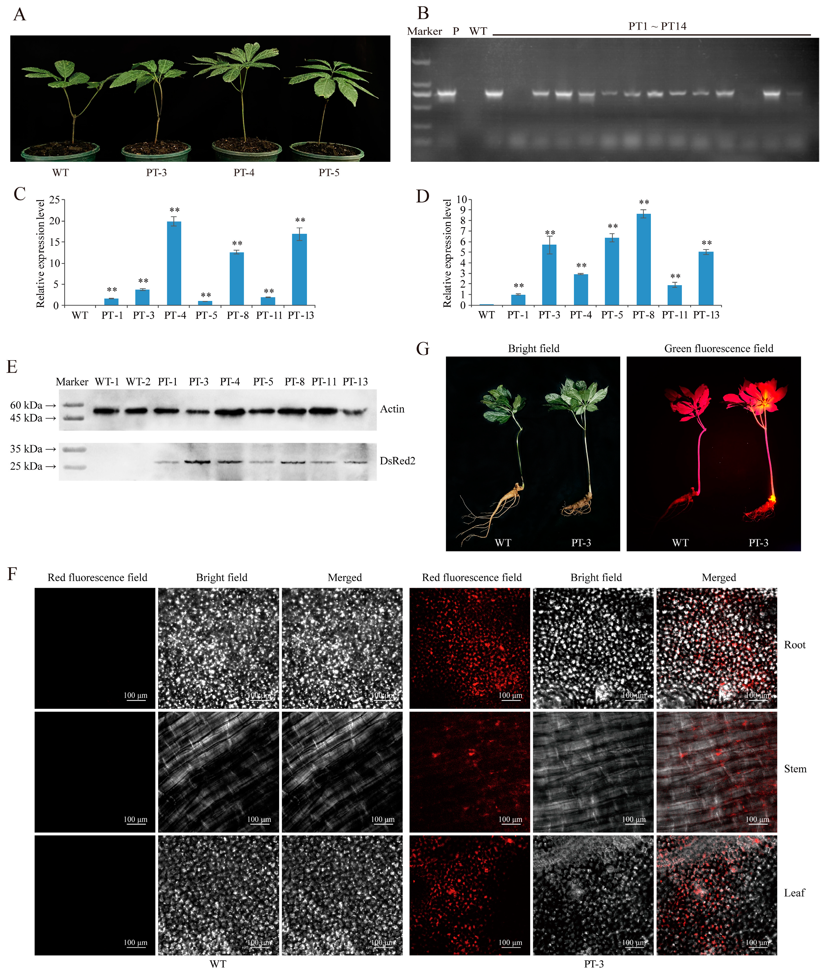
2.1. Establishment of Agrobacterium-Based Injection Rhizome Transformation Method in P. notoginseng

P. notoginseng is a well-known Chinese traditional herbal medicine plant, but its functional genomics and genetic breeding features, including the improvement of disease resistance and active compound biosynthesis, have been limited by the lack of a stable genetic transformation system. Notably, P. notoginseng is a perennial plant with meristems in the rhizome, where new plants are produced every spring. In this study, we developed a genetic transformation method for P. notoginseng by injecting A. tumefaciens LBA4404 containing DsRed2 into the rhizome (Figure 1). All the tested P. notoginseng plants sprouted on the rhizome after 30–40 days of injection, and the P. notoginseng plants showed a similar phenotype as the wild-type (WT) plants (Figure 2A).
The leaves from 71 plants derived from injected rhizomes were used to extract the genomic DNA, and the wild-type P. notoginseng was included as the negative control in the PCR analysis and subsequent experiments. Gel electrophoresis of the PCR products revealed that 61 plants contained the DsRed2 gene in their genomes (Figure 2B and Figure S1). The transformation rate was approximately 85.91%. Furthermore, qRT-PCR analysis confirmed the accumulation of a large number of DsRed2 transcripts in both the leaves and stems of seven positive transgenic P. notoginseng plants (Figure 2C,D), with PT-4 and PT-8 showing the highest expression levels in the stems and leaves, respectively.
Moreover, Western blot analysis further confirmed the expression of the red fluorescent protein in the leaves of all tested DsRed2 transgenic P. notoginseng plants, including PT-1/3/4/5/8/11/13 (Figure 2E). Additionally, red fluorescence was detected in the leaves, stems, and roots of the DsRed2 transgenic P. notoginseng plant (PT-3) using a laser scanning confocal microscope, but no specific red fluorescence was found in the WT plant, indicating that the red fluorescent protein expressed by DsRed2 in transgenic P. notoginseng possesses normal biological activity (Figure 2F). Furthermore, in the second year after injection, bright orange fluorescence was detected in the rhizome and leaves of DsRed2 transgenic P. notoginseng, but this specific fluorescence was not present in the WT plant (Figure 2G). These results demonstrate the successful integration of DsRed2 into the genome of P. notoginseng and the stable expression achieved through the injection of A. tumefaciens into the P. notoginseng rhizome. This method only takes 50–60 days from transgenic operation to the acquisition of positive transgenic plants, and its use means that complicated aseptic operations and tissue culture can be avoided.

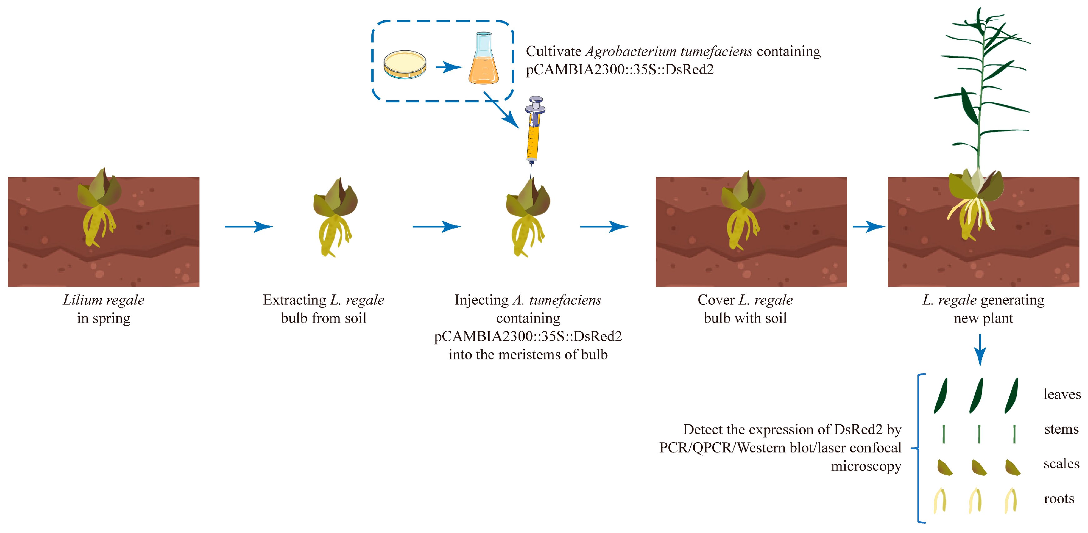
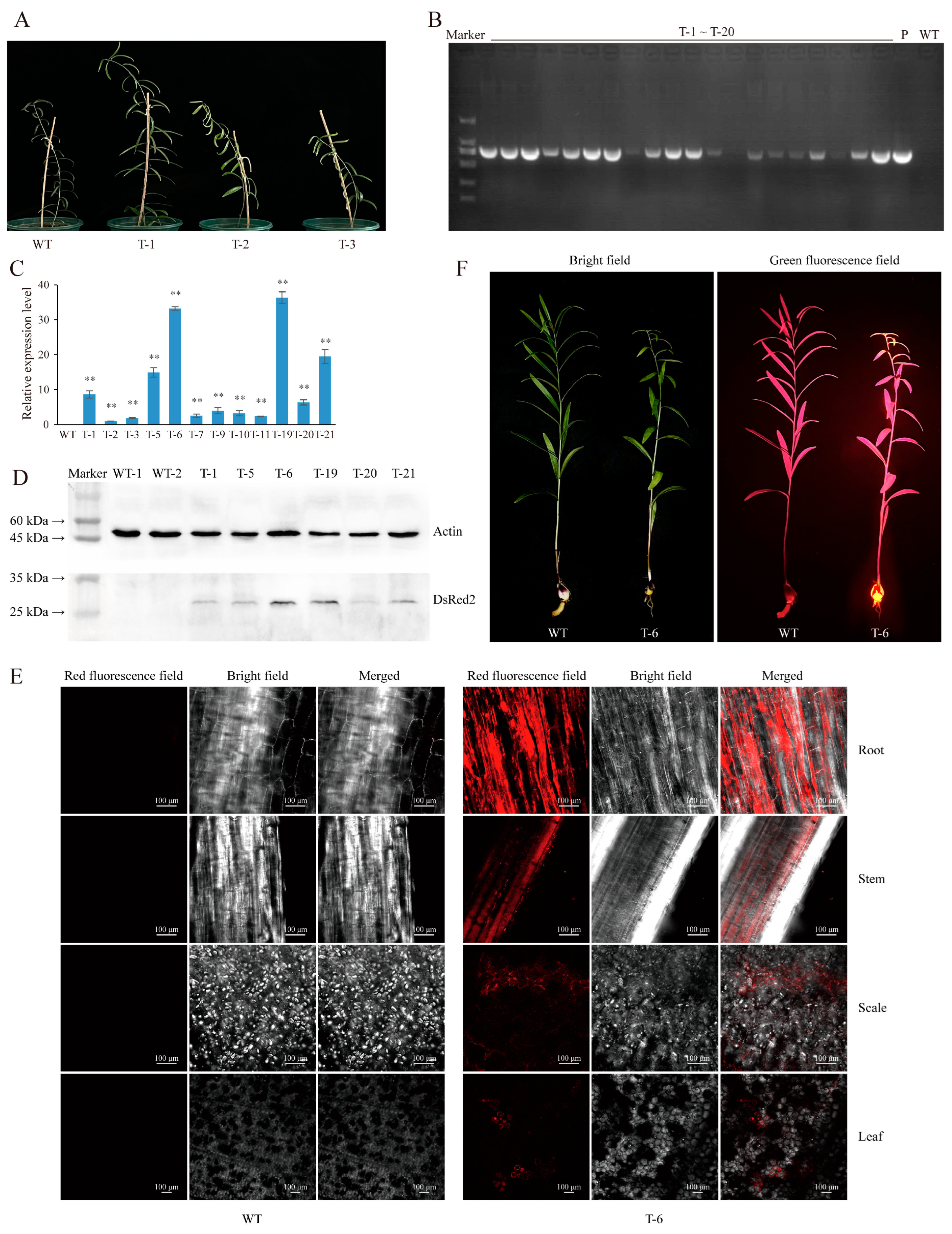
2.2. Agrobacterium-Based Injection in the Bulb Meristems Is a Fast and Efficient L. regale Genetic Transformation Method

Lilies are globally important fresh-cut flowers, but explorations of the molecular biology of lilies and breeding research have been hindered by the low efficiency of stable genetic transformation. Lily bulbs have a high reproduction ability due to the presence of the meristems in the innermost bulb layer. To explore a more efficient method for stable genetic transformation in lilies, in this study, we injected A. tumefaciens carrying DsRed2 into the bulb meristems of L. regale plants (Figure 3). All L. regale bulbs sprouted new plants from the meristems within 25–30 days after injection, and the L. regale plants from injected bulbs showed the same phenotype as the WT plant (Figure 4A). Genomic DNA was extracted from the leaves of L. regale plants, and PCR analysis indicated that 71 out of 82 tested plants showed the same specific DNA fragments as the positive control (plasmids of pCAMBIA2300::35S::DsRed2) (Figure 4B and Figure S2). The transformation rate was approximately 86.58%. Moreover, a high expression level of DsRed2 was detected in the leaves of positive transgenic L. regale plants, with plants T-5/6/19/21 showing higher expression levels compared to other plants (Figure 4C).
Additionally, Western blot analysis with the DsRed2 antibody revealed that the accumulation of the DsRed2 protein in the leaves of positive transgenic L. regale plants (T-6/19/21) was higher than that in the leaves of T-1/5/20. On the contrary, DsRed2 was not found in the leaves of the WT L. regale upon Western blot analysis (Figure 4D). Furthermore, observation with a laser scanning confocal microscope confirmed the presence of red fluorescence in the leaves, stems, roots, and scales of transgenic L. regale plants (T-6), while no specific red fluorescence was observed in the WT L. regale (Figure 4E). Using a handheld fluorescence detector, we found that bright orange fluorescence specifically accumulated in the bulb and shoots of DsRed2 transgenic L. regale plants in the second year after injection (Figure 4F). Generally, through using this method, positive transgenic L. regale can be obtained after 40–50 days. These results, obtained using a method involving Agrobacterium-based injection in the bulb meristems, which proved to be a rapid and efficient approach suitable for the genetic transformation of L. regale, demonstrate the successful integration and stable expression of DsRed2 in transgenic L. regale plants.

3. Discussion

Functional genomics and molecular breeding studies require efficient and rapid genetic transformation systems. However, traditional Agrobacterium-based transformation methods and gene gun techniques have some limitations [19]. The traditional Agrobacterium-based method is characterized by a long transformation cycle and low efficiency, and it is unsuitable for plants with limited regeneration capabilities [20]. On the other hand, the gene gun method relies on plants’ regenerative abilities and requires expensive equipment [19]. These methods are not ideal for plants like P. notoginseng, which have limited regeneration abilities [21]. Additionally, traditional genetic transformation methods require stringent sterile operations [22], which can significantly reduce plant survival, regeneration, and transformation rates, thereby limiting their application in plants like lilies [3].
For Panax spp., there is currently no research indicating that transgenic plants can be successfully developed through the traditional genetic transformation method. It is important to note that the transformation method established in this study showed a transformation efficiency of up to 85.7% in P. notoginseng, only taking 50–60 days to achieve this. For Lilium spp., it takes at least 90–120 days to obtain the conventional transgenic plants, and the transformation rate is on average 65.6% [13]. In this study, it only took 40–50 days from the injection of Agrobacterium to the acquisition of transgenic L. regale, and we avoided the complex steps of aseptic operation and tissue culture. At the same time, its efficiency is higher than that of the traditional Agrobacterium-based transformation method, reaching 86.58%, making it a more suitable transformation method in terms of applicability to Lilium spp.
Recently, Cao et al. [23] established a novel Agrobacterium-based transformation method named cut–dip–budding that does not need sterile operations and developed transgenic herbaceous and tuberous plants. Moreover, this method was also used to generate transgenic succulent plants [24]. The injection of A. tumefaciens into the meristems in stems successfully generated some stable transgenic plants, such as the sweet potato (Ipomoea batatas), potato (Solanum tuberosum), and bayhops (Ipomoea pes-caprae) [25]. The above two methods are practicable in the plants which have high redifferentiation capabilities, but the transformation methods for plants with low differentiation abilities need further exploration. In this study, we introduced a fast and efficient Agrobacterium transformation method based on the injection of A. tumefaciens LBA4404 containing DsRed2 into the meristems of the L. regale bulb and P. notoginseng rhizome. This method eliminates the need for sterile operations and meets the requirements for plants with low regeneration abilities. The successful expression of the reporter gene DsRed2 was demonstrated in multiple organs of the P. notoginseng and L. regale plants (Figure 2 and Figure 4). Protein expression of DsRed2 was confirmed via Western blot analysis, and the presence of red fluorescence in multiple organs of positive transgenic P. notoginseng and L. regale plants indicated normal activity of the red fluorescent protein (Figure 2E,F and Figure 4D,E).
Most importantly, in the second year after injection, specifically, bright orange fluorescence was observed in the transgenic P. notoginseng and L. regale plants (Figure 2G and Figure 4F), evidencing the stable expression of DsRed2. This study provides robust technical support for fundamental research on plants with low transformation rates and low regeneration abilities. In addition, as perennial plants, it takes 2–3 and 6–8 years for P. notoginseng and L. regale to generate seeds, respectively; therefore, the transgenic plants featured in this study are the chimera plants. Nonetheless, the genetic features of DsRed2 require further investigation in order to clearly understand the stability of the transgenic method established in this study.
Furthermore, the gene editing technique based on CRISPR/Cas has been extensively used for plant genome editing to improve important traits without the need for inserting exogenous genes [26]. However, CRISPR/Cas-mediated genome editing relies on a gene delivery system, such as the Agrobacterium-based transformation method [27]. Therefore, the development of a rapid and efficient transformation method will facilitate the application of CRISPR/Cas technology in a variety of plant species. There have been no reports on gene editing in P. notoginseng, while there are two studies reporting on the use of a CRISPR/Cas9-based system in relation to lilies, with these being published in 2019 and 2020, respectively [13,28]. However, the low transformation rate remains a serious issue affecting the efficiency of lily CRISPR/Cas9 technology. With its high efficiency, this Agrobacterium transformation method, based on meristem injection, may bring hope for the application of CRISPR/Cas technology in P. notoginseng and L. regale.

4. Materials and Methods

4.1. Plant Materials

After collection from three-year-old plants, the coat of P. notoginseng seeds were removed and sown in damp sands with 20–30% humidity before being placed in a refrigerator (8 °C) for low-temperature treatment. After 30 days, the seeds were sown in a shed with a sunshade net. In addition, the L. regale seeds were sown in nutrient soil (vermiculite/perlite/nutrient soil = 2:1:1) and cultivated in a greenhouse (25 °C, natural light). One-year-old P. notoginseng and L. regale plants were used for gene transformation.

4.2. A. tumefaciens Strain, Binary Vector, and Culture Preparation

pCAMBIA2300::35S::DsRed2 (Figure S3), composed of a reporter gene, DsRed2, driven by a 35S promoter, which was gifted to us from Prof. ShuangXia Jin of Huazhong Agricultural University (Wuhan, China), was transferred into A. tumefaciens LBA4404. Positive clones were streaked onto LB solid medium supplemented with 20 μg/mL rifampicin and 50 μg/mL kanamycin and then incubated at 28 °C for two days. The bacterial lawn was scraped using an inoculation ring and inoculated into MGL liquid medium (5.0 g/L peptone, 0.25 g/L KH2PO4, 5 g/L NaCl, 0.1 g/L MgSO4 7H2O, 1.0 g/L glycine, 5.0 g/L D-mannitol) containing 30 mg/mL acetosyringone. The bacterial solution was cultivated using a rotatory shaker at 200 rpm and 28 °C for four hours until the OD600 reached 0.8, which was used for transformation.

4.3. Plant Preparation and Infection

For perennial plants like L. regale and P. notoginseng, the aboveground parts will wither during winter. When the next spring arrives, the tender shoot (stem and leaves) will sprout again from the meristems of its rhizome or bulb and grow into a complete plant. At the same time, some new tuberous roots are generated from the P. notoginseng old taproot, and some new roots and scales also generate from the meristems in the L. regale bulb.
In spring, the soil above the P. notoginseng rhizome was gently removed before the germination of tender shoots. A. tumefaciens LBA4404 containing pCAMBIA2300::35S::DsRed2 was injected into the rhizome of P. notoginseng using a syringe at two positions with one injection per position (Figure S4). A total of 5 μL of bacterial solution was injected into each position (Figure 1). Then, the soil was moved so that it gently covered the rhizome. In total, 71 P. notoginseng rhizomes were injected with A. tumefaciens.
Similarly, one-year-old L. regale bulbs were collected from the soil before sprouting. A 5 μL bacterial solution was sucked with a syringe, and the needle was vertically inserted into the meristems of the L. regale bulb to inject the A. tumefaciens (Figure 3 and Figure S5). After this injection, each L. regale bulb was cultured at 28 °C in the dark for 2 days before being planted in the nutrient soil. In total, 82 L. regale bulbs were subjected to injection.

4.4. PCR Screening of Positive Transgenic P. notoginseng and L. regale Plants

New seedlings of P. notoginseng and L. regale (candidate transgenic plant) were produced after injection. One leaf of one candidate plant was collected for genomic DNA extraction. DsRed2-specific primers (F: 5′-ACAGAACTCGCCGTAAAGAC-3′; R: 5′-CCGTCCTCGAAGTTCATCAC-3′) were used for PCR, with the genomic DNA serving as the template. The PCR reaction mix consisted of 12.5 μL of 2× GS Taq PCR Mix, 0.2 μg genomic DNA, 0.1 μL forward primer (10 μM), 0.1 μL reverse primer (10 μM), and double-distilled water (total volume: 25 μL). The PCR conditions were as follows: 94 °C for 5 min; 94 °C for 30 s, 56 °C for 30 s, and 72 °C for 12 s for 32 cycles; and 72 °C for 10 min. The PCR products were analyzed using 1.2% agarose gel electrophoresis. Moreover, the PCR products from the genomic DNA of the wild-type P. notoginseng and L. regale leaves, as well as the pCAMBIA2300::35S::DsRed2 plasmids, were used as negative and positive controls, respectively.

4.5. qRT-PCR-Based Detection of DsRed2 Expression

Total RNA was extracted from the leaves and stems of positive transgenic P. notoginseng plants, as well as the leaves of transgenic L. regale, and then the RNA was reverse-transcribed into cDNA. qRT-PCR was performed as described by Su et al. [29] using DsRed2-specific primers (F: 5′-CCAGTTCCAGTACGGCTCCAA-3′; R: 5′-AGGAGTCCTGGGTCACGGTC-3′), and the relative expression level of DsRed2 was calculated using the −2ΔΔCt method [30].

4.6. Analysis of Red Fluorescent Protein Expression through Western Blot

Total protein was extracted from the leaves of transgenic P. notoginseng and L. regale using a plant total protein extraction kit (Sangon, Shanghai, China). The concentration of total protein was quantified at 10 μg/μL and subjected to sodium dodecyl sulfate polyacrylamide gel electrophoresis (SDS-PAGE) using a 5% concentrated gel and a 12% separating gel. Electrophoresis was performed at 80 V for 40 min, followed by 120 V for 80 min.
A polyvinylidene fluoride (PVDF) membrane (0.45 μm, Sigma-Aldrich, St. Louis, MO, USA) was used to transfer the proteins from the gel using a current of 400 mA in an ice bath for 60 min. After membrane transfer, the PVDF membrane was divided into upper and lower parts. The upper membrane, containing proteins with a molecular weight greater than 45 kDa, was incubated with the primary antibody (Affinity, Shanghai, China) against the actin protein, serving as an internal reference protein. The lower half of the membrane, containing proteins with a molecular weight less than 45 kDa, was incubated with the primary antibody (Affinity, China) against the red fluorescent protein encoded by DsRed2. Both membranes were incubated in a rotary shaker at 80 rpm and 28 °C for 2 h, followed by two washes with 1X TBST (Tris-HCl with Tween-20) for 10 min each. Subsequently, the membranes were incubated with the secondary antibody (anti-mouse IgG H&L/HRP antibody, Affinity, China) in a rotary shaker at 80 rpm and 28 °C for 1 h. After incubation, the membranes were washed twice with 1X TBST for 10 min each. Finally, superKine™ ultra-sensitive ECL luminescent liquid (Abbkine Scientific, Wuhan, China) was used for visualization, and exposure was performed using a transilluminator (Bio-Rad, Hercules, CA, USA).

4.7. Observation of Specific Red Fluorescence in Different Organs of DsRed2 Transgenic Plants Using Laser Confocal Microscopy

Three transgenic plants with a high expression of DsRed2 were selected for fluorescence observation. To confirm the biological activity of the red fluorescent protein expressed in the transgenic plants, the leaves, stems, and newly grown tuberous roots of DsRed2 transgenic P. notoginseng plants were collected. Subsequently, they were sliced using a surgical knife. The samples were placed on a glass slide and covered with a cover slip before being observed using a laser confocal microscope (Nikon, Tokyo, Japan) at an excitation wavelength of 560 nm and emission wavelength of 575 nm [17]. Similarly, the leaves, stems, newly grown roots and scales of DsRed2 transgenic L. regale plants were harvested for fluorescence observation using the same method.

4.8. Observing Red Fluorescence in Transgenic Plants Using a Handheld Fluorescence Detector

In the second year after injection, wild-type plants, as well as the transgenic plants without additional injection of A. tumefaciens LBA4404 that grew normally and had a high expression of DsRed2, were collected to observe the bright orange fluorescence derived from DsRed2 expression using handheld fluorescence observation equipment (LUYOR-3415RG, Shanghai, China), with a green light indicating excitation [18]. Fluorescence observations were recorded with a camera (Canon, Tokyo, Japan) with a red filter lens.

5. Conclusions

The tissue-culture-independent genetic transformation method presented in this study is a fast and efficient transformation method for P. notoginseng and L. regale. The reporter gene DsRed2 was successfully integrated into the genome of P. notoginseng and L. regale and was normally expressed in multiple organs. The red fluorescence of DsRed2 was detected in transgenic P. notoginseng and L. regale. Most importantly, the DsRed2 transgenic P. notoginseng and L. regale plants still maintained red fluorescence activity in the second year after injection. This transformation method is suitable for plants with a low regeneration ability or those with a low transformation rate, such as P. notoginseng and L. regale.

Supplementary Materials

The following supporting information can be downloaded at: https://www.mdpi.com/article/10.3390/plants13172509/s1, Figure S1: Gel electrophoresis results of DsRed2 amplification from genomic DNA of injected P. notoginseng leaves. Figure S2: Gel electrophoresis showing DsRed2 amplification from injected L. regale leaves. Figure S3: The schematic representation of pCAMBIA2300::35S::DsRed2 vector. Figure S4: The injection position diagram of the P. notoginseng rhizome. Figure S5: The injection position diagram of the L. regale bulb.

Author Contributions

Conceptualization, J.D.; methodology, J.D. and X.L.; formal analysis, X.L. and W.L.; investigation, J.D.; data curation, J.D. and W.L.; writing—original draft preparation, J.D.; writing—review and editing, D.L. and G.L.; visualization, J.D.; supervision, D.L. and G.L.; funding acquisition, D.L. and G.L. All authors have read and agreed to the published version of the manuscript.

Funding

This research was funded by the National Natural Science Foundation of China, grant number 82060693 and Yunnan Seed Laboratory, grant number 202205AR070001-13.

Data Availability Statement

Data are contained within the article and Supplementary Materials.

Acknowledgments

We would like to express our gratitude to Shuangxia Jin of Huazhong Agricultural University for the gift of the pCAMBIA2300::35S::DsRed2 vector.

Conflicts of Interest

The authors declare no conflicts of interest.

References

  1. Krenek, P.; Samajova, O.; Luptovciak, I.; Doskocilova, A.; Komis, G.; Samaj, J. Transient plant transformation mediated by Agrobacterium tumefaciens: Principles, methods and applications. Biotechnol. Adv. 2015, 33, 1024–1042. [Google Scholar] [CrossRef]
  2. Azizi-Dargahlou, S.; Pouresmaeil, M. Agrobacterium tumefaciens-mediated plant transformation: A review. Mol. Biotechnol. 2024, 66, 1563–1580. [Google Scholar] [CrossRef] [PubMed]
  3. Shrawat, A.K.; Lörz, H. Agrobacterium-mediated transformation of cereals: A promising approach crossing barriers. Plant Biotechnol. J. 2006, 4, 575–603. [Google Scholar] [CrossRef]
  4. Rizwan, H.M.; Yang, Q.; Yousef, A.F.; Zhang, X.; Sharif, Y.; Kaijie, J.; Shi, M.; Li, H.; Munir, N.; Yang, X.; et al. Establishment of a novel and efficient Agrobacterium-Mediated in planta transformation system for passion fruit (Passiflora edulis). Plants 2021, 10, 2459. [Google Scholar] [CrossRef] [PubMed]
  5. Wang, Y.R.; Sun, X.; Xie, Y.M.; Du, A.; Chen, M.; Lai, S.S.; Wei, X.H.; Ji, L.L.; Wang, C.H. Panax notoginseng saponins alleviate diabetic retinopathy by inhibiting retinal inflammation: Association with the NF-κB signaling pathway. J. Ethnopharmacol. 2024, 319, 117135. [Google Scholar] [CrossRef] [PubMed]
  6. Kim, Y.J.; Zhang, D.; Yang, D.C. Biosynthesis and biotechnological production of ginsenosides. Biotechnol. Adv. 2015, 33, 717–735. [Google Scholar] [CrossRef] [PubMed]
  7. Choi, H.S.; Koo, H.B.; Jeon, S.W.; Han, J.Y.; Kim, J.S.; Jun, K.M.; Choi, Y.E. Modification of ginsenoside saponin composition via the CRISPR/Cas9-mediated knockout of protopanaxadiol 6-hydroxylase gene in Panax ginseng. J. Ginseng Res. 2022, 46, 505–514. [Google Scholar] [CrossRef]
  8. Liu, S.Z.; Chen, X.X.; Zhao, T.Q.; Yu, J.H.; Chen, P.; Wang, Y.F.; Wang, K.Y.; Zhao, M.Z.; Jiang, Y.; Wang, Y.; et al. Identification of PgRg1-3 gene for ginsenoside Rg1 biosynthesis as revealed by combining genome-wide association study and gene co-expression network analysis of jilin ginseng core collection. Plants 2024, 13, 1784. [Google Scholar] [CrossRef] [PubMed]
  9. Jiang, Y.; Zhang, Q.; Zeng, Z.X.; Wang, Y.; Zhao, M.Z.; Wang, K.G.; Zhang, M.P. The AP2/ERF transcription factor PgERF120 regulates ginsenoside biosynthesis in ginseng. Biomolecules 2024, 14, 345. [Google Scholar] [CrossRef]
  10. Zhang, X.; Yu, Y.L.; Jiang, S.; Yu, H.; Xiang, Y.Y.; Liu, D.Q.; Qu, Y.; Cui, X.M.; Ge, F. Oleanane-type saponins biosynthesis in Panax notoginseng via transformation of β-Amyrin synthase gene from Panax japonicus. J. Agric. Food Chem. 2019, 67, 1982–1989. [Google Scholar] [CrossRef]
  11. Shi, Y.; Man, J.H.; Huang, Y.Y.; Zhang, J.H.; Zhang, Z.F.; Yin, G.Y.; Wang, X.; Liu, S.H.; Chen, Y.; Wang, X.H.; et al. Overexpression of PnMYB2 from Panax notoginseng induces cellulose and lignin biosynthesis during cell wall formation. Planta 2022, 255, 107. [Google Scholar] [CrossRef]
  12. Sui, J.J.; Jia, W.J.; Xin, Y.; Zhang, Y.Y. Transcriptomics-based identification of genes related to tapetum degradation and microspore development in lily. Genes 2022, 13, 366. [Google Scholar] [CrossRef] [PubMed]
  13. Yan, R.; Wang, Z.P.; Ren, Y.M.; Li, H.Y.; Liu, N.; Sun, H.M. Establishment of efficient genetic transformation systems and application of CRISPR/Cas9 genome editing technology in Lilium pumilum DC. Fisch. and Lilium longiflorum White Heaven. Int. J. Mol. Sci. 2019, 20, 2920. [Google Scholar] [CrossRef] [PubMed]
  14. Wei, C.; Cui, Q.; Yuan, L.; Jia, G.X. Efficient Agrobacterium tumefaciens (Smith & Towns.) conn-mediated transformation of Lilium ‘Sorbonne’ with genes encoding anthocyanin regulators. Can. J. Plant Sci. 2017, 97, 796–807. [Google Scholar]
  15. Fu, Y.Y.; Shu, L.L.; Li, H.Y.; Zhang, X.M.; Liu, X.; Ou, Z.Y.; Liang, X.M.; Qi, X.Y.; Yang, L.P. Establishment of highly efficient plant regeneration, callus transformation and analysis of Botrytis cinerea-responsive PR promoters in Lilium brownii var. viridulum. Plants 2023, 12, 1992. [Google Scholar] [CrossRef] [PubMed]
  16. Zhang, M.F.; Ma, X.; Jin, G.; Han, D.Y.; Xue, J.; Du, Y.P.; Chen, X.Q.; Yang, F.P.; Zhao, C.N.; Zhang, X.H. A modified method for transient transformation via pollen magnetofection in Lilium germplasm. Int. J. Mol. Sci. 2023, 24, 15304. [Google Scholar] [CrossRef]
  17. Sun, L.; Alariqi, M.; Zhu, Y.; Li, J.Y.; Li, Z.L.; Wang, Q.; Li, Y.J.; Rui, H.P.; Zhang, X.L.; Jin, S.X. Red fluorescent protein (DsRed2), an ideal reporter for cotton genetic transformation and molecular breeding. Crop J. 2018, 6, 366–376. [Google Scholar] [CrossRef]
  18. Huai, D.X.; Wu, J.; Xue, X.M.; Hu, M.L.; Zhi, C.Y.; Pandey, M.K.; Liu, N.; Huang, L.; Bai, D.M.; Yan, L.Y.; et al. Red fluorescence protein (DsRed2) promotes the screening efficiency in peanut genetic transformation. Front. Plant Sci. 2023, 14, 1123644. [Google Scholar] [CrossRef] [PubMed]
  19. Nandy, D.; Maity, A.; Mitra, A.K. Target-specific gene delivery in plant systems and their expression: Insights into recent developments. J. Biosci. 2020, 45, 30. [Google Scholar] [CrossRef]
  20. Filipecki, M.; Malepszy, S. Unintended consequences of plant transformation: A molecular insight. J. Appl. Genet. 2006, 47, 277–286. [Google Scholar] [CrossRef]
  21. Dong, O.X.; Ronald, P.C. Targeted DNA insertion in plants. Proc. Natl. Acad. Sci. USA 2021, 118, e2004834117. [Google Scholar] [CrossRef]
  22. Su, W.B.; Xu, M.Y.; Radani, Y.; Yang, L.M. Technological development and application of plant genetic transformation. Int. J. Mol. Sci. 2023, 24, 10646. [Google Scholar] [CrossRef] [PubMed]
  23. Cao, X.S.; Xie, H.T.; Song, M.L.; Lu, J.H.; Ma, P.; Huang, B.Y.; Wang, M.G.; Tian, Y.F.; Chen, F.; Peng, J.; et al. Cut-dip-budding delivery system enables genetic modifications in plants without tissue culture. Innovation 2022, 4, 100345. [Google Scholar] [CrossRef] [PubMed]
  24. Lu, J.H.; Li, S.S.; Deng, S.A.; Wang, M.G.; Wu, Y.H.; Li, M.; Dong, J.S.; Lu, S.H.; Su, C.L.; Li, G.F.; et al. A method of genetic transformation and gene editing of succulents without tissue culture. Plant Biotechnol. J. 2024, 22, 1981–1988. [Google Scholar] [CrossRef]
  25. Mei, G.G.; Chen, A.; Wang, Y.R.; Li, S.Q.; Wu, M.Y.; Hu, Y.L.; Liu, X.; Hou, X.L. A simple and efficient in planta transformation method based on the active regeneration capacity of plants. Plant Commun. 2024, 5, 100822. [Google Scholar] [CrossRef]
  26. Shah, T.; Andleeb, T.; Lateef, S.; Noor, M.A. Genome editing in plants: Advancing crop transformation and overview of tools. Plant Physiol. Biochem. 2018, 131, 12–21. [Google Scholar] [CrossRef] [PubMed]
  27. Zhang, D.Q.; Zhang, Z.Y.; Unver, T.; Zhang, B.H. CRISPR/Cas: A powerful tool for gene function study and crop improvement. J. Adv. Res. 2020, 29, 207–221. [Google Scholar] [CrossRef]
  28. Song, S.L.; Yan, R.; Wang, C.X.; Wang, J.X.; Sun, H.M. Improvement of a genetic transformation system and preliminary study on the function of LpABCB21 and LpPILS7 based on somatic embryogenesis in Lilium pumilum DC. Fisch. Int. J. Mol. Sci. 2020, 21, 6784. [Google Scholar] [CrossRef]
  29. Su, L.L.; Li, W.Y.; Chen, X.H.; Wang, P.C.; Liu, D.Q. Proline-rich protein PRPL1 enhances Panax notoginseng defence against Fusarium solani by regulating reactive oxygen species balance and strengthening the cell wall barrier. Plant Cell Environ. 2024, 47, 2377–2395. [Google Scholar] [CrossRef]
  30. Rao, X.Y.; Huang, X.L.; Zhou, Z.C.; Lin, X. An improvement of the 2^(-delta delta CT) method for quantitative real-time polymerase chain reaction data analysis. Biostat. Bioinform. Biomath. 2013, 3, 71–85. [Google Scholar]
Figure 1. Schematic diagram illustrating Agrobacterium tumefaciens injection into the rhizome of Panax notoginseng.
Plants 13 02509 g001
Figure 2. A. tumefaciens injection into the rhizome of P. notoginseng is a stable and efficient transformation method. (A) Comparison of phenotype between the injected P. notoginseng plants (PT-3/4/5) and wild-type (WT) plant. (B) Gel electrophoresis showing DsRed2 (Discosoma striata red fluorescence protein 2) amplification from genomic DNA of injected P. notoginseng leaves. P, WT, and PT1-PT14 represent PCR reaction of wild-type P. notoginseng, pCAMBIA2300S-DsRed2 plasmids, and DsRed2 transgenic P. notoginseng, respectively. (C) Expression of DsRed2 in leaves of transgenic P. notoginseng as determined by qPCR. WT and PT-1/3/4/5/8/11/13 represent the qPCR reaction with the leaves of wild-type and DsRed2 transgenic P. notoginseng plants, respectively. (D) Expression of DsRed2 in the stems of transgenic P. notoginseng as determined by qPCR. WT and PT-1/3/4/5/8/11/13 represent the qPCR reaction with the stems of wild-type and DsRed2 transgenic P. notoginseng plants, respectively. (E) Detection of DsRed2 protein expression in leaves of transgenic P. notoginseng through Western blot. WT-1/2 and PT-1/3/4/5/8/11/13 represent the leaf total protein of wild-type and DsRed2 transgenic P. notoginseng plants, respectively. (F) Observation of specific red fluorescence in leaves, stems, and roots of transgenic P. notoginseng under a laser scanning confocal microscope. WT and PT-3 represent the wild-type and DsRed2 transgenic P. notoginseng plants, respectively. (G) Observation of specific fluorescence in transgenic P. notoginseng with a handle fluorescence detector. WT and PT-3 represent the wild-type and DsRed2 transgenic P. notoginseng plants in the second year after injection, respectively. T-test was used to reveal the statistical difference of DsRed2 expression level between wild type and transgenic plants (** p < 0.01).
Plants 13 02509 g002
Figure 3. Schematic diagram illustrating A. tumefaciens injection into the meristems of Lilium regale bulb.
Plants 13 02509 g003
Figure 4. A. tumefaciens injection into the meristems of the L. regale bulb is a fast and efficient transformation method. (A) Comparison of phenotype between the injected L. regale plants (T-1/2/3) and wild-type (WT) plant. (B) Gel electrophoresis showing DsRed2 amplification from injected L. regale leaves. T-1~T-20, P, and WT represent PCR reaction of DsRed2 transgenic L. regale, pCAMBIA2300S-DsRed2 plasmids, and wild-type, respectively. (C) Expression of DsRed2 in leaves of transgenic L. regale as determined by qPCR. WT and T-1/2/3/5/6/7/9/10/11/19/20/21 represent qPCR reaction of the wild-type and DsRed2 transgenic L. regale plants. (D) Detection of DsRed2 protein expression in leaves of transgenic L. regale through Western blot. WT and T-1/5/6/19/20/21 represent the leaf total protein of wild-type and DsRed2 transgenic L. regale plants. (E) Observation of specific red fluorescence in leaves, stems, roots, and scales of transgenic L. regale under a laser scanning confocal microscope. WT and T-6 represent the wild-type and DsRed2 transgenic L. regale plants, respectively. (F) Observation of specific fluorescence in transgenic L. regale with a handle fluorescence detector. WT and T-6 represent the wild-type and DsRed2 transgenic L. regale in the second year after injection, respectively. T-test was used to reveal the statistical difference of DsRed2 expression level between wild type and transgenic plants (** p < 0.01).
Plants 13 02509 g004
Disclaimer/Publisher’s Note: The statements, opinions and data contained in all publications are solely those of the individual author(s) and contributor(s) and not of MDPI and/or the editor(s). MDPI and/or the editor(s) disclaim responsibility for any injury to people or property resulting from any ideas, methods, instructions or products referred to in the content.

