Article

Genome-Wide Identification and Characterization of the Salvia miltiorrhiza Histone Deacetylase (HDAC) Family in Response to Multiple Abiotic Stresses

1
School of Pharmaceutical Sciences, Academy of Chinese Medical Sciences, Zhejiang Chinese Medical University, Hangzhou 310053, China
2
College of Food and Health, Zhejiang A & F University, Hangzhou 311300, China
*
Authors to whom correspondence should be addressed.
These authors contributed equally to this work.
Plants 2024, 13(5), 580; https://doi.org/10.3390/plants13050580
Submission received: 8 January 2024 / Revised: 18 February 2024 / Accepted: 19 February 2024 / Published: 21 February 2024

Abstract

:
Salvia miltiorrhiza is a plant commonly used in traditional Chinese medicine. Its material bases for treating diseases are tanshinones and phenolic acids, including salvianolic acids. Histone deacetylase proteins (HDACs) are a class of specific functional enzymes that interact with acetylation groups on the N-terminal lysine of histone proteins further regulate gene transcription through structural changes at the chromatin level. HDACs involved in the growth and development of various plants, and induced by plant hormones to regulate the internal environment of plants to resist stress, at the same time affect the accumulation of some secondary metabolites. However, the role of SmHDACs on the accumulation of salvianolic acid in S. miltiorrhiza remains unclear. In this study, 16 SmHDACs genes were identified from the high-quality S. miltiorrhiza genome, their physicochemical properties were predicted. In phylogenetic trees co-constructed with HDACs proteins from other plants, SmHDACs was divided into three subfamilies, each with similar motif and conserved domain composition. The distribution of the three subfamilies is similar to that of dicotyledonous plants. Chromosome localization analysis showed that SmHDACs genes were randomly located. Cis-acting element analysis predicted that SmHDACs gene expression may be related to and induced by various phytohormones, such as MeJA and ABA. By combining the expression pattern and co-expression network induced by phytohormones, we speculate that SmHDACs may further influence the synthesis of salvianolic acid, and identified SmHDA5, a potential functional gene, then speculate its downstream target based on the co-expression network. In summary, we analyzed the SmHDACs gene family of S. miltiorrhiza and screened out the potential functional gene SmHDA5. From the perspective of epigenetics, we proposed the molecular mechanism of plant hormone promoting salvianolic acid synthesis, which filled the gap in the subdivision of histone deacetylase in S. miltiorrhiza research, provided a theoretical basis for the culture and transformation of S. miltiorrhiza germplasm resources.

1. Introduction

Salvia miltiorrhiza is a Chinese traditional medicinal plant that has been used for thousands of years. It is rich in water-soluble component salvianolic acids and lipid-soluble component tanshinones [1,2], which is used to combat the increasing incidence of cardiovascular disease [3,4]. However, the wild resources of S. miltiorrhiza are decreasing continuously, which cannot meet the market demand. Therefore, it is urgent to innovate the cultivation technology of S. miltiorrhiza, especially to improve the content of medicinal components by metabolic engineering [5]. Salvianolic acid synthetic pathway has been improved with the efforts of researchers, simply put, the phenylpropanoid and tyrosine-derived pathways work simultaneously to generate two critical precursors, 4-coumaroyl-CoA and 3, 4-dihydroxyphenyllactic acid, consequently under the catalysis of rosmarinic acid synthetase (RAS) and cytochrome P450-dependent monooxygenase (CYP98A14) to product rosmarinic acid, but the subsequent synthesis of Salvianolic acid B is uncertain [6,7,8].
Based on the above synthetic pathways, the regulation mechanism of salvianolic acid synthesis has been studied extensively. Various signaling pathways specifically activate certain transcription factors for activating target genes to promote salvianolic acid synthesis, and this kind of metabolic mechanism has been extensively studied. For example, JA signaling pathway induced by jasmonic acid (JA) has been proved to significantly increase the content of salvianolic acid [9]. In-depth study on JA pathway found that some transcription factors activated by JA pathway can enhance the transcription of salvianolic acid key enzyme genes, such as basic helix-loop-helix (bHLH), R2R3-MYB transcription factor MYB76, MYB1, MYB2 [2,10,11]; APETALA2/ERF (AP2/ERF) transcription factor ERF115 [12]. However, in addition to transcription factors, there are many other factors affecting gene expression such as post-translational modifications (PTMs). These modifications occur after transcription and work through structural changes at the chromatin level, known as epigenetics.
DNA wraps around octamer protein core of histones (two copies of H2A, H2B, H3, H4) to form a nucleosome [13]. N-terminal tails of histone proteins protrude from nucleosome, where PTMs events occurred, such as acetylation, methylation and phosphorylation [14]. Among them, acetylation of histone lysine affects the structure of the nucleosome, thus affect the expression of the gene [15]. The level of histone acetylation is a dynamic equilibrium process, which is dominated by histone acetyltransferase and histone deacetylases (HDACs) [16], when the balance upset, histone acetylation levels rise/fall, generally means that gene transcription is activated/suppressed [17]. In plants, HDACs are divided into three subfamilies, Reduced Potassium Dependency 3/histone deacetylase 1 (RPD3/HDA1), Silent Information Regulator 2 (SIR2) and the plant-specific histone deacetylase 2 (HD2) [18]. The effects of histone acetylation on plants are comprehensive, including the regulation of plant growth and development, as well as various secondary metabolism. For example, in Arabidopsis thaliana, PIF1/PIF3 interact with HDA19 and MED25 to mediate transcriptional repression in the phytochrome signaling pathway [19]; HDA5 is involved in the control of cytosolic metabolism and cell function [20]. Moreover, HDACs showed sensitivity to various phytohormones in recent reports, OsHDA710 in rice is sensitive to ABA and JA, which regulates the expression of salt-tolerant genes [21]; AtHDA6 in Arabidopsis thaliana direct deacetylation of TOPLESS protein (JA pathway suppressor), under JA induction, AtHDA6 expression increases and breaks homeostasis, the inhibitory effect of TOPLESS is weakened thus activating JA pathway [22]. It is known that increased expression of DNA methyltransferase related genes contributes to increased DNA methylation levels during root growth of S. miltiorrhiza and changes the accumulation of secondary metabolites [23]. However, the role of histone deacetylation and expression of HDACs in salvianolic acid accumulation in S. miltiorrhiza are still unclear.
In this study, we identified SmHDACs (Salvia miltiorrhiza Histone Deacetylase) genes from high-quality genomes for bioinformatics analysis. Muti-transcriptome data were used to analyze the expression profiles of SmHDACs under the influence of hormones, further potential functional genes were screened. Our results confirmed that the expression of SmHDACs gene changes under various stresses, indicating that histone deacetylation plays an important role in S. miltiorrhiza under stress. The potential role of SmHDACs in the synthesis of salvianolic acid were further discussed. These conclusions are of great significance for further research on the epigenetics of S. miltiorrhiza.

2. Results

2.1. Identification of SmHDACs

Through HMMER server, there are 12 RPD3/HDA1 subfamily and 2 SIR2 subfamily SmHDACs, respectively. By BLAST of AtHDTs, two SmHDTs are found, both of which contain NPL conserved domains verified by HMMER server. All the above 16 SmHDACs passed the conserved domain integrity verification, and named SmHDA1-12, SmSRT1-2, and SmHDT1-2 according to subfamily affiliation, respectively, as shown in Table 1. Their protein size ranges from 284 to 776 (aa), molecular weight from 30.699 to 85.280 (kDa), isoelectric point from 5.01 to 9.40. Other physical and chemical properties are shown in Table 1. Subcellular localization prediction found that most SmHDAs were predicted in the nucleus and cytoplasm, while all SmSRTs were predicted in chloroplasts, may be able to act on proteins that respond to light. Only SmHDT2 is predicted to be located in the vacuole (Table 1).

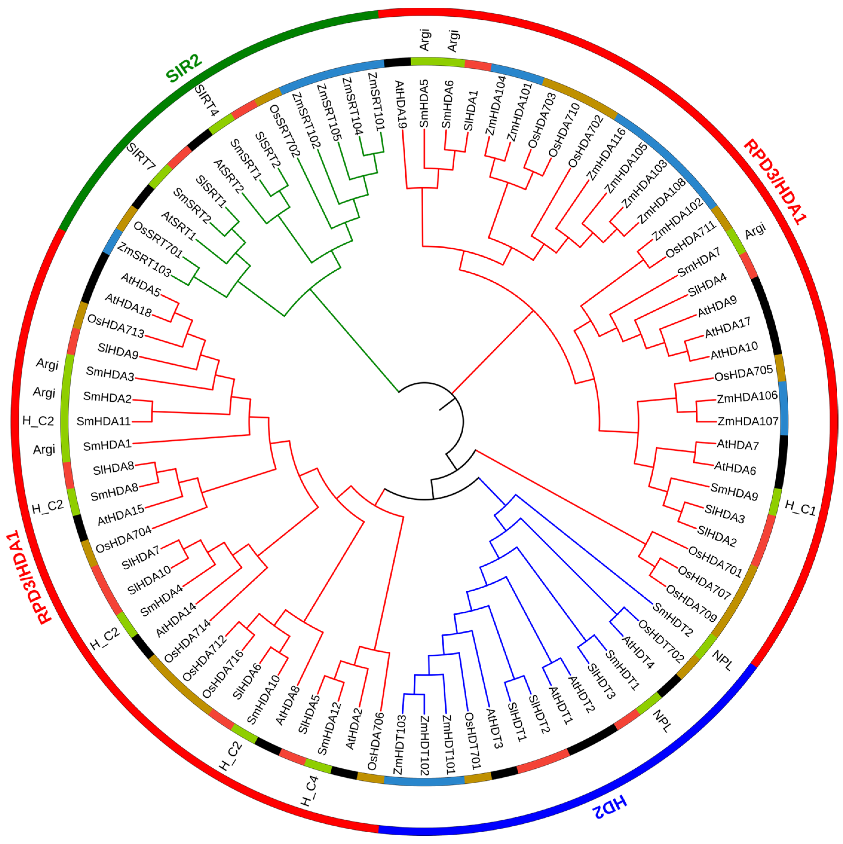
2.2. Phylogenetic Tree of SmHDACs

SmHDACs and HDACs from four species (Arabidopsis thaliana, Oryza sativa, Zea mays, Solanum lycopersicum), were used to build phylogenetic tree, the conservative domain information of SmHDACs is also added, as shown in Figure 1. The grouping of all proteins was exactly as expected, and the three subfamilies were completely separated. The largest subfamily of SmHDACs is RPD3/HDA1 subfamily, with a total of 12. The number of SIR2 and HD2 subfamilies is equal, both with 2. The conserved domains of each subfamily also conform to the initial description, that is, the SIRT4 and SIRT7 domain is owned specifically by the SIR2 subfamily, the NPL domain is owned specifically by the HD2 subfamily, and the HDA subfamily contains multiple HDAC domains, such as Arginase_HDAC superfamily (Accession = cl17011), HDAC_classII (Accession = cd09992) et al., and the aggregation of SmHDACs is independent of the conserved domain. Different from other species, there are more HDACs in the HD2 and SIR2 subfamilies in Z. maize, which shows that monocotyledonous plants and dicotyledonous plants have different evolutionary trends of HDACs.

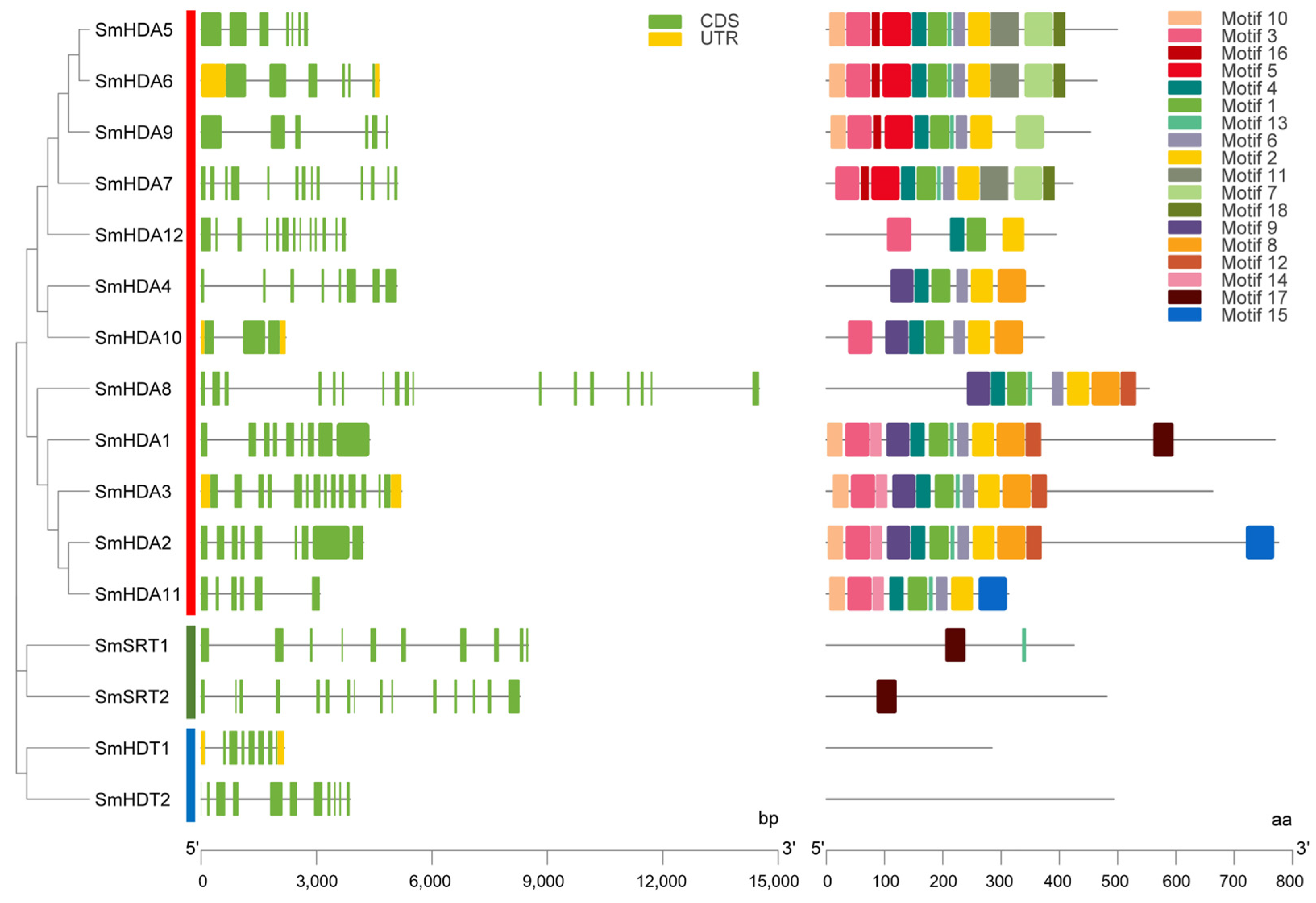
2.3. Gene Structure and Motif Analysis of SmHDACs

The gene structure and motif information of SmHDACs are shown in Figure 2. There were no significant association between gene structure and SmHDACs grouping. A total of 17 motifs have been excavated, named Motif 1-17. Their distribution is obviously related to grouping. SmHDACs include all motif 1-17, clustered SmHDACs have similar motifs (Specific motif sequence information is shown in Figure S1). Interestingly, the motif analysis results of SmHDA5 and SmHDA6 are consistent, they are also grouped into clusters in phylogenetic tree, indicating that their conserved domains are similar, which indicated they may further lead to functional similarities. A similar situation also occurs for SmHDA1-3, and the results showed that they differ only in the terminal motifs. On the other hand, SmHDTs does not contain any motifs, a similar pattern has been observed in Dendrobium officinale SRTs [24].

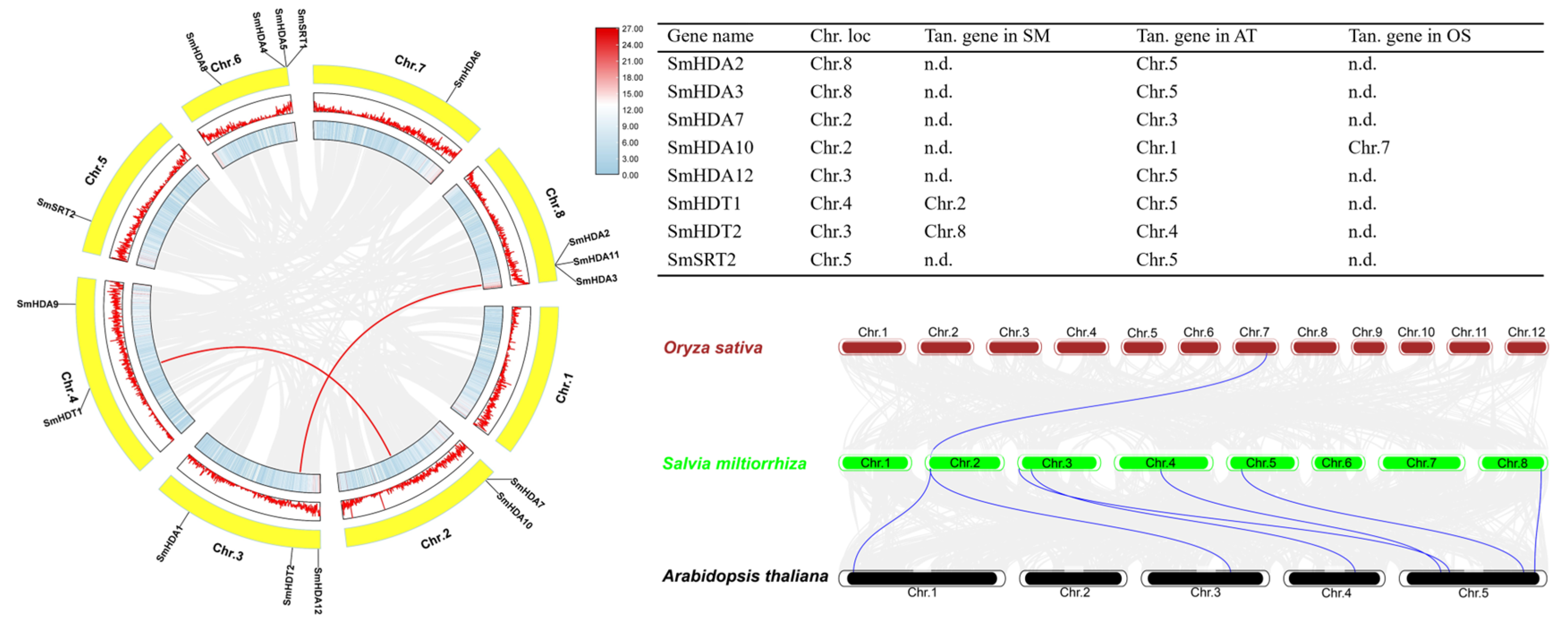
2.4. Chromosome Localization and Collinearity Analysis of SmHDACs

The distribution of 16 SmHDACs on the chromosome map is shown in Figure 3. Meanwhile, we also labeled the density of chromosomes and the collinearity analysis among SmHDACs. In order of number, chromosome (Chr.) 6, 8, 3, 4, 2, 5, 7 distributed 4, 3, 3, 2, 2, 1, 1 gene, respectively, while SmHDACs were not distributed on Chr.1. SmHDA4, 5, and SmSRT1 are clustered on Chr.6, and SmHDA2, 3, 11 are clustered on Chr.8. Most SmHDACs are found in areas with high gene density, and histone deacetylated proteins may play a role in many gene clusters. SmHDT1 and SmHDA2 have collinear genes on Chr.2 and Chr.3, respectively, suggests that these genes may have duplicated along the way to take on important functions. Collinearity analysis with other species showed that SmHDACs had collinear gene with A. thaliana, but there were few genes in rice genome, indicating that S. miltiorrhiza and A. thaliana were closely related.

2.5. Cis-Acting Elements Analysis of SmHDACs

The upstream 2000 bp of a gene is regarded as a promoter region, it was extracted from the genome for cis-acting element (CAEs) analysis. We visualized the distribution of CAEs of 16 SmHDACs on promoters and categorized them as growth and development, phytohormone responsiveness, and stress responsiveness, as shown in Figure 4. A total of 27 CAEs were randomly distributed on the promoters of all SmHDACs, involving many events such as plant growth and development, phytohormone responsiveness and so on (Figure 4A). Further study found that the number of CAEs related to phytohormone response was the highest, while the number related to growth and development was the lowest (Figure 4B). G-Box, CGTCA-motifs and TGACG-motifs related to MeJA response accounted for 37.3% of all CAEs, indicating that SmHDACs may be strongly associated with phytohormone MeJA. On the other hand, a total of 46 ABRE were found, suggesting that SmHDACs may play a role in responding to ABA. In addition, SmHDACs may respond to gibberellin (P-box, TATC-box, GARE-motif), salicylic acid (TCA-element), and auxin (TGA-element). SmHDACs may also play a role in the following stresses: low temperature (LTR), hypoxia (ARE), light (GT1-motif, G-box, sp1, MRE), drought (MBS), and mechanical injury (WUN-motif), Low temperature and hypoxia are the most likely stressors to induce SmHDACs expression. Under these two conditions, SmHDACs protein may enhance its histone deacetylation function. SmHDA5, 6, 9 and SmSRT2 have a large number of CAES, which may be active in plant metabolic regulation species, and may be potential functional genes.

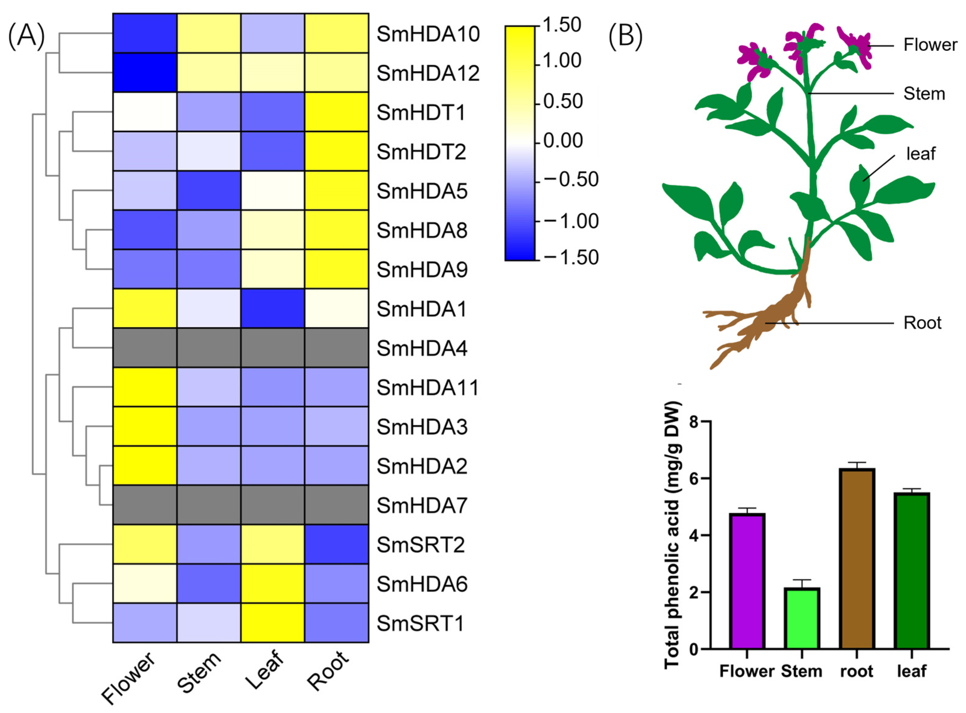
2.6. Tissue-Specific Analysis of SmHDACs

The expression of genes in individual plant tissues is significantly correlated with their functions. Here, we analyzed the tissue specificity of SmHDACs in roots, stems, leaves, and flowers, as shown in Figure 5A. Half of the genes are highly expressed in the root, which may be involved in the synthesis of salvianolic acid. SmHDA6 and SmSRT1, 2 are highly expressed in leaves, which may be related to photomorphogenesis or anthocyanin synthesis. SmHDA1, 2, 3, 11 and SmSRT2 are highly expressed in flowers, which may play a role in plant maturation. Some of the functions of these hypotheses can be matched with the results of CAEs analysis (Figure 4A). For example, SmHDA6 and SmSRT2 are highly expressed in leaves and contain GT1-motif (light responsiveness), indicating that they may participate in plant photosynthesis. Total phenolic acid content was highest in roots (Figure 5B), suggesting that SmHDACs such as SmHDA10, 12, 5, 8, 9 and SmHDT1, 2, which are specifically expressed in roots, may be involved in the synthesis of phenolic acids.

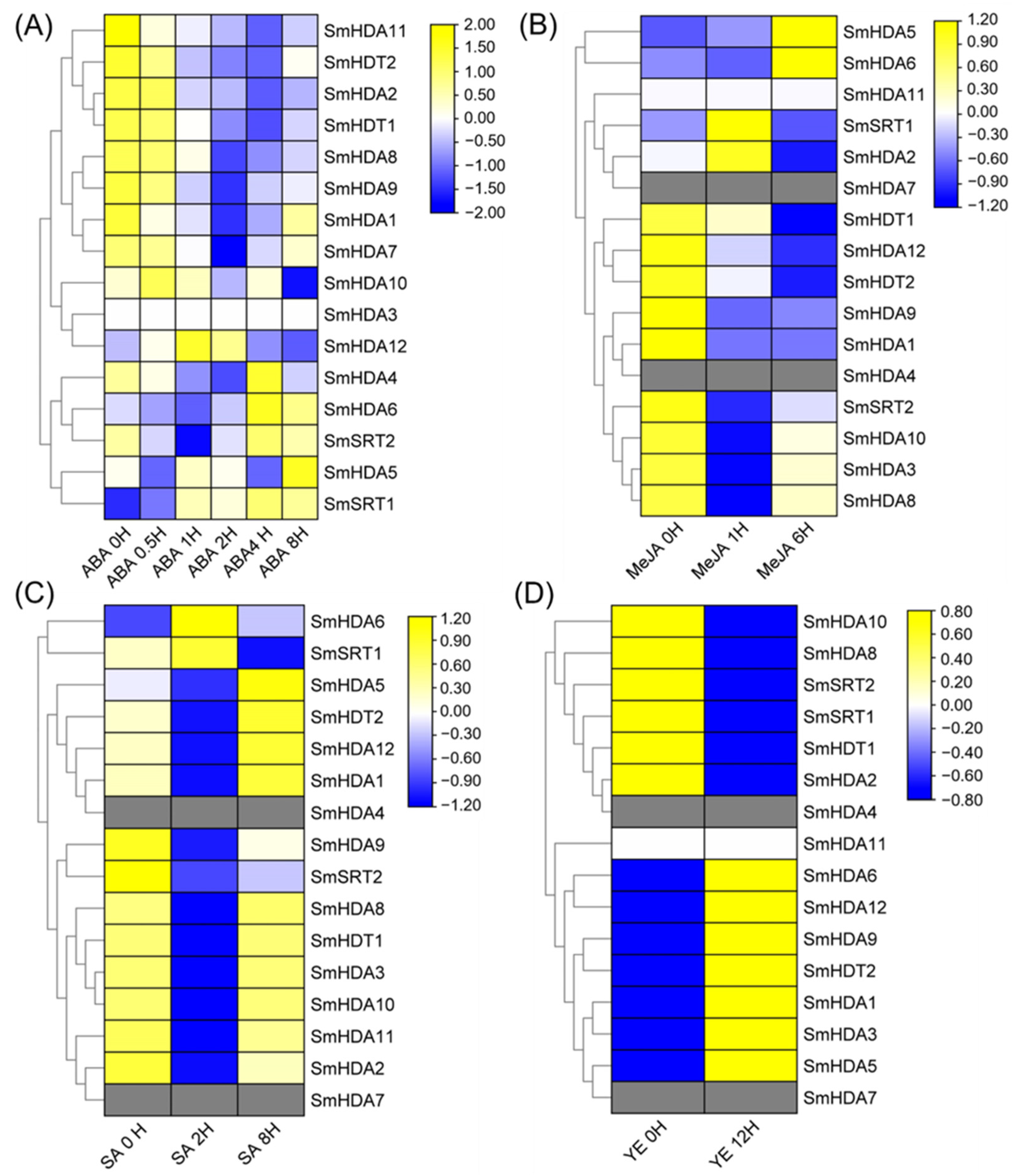
2.7. Induction of SmHDACs Expression Patterns by Each Inducer

Since CAEs analysis indicated that the promoters of SmHDACs may have CAEs in response to various phytohormones, we analyzed the expression pattern of SmHDACs under multi-medium inducer treatment by accessing the transcriptome database, include ABA, MeJA, SA, YE (Figure 6).
Under Abscisic acid induction, SmHDA3 remained unexpressed, while SmHDA5, 6, and SmSRT1, 2 tended to be activated by ABA (Figure 6A). The expression of SmHDACs decreased in more than half of the patients, indicating the overall inhibition of ABA on SmHDACs.
Methyl jasmonate (MeJA) is one of the metabolites that plants biosynthesis when Subject to biological or abiotic stress, it is an important hormone in S. miltiorrhiza which has been shown to promote the accumulation of salvianolic acid [9]. According to the results of MeJA induction, the expressions of SmHDA2, 5, 6 and SmSRT1 were significantly up-regulated, while others were down-regulated, and SmHDA11 was not expressed (Figure 6B). Among them, only SmHDA5 and 6 were continuously activated by MeJA, indicating that they may play a role in the JA response pathway related proteins under MeJA induction.
Salicylic acid can inhibit almost all SmHDACs except SmHDA6 and SmSRT1 (Figure 6C). After treated with yeast extract that was shown to induce the production of specialized metabolites [9], half of the genes, including SmHDA6, were activated and the other half were suppressed (Figure 6D). According to the above conclusions, it can be seen that the most likely functional gene of SmHDA5, 6 in SmHDACs is the core histone deacetylation protein of S. miltiorrhiza in response to stress.

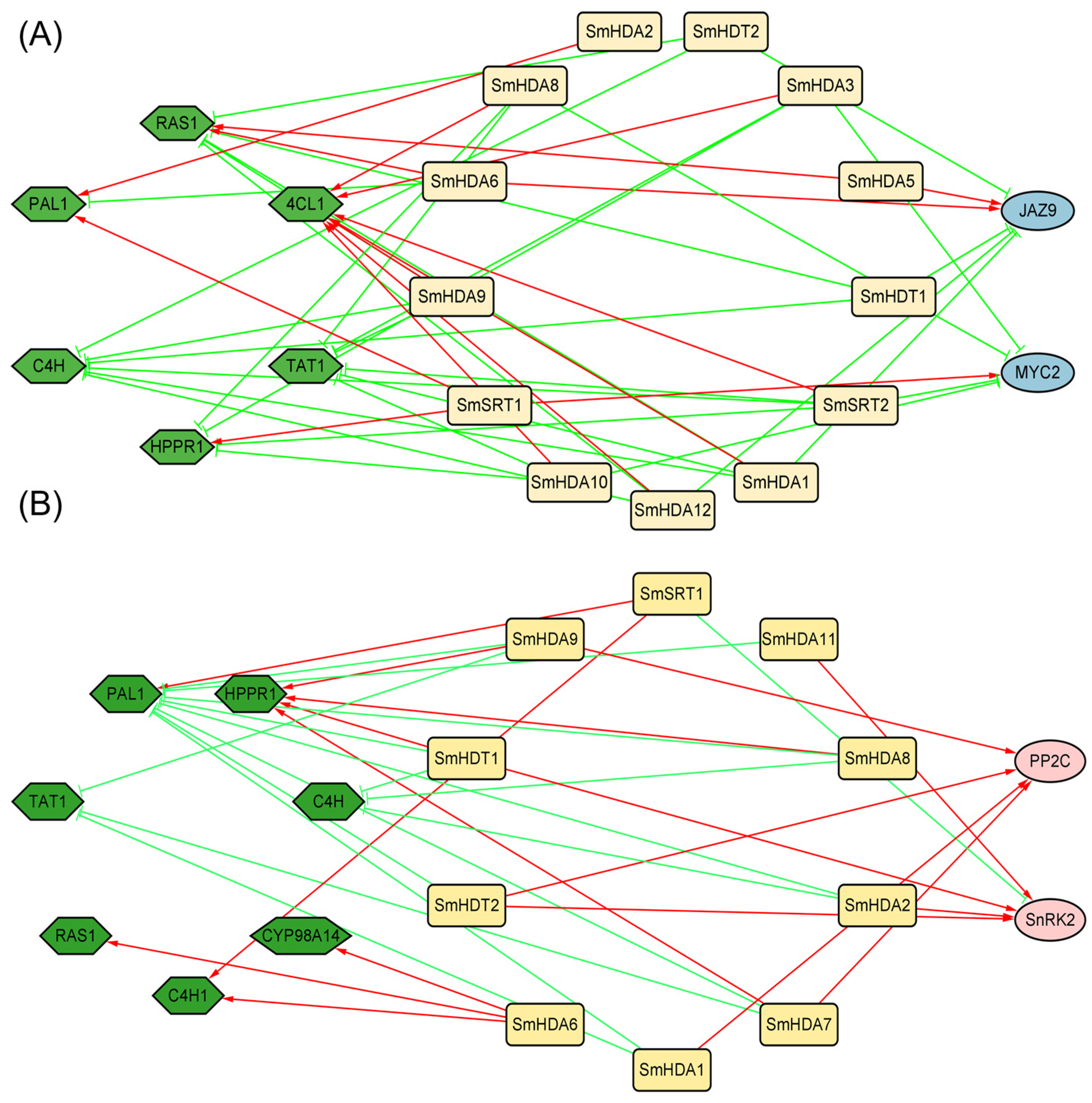
2.8. Co-Expression Network Analysis by MeJA and ABA Treatment

In order to further explore the molecular mechanism of how SmHDACs mediates hormone signaling pathway to regulate salvianolic acid synthesis, we established a co-expression network of salvianolic acid pathway synthase genes and SmHDACs by MeJA and ABA treatment (Figure 7). At the same time, the core transcription factors of JA pathway and ABA pathway were also added, as a preliminary prediction of downstream targets of SmHDACs (Figure 7A). At the three time points after MeJA treatment, the highest number of SmHDACs were close to and positively correlated with the expression trend of the key enzyme gene Sm4CL1, suggesting that Sm4CL may be the final target for SmHDAC to affect salvianolic acid synthesis. SmJAZ9 is negatively correlated with more SmHDACs, may be one of the main targets of histone deacetylation.
Under ABA induction, SmPAL1 was inversely associated with the most SmHDACs and most likely the ultimate target (Figure 7B). Compared with PP2C, SnRK2 is more likely to be the downstream target of SmHDACs, which may be deacetylated by SmSRT1, resulting in repression of gene expression. ABA hormones are often biosynthesized by plants in response to stresses such as drought, low temperature [25,26], among which SnRK2 acts as an intermediate transcription factor during the occurrence of these stresses. SmHDA2, 11 and SmHDT1, 2 were positively correlated with the expression of SnRK2, indicating that they may play a role in the stress of S. miltiorrhiza, while SmSRT1 is negatively correlated. It may be the upstream acting protein of SnRK2.

3. Discussion

Histone deacetylation is an important post-translational modification that regulates gene expression by regulating the acetylation level of histone terminal lysine. After the deacetylation of histones terminal lysine by HDACs, the structure tends to compress, making it difficult for DNA to be transcribed, thus inhibiting and even silencing the genes [14]. It has been widely studied in plants and can be divided into three subfamilies: RPD3/HDA1, SIR2 and HD2 [18]. In this study, 16 SmHDACs proteins were discovered from the high-quality genome of S. miltiorrhiza [27], and named according to subfamilies, with 12 RPD3/HDA1,2 SIR2 and 2 HD2, respectively (Table 1), which ratio was similar to other dicotyledonous plants such as A. thaliana, O. sativa, et al., the RPD3/HDA1 subfamily of HDACs is dominant in numbers, and this pattern is also reflected in more species [28,29]. However, although the RPD3/HDA1 subfamily is the most numerous, many of the genes in this subfamily function were similar [30]. According to motif analysis (Figure 2), motifs of SmHDAs were more conserved, while motifs were not detected by SmSRTs, indicating that the number of SmSRT mutations during evolution may be more. From the perspective of collinear analysis (Figure 3), SmHDT1 and SmHDA2 have collinear genes on Chr.2 and Chr.3, respectively, suggests that SmHDTs may be copied by other genes during evolution, and may play an important role in the adaptation of S. miltiorrhiza to certain environmental changes. Although HDACs functions in so many different ways, unfortunately, the molecular mechanisms of phytohormones promote salvianolic acid accumulation on histone deacetylation have not received sufficient attention.
Under biological or abiotic stress, S. miltiorrhiza may coordinate homeostasis by secreting phytohormones and other ways, thereby some important secondary metabolites are synthesized in large quantities during this process, including salvianolic acid, an important medicinal ingredient [1,10]. In this study, the content of total phenolic acid in various tissues of S. miltiorrhiza was quantitatively determined, results demonstrated that the root is the main accumulation site of salvianolic acids (Figure 5B), therefore, the relationship between SmHDACs with higher expression in the root and salvianolic acid synthesis is most valuable to discuss. Compared with other tissues, SmHDT1, 2 and SmHDA5, 8, 9, 10, 12 have the highest expression levels in roots, and the proteins encoded by them may be the most active in roots. The amount of salvianolic acid was increased by ABA, and the expression of SmHDACs activated at certain time nodes under ABA treatment included SmHDA1, 4, 5, 6, 7, 10, 12 and SmSRT1, 2. Combining the two expression profiles, we can preliminatively conclude that SmHDA5, 10, and 12 are potential ABA-induced genes that increase salvianolic acid content. On the other hand, co-expression networks can also help search for potential functional genes (Figure 6). For example, SmSRT2 is the only gene negatively related to SnRK2, which is a positive regulator of ABA pathway and plays a central role in the entire transcriptional network. Moreover, SmSRT2 was induced by ABA, suggested that SmSRT2 may be induced after ABA secretion, one of the restrictors of this pathway, and may directly deacetylate SnRK2. Similarly, the expression of SmHDA2, 5, 6 and SmSRT1 was upregulated under MeJA induction, among which SmHDA5 was the root gene with high expression, therefore it can be preliminarily speculated that SmHDA5 might have an effect on salvianolic acid synthesis. It is worth noting that SmHDA5 also positively correlated with the expression of SmJAZ9. In previous reports, SmJAZ9 was shown that it can form the complex with SmMYB76, further inhibit the expression of the synthetic gene SmRAS1, SmMYB76 was also associated with SmHDA5 [2]. The addition of SmHDA5 may provide a new idea for the molecular mechanism of SmMYB76 inhibiting salvianolic acid synthesis. According to the above reasoning, SmHDA5 may have multiple downstream targets, which can inhibit the expression of SmMYB76, SmJRB1 [1] and SmJAZ9 through histone deacetylation, weaken the inhibitory effect of the latter on synthase, and promote the synthesis of salvianolic acid.
Phytohormone is a special metabolite secreted by plants to fight against external biological or abiotic stress, which can maintain the homeostasis of the plant internal environment [31]. In the CAEs analysis (Figure 4), we found that the response to plant hormones may be the main functional pathway of SmHDACs, compared with plant growth and development and stress responsiveness. CAEs related to phytohormone responsiveness dominated in number, especially in response to MeJA and ABA. It is worth noting that both MeJA and ABA have been reported that can promote medical compounds salvianolic acid accumulation [9,32], the complex molecular mechanisms involved have been extensively studied. In addition to the role of transcription factors, SmHDACs provide a potential molecular mechanism at the epigenetic level. SmHDACs may have deacetylated effect on some key transcription factors in plant hormone signaling pathway, thus regulating the whole pathway, such mechanisms have been resolved in other plants. For example, in tea, CsHDA6 deacetylates histone proteins of CsMYC2, affecting the latter’s ability to enhance the transcription of CsAFS and inhibiting the accumulation of α-farnesene in tea, while the intervention of MeJA inhibits this effect [33], a similar mechanism occurs in CsHDA2 and ultimately affects the pest resistance of tea [34]. Furthermore, OsHDA710 has been shown to enhance the sensitivity of rice to ABA, which is reflected in the salt tolerance of rice [21]. Even further studies have found that HDACs can directly act on some proteins, but not histones, for deacetylation, such as Arabidopsis HDA6 can deacetylate TPL protein, which is the restriction of JA pathway activation, HDA6 and GCN5 jointly maintain the acetylation homeostasis of TPL protein, thereby affecting the expression of subsequent transcription factors on the JA pathway, such as MYC2, JAZ, et al. [22]. To explore the role of SmHDACs in response to phytohormones, and to look for potential functional genes, we used a series of bioinformatics analysis methods such as CAEs analysis, phylogenetic tree, and induced expression pattern. Genes assigned to one branch of the phylogenetic tree may have similar functions, for example, AtHDA19 is a protein with multiple functions, which can regulate photomorphogenesis and seed dormancy et al. [35,36], and can be regulated by MeJA and ABA [37,38], the homologous SmHDA5, 6 may also play the similar role. CAEs analysis showed that SmHDA5, 6 were one of the SmHDACs with the largest number of hormone-responsive elements (Figure 4), and the expression of these two genes were activated under the induction of MeJA and ABA (Figure 6), indicating that they are likely to be able to respond to these two phytohormones to play more subsequent functions, this confirms previous predictions of their function by the phylogenetic tree. Similarly, it is speculated that the AtHDA6 homologous SmHDA9 may have the function of non-histone deacetylation [22], however, it is inhibited by MeJA (Figure 6), suggesting that the target of SmHDA9 may be a negative regulator in the pathway. SmSRTs is also a noteworthy gene. In A. thaliana, AtSRTs is usually associated with the growth and development process. For example, AtSRT2 can regulate the salt-stress resistance of Arabidopsis seed during germination [30], AtSRT1 can regulates primary metabolism by modulation of AtMBP-1 transcriptional activity [39]. Their homologous SmSRT1, 2 probably similar to AtSRTs. The expression levels of them changed little under the treatment of TSA, and the changes induced by hormones were not obvious, indicating that their expression was stable. In general, the SmHDACs family may play a role in various growth stages of S. miltiorrhiza, and regulate the expression and affect the function of some transcription factors under the induction of plant hormones.

4. Materials and Methods

4.1. Plant Materials, Hormonal/Elicitor Treatment

Three-mouth-old aseptic plant of S. miltiorrhiza cultured in a greenhouse at 26 °C with Murashige and Skoog medium, exposure to 16-h light/8-h dark. The hairy roots of S. miltiorrhiza were obtained by infecting leaves or stems from aseptic plant with Agrobacterium rhizogenes strain C58C1 (pRi A4) (Weidi Biotechnology Co., Shanghai, China), then subcultured in half-strength MS medium. The young hairy roots were cut and cultured in liquid medium with persistent darkness at 25 °C and 120 rpm for 50 days, therewith treated with various exogenous inducers. Additional transcriptome data for this article can be downloaded by BioProject access numbers: PRJNA703309 (ABA), PRJNA393563 (MeJA), PRJNA301529 (SA), and PRJNA393563 (YE) [40].
Mature one-year raw S. miltiorrhiza planted in the greenhouse of our laboratory was used to determine the total phenolic acid content of each tissue. The roots, stems, leaves and flowers of S. miltiorrhiza were collected, dried in an oven at 65 °C, and ground into powder for subsequent experiments.

4.2. Identification of the HDACs Family in S. miltiorrhiza

The newly assembled annotated high-quality genomic data is used to mine SmHDACs [27]. To get the subfamily of RPD3/HDA1 and SIR2, Hist_deacetyl domain (PF00850) and SIR2 domain (PF02146) were utilized as query to search against S. miltiorrhiza protein database by Hidden Markov Model (HMM) function module of TBtools software (v2.026) [41], e-value > 0.05 is used as a threshold filter. Moreover, HD2 subfamily was discovered by BLSAT to AtHDT (AT3G44750, AT5G22650, AT5G03740, AT2G27840) protein sequence in model plant Arabidopsis thaliana, the result is compared to verify the NPL domain (PF17800) by HMM. All the SmHDACs obtained above were verified by Conserved Domain Database (http://www.ncbi.nlm.nih.gov/Structure/cdd/wrpsb.cgi, (accessed on 5 August 2023)) to ensure their integrity, and the incomplete sequences in the conserved domain were deleted. Finally, 16 SmHDACs were identified (Table S1). Furthermore, the physical and chemical properties of SmHDACs were predicted by ExPASy tool (https://web.expasy.org/, (accessed on 6 August 2023)), and the subcellular localization information was predicted by Plant-mPLoc predictor (http://www.csbio.sjtu.edu.cn/bioinf/plant-multi/, (accessed on 6 August 2023)).

4.3. Phylogenetic Tree, Motif and Gene Structure Analysis of SmHDACs

To understand the evolutionary relationship of SmHDACs, HDACs of Arabidopsis thaliana collected from TAIR database (https://www.arabidopsis.org/, (accessed on 2 August 2023)), Oryza sativa, Zea mays, and Solanum lycopersicum collected from Phytozome database (https://phytozome-next.jgi.doe.gov/, (accessed on 20 July 2023)) (the protein sequence information is shown in Table S2) were aligned by ClustalW and construct phylogenetic tree by MEGA 11.0 software (https://megasoftware.net/, (accessed on 15 June 2022)) with Maximum likelihood algorithm. iTOL online tool (https://itol.embl.de) was employed for modifying the phylogenetic tree. The analysis of conserved motif was implemented by MEME tool (https://meme-suite.org/, (accessed on 10 August 2023)), with parameters as motif width 6–50 and max number is 20. The visualization of conserved motifs and gene structure of SmHDACs were employed by TBtools [41].

4.4. Chromosome Localization and Collinearity Analysis of SmHDACs

The distribution of SmHDACs at the chromosomes was analyzed through the annotation information. To explore gene duplication information between SmHDACs, collinearity analysis of members was performed by MCScanX [42] and visualized by Tbtools [41].

4.5. Cis-Acting Elements Analysis of SmHDACs

In order to search for the possible molecular mechanism of SmHDACs, the cis-acting elements on their promoters were analyzed. The upstream 2000 bp long sequences of SmHDACs proteins were extracted from S miltiorrhiza genome, then input into PlantCARE (http://bioinformatics.psb.ugent.be/webtools/plantcare/html/, (accessed on 13 August 2023)) [43] to predict potential cis-acting elements.

4.6. Organ-Specific and Each Inducer Treated Expression Pattern of SmHDACs

In order to study the ability of SmHDACs to respond to stress, we obtained multiple transcriptome data and analyzed their expression profiles. All SmHDACs were identified in muti-transcriptome by BLAST. The transcriptome of four organs (leaf, flower, root, stem) of S. miltiorrhiza were used for organ-specific analysis. The results are visualized by heatmap. In order to search for potential downstream targets of SmHDACs, key enzyme gene in the salvianolic acid synthesis pathway and core transcription factor genes (Table S3) in the MeJA pathways and ABA pathway were used as co-expression networks with SmHDACs, correlation coefficients test (Pearson’s coefficients, take its absolute value) >0.8 is considered as associated, the result is presented via cytoscape 3.9.1 software.

4.7. Determination of Total Phenolic Acid

To investigate the optimum tissue for the accumulation of total phenolic acid, roots, stems, leaves, and flowers of one-year-old S. miltiorrhiza were sampled. Afterwards, 100 mg of dry powder were obtained through mSini-pulverizer (Xinda Machinery Co., Jiangyin, China), extracted with an ultrasound-assisted method, and utilized 10 mL 80% ethanol solution as a solvent. Total phenolic acid content of four different tissues were quantitatively measured by an improved Folin-Ciocalteu method. In brief, 1 mL of alcohol extract solution was evenly mixed with 5 mL of 0.1 M Folin-Ciocalteu reagent (Solarbio Science & Technology Co., Beijing, China), 4 mL of 20% (m/v) Na2CO3 solution within 60 s, and keep the mixed solution away from light for 1 h. Gallic acid (Sigma-Aldrich, St. Louis, MO, USA) was utilized as reference for a standard curve (y = 8.068 x + 0.015, R2 = 0.999). The content of total phenolic acid was photometrically detected at 760 nm with UV spectrophotometer (Metash Instruments Co., Shanghai, China), and expressed as mg of gallic acid equivalents per 100 mg of dry weigh (DW).

5. Conclusions

In this paper, 16 SmHDACs were obtained from the high-quality genome of Salvia miltiorrhiza, and their biological characteristics were analyzed, including phylogenetic tree, cis-acting element analysis, prediction of subcellular localization, and collinearity analysis. The above analysis suggests that SmHDACs respond to a variety of phytohormones, and further affect the accumulation of salvianolic acid, the medicinal component of S. miltiorrhiza. The expression profiles of SmHDACs under various inducers were used to evaluate the ability of SmHDACs to respond to stress. Combined with the co-expression analysis network and previous reports, several potential functions of SmHDACs were speculated, and a molecular mechanism of SmHDA5 mediated JA pathway in promoting salvianolic acid accumulation was further proposed. This study provides a strategy to improve the quality of S. miltiorrhiza from the perspective of epigenetic histone deacetylase.

Supplementary Materials

The following supporting information can be downloaded at: https://www.mdpi.com/article/10.3390/plants13050580/s1, Figure S1: Seventeen motif logos in SmHDAC protein. Table S1: Protein sequence of all SmHDACs; Table S2: Protein sequences used to construct the phylogenetic tree; Table S3: Gene ID used to construct a co-expression network.

Author Contributions

Z.Y. conceived and designed the project. J.C., Y.Y., L.Y., Z.X. and Z.Y. analysed the data. J.C. and Z.Y. co-wrote the manuscript. G.K. and Z.Y. revised and edited the manuscript. All authors have read and agreed to the published version of the manuscript.

Funding

This work was supported by National Natural Science Foundation of China (32000257), Guangzhou Basic and Applied Basic Research Scheme (202201010122), Zhejiang Provincial Natural Science Foundation of China (LY23H280003), the Research Project of Zhejiang Chinese Medical University (2022JKZKTS15), and Zhejiang Xin-miao Talents Program (2023R410044).

Data Availability Statement

The datasets analyzed during the current study are available at the NCBI under the accession number PRJNA703309, PRJNA393563, PRJNA301529, and PRJNA393563 for the ABA, MeJA, SA and YE treatments [40], respectively. All methods were carried out in accordance with relevant guidelines and regulations. All experimental studies on plants were complied with relevant institutional, national, and international guidelines and legislation.

Acknowledgments

We appreciate the considerable help/technical support/experimental support from the Public Platform of Pharmaceutical Research Center, Academy of Chinese Medical Science, Zhejiang Chinese Medical University.

Conflicts of Interest

The authors declare that the research was conducted in the absence of any commercial or financial relationships that could be construed as a potential conflict of interest.

References

  1. Zhou, W.; Li, S.; Maoz, I.; Wang, Q.; Xu, M.; Feng, Y.; Hao, X.L.; Du, Z.Y.; Kai, G.Y. SmJRB1 positively regulates the accumulation of phenolic acid in Salvia miltiorrhiza. Ind. Crop. Prod. 2021, 164, 113417. [Google Scholar] [CrossRef]
  2. Liu, S.C.; Gao, X.K.; Shi, M.; Sun, M.H.; Li, K.L.; Cai, Y.; Chen, C.A.; Wang, C.; Maoz, I.; Guo, X.H.; et al. Jasmonic acid regulates the biosynthesis of medicinal metabolites via the JAZ9-MYB76 complex in Salvia miltiorrhiza. Hortic. Res. 2023, 10, uhad004. [Google Scholar] [CrossRef]
  3. Sun, C.T.; Han, B.; Zhai, Y.F.; Zhao, H.; Li, X.; Qian, J.; Hao, X.L.; Liu, Q.; Shen, J.Y.; Kai, G.Y. Dihydrotanshinone I inhibits ovarian tumor growth by activating oxidative stress through Keap1-mediated Nrf2 ubiquitination degradation. Free Radic. Biol. Med. 2022, 180, 220–235. [Google Scholar] [CrossRef] [PubMed]
  4. Jia, Q.Q.; Zhu, R.Y.; Tian, Y.M.; Chen, B.B.; Li, R.; Li, L.; Wang, L.L.; Chen, Y.W.; Zhao, D.D.; Mo, F.F.; et al. Salvia miltiorrhiza in diabetes: A review of its pharmacology, phytochemistry, and safety. Phytomedicine 2019, 58, 152871. [Google Scholar] [CrossRef] [PubMed]
  5. Shi, M.; Huang, F.F.; Deng, C.P.; Wang, Y.; Kai, G.Y. Bioactivities, biosynthesis and biotechnological production of phenolic acids in Salvia miltiorrhiza. Crit. Rev. Food Sci. Nutr. 2019, 59, 953–964. [Google Scholar] [CrossRef]
  6. Fu, R.; Shi, M.; Deng, C.P.; Zhang, Y.; Zhang, X.C.; Wang, Y.; Kai, G.Y. Improved phenolic acid content and bioactivities of Salvia miltiorrhiza hairy roots by genetic manipulation of RAS and CYP98A14. Food Chem. 2020, 331, 127365. [Google Scholar] [CrossRef]
  7. Lu, S. Biosynthesis and regulatory mechanisms of bioactive compounds in Salvia miltiorrhiza, a model system for medicinal plant biology. Crit. Rev. Food Sci. Nutr. 2021, 40, 243–283. [Google Scholar] [CrossRef]
  8. Xu, Y.P.; Geng, L.J.; Zhang, Y.W.; Jones, J.A.; Zhang, M.H.; Chen, Y.; Tan, R.H.; Koffas, M.A.G.; Wang, Z.T.; Zhao, S.J. De novo biosynthesis of salvianolic acid B in Saccharomyces cerevisiae engineered with the rosmarinic acid biosynthetic pathway. J. Agric. Food Chem. 2022, 70, 2290–2302. [Google Scholar] [CrossRef]
  9. Zhou, W.; Huang, Q.; Wu, X.; Zhou, Z.W.; Ding, M.Q.; Shi, M.; Huang, F.F.; Li, S.; Wang, Y.; Kai, G.Y. Comprehensive transcriptome profiling of Salvia miltiorrhiza for discovery of genes associated with the biosynthesis of tanshinones and phenolic acids. Sci. Rep. 2017, 7, 10554. [Google Scholar] [CrossRef] [PubMed]
  10. Zhou, W.; Shi, M.; Deng, C.P.; Lu, S.C.; Huang, F.F.; Wang, Y.; Kai, G.Y. The methyl jasmonate-responsive transcription factor SmMYB1 promotes phenolic acid biosynthesis in Salvia miltiorrhiza. Hortic. Res. 2021, 8, 10. [Google Scholar] [CrossRef]
  11. Deng, C.P.; Wang, Y.; Huang, F.F.; Lu, S.J.; Zhao, L.M.; Ma, X.Y.; Kai, G.Y. SmMYB2 promotes salvianolic acid biosynthesis in the medicinal herb Salvia miltiorrhiza. J. Integr. Plant Biol. 2020, 62, 1688–1702. [Google Scholar] [CrossRef]
  12. Sun, M.H.; Shi, M.; Wang, Y.; Huang, Q.; Yuan, T.P.; Wang, Q.; Wang, C.; Zhou, W.; Kai, G.Y. The biosynthesis of phenolic acids is positively regulated by the JA-responsive transcription factor ERF115 in Salvia miltiorrhiza. J. Exp. Bot. 2019, 70, 243–254. [Google Scholar] [CrossRef] [PubMed]
  13. Li, S.; He, X.; Gao, Y.; Zhou, C.G.; Chiang, V.L.; Li, W. Histone acetylation changes in plant response to drought stress. Genes 2021, 12, 1409. [Google Scholar] [CrossRef] [PubMed]
  14. Garske, A.L.; Oliver, S.S.; Wagner, E.K.; Musselman, C.A.; LeRoy, G.; Garcia, B.A.; Kutateladze, T.G.; Denu, J.M. Combinatorial profiling of chromatin binding modules reveals multisite discrimination. Nat. Chem. Biol. 2010, 6, 283–290. [Google Scholar] [CrossRef] [PubMed]
  15. Lusser, A.; Kölle, D.; Loidl, P. Histone acetylation: Lessons from the plant kingdom. Trends Plant Sci. 2001, 6, 59–65. [Google Scholar] [CrossRef] [PubMed]
  16. Murray, S.L.; Ingle, R.A.; Petersen, L.N.; Denby, K.J. Basal resistance against Pseudomonas syringae in Arabidopsis involves WRKY53 and a protein with homology to a nematode resistance protein. Mol. Plant Microbe Interact. 2007, 20, 1431–1438. [Google Scholar] [CrossRef] [PubMed]
  17. Chen, Z.J.; Tian, L. Roles of dynamic and reversible histone acetylation in plant development and polyploidy. Biochim. Biophys. Acta 2007, 1769, 295–307. [Google Scholar] [CrossRef]
  18. Pandey, R.; Müller, A.; Napoli, C.A.; Selinger, D.A.; Pikaard, C.S.; Richards, E.J.; Bender, J.; Mount, D.W.; Jorgensen, R.A. Analysis of histone acetyltransferase and histone deacetylase families of Arabidopsis thaliana suggests functional diversification of chromatin modification among multicellular eukaryotes. Nucleic Acids Res. 2002, 30, 5036–5055. [Google Scholar] [CrossRef] [PubMed]
  19. Guo, Q.; Jing, Y.J.; Gao, Y.; Liu, Y.T.; Fang, X.F.; Lin, R.C. The PIF1/PIF3-MED25-HDA19 transcriptional repression complex regulates phytochrome signaling in Arabidopsis. New Phytol. 2023, 240, 1097–1115. [Google Scholar] [CrossRef]
  20. Tilak, P.; Kotnik, F.; Née, G.; Seidel, J.; Sindlinger, J.; Heinkow, P.; Eirich, J.; Schwarzer, D.; Finkemeier, I. Proteome-wide lysine acetylation profiling to investigate the involvement of histone deacetylase HDA5 in the salt stress response of Arabidopsis leaves. Plant J. 2023, 115, 275–292. [Google Scholar] [CrossRef]
  21. Ullah, F.; Xu, Q.; Zhao, Y.; Zhou, D.X. Histone deacetylase HDA710 controls salt tolerance by regulating ABA signaling in rice. J. Integr. Plant Biol. 2020, 63, 451–467. [Google Scholar] [CrossRef]
  22. An, C.P.; Deng, L.; Zhai, H.W.; You, Y.R.; Wu, F.M.; Zhai, Q.Z.; Goossens, A.; Li, C.Y. Regulation of jasmonate signaling by reversible acetylation of TOPLESS in Arabidopsis. Mol. Plant 2022, 15, 1329–1346. [Google Scholar] [CrossRef] [PubMed]
  23. He, X.Y.; Chen, Y.W.; Xia, Y.T.; Hong, X.Y.; You, H.Q.; Zhang, R.; Liang, Z.S.; Cui, Q.; Zhang, S.C.; Zhou, M.; et al. DNA methylation regulates biosynthesis of tanshinones and phenolic acids during growth of Salvia miltiorrhiza. Plant Physiol. 2023, kiad573. [Google Scholar] [CrossRef] [PubMed]
  24. Zhang, M.Z.; Teixeira, S.J.A.; Yu, Z.M.; Wang, H.B.; Si, C.; Zhao, C.H.; He, C.M.; Duan, J. Identification of histone deacetylase genes in Dendrobium officinale and their expression profiles under phytohormone and abiotic stress treatments. PeerJ 2020, 8, e10482. [Google Scholar] [CrossRef] [PubMed]
  25. Fujii, H.; Verslues, P.E.; Zhu, J.K. Arabidopsis decuple mutant reveals the importance of SnRK2 kinases in osmotic stress responses in vivo. Proc. Natl. Acad. Sci. USA 2011, 108, 1717–1722. [Google Scholar] [CrossRef] [PubMed]
  26. Fujita, Y.; Nakashima, K.; Yoshida, T.; Katagiri, T.; Kidokoro, S.; Kanamori, N.; Umezawa, T.; Fujita, M.; Maruyama, K.; Ishiyama, K.; et al. Three SnRK2 protein kinases are the main positive regulators of abscisic acid signaling in response to water stress in Arabidopsis. Plant Cell Physiol. 2009, 50, 2123–2132. [Google Scholar] [CrossRef] [PubMed]
  27. Ma, Y.; Cui, G.H.; Chen, T.; Ma, X.H.; Wang, R.S.; Jin, B.L.; Yang, J.; Kang, L.P.; Tang, J.F.; Lai, C.J.S.; et al. Expansion within the CYP71D subfamily drives the heterocyclization of tanshinones synthesis in Salvia miltiorrhiza. Nat. Commun. 2021, 12, 685. [Google Scholar] [CrossRef]
  28. Jin, P.; Gao, S.Q.; He, L.; Xu, M.Z.; Zhang, T.Y.; Zhang, F.; Jiang, Y.Y.; Liu, T.T.; Yang, J.; Yang, J.; et al. Genome-wide identification and expression analysis of the histone deacetylase gene family in wheat (Triticum aestivum L.). Plants 2020, 10, 19. [Google Scholar] [CrossRef]
  29. Zhao, L.M.; Lu, J.X.; Zhang, J.X.; Wu, P.Y.; Yang, S.G.; Wu, K.Q. Identification and characterization of histone deacetylases in tomato (Solanum lycopersicum). Front. Plant Sci. 2015, 5, 760. [Google Scholar] [CrossRef]
  30. Tang, W.S.; Zhong, L.; Ding, Q.Q.; Dou, Y.N.; Li, W.W.; Xu, Z.S.; Zhou, Y.B.; Chen, J.; Chen, M.; Ma, Y.Z. Histone deacetylase AtSRT2 regulates salt tolerance during seed germination via repression of vesicle-associated membrane protein 714 (VAMP714) in Arabidopsis. New Phytol. 2022, 234, 1278–1293. [Google Scholar] [CrossRef]
  31. Waadt, R. Phytohormone signaling mechanisms and genetic methods for their modulation and detection. Curr. Opin. Plant Biol. 2020, 57, 31–40. [Google Scholar] [CrossRef]
  32. Deng, C.P.; Shi, M.; Fu, R.; Zhang, Y.; Wang, Q.; Zhou, Y.; Wang, Y.; Ma, X.Y.; Kai, G.Y. ABA-responsive transcription factor bZIP1 is involved in modulating biosynthesis of phenolic acids and tanshinones in Salvia miltiorrhiza. J. Exp. Bot. 2020, 71, 5948–5962. [Google Scholar] [CrossRef] [PubMed]
  33. Wu, S.H.; Yang, Y.H.; Chen, J.M.; Li, J.L.; Jian, G.T.; Yang, J.; Mao, K.Q.; Zeng, L.T.; Gu, D.C. Histone deacetylase CsHDA6 mediates the regulated formation of the anti-insect metabolite α-farnesene in tea (Camellia sinensis). Plant Sci. 2023, 326, 111501. [Google Scholar] [CrossRef] [PubMed]
  34. Gu, D.C.; Wu, S.H.; Yu, Z.M.; Zeng, L.T.; Qian, J.J.; Zhou, X.J.; Yang, Z.Y. Involvement of histone deacetylase CsHDA2 in regulating (E)-nerolidol formation in tea (Camellia sinensis) exposed to tea green leafhopper infestation. Hortic. Res. 2022, 9, uhac158. [Google Scholar] [CrossRef] [PubMed]
  35. Jing, Y.; Guo, Q.; Lin, R. The SNL-HDA19 histone deacetylase complex antagonizes HY5 activity to repress photomorphogenesis in Arabidopsis. New Phytol. 2021, 229, 3221–3236. [Google Scholar] [CrossRef] [PubMed]
  36. Zhou, Y.; Yang, P.; Zhang, F.; Luo, X.; Xie, J. Histone deacetylase HDA19 interacts with histone methyltransferase SUVH5 to regulate seed dormancy in Arabidopsis. Plant Biol. 2020, 22, 1062–1071. [Google Scholar] [CrossRef] [PubMed]
  37. Chen, L.T.; Wu, K. Role of histone deacetylases HDA6 and HDA19 in ABA and abiotic stress response. Plant Signal. Behav. 2010, 5, 1318–1320. [Google Scholar] [CrossRef] [PubMed]
  38. Zhou, C.H.; Zhang, L.; Duan, J.; Miki, B.; Wu, K.Q. HISTONE DEACETYLASE19 is involved in jasmonic acid and ethylene signaling of pathogen response in Arabidopsis. Plant Cell 2005, 17, 1196–1204. [Google Scholar] [CrossRef] [PubMed]
  39. Liu, X.Y.; Wei, W.; Zhu, W.J.; Su, L.F.; Xiong, Z.Y.; Zhou, M.; Zhen, G.Y.; Zhou, D.X. Histone deacetylase AtSRT1 links metabolic flux and stress response in Arabidopsis. Mol. Plant 2017, 10, 1510–1522. [Google Scholar] [CrossRef]
  40. Chen, C.A.; Wang, C.; Li, J.B.; Gao, X.K.; Huang, Q.K.; Gong, Y.F.; Hao, X.L.; Maoz, I.; Kai, G.Y. Genome-wide analysis of U-box E3 ubiquitin ligase family in response to ABA treatment in Salvia miltiorrhiza. Front. Plant Sci. 2022, 13, 829447. [Google Scholar] [CrossRef]
  41. Chen, C.J.; Chen, H.; Zhang, Y.; Thomas, H.R.; Frank, M.H.; He, Y.H.; Xia, R. TBtools: An integrative toolkit developed for interactive analyses of big biological data. Mol. Plant 2020, 13, 1194–1202. [Google Scholar] [CrossRef] [PubMed]
  42. Wang, Y.P.; Tang, H.B.; Debarry, J.D.; Tan, X.; Li, J.P.; Wang, X.Y.; Lee, T.H.; Jin, H.Z.; Marler, B.; Guo, H.; et al. MCScanX: A toolkit for detection and evolutionary analysis of gene synteny and collinearity. Nucleic Acids Res. 2012, 40, e49. [Google Scholar] [CrossRef] [PubMed]
  43. Lescot, M.; Déhais, P.; Thijs, G.; Marchal, K.; Moreau, Y.; de Peer, V.V.; Rouzé, P.; Rombauts, S. PlantCARE, a database of plant cis-acting regulatory elements and a portal to tools for in silico analysis of promoter sequences. Nucleic Acids Res. 2002, 30, 325–327. [Google Scholar] [CrossRef] [PubMed]
Figure 1. Phylogenetic tree of the SmHDACs in Salvia miltiorrhiza and in four other plant species. The color of the inner ring represents the species: green, S. miltiorrhiza; red, S. lycopersicum; black, A. thaliana; brown, O. sativa; blue, Z. mays. Annotate the conserved domain in SmHDACs above S. miltiorrhiza. The outer ring represents the subfamily: red, RPD3/HDA1 subfamily; Blue, HD2 subfamily; Green, SIR2 subgroup. Between the two rings, the comments on Salvia miltiorrhiza are conserved domain types, which are: Agri, Arginase_HDAC, cl17011; H_C1, HDAC_classII_1, cd09996; H_C2, HDAC_classII, cd09992; H_C4, HDAC_classIV, cd09993, so far, they are all conserved domains owned by the RPD3/HDA1 subfamily; NPL, cl03870, a conservative domain unique to HD2 subfamily; SIRT4, cd01409, SIRT7, cd01410, a conservative domain unique to the SIR subfamily.
Plants 13 00580 g001
Figure 2. Analysis of gene structure and motifs of the SmHDACs. All motifs of SmHDACs and their gene locations were labeled and classified according to subfamilies. Motifs of the same subfamily have similarities. SmHDTs does not contain motif. Motif logo as show in Figure S1.
Plants 13 00580 g002
Figure 3. Chromosome localization and collinearity analysis of SmHDACs. The 16 genes were randomly distributed on 8 chromosomes, and tended to be in the locations with high gene density. SmHDT1 and SmHDT2 have collinear genes and are labeled with red lines. The collinear genes between multiple species are shown as blue lines, and all the results are counted. Ten. Gene in AT means collinear genes in A. thaliana, SM means S. miltiorrhiza, OS means O. sativa. n.d. indicates that there are no collinear genes in the species.
Plants 13 00580 g003
Figure 4. Cis-acting element analysis of SmHDACs. (A), The position of each CAEs on the SmHDACs promoter. (B), The statistical heatmap of each CAEs on the promoter of SmHDACs, all CAEs were divided into three parts: growth and development, phytohormone responsiveness and stress responsiveness, among which the number of CAEs responding to phytohormone MeJA was the largest.
Plants 13 00580 g004
Figure 5. Tissue-specific analysis of SmHDACs. (A), Total phenolic acid content in roots, stems, leaves, and flowers; (B), Schematic diagram of S. miltiorrhiza tissues and expression profile of SmHDACs genes in flowers, leaves, stems and roots. DW, dry weight.
Plants 13 00580 g005
Figure 6. Analysis of the expression profile of the SmHDACs induced by exogenously applied phytohormones. (A) Expression of SmHDACs induced by ABA at six time points (0, 0.5, 1, 2, 4, and 8 h); (B) Expression of SmHDACs induced by MeJA at three time points (0, 6, and 12 h); (C) Expression of SmHDACs induced by SA at three time points (0, 2, and 8 h); (D) Expression of SmHDACs induced by YE at two time points (1 and 12 h). ABA, abscisic acid; MeJA, methyl jasmonate; SA, salicylic acid; YE, yeast extract. Different elicitation was assessed at different time point.
Plants 13 00580 g006
Figure 7. Co-expression network analysis of the SmHDACs. After phytohormony treatment, a co-expression network between all 16 SmHDACs and the critical salvianolic acid biosynthetic genes were carried out using Cytoscape software v3.9.1 (|r| > 0.8, p < 0.05). (A) SmMYC2 and SmJAZ9 of MeJA pathway. (B) SnRK2 and PP2C of ABA pathway. Four core transcription factors or core protein coding genes on two phytohormone pathways were added for further downstream target prediction. Red/Green arrows indicate positive/negative correlations.
Plants 13 00580 g007
Table 1. Physiochemical properties of the SmHDACs.
NameSize/aaMolecular Weight/kDaPIGRAVYAliphatic IndexInstability Index (II)Subcellular Localization
SmHDA177083.8735.81−0.24983.1440.40Nucleus
SmHDA277685.2806.39−0.38584.3653.08Nucleus
SmHDA366373.8145.27−0.24885.7849.16Chloroplast and Nucleus
SmHDA437440.6195.69−0.09787.1937.65Nucleus
SmHDA549956.2765.27−0.48975.9938.73Cytoplasm and Nucleus
SmHDA646452.4365.15−0.54472.8738.75Cytoplasm and Nucleus
SmHDA742347.7425.01−0.36581.8231.80Cytoplasm and Nucleus
SmHDA855460.5715.68−0.22979.8241.16Nucleus
SmHDA945351.4545.82−0.51376.1647.61Nucleus
SmHDA1037440.4115.38−0.12486.5534.18Nucleus
SmHDA1131335.3056.74−0.41686.3642.16Nucleus
SmHDA1239443.1737.21−0.11294.8536.19Nucleus
SmSRT142547.9839.40−0.45077.1345.82Chloroplast
SmSRT248153.4339.34−0.18089.5839.70Chloroplast
SmHDT128430.6995.07−1.05950.2846.80Nucleus
SmHDT249353.8225.09−0.99261.0847.81Vacuole and Nucleus
Disclaimer/Publisher’s Note: The statements, opinions and data contained in all publications are solely those of the individual author(s) and contributor(s) and not of MDPI and/or the editor(s). MDPI and/or the editor(s) disclaim responsibility for any injury to people or property resulting from any ideas, methods, instructions or products referred to in the content.

