Article

Comparative Phytoprofiling of Achillea millefolium Morphotypes: Assessing Antioxidant Activity, Phenolic and Triterpenic Compounds Variation across Different Plant Parts

1
Department of Pharmacognosy, Lithuanian University of Health Sciences, Sukileliu Avenue 13, 50162 Kaunas, Lithuania
2
Laboratory of Biopharmaceutical Research, Institute of Pharmaceutical Technologies, Lithuanian University of Health Sciences, Sukileliu Avenue 13, 50162 Kaunas, Lithuania
3
Department of Analytical and Toxicological Chemistry, Lithuanian University of Health Sciences, Sukileliu Avenue 13, 50162 Kaunas, Lithuania
4
Laboratory of Economic Botany, Nature Research Centre, Akademijos Street 2, 08412 Vilnius, Lithuania
*
Author to whom correspondence should be addressed.
Plants 2024, 13(7), 1043; https://doi.org/10.3390/plants13071043
Submission received: 28 February 2024 / Revised: 29 March 2024 / Accepted: 5 April 2024 / Published: 8 April 2024

Abstract

:
Achillea millefolium L., commonly known as yarrow, is a versatile and widely distributed plant species with a rich history of ethnopharmacological significance. This study aimed to evaluate the comparative differences of A. millefolium inflorescence morphotypes. The phytochemical profile of white and pink inflorescence morphotypes was characterised by a complex of thirty-four phenolic and triterpene compounds. The species has distinct morphotypes of white and pink inflorescence. Phenolic and triterpenic profiles were determined, and individual compounds were quantified in inflorescence, leaf, and stem samples of two morphotypes tested. The antioxidant activity of plant extracts was evaluated by free radical scavenging (ABTS) and ferric-reducing antioxidant power (FRAP) assays. Caffeoylquinic acids predominated in all parts of the plant tested. Chlorogenic acid and 3,5-dicaffeoylquinic acid were the principal compounds in the phenolic profile. Betulin, betulinic acid, and α-amyrin were the prevailing triterpenic components in the triterpenic profiles of Achillea millefolium morphotypes. The predominant flavonoids in inflorescences were flavones, while in leaves, flavonols were the organ-specific compounds. The quantitative differences were observed between plant parts of morphotypes. Leaves consistently displayed the highest amounts of identified compounds and have been testified as the main source of antioxidant activity. Overall, white inflorescences accumulated a higher total amount of compounds compared to pink ones. The observed differences between morphotypes derived from the same population reflect the differences in specialised metabolites and their chemotypes. This study addresses gaps in knowledge, particularly in phenolic and triterpenic profiling of coloured inflorescence morphotypes, enhancing our understanding of chemotypes and morphotypes within the species.

1. Introduction

As the prevalence of chronic diseases continuously increases, there is a rising interest in the research of natural compounds and their sources, providing substantial challenges for the pharmaceutical industry. Additionally, there is a growing focus on using medicinal plants with profound antioxidant activity to enhance the value, safety, and application of products in many other fields, such as nutraceuticals, foods, and cosmetics [1,2,3,4]. Moreover, there is still a significant gap in understanding the pharmacological potential of plant extracts from complex botanical matrices and coupling the morphological and phytochemical markers to bioactivity markers. Using herbs as medicine is essential to human history and cultural heritage.
One of the oldest known plants used by humans for thousands of years and considered ethnopharmacologically relevant is Achillea millefolium L. [5,6]. Achillea millefolium s.l., a member of the Asteraceae family, includes a group of closely related taxa commonly known as yarrows, widespread throughout the temperature and boreal zones of the Northern Hemisphere [7,8]. These are hardy perennial herbs, about 30–60 cm high, with feathery leaves and clusters of small, white, pink or reddish flowers with a strong aroma and bitter taste [6,9,10]. The taxonomy of A. millefolium is highly complicated due to its infraspecific variability, hybridisation, genetic polymorphism and ecological plasticity, and there is no single accepted classification of the A. millefolium complex [8].
Studies on the chemical constituents of A. millefolium showed the presence of a wide range of bioactive compounds, namely mono- and sesquiterpenoids, lactones, phenolic compounds, amino acids, fatty acids, organic acids, sugars, saponins, and coumarins. Among them, flavonoids (derivatives of apigenin and luteolin), caffeoylquinic acids, and essential oils monoterpenes were suggested as the most representative metabolites associated with health benefits [5,11,12,13]. Due to its safety and multifunctionality, A. millefolium has many applications in medicine, veterinary science, and cosmetics. It has been used in the treatment and management of pains, bleeding, dermatological, inflammatory, hepatobiliary, cardiovascular, respiratory, and gastrointestinal disorders, as well as diabetes and neurodegenerative diseases regarding the antioxidant properties and related anti-ageing and rejuvenation mechanisms of active ingredients [9,14,15,16,17].
Regarding medicinal plants’ safety and efficacy assurance, phytochemical standardisation and reproducibility are important. The genetic factors, climatic conditions, growing environment, origin, and phenological stage were considered to affect the chemical composition of plants [18,19]. Changes in abiotic conditions can lead to differences in the pharmacological and therapeutic properties of wild plant materials and determine the value and quality of the derived products. Observations of phytochemical content in plant raw materials under the influence of relevant factors can help to understand and control the quality of plant material by selecting the most promising plant wild clones and favourable growth conditions to determine the highest levels of targeted bioactive compounds [20,21,22].
Previous studies have mainly focused on the characterisation of essential oils and flavonoid components of the typical white morphotype of A. millefolium in relation to external and internal cues [13,23,24,25,26]. However, to date, no study has reported a detailed investigation of the triterpenoid profile in A. millefolium parts of the plant. Since triterpenic compounds have many potential applications and a broad spectrum of pharmacological activities [27,28], this study would substantially complement the comprehensive phytochemical characterisation of bioactive compounds in A. millefolium and would indicate a possible contribution of triterpenoids to the potent antioxidant potential. As far as we are aware, this is the first study to report the composition of triterpenoids and phenolic compounds as well as antioxidant activity in inflorescences, stems, and leaves of two different A. millefolium morphotypes with white (W) and pink (P) inflorescences. The research couples the assessment of triterpenoids, a relatively unexplored area, with phenolic compounds and antioxidant activity. This study aimed to evaluate the phytochemical composition of A. millefolium white and pink inflorescence morphotypes and to provide insights into the accumulation trends of phenolic and triterpenic compounds across morphotypes and parts of the plant in relation to the potential of antioxidant activity. The results of the comparative study would have an indicative function, contribute to the assessment of genetic and geographical diversity data, and support the integrated use of different parts of A. millefolium.

2. Results

2.1. Phenolic and Triterpenic Profiles of Two Morphotypes: Inflorescences, Leaves and Stems

The complex of thirty-four phenolic and triterpenic compounds was identified in the inflorescences, leaf, and stem samples of two A. millefolium morphotypes (Table 1). The phenolic profile was principally composed of caffeoylquinic acids and flavones, namely luteolin and apigenin glycosides. The flavonols were minor compounds, and their variability strongly depended on the tested plant materials. The triterpenic profile was comprised of triterpenic acids (oleanolic acid, ursolic acid, maslinic acid, corosolic acid, betulinic acid), neutral triterpenoids (betulin, uvaol, α-amyrin, β-amyrin), phytosterols (β-sitosterol), and triterpene esters (betulinic acid methyl ester). Betulin, betulinic acid, and α-amyrin were the prevailing triterpenic compounds in all samples tested.
Ten quantitatively prevailing (>1000 µg/g, DW) compounds belonging to the groups of caffeoylquinic acid, flavone glycosides, and neutral triterpenoids were determined in the samples of A. millefolium inflorescences. Chlorogenic acid and 3,5-O-dicaffeoylquinic acid were the predominant compounds with the highest amounts (p < 0.05) found in all the tested inflorescence samples of white (7969.08 ± 226.96 µg/g and 9014.24 ± 732.64 µg/g, respectively) and pink (7848.88 ± 346.64 µg/g and 8502.93 ± 762.67 µg/g, respectively) morphotypes. 3,5-O-Dicaffeoylquinic acid prevailed over chlorogenic acid in all inflorescence samples tested. The amounts of other major compounds ranged from 1000 to 3600 µg/g, DW. However, the quantitative differences between the predominant compounds in W and P morphotype samples were not detected, with the exception of significantly higher levels of betulin and 4-O-caffeoylquinic acid in white inflorescences. The amounts of other prevailing compounds were higher in W inflorescences, although no significant differences were found. The amounts of other identified compounds were in a range of 8–760 µg/g. Significant differences between the amounts of compounds in W and P inflorescences were determined only for triterpenic compounds, namely oleanolic acid, maslinic acid, corosolic acid, betulinic acid, β-amyrin, and α-amyrin, with greater (p < 0.05) amounts in P and W inflorescences, respectively. Overall, the total amount of identified compounds in W inflorescences was higher compared to P inflorescences, 39,680.41 µg/g and 37,193.72 µg/g, respectively.
Leaves of both A. millefolium morphotypes contained the greatest (p < 0.05) amounts of identified compounds compared to inflorescences and stems. Thirty-three compounds’ phytochemical profiles were characterised. The qualitative profile corresponded with that of inflorescences, except that leaf samples additionally contained ursolic acid, nicotiflorin, isoquercitrin and cynarin, which were absent in inflorescences. The total amount of identified compounds did not show significant differences between the leaves of W and P morphotypes and accounted for 71,657.67 and 70,748.81 µg/g, respectively. The quantitatively prevailing compounds were phenolic compounds with the highest amounts of chlorogenic acid—28,123.17 ± 1013.01 µg/g and 28,934.28 ± 1864.98 µg/g in W and P morphotypes, respectively (Table 1). The prevailing triterpenic compounds in the leaves were betulin (956.93 ± 110.63 µg/g and 992.32 ± 153.83 µg/g in W and P, respectively) and betulinic acid (440.54 ± 78.94 µg/g and 519.01 ± 51.77 µg/g in W and P, respectively). The amounts of individual compounds varied among leaf samples of morphotypes, showing compound-specific predominance in the respective W or P morphotype of A. millefolium. The leaves of both W and P morphotypes accumulated the highest total amount of determined phenolics compared to inflorescences and stems. Furthermore, the total amount of triterpenic compounds was consistent in leaf and inflorescence samples, with the highest total amount in W inflorescences (p < 0.05). Moreover, the key differences in the phenolic profiles in inflorescence and leaf samples were between flavone and flavonol composition; although no significant differences were found between morphotypes for the total amount of compounds, flavones predominated in inflorescences and flavonols in leaves. Consequently, the leaves and inflorescences have their plant organ-specific profiles with a characteristic composition of triterpenic and phenolic compounds.
The total amount of identified compounds in W and P stems was six-fold lower than in inflorescence and leaf samples. Stems accumulated the lowest amounts (p < 0.05) of all identified compounds. Chlorogenic acid, 3,5-dicaffeoylquinic acid, 1,5-caffeoylquinic acid, and rutin were predominant phenolics found in the stems (Table 1). Betulin pre-vailed in the triterpenic profile, accounting for up to 83 and 84% of total triterpenic compounds in stem samples of P and W morphotypes, respectively. The quantitative profile of flavones and flavonols corresponded to the profile of leaves with a significant predominance of flavonols (p < 0.05). The stems were distinguished by the absence of some minor compounds, such as oleanolic acid, ursolic acid, betulinic acid, uvaol, nicotiflorin, isovitexin, and apigenin, which were found in other parts of the plant (Table 1).
The phytochemical composition of phenolic and triterpenic compounds of A. millefolium was not appreciably affected by habitat. The analysis did not reveal statistical differences in the amounts of chemical compounds in inflorescences, leaves, and stems between different populations. The pink and white morphotypes exhibited different phytochemical profiles, but these differences were morphotype-dependent and were not influenced by the growing site of populations. Overall, the study suggests that the intricate chemical makeup of A. millfolium is mainly determined by internal factors, such as morphotype, rather than external environmental conditions.

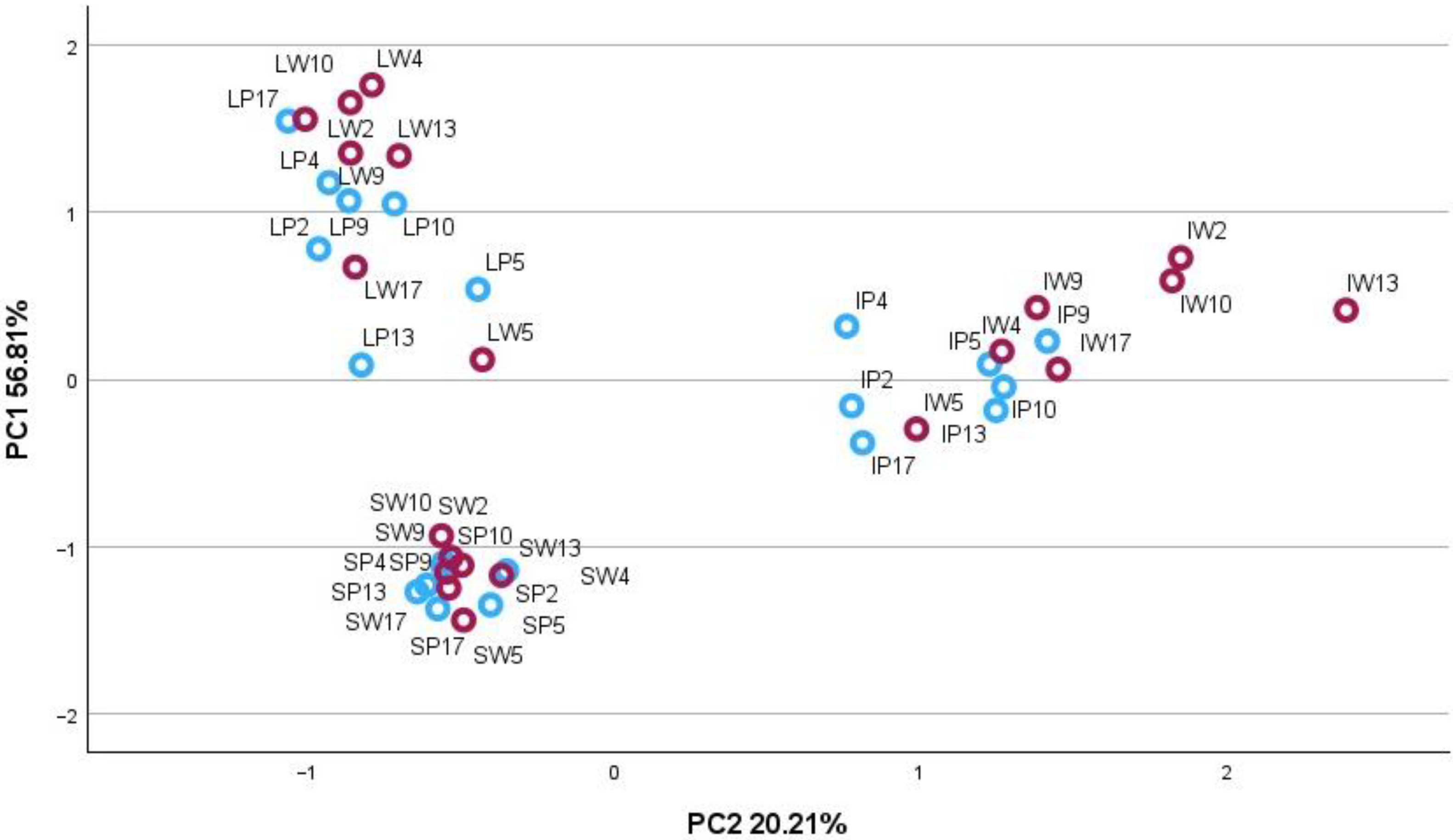
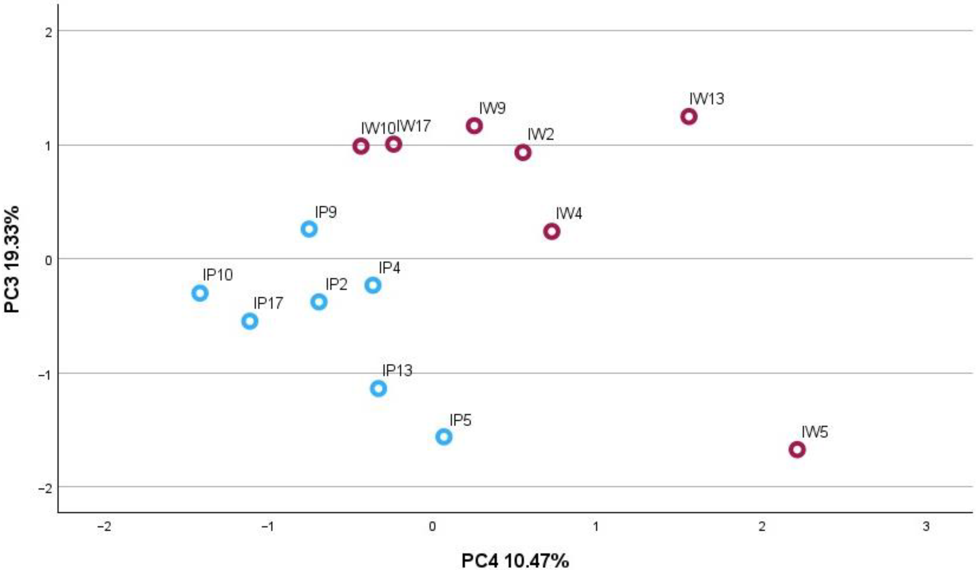
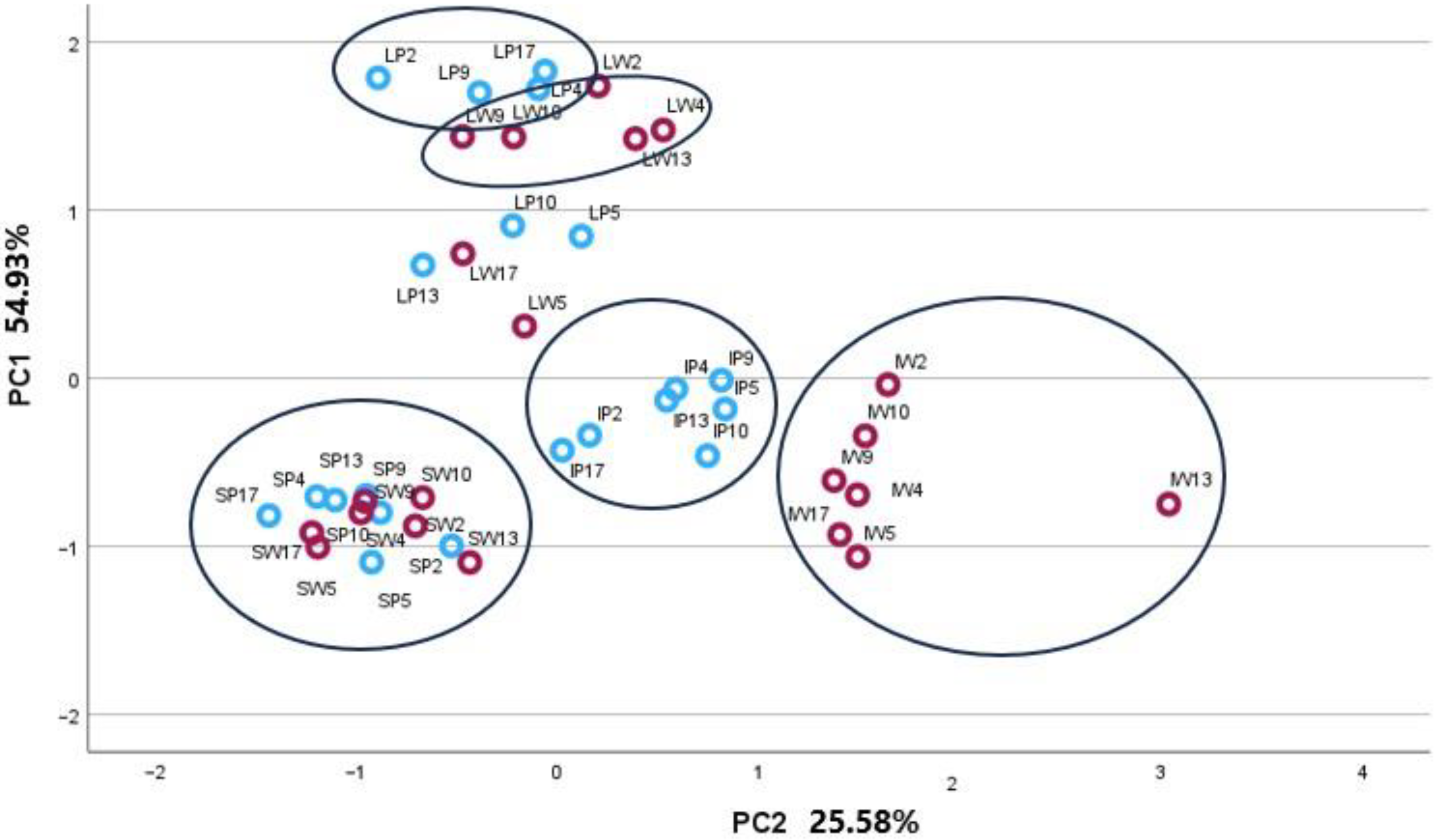
2.2. Principal Component Analysis

Principal component analysis (PCA) was applied to determine the phytochemical relationships between morphotypes in individual plant organs. Individual phenolic and triterpenic compounds and their phytochemical groups were used as variables to create two-dimensional PCA matrix models to visualise the available plant organ-specific patterns of two A. millefolium morphotypes.
The correlation matrix for the variables and principal components of the PCA-1 model accounted for 77.02% of the total variance of the data set of inflorescence, leaf, and stem samples (Figure 1). The PC1 explained for 56.81% of the total variance and was highly correlated with β-sitosterol (0.915), α-amyrin (0.637), luteolin-7-glucuronide (0.882), chlorogenic acid (0.858), 3,4-dicaffeoylquinic acid (0.964), 3,5-dicaffeoylquinic acid (0.937), 1,5-caffeoylquinic acid (0.750), 4,5-dicaffeoylquinic acid (0.854), and luteolin-7-rutinoside (0.801). The PC2 explained 20.21% of the total variance and was positively correlated with betulin (0.875), β-amyrin (0.721), α-amyrin (0.560), luteolin (0.696), luteolin-7-glucoside (0.848), apigenin (0.958), apigenin-7-glucoside (0.956) and negatively with rutin (—0.687). The score plot showed the arrangement of plant organ samples of W and P A. millefolium morphotypes (Figure 1). All samples fell into three distinct plant organ-specific groups of overlapping morphotypes. On the other hand, inflorescence samples tended to group into clusters, coupling the distinct morphotypes. Therefore, the inflorescences were subjected to the PCA-2 model.
The PCA-2 model correlation matrix was constructed for inflorescence samples using triterpenic compounds (Figure 2) and covered 85.56% of the total variance, resulting in four components. The PC1 explained 31.51% of the total variance in the data set and was highly correlated with corosolic acid (0.930), betulin (0.806), and uvaol (0.854). The PC2 accounted for 24.25% of the total variance and was correlated with oleanolic acid (0.821), ursolic acid (0.924), and maslinic acid (0.823). The PC3 described 19.33% of the total variance and was correlated with betulinic acid methyl ester (0.870), β-amyrin (0.807), and β-sitosterol (0.701). The PC4 presented 10.47% of the total variance and was correlated with betulinic acid (0.970) and α-amyrin (0.922). The plotting of PC3 and PC4 presented the best separations between the pink and white morphotype samples. The inflorescences of P A. millefolium were positioned on the positive side of the scatterplot of PC3 and PC4, indicating the highest levels of α, β-amyrins, β-sitosterol, betulin, and betulinic acid methyl ester compared to W samples.
The PCA-3 model was constructed to identify the variance in concentrations of specialised metabolite groups in plant organs and morphotypes. Groups of chemical compounds of similar origin were as follows: sum of titerpenic compounds, sum of triterpenic acids, sum of neutral triterpenes, sum of phytosterols, sum of triterpene esters, sum of phenolic compounds, sum of flavonoids, sum of flavones, sum of flavonols, sum of flavanones, and sum of phenolic acids. The matrix accounted for 80.50% of the total variance of the data set. The PC1 explained 54.93% of the total variance and was correlated with the sum of triterpenic compounds (0.824), the sum of phenolic compounds (0.918), the sum of flavonoids (0.601), the sum of phenolic acids (0.942), the sum of flavonols (0.836), and the sum of flavanones (0.946). The PC2 presented 25.58% of the total variance and was correlated with the sum of triterpenic acids (0.896), the sum of neutral triterpenes (0.839), the sum of phytosterols (0.676), the sum of flavonoids (0.718), and the sum of flavones (0.856). The arrangement of inflorescence and leaf samples in PC1 and PC2 plot space revealed their clustering according to P and W morphotypes, although the stem morphotypes remained in an overlapping position (Figure 3).
Overall, the distant position towards the positive PC2 axis demonstrated the unique peculiarities of the P morphotype inflorescences. Pink inflorescences contained more triterpenic acids, neutral triterpenes, and phytosterols and were marked by high levels of flavones. Several leaf samples tended to cluster by morphotypes, as W samples showed higher amounts of total triterpenic compounds and flavonoids than P ones. Stem samples were positioned at the negative sides of both components and indicated the lowest amounts of all compounds.

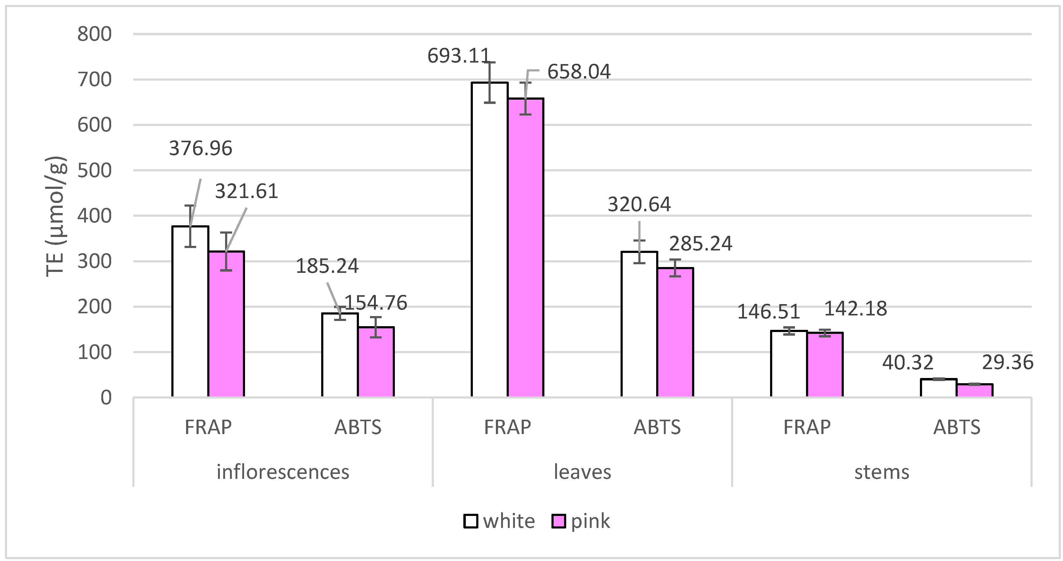
2.3. Assesment of Radical Scavenging and Reducing Activity of Achillea millefolium Extracts

The antioxidant activity of A. millefolium morphotypes was evaluated using two action assays: Ferric Reducing Antioxidant Power (FRAP) and ABTS radical cation decolourization assay (ABTS). The results are presented in Figure 4.
Antioxidant activity values varied significantly between different plant organs tested (p < 0.05). The highest values of radical scavenging and reducing activity were determined for leaf samples of both morphotypes. The mean FRAP values for the pink morphotype were 321.61, 658.04, and 142.18 µmol TE/g for inflorescences, leaves, and stems, respectively. The corresponding ABTS values were 154.76, 285.24, and 29.36 µmol TE/g. The results indicate that the samples of white A. millefolium morphotype exhibit higher antioxidant activity in all three parts of the plant studied as measured by both FRAP and ABTS assays. The antioxidant activity of inflorescences was intermediate, while stems showed the lowest activity, within the range of their variation. These findings suggest that the leaves are the principal source of antioxidant active compounds. The detailed phytochemical analysis of bioactive compounds could contribute to understanding the underlying factors determining these differences. The correlations were established between the antioxidant values and identified compounds. Compounds, namely oleanolic, ursolic, maslinic, corosolic acid, uvaol, β-sitosterol, nicotiflorin, isovitexin, hesperidin, luteolin-7-glucuronide, neochlorogenic acid, chlorogenic acid, cryptochlorogenic acid, 3,4-dicaffeoylquinic acid, 3,5-dicaffeoylquinic acid, 1,5-caffeoylquinic acid, 4,5-dicaffeoylquinic acid, caffeic acid, quercitrin, rutin, isoquercitrin, luteolin-7-rutinoside, santin, and cynarin showed a positive correlation (p < 0.0001) varying in the range of 0.527–0.824 and 0.577–0.923 for the radical scavenging and reducing activities, respectively. The correlation between the total number of determined phenolic compounds was 0.815 and 0.946 (p < 0.0001) for ABTS and FRAP assays, respectively. Furthermore, the correlation between the total amount of triterpenic compounds detected and the ABTS and FRAP assay results was found to be 0.769 and 0.841, respectively.

3. Discussion

The exploration of A. millefolium is still relevant because it represents a plant species of considerable ethnopharmacological and classical medicinal importance [29]. Our specific focus on Achillea species in this context is driven by the potential therapeutic benefits and ecological significance associated with the diverse compounds present in this plant, making it a valuable subject for comprehensive investigation. Although literature data covers various aspects of the phytochemical profiles, there are still undescribed aspects that would add to the scientific knowledge of A. millefolium phytochemical maps [5]. The plant’s ability to grow in different climates and soil types has contributed to its successful establishment in various geographic regions around the world [3]. Our previous study assessed the phytochemical diversity of A. millefolium along geographical gradients. The content of phenolic compounds in yarrow raw material from northern latitudes was found to be more than twice as high as that of southern latitudes, potentially reflecting greater plant adaptation to the respective conditions.
The present study encompasses the colour of flowers used as a differential intraspecious character to discriminate morphotypes as taxa of A. millefolium. The morphological diversity of A. millefolium covers various morphological characters, the most prominent of which is the different colours of flowers. In this context, a previous morphological study of wild A. millefolium showed that among 3345 evaluated plants from 147 cenopopulations, plants with white inflorescences accounted for 78.8% and the rest pink ones [30]. Furthermore, a correlation was found between the inflorescence colour and other morphological characters, which indicated that the colour of inflorescences can be used as a marker to identify intraspecific taxa [30]. In this way, when assessing the diversity potential of yarrow, it was important to find out the relationship between flower colour and the phytochemical composition of the plant material. In this case, chemophenetics is useful for coupling chemotypic and morphotypic variation [31]. Our investigations reveal some variations in the underlying phytochemical profiles of the inflorescence colour-specific morphotypes. Available literature data suggested that the white-flowered A. millefolium morphotype exhibited a different accumulation of certain compounds, such as flavonoids and essential oils, compared to its pink-flowered morphotype [32]. Garzoli et al. found that the aerial parts of the pink morphotype A. millefolium accumulated higher amounts of monoterpenes than the white morphotype [33]. According to the authors, these differences in phytochemical composition between morphotypes suggested a potential adaptation to different ecological conditions or reproductive strategies. However, this assumption contradicts our findings, as white and pink plant samples were collected from the same mixed stands, eliminating the influence of habitat conditions on morphotype diversity. Thus, the observed phytochemical diversity of A. millefolium morphotypes potentially reflected genetic diversity. The colour of the inflorescences can be considered an indicator of a certain chemotype of A. milllefolium plant material.
Our findings revealed that the caffeoylquinic acid complex predominantly shaped the phenolic profile in both pink and white morphotypes of A. millefolium. Mono- and dicaffeoylquinic acids are fast-acting antioxidants and contribute to the total antioxidant activity of plant extracts [5,34]. Moreover, they are also considered the key compounds of hepatoprotective and choleretic activity [35]. Achillea spp. inflorescences were characterised by flavones as organ-specific compounds, while leaves were characterised by flavonols, especially rutin, which is in agreement with our previous studies [23,36]. Achillea spp. flavonoids have been linked with antispasmodic, anti-ulcer, antiproliferative, estrogenic, antioxidant, and anti-inflammatory properties [5]. Antioxidant and anti-inflammatory properties are closely related and play a significant role in the pathogenesis of chronic degenerative diseases [9]. Determining the antioxidant activity of plant extracts is crucial to elucidate their therapeutic and nutritional benefits, as antioxidants play a key role in mitigating oxidative stress. There is no universal method that can accurately assess antioxidant capacity and obtain an unambiguous result. Applying assays with diverse modes of action can provide a deeper understanding of the action of chemical compounds and their potential to counteract oxidative stress through varied mechanisms [37]. Antioxidants can potentially influence the activity of transcription factors related to the immune response, triggering the reduction of pro-inflammatory cytokines [38]. Antioxidants in target sites are capable of free radical quenching or oxidation chain termination, thus participating in cancer chemoprevention [39]. However, significant variations in the concentrations of phenolic compounds and total antioxidant activity were observed between the morphotypes and parts of the plant. These variations highlight the dynamic and adaptive nature of the plant in response to its local environment. Plants alter the phenolic composition and antioxidant capacities of their tissues as an adaptive response to regulate environmental stresses [40].
The analysis of triterpenic profiles uncovered the presence of several biologically active compounds, including oleanolic acid, maslinic acid, corosolic acid, betulinic acid, betulin, uvaol, betulinic acid methyl ester, β-amyrin, β-sitosterol, and α-amyrin. The concentrations of these triterpenic compounds exhibited variability in the tested samples, with potential implications for the medicinal properties of A. millefolium raw material. The available data on compounds of triterpenic origin in the Achillea spp. are scarce. Fifteen compounds of triterpenic origin, including α-amyrin, β-amyrin, and β-sitosterol, were previously identified in A. alexandri-regis [41], while magnificol, β-sitosterol, α-amyrine, and lupeol were detected in A. magnifica. In the methanolic extracts of flowering parts of A. ligustica, the triterpene moretenol was detected as the principal compound, followed by stigmast-6-en-3β-ol, veridiflorol, and β-amyrin [42]. Betulin was identified as the essential oil of endemic A. eriophora from Iran [43]. In addition, four sterols, β-sitosterol, cholesterol, campesterol, and stigmasterol and α and β-amyrins, have been reported in A. millefolium [44]. A fraction of A. millefolium rich in stigmasterol and β-sitosterol has been shown to have skin-calming and anti-inflammatory effects [39]. In addition, triterpene components have significant anti-inflammatory activity and may also contribute to specific pharmacological activity with implications for inflammation and cancerogenesis [45,46]. However, most of the studies were performed on the whole areal part of Achillea spp., without dissecting it into individual plant organs. Studies of individual plant organs allow us to reveal a more targeted use of plant material. In this regard, the leaves of the morphotypes revealed a qualitatively different profile with a characteristic composition of triterpene and phenolic compounds, indicating their additional value. Hence, in the pharmaceutical raw material of A. millefolium, which is defined and used both in the European pharmacopoeia and in folk medicine as a herb (Millefolii herba) or flowers (Millefolii flos) [47,48], leaves with additional value, should be given more attention.
In the current study, phytochemical differences between individual plant organs and morphotypes of A. millefolium were determined using PCA, which highlights the morphotype-dependent variations in phytochemical profiles. This separation is particularly pronounced regarding triterpenic acids, neutral triterpenes, and phytosterols, emphasising the unique chemical makeup of morphotype inflorescences. This work has expanded our understanding of bioactive compounds and their chemical diversity in A. millefolium inflorescence morphotypes, which may contribute to the additional application of this medicinal plant. Differences in specialised metabolites and their chemotypes likely reflect their genetic differences, as both morphotypes originated from the same population. The morphotype can serve as a marker for the selection of a certain chemotype.
To the best of our knowledge, no data have been found regarding phenolic and triterpenic profiling in coloured inflorescence morphotypes of A. millefolium. Furthermore, the databases do not contain information on the pigment composition of coloured inflorescences of A. millefolium morphotypes. Given these gaps in knowledge, future research could focus on investigating the triterpenic profiles and anthocyanin content of Achillea spp. inflorescences, particularly focusing on flower colour variations.

4. Materials and Methods

4.1. Chemicals and Reagents
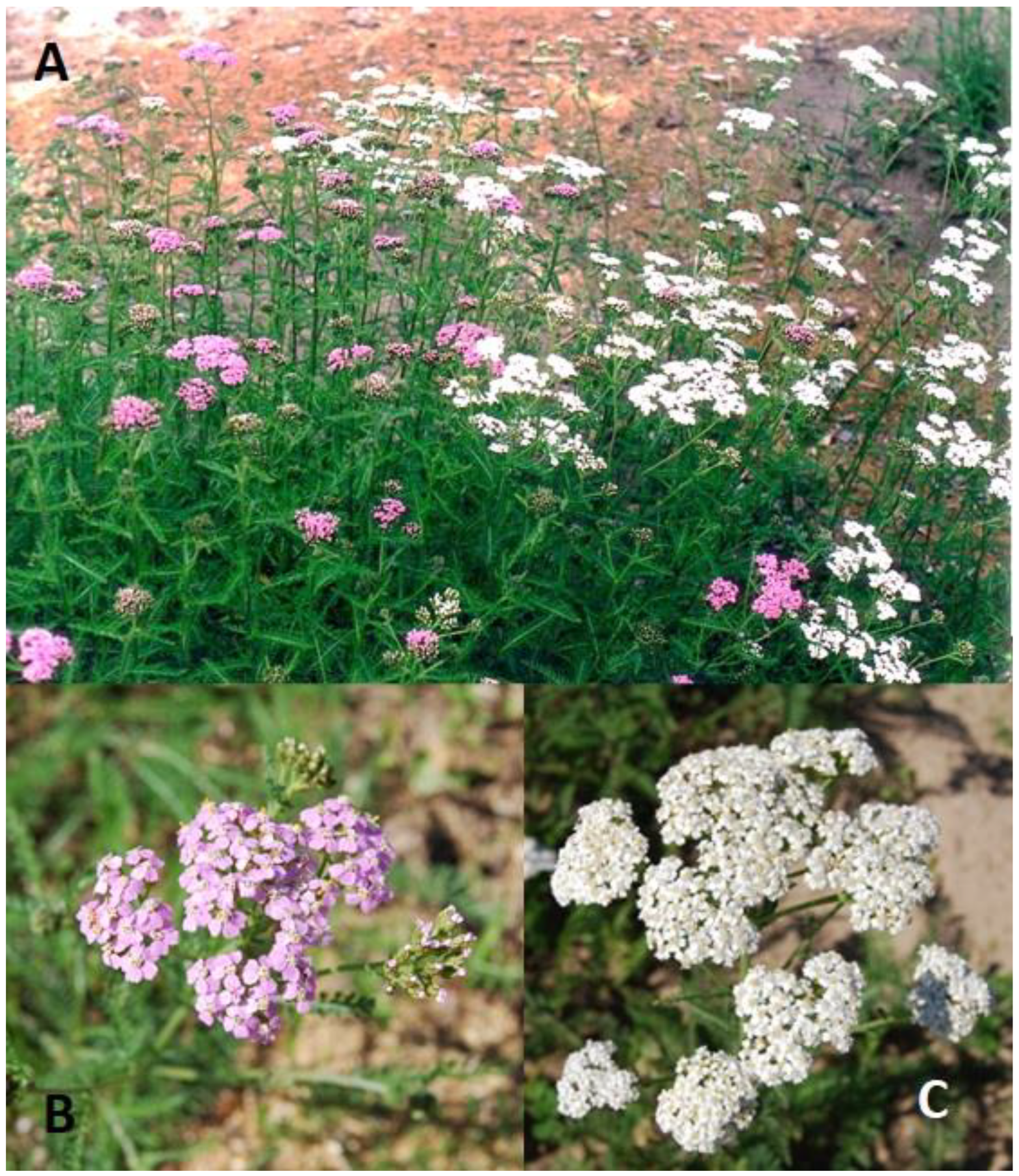
The following analytical or chromatographic grade solvents were used in the study: 99.9% acetonitrile, 99.9% methanol, 99.8% anhydrous acetic acid, and 37% hydrochloric acid from Sigma-Aldrich (Steinheim, Germany); 99.8% trifluoroacetic acid from Merck (Darmstadt, Germany); and purified water was prepared using Milli–Q (Millipore, Bedford, MA, USA) water purification system. The following high purity substances were used: rutin, santin, quercetin, quercetin-3-O-glucoside, nicotiflorin (kaempferol-3-O-rutinoside), luteolin, luteolin-7-O-glucoside, luteolin-7-O-rutinoside, luteolin-7-O-glucuronide, apigenin, apigenin-7-O-glucoside, 3-O-caffeoylquinic acid (chlorogenic acid), 5-O-caffeoylquinic acid (neochlorogenic acid), 4-O-caffeoylquinic acid, 1,3-O-dicaffeoylquinic acid (cynarin), 3,4-O-dicaffeoylquinic acid, 3,5-O-dicaffeoylquinic acid, 1,5-O-dicaffeoylquinic acid, 4,5-O-dicaffeoylquinic acid, α-amyrin, β-amyrin, β-sitosterol, maslinic acid, and oleanolic acid from Sigma-Aldrich; uvaol, betulin, betulinic acid, corosolic acid, and betulinic acid methyl ester from Extrasynthese (Genay, France); ursolic acid from Carl Roth (Karlsruhe, Germany). The following reagents for antioxidant activity determination were used: 6-hydroxy-2,5,7,8-tetramethylchroman-2-carboxylic acid (Trolox), 2,2′-azino-bis(3-ethylbenzothiazoline-6-sulfonic acid) diammonium salt (ABTS), 2,4,6-tri-(2-pyridyl)-S-triazine (TPTZ), ferric chloride (FeCl3), and sodium acetate from Sigma-Aldrich (Steinheim, Germany); and potassium persulfate from Alfa Aesar (Karlsruhe, Germany).

4.2. Plant Material

Aerial parts of the plant of two A. millefolium morphotypes with white (W) and pink (P) inflorescences (Figure 5) were sampled from seven mixed morphotype stand populations (Table 2) during full bloom in June and August 2022. Plant material consisting of 30 single shoots per sample was dissected into inflorescences, leaves, and stems and then dried in a drying cabinet type SSO-80 (Isoterma, Wroclaw, Poland) at 25 °C temperature and 10% relative humidity for 24 h. The botanical identification of species was performed on morphological characters according to descriptors [7,47].
The air-dried plant material samples were ground to homogenous powder using a Retsch 200 mill (Haan, Germany); the material was crushed to particles passing through a 355 μm sieve and kept in the dark in sealed containers until extraction.

4.3. Preparation of Plant Extracts

Extraction of phenolic compounds was performed as follows: 0.1 g of fine powder of dried stems, leaves, or inflorescences were extracted with 10 mL of 70% v/v methanol in water for 30 min at 40 °C in an ultrasonic bath Elmasonic P (Singen, Germany). Extraction of triterpenoids was performed by extracting 1 g of powdered raw material with 10 mL of pure methanol using ultrasound-assisted extraction for 25 min. All obtained extracts were filtered through 0.22 µm pore size membrane filters (Carl Roth GmbH, Karlsruhe, Germany) and stored at 4 °C until analyses.

4.4. HPLC-PDA Conditions

The HPLC-PDA (Waters e2695 Alliance system, Milford, MA, USA) system was used to analyse phenolic and triterpenic compounds. The identification was performed by comparing retention times and spectra of samples to those of commercially available standard substances. The concentration of each compound was calculated based on the external standard method using linear regression models. Quantitative phytochemical profiles were expressed as the mean and standard deviation (SD) from three replicates.
For phenolic compounds analysis, ACE Super C18 (250 mm × 4.6 mm, 3 µm) reversed-phase column (ACT, Aberdeen, UK), maintained at 35 °C, was used. The gradient consisting of eluent A (0.1% trifluoroacetic acid in water) and eluent B (100% acetonitrile) was formed as follows: 0 min, 90% A; 0–40 min, 70% A; 40–60 min, 30% A; 60–64 min, 10% A; 64–70 min, 90% A. The flow rate of 0.5 mL/min and injection volume of 10 µL were set.
Analysis of all triterpenic compounds was carried out on the ACE C18 column (150 mm × 4.6 mm, 3 µm) reversed-phase column (ACT, Aberdeen, UK). For evaluation of triterpenoid acids (maslinic, corosolic, betulinic, oleanolic, and ursolic acids) and neutral triterpenoids with chromophores (betulin and uvaol), isocratic elution consisting of acetonitrile and water (89:11, v/v) at a flow rate of 0.7 mL/min was used. The column temperature was maintained at 20 °C. Chromatographic separation of neutral triterpenoids, which lack chromophores (betulinic acid methyl ester, α-amyrin, and β-amyrin) and phytosterol (β-sitosterol) was performed at isocratic elution consisting of methanol and acetonitrile (90:10, v/v), at a flow rate of 1 mL/min when column temperature was set at 35 °C. The injection volume of triterpenoid extracts was 10 µL.
More data on the methods used were presented in previous papers and in Table S1 [36,49].

4.5. Antioxidant Activity Assays

The antioxidant activity of A. millefolium extracts was analysed using the ABTS radical cation decolourization assay (ABTS) and ferric reducing antioxidant power (FRAP) assays. The ABTS radical cation is described by Re et al. [49]. FRAP was performed using the method described by Benzie and Strain [50] with the modifications as detailed in our previous paper [51,52] using a spectrophotometer (Spectronic CamSpec M550, Garforth, UK). Antiradical and reducing activity results were expressed as micromolar of Trolox equivalents per gram of dry weight of plant material (μmol TE/g, DW).

4.6. Statistical Analysis

Results were expressed as the mean ± standard deviation (SD) from three replicates. Significant differences in chemical compounds among populations and plant organs were tested using one-way analysis of variance (ANOVA) at a confidence level of p < 0.05, followed by the posthoc Duncan’s Multiple Range tests [53]. Pearson’s correlation analysis was performed to explain the relationship between variables and principal components and the p-value obtained by testing the hypothesis on nonlinear regression. The data underwent principal component analysis (PCA), ensuring adequacy through Bartlett’s test [54] of sphericity and the Kaiser–Meyer–Olkin measure of sampling adequacy [55]. Factors with eigenvalues surpassing 1 were taken into consideration. The data were processed using Microsoft Office Excel for 365 Version 2403 (Microsoft, Redmond, WA, USA) and SPSS 29 Version 29.0.1.0 (171) (IBM, Armonk, NY, USA) software.

5. Conclusions

Research on Achillea millefolium remains relevant due to its potential therapeutic benefits and ecological significance associated with its diverse compounds. This study highlighted the morphological diversity within the species, particularly in inflorescence colour-specific morphotypes. The observed variations in phenolic compounds and triterpenic profiles between morphotypes and plant parts potentially reflect the adaptive nature of A. millefolium in response to its local environment and genetic diversity. This study highlighted the chemical differences between A. millefolium morphotypes based on inflorescence colour-specific differences. The phytochemical profile of white and pink inflorescence morphotypes of A. millefolium was characterised by a complex of thirty-four phenolic and triterpene compounds. Significant differences in morphotype-dependent phytochemical profiles were expressed only for triterpenic compounds. In general, white inflorescences accumulated a higher amount of compounds compared to pink inflorescences. The leaves revealed qualitative differences from other plant parts due to the additional presence of ursolic acid, nicoflorin, isoquercitrin, and cynarin, indicating their higher value. Furthermore, the leaves of both morphotypes have been testified as the main source of antioxidant active compounds.
Notably, our search revealed a lack of data on phenolic and triterpenic profiling in the coloured inflorescence of A. millefolium morphotypes, suggesting promising future research to better understand the chemical and morphological diversity of the target species. We assume that the differences in the phytochemical profiles of A. millefolium morphotypes were not significantly affected by habitat, as both morphotypes originated from the same population. Differences in specialised metabolites and their chemotypes likely reflect their genetic differences. The morphotype can serve as a marker for the selection of a certain chemotype. Mapping of chemotypic and morphotypic data with pharmacological activity contributes to drug discovery and development and promotes the responsible and evidence-based use of herbal medicines in healthcare.

Supplementary Materials

The following supporting information can be downloaded at: https://www.mdpi.com/article/10.3390/plants13071043/s1, Table S1: HPLC-PDA methods phenolic and triterpenic compounds identification and quantification parameters.

Author Contributions

Conceptualization, L.R.; methodology, L.R., G.V. and M.M.; software, G.V.; validation, G.V.; formal analysis, L.R.; investigation, G.V. and M.M.; resources, J.R.; data curation, L.R.; writing—original draft preparation, L.R. and G.V.; writing—review and editing, L.R., G.V. and J.R.; visualisation, L.R. and G.V.; supervision, L.R. and J.R. All authors have read and agreed to the published version of the manuscript.

Funding

This research received no external funding.

Data Availability Statement

All data generated during this study are included in this article.

Conflicts of Interest

The authors declare no conflicts of interest.

References

  1. Lourenço, S.C.; Moldão-Martins, M.; Alves, V.D. Antioxidants of Natural Plant Origins: From Sources to Food Industry Applications. Molecules 2019, 24, 4132. [Google Scholar] [CrossRef] [PubMed]
  2. Marino, A.; Battaglini, M.; Moles, N.; Ciofani, G. Natural Antioxidant Compounds as Potential Pharmaceutical Tools against Neurodegenerative Diseases. ACS Omega 2022, 7, 25974–25990. [Google Scholar] [CrossRef] [PubMed]
  3. Hoang, H.T.; Moon, J.Y.; Lee, Y.C. Natural Antioxidants from Plant Extracts in Skincare Cosmetics: Recent Applications, Challenges and Perspectives. Cosmetics 2021, 8, 106. [Google Scholar] [CrossRef]
  4. Nasim, N.; Sandeep, I.S.; Mohanty, S. Plant-Derived Natural Products for Drug Discovery: Current Approaches and Prospects. Nucleus 2022, 65, 399–411. [Google Scholar] [CrossRef] [PubMed]
  5. Ali, S.I.; Gopalakrishnan, B.; Venkatesalu, V. Pharmacognosy, Phytochemistry and Pharmacological Properties of Achillea millefolium L.: A Review. Phytother. Res. 2017, 31, 1140–1161. [Google Scholar] [CrossRef] [PubMed]
  6. Sharma, A.; Nayik, G.A. Immunity Boosting Medicinal Plants of the Western Himalayas; Springer: Singapore, 2023; pp. 1–614. [Google Scholar] [CrossRef]
  7. Richardson, I.B.K. Achillea L. In Flora Europaea; Tutin, T.G., Heywood, V.H., Burges, N.A., Moore, D.M., Valentine, D.H., Walters, S.M., Webb, D.A., Eds.; Cambridge University Press: London, UK, 1976; Volume 4, pp. 159–165. [Google Scholar]
  8. Ehrendorfer, F.; Guo, Y.-P. Multidisciplinary Studies on Achillea Sensu Lato (Compositae-Anthemideae): New Data on Systematics and Phylogeography. Willdenowia 2006, 36, 69–87. [Google Scholar] [CrossRef]
  9. Ayoobi, F.; Shamsizadeh, A.; Fatemi, I.; Vakilian, A.; Allahtavakoli, M.; Hassanshahi, G.; Moghadam-Ahmadi, A. Bio-Effectiveness of the Main Flavonoids of Achillea Millefolium in the Pathophysiology of Neurodegenerative Disorders—A Review. Iran. J. Basic Med. Sci. 2017, 20, 604–612. [Google Scholar] [CrossRef] [PubMed]
  10. Masoodi, M.H.; Rehman, M.U. Edible Plants in Health and Diseases: Volume II: Phytochemical and Pharmacological Properties; Springer Nature: Berlin, Germany, 2022; Volume 2, pp. 1–522. [Google Scholar]
  11. Villalva, M.; Jaime, L.; Villanueva-Bermejo, D.; Lara, B.; Fornari, T.; Reglero, G.; Santoyo, S. Supercritical Anti-Solvent Fractionation for Improving Antioxidant and Anti-Inflammatory Activities of an Achillea millefolium L. Extract. Food Res. Int. 2019, 115, 128–134. [Google Scholar] [CrossRef] [PubMed]
  12. Ahmadi-Dastgerdi, A.; Ezzatpanah, H.; Asgary, S.; Dokhani, S.; Rahimi, E. Phytochemical, Antioxidant and Antimicrobial Activity of the Essential Oil from Flowers and Leaves of Achillea millefolium Subsp. millefolium. J. Essent. Oil Bear. Plants 2017, 20, 395–409. [Google Scholar] [CrossRef]
  13. Farhadi, N.; Babaei, K.; Farsaraei, S.; Moghaddam, M.; Ghasemi Pirbalouti, A. Changes in Essential Oil Compositions, Total Phenol, Flavonoids and Antioxidant Capacity of Achillea millefolium at Different Growth Stages. Ind. Crops Prod. 2020, 152, 112570. [Google Scholar] [CrossRef]
  14. Syakri, S.; Ismail, I.; Amal, N.M.; Masjidi, N.A.; Tahir, K.A. Characterization and Anti-Aging Tests of Peel-Off Gel Masks Made from Ethanolic Extract of Yarrow (Achillea millefolium). Open Access Maced. J. Med. Sci. 2021, 9, 1156–1161. [Google Scholar] [CrossRef]
  15. Villalva, M.; Silvan, J.M.; Alarcón-Cavero, T.; Villanueva-Bermejo, D.; Jaime, L.; Santoyo, S.; Martinez-Rodriguez, A.J. Antioxidant, Anti-Inflammatory, and Antibacterial Properties of an Achillea millefolium L. Extract and Its Fractions Obtained by Supercritical Anti-Solvent Fractionation against Helicobacter Pylori. Antioxidants 2022, 11, 1849. [Google Scholar] [CrossRef] [PubMed]
  16. Fierascu, I.; Ungureanu, C.; Avramescu, S.; Fierascu, R.C.; Ortan, A.; Soare, L.C.; Paunescu, A. In Vitro Antioxidant and Antifungal Properties of Achillea millefolium L. Rom. Biotechnol. Lett. 2015, 20, 10626–10636. [Google Scholar]
  17. Georgieva, L.; Gadjalova, A.; Mihaylova, D.; Pavlov, A. Achillea millefolium L.—Phytochemical Profile and in Vitro Antioxidant Activity. Int. Food Res. J. 2015, 22, 1347–1352. [Google Scholar]
  18. Pant, P.; Pandey, S.; Dall’Acqua, S. The Influence of Environmental Conditions on Secondary Metabolites in Medicinal Plants: A Literature Review. Chem. Biodivers. 2021, 18, e2100345. [Google Scholar] [CrossRef] [PubMed]
  19. Li, Y.; Kong, D.; Fu, Y.; Sussman, M.R.; Wu, H. The Effect of Developmental and Environmental Factors on Secondary Metabolites in Medicinal Plants. Plant Physiol. Biochem. 2020, 148, 80–89. [Google Scholar] [CrossRef] [PubMed]
  20. Ul Hassan, M.; Rasool, T.; Iqbal, C.; Arshad, A.; Abrar, M.; Abrar, M.M.; Habib-ur-Rahman, M.; Noor, M.A.; Sher, A.; Fahad, S. Linking Plants Functioning to Adaptive Responses Under Heat Stress Conditions: A Mechanistic Review. J. Plant Growth Regul. 2022, 41, 2596–2613. [Google Scholar] [CrossRef]
  21. Ncube, B.; Finnie, J.F.; Van Staden, J. Quality from the Field: The Impact of Environmental Factors as Quality Determinants in Medicinal Plants. S. Afr. J. Bot. 2012, 82, 11–20. [Google Scholar] [CrossRef]
  22. Yang, L.; Wen, K.S.; Ruan, X.; Zhao, Y.X.; Wei, F.; Wang, Q. Response of Plant Secondary Metabolites to Environmental Factors. Molecules 2018, 23, 762. [Google Scholar] [CrossRef]
  23. Radušienė, J.; Karpavičienė, B.; Raudone, L.; Vilkickyte, G.; Çırak, C.; Seyis, F.; Yayla, F.; Marksa, M.; Rimkienė, L.; Ivanauskas, L. Trends in Phenolic Profiles of Achillea millefolium from Different Geographical Gradients. Plants 2023, 12, 746. [Google Scholar] [CrossRef]
  24. Mazandarani, M.; Behmanesh, B.; Rezaei, M.B.; Ghaeimi, E.O. Ecological Factors, Chemical Composition and Antibacterial Activity of the Essential Oil from Achillea millefolium L. in the North of Iran. Planta Med. 2007, 73, 179. [Google Scholar] [CrossRef]
  25. Aziz, A.; Badawy, E.M.; Zheljazkov, V.D.; Nicola, S.M.; Fouad, H. Yield and Chemical Composition of Essential Oil of Achillea millefolium L. as Affected by Harvest Time. Egypt J. Chem. 2019, 62, 533–540. [Google Scholar] [CrossRef]
  26. Radzhabov, G.K.; Aliev, A.M.; Musaev, A.M.; Islamova, F.I. Variability of the Constituent Composition of Achillea millefolium Essential Oils in the Wild Flora of Dagestan. Pharm. Chem. J. 2022, 56, 661–666. [Google Scholar] [CrossRef]
  27. Yang, W.; Chen, X.; Li, Y.; Guo, S.; Wang, Z.; Yu, X. Advances in Pharmacological Activities of Terpenoids. Nat. Prod. Commun. 2020, 15, 1934578X20903555. [Google Scholar] [CrossRef]
  28. Żwawiak, J.; Pawełczyk, A.; Olender, D.; Zaprutko, L. Structure and Activity of Pentacyclic Triterpenes Codrugs. A Review. Mini Rev. Med. Chem. 2021, 21, 1509–1526. [Google Scholar] [CrossRef]
  29. Mohammadhosseini, M.; Sarker, S.D.; Akbarzadeh, A. Chemical Composition of the Essential Oils and Extracts of Achillea Species and Their Biological Activities: A Review. J. Ethnopharmacol. 2017, 199, 257–315. [Google Scholar] [CrossRef] [PubMed]
  30. Radusiene, J.; Gudaityte, O. Analysis of Phenotypic Variation in Cenopopulations of Achillea millefolium. Bot. Lith 2005, 11, 205–210. [Google Scholar]
  31. Ovile Mimi, C.; De-la-Cruz-Chacón, I.; Caixeta Sousa, M.; Aparecida Ribeiro Vieira, M.; Ortiz Mayo Marques, M.; Ferreira, G.; Silvia Fernandes Boaro, C. Chemophenetics as a Tool for Distinguishing Morphotypes of Annona emarginata (Schltdl.) H. Rainer. Chem. Biodivers. 2021, 18, e2100544. [Google Scholar] [CrossRef] [PubMed]
  32. Bimbiraite, K.; Ragazinskiene, O.; Maruska, A.; Kornysova, O. Comparison of the Chemical Composition of Four Yarrow (Achillea millefolium L.) Morphotypes. Biologija 2008, 54, 208–2012. [Google Scholar] [CrossRef]
  33. Garzoli, S.; Cicaloni, V.; Salvini, L.; Trespidi, G.; Iriti, M.; Vitalini, S. SPME-GC-MS Analysis of the Volatile Profile of Three Fresh Yarrow (Achillea millefolium L.) Morphotypes from Different Regions of Northern Italy. Separations 2023, 10, 51. [Google Scholar] [CrossRef]
  34. Marksa, M.; Zymone, K.; Ivanauskas, L.; Radušienė, J.; Pukalskas, A.; Raudone, L. Antioxidant Profiles of Leaves and Inflorescences of Native, Invasive and Hybrid Solidago Species. Ind. Crop. Prod. 2020, 145, 112123. [Google Scholar] [CrossRef]
  35. Pereira, C.; Barros, L.; Ferreira, I.C. Extraction, Identification, Fractionation and Isolation of Phenolic Compounds in Plants with Hepatoprotective Effects. J. Sci. Food Agric. 2016, 96, 1068–1084. [Google Scholar] [CrossRef] [PubMed]
  36. Raudone, L.; Radušiene, J.; Seyis, F.; Yayla, F.; Vilkickyte, G.; Marksa, M.; Ivanauskas, L.; Cırak, C. Distribution of Phenolic Compounds and Antioxidant Activity in Plant Parts and Populations of Seven Underutilized Wild Achillea Species. Plants 2022, 11, 447. [Google Scholar] [CrossRef] [PubMed]
  37. Kotha, R.R.; Tareq, F.S.; Yildiz, E.; Luthria, D.L. Oxidative Stress and Antioxidants—A Critical Review on In Vitro Antioxidant Assays. Antioxidants 2022, 11, 2388. [Google Scholar] [CrossRef] [PubMed]
  38. Mucha, P.; Skoczyńska, A.; Małecka, M.; Hikisz, P.; Budzisz, E. Overview of the Antioxidant and Anti-Inflammatory Activities of Selected Plant Compounds and Their Metal Ions Complexes. Molecules 2021, 26, 4886. [Google Scholar] [CrossRef] [PubMed]
  39. Strzępek-Gomółka, M.; Gaweł-Bęben, K.; Kukula-Koch, W. Achillea Species as Sources of Active Phytochemicals for Dermatological and Cosmetic Applications. Oxid. Med. Cell Longev. 2021, 2021, 6643827. [Google Scholar] [CrossRef] [PubMed]
  40. Naikoo, M.I.; Dar, M.I.; Raghib, F.; Jaleel, H.; Ahmad, B.; Raina, A.; Khan, F.A.; Naushin, F. Role and Regulation of Plants Phenolics in Abiotic Stress Tolerance: An Overview. In Plant Signaling Molecules: Role and Regulation under Stressful Environments; Elsevier: Amsterdam, The Netherlands, 2019; pp. 157–168. [Google Scholar] [CrossRef]
  41. Kundakovic, T.; Fokialakis, N.; Magiatis, P.; Kovacevic, N.; Chinou, I. Triterpenic Derivatives of Achillea Alexandri-Regis BORNM. & RUDSKI. Chem. Pharm. Bull. 2004, 52, 1462–1465. [Google Scholar] [CrossRef]
  42. Conforti, F.; Loizzo, M.R.; Statti, G.A.; Menichini, F. Comparative Radical Scavenging and Antidiabetic Activities of Methanolic Extract and Fractions from Achillea ligustica ALL. Biol. Pharm. Bull. 2005, 28, 1791–1794. [Google Scholar] [CrossRef]
  43. Etehadpour, M.; Tavassolian, I. Ecological Factors Regulate Essential Oil Yield, Percent and Compositions of Endemic Yarrow (Achillea eriophora DC.) in Southeast Iran. Int. J. Hortic. Sci. Technol. 2019, 6, 201–215. [Google Scholar]
  44. Chandler, R.F.; Hooper, S.N.; Safe, L.M.; Hooper, D.L.; Jamieson, W.D.; Flinn, C.G. Herbal Remedies of the Maritime Indians: Sterols and Triterpenes of Achillea millefolium L. (Yarrow). J. Pharm. Sci. 1982, 71, 690–693. [Google Scholar] [CrossRef]
  45. Faustino, C.; Pinheiro, L.; Duarte, N. Triterpenes as Potential Drug Candidates for Rheumatoid Arthritis Treatment. Life 2023, 13, 1514. [Google Scholar] [CrossRef] [PubMed]
  46. Vilkickyte, G.; Petrikaite, V.; Marksa, M.; Ivanauskas, L.; Jakstas, V.; Raudone, L. Fractionation and Characterization of Triterpenoids from Vaccinium Vitis-idaea L. Cuticular Waxes and Their Potential as Anticancer Agents. Antioxidants 2023, 12, 465. [Google Scholar] [CrossRef] [PubMed]
  47. European Pharmacopoeia 11. Yarrow. Millefolii herba. 2023. Available online: https://www.edqm.eu/en/european-pharmacopoeia-ph.-eur.-11th-edition (accessed on 26 February 2024).
  48. Applequist, W.L.; Moerman, D.E. Yarrow (Achillea millefolium L.): A Neglected Panacea? A Review of Ethnobotany, Bioactivity, and Biomedical Research. Econ. Bot. 2011, 65, 209–225. [Google Scholar] [CrossRef]
  49. Vilkickyte, G.; Raudone, L. Optimization, Validation and Application of HPLC-PDA Methods for Quantification of Triterpenoids in Vaccinium Vitis-idaea L. Molecules 2021, 26, 1645. [Google Scholar] [CrossRef]
  50. Re, R.; Pellegrini, N.; Proteggente, A.; Pannala, A.; Yang, M.; Rice-Evans, C. Antioxidant Activity Applying An Improved ABTS Radical Cation Decolorization Assay. Free Radic. Biol. Med. 1999, 26, 1231–1237. [Google Scholar] [CrossRef]
  51. Benzie, I.; Strain, J. The Ferric Reducing Ability of Plasma (FRAP) As A Measure Of “Antioxidant Power”: The FRAP Assay. Anal. Biochem. 1996, 239, 70–76. [Google Scholar] [CrossRef]
  52. Cirak, C.; Radusiene, J.; Raudone, L.; Vilkickyte, G.; Seyis, F.; Marksa, M.; Ivanauskas, L.; Yayla, F. Phenolic Compounds and Antioxidant Activity of Achillea arabica Populations. South Afr. J. Bot. 2022, 147, 425–433. [Google Scholar] [CrossRef]
  53. Midway, S.; Robertson, M.; Flinn, S.; Kaller, M. Comparing multiple comparisons:practical guidance for choosing the best multiple comparisons test. PeerJ 2020, 8, e10387. [Google Scholar] [CrossRef]
  54. Arsham, H.; Lovric, M. Bartlett’s Test. In International Encyclopedia of Statistical Science; Lovric, M., Ed.; Springer: Berlin/Heidelberg, Germany, 2011; pp. 87–88. [Google Scholar] [CrossRef]
  55. Kaiser, H.F. An Index of Factorial Simplicity. Psychometrika 1974, 39, 31–36. [Google Scholar] [CrossRef]
Figure 1. PCA-1 score plot model presenting the amounts of predominant phenolic and triterpenic compounds in plant organs (I−inflorescences; L−leaves; S−stems) of P (blue circles) and W (red circles) of A. millefolium morphotypes.
Plants 13 01043 g001
Figure 2. PCA-2 score plot model presenting the amounts of predominant phytochemical compounds in inflorescences (I−inflorescences) of P (blues circles) and W (red circles) A. millefolium morphotypes.
Plants 13 01043 g002
Figure 3. PCA-3 score plot model presenting phytochemical compound groups in plant organs (I−inflorescences; L−leaves; S−stems) of P (blue circles) and W (red circles) Achillea millefolium morphotypes.
Plants 13 01043 g003
Figure 4. Antioxidant activity mean Trolox equivalent (TE) values (µmol/g, DW) of inflorescences, leaves and stems of white and pink A. millefolium morphotypes.
Plants 13 01043 g004
Figure 5. Two wild morphotypes of Achillea millefolium ((A)—mixed stands; (B)—pink morphotype; (C)—white morphotype). Photo by authors.
Plants 13 01043 g005
Table 1. Mean quantities (µg/g, DW) of phenolic and triterpenic compounds in the inflorescence, leaf and stem samples of Achillea milefollium morphotypes.
CompoundIW 1IPLWLPSWSP
Oleanolic acid29.22 ± 14.64 280.03 ± 15.35102.68 ± 44.53139.25 ± 44.530 ± 00 ± 0
ursolic0 ± 00 ± 016.78 ± 00 ± 00 ± 00 ± 0
Maslinic acid18.02 ± 5.1832.96 ± 9.6632.18 ± 13.7158.27 ± 13.7114.31 ± 2.9216.04 ± 6.26
Corosolic acid46.1 ± 15.4478.79 ± 12.1486.81 ± 391.1 ± 35.36 ± 0.551.39 ± 0.86
Betulinic acid72.38 ± 18.52197.27 ± 66.68440.54 ± 78.94519.01 ± 51.770 ± 00 ± 0
Betulin2327.85 ± 726.911114.17 ± 257.8956.93 ± 110.63992.32 ± 153.831016.3 ± 5.5888.95 ± 0
Uvaol24.87 ± 14.4523.38 ± 6.680 ± 00 ± 00 ± 00 ± 0
Betulinic acid methyl ester 226.15 ± 77.43239.78 ± 75.7230.81 ± 63.13225.74 ± 63.1323.84 ± 6.7219.12 ± 2.69
beta-Amyrin571.63 ± 56.64388.28 ± 97.07153.61 ± 33.32115.71 ± 33.3215.15 ± 3.916.31 ± 6.8
beta-Sitosterol283.77 ± 68.47211.71 ± 53.62166.02 ± 50.38165.21 ± 50.38118.36 ± 16.33125.77 ± 31.3
Alfa-amyrin153.83 ± 37.4284.95 ± 10.24297.29 ± 33.4250.44 ± 33.419.41 ± 11.619.08 ± 6.67
Nicotiflorin 0 ± 00 ± 0237.18 ± 33.8138.88 ± 33.80 ± 00 ± 0
Isovitexin14.03 ± 4.0525.18 ± 12.227.15 ± 12.4624.92 ± 8.460 ± 00 ± 0
Hesperidin491.5 ± 152.82670.9 ± 188.851104.06 ± 35.511380.83 ± 50.58238.54 ± 113.48285.37 ± 121.65
Luteolin-7-O-glucuronide2176.98 ± 444.591540.17 ± 310.575068.57 ± 345.753459.83 ± 345.75611.39 ± 302.49292.64 ± 199.77
Neochlorogenic acid402.67 ± 53.65482.4 ± 61.51184.96 ± 99.821190.55 ± 99.82263.4 ± 47.24268.3 ± 52.92
Chlorogenic acid7969.08 ± 226.967848.88 ± 346.6428,123.17 ± 1013.0128,934.28 ± 1864.984583.7 ± 579.724076.34 ± 333.15
4-O-caffeoylquinic acid1313.7 ± 229.52212.6 ± 69.91405.28 ± 393.891058.9 ± 393.89685.87 ± 46.0096 ± 51.24
3.4-O-dicaffeoylquinic acid2545.78 ± 540.082546.82 ± 340.773749.7 ± 784.633338.91 ± 784.63651.83 ± 188.26714.49 ± 184.64
3.5-O-dicaffeoylquinic acid9014.24 ± 732.648502.93 ± 762.6716,064.82 ± 984.8814,974.35 ± 984.881486.04 ± 245.461344.03 ± 503.54
1.5-O-dicaffeoylquinic acid2595.3 ± 213.952414.05 ± 100.414957.9 ± 807.785765.53 ± 807.781070.17 ± 448.67932.41 ± 481.21
4.5-O-dicaffeoylquinic acid1005.02 ± 239.561310.5 ± 188.352075.26 ± 702.212402.77 ± 702.21590.36 ± 230.26605.63 ± 217.05
Caffeic acid17.46 ± 7.658.48 ± 3.9464.14 ± 37.4834.86 ± 17.4833.93 ± 15.5212.54 ± 9.12
Quercitrin174.5 ± 23.63106.46 ± 19.88306.19 ± 134.67229.69 ± 134.67110.86 ± 56.2242.19 ± 19.23
Rutin176.2 ± 84.9304.7 ± 42.422634.46 ± 862.932965.2 ± 1397.95734.99 ± 414.211095.84 ± 440.83
Quercetin27.61 ± 327.8 ± 518.76 ± 8.0121.17 ± 8.0119.83 ± 2.7419.46 ± 3.05
Isoquercitrin0 ± 00 ± 086.01 ± 9.83137.9 ± 27.875.3 ± 0.2542.76 ± 19.14
Luteolin110.98 ± 3.132685.86 ± 384.43144.96 ± 27.41128.29 ± 27.41137.77 ± 7.54133.14 ± 5.5
Luteolin-7-O-glucoside2792.46 ± 446.22016.26 ± 376.031032.26 ± 344.69749.36 ± 344.6989.37 ± 34.8163.85 ± 28.59
Luteolin-7-O-rutinoside756.05 ± 156.91680.43 ± 99.551296.21 ± 655.24780.66 ± 655.24232.89 ± 97.1350.41 ± 34.16
Apigenin559.24 ± 65.3413.08 ± 69.6311.03 ± 2.152.49 ± 0.150 ± 00 ± 0
Apigenin-O-7-glucoside3535.22 ± 462.022724.33 ± 338.68258.94 ± 102.82184.97 ± 102.8227.1 ± 11.622.21 ± 9.07
Santin249.57 ± 25.88220.59 ± 11.77248.04 ± 84.09257.24 ± 84.09207.03 ± 0.93147.97 ± 93.59
Cynarin0 ± 00 ± 074.99 ± 34.5930.2 ± 34.5928.49 ± 23.712.87 ± 6.75
1 IW—White inflorescences; IP—pink inflorescences; LW—white leaves; LP—pink leaves; SW—white stems; SP—pink stems; 2 Values are expressed as mean ± SE.
Table 2. Description of Achillea millefolium collecting locations.
Pop. NoAdministrative LocationLatitude (°N)Longitude (°E)Elevation (m.a.s.l.)Habitat
1Einororys, Alytus distr.54.4461424.38971120Mesophytic grassland
2Geruliai, Alytus distr.54.5312024.27056130Mesophytic grassland
3Užugriovis, Vilnius distr.54.8278225.24644161Dry grassland
4Bernatonys, Vilnius distr.54.9093425.32271160Mesophytic grassland
5Vorėnai, Molėtai distr.55.3577925.61012158Mesophytic grassland
6Juodžionys, Biržai distr.56.2435324.8791560Mesophytic grassland
7Dubingiai, Molėtai distr.55.0591125.43509175Pine forest, roadside
Disclaimer/Publisher’s Note: The statements, opinions and data contained in all publications are solely those of the individual author(s) and contributor(s) and not of MDPI and/or the editor(s). MDPI and/or the editor(s) disclaim responsibility for any injury to people or property resulting from any ideas, methods, instructions or products referred to in the content.

