AI-Assisted Assessment of Wound Tissue with Automatic Color and Measurement Calibration on Images Taken with a Smartphone
Abstract
:1. Introduction
2. Literature Review
2.1. Physiology of Wound Healing
2.2. Wound Tissue Types
2.3. Wound Assessment with Artificial Intelligence
3. Materials and Methods
3.1. Clinical Study
3.2. Data Collection
3.3. Data Annotation
3.4. Wound Assessment Framework
3.5. Automatic Color Calibration
3.5.1. Detection of ArUco Markers
3.5.2. Color Correction
3.6. Deep Learning for Wound Segmentation
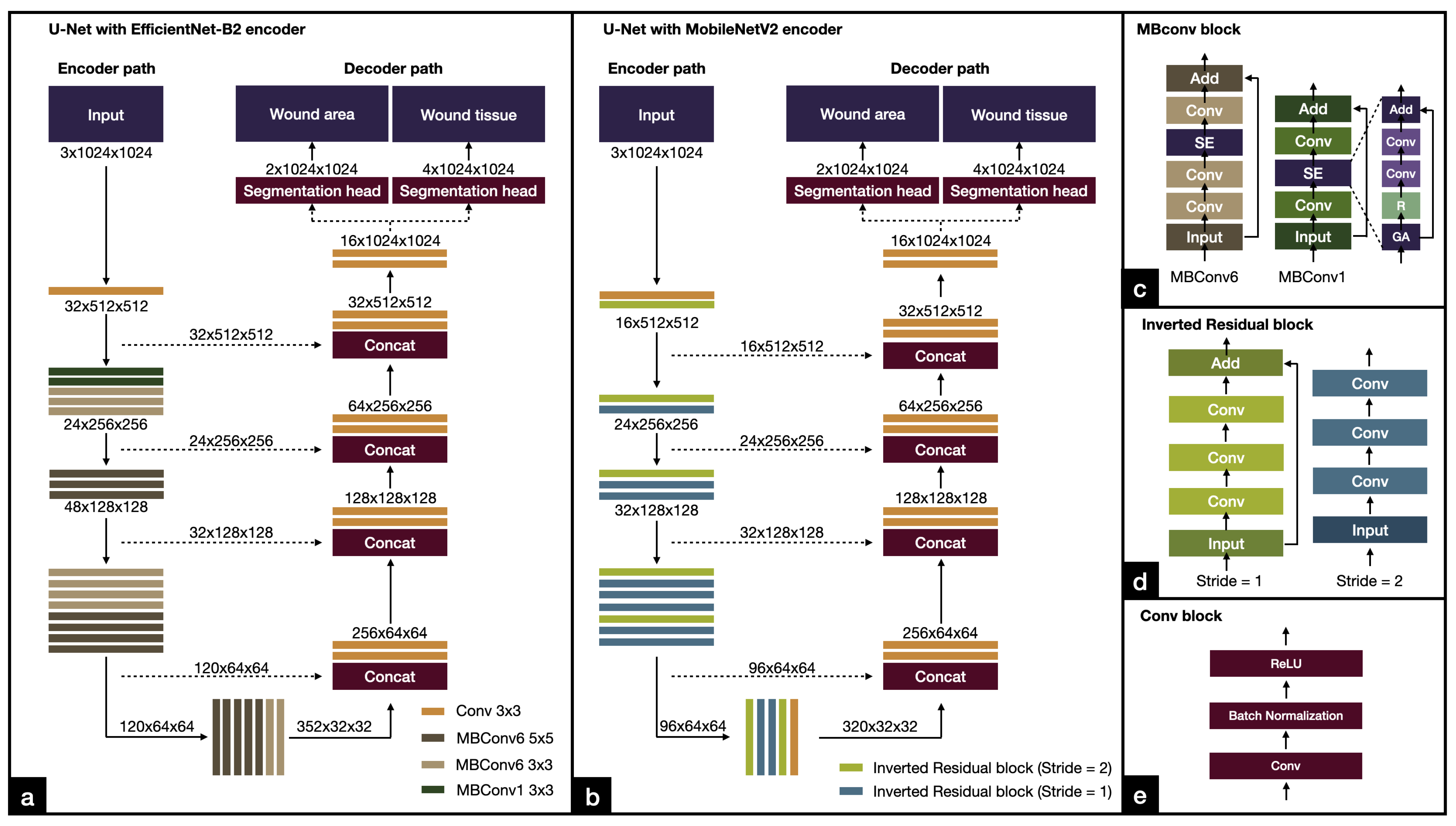
3.6.1. EfficientNet
3.6.2. MobileNetV2
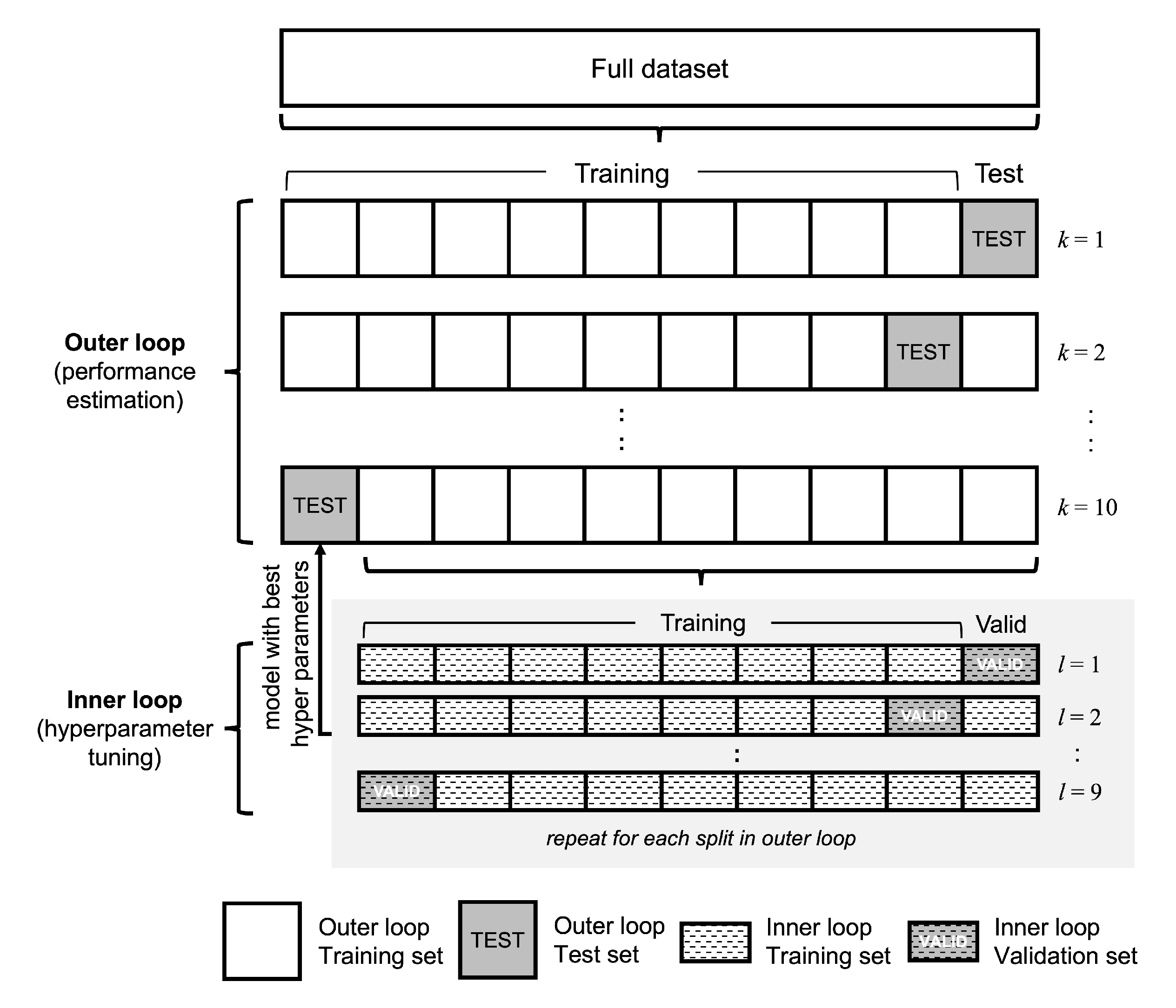
3.6.3. Network Training
3.6.4. Model Evaluation
3.7. Evaluation Metrics
Environment Setup
4. Results
4.1. Inter-Rater Agreement
4.2. Wound Area Segmentation
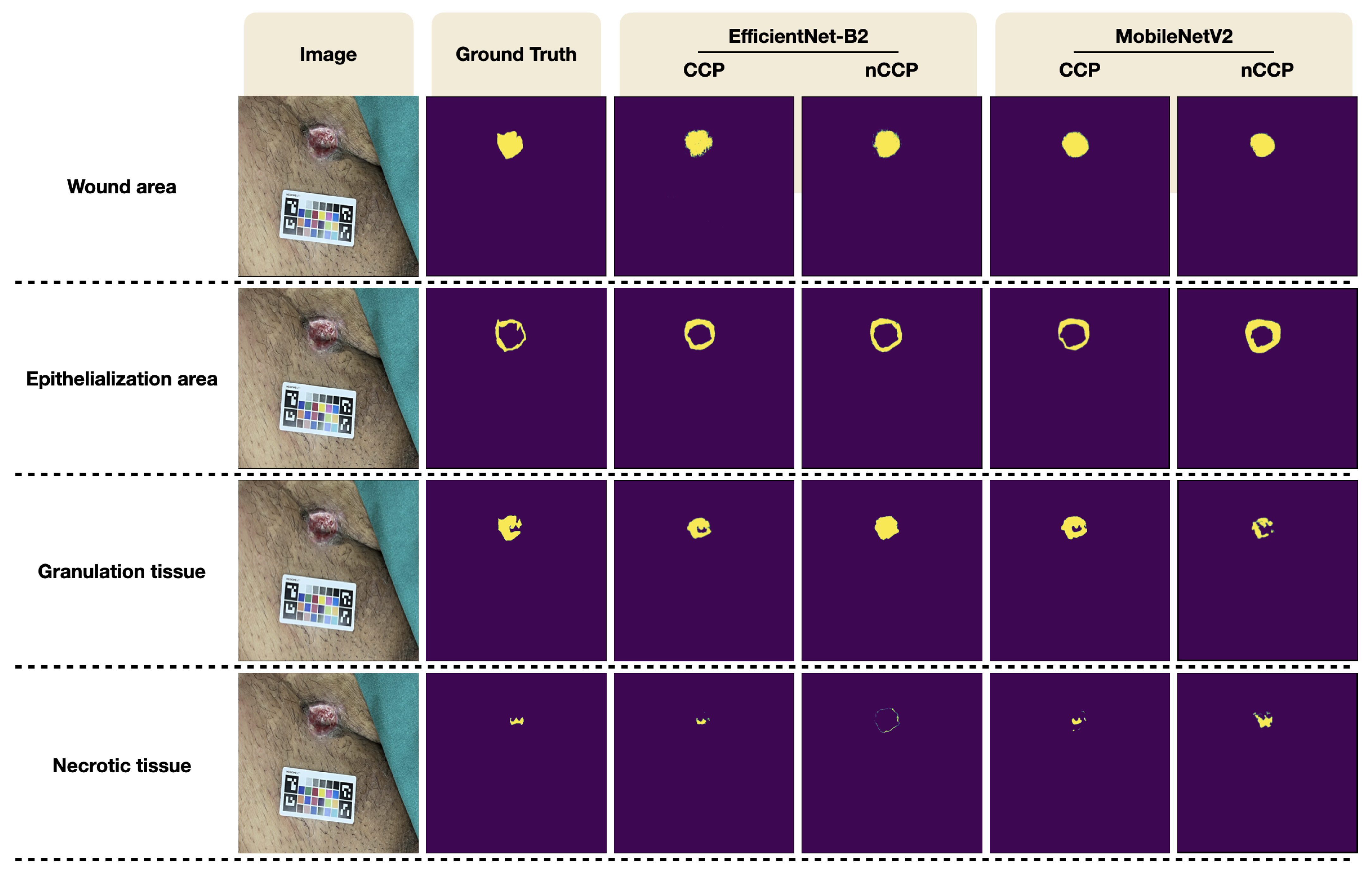
4.3. Wound Tissue Segmentation
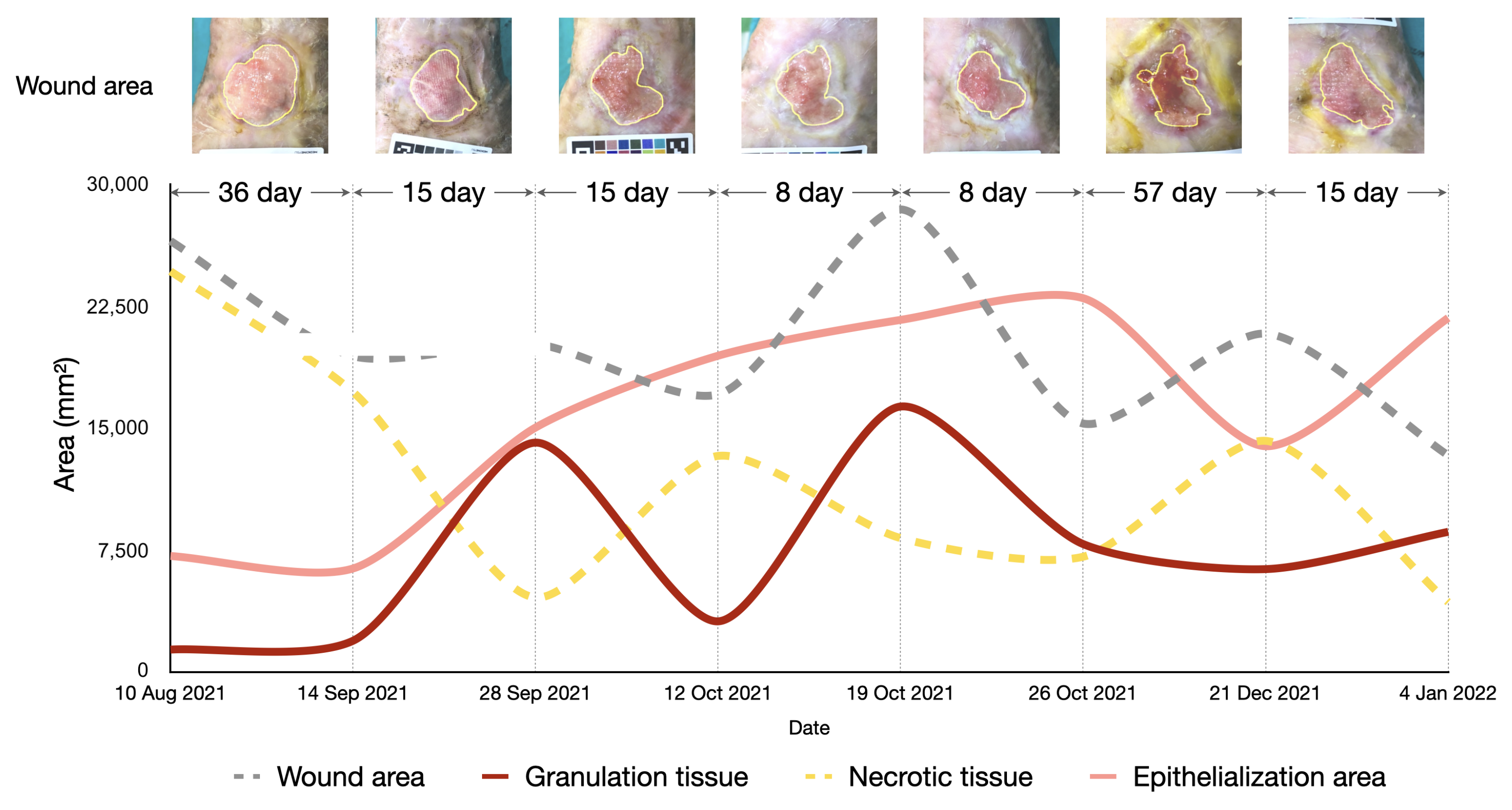
4.4. Longitudinal Wound Assessment
5. Discussion
5.1. Wound Segmentation
5.2. Effects of Color and Measurement Calibration
5.3. Longitudinal Wound Assessment
5.4. Comparison to Other Studies
5.5. Limitations
5.6. Future Work
6. Conclusions
Author Contributions
Funding
Institutional Review Board Statement
Informed Consent Statement
Data Availability Statement
Conflicts of Interest
References
- Martinengo, L.; Olsson, M.; Bajpai, R.; Soljak, M.; Upton, Z.; Schmidtchen, A.; Car, J.; Järbrink, K. Prevalence of chronic wounds in the general population: Systematic review and meta-analysis of observational studies. Ann. Epidemiol. 2019, 29, 8–15. [Google Scholar] [CrossRef] [PubMed]
- Marijanović, D.; Filko, D. A Systematic Overview of Recent Methods for Non-Contact Chronic Wound Analysis. Appl. Sci. 2020, 10, 7613. [Google Scholar] [CrossRef]
- Bowers, S.; Franco, E. Chronic Wounds: Evaluation and Management. Am. Fam. Physician 2020, 101, 159–166. [Google Scholar] [PubMed]
- Keast, D.H.; Bowering, C.K.; Evans, A.W.; Mackean, G.L.; Burrows, C.; D’Souza, L. Contents. MEASURE: A proposed assessment framework for developing best practice recommendations for wound assessment. Wound Repair Regen. 2004, 12, s1–s17. [Google Scholar] [CrossRef] [PubMed]
- Wang, C.; Anisuzzaman, D.M.; Williamson, V.; Dhar, M.K.; Rostami, B.; Niezgoda, J.; Gopalakrishnan, S.; Yu, Z. Fully automatic wound segmentation with deep convolutional neural networks. Sci. Rep. 2020, 10, 21897. [Google Scholar] [CrossRef]
- Gurkan, A.; Kirtil, I.; Aydin, Y.D.; Kutuk, G. Pressure injuries in surgical patients: A comparison of Norton, Braden and Waterlow risk assessment scales. J. Wound Care 2022, 31, 170–177. [Google Scholar] [CrossRef]
- Biagioni, R.B.; Carvalho, B.V.; Manzioni, R.; Matielo, M.F.; Neto, F.C.B.; Sacilotto, R. Smartphone application for wound area measurement in clinical practice. J. Vasc. Surg. Cases Innov. Tech. 2021, 7, 258–261. [Google Scholar] [CrossRef]
- Howell, R.S.; Liu, H.H.; Khan, A.A.; Woods, J.S.; Lin, L.J.; Saxena, M.; Saxena, H.; Castellano, M.; Petrone, P.; Slone, E.; et al. Development of a Method for Clinical Evaluation of Artificial Intelligence–Based Digital Wound Assessment Tools. JAMA Netw. Open 2021, 4, e217234. [Google Scholar] [CrossRef]
- Ramachandram, D.; Ramirez-GarciaLuna, J.L.; Fraser, R.D.J.; Martínez-Jiménez, M.A.; Arriaga-Caballero, J.E.; Allport, J. Fully Automated Wound Tissue Segmentation Using Deep Learning on Mobile Devices: Cohort Study. JMIR mHealth uHealth 2022, 10, e36977. [Google Scholar] [CrossRef]
- Kim, P.J.; Homsi, H.A.; Sachdeva, M.; Mufti, A.; Sibbald, R.G. Chronic Wound Telemedicine Models Before and During the COVID-19 Pandemic: A Scoping Review. Adv. Skin Wound Care 2022, 35, 87–94. [Google Scholar] [CrossRef]
- Lathan, R.; Sidapra, M.; Yiasemidou, M.; Long, J.; Totty, J.; Smith, G.; Chetter, I. Diagnostic accuracy of telemedicine for detection of surgical site infection: A systematic review and meta-analysis. NPJ Digit. Med. 2022, 5. [Google Scholar] [CrossRef] [PubMed]
- Chang, C.W.; Christian, M.; Chang, D.H.; Lai, F.; Liu, T.J.; Chen, Y.S.; Chen, W.J. Deep learning approach based on superpixel segmentation assisted labeling for automatic pressure ulcer diagnosis. PLoS ONE 2022, 17, e0264139. [Google Scholar] [CrossRef] [PubMed]
- Veredas, F.J.; Mesa, H.; Morente, L. Efficient detection of wound-bed and peripheral skin with statistical colour models. Med. Biol. Eng. Comput. 2015, 53, 345–359. [Google Scholar] [CrossRef] [PubMed]
- Casal-Guisande, M.; Comesaña-Campos, A.; Cerqueiro-Pequeño, J.; Bouza-Rodríguez, J.B. Design and Development of a Methodology Based on Expert Systems, Applied to the Treatment of Pressure Ulcers. Diagnostics 2020, 10, 614. [Google Scholar] [CrossRef]
- Kujath, P.; Michelsen, A. Wounds—From Physiology to Wound Dressing. Deutsch. Ärztebl. Int. 2008, 105, 239–248. [Google Scholar] [CrossRef]
- Singh, M. Incorporating vascular-stasis based blood perfusion to evaluate the thermal signatures of cell-death using modified Arrhenius equation with regeneration of living tissues during nanoparticle-assisted thermal therapy. Int. Commun. Heat Mass Transf. 2022, 135, 106046. [Google Scholar] [CrossRef]
- Percival, N.J. Classification of Wounds and their Management. Surgery 2002, 20, 114–117. [Google Scholar] [CrossRef]
- Dhivya, S.; Padma, V.V.; Santhini, E. Wound dressings—A review. BioMedicine 2015, 5. [Google Scholar] [CrossRef]
- Lee, C.K.; Hansen, S.L. Management of Acute Wounds. Surg. Clin. N. Am. 2009, 89, 659–676. [Google Scholar] [CrossRef]
- Goldberg, E.; Beitz, J.M. The lived experience of diverse elders with chronic wounds. Ostomy Wound Manag. 2010, 56, 36–46. [Google Scholar]
- Wild, T.; Rahbarnia, A.; Kellner, M.; Sobotka, L.; Eberlein, T. Basics in nutrition and wound healing. Nutrition 2010, 26, 862–866. [Google Scholar] [CrossRef] [PubMed]
- Gale, A.J. Current Understanding of Hemostasis. Toxicol. Pathol. 2011, 39, 273–280. [Google Scholar] [CrossRef] [PubMed] [Green Version]
- Baum, C.L.; Arpey, C.J. Normal Cutaneous Wound Healing: Clinical Correlation with Cellular and Molecular Events. Dermatol. Surg. 2006, 31, 674–686. [Google Scholar] [CrossRef] [PubMed]
- Dalisson, B.; Barralet, J. Bioinorganics and Wound Healing. Adv. Healthc. Mater. 2019, 8, 1900764. [Google Scholar] [CrossRef]
- Pastar, I.; Stojadinovic, O.; Yin, N.C.; Ramirez, H.; Nusbaum, A.G.; Sawaya, A.; Patel, S.B.; Khalid, L.; Isseroff, R.R.; Tomic-Canic, M. Epithelialization in Wound Healing: A Comprehensive Review. Adv. Wound Care 2014, 3, 445–464. [Google Scholar] [CrossRef] [Green Version]
- Sood, A.; Granick, M.S.; Tomaselli, N.L. Wound Dressings and Comparative Effectiveness Data. Adv. Wound Care 2014, 3, 511–529. [Google Scholar] [CrossRef] [Green Version]
- Kalogeris, T.; Baines, C.P.; Krenz, M.; Korthuis, R.J. Cell Biology of Ischemia/Reperfusion Injury. Int. Rev. Cell Mol. Biol. 2012, 298, 229–317. [Google Scholar] [CrossRef] [Green Version]
- Sheehan, P.; Jones, P.; Caselli, A.; Giurini, J.M.; Veves, A. Percent Change in Wound Area of Diabetic Foot Ulcers Over a 4-Week Period Is a Robust Predictor of Complete Healing in a 12-Week Prospective Trial. Diabetes Care 2003, 26, 1879–1882. [Google Scholar] [CrossRef] [Green Version]
- Howard, A.G.; Zhu, M.; Chen, B.; Kalenichenko, D.; Wang, W.; Weyand, T.; Andreetto, M.; Adam, H. MobileNets: Efficient Convolutional Neural Networks for Mobile Vision Applications. arXiv 2017, arXiv:1704.04861. [Google Scholar] [CrossRef]
- Ronneberger, O.; Fischer, P.; Brox, T. U-Net: Convolutional Networks for Biomedical Image Segmentation. arXiv 2015, arXiv:1505.04597. [Google Scholar] [CrossRef]
- He, K.; Zhang, X.; Ren, S.; Sun, J. Deep Residual Learning for Image Recognition. arXiv 2015, arXiv:1512.03385. [Google Scholar] [CrossRef]
- Tan, M.; Le, Q.V. EfficientNet: Rethinking Model Scaling for Convolutional Neural Networks. arXiv 2019, arXiv:1905.11946. [Google Scholar] [CrossRef]
- Liu, X.; Wang, C.; Li, F.; Zhao, X.; Zhu, E.; Peng, Y. A framework of wound segmentation based on deep convolutional networks. In Proceedings of the 2017 10th International Congress on Image and Signal Processing, BioMedical Engineering and Informatics (CISP-BMEI), Shanghai, China, 14–16 October 2017; pp. 1–7. [Google Scholar] [CrossRef]
- Pholberdee, N.; Pathompatai, C.; Taeprasartsit, P. Study of Chronic Wound Image Segmentation: Impact of Tissue Type and Color Data Augmentation. In Proceedings of the 2018 15th International Joint Conference on Computer Science and Software Engineering (JCSSE), Nakhonpathom, Thailand, 11–13 July 2018; IEEE: Piscataway, NJ, USA, 2018. [Google Scholar] [CrossRef]
- Wannous, H.; Lucas, Y.; Treuillet, S. Enhanced Assessment of the Wound-Healing Process by Accurate Multiview Tissue Classification. IEEE Trans. Med Imaging 2011, 30, 315–326. [Google Scholar] [CrossRef] [PubMed] [Green Version]
- Liu, C.; Fan, X.; Guo, Z.; Mo, Z.; Chang, E.I.C.; Xu, Y. Wound area measurement with 3D transformation and smartphone images. BMC Bioinform. 2019, 20. [Google Scholar] [CrossRef] [PubMed] [Green Version]
- Barbosa, F.M.; Carvalho, B.M.; Gomes, R.B. Accurate Chronic Wound Area Measurement using Structure from Motion. In Proceedings of the 2020 IEEE 33rd International Symposium on Computer-Based Medical Systems (CBMS), Rochester, MN, USA, 28–30 July 2020; IEEE: Piscataway, NJ, USA, 2020; pp. 208–213. [Google Scholar] [CrossRef]
- McCamy, C.S.; Marcus, H.; Davidson, J.G. A color-rendition chart. J. Appl. Photogr. Eng. 1976, 2, 95–99. [Google Scholar]
- Garrido-Jurado, S.; Muñoz-Salinas, R.; Madrid-Cuevas, F.; Marín-Jiménez, M. Automatic generation and detection of highly reliable fiducial markers under occlusion. Pattern Recognit. 2014, 47, 2280–2292. [Google Scholar] [CrossRef]
- Gehan, M.A.; Fahlgren, N.; Abbasi, A.; Berry, J.C.; Callen, S.T.; Chavez, L.; Doust, A.N.; Feldman, M.J.; Gilbert, K.B.; Hodge, J.G.; et al. PlantCV v2: Image analysis software for high-throughput plant phenotyping. PeerJ 2017, 5, e4088. [Google Scholar] [CrossRef]
- Sandler, M.; Howard, A.; Zhu, M.; Zhmoginov, A.; Chen, L.C. MobileNetV2: Inverted Residuals and Linear Bottlenecks. arXiv 2018, arXiv:1801.04381. [Google Scholar] [CrossRef]
- Scebba, G.; Zhang, J.; Catanzaro, S.; Mihai, C.; Distler, O.; Berli, M.; Karlen, W. Detect-and-segment: A deep learning approach to automate wound image segmentation. Inform. Med. Unlocked 2022, 29, 100884. [Google Scholar] [CrossRef]
- Sunoj, S.; Igathinathane, C.; Saliendra, N.; Hendrickson, J.; Archer, D. Color calibration of digital images for agriculture and other applications. ISPRS J. Photogramm. Remote Sens. 2018, 146, 221–234. [Google Scholar] [CrossRef]
- Rogers, L.C.; Bevilacqua, N.J.; Armstrong, D.G.; Andros, G. Digital Planimetry Results in More Accurate Wound Measurements: A Comparison to Standard Ruler Measurements. J. Diabetes Sci. Technol. 2010, 4, 799–802. [Google Scholar] [CrossRef] [PubMed] [Green Version]
- Chan, K.S.; Chan, Y.M.; Tan, A.H.M.; Liang, S.; Cho, Y.T.; Hong, Q.; Yong, E.; Chong, L.R.C.; Zhang, L.; Tan, G.W.L.; et al. Clinical validation of an artificial intelligence-enabled wound imaging mobile application in diabetic foot ulcers. Int. Wound J. 2021, 19, 114–124. [Google Scholar] [CrossRef] [PubMed]
- Wagh, A.; Jain, S.; Mukherjee, A.; Agu, E.; Pedersen, P.C.; Strong, D.; Tulu, B.; Lindsay, C.; Liu, Z. Semantic Segmentation of Smartphone Wound Images: Comparative Analysis of AHRF and CNN-Based Approaches. IEEE Access 2020, 8, 181590–181604. [Google Scholar] [CrossRef] [PubMed]
- Singh, M.; Singh, T.; Soni, S. Pre-operative Assessment of Ablation Margins for Variable Blood Perfusion Metrics in a Magnetic Resonance Imaging Based Complex Breast Tumour Anatomy: Simulation Paradigms in Thermal Therapies. Comput. Methods Programs Biomed. 2021, 198, 105781. [Google Scholar] [CrossRef]


| Phase | Description |
|---|---|
| Hemostasis | During wound development, the loss of tissue integrity results in bleeding that exposes the blood to various extracellular matrix (ECM) components. The bleeding is stopped by vasoconstriction and platelet plugging, which causes blood vessels to constrict, blood flow to slow or block, and platelets to aggregate. This leads to hemostasis and fibrin formation. Fibrin acts as a temporary matrix for cell migration. Fibrinolysis is then activated to dissolve the fibrin and provide space for further cell migration into the next phase of wound healing [22]. |
| Inflammation | At the onset of inflammation, fibrin is broken down and vessels become permeable, releasing plasma and allowing inflammatory cells (neutrophils and macrophages) to migrate to the wound site. Neutrophils arrive within 24–48 h and help defend against infection by phagocytizing debris and pathogens. Macrophages, derived from blood monocytes, arrive 24–36 h later and aid in the removal of bacteria and debris by phagocytosis. They also secrete growth factors and cytokines that stimulate new blood vessel formation, granulation, and re-epithelialization [23]. |
| Proliferation | The proliferation phase mainly involves the formation of granulation tissue and the immigration of keratinocytes on the wound bed. Keratinocytes stimulate the regeneration of epithelial tissue and restore the continuity of the epidermal layer. New tissue forms from a matrix of collagen, elastin, glycosaminoglycans, and other fibrous proteins, as well as fibrin and fibronectin. Fibroblasts produce and deposit extracellular proteins, including growth factors and angiogenic factors, which control the cell proliferation and angiogenesis [23]. |
| Remodeling | The remodeling phase begins when collagen synthesis and degradation are balanced and the wound has closed. During this phase, the body replaces the temporary tissue formed during the inflammation and proliferation phases with new, stronger tissue. It also works to improve the appearance of the scar and adjust the tension of the scar tissue to match the surrounding tissue [24]. |
| Tissue Type | Description |
|---|---|
| Granulation tissue | New connective tissue and microvessels form the granulation tissue, which is the main carrier of oxygen and nutrients during the healing process. The characteristic of granulation tissue is a rough and moist surface on the wound bed. Granulation tissue appears pale pink in the wound bed or bright, beefy red as the depth of the wound bed increases. In addition, granulation tissue provides a scaffold for the epithelialization process to form and cover the wound. |
| Epithelial tissue | Epithelialization is a complex process involving morphogenesis, cell proliferation, cell differentiation, and migration with substantial mitotic activity at the wound edge [25]. New epithelial tissue appears deep pink at the wound site as basal keratinocytes migrate to the wound surface or the epidermis regenerates. New epithelial tissue covers the wound from the edge to the center. Postoperative wound dressings have been used to accelerate and optimize the healing process by providing protection from contaminants, absorbing exudate and humidity, and promoting autolytic debridement [26]. |
| Necrotic tissue | Necrosis is the death of body tissue. Cell death occurs due to lack of oxygen and interrupted blood supply for a long enough time [27]. Necrotic tissue can be divided into two main types: eschar is a dry, dark tissue that can thickly cover a wound bed; and slough is a soft, moist tissue that contains dead tissue and bacteria. Its color may range from yellow to green to tan to brown. Necrosis tissue cannot be salvaged and must be removed by appropriate wound debridement techniques to allow wound healing to occur [16,26]. |
| Characteristic | Value |
|---|---|
| Age, mean ± SD [Range] | 64.61 ± 20.41 |
| [17–85] | |
| Sex, N (%) | |
| Male | 11 (55.00) |
| Female | 9 (45.00) |
| Wound type, N (%) | |
| Infection/inflammation | 14 (45.16) |
| Pressure ulcer | 8 (25.81) |
| Burn | 4 (12.90) |
| Trauma | 4 (12.90) |
| Diabetics | 1 (3.23) |
| Wound location, N (%) | |
| Lower extremity | 17 (54.84) |
| Foot (excluding heel) | 5 (16.13) |
| Sacrum/coccyx | 5 (16.13) |
| Upper/lower back | 3 (9.68) |
| Chest/breast | 1 (3.23) |
| Exudate, N (%) | |
| Serous | 7 (22.58) |
| Purulent | 1 (3.23) |
| Serosanguinous | 1 (3.23) |
| Amount, N (%) | |
| Minimal | 14 (45.16) |
| Moderate | 3 (9.68) |
| Dressing material, N (%) | |
| Foam | 8 (25.81) |
| Gel | 7 (22.58) |
| Non-adhesive mesh | 6 (19.35) |
| Aquacel Ag | 3 (9.68) |
| Foam/non-adhesive mesh | 1 (3.23) |
| Gauze | 1 (3.23) |
| Granudacyn solution | 1 (3.23) |
| Feature | Physicians | Intersection over Union | |
|---|---|---|---|
| Mean | S.D. | ||
| Wound area | A vs. B | 0.6739 | 0.2374 |
| A vs. C | 0.5143 | 0.2303 | |
| B vs. C | 0.6559 | 0.2483 | |
| Epithelialization area | A vs. B | 0.3248 | 0.2087 |
| A vs. C | 0.2850 | 0.2609 | |
| B vs. C | 0.2368 | 0.2261 | |
| Granulation tissue | A vs. B | 0.5438 | 0.3316 |
| A vs. C | 0.5561 | 0.2303 | |
| B vs. C | 0.6201 | 0.2877 | |
| Necrotic tissue | A vs. B | 0.2680 | 0.2763 |
| A vs. C | 0.1635 | 0.2473 | |
| B vs. C | 0.1125 | 0.1991 | |
| Feature | Architecture | Encoder | Color Calibration | Intersection over Union | Pixel Accuracy | ||
|---|---|---|---|---|---|---|---|
| Mean | S.D. | Mean | S.D. | ||||
| Wound area | U-Net | MobileNetV2 | Yes | 0.6826 | 0.1987 | 0.9881 | 0.0105 |
| No | 0.6624 | 0.2307 | 0.9854 | 0.0219 | |||
| EfficientNet-B2 | Yes | 0.6964 | 0.1359 | 0.9866 | 0.0114 | ||
| No | 0.6878 | 0.1665 | 0.9871 | 0.0101 | |||
| Epithelialization area | U-Net | MobileNetV2 | Yes | 0.3186 | 0.1630 | 0.9792 | 0.0153 |
| No | 0.2854 | 0.1552 | 0.9770 | 0.0179 | |||
| EfficientNet-B2 | Yes | 0.3957 | 0.1869 | 0.9808 | 0.0157 | ||
| No | 0.3672 | 0.1517 | 0.9812 | 0.0145 | |||
| Granulation tissue | U-Net | MobileNetV2 | Yes | 0.5997 | 0.2885 | 0.9899 | 0.0086 |
| No | 0.5879 | 0.2633 | 0.9921 | 0.0048 | |||
| EfficientNet-B2 | Yes | 0.6421 | 0.2444 | 0.9920 | 0.0064 | ||
| No | 0.6297 | 0.2529 | 0.9915 | 0.0084 | |||
| Necrotic tissue | U-Net | MobileNetV2 | Yes | 0.1386 | 0.1467 | 0.9831 | 0.0133 |
| No | 0.1185 | 0.1754 | 0.9858 | 0.0125 | |||
| EfficientNet-B2 | Yes | 0.1552 | 0.1428 | 0.9853 | 0.0123 | ||
| No | 0.1195 | 0.1040 | 0.9896 | 0.0073 | |||
Disclaimer/Publisher’s Note: The statements, opinions and data contained in all publications are solely those of the individual author(s) and contributor(s) and not of MDPI and/or the editor(s). MDPI and/or the editor(s) disclaim responsibility for any injury to people or property resulting from any ideas, methods, instructions or products referred to in the content. |
© 2023 by the authors. Licensee MDPI, Basel, Switzerland. This article is an open access article distributed under the terms and conditions of the Creative Commons Attribution (CC BY) license (https://creativecommons.org/licenses/by/4.0/).
Share and Cite
Chairat, S.; Chaichulee, S.; Dissaneewate, T.; Wangkulangkul, P.; Kongpanichakul, L. AI-Assisted Assessment of Wound Tissue with Automatic Color and Measurement Calibration on Images Taken with a Smartphone. Healthcare 2023, 11, 273. https://doi.org/10.3390/healthcare11020273
Chairat S, Chaichulee S, Dissaneewate T, Wangkulangkul P, Kongpanichakul L. AI-Assisted Assessment of Wound Tissue with Automatic Color and Measurement Calibration on Images Taken with a Smartphone. Healthcare. 2023; 11(2):273. https://doi.org/10.3390/healthcare11020273
Chicago/Turabian StyleChairat, Sawrawit, Sitthichok Chaichulee, Tulaya Dissaneewate, Piyanun Wangkulangkul, and Laliphat Kongpanichakul. 2023. "AI-Assisted Assessment of Wound Tissue with Automatic Color and Measurement Calibration on Images Taken with a Smartphone" Healthcare 11, no. 2: 273. https://doi.org/10.3390/healthcare11020273
APA StyleChairat, S., Chaichulee, S., Dissaneewate, T., Wangkulangkul, P., & Kongpanichakul, L. (2023). AI-Assisted Assessment of Wound Tissue with Automatic Color and Measurement Calibration on Images Taken with a Smartphone. Healthcare, 11(2), 273. https://doi.org/10.3390/healthcare11020273


