3.1. Spectrometer Delay and Spectral Stability
When high-energy laser pulses interact with the sample, atoms and molecules within the sample become excited, resulting in the formation of a high-temperature, high-density plasma. Particles within the plasma, possessing elevated energy levels, emit characteristic spectral signals upon returning to their ground state. Closely associated with this process is the collection delay of the spectrometer, which is the time from the emission of the laser pulse to the initiation of spectrum collection for the plasma emission. Due to the relatively short lifetime of the laser-induced plasma, the precise selection of the spectrometer’s collection delay is directly related to the timing of interactions, such as the laser pulse with the sample, the plasma with the sample, and the plasma with the surrounding gas. Both excessively short or long delays may influence the incomplete formation or premature diffusion of the collected plasma emission spectrum region, consequently directly impacting the intensity, stability, and shape of the spectral signal [
2,
3,
17].
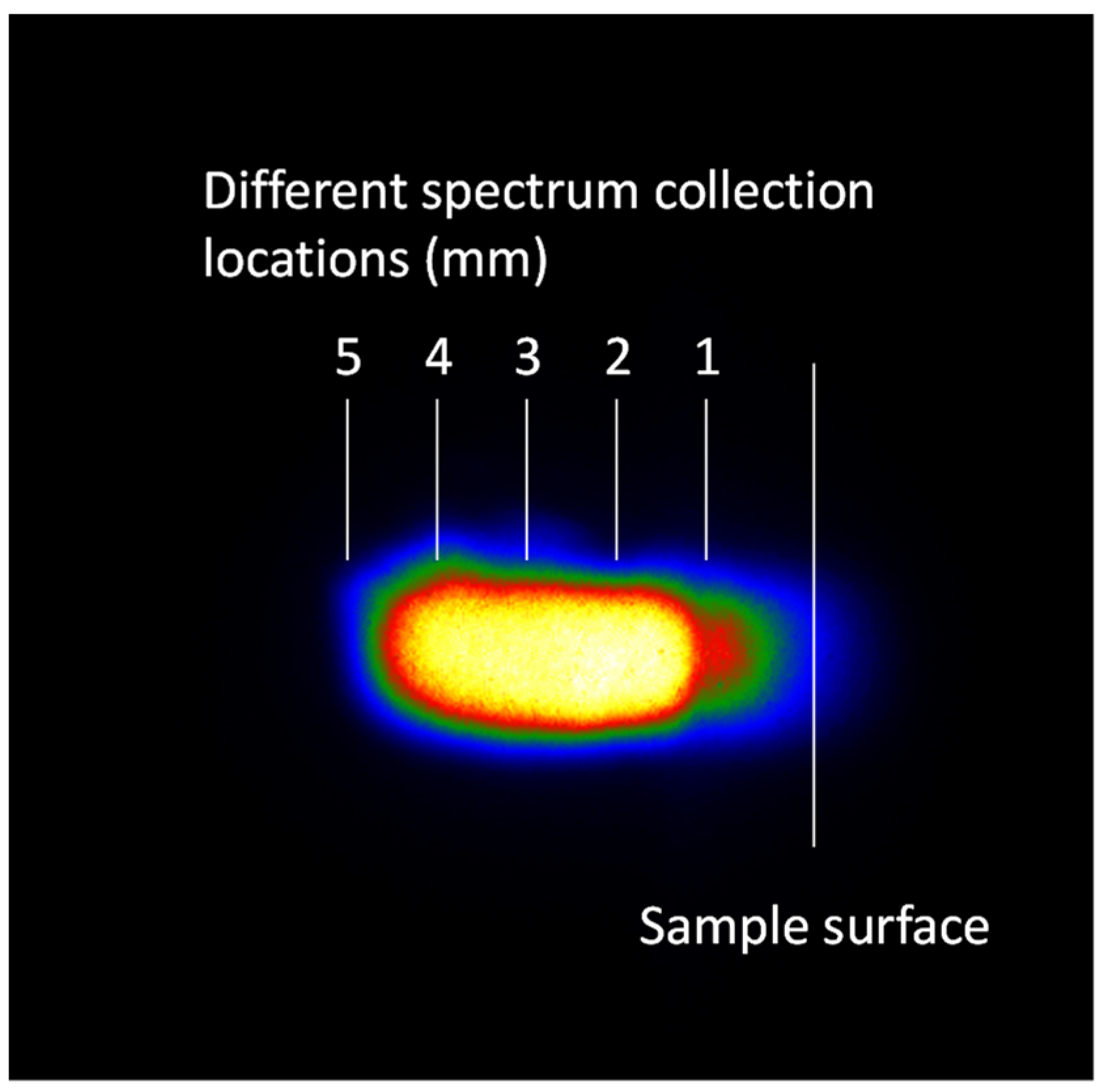
To examine spectral stability as the plasma evolves, this study investigates the relationship between spectral intensity and stability under varying acquisition delays, while keeping the laser focal depth at 0 mm and aligning the spectral acquisition position with position 2 in
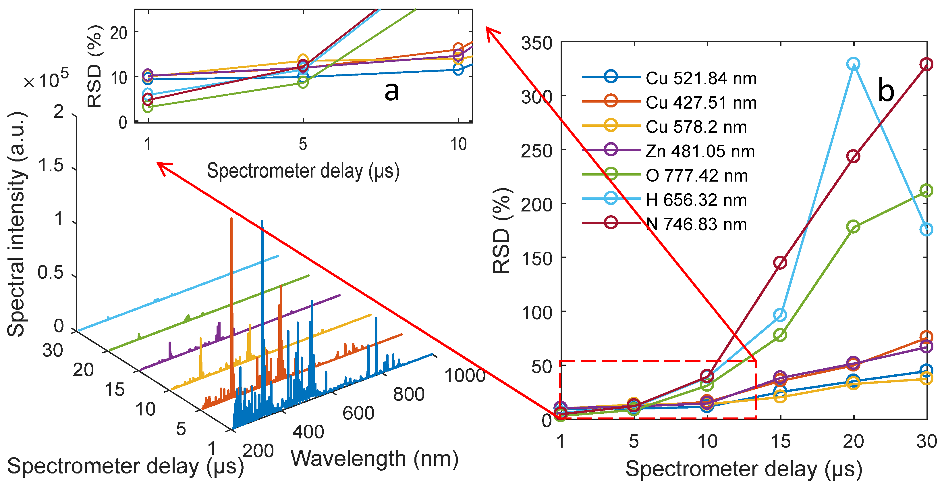
Figure 2. As shown in
Figure 3, at a spectrometer delay of 1 μs, the collected spectral lines exhibit strong and stable intensity. The standard deviation for the single spectrum of O 777.42 nm is within 5%. However, with a spectrometer delay of 5 μs, the relative standard deviation (RSD) of the single spectrum increases to about 10% for matrix elements (Cu and Zn) and air elements (N, H, and O). In addition, as the laser delay continues to increase, two trends emerge. Firstly, the intensity of the spectrum signal collected by the spectrometer weakens. Secondly, the spectrum stability experiences a sharp decline.
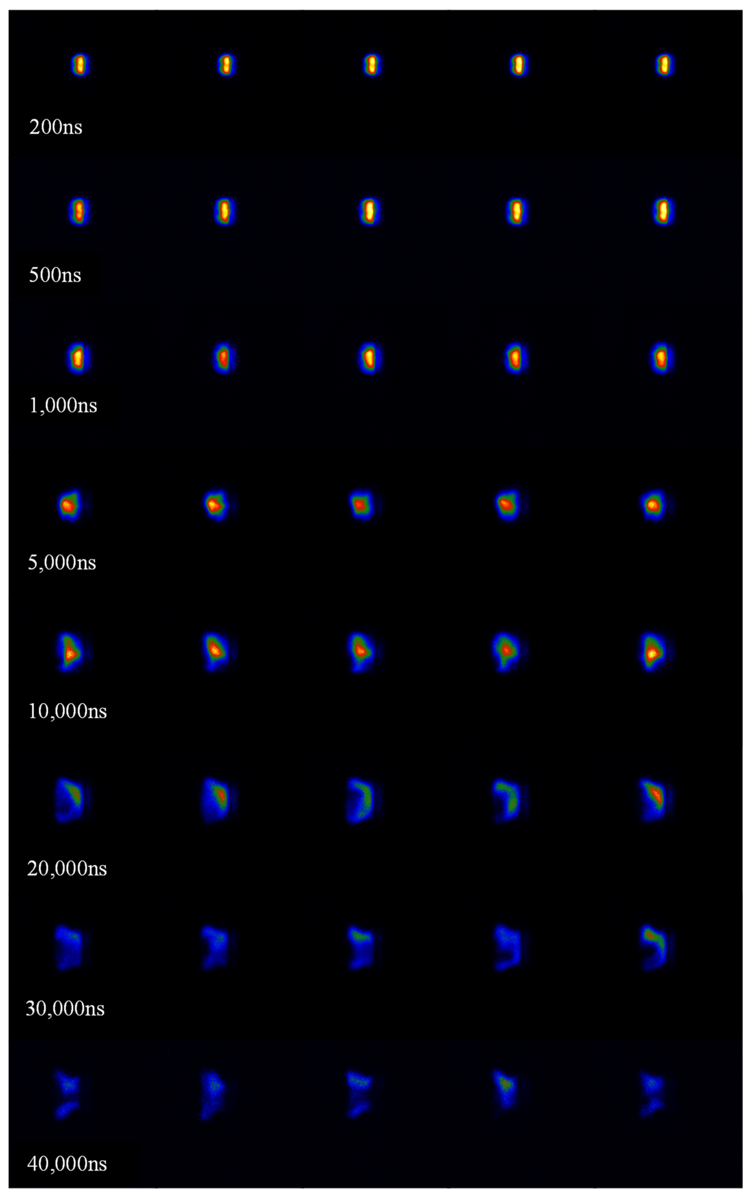
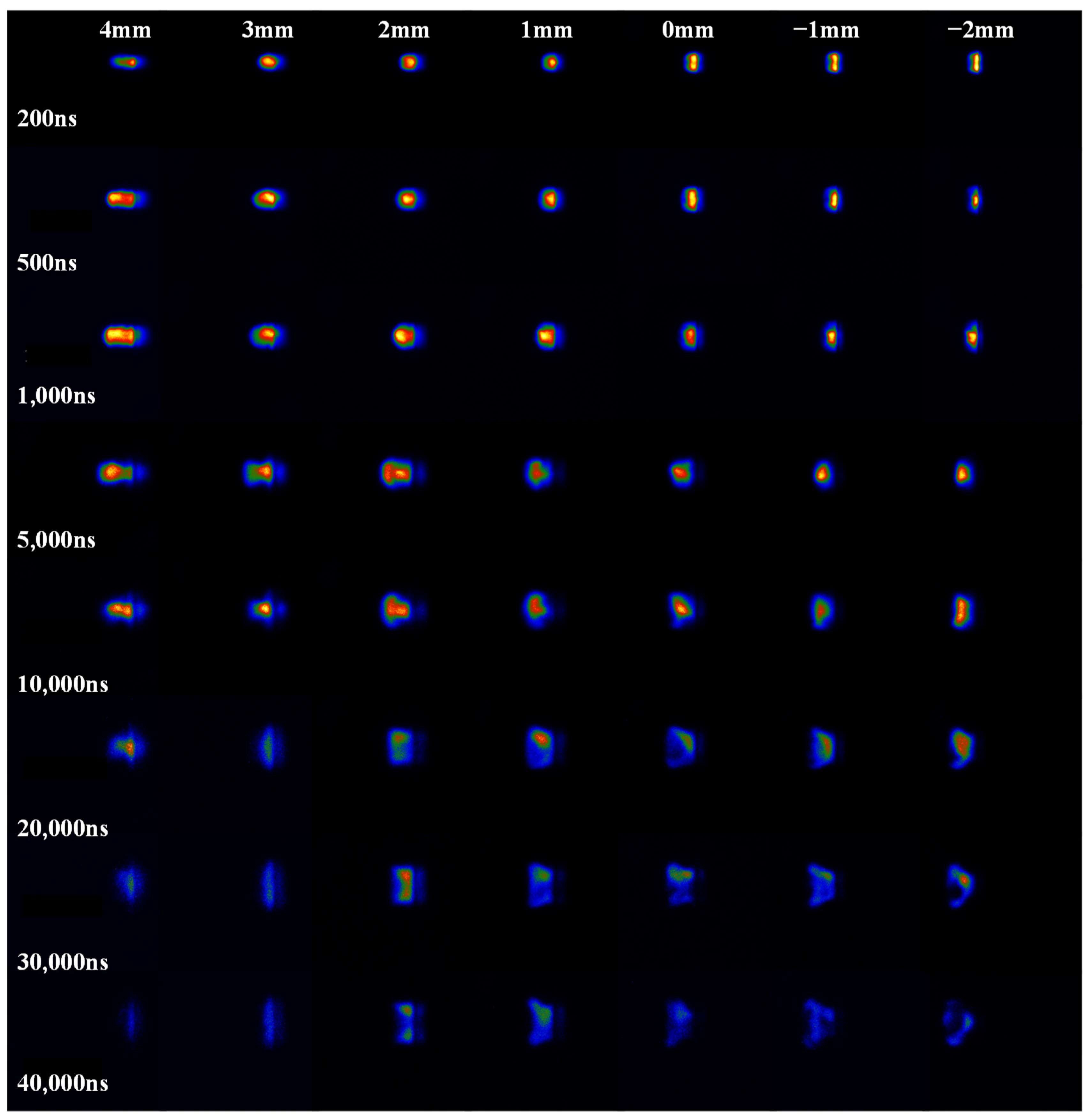
As shown in
Figure 4, plasma patterns were captured under different laser delays. From this Figure, it can be seen that the plasma morphology differences captured under the same delay in the early stages were relatively small. The initial morphology of the plasma is relatively stable. During the period of 200 ns to 10 μs, the obtained plasma morphology is relatively stable. Due to diffusion and ablation, there are significant differences in the morphology of plasma at the same delay in the later stage. At 20~40 μs, the differences in plasma morphology captured at the same time become increasingly significant. Moreover, as a result of plasma shock waves, a distinct trend was observed wherein the plasma moved in the opposite direction of the laser.
Considering the spectrometer’s integration time is 3 μs and the stability of the generated plasma emission spectrum, this phenomenon indicates that within 1–4 μs after the laser is generated, the number of luminescent particles in the collected signal region of the plasma remains relatively constant. However, as the collection delay increases, the collected spectra show weakened intensity and decreased stability, as shown in
Figure 3a,b, respectively. The reason for this phenomenon was that as the collection delay increases, the number of particles in the spectral collection area decreased and the fluctuation increased.
From
Figure 5b, it can be seen that when the spectral acquisition delay is 1 µs, the relative standard deviation of the collected spectral lines for the three elements C, H, and O is about 5%. For the spectral lines of Cu and Zn, the relative standard deviation of the collected spectral lines is about 10%. When the delay of the spectrometer is 5 µs, the relative standard deviations of the spectral lines of C, H, O, Cu, and Zn are all about 10%. However, when the delay of the spectrometer exceeds 10 µs, the relative standard deviation of the spectral line intensities corresponding to C, H, and O suddenly increases, even exceeding 100%, while the trend of the relative standard deviation of the spectral lines for Cu and Zn is not very obvious. The observed phenomenon may be attributed to the fact that when the collection delay of the spectrometer exceeds 10 μs, the spectral signal emitted by air particles gradually becomes submerged in the spectral background.
3.2. Laser Defocus and Spectral Stability
The different focusing depths of the laser on the sample surface will lead to inconsistent power density of the laser on the sample surface, directly affecting the interaction process between the laser and the sample. Appropriate focusing depth helps to ensure that the laser pulse can fully excite the elements in the sample, generating stable plasma emission signals. Focusing too shallow or too deep may lead to incomplete plasma formation or interaction with the surrounding environment, thereby affecting the stability of spectral signals [
18].
In this study, when the spectrometer’s delay time is set to 1 μs and the spectrum collection position is 1 as indicated in
Figure 2, spectral collection is carried out. In order to ensure that the spectral acquisition position remained unchanged relative to the sample surface, the spectral acquisition device moved simultaneously with the sample surface when adjusting the focusing depth of the laser on the sample surface. The relationship between spectral stability and the depth of laser focus on the sample surface has been studied.
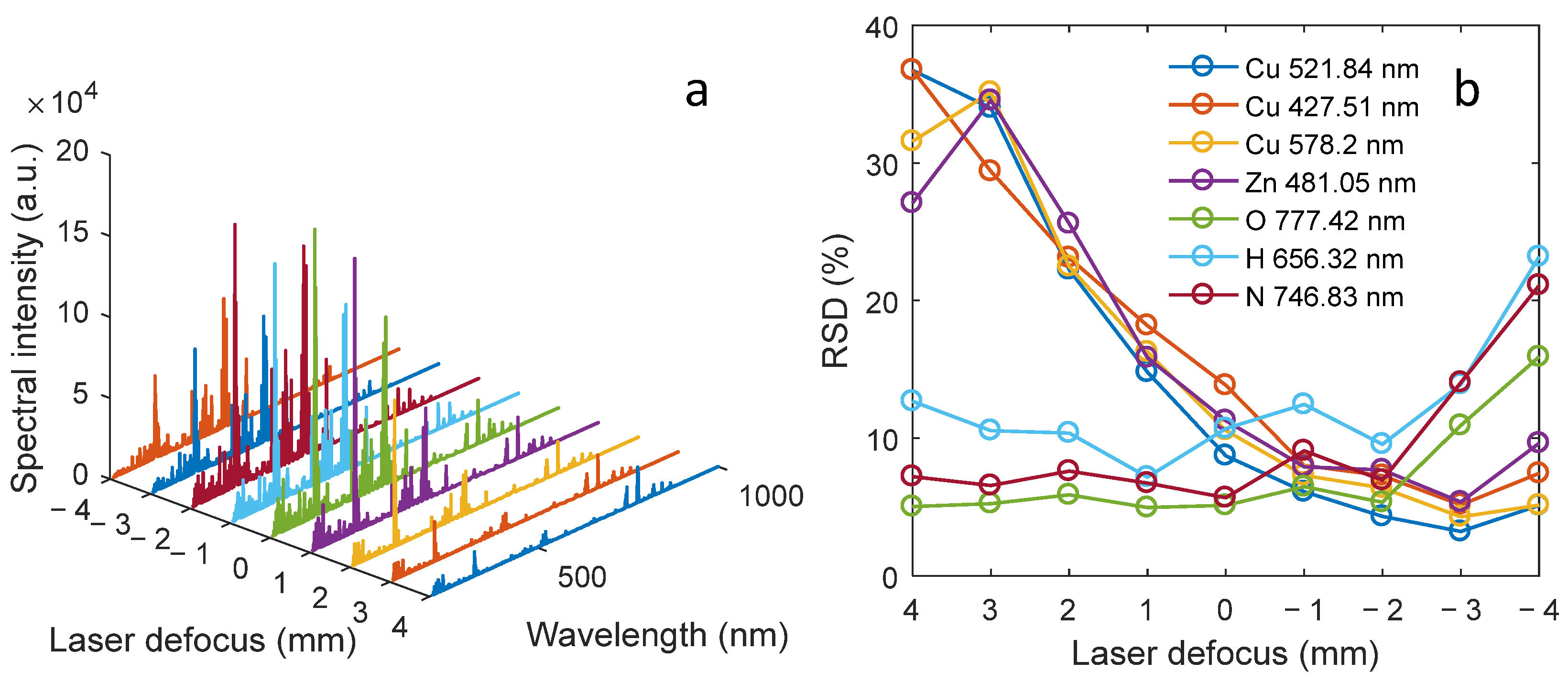
The obtained spectrum intensity under different defocus conditions are shown in
Figure 5a. The variation of the relative standard deviation of the seven spectral lines of the selected Cu, Zn, O, H and N elements with the laser defocusing amount is shown in
Figure 5b.
From
Figure 5a, when the laser focusing depth is between −4 mm and 1 mm, the overall intensity of the collected spectra is basically consistent. When the laser focusing depth is between 2~4 mm, the spectral intensity values show a gradually decreasing trend. When the defocus of the laser is 4 mm, most of the energy is used to break down the air. Due to the higher breakdown threshold of the air sample compared to brass, and the relatively low air density, the total density of luminescent particles in the plasma decreases, resulting in a relatively weak spectral intensity collected. As the defocus of the laser decreases, more and more energy is used to excite the surface material of the sample, and increases the overall density of luminescent particles in the plasma, ultimately resulting in a stronger spectral intensity collected.
Figure 5b shows the fluctuation of the relative standard deviation of the seven selected spectral lines with the defocusing amount of the laser. For the spectral line intensities of the three elements of O, H and N, the spectral intensity is relatively stable when the laser defocus is changed between −2 and 4 mm. However, the stability of the three spectra of N, H and O decreases when the laser defocusing distance changes from −2 to −4 mm. As for the spectral line of Cu and Zn in brass, the stability of spectral line is enhanced with the decrease in laser defocusing. The relative standard deviation of spectral lines can be reduced from above 35% to less than 10%.
The reason for this phenomenon may be that as the defocusing amount of the laser gradually decreases, more and more laser energy is used to excite the surface of the sample, resulting in an increase in the composition of Cu and Zn elements in the center area of generated plasma. Throughout this interval, the primary particles within the plasma’s central region undergo a gradual transition from air components to components of the sample material. When the defocusing amount of the laser is positive, the vast majority of the laser energy is used to break down the air. In the generated plasma, the plasma generated on the sample surface at the edge of the plasma fluctuates greatly in the interaction process with the center plasma and environmental gas. Therefore, in this case, the emission spectra of the three elements H, N, and O obtained are relatively stable, while the emission spectra of the two elements Cu and Zn have poor stability. When the defocusing amount of the laser gradually becomes negative, the vast majority of the laser energy is used to excite the surface of the sample, making the central region of the plasma mainly composed of the sample components. During the interaction between the air components located at the edge of the plasma and the center plasma and environmental gas, there are large fluctuations. As a result, the stability of the emission spectra of the H, N, and O elements is poor.
Moreover, under different defocusing amounts, for the seven selected element spectrum lines, there is always a certain element’s spectrum line stability about 5%. This phenomenon indicates that the plasma generated at different laser focusing depths is relatively stable. To verify this idea, the plasma morphology variation characteristics of the laser defocusing amount at relative positions ranging from 4 mm to −2 mm were studied, as shown in
Figure 6.
From
Figure 6, it is found that the morphological changes in the plasma are relatively stable, without significant changes, despite varying depths of laser focus on the sample surface. In addition, within 500 ns after the plasma is generated, there is no plasma split [
13]. When the plasma morphology is within 10,000 ns, the plasma main body morphology presents a good consistency without violent fluctuations. This phenomenon also confirms the stability of the above spectral collection. Between 10,000 ns~40,000 ns, the uniformity of the plasma morphology becomes worse.
In addition, there are significant differences in the evolution of plasma under different laser defocusing amounts. When the relative laser output delay is 200 ns and the laser focusing depth is 4 mm, the obtained plasma side profile is conical; when the laser is focused at a depth of 1 mm, the obtained plasma profile is circular; when the laser defocus is −2 mm, the obtained plasma profile is linear. In addition, when the relative laser output delay is 1000 ns and the laser focusing depth is 4 mm, the obtained plasma profile is approximately rectangular; at a laser focusing depth of 1 mm, the obtained plasma profile is approximately square in shape; when the laser defocus is −2 mm, the obtained plasma profile is approximately shuttle shaped. The reason for the significant difference in plasma morphology under different laser focusing depths may be that when the laser defocus is positive, the laser first penetrates the air and excites the sample. Due to the higher density of the sample plasma compared to the air plasma, a relatively thin gas environment is formed in front of the generated sample plasma, which has a certain drag effect on the generated sample plasma. Therefore, as the plasma evolves over time, the lateral morphology will become longer. When the defocusing amount of the laser is negative, the laser beam directly excites the surface of the sample to produce plasma, without the drag effect of the thin gas environment. It only expands under the action of the plasma shock wave. Therefore, during the evolution process of the plasma, the length of the lateral morphology of the plasma does not change much. In order to explore the stability characteristics of spectra in different regions of plasma, the spectral stability of different defocusing amounts and collection positions was studied.
3.3. Collection Location and Spectral Stability
To investigate spectral line stability across various collection positions, the experiment recorded changes in spectral stability with a spectrometer delay of 1 μs and relative positions of spectrum collection ranging from 1 to 5, as depicted in
Figure 2.
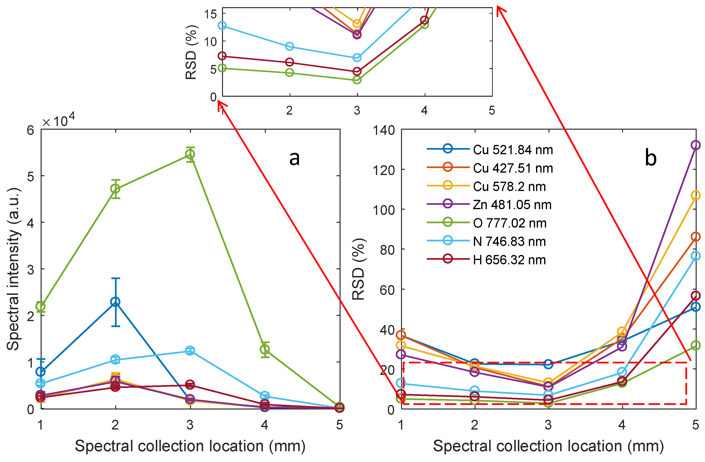
Figure 7 illustrates the spectral line intensity and stability at different spectral acquisition positions when the laser is defocused by 4 mm.
From
Figure 7a, the spectral line intensities of the detected elements Cu, Zn, O, N, and H all show a trend of first increasing and then decreasing. Among them, the spectral line intensities of Cu and Zn are the strongest at relative position 2, while the spectral line intensities of O, N, and H are the strongest at relative position 3. This indicates that when the laser is focused at a depth of 4 mm, the spectral acquisition delay is 1 us, and the integration time is 3 μs, the region where the matrix element is located is mainly at position 2, and the region where the air plasma element is located is mainly at position 3. In addition, from
Figure 7b, as the spectral collection positions change from 1 to 5, the relative standard deviations of the spectral line intensities corresponding to the elements Cu, Zn, O, N, and H show a trend of first weakening and then strengthening, and the relative standard deviations of the spectral lines obtained at position 3 are all the smallest. The reason for this phenomenon may be that in the early stage of plasma evolution, the position where the spectrum collection position 1 or 2 is located is the edge of the plasma. Due to the fact that particles in the edge region of the main plasma particles interact with both air and the main plasma, the fluctuation of luminescent particles in the spectral collection area is relatively large. Therefore, the stability of the spectrum is relatively poor. In addition, when the relative position is 5, because the number of luminescent particles in the collected area is small, the effective spectrum is submerged in the spectrum background, and the stability of the obtained spectrum is poor. At the relative position 3, the spectrum collection position is in the central area of the plasma, and the overall density of the particles is relatively constant within the spectrum acquisition time range. The stability of the seven obtained spectral lines has been enhanced in this region, and the RSD of O 777.02 nm is 2.87%.
In order to explore the spectral stability of different focusing depths and collection positions, the changes in laser defocus between −4~4 mm and the spectral stability collected at 1~5 spectral collection positions were analyzed. The relative standard deviations of the spectral line intensities of O and Cu elements obtained are shown in
Table 1 and
Table 2, respectively.
As shown in
Table 1, the RSD changes in the O 777.02 nm at different spectral collection positions under different laser defocusing amounts are recorded. Experiments have found that as the defocusing amount of the laser decreases, the best collection position of the O 777.02 nm moves toward the direction of the sample. When the laser defocus is 4 mm, the most stable collection position for O element spectral lines is 3. However, when the laser defocus is −1~3 mm, the most stable collection position for O element spectral line intensity obtained is 2. When the laser defocus is −2, the most stable collection position for O element spectral lines obtained is 1.
Furthermore, for Cu 578.02 nm, the variation trend between the focusing depth of the laser on the sample surface and the optimal spectral acquisition position is not very obvious. When the laser is focused at a depth of 2 mm~4 mm on the sample surface, a relatively stable plasma emission signal can be obtained at the spectral acquisition position 3. When the laser is focused at a depth of 1 mm on the sample surface, a relatively stable spectral signal acquisition position of 2 is obtained. When the focusing depth of the laser on the sample surface is 0, the acquisition position of the relatively stable spectral signal obtained is 1. When the laser focuses at a depth of −1 mm or −2 mm, a relatively stable plasma emission signal can be obtained at the spectral acquisition position 2. When the laser focuses at a depth of −3 mm or −4 mm, a relatively stable plasma emission signal can be obtained at the spectral acquisition position 1.
By comparing the stability of spectral lines corresponding to O and Cu elements, it has been partially verified that differences exist in the emission spectral lines of various elements within the spatial distribution of plasma. In addition, by comparing the data in
Table 1 and
Table 2, as well as the plasma morphology captured in
Figure 6, it can be observed that stable plasma emission spectral signals can be obtained near the plasma center region. When the focusing depth of the laser on the sample surface is negative, the center position of the plasma is approximately at the spectral acquisition position 1 or 2. Therefore, a relatively stable plasma emission spectrum signal can be obtained at the spectral acquisition position 1 or 2. When the laser focusing depth is positive, the obtained plasma center position is approximately at the spectral acquisition position 3, so a relatively stable plasma emission spectrum signal can also be obtained at that position.


