Review

Organelle Targeting Self-Assembled Fluorescent Probe for Anticancer Treatment

by
Md Sajid Hasan
,
Sangpil Kim
,
Chaelyeong Lim
,
Jaeeun Lee
,
Min-Seok Seu
and
Ja-Hyoung Ryu
*
Department of Chemistry, Ulsan National Institute of Science and Technology (UNIST), Ulsan 44919, Republic of Korea
*
Author to whom correspondence should be addressed.
These authors contributed equally to this work.
Chemosensors 2024, 12(7), 138; https://doi.org/10.3390/chemosensors12070138
Submission received: 12 June 2024 / Revised: 6 July 2024 / Accepted: 9 July 2024 / Published: 11 July 2024

Abstract

:
Organic fluorescent probes have attracted attention for bioimaging due to their advantages, including high sensitivity, biocompatibility, and multi-functionality. However, some limitations related to low signal-to-background ratio and false positive and negative signals make them difficult for in situ target detection. Recently, organelle targeting self-assembled fluorescent probes have been studied to meet this demand. Most of the dye molecules suffer from a quenching effect, but, specifically, some dyes like Pyrene, Near-Infrared (NIR), Nitrobenzoxadiazole (NBD), Fluorescein isothiocyanate (FITC), Naphthalenediimides (NDI), and Aggregation induced emission (AIE) show unique characteristics when they undergo self-assembly or aggregation. Therefore, in this review, we classified the molecules according to the dye type and provided an overview of the organelle-targeting strategy with an emphasis on the construction of fluorescent nanostructures within complex cellular environments. Results demonstrated that fluorescent probes effectively target and localized inside the organelles (mitochondria, lysosome, and golgi body) and undergo self-assembly to form various nanostructures that possess bio-functionality with long retention time, organelles membrane disruption/ROS generation/enzyme activity suppression ability, and enhanced photodynamic properties for anticancer treatment. Furthermore, we systematically discussed the challenges that remain to be resolved for the high performance of these probes and mentioned some of the future directions for the design of molecules.

Graphical Abstract

1. Introduction

Fluorescent probes have been attracted as biological techniques for visualizing cellular organelles with high sensitivity and specificity due to their non-invasiveness, fast response, and real-time detection features [1,2,3,4]. In particular, organic fluorophore-based fluorescent probes have attracted attention for their application in imaging biological systems owing to their intrinsic biocompatibility and high performance with stable fluorescence [5,6,7,8]. Representatively, rhodamine organic dyes are one of the most widely used dyes and have been extensively used in biotechnology for fluorescent markers of mitochondria or small molecule detection including Cu(II), inorganic phosphate, and nitric oxide [9,10,11,12,13,14,15]. However, there are still challenging issues, such as their tendency to move away from their targets because they are made mostly of water-soluble fluorescent dyes and react with the living systems’ water, protein, and lipids components. These lead to adverse outcomes, such as low signal-to-background ratio, and false positive and negative signals, and ultimately this fluorescent probe cannot achieve accurate target detection within living systems [16,17,18,19]. Thus, it is necessary to develop novel imaging strategies to improve the capability of organic fluorescent probes in biological applications.
Self-assembly is a common process whereby small building blocks thermodynamically form three-dimensional ordered functional material through non-covalent interactions such as hydrogen bonding, hydrophobic, electrostatic, and pi-pi (π-π) interactions [20,21,22,23]. Interestingly, self-assemblies have distinct physical and chemical properties that building blocks do not possess, which give directions to design advanced functional materials in biotechnology [24,25]. In this regard, the self-assembly of organic fluorescent probes has been the focus of many investigations because of their thermodynamics, kinetics, and optical changes by J-aggregation or H-aggregation fluorophore [26,27,28,29]. Especially, these self-assembled fluorophores, with the size of nanometers, exhibit extended residence time within cells, thereby enabling high-functionality and in situ long-retention bioimaging benefits [30,31,32,33,34]. Despite the great outcomes of numerous studies, prefabricated nanostructures could encounter difficulties in internalizing into cells, typically requiring processes such as energy-dependent endocytosis [35,36]. Recognizing that such challenges may compromise the efficiency of bioimaging, several research groups have focused on introducing in situ self-assembly strategies, wherein nanostructures are formed directly within the cellular environment.
Recently, our group developed organelle-targeting self-assembly strategies to fabricate the nanostructures inside intracellular confined space, including cytosol, mitochondria, golgi body, and lysosome. The designed monomers would be accumulated into the organelle, which provides an effective condition to occur covalent/non-covalent interactions for inducing self-assembly behavior. Although it is difficult to control the molecular interaction in a dynamic and complicated intracellular environment, a confined space of organelles can be utilized as a chemical reactor, which facilitates in situ construction of assembled structures even within cells [37]. In this regard, organelle targeting fluorescent probes have been demonstrated as self-assembly into nanostructures inside organelle, which would envision bio-functionality with long retention time, enhanced photodynamic properties, and mechanical rigidity. In particular, diverse organelle-targeting fluorescent probes have been studied in various approaches involving imaging of organelles and photodynamic therapy for anticancer treatment because the organelle of cancer cells has distinct characteristics that can apply to target organelles specifically [38,39]. From a feature perspective, we provide an overview of organelle-targeting fluorescence probes in the field of anticancer treatment, emphasizing the construction of fluorescent nanostructures within the complex cellular environment to develop bio-functionalities. Typically, molecular self-assembly or aggregation behavior induces a decrease in the fluorescence rate and an increased nonradiative rate, which causes an emission-quenching effect on fluorescent probes [40,41].
However, it is known that some fluorescent probes would exhibit different properties in their aggregation state or after self-assembly; for instance, Pyrene shows excimer emission fluorescence in the aggregation state [42], Near-Infrared (NIR) dyes offer enhancing imaging resolution and depth penetration [43], the strong green fluorescence of Nitrobenzoxadiazole (NBD) in hydrophobic environments [44], enhancing the selectivity and stability of Fluorescein isothiocyanate (FITC) [45], self-assembly and fluorescent enhancement of Naphthalenediimides (NDI) [46], and stronger emission, enhanced ROS generation with excellent PDT characteristics of Aggregation induced emission (AIE) dye in the cellular environment compare with the non-aggregate dye [47]. These above-mentioned fluorescent probes could be a great choice to design organelle targeting self-assembly precursor molecules that could avoid the emission-quenching effect, low signal-to-background ratio, false positive and negative signals, and enhance the long-time cellular imaging properties. From the literature survey, we found that organelle targeting self-assembly strategy deals with fluorescent probes (Pyrene, NIR, NBD, FITC, NDI, and AIE) remain as scattering therefore here we have summarized them by categorizing the organelle targeting fluorescent monomers under the type of fluorescent dye e.g., Pyrene-based monomer, NIR-based monomer, NBD-based monomer, FITC-based monomer, NDI-based monomer, and AIE-based monomers shown in Table 1. In brief, we discuss their design strategy, organelle targeting techniques, self-assembly behavior by cancer stimuli, cellular imaging technique (uptake, colocalization, self-assembly, etc.), and the mechanism of action of these probes within the complex cellular environment as well as their application in cancer treatment.

2. Key Milestones of Organelle Targeting Self-Assembled Fluorescent Probe in Cancer Treatment

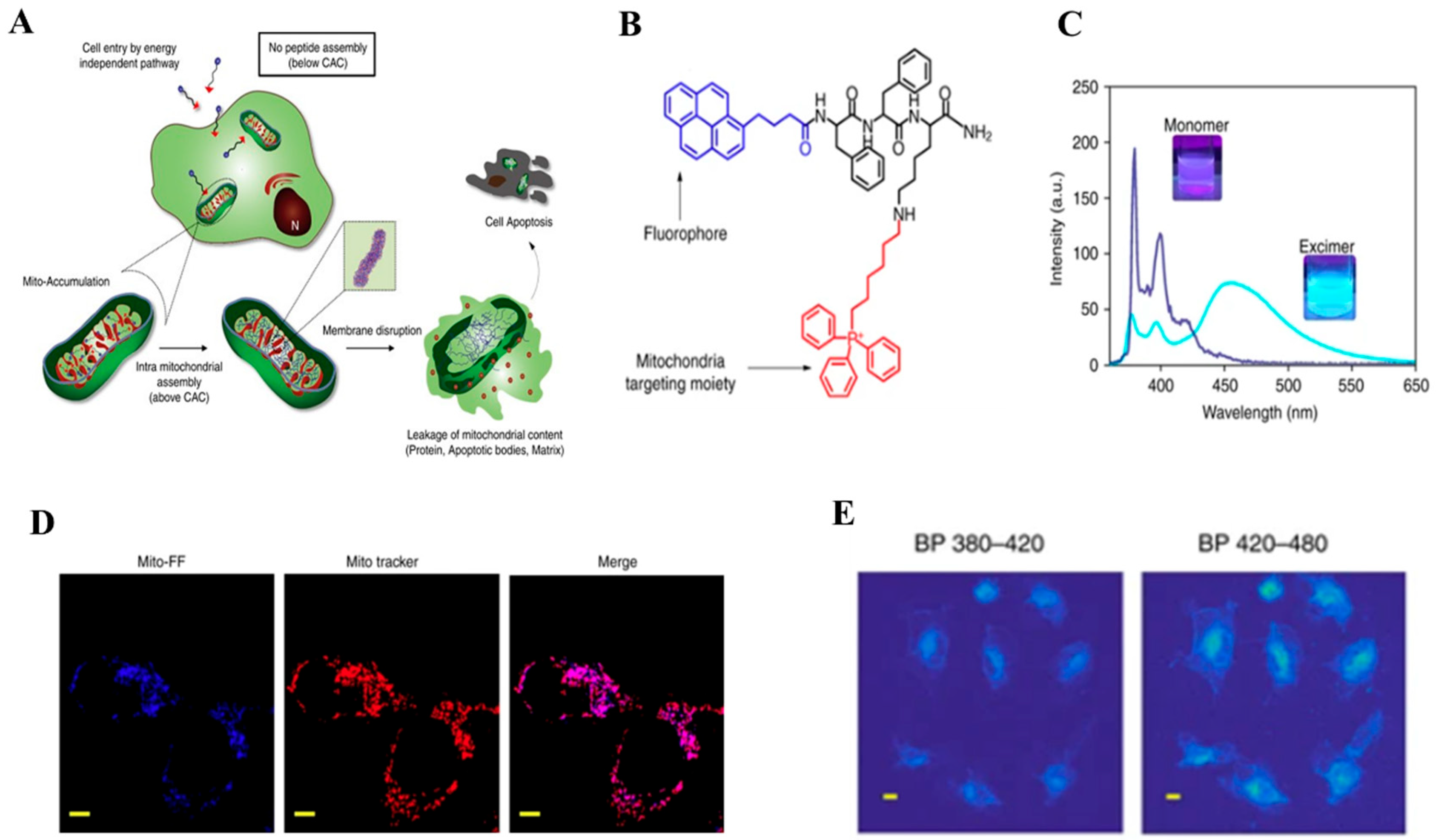
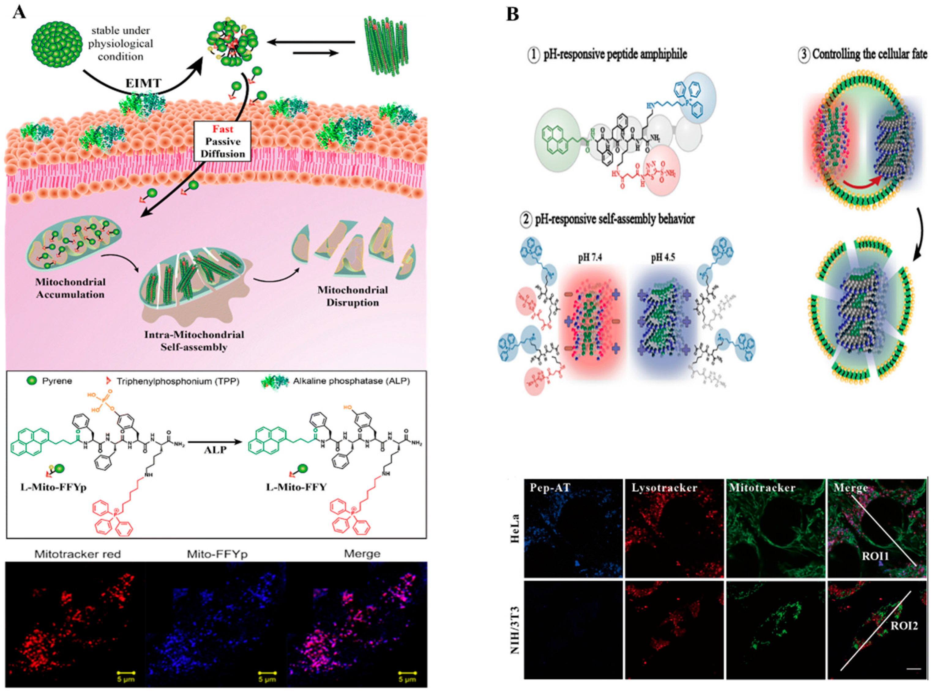
2.1. Pyrene-Based Monomer

Pyrene is a polycyclic aromatic hydrocarbon consisting of four fused benzene rings, resulting in a flat aromatic system. It is a spatially sensitive probe that displays fluorescence emission peaks with 375–405 nm at the monomeric state. Interestingly, they have an additional emission band, which is known as excimers, at ~460 nm when two fluorophores are spatially proximal, showing not only large Stokes shifts (~140 nm) but also long fluorescence lifetimes (~40–60 ns) [63,64,65]. Therefore, the excimer band could be enhanced when its local concentration increases (aggregates/self-assembles). Moreover, pyrene can form highly ordered π-π stacks, facilitating aggregation/self-assembly, thus making these characteristics useful for bioimaging and theranostic applications [66]. For example, Zhang et al. reported a label-free nucleic acid sensing strategy by employing aggregation-induced pyrene excimer fluorescence. They designed and synthesized a positively charged pyrene probe that shows an emission of 377 nm at a monomeric state. When mixed with the oligonucleotides in an aqueous buffer, it undergoes aggregation and forms an excimer that shows significant changes in the fluorescence emission at 485 nm [67]. Due to this rationale, pyrene-based building blocks are being engineered as self-assembling fluorescence probes for targeting cellular organelles like mitochondria, and lysosomes and real-time monitoring of the bioimaging within the complex cellular environment.
Mitochondria are double-layer membrane-encapsulated subcellular dynamic organelles. Its inner and outer membrane separates the matrix and the region of the intermembrane. It acts as the cell’s powerhouse, i.e., supplying the energy to maintain the physiological processes. Also, it plays essential roles in many cellular processes, for instance, energy production, metabolic regulation, ROS generation, calcium homeostasis, immune system regulation, controlling cellular function, and programmed cell death [68,69,70]. However, compared to healthy mitochondria, there are significant characteristic differences in cancerous mitochondria such as elevated levels of ROS, stronger negative transmembrane potential (−220 mV), and upregulation and down-regulation of some proteins [71,72]. Therefore, lipophilic cationic molecules and fluorophores including triphenylphosphonium (TPP), pyridinium, guanidium, Szeto-Schiller (SS) peptides, indocyanine, and tetramethylrhodamine- and benzothiazole-based fluorophores can effectively accumulate inside the mitochondria [73,74]. After the direct translocation of these probes into the mitochondria, they undergo morphological transformation via the self-assembly process and form nanostructures with or without the cancerous stimuli. Mitochondria localization-induced self-assembly is an example of without stimulus where these monomeric building blocks accumulate into the mitochondria, reach their critical aggregation concentration, and undergo self-assembly to form various nanostructures. Enzyme-instructed self-assembly and polymerization-induced self-assembly involve the enzyme or redox stimuli (enterokinase, SIRT5, alkaline phosphatase, or reactive oxygen species, ROS) mediated self-assembly process [48,49,75,76,77]. Nanostructures that are formed by this self-assembly process take charge of determining the fate of cancer cells via the induction of hypoxic or normoxic conditions, depolarization of mitochondrial membrane potential, ATP depletion, or mitochondrial dysfunction. Thus, the mitochondria-targeting self-assembly strategy of fluorescent probes has been extensively investigated in anticancer therapies with these unique characteristics.
However, by utilizing the mitochondria targeting moieties together with a pyrene fluorescent probe, the pyrene-based building blocks can target and easily be translocated inside the mitochondria by passive diffusion. After the effective accumulation of this pyrene-based monomer, it reaches the critical aggregation concentration and undergoes aggregation/self-assembly to form various nanostructures with enhanced excimer fluorescence properties. This unique fluorescence behavior of pyrene excimer can be employed to monitor the co-localization experiments of the monomer, self-assembly process, and selective organelle dysfunction in cancer cells. Thus, in recent years, pyrene-based mitochondria-targeting self-assembling probes have been extensively investigated in anticancer therapies. For example, Ryu et al. developed, for the first time, the mitochondria-localization-induced self-assembly strategy for controlling the fate of cancer cells (Figure 1A) [48]. The designed building block (Mito-FF) consists of a tripeptide Phe-Phe-Lys (self-assembled unit), pyrene (fluorescent probe), and triphenylphosphonium (TPP, mitochondria-targeting) moiety (Figure 1B). Here the pyrene unit serves a dual role i.e., as a fluorophore, and increases the self-assembly propensity via enhancing hydrophobic and π–π interactions. The Mito-FF monomer readily diffuses through the cell membrane and accumulates in the mitochondria of cancer cells due to the high negative membrane potential. After that, it reaches the CAC and causes Mito-FF to self-assemble into a fibrous structure with a width of 9.6 ± 1.1 nm. The CAC value (60 µM) was determined by using pyrene excitation spectra and emission at 450 nm (intense sky blue) in PBS indicates the formation of pyrene excimer in its aggregation state while pale blue emission in the range of 370–410 nm reflects the monomeric state of pyrene (Figure 1C). Again, the mitochondrial co-localization experiment of this probe was confirmed by the confocal microscopy, which showed a good overlap between the red fluorescence of Mito Tracker Red FM (MT-Red) and the blue fluorescence of the pyrene building unit with a Pearson’s Coefficient of +0.8 (Figure 1D). Furthermore, the Mito-FF fibrils formation inside mitochondria was checked by two-photon microscopic analysis indicating pyrene excimer emission at a 420–480 nm filter and monomeric emission at a 380–420 nm filter (Figure 1E). In addition, to achieve high selectivity with reduced side effects, they reported alkaline phosphatase (ALP) mediated morphology transformable pyrene-based mitochondria-targeting peptide molecule, L-Mito-FFYp [50]. The blue fluorescence of this pyrene probes good overlap with MT-Red implying cellular internalization and effective mitochondrial colocalization (Figure 2A). In the presence of endocytosis inhibitors, intracellular mitochondrial dysfunction was also monitored by utilizing the fluorescent behavior of this pyrene monomer. Moreover, they extend this technique for supramolecular cancer therapy by modifying the pyrene-based monomer with D and L isomers [49]. In this technique, the monomer design involves the conjugation of TTP with both D and L isomer and a pyrene fluorescent probe. The π–π interactions between pyrenes are stronger than phenylalanine units for one enantiomeric assembly while two enantiomers of opposite chiral are assembled; this interaction becomes stronger for phenylalanine units. Thus, pyrene units bind to the surrounding fibers to form a superfibril structure inside mitochondria inducing rapid kinetics, enhanced mitochondrial disruption, and higher cellular cytotoxicity. Furthermore, they construct a tumoral pH-responsive mitochondria-targeting strategy by conjugating a succinic amide and FF peptide with a pyrene fluorescent probe [51]. The cleavage of the succinic amide in an acidic condition was confirmed through the fluorescence spectra where the excitation band of pyrene and emission band of naphthalene showed good overlaps via the fluorescence resonance energy transfer (FRET).
After the successful results of bioimaging in cancer treatment based on the mitochondria targeting strategy, the Ryu group extended the application of the pyrene fluorescent probe to another cellular organelle, namely the lysosome. As we know, lysosomes are considered one of the most important cellular organelles involved in autophagy, intracellular signal transduction, cell division, and cell death pathways [79]. Therefore, targeting lysosomes and their intracellular bioimaging as well as monitoring their behavior has attracted significant attention in recent years. However, unlike mitochondria, lysosomes are single membrane (membrane permeabilization)-enclosed vesicles with an acidic nature. Compared to normal cell lysosomes, the de-stabilized thinner membranes and volume enlargement in cancer lysosomes make them selective for organelle-targeting cancer treatment [80]. Thus, lipophilic amines, for example, 4-(2-Hydroxyethyl) morpholine and N,N-dimethylethylenediamin, are extensively employed together with the fluorescent probe to drive the molecule into the lysosomes. These probes, after accumulating into the lysosomes, are protonated and are membrane impermeable, resulting in the entrapping there [52]. Therefore, fluorescent probes possessing lysosome targeting moiety can be used to study lysosomal function and organelle targeting strategy in cancer treatment. Motivated by this, the Ryu group reported a lysosome targeting pyrene-based molecule to control the fate of cancer with a low therapeutic dose [78]. They labeled the building block Pep-AT with a pyrene fluorescent unit to verify the self-assembly and cellular co-localization behavior of this molecule, an acetazolamide unit for targeting carbonic anhydrase IX enzymes, and TPP for increasing the membrane interactions. Results demonstrate the bathochromic shift for the pyrene excimer peak at 460 nm (λex = 360 nm), indicating the formation of supramolecular J-aggregation in pH 4.5. Then the CAIX-mediated endocytosis into the lysosomes was confirmed by observing the blue fluorescence of pyrene inside the cells by CLSM. Furthermore, the lysosomal co-localization of this probe molecule was determined by the overlaps image of the blue fluorescence of pyrene monomers (Pep-AT) with the red fluorescence of Lysotracker Red (Figure 2B). Thus, the pyrene fluorescent unit is employed extensively in bioimaging techniques to monitor dynamic and complex cellular compartments.

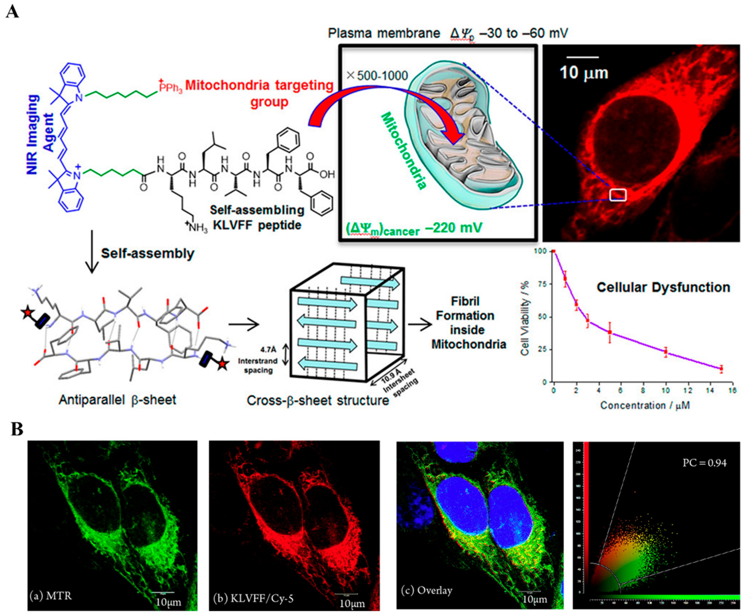
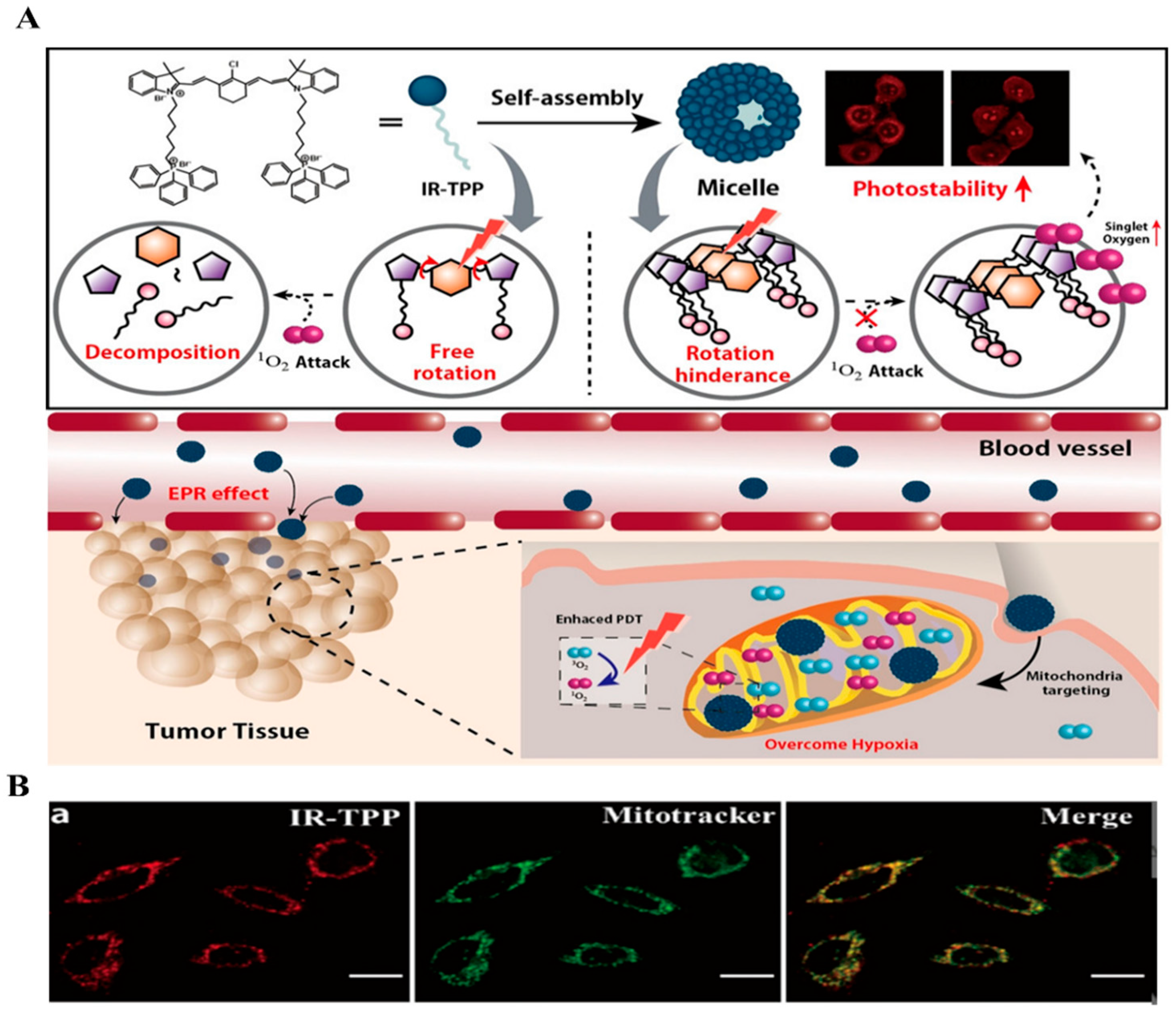
2.2. NIR-Based Monomer

Near-infrared (NIR) fluorescence probes, including Cyanine derivatives, Dithiolene complexes, and Phthalocyanines, have a long excitation wavelength range of 650–900 nm. They offer several advantages, such as non-invasiveness, deep tissue penetration, and high sensitivity [81,82]. These characteristics make them highly valuable in medical diagnostics, drug delivery, and intracellular signaling [83,84,85]. In particular, Indocyanine dyes have emerged as promising candidates for photodynamic therapy (PDT) due to their strong absorption and emission in the NIR region and their preferential accumulation in the mitochondria of tumor cells. Notably, self-assembly enhances chemical stability and fluorescence, leading to a more reliable and robust fluorescent signal and improving the effectiveness of NIR [86,87,88]. Through these insights, recent studies have been actively exploring the combination of self-assembly techniques for organelle targeting with near-infrared (NIR) fluorescent probes focusing on applications in imaging and photodynamic therapy (PDT). For instance, Guha et al. reported a cellular mitochondria-targeted imaging strategy based on Near-Infrared fluorescent nanotubes where the molecular design consists of unsymmetrical Cy-3 and NIR Cy-5 chromophores with a mitochondria-specific triphenylphosphonium (TPP+) group [89]. These lipophilic cationic fluorescent molecules spontaneously and selectively accumulate inside the mitochondria reach the critical aggregation concentration and self-assemble into nanotubes that cause the early-stage apoptosis of malignant cells. Molecular self-assembly behavior was confirmed by the confocal laser scanning microscopy (CLSM) image by 645 nm laser excitation (laser λem = 664 nm). Results demonstrate the self-assembled NIR bright fluorescent nanotubes with negligible photobleaching. Furthermore, the narrow NIR emission bands, high fluorescence quantum yield, and high fluorescence lifetime of this NIR dye make it a great choice for live cell mitochondrial imaging. Confocal results show the remarkable colocalizations of Cy-3-TPP/FF and Cy-5-TPP/FF with Pearson’s correlation coefficient (PCC) calculated to be 0.87 and 0.91, respectively. Additionally, the same research group introduced another method for selective targeting, imaging, and dysfunction of mitochondria based on Near-Infrared chromophores [53]. The molecular structure consists of an amyloid-β (Aβ) peptide, Lys-Leu-Val-Phe-Phe (KLVFF, fragment of Aβ16–20), an unsymmetrical near-infrared (NIR) cyanine-5 (Cy-5) chromophore unit, and mitochondria targeting triphenylphosphonium moiety. This combination promotes the mitochondrial dysfunction and cytotoxicity in cancer cells via the formation of amyloid fibers. The KLVFF/Cy-5 conjugate demonstrates high photostability, strong NIR absorption and emission, a high molar extinction coefficient, and a high fluorescence quantum yield, making it an effective bioimaging tool with specific mitochondrial targeting capabilities. Confocal microscopy confirmed the precise localization of the KLVFF/Cy-5 conjugate within the mitochondria of both HeLa and A549 cells, substantiated by the cytotoxic effects demonstrated via the MTT assay. Fluorescence assay results further underscored a significant accumulation of KLVFF/Cy-5 molecules within the mitochondria, with concentrations measuring 3.2 ± 0.7 mM and 6.1 ± 0.5 mM in HeLa and A549 cells, respectively. This approach illustrates the potential of KLVFF/Cy-5 for precise mitochondrial targeting and cancer cell disruption, showcasing a promising strategy for anti-cancer therapy (Figure 3A,B).
Moreover, the Ryu group reported a mitochondria-targeting NIR photosensitizer to increase photostability and photodynamic therapeutic efficacy strategy based on indocyanine dyes [54]. Indocyanine dyes are used for imaging and photodynamic therapy due to their capacity for reduced background interference from biological tissues. Triphenylphosphonium (TPP) has a high affinity for mitochondria in tumor cells, which allows for its preferential accumulation in these organelles. This characteristic can be leveraged for targeted therapy or imaging applications. The designed molecule has triphenylphosphonium cation linked with IR dye (IR-TPP). In a photobleaching experiment, the luminescence intensity of IR-TPP remained stable even with extended laser irradiation, indicating high photostability. Fluorescence microscopy images demonstrated the co-localization of the red fluorescence from IR-TPP with MitoTracker Green FM, suggesting that the photosensitizer successfully targets mitochondria. This co-localization further confirms the potential of IR-TPP as a mitochondria-targeted photosensitizer with high photostability, making it a promising candidate for photodynamic therapy (Figure 4A,B). Furthermore, Li et al. developed an enzyme-responsive lysosome localized self-assembly strategy for intrinsic apoptosis of breast cancer [55]. The precursor molecule consists of four parts: IR808 as a fluorophore, Phe-Phe dipeptide (FF) acts as a self-assembled backbone, Arg-Arg-Gly-Lys (RRGK) peptide sequence acts as cathepsin B (CTSB) restriction site, and a neuropeptide Y1L. Y1L-KGRR-FF-IR is cleaved by CTSB in the lysosomes, forms nanofiber, and leads to lysosomal enlargement and damage as well as mitochondria-mediated apoptosis of breast cancer cells. Fluorescent enhancement results by CLSM demonstrated that the enzymatic cleavage and the green fluorescent of Lysol-Tracker overlapped with the precursor’s red fluorescence, which again confirmed lysosomal colocalization. Furthermore, lysosomal swelling and enlargement were also determined by the fluorescent properties of this probe.

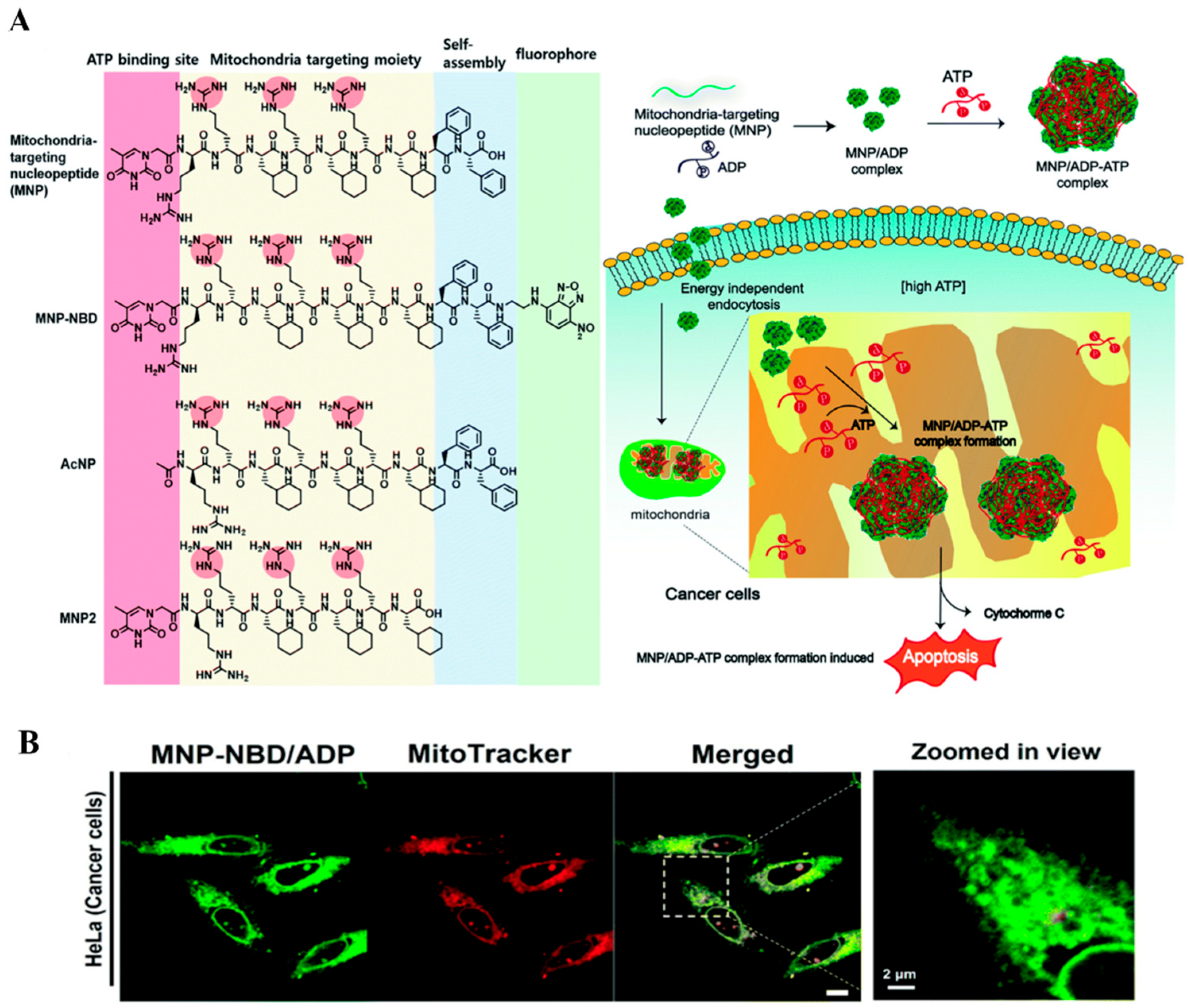
2.3. NBD Based Monomer

Nitrobenzoxadiazole (NBD) is a fluorescent compound with an excitation peak at 466 nm and an emission peak at 539 nm. Its large Stokes shift of approximately 73 nm helps minimize interference between the excitation light and the fluorescence signal, resulting in clearer fluorescence imaging. Additionally, NBD, which has a small molecular weight of 165.11 g/mol, is advantageous for functionalization. Interestingly, NBD is virtually non-fluorescent in hydrophilic media but exhibits strong green fluorescence in hydrophobic environments because the hydrophobic microenvironment reduces the diffusion-originated non-radiative decay pathways. The self-assembly behavior contributes to the well-ordered, rigid, and hydrophobic microenvironment, enhancing the stability and fluorescence properties of NBD [90,91,92]. Consequently, NBD is a powerful tool for studying self-assembly and other complex biological and chemical systems, providing clearer fluorescence imaging in self-assembled structures. This capability is valuable for real-time monitoring of cellular activities and evaluating the efficiency of drug delivery systems. Additionally, NBD can be used in optically transparent samples, adding to its versatility across different experimental settings. This flexibility enables precise tracking of subtle changes in self-assembly processes and a better understanding of the final structures [93,94]. For example, Sun et al. reported NBD-based program supramolecular self-assembly to target mitochondria and live cell imaging strategy [56]. The precursor monomer consists of an environment-sensitive NBD fluorophore, a phenylalanine-rich peptide fragment, and a succinylated lysine (Ksucc) switch module. The formation of nanofibers inside the cell was confirmed by CLSM, showing the strong fluorescent emission of NBD dyes due to the hydrophobic environment. Further, the colocalization experiment of this probe inside mitochondria was checked by the strong overlap of a fluorescence image of NBD-containing nanofibers and that of MitoTracker Red with a Pearson correlation coefficient of 0.90 ± 0.006. Again, Xu et al. reported NBD fluorescent-based branched peptides that can form micelles and, after being uptaken by the cell transform into nanofibers through enterokinase (ENTK) cleavage, enabling targeted delivery to mitochondria [57]. These branched peptides contain a FLAG-tag (DYKDDDDK) recognized and cleaved by ENTK, a self-assembling peptide sequence with a 2-acetylnaphthyl (Nap) group, a d-tripeptide (D-Phe-D-Phe-D-Lys), and a nitrobenzoxadiazole ethylenediamine (NBD-EA) moiety that enhances fluorescence during self-assembly, along with a glycine spacer linking the FLAG-tag to the self-assembling peptide. The cellular uptake pathway was confirmed by the strong fluorescent of NBD and a good overlap with the red fluorescence of MitoTracker showed mitochondrial colocation (Figure 5A,B). Besides, the same research group developed an imaging strategy for measuring the dynamics of cancer cells’ golgi bodies based on the self-assembling NBD probes [58]. The golgi body, serving as an intracellular transportation portal and regulating the various cellular functions, has been considered the signaling hub in mammalian cells. Thus, real-time monitoring of the dynamics and conditions of the cancer golgi body may offer potential data for the mechanistic behavior of cancer cells [95]. However, they constructed an enzyme-responsive and redox-active NBD-thiophosphopeptide self-assembly system that selectively targets the cancer golgi body and reveals its dynamic behavior. The designed monomer molecule consists of a fluorescent probe (NBD), a self-assembly moiety (peptide ff), and a substrate for alkaline phosphatase enzyme (thiophosphate group). This probe enters the cell above its critical micelle concentration and undergoes dephosphorylation by alkaline phosphatase at the site of the golgi body. Then it undergoes self-assembly to form covalent dimers and attach to the cysteine-rich proteins. Results show that the green fluorescence of this probe merged well with the red fluorescence of Golgi-RFP in HeLa cells, confirming its localization into the cancer golgi body. The self-assembly behavior inside the golgi body is also determined by the bright fluorescent of NBD. Then the morphological differences of the cancer golgi body (its fragmentation) were checked by the fluorescent intensity of the probes in various cell lines like Saos-2, HeLa, OVSAHO, HCC1937, HepG2, Capan-2, OVCAR4, PC-3, and B16F10. Thus, utilizing this fluorescent probe, they effectively monitored the state and dynamics of the cancer cells’ golgi body. The Ryu group also reported an NBD-based intramitochondrial co-assembly strategy between ATP and mitochondria-targeting nucleopeptide (MNP-NBD) to develop cancer therapeutics that rely on molecular assemblies [59]. This nucleopeptide contains a nucleobase (thymine) that interacts with ATP and guanidinium groups that bind to the negatively charged triphosphate group of ATP. Additionally, it has a structure capable of penetrating mitochondria, where it forms large self-assembled structures. This leads to physical stress and metabolic disruption in cancer cells, ultimately causing apoptosis. In confocal microscopy images, the MNP–NBD/ADP complex exhibited intense green fluorescence, which significantly overlapped with MitoTracker Deep Red in HeLa cells. This suggests that the MNP-NBD/ADP complex was internalized by the cells and had accumulated inside mitochondria through the mitochondria-penetrating peptides (MPPs). Moreover, the micellar structure formation of this molecule was confirmed by the green fluorescence of NBD probes (Figure 6A,B).

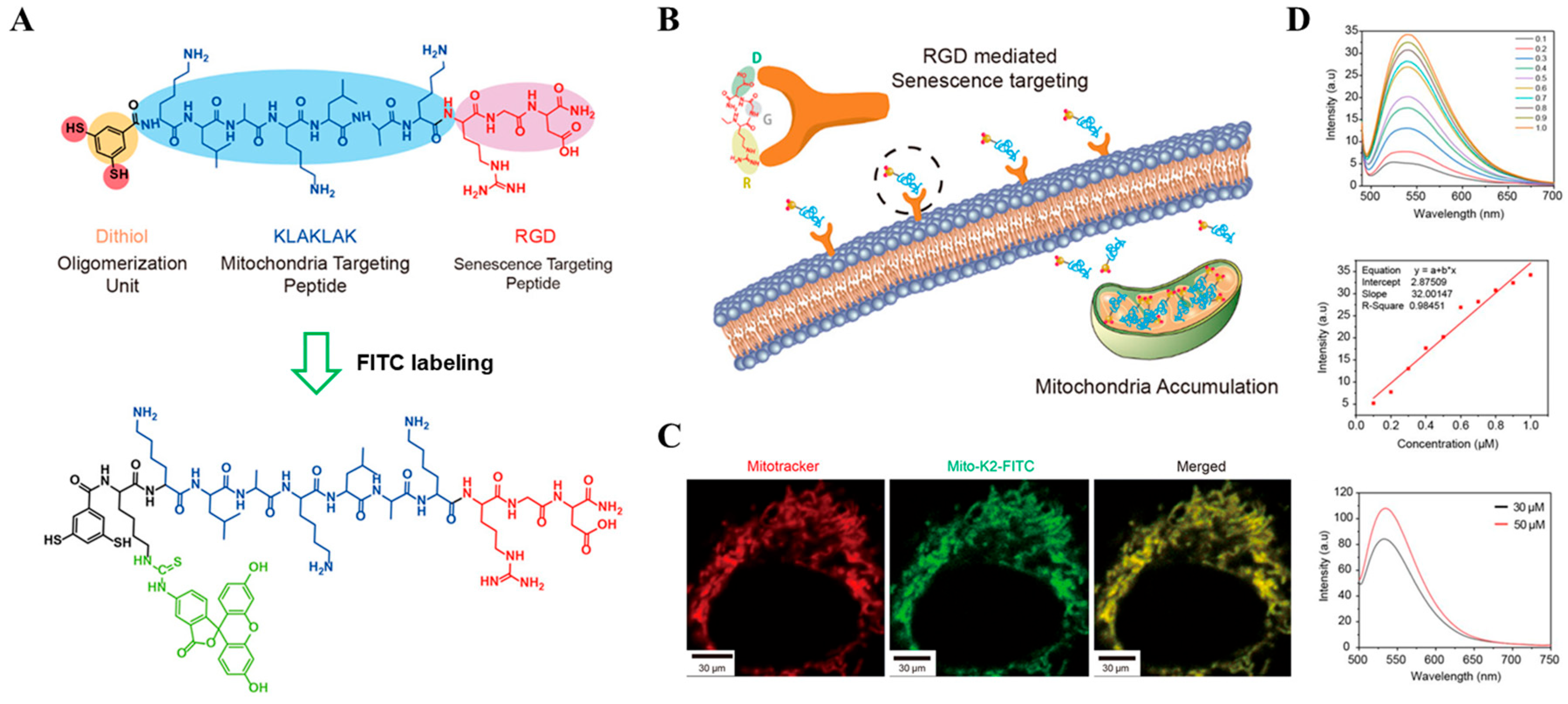
2.4. FITC Based Monomer

Fluorescein Isothiocyanate (FITC) is a widely used fluorescent dye in biological research, primarily for labeling proteins, antibodies, and other biomolecules. It emits a distinctive green fluorescence upon excitation at 495 nm, with peak emission at 519 nm. FITC forms covalent bonds with amine groups (-NH2) present in functional building blocks, thereby enhancing the selectivity, stability, and overall efficacy of these molecular constructs [96,97]. When tailored to self-assembling functional building blocks, the compound can improve the stability of these assemblies and modulate their formation and disassembly at targeted sites. This adaptability is particularly advantageous in the design of targeted therapeutic interventions. For instance, when linked to tumor cell-targeting moieties, FITC-labeled compounds can significantly reduce off-target effects. Additionally, by attaching organelle-specific targeting blocks, the reactivity can be confined to desired cellular compartments such as lysosomes or mitochondria, thus enhancing the specificity and effectiveness of the intervention. The reactivity of FITC with amine groups has spurred extensive research involving peptides, which are employed to target specific organelles or cellular structures. In scenarios where FITC is conjugated to lysosome-targeting peptides, it can facilitate the degradation of proteins overexpressed in tumor cells. Conversely, when linked with mitochondria-targeting peptides, FITC can exploit the mitochondrial environment to accelerate polymerization processes, shifting the site of reaction from the cytosol to the mitochondria. This functionality renders FITC an invaluable tool in fluorescence microscopy and flow cytometry, enabling the precise identification and quantification of specific molecular entities within cells or tissues, as well as distinguishing between different cell or organelle populations [98,99,100]. The Ryu group utilized FITC-labeled self-assembling peptides aimed at mitochondria in senescent cells, which are known to exhibit elevated levels of reactive oxygen species [60]. These peptides are engineered to form alpha-helix structures stabilized by disulfide bonds, disrupting mitochondrial membranes through multivalent interactions and triggering apoptosis. To validate the efficacy of these molecules, it is crucial to confirm their self-assembly within the mitochondria. Comparative analyses with other organelle trackers have substantiated that the FITC-labeled peptides localize and assemble within the mitochondria, disrupting the mitochondrial membrane as demonstrated in Figure 7A–D. This study underscores the potential of FITC-labeled peptides in targeted cellular interventions, exemplifying their utility in advanced therapeutic strategies.

2.5. NDI Based Monomer

Fluorescent probes are indispensable in the exploration of complex biological systems, providing dynamic insights into cellular processes. Among various compounds, naphthalene diimide (NDI) derivatives are notable for their robust photophysical properties, serving as a versatile scaffold for probe design. They have strong absorption in the ultraviolet to visible light spectrum, typically around 300 nm to 400 nm. These compounds emit light in a wide range of colors, from blue to red, depending on their specific chemical modifications, with emission wavelengths typically falling between 400 nm and 650 nm. This versatility makes NDIs valuable for designing fluorescent probes tailored to specific research and diagnostic applications [101,102,103,104]. Specifically, NDIs can be engineered to target certain cellular organelles like lysosomes, which is crucial in the development of precise therapeutic agents. For example, the Ryu group reported NDI-based lysosomal targeting peptide (NDI-Lyso-RGD) assembly techniques for high selectivity against cancer cells [61]. The probe design consisted of a self-assembling motif (Phe–Phe FF), self-assembling enhancing, and fluorophore (NDI), cell-penetrating peptide sequence (cathepsin B substrate). This derivative initially forms micelles that subsequently assemble into fibers within the lysosomes, driven by their hydrophobic properties (Figure 8A,B). This transformation disrupts the structural integrity of the lysosomes, leading to the induction of apoptosis in cancer cells. The targeting mechanism of NDI derivatives involves the activation by specific enzymes that are overexpressed in target cells. The activation of NDI derivatives in cancer cells can be triggered by cathepsin B, an enzyme abundantly present in these cells. This selective activation exposes the lysosome targeting moiety, allowing for precise delivery and controlled activation of the therapeutic compounds within specific cellular compartments. Furthermore, the assembly and functional transformation of NDI structures within targeted organelles can be monitored and verified through advanced imaging techniques. Using fluorescent comparisons with organelle-specific trackers, such as a red lysotracker, researchers can visually confirm the successful localization and activity of NDI derivatives inside the lysosomes. This capability not only provides a direct method to assess the effectiveness of NDIs but also showcases their potential in designing advanced diagnostic and therapeutic strategies specifically aimed at organelle dysfunctions in disease contexts.

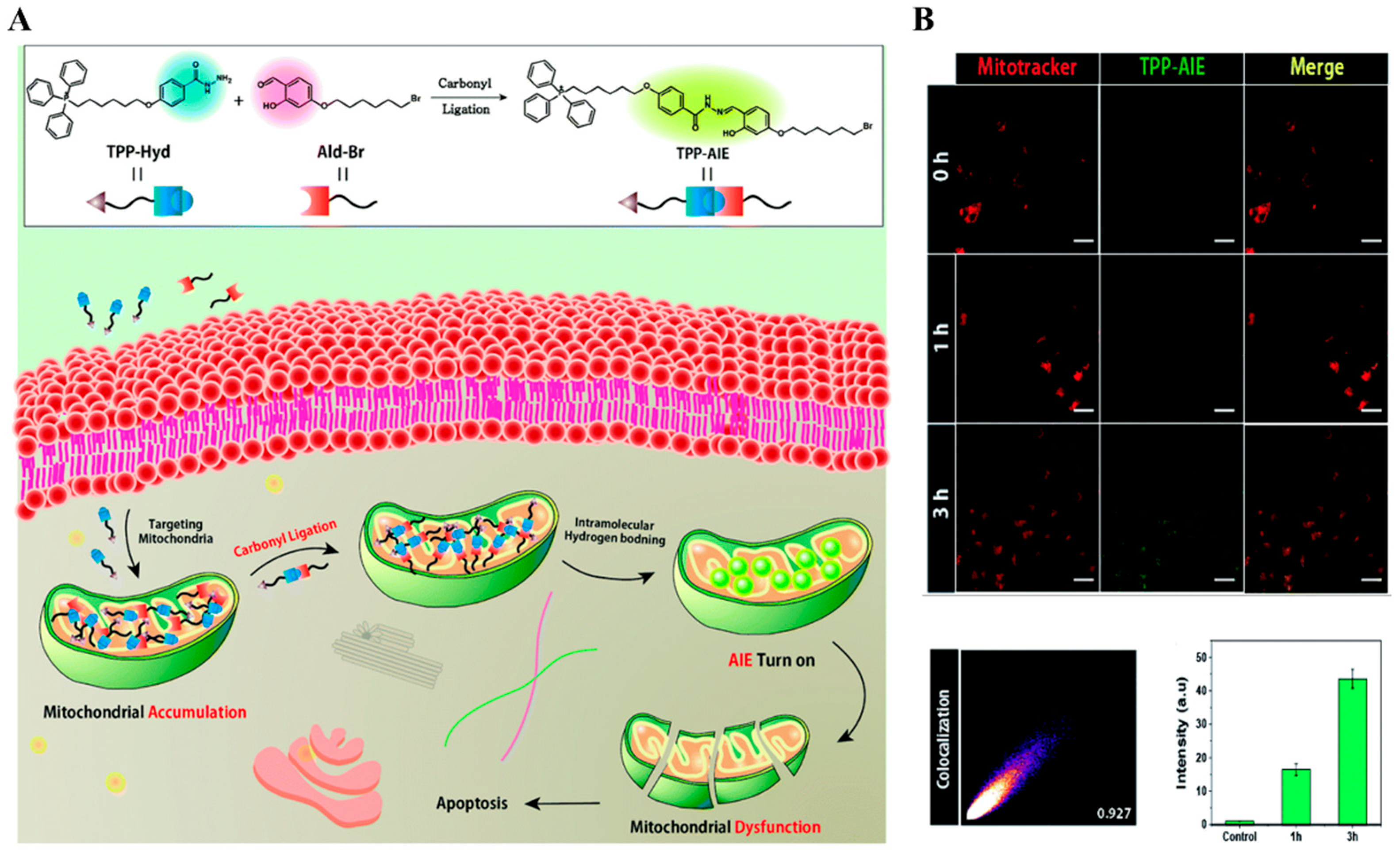
2.6. AIE Based Monomer

Aggregation-induced emission (AIE) constitutes a notable phenomenon observed in select molecular species, wherein their fluorescence undergoes augmentation upon aggregation or consolidation in a condensed phase. Unlike conventional fluorescent molecules, which typically experience fluorescence quenching upon aggregation due to molecular interactions, AIE molecules exhibit a converse behavior, wherein their fluorescence intensifies. This distinctive attribute endows AIE molecules with considerable utility across diverse domains, including but not limited to biological imaging, optoelectronic devices, and sensor technologies. [105,106]. For example, Liu et al. reported lysosomes and mitochondria dual-targeting aggregation-induced emission (AIE) photosensitizer for enhanced antitumor activity and two-photon imaging strategy [107]. The positively charged amphiphilic organic compound (ADB) was rationally designed based on D-A-π-A skeleton by combining an electron donor (triphenylamine) and electron acceptor (2,1,3-benzothiadiazole and 2,3-dimethylbenzo[d]thiazol-3-ium iodide). The cationic lipophilic segment of the molecule enhanced its mitochondria-targeting ability and further facilitated into the lysosomes. Results show that its fluorescent intensity increases (16 times) in toluene/DMSO (toluene fraction reached 95%) indicating its AIE characteristics. Its organelle targeting ability was also determined by its overlapped fluorescence (NIR fluorescence) signal with the green fluorescence of Lyso-Tracker Green (Pearson’s correlation coefficients 0.88), and with the orange fluorescence of Mito-Tracker Orange (Pearson’s correlation coefficients 0.81). Cellular apoptosis by producing ROS and PDT effect was also measured by the confocal images of these probes where it exhibited bright red fluorescence (upon white light (400–700 nm irradiation) in the PI channel, and no green fluorescence was observed in the AM channel, implying that almost all cells were dead. Moreover, in vivo, fluorescence imaging of tumors and metabolic capability were performed to verify its selectivity of cancer and application in PDT therapy. Again, the Ryu group applied the properties in the organelle for imaging organelle and inducing dysfunction of the organelle [62]. They designed two species of molecules: triphenylphosphonium-hydrazine (TPP-Hyd) and aldehyde-Br (Ald-Br). Those molecules are accumulated in mitochondria by mitochondrial targeting moiety. Inside of mitochondria, the carbonyl ligation reaction occurred between TPP-Hyd and Ald-Br and resulted in the formation of TPP-AIE, which facilitates self-assembly behavior due to increased hydrophobicity. The assembly of TPP-AIE molecules induces aggregation-induced emission property to emit green fluorescence (Figure 9A,B). Interestingly, the fluorescence was found to be dependent on the concentration of reactant because carbonyl ligation required a critical concentration of TPP-Hyd and Ald-Br. Under diluted conditions, the reaction could not occur enough, resulting in insufficient generation of TPP-AIE to induce self-assembly behavior. However, when the reactants accumulate inside mitochondria, high concentration facilitates the valid formation of TPP-AIE for the construction of nanostructure. In this regard, TPP-AIE formed inside the mitochondria of cancer cells, leading to selective fluorescence and mitochondrial dysfunction in cancer cells compared to normal cells. Furthermore, the formation of fluorescent nanostructure leads to mitochondrial dysfunction, suggesting its potential as a therapeutic agent for cancer treatment. Thus, AIE-based monomer demonstrates the great potential of bioimaging, selective organelle dysfunction, and clinical imaging-guided photodynamic therapy applications.

3. Conclusions and Future Outlook

This review summarizes the organelle-targeting self-assembled fluorescent probes in cancer treatment that have been developed in the last decade. In brief, we mostly focused on the design of the fluorescent probe-based monomer/precursors, their specific organelle targeting strategy, self-assembly behavior by the cancer stimuli with unique chemical or physical properties, cellular uptake, colocalization, and imaging technique, and the mechanism of action for the dysfunction of distinct cellular organelles. However, results demonstrated that fluorescent probes effectively target and localized inside the organelles and undergo self-assembly or aggregation by the overexpressed stimuli to form various nanostructures that possess bio-functionality with long retention time, organelles membrane disruption or swelling/ROS generation/enzyme activity suppression ability and enhanced photodynamic properties for anticancer treatment. Thus, this strategy can be effective for the in situ long-time cellular imaging technique with reduced low signal-to-background ratio, and false positive and negative signals as well as overcoming the aggregation-caused quenching effect of dyes. Despite the great advantages of this strategy, many challenges remain to be resolved for the high performance of these probes. For example,
(i)
Most of the mitochondria targeting units are positive so it may induce undesirable interaction with the negatively charged biomolecule inside the complex cellular environments.
(ii)
Sometimes nonspecific accumulation of the precursor molecule inside the cellular organelle may happen which leads to unexpected self-assembly and causes the breakdown of cellular immune systems.
(iii)
Off-target accumulation also leads to the toxicity of the probe molecule for the normal cells.
(iv)
Many fluorescent probes face the aggregation caused-quenching effect (ACQ) or photobleaching after self-assembly and thus desired activity of the probe cannot be achieved.
However, the above-mentioned challenges could be overcome by undertaking the following considerations during the designing of the monomer molecules. (i) instead of TPP, a charged-free amphiphilic molecule could be a good choice, (ii) a more specific organelle targeting unit or stimuli-responsive monomer may designed, (iii) only cancer cell-selective molecular design based on cancer cell characteristics and its stimuli, and (iv) sometimes modifying the dye structure or using AIE dye may be one choice. We hope and believe that this feature review will serve as a valuable resource for researchers to design better organelle-targeted self-assembled fluorescent probes in the field of cancer nanomaterials in the near future.

Author Contributions

Conceptualization: J.-H.R., S.K. and M.S.H.; writing—original draft preparation: J.-H.R., S.K., M.S.H., C.L., M.-S.S. and J.L.; writing—review and editing: J.-H.R., S.K. and M.S.H.; project administration: J.-H.R.; funding acquisition: J.-H.R. All authors have read and agreed to the published version of the manuscript.

Funding

This work was supported by National Research Foundation of Korea (NRF) grants funded by the Korean Government (MSIP) (RS-2023-00208386, 2020M3A9D8038192, RS-2023-00255698).

Conflicts of Interest

The authors declare no conflict of interest.

References

  1. Ntziachristos, V. Fluorescence molecular imaging. Annu. Rev. Biomed. Eng. 2006, 8, 1–33. [Google Scholar] [CrossRef] [PubMed]
  2. Kim, J.S.; Quang, D.T. Calixarene-Derived Fluorescent Probes. Chem. Rev. 2007, 107, 3780–3799. [Google Scholar] [CrossRef] [PubMed]
  3. Chan, J.; Dodani, S.; Chang, C. Reaction-based small-molecule fluorescent probes for chemoselective bioimaging. Nat. Chem. 2012, 4, 973–984. [Google Scholar] [CrossRef] [PubMed]
  4. Yue, Y.; Huo, F.; Cheng, F.; Zhu, X.; Mafireyi, T.; Strongin, R.M.; Yin, C. Correction: Functional synthetic probes for selective targeting and multi-analyte detection and imaging. Chem. Soc. Rev. 2019, 48, 4336–4337. [Google Scholar] [CrossRef] [PubMed]
  5. Yang, Y.; Gao, F.; Wang, Y.; Li, H.; Zhang, J.; Sun, Z.; Jiang, Y. Fluorescent Organic Small Molecule Probes for Bioimaging and Detection Applications. Molecules 2022, 27, 8421. [Google Scholar] [CrossRef] [PubMed]
  6. Han, H.; Zhong, Y.; He, C.; Fu, L.; Huang, Q.; Kuang, Y.; Yi, X.; Zeng, W.; Zhong, H.; Yang, M. Recent advances in organic fluorescent probes for tumor related enzyme detection. Dye. Pigm. 2022, 204, 110386. [Google Scholar] [CrossRef]
  7. Balleza, E.; Kim, J.; Cluzel, P. Systematic characterization of maturation time of fluorescent proteins in living cells. Nat. Methods 2018, 15, 47–51. [Google Scholar] [CrossRef] [PubMed]
  8. Zhang, J.; Huang, Y.; Wang, D.; Pollard, A.C.; Chen, Z.G.; Egap, E. Triblock near-infrared fluorescent polymer semiconductor nanoparticles for targeted imaging. J. Mater. Chem. C 2017, 5, 5685–5692. [Google Scholar] [CrossRef]
  9. Bucevičius, J.; Gerasimaitė, R.; Kiszka, K.A.; Pradhan, S.; Kostiuk, G.; Koenen, T.; Lukinavičius, G. A general highly efficient synthesis of biocompatible rhodamine dyes and probes for live-cell multicolor nanoscopy. Nat. Commun. 2023, 14, 1306. [Google Scholar] [CrossRef]
  10. Fuente-Herreruela, D.D.L.; Gónzalez-Charro, V.; Almendro-Vedia, V.G.; Morán, M.; Martín, M.Á.; Lillo, M.P.; Natale, P.; López-Montero, I. Rhodamine-based sensor for real-time imaging of mitochondrial ATP in living fibroblasts. Biochim. Biophys. Acta Bioenerg. 2017, 1858, 999–1006. [Google Scholar] [CrossRef]
  11. Karakuş, E. A rhodamine based fluorescent chemodosimeter for the selective and sensitive detection of copper (II) ions in aqueous media and living cells. J. Mol. Struct. 2021, 1224, 129037. [Google Scholar] [CrossRef]
  12. Sun, S.; Wu, X.; Huang, Y.; Jiang, Q.; Zhu, S.; Sun, S. Visual detection of Cu2+ in high-copper feed based on a fluorescent derivative of rhodamine B. Microchem. J. 2021, 171, 106858. [Google Scholar] [CrossRef]
  13. Okoh, M.P.; Hunter, J.L.; Corrie, J.E.T.; Webb, M.R. A Biosensor for Inorganic Phosphate Using a Rhodamine-Labeled Phosphate Binding Protein. Biochemistry 2006, 45, 14764. [Google Scholar] [CrossRef] [PubMed]
  14. Patra, C.R.; Bhattacharya, R.; Patra, S.; Basu, S.; Mukherjee, P.; Mukhopadhyay, D. Inorganic phosphate nanorods are a novel fluorescent label in cell biology. J. Nanobiotechnol. 2006, 4, 11. [Google Scholar] [CrossRef] [PubMed]
  15. Wen, L.; Ma, X.; Yang, J.; Jiang, M.; Peng, C.; Ma, Z.; Yu, H.; Li, Y. A New Ratiometric Design Strategy Based on Modulation of π-Conjugation Unit for Developing Fluorescent Probe and Imaging of Cellular Peroxynitrite. Anal. Chem. 2022, 94, 4763. [Google Scholar] [CrossRef]
  16. Li, Z.; Liang, P.Z.; Ren, T.-B.; Yuan, L.; Zhang, X.-B. Orderly Self-Assembly of Organic Fluorophores for Sensing and Imaging. Angew. Chem. Int. Ed. 2023, 62, e202305742. [Google Scholar] [CrossRef]
  17. Wang, J.; Jiang, Z.; Huang, C.; Zhao, S.; Zhu, S.; Liu, R.; Zhu, H. Self-Assembled BODIPY Nanoparticles for Near-Infrared Fluorescence Bioimaging. Molecules 2023, 28, 2997. [Google Scholar] [CrossRef]
  18. Ding, F.; Fan, Y.; Sun, Y.; Zhang, F. Beyond 1000 nm Emission Wavelength: Recent Advances in Organic and Inorganic Emitters for Deep-Tissue Molecular Imaging. Adv. Healthc. Mater. 2019, 8, e1900260. [Google Scholar] [CrossRef]
  19. Li, K.; Xu, S.; Xiong, M.; Huan, S.-Y.; Yuan, L.; Zhang, X.-B. Molecular engineering of organic-based agents for in situ bioimaging and phototherapeutics. Chem. Soc. Rev. 2021, 50, 11766–11784. [Google Scholar] [CrossRef]
  20. Kim, S.; Park, G.; Kim, D.; Hasan, M.S.; Lim, C.; Seu, M.-S.; Ryu, J.-H. Intracellular Chemical Reaction-Induced Self-Assembly for the Construction of Artificial Architecture and Its Functions. Adv. NanoBiomed Res. 2024, 4, 2300137. [Google Scholar] [CrossRef]
  21. Okamoto, K.; Chithra, P.; Richards, G.J.; Hill, J.P.; Ariga, K. Self-Assembly of Optical Molecules with Supramolecular Concepts. Int. J. Mol. Sci. 2009, 10, 1950–1966. [Google Scholar] [CrossRef] [PubMed]
  22. Hu, L.; Li, Y.; Lin, X.; Huo, Y.; Zhang, H.; Wang, H. Structure-Based Programming of Supramolecular Assemblies in Living Cells for Selective Cancer Cell Inhibition. Angew. Chem. Int. Ed. 2021, 60, 21807–21816. [Google Scholar] [CrossRef]
  23. Huang, F.; Anslyn, E.V. Introduction: Supramolecular Chemistry. Chem. Rev. 2015, 115, 6999–7000. [Google Scholar] [CrossRef]
  24. Verduzco, R.; Li, X.; Peseka, S.L.; Stein, G.E. Structure, function, self-assembly, and applications of bottlebrush copolymers. Chem. Soc. Rev. 2015, 44, 2405–2420. [Google Scholar] [CrossRef] [PubMed]
  25. Wang, L.-J.; Li, X.; Bai, S.; Wang, Y.-Y.; Han, Y.-F. Self-Assembly, Structural Transformation, and Guest-Binding Properties of Supramolecular Assemblies with Triangular Metal-Metal Bonded Units. J. Am. Chem. Soc. 2020, 142, 2524–2531. [Google Scholar] [CrossRef] [PubMed]
  26. Kaiser, T.E.; Stepanenko, V.; Würthner, F. Fluorescent J-Aggregates of Core-Substituted Perylene Bisimides: Studies on Structure−Property Relationship, Nucleation−Elongation Mechanism, and Sergeants-and-Soldiers Principle. J. Am. Chem. Soc. 2009, 131, 6719–6732. [Google Scholar] [CrossRef] [PubMed]
  27. Kaiser, T.E.; Stepanenko, V.; Würthner, F. Supramolecular Construction of Fluorescent J-Aggregates Based on Hydrogen-Bonded Perylene Dyes. Angew. Chem. Int. Ed. 2007, 46, 5541–5544. [Google Scholar] [CrossRef]
  28. Song, X.; Zhang, R.; Liang, C.; Chen, Q.; Gong, H.; Liu, Z. Nano-assemblies of J-aggregates based on a NIR dye as a multifunctional drug carrier for combination cancer therapy. Biomaterials 2015, 57, 84–92. [Google Scholar] [CrossRef]
  29. Cai, K.; Xie, J.; Zhang, D.; Shi, W.; Yan, Q.; Zhao, D. Concurrent Cooperative J-Aggregates and Anticooperative H-Aggregates. J. Am. Chem. Soc. 2018, 140, 5764–5773. [Google Scholar] [CrossRef]
  30. Li, Z.; Liang, P.Z.; Xu, L.; Zhang, X.-X.; Li, K.; Wu, Q.; Lou, X.-F.; Ren, T.-B.; Yuan, L.; Zhang, X.-B. In situ orderly self-assembly strategy affording NIR-II-J-aggregates for in vivo imaging and surgical navigation. Nat. Commun. 2023, 14, 1843. [Google Scholar] [CrossRef]
  31. Sivagnanam, S.; Das, K.; Basak, M.; Mahata, T.; Stewart, A.; Maity, B.; Das, P. Self-assembled dipeptide based fluorescent nanoparticles as a platform for developing cellular imaging probes and targeted drug delivery chaperones. Nanoscale Adv. 2022, 4, 1694–1706. [Google Scholar] [CrossRef] [PubMed]
  32. Wei, H.; Wu, G.; Tian, X.; Liu, Z. Smart fluorescent probes for in situ imaging of enzyme activity: Design strategies and applications. Future Med. Chem. 2018, 10, 2729–2744. [Google Scholar] [CrossRef] [PubMed]
  33. Ren, C.; Zhang, J.; Chen, M.; Yang, Z. Self-assembling small molecules for the detection of important analytes. Chem. Soc. Rev. 2014, 43, 7257–7266. [Google Scholar] [CrossRef] [PubMed]
  34. Hai, Z.; Liang, C. Intracellular Self-Assembly of Nanoprobes for Molecular Imaging. Adv. Biosyst. 2018, 2, 1800108. [Google Scholar] [CrossRef]
  35. Ishii, M.; Fukuoka, Y.; Deguchi, S.; Otake, H.; Tanino, T.; Nagai, N. Energy-Dependent Endocytosis is Involved in the Absorption of Indomethacin Nanoparticles in the Small Intestine. Int. J. Mol. Sci. 2019, 20, 476. [Google Scholar] [CrossRef] [PubMed]
  36. Zhang, B.; Feng, X.; Yin, H.; Ge, Z.; Wang, Y.; Chu, Z.; Raabova, H.; Vavra, J.; Cigler, P.; Liu, R.; et al. Anchored but not internalized: Shape dependent endocytosis of nanodiamond. Sci. Rep. 2017, 7, 46462. [Google Scholar] [CrossRef] [PubMed]
  37. Jang, Y.H.; Jin, X.; Shankar, P.; Lee, J.H.; Jo, K.; Lim, K.I. Molecular-Level Interactions between Engineered Materials and Cells. Int. J. Mol. Sci. 2019, 20, 4142. [Google Scholar] [CrossRef]
  38. Zhu, H.; Fan, J.; Du, J.; Peng, X. Fluorescent Probes for Sensing and Imaging within Specific Cellular Organelles. Acc. Chem. Res. 2016, 49, 2115–2126. [Google Scholar] [CrossRef]
  39. Wang, X.; Luo, D.; Basilion, J.P. Photodynamic Therapy: Targeting Cancer Biomarkers for the Treatment of Cancers. Cancers 2021, 13, 2992. [Google Scholar] [CrossRef]
  40. Elsyed, A.F.N.; Wong, G.-L.; Ameen, M.; Wu, M.-W.; Chang, C.-C. Tunable Fluorescence via Self-Assembled Switching of AIE-Active Micelle-like Nanoaggregates. Int. J. Mol. Sci. 2023, 24, 9941. [Google Scholar] [CrossRef]
  41. Cheung, S.; O’Shea, D.F. Directed self-assembly of fluorescence responsive nanoparticles and their use for real-time surface and cellular imaging. Nat. Commun. 2017, 8, 1885. [Google Scholar] [CrossRef] [PubMed]
  42. Shao, L.; Yu, H.; Song, J.; Liu, S.; Li, G. Pyrene-Based Self-Assembling Peptide for Ratiometric Detection of Heparin. ChemBioChem 2023, 24, e202200652. [Google Scholar] [CrossRef] [PubMed]
  43. Chen, X.; Li, J.; Roy, S.; Ullah, Z.; Gu, J.; Huang, H.; Yu, C.; Wang, X.; Wang, H.; Zhang, Y.; et al. Development of Polymethine Dyes for NIR-II Fluorescence Imaging and Therapy. Adv. Healthc. Mater. 2024, 13, 2304506. [Google Scholar] [CrossRef] [PubMed]
  44. Sarkar, P.; Harikumar, K.G.; Rawat, S.S.; Das, S.; Chakraborty, T.K.; Chattopadhyay, A. Environment-Sensitive Fluorescence of 7-Nitrobenz-2-oxa-1,3-diazol-4-yl (NBD)-Labeled Ligands for Serotonin Receptors. Molecules 2021, 26, 3848. [Google Scholar] [CrossRef] [PubMed]
  45. Wang, Z.; Mizoguchi, T.; Kuribara, T.; Nakajima, M.; Iwata, M.; Sakamoto, Y.; Nakamura, H.; Murayama, T.; Nemoto, T.; Itoh, M. Py3-FITC: A new fluorescent probe for live cell imaging of collagen-rich tissues and ionocytes. Open Biol. 2021, 11, 200241. [Google Scholar] [CrossRef] [PubMed]
  46. Bhusanur, D.I.; More, K.S.; Al Kobaisi, M.; Singh, P.K.; Bhosale, S.V.; Bhosale, S.V. Synthesis, Photophysical Properties and Self-Assembly of a Tetraphenylethylene-Naphthalene Diimide Donor-Acceptor Molecule. Chem. Asian J. 2024, 19, e202301046. [Google Scholar] [CrossRef] [PubMed]
  47. Feng, G.; Zhang, G.-Q.; Ding, D. Design of superior phototheranostic agents guided by Jablonski diagrams. Chem. Soc. Rev. 2020, 49, 8179–8234. [Google Scholar] [CrossRef] [PubMed]
  48. Jeena, M.T.; Palanikumar, L.; Go, E.M.; Kim, I.; Kang, M.G.; Lee, S.; Park, S.; Choi, H.; Kim, C.; Jin, S.-M.; et al. Mitochondria localization induced self-assembly of peptide amphiphiles for cellular dysfunction. Nat. Commun. 2017, 8, 26. [Google Scholar] [CrossRef] [PubMed]
  49. Jeena, M.T.; Jeong, K.; Go, E.M.; Cho, Y.; Lee, S.; Jin, S.; Hwang, S.-W.; Jang, J.H.; Kang, C.S.; Bang, W.-Y.; et al. Heterochiral Assembly of Amphiphilic Peptides inside the Mitochondria for Supramolecular Cancer Therapeutics. ACS Nano 2019, 13, 11022. [Google Scholar] [CrossRef]
  50. Jeena, M.T.; Jin, S.; Jana, B.; Ryu, J.-H. Enzyme-instructed morphology transformation of mitochondria-targeting peptide for the selective eradication of osteosarcoma. RSC Chem. Biol. 2022, 3, 1416–1421. [Google Scholar] [CrossRef]
  51. Jeena, M.T.; Jin, S.; Jeong, K.; Cho, Y.; Kim, J.C.; Lee, J.H.; Lee, S.; Hwang, S.-W.; Kwak, S.K.; Kim, S.; et al. Cancer-Selective Supramolecular Chemotherapy by Disassembly-Assembly Approach. Adv. Funct. Mater. 2022, 32, 2208098. [Google Scholar] [CrossRef]
  52. Zhu, M.; Xing, P.; Zhou, Y.; Gong, L.; Zhang, J.; Qi, D.; Bian, Y.; Du, H.; Jiang, J. Lysosome-targeting ratiometric fluorescent pH probes based on long-wavelength BODIPY. J. Mater. Chem. B 2018, 6, 4422. [Google Scholar] [CrossRef] [PubMed]
  53. Saha, P.C.; Das, R.S.; Chatterjee, T.; Bhattacharyya, M.; Guha, S. Supramolecular β-Sheet Forming Peptide Conjugated with Near-Infrared Chromophore for Selective Targeting, Imaging, and Dysfunction of Mitochondria. Bioconjug. Chem. 2020, 31, 1301–1306. [Google Scholar] [CrossRef] [PubMed]
  54. Jana, B.; Thomas, A.P.; Kim, S.; Lee, I.S.; Choi, H.; Jin, S.; Park, S.A.; Min, S.K.; Kim, C.; Ryu, J.-H. Self-Assembly of Mitochondria-Targeted Photosensitizer to Increase Photostability and Photodynamic Therapeutic Efficacy in Hypoxia. Chem. Eur. J. 2020, 26, 10695. [Google Scholar] [CrossRef] [PubMed]
  55. Ge, X.; Cao, Y.; Zhu, X.; Yuan, B.; He, L.; Wu, A.; Li, J. Self-Assembly of Organelle-Localized Neuropeptides Triggers Intrinsic Apoptosis against Breast Cancer. Adv. Healthc. Mater. 2023, 12, 2300265. [Google Scholar] [CrossRef] [PubMed]
  56. Yang, L.; Peltier, R.; Zhang, M.; Song, D.; Huang, H.; Chen, G.; Chen, Y.; Zhou, F.; Hao, Q.; Bian, L.; et al. Desuccinylation-Triggered Peptide Self-Assembly: Live Cell Imaging of SIRT5 Activity and Mitochondrial Activity Modulation. J. Am. Chem. Soc. 2020, 142, 18150–18159. [Google Scholar] [CrossRef] [PubMed]
  57. He, H.; Wang, J.; Wang, H.; Zhou, N.; Yang, D.; Green, D.R.; Xu, B. Enzymatic Cleavage of Branched Peptides for Targeting Mitochondria. J. Am. Chem. Soc. 2018, 140, 1215. [Google Scholar] [CrossRef] [PubMed]
  58. Tan, W.; Zhang, Q.; Hong, P.; Xu, B. A Self-Assembling Probe for Imaging the States of Golgi Apparatus in Live Single Cells. Bioconjug. Chem. 2022, 33, 1983. [Google Scholar] [CrossRef]
  59. Choi, H.; Park, G.; Shin, E.; Shin, S.W.; Jana, B.; Jin, S.; Kim, S.; Wang, H.; Kwak, S.K.; Xu, B.; et al. Intramitochondrial co-assembly between ATP and nucleopeptides induces cancer cell apoptosis. Chem. Sci. 2022, 13, 6197–6204. [Google Scholar] [CrossRef]
  60. Kim, S.; Chae, J.-B.; Kim, D.; Park, C.-W.; Sim, Y.; Lee, H.; Park, G.; Lee, J.; Hong, S.; Jana, B.; et al. Supramolecular Senolytics via Intracellular Oligomerization of Peptides in Response to Elevated Reactive Oxygen Species Levels in Aging Cells. J. Am. Chem. Soc. 2023, 145, 21991–22008. [Google Scholar] [CrossRef]
  61. Jana, B.; Jin, S.; Go, E.M.; Cho, Y.; Kim, D.; Kim, S.; Kwak, S.K.; Ryu, J.-H. Intra-Lysosomal Peptide Assembly for the High Selectivity Index against Cancer. J. Am. Chem. Soc. 2023, 145, 18414–18431. [Google Scholar] [CrossRef] [PubMed]
  62. Kim, S.; Kim, J.; Jana, B.; Ryu, J.-H. Intra-mitochondrial reaction for cancer cell imaging and anti-cancer therapy by aggregation-induced emission. RSC Adv. 2020, 10, 43383. [Google Scholar] [CrossRef]
  63. Ji, X.; Wang, N.; Wang, J.; Wang, T.; Huang, X.; Hao, H. Non-destructive real-time monitoring and investigation of the self-assembly process using fluorescent probes. Chem. Sci. 2024, 15, 3800–3830. [Google Scholar] [CrossRef] [PubMed]
  64. Agafontsev, A.M.; Shumilova, T.A.; Oshchepkov, A.S.; Hampel, F.; Kataev, E.A. Ratiometric Detection of ATP by Fluorescent Cyclophanes with Bellows-Type Sensing Mechanism. Chem. Eur. J. 2020, 26, 9991. [Google Scholar] [CrossRef] [PubMed]
  65. Chen, Y. Recent Advances in Excimer-Based Fluorescence Probes for Biological Applications. Molecules 2022, 27, 8628. [Google Scholar] [CrossRef] [PubMed]
  66. Shellaiah, M.; Venkatesan, P.; Thirumalaivasan, N.; Wu, S.-P.; Sun, K.-W. Pyrene-Based Fluorescent Probe for “Off-on-Off” Sequential Detection of Cu2+ and CN− with HeLa Cells Imaging. Chemosensors 2023, 11, 115. [Google Scholar] [CrossRef]
  67. Zhang, R.; Tang, D.; Lu, P.; Yang, X.; Liao, D.; Zhang, Y.; Zhang, M.; Yu, C.; Yam, V.W.W. Nucleic Acid-Induced Aggregation and Pyrene Excimer Formation. Org. Lett. 2009, 11, 4302–4305. [Google Scholar] [CrossRef] [PubMed]
  68. Bonora, M.; Patergnani, S.; Rimessi, A.; Marchi, E.D.; Suski, J.M.; Bononi, A.; Giorgi, C.; Marchi, S.; Missiroli, S.; Poletti, F.; et al. ATP synthesis and storage. Purinergic Signal. 2012, 8, 343–357. [Google Scholar] [CrossRef] [PubMed]
  69. Özalp, V.C.; Pedersen, T.R.; Nielsen, L.J.; Olsen, L.F. Time-resolved Measurements of Intracellular ATP in the Yeast Saccharomyces cerevisiae using a New Type of Nanobiosensor. J. Biol. Chem. 2010, 285, 37579–37588. [Google Scholar] [CrossRef]
  70. Li, Z.; Zou, J.; Chen, X. In Response to Precision Medicine: Current Subcellular Targeting Strategies for Cancer Therapy. Adv. Mater. 2023, 35, 2209529. [Google Scholar] [CrossRef]
  71. Mills, E.; Kelly, B.; O’Neill, L. Mitochondria are the powerhouses of immunity. Nat. Immunol. 2017, 18, 488–498. [Google Scholar] [CrossRef] [PubMed]
  72. Sweetwyne, M.T.; Pippin, J.W.; Eng, D.G.; Hudkins, K.L.; Chiao, Y.A.; Campbell, M.D.; Marcinek, D.J.; Alpers, C.E.; Szeto, H.H.; Rabinovitch, P.S.; et al. The mitochondrial-targeted peptide, SS-31, improves glomerular architecture in mice of advanced age. Kidney Int. 2017, 91, 1126–1145. [Google Scholar] [CrossRef] [PubMed]
  73. Sibrian-Vazquez, M.; Nesterova, I.V.; Jensen, T.J.; Vicente, M.G.H. Mitochondria Targeting by Guanidine– and Biguanidine–Porphyrin Photosensitizers. Bioconjug. Chem. 2008, 19, 705. [Google Scholar] [CrossRef]
  74. Jeena, M.T.; Kim, S.; Jin, S.; Ryu, J.-H. Recent Progress in Mitochondria-Targeted Drug and Drug-Free Agents for Cancer Therapy. Cancers 2020, 12, 4. [Google Scholar] [CrossRef]
  75. Shi, J.; Du, X.; Yuan, D.; Zhou, J.; Zhou, N.; Huang, Y.; Xu, B. D-Amino Acids Modulate the Cellular Response of Enzymatic-Instructed Supramolecular Nanofibers of Small Peptides. Biomacromolecules 2014, 15, 3559. [Google Scholar] [CrossRef] [PubMed]
  76. Seia, M.; Zisman, E. Different roles of D-amino acids in immune phenomena. FASEB J. 1997, 11, 449. [Google Scholar] [CrossRef] [PubMed]
  77. Jeena, M.T.; Lee, S.; Barui, A.K.; Jin, S.; Cho, Y.; Hwang, S.-W.; Kim, S.; Ryu, J.-H. Intra-mitochondrial self-assembly to overcome the intracellular enzymatic degradation of l-peptides. Chem. Commun. 2020, 56, 6265. [Google Scholar] [CrossRef] [PubMed]
  78. Kim, D.; Kim, S.; Park, G.; Choi, H.; Ryu, J.-H. Spatiotemporal Self-Assembly of Peptide Amphiphiles by Carbonic Anhydrase IX-Targeting Induces Cancer-Lysosomal Membrane Disruption. JACS Au 2022, 2, 2539–2547. [Google Scholar] [CrossRef]
  79. Platt, F.M.; d’Azzo, A.; Davidson, B.L.; Neufeld, E.F.; Tifft, C.J. Lysosomal storage diseases. Nat. Rev. Dis. Primers 2018, 4, 27. [Google Scholar] [CrossRef]
  80. Dielschneider, R.F.; Henson, E.S.; Gibson, S.B. Lysosomes as Oxidative Targets for Cancer Therapy. Oxidative Med. Cell. Longev. 2017, 2017, 3749157. [Google Scholar] [CrossRef]
  81. Fan, Z.; Chang, Y.; Cui, C.; Sun, L.; Wang, D.H.; Pan, Z.; Zhang, M. Near infrared fluorescent peptide nanoparticles for enhancing esophageal cancer therapeutic efficacy. Nat. Commun. 2018, 9, 2605. [Google Scholar] [CrossRef]
  82. Zhou, J.; Du, X.; Xu, B. Regulating the Rate of Molecular Self-Assembly for Targeting Cancer Cells. Angew. Chem. Int. Ed. 2016, 55, 5770–5775. [Google Scholar] [CrossRef]
  83. Yan, R.; Hu, Y.; Liu, F.; Wei, S.; Fang, D.; Shuhendler, A.J.; Liu, H.; Chen, H.-Y.; Ye, D. Activatable NIR Fluorescence/MRI Bimodal Probes for in Vivo Imaging by Enzyme-Mediated Fluorogenic Reaction and Self-Assembly. J. Am. Chem. Soc. 2019, 141, 10331–10341. [Google Scholar] [CrossRef]
  84. Escobedo, J.O.; Rusin, O.; Lim, S.; Strongin, R.M. NIR dyes for bioimaging applications. Curr. Opin. Chem. Biol. 2010, 14, 64–70. [Google Scholar] [CrossRef]
  85. Yue, C.; Liu, P.; Zheng, M.; Zhao, P.; Wang, Y.; Ma, Y.; Cai, L. IR-780 dye loaded tumor targeting theranostic nanoparticles for NIR imaging and photothermal therapy. Biomaterials 2013, 34, 6853–6861. [Google Scholar] [CrossRef]
  86. Zhou, Y.; Li, Q.; Wu, Y.; Li, X.; Zhou, Y.; Wang, Z.; Liang, H.; Ding, F.; Hong, S.; Steinmetz, N.F.; et al. Molecularly Stimuli-Responsive Self-Assembled Peptide Nanoparticles for Targeted Imaging and Therapy. ACS Nano 2023, 17, 8004–8025. [Google Scholar] [CrossRef]
  87. Wang, Q.; Jiang, N.; Fu, B.; Huang, F.; Liu, J. Self-assembling peptide-based nanodrug delivery systems. Biomater. Sci. 2019, 7, 4888–4911. [Google Scholar] [CrossRef] [PubMed]
  88. An, H.-W.; Hou, D.; Zheng, R.; Wang, M.-D.; Zeng, X.-Z.; Xiao, W.-Y.; Yan, T.-D.; Wang, J.-Q.; Zhao, C.-H.; Cheng, L.-M.; et al. A Near-Infrared Peptide Probe with Tumor-Specific Excretion-Retarded Effect for Image-Guided Surgery of Renal Cell Carcinoma. ACS Nano 2020, 14, 927–936. [Google Scholar] [CrossRef] [PubMed]
  89. Saha, P.C.; Bera, T.; Chatterjee, T.; Samanta, J.; Sengupta, A.; Bhattacharyya, M.; Guha, S. Supramolecular Dipeptide-Based Near-Infrared Fluorescent Nanotubes for Cellular Mitochondria Targeted Imaging and Early Apoptosis. Bioconjug. Chem. 2021, 32, 833–841. [Google Scholar] [CrossRef] [PubMed]
  90. Wang, H.; Feng, Z.; Signore, S.J.D.; Rodal, A.A.; Xu, B. Active Probes for Imaging Membrane Dynamics of Live Cells with High Spatial and Temporal Resolution over Extended Time Scales and Areas. J. Am. Chem. Soc. 2018, 140, 3505–3509. [Google Scholar] [CrossRef]
  91. Cai, Y.; Shi, Y.; Wang, H.; Wang, J.; Ding, D.; Wang, L.; Yang, Z. Environment-Sensitive Fluorescent Supramolecular Nanofibers for Imaging Applications. Anal. Chem. 2014, 86, 2193–2199. [Google Scholar] [CrossRef] [PubMed]
  92. Jiang, C.; Huang, H.; Kang, X.; Yang, L.; Xi, Z.; Sun, H.; Pluth, M.D.; Yi, L. NBD-Based Synthetic Probes for Sensing Small Molecules and Proteins: Design, Sensing Mechanisms and Biological Applications. Chem. Soc. Rev. 2021, 50, 7436–7495. [Google Scholar] [CrossRef] [PubMed]
  93. Meglio, P.D.; Ianaro, A.; Ghosh, S. Amelioration of acute inflammation by systemic administration of a cell-permeable peptide inhibitor of NF-κB activation. Arthritis Rheum. 2005, 52, 951–958. [Google Scholar] [CrossRef] [PubMed]
  94. Long, Y.-M.; Chen, K.; Liu, X.-J.; Xie, W.-R.; Wang, H. Cell-permeable Tat-NBD peptide attenuates rat pancreatitis and acinus cell inflammation response. World J. Gastroenterol. 2009, 15, 561–569. [Google Scholar] [CrossRef] [PubMed]
  95. Farber-Katz, S.E.; Dippold, H.C.; Buschman, M.D.; Peterman, M.C.; Xing, M.; Noakes, C.J.; Tat, J.; Ng, M.M.; Rahajeng, J.; Cowan, D.M.; et al. DNA damage triggers golgi dispersal via DNA-PK and GOLPH3. Cell 2014, 156, 413. [Google Scholar] [CrossRef]
  96. Kirkham, S.; Hamley, I.W.; Smith, A.M.; Gouveia, R.M.; Connon, C.J.; Reza, M.; Ruokolainen, J. A self-assembling fluorescent dipeptide conjugate for cell labelling. Colloids Surf. B Biointerfaces 2016, 137, 104–108. [Google Scholar] [CrossRef]
  97. Cheng, J.; Fu, S.; Qin, Z.; Han, H.; Yang, X. Self-assembled natural small molecule diterpene acids with favorable anticancer activity and biosafety for synergistically enhanced antitumor chemotherapy. J. Mater. Chem. B 2021, 9, 2674–2687. [Google Scholar] [CrossRef] [PubMed]
  98. Zhang, P.; Cheetham, A.G.; Lock, L.L.; Cui, H. Cellular Uptake and Cytotoxicity of Drug–Peptide Conjugates Regulated by Conjugation Site. Bioconjug. Chem. 2013, 24, 604–613. [Google Scholar] [CrossRef] [PubMed]
  99. Zhang, P.; Lock, L.L.; Cheetham, A.G.; Cui, H. Enhanced Cellular Entry and Efficacy of Tat Conjugates by Rational Design of the Auxiliary Segment. Mol. Pharm. 2014, 11, 964–973. [Google Scholar] [CrossRef]
  100. Choi, S.W.; Hong, H.K.; Jeon, J.; Choi, J.Y.; Kim, M.; Kim, P.; Lee, B.C.; Woo, S.J. FITC-Labeled RGD Peptides as Novel Contrast Agents for Functional Fluorescent Angiographic Detection of Retinal and Choroidal Neovascularization. Cells 2023, 12, 1902. [Google Scholar] [CrossRef]
  101. Das, A.; Ghosh, S. Stimuli-Responsive Self-Assembly of a Naphthalene Diimide by Orthogonal Hydrogen Bonding and Its Coassembly with a Pyrene Derivative by a Pseudo-Intramolecular Charge-Transfer Interaction. Angew. Chem. Int. Ed. 2014, 53, 1092–1097. [Google Scholar] [CrossRef]
  102. Bhosale, R.S.; Al Kobaisi, M.; Bhosale, S.V.; Bhargava, S.; Bhosale, S.V. Flower-like supramolecular self-assembly of phosphonic acid appended naphthalene diimide and melamine. Sci. Rep. 2015, 5, 14609. [Google Scholar] [CrossRef] [PubMed]
  103. Markiewicz, G.; Smulders, M.M.J.; Stefankiewicz, A.R. Steering the Self-Assembly Outcome of a Single NDI Monomer into Three Morphologically Distinct Supramolecular Assemblies, with Concomitant Change in Supramolecular Polymerization Mechanism. Adv. Sci. 2019, 6, 1900577. [Google Scholar] [CrossRef] [PubMed]
  104. Kolhe, N.B.; Devi, R.N.; Senanayak, S.P.; Jancy, B.; Narayan, K.S.; Asha, S.K. Structure engineering of naphthalene diimides for improved charge carrier mobility: Self-assembly by hydrogen bonding, good or bad? J. Mater. Chem. 2012, 22, 15235–15246. [Google Scholar] [CrossRef]
  105. Peng, Y.; Jin, X.; Zheng, Y.; Han, D.; Liu, K.; Jiang, L. Direct Imaging of Superwetting Behavior on Solid–Liquid–Vapor Triphase Interfaces. Adv. Mater. 2017, 29, 1703009. [Google Scholar] [CrossRef] [PubMed]
  106. Shellaiah, M.; Sun, K.-W. Pyrene-Based AIE Active Materials for Bioimaging and Theranostics Applications. Biosensors 2022, 12, 550. [Google Scholar] [CrossRef]
  107. Wang, S.; Liao, Y.; Wu, Z.; Peng, Y.; Liu, Y.; Chen, Y.; Shao, L.; Zeng, Z.; Liu, Y. A lysosomes and mitochondria dual-targeting AIE-active NIR photosensitizer: Constructing amphiphilic structure for enhanced antitumor activity and two-photon imaging. Mater. Today Bio 2023, 21, 100721. [Google Scholar] [CrossRef]
Figure 1. (A) Mito-LISA strategy of Mito FF building block to form self-assembled nanofibrils, resulting in damage of the mitochondrial membrane and apoptosis (B) design of Mito FF and (C) Emission spectra for Mito-FF showing the excimeric emission above the CAC (sky blue line) and lack of excimer below the CAC (deep blue line) (the inset shows optical images of monomeric (left) and excimeric (right) emission) (D) Mitochondrial co-localization of Mito-FF measured with MitoTracker Red FM shows high localization inside mitochondria (scale bar, 5 μm) (E) Two-photon analysis of Mito-FF-treated HeLa cells showing high-intensity excimer emission with a 420–480 nm filter, compared with a 380–420 nm filter (which corresponds to monomeric emission). Reproduced with permission from Ref. [48]. Copyright 2017 Nature Publishing Group.
Chemosensors 12 00138 g001
Figure 2. (A) Schematic illustration of the enzyme-instructed morphology transformation of an ALP-responsive pyrene monomer L-Mito-FFYp and Cellular internalization of Mito-FFYp in the SaOS-2 cell lines showing their co-localization (B) Schematic Illustration Showing the Mechanism of Pep-AT and the CLSM image for the co-localization study. Reproduced with permission from Refs. [50,78] Copyright 2022 The Royal Society of Chemistry and Copyright 2022 American Chemical Society.
Chemosensors 12 00138 g002
Figure 3. (A) Schematic illustration of mitochondria targeting process of KLVFF/Cy-5 conjugate in the presence of an anticancer agent, resulting in self-aggregation and (B) confocal microscopic image shows high colocalization in mitochondria with KLVFF/Cy-5 in HeLa cells. Reproduced with permission from Ref. [53] Copyright 2020 American Chemical Society.
Chemosensors 12 00138 g003
Figure 4. (A) Schematic illustration depicts the photodynamic therapy process of IR-TPP, illustrating its formation into micelle nanoparticles and (B) subsequent accumulation in the mitochondria experiment of IR-TPP in cancer HeLa cells. Reproduced with permission from Ref. [54] Copyright 2020 Wiley-VCH.
Chemosensors 12 00138 g004
Figure 5. (A) This figure illustrates the structure of a representative branched peptide and the action of ENTK in cleaving the branch, resulting in the conversion of micelles to nanofibers on mitochondria and (B) fluorescence microscopy images describe D −1TFLAG, Mito Tracker staining, and combination of RPE (1 μg/mL) and D −1TFLAG in HeLa cells, Reproduced with permission from Ref. [57] Copyright 2018 American Chemical Society.
Chemosensors 12 00138 g005
Figure 6. (A) The schematic illustrates chemical structures, such as the fluorophore, mitochondria-targeting moiety, ATP binding part, and self-assembly, along with their function in sequestering adenosine triphosphate (ATP) within cancer cell mitochondria, leading to apoptosis and (B) confocal imaging confirms the mitochondrial localization of the MNP–NBD/ADP complex in HeLa cells. Reproduced with permission from Ref. [59] Copyright 2022 The Royal Society of Chemistry.
Chemosensors 12 00138 g006
Figure 7. (A) Chemical structure of FITC labeled Mito-K2 building blocks to induce self-assembly inside senescent cells. (B) Selective accumulation of designed molecules in the senescent cell’s mitochondria (C) High mitochondrial colocalization of Mito-K2-FITC and MitoTracker Deep Red in senescence cell. (D) Intramitochondrial monomer concentrations of Mito-K2-FITC in senescent cells with different concentrations. Reproduced with permission from Ref. [60] Copyright 2023 American Chemical Society.
Chemosensors 12 00138 g007
Figure 8. (A) Chemical structure of NDI-based peptide amphiphile before (NDI-Lyso-RGD) and after enzymatic cleavage (NDI-R3) and Schematic illustration of intra-lysosomal self-assembly induced cancer cell apoptosis. (B) Colocalization study of NDI-Lyso with LysoTracker Red in HeLa and HEK293 cells. (Scale bar, 10 μm). Reproduced with permission from Ref. [61] Copyright 2023 American Chemical Society.
Chemosensors 12 00138 g008
Figure 9. (A) Chemical design and carbonyl ligation scheme of TPP-Br and Ald-Br, (B) Schematic illustration of molecules showing mitochondria targeting (colocalization) carbonyl ligation for formation of nanoparticle with AIE property. Reproduced with permission from Ref. [62] Copyright 2020 The Royal Society of Chemistry.
Chemosensors 12 00138 g009
Table 1. Summary of the organelle targeting self-assembly fluorescent probe. 
Types of
Monomer
λex and λemiMolecular Design
(Target-Organelle)
Shape of
Structure
Functional Mechanism Ref.
Pyrene-based343 nm
375–405 nm
Pyrene + FF + TPP
(Mitochondrial)
FiberMitochondrial membrane disruption[48]
Pyrene + FF(D/L) + TPP
(Mitochondrial)
Super fibrilMitochondrial membrane disruption[49]
Pyrene + FFYp + TPP
(Mitochondrial)
Micelle to fiberMitochondrial membrane disruption[50]
Pyrene + FF(SA) + TPP
(Mitochondrial)
Micelle to fiberMitochondrial membrane disruption[51]
Pyrene + FF + AZ
(Lysosome)
Nanofiber Lysosomal membrane disruption[52]
NIR-based651 nm
672 nm
NIR + KLVFF + TPP
(Mitochondrial)
Amyloid fibrilsMitochondrial dysfunction[53]
785 nm
810 nm
NIR + TPP
(Mitochondrial)
MicelleROS generation, PDT[54]
745 nm
810 nm
NIR + FF + KGRR + Y1L
(Lysosome)
Nanofiber Lysosomal enlargement and damages[55]
NBD-based466 nm
539 nm
NBD + FFGKsuccG
(Mitochondrial)
NanofibersDepolarization of mitochondria membrane, ROS generation,[56]
NBD + Nap-ffk
(Mitochondrial)
Micelle to fiberAnticancer drug delivery[57]
NBD + ff + pS1
(Golgi body)
NanofiberReveals the dynamics of cancer cells golgi body[58]
NBD + MNP
(Mitochondrial)
Micelle ROS generation, metabolic disorder[59]
FITC-based498 nm
519 nm
FITC + dithiol + RGD
(Mitochondrial)
α-helixMitochondrial membrane disruption[60]
NDI-based395 nm
420 nm
NDI + Lyso + RGD
(Lysosome)
FiberLysosomal swelling and damage[61]
AIE-basedvariableAIE + TPP
(Mitochondrial)
NanoaggregateMitochondrial dysfunction[62]
FF = phenylalanine dipeptide, TPP = triphenylphosphonium, D/L = dextrorotatory/laevorotatory, SA = succinic amide, p = phosphate, AZ = acetazolamide, KGRR = Arg-Arg-Gly-Lys, Y1L = neuropeptide peptide, MNP = mitochondria-targeting nucleopeptide, pS1 = thiophosphate, RGD = Arg-Gly-Asp.
Disclaimer/Publisher’s Note: The statements, opinions and data contained in all publications are solely those of the individual author(s) and contributor(s) and not of MDPI and/or the editor(s). MDPI and/or the editor(s) disclaim responsibility for any injury to people or property resulting from any ideas, methods, instructions or products referred to in the content.

