Review

The Use of Drosophila to Understand Psychostimulant Responses

by
Travis James Philyaw
1,
Adrian Rothenfluh
2,3,4,5,* and
Iris Titos
3,*
1
Molecular Biology Graduate Program, University of Utah, Salt Lake City, UT 84112, USA
2
Department of Psychiatry, Huntsman Mental Health Institute, University of Utah, Salt Lake City, UT 84108, USA
3
Molecular Medicine Program, University of Utah, Salt Lake City, UT 84112, USA
4
Department of Neurobiology, University of Utah, Salt Lake City, UT 84132, USA
5
Department of Human Genetics, University of Utah, Salt Lake City, UT 84112, USA
*
Authors to whom correspondence should be addressed.
Biomedicines 2022, 10(1), 119; https://doi.org/10.3390/biomedicines10010119
Submission received: 3 December 2021 / Revised: 31 December 2021 / Accepted: 31 December 2021 / Published: 6 January 2022
(This article belongs to the Special Issue Biological Aspects of Drug Addiction)

Abstract

:
The addictive properties of psychostimulants such as cocaine, amphetamine, methamphetamine, and methylphenidate are based on their ability to increase dopaminergic neurotransmission in the reward system. While cocaine and methamphetamine are predominately used recreationally, amphetamine and methylphenidate also work as effective therapeutics to treat symptoms of disorders including attention deficit and hyperactivity disorder (ADHD) and autism spectrum disorder (ASD). Although both the addictive properties of psychostimulant drugs and their therapeutic efficacy are influenced by genetic variation, very few genes that regulate these processes in humans have been identified. This is largely due to population heterogeneity which entails a requirement for large samples. Drosophila melanogaster exhibits similar psychostimulant responses to humans, a high degree of gene conservation, and allow performance of behavioral assays in a large population. Additionally, amphetamine and methylphenidate reduce impairments in fly models of ADHD-like behavior. Therefore, Drosophila represents an ideal translational model organism to tackle the genetic components underlying the effects of psychostimulants. Here, we break down the many assays that reliably quantify the effects of cocaine, amphetamine, methamphetamine, and methylphenidate in Drosophila. We also discuss how Drosophila is an efficient and cost-effective model organism for identifying novel candidate genes and molecular mechanisms involved in the behavioral responses to psychostimulant drugs.

1. Introduction

Psychostimulants with abuse potential, such as cocaine, amphetamine, methamphetamine, and methylphenidate, are drugs that increase central nervous system activity and arousal [1]. These drugs also elevate mood by activating the neural substrates associated with reward [1]. Stimulant drugs have a long history of use in humans and evidence suggests cocaine, in its unpurified form in the coca leaf, has been ingested for more than 3000 years [2]. More potent stimulants, such as processed cocaine and pharmaceutical amphetamine, are relatively new psychoactive substances. Processed cocaine was not available until the late 19th century, when it gained popularity as an additive in medicines, drinks, and cigarettes [2]. Pharmaceutical amphetamine and methamphetamine were not available until the early 20th century when they enjoyed a similar phase of popularity and broad application [3]. The recognition of abuse liability and harm potential of these substances led to their prohibition. While cocaine, amphetamine, and methamphetamine have all been regulated and restricted [2,3,4], their abuse continues to pose a global health concern, with an estimated of 47 million global users of cocaine and amphetamine-type stimulants as of 2019 [5]. The recreational doses of psychostimulant drugs that produce euphoria can lead to addiction and cognitive impairment, while overdoses can cause psychosis, circulatory collapse, and death. In contrast, lower doses produce cognitive enhancement and have a range of therapeutic uses [6,7]. These include treatment of the cognitive and behavioral impairments associated with attention deficit and hyperactivity disorder (ADHD) [8] and autism spectrum disorder (ASD) [9,10]. Despite successful clinical application, the molecular mechanisms mediating the different therapeutic actions of psychostimulant drugs remain unknown. Therefore, two main questions are the focus of continuing research (1) what molecular mechanisms are involved in the transition from recreational psychostimulant use to habitual maladaptive use, and (2) what are the molecular mechanisms that determine the therapeutic success of psychostimulant drugs for treating the symptoms of psychiatric disorders such as ADHD and ASD.
One approach to answering these questions is to take advantage of model organisms that are experimentally amenable and frequently used for their powerful genetics. Genes are a primary determinant of variation in behavioral responses to the addictive or therapeutic effects of psychostimulant drugs [11,12], and family studies show genes influence drug initiation [13] and addiction [14]. Addictions are among the most inherited psychiatric disorders, with an estimated genetic heritability component of ~72% for cocaine [15] and ~42% for non-cocaine psychostimulants [16]. Despite all the evidence from family studies, it has been challenging to identify the specific genes or genetic modifications that make an individual more susceptible to becoming a compulsive user [11]. Similarly, while it is known that genetic variation underlies differences in response to therapeutic drugs [17] little is known about the specific genes and molecules that impact therapeutic response to psychostimulants. Heterogeneity in the population and the subsequent need for a large sample size make it difficult to use human studies for identifying genes that impact psychostimulant response. In contrast to mammals, the model organism Drosophila melanogaster enables high-throughput analysis of behavior in assays that can scale to accommodate thousands of flies while supporting targeted genetic and molecular manipulations [18,19]. This review discusses how the model organism Drosophila can help elucidate the genetic basis of behavioral response to psychostimulant drugs to improve our understanding of human addiction and psychopathology.

1.1. Drosophila as a Model Organism to Study Addiction

Drosophila is a powerful model organism for studying the genetic and molecular basis of behavioral responses to drugs of abuse. A fast generation time, excellent genetic tractability, and a broad range of molecular tools enable precise spatiotemporal manipulation of gene expression, protein function, and cellular activity in Drosophila [18,20,21,22,23]. Around 75% of human disease-causing genes are conserved in flies [24], highlighting the translational application of Drosophila research [25]. Additionally, Drosophila is a valuable model for studying complex addictive disorders such as alcoholism [26]. Studies over the last 25 years have shown that flies express a range of alcohol-induced behaviors similar to those observed in intoxicated humans [27], and numerous genes isolated based on their involvement in Drosophila alcohol responses have helped identify corresponding genes that contribute to alcohol use disorder (AUD) in humans [28]. Additionally, the neurotransmitters important for mammalian alcohol-induced behaviors are also involved in Drosophila alcohol responses [29], including dopamine, which is required for reward in flies [26] and in humans [30]. The successful use of Drosophila to identify novel genes and mechanisms involved in human AUD provides the rationale for performing similar studies to investigate other addictive drugs. Psychostimulants increase dopaminergic signaling in flies as well as humans, but little is known about the genes, molecules, and pathways that influence the behavioral response to psychostimulants, making Drosophila a great model organism to answer these questions and provide valuable translational information.

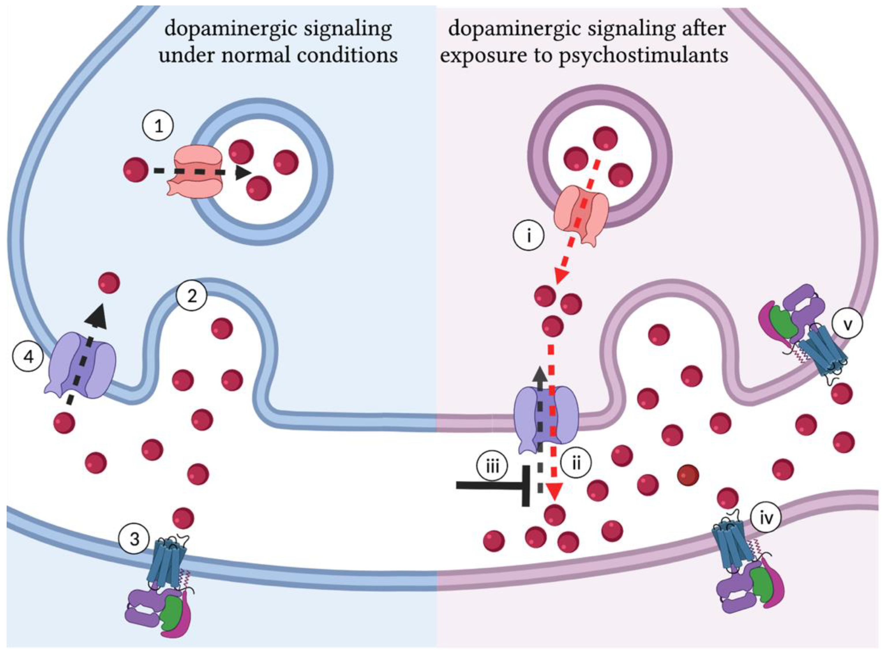
1.2. Dopamine Is Central to the Molecular Mechanisms of Psychostimulant Response

Experiences that benefit biological fitness, such as food or sex, are perceived as rewarding [31]. The association of salient stimuli with reward requires dopamine (DA), a monoamine neurotransmitter central to reward prediction and the reinforcement of motivated behavior [32,33,34]. Natural rewards such as food and sex, along with signals that predict natural rewards, are associated with phasic activation of distinct striatal dopaminergic neurons [35]. The bursts of dopaminergic signaling associated with natural rewards involve increased release of dopamine stored by vesicular monoamine transporter 2 (VMAT2) [36]. Dopamine released into the synaptic cleft binds D1-like (DRD1) and D2-like (DRD2) dopamine receptors, activating downstream G-protein coupled signaling cascades [37]. The plasma membrane dopamine transporter (DAT) removes dopamine from the synapse and indirectly reduces the activation of dopamine receptors [38]. Like natural rewards, drugs of abuse increase dopaminergic signaling, effectively hijacking the endogenous reward system (Figure 1) [39]. Cocaine and amphetamine-like psychostimulants activate the dopaminergic pathways associated with reward through interactions with the DAT [40] and VMAT2 [41] that increase synaptic dopamine [42,43,44,45]. Human brain imaging studies confirm that psychostimulants induce phasic firing of dopaminergic neurons in the brain regions associated with reward [46]. Additionally, this activity correlates with the reported euphoric effects of psychostimulants drugs [47].

1.3. Behavioral Responses to Psychostimulants

Psychostimulant administration in rodents leads to dose-dependent changes in motor behaviors including grooming, locomotion, dyskinesia, and seizures [48]. Flies exposed to cocaine exhibit dose-dependent changes in stereotyped behaviors that, as in mammals, range from increased grooming and hyperlocomotion to seizures and death [49]. Psychostimulants also influence social behavior in rodents, including courtship, where drug exposure promotes sexual behavior [50,51]. Flies exposed to vaporized cocaine or methamphetamine also display an increase sexual behavior and spend more time performing courtship displays [52]. These parallels demonstrate that Drosophila is a model with high face validity for studying the behavioral response to psychostimulants.
Dopaminergic signaling is central to psychostimulant-induced behavioral responses. In mammals, the locomotor activating effects of psychostimulants require increased dopaminergic signaling in the neural substrates associated with reward [53]. Disrupting psychostimulant interactions with DAT and VMAT2, or blocking the activation of downstream dopamine receptors in psychostimulant-activated circuits disrupts the effects of psychostimulant drugs. Wild-type mice, for example, respond to cocaine with increased locomotion, while mutant mice with a cocaine-insensitive dopamine transporter do not display an increase in locomotion after cocaine exposure [54]. Similarly, the VMAT2 inhibitor reserpine reduces amphetamine-induced increases in dopamine in mice [55], and DRD1 antagonists disrupt self-administration of methamphetamine in rats [56]. In flies, psychostimulants also increase dopaminergic signaling by binding to the Drosophila dopamine transporter (dDAT) and vesicular dopamine transporter (dVMAT) [57]. Disrupting dopamine synthesis in Drosophila with 3-iodo-tyrosine reduces the motor-activating effects of cocaine [58], blocking synaptic release of dopamine with the VMAT2 inhibitor reserpine disrupts the locomotor-activating effects of cocaine and amphetamine-like stimulants [59], and mutation of the D1-like dopamine receptor in Drosophila reduces self-administration of methamphetamine [60]. These experiments show that the molecular and pharmacological basis of psychostimulant-induced behaviors are conserved in flies and mammals, highlighting the mechanistic validity of Drosophila as a model organism for studying the genetic underpinnings of response to psychostimulant drugs.

2. Measuring Behavioral Responses to Psychostimulants in Drosophila

Exposure to psychostimulants impacts many behaviors, including locomotion, sleep, arousal, attention-like processes, learning, memory, and social interactions. These behaviors have been studied using assays that quantify motor behaviors, feeding behavior, and attention-like processes. Psychostimulant-induced changes in fly motor behavior produce motor-activating and motor-impairing effects. Assays corresponding to each of these responses allow measurement of sensitivity and sensitization to the activating or impairing effects of psychostimulants. Sensitization involves the development of increased sensitivity to the effects of a drug across subsequent exposures, and is associated with neuroadaptations that increase the salience of stimuli associated with drug reward [61]. In assays of feeding behavior, voluntary consumption of psychostimulant-containing food can be used to monitor changes in self-administration and preference over time. These behaviors are especially relevant to modeling the progression of drug-taking behavior observed in addiction, allowing interpretation of the reinforcing effects of each drug. Attention-like assays, on the other hand, can model the therapeutic application of psychostimulant drugs in rescuing impairments in flies exhibiting behavioral features of ADHD. Here, we discuss the advantages and shortcomings of these assays of motor activity, feeding, and attention-like processes, highlighting techniques that effectively decrease labor time, reduce subjectivity, and take full advantage of the fast generation time and prolific reproduction of Drosophila.

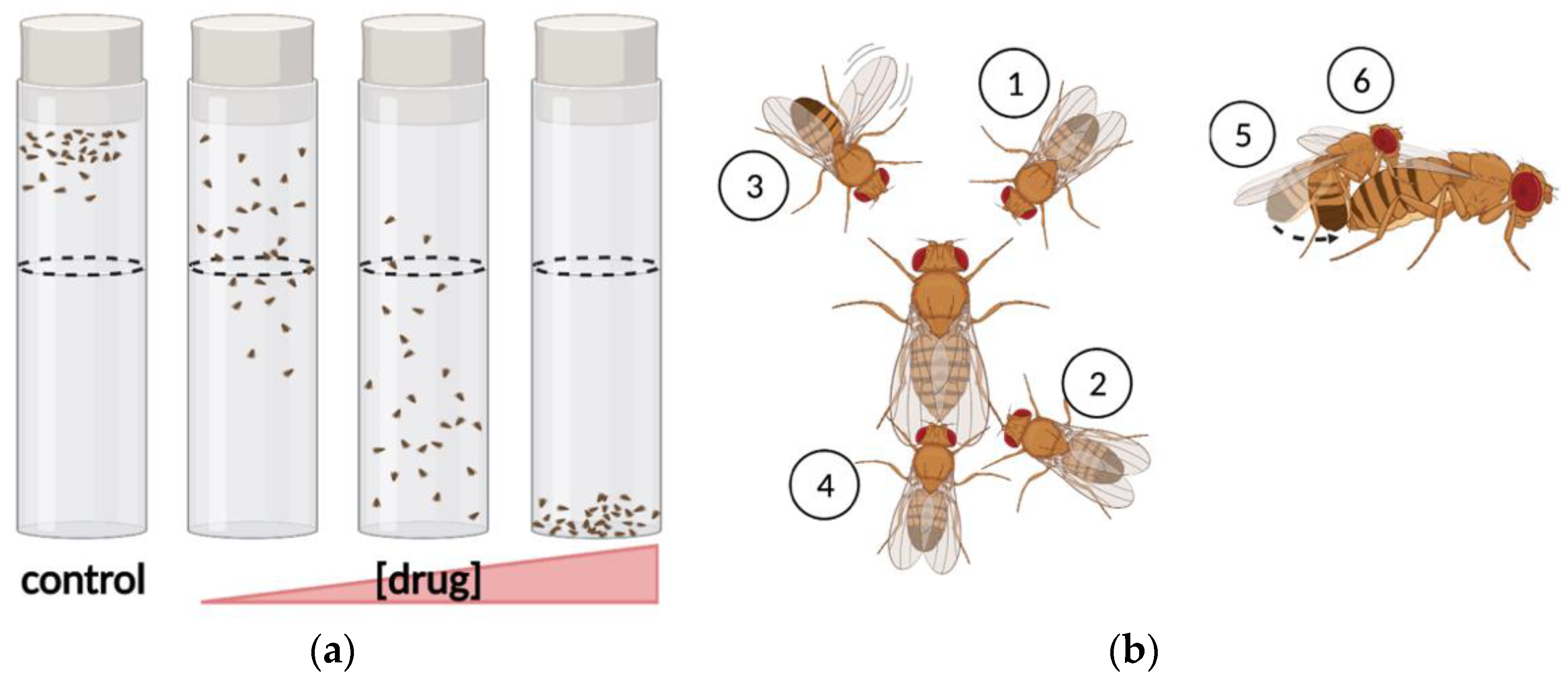
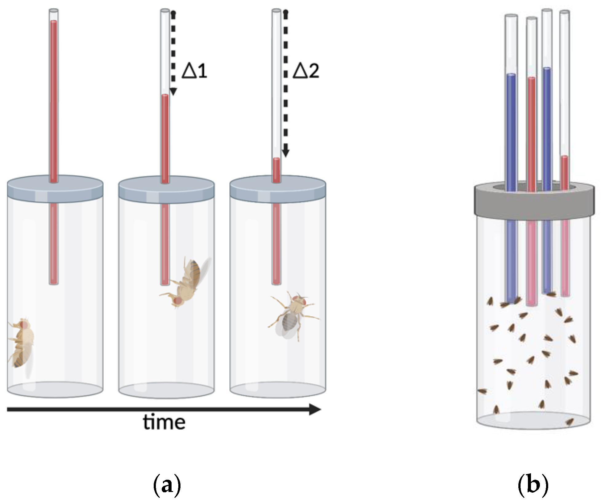
2.1. Assays of Motor-Activity

One of the most obvious effect of psychostimulants is the activation of motor-related behaviors, an effect that is highly conserved across model organisms. Flies exposed to increasing concentrations of cocaine transition through phases of increased grooming behavior, hyperlocomotion, loss of negative geotaxis, stereotyped circling behaviors, erratic movements such as twirling, akinesia, seizures, and even death. One of the first ways this behavior was quantified was assigning a behavioral severity score using a behavioral scoring assay. Experimenters rated the activity of drug exposed flies from 0 to 7 using an ordinal scale of defined behavioral responses (Figure 2a). This behavioral scoring assay can also characterize the temporal dynamics of cocaine response by measuring latency to the peak of drug-induced behavioral effects and the time required to return to baseline. The peak response values recorded for individual flies can be used to generate a drug effect score, which represents the percent of flies that exceed an intoxication associated score during a defined observation period. In addition to defining the progression of cocaine-related behaviors in flies, this method allowed the analysis of how different doses or administration schedules impact sensitivity [49] and sensitization [62,63]. The development of behavioral sensitization is measured by performing repeated exposures to generate a time-course of drug effect scores. Exposures where the drug effect score is greater than the previous recorded score indicate the development of sensitization. The development of sensitization is a function of the interval after the first exposure, and for flies exposed to 75 μg vaporized cocaine, sensitization is only observed after exposure to a second dose 6–24 h after the initial exposure, but not before [62,63]. A similar interval is required for sensitization in mice, where cocaine-induced locomotion increases when the second dose is administered 24 h after initial drug exposure [64]. While the behavioral scoring assay can effectively measure sensitization, a critical psychostimulant response, scoring requires detailed observation of Drosophila behavior, is time-intensive, and subjective. One automated option that provides objectivity and can decrease the time spent scoring the motor activating effects of psychostimulants is the Drosophila activity monitor (DAM).
The DAM system is a high-throughput automated method of behavioral analysis that uses an infrared beam to detect motor activity [65]. Flies in the DAM system are individually housed in glass cuvettes transected by an infrared beam, and locomotor activity is analyzed by quantifying beam breaks (Figure 2b). The rate of beam breaks in the DAM measures activity as well as sleep and can be used to infer arousal state. Additionally, the software for processing DAM system data supports analysis of customized variables such as night time or day time activity, providing information on circadian patterns of response [66]. The DAM system has been used to measure the increases in locomotor activity that occur after injection [67], vaporization [68,69], or oral administration [52] of psychostimulant drugs. Every method of administration for cocaine or methamphetamine increases locomotor activity and decreases sleep in the DAM. The DAM also effectively measures sensitization, which is observed when a second dose of vaporized cocaine or methamphetamine is administered 6 or 10 h after the first dose, respectively [68,69]. The DAM also shows the wakefulness-promoting effects of methamphetamine in sleep-deprived flies, which sleep the same amount as flies that have not been sleep deprived, while sleep-deprived flies that are not given methamphetamine display significant rebound rest [52].
While each psychostimulant administration method produces measurable changes in behavior, it is important to consider tradeoffs in speed of delivery, precision of dosing, and how disruptive each method is to fly behavior. Drug-supplemented food provides the least control of speed and precision, but does not disrupt fly behavior. Drug injection is time consuming and disturbs normal fly behavior, but provides strict control of the dose. The FlyBong [68], designed to administer vaporized drugs in the DAM, provides a balance of speed and precision that enables simultaneous exposure for all flies without disrupting data collection, making it a valuable method for standardizing administration of psychostimulant drugs. Acute responses, in the hour following treatment, can be analyzed by recording beam break to measure activity rate (beam breaks/min) and number of activity peaks, where activity rate is at least double that of untreated controls [67]. Sensitization can be quantified based on relative changes in activity rate and number of peaks observed during successive psychostimulant exposures. For longitudinal assays, or experiments with orally administered psychostimulants, periods of inactivity can be measured to characterize the effects on sleep duration and architecture [52]. Regardless of drug administration method, the DAM system is a reliable and high-throughput method for measuring the locomotor response to psychostimulant drugs.
Multiple methods of drug administration can be combined with the DAM system, as demonstrated in a modified DAM where flies in each cuvette have access to capillary tubes filled with feeding solutions of 100 mM sucrose and 100 mM sucrose supplemented with methamphetamine [69]. Out of a range of concentrations, this experiment showed flies develop the strongest preference for 0.15 mg/mL methamphetamine, which is evident by the second day and remains stable for the remainder of the 7-day assay. Flies exposed to 75 μg vaporized methamphetamine in the FlyBong display increased locomotor activity after exposure, this activity more than doubles for flies subjected to a second exposure 10 h later. Transferring these sensitized flies to the capillary feeding DAM changes the outcome for preference, which no longer develops during the 3-day period of measurement [69]. These results are similar to those observed in rats, where methamphetamine self-administration is reduced after the development of sensitization following injections of methamphetamine [70]. Reciprocally, flies that develop preference in 3-day capillary feeding DAM performed prior to vaporized methamphetamine exposure in the FlyBong do not develop sensitization after the second 75 μg dose [69]. The relationship between preferential drug-consumption and locomotor sensitization, two behaviors with face validity for modeling addiction, demonstrates how seemingly unrelated endophenotypes can be intertwined at a mechanistic level. Accordingly, uncovering the molecular mechanisms involved in a simple response like sensitization can be informative about the molecular mechanisms that influence preferential self-administration.
While the constrained analysis of locomotion in the DAM is a reliable automated method for measuring the motor activating effects of psychostimulant drugs, some behaviors cannot be measured within the interior of a glass cuvette. Several assays that address this issue allow measurement of locomotion in freely moving Drosophila. Locomotor responses to psychostimulant drugs in adult or larval Drosophila can be measured by video recording responses in an observation chamber. Adult Drosophila [71] and 3rd instar larvae [72] fed cocaine display significantly increased rates of locomotion. These studies quantify locomotion rate by placing the observation chambers over a grid and counting the lines crossed during an observation period (Figure 2c). The availability of 2D-tracking and video processing software support automated processing of video recorded assays, which reduces the time required for analysis and limits interpretive bias [73,74,75].
Psychostimulant responses in Drosophila have also been studied by analysis of repetitive startle-induced hyperactivity (ReSH), which is based on locomotor response to recurrent puffs of air [76]. Air puffs are delivered to 8 groups of flies using the “puff-o-mat”, a device based on a similar apparatus used for the delivery of ethanol vapor (now appropriately known as the “booze-o-mat”) [77,78]. Recordings of the assay are analyzed using a custom software that measures changes in fly position, acceleration, velocity, and trajectory following mechanical stress from puffed-air (Figure 2d) [76]. Activity peaks immediately after the startle stimulus, and then slowly returns to baseline. Measurement of ReSH in untreated control flies shows large increase in velocity followed by a slow decay toward baseline [76]. Cocaine leads to a dose-dependent decrease in ReSH, reducing peak velocity after startle and reducing the decay period. Interestingly, the state of arousal measured during ReSH differs from the locomotor arousal associated with spontaneous activity quantified in the DAM, and is inversely impacted by cocaine, which reduces rather than increases locomotor activity [76].

2.2. Assays of Motor-Impairment

While psychostimulants activate some motor behaviors, they can impair others. This is the case for negative geotaxis [58] and some aspects of courtship behavior [52]. One possible explanation for this impairment might be the complexity of these behaviors. Negative geotaxis is a complex behavior that requires flies to integrate sensory information about light as well as gravity [79], and is even affected by electromagnetic fields [80]. Similarly, Drosophila courtship is a complex social behavior, with successful copulation requiring the integration of audible [81] visual [82] olfactory [83] and gustatory [84] stimuli. For some Drosophila behaviors, such as response to visual stimuli, optimal responses occur within a specific range of dopaminergic activity and are disrupted when dopamine levels are outside of that range [52]. It is likely that psychostimulant-induced increases in dopamine similarly impair negative geotaxis and courtship behaviors, which require complex processing and integration of sensory information across several modalities to mount an appropriate response.
Startle-induced negative geotaxis (SING) was one of the first behavioral assays developed in Drosophila, dating back to Seymour Benzer’s experiments in the 1960s [85]. Benzer joined two test tubes, forced the flies to the bottom of the enclosure, and scored the portion of flies that move from the proximal tube to the distal tube within a brief recovery period. SING condenses the analysis of motor response for multiple flies to one value, defined by the proportion of flies that make it past a defined height along the tube. The efficient quantification of this assay makes it amenable to automation: recordings can be processed with video analysis software. Additionally, this assay is easily scaled up allowing simultaneous measurement of locomotor responses across several groups of flies, exploiting the economy of scale that Drosophila offer. Untreated control flies quickly climb to the top of the tube, whereas flies exposed to cocaine exhibit dose-dependent impairments in negative geotaxis [58] (Figure 3a). Several variations of this assay have been developed, and provide similar measures of intoxication. A drug effect score can be determined with consecutive climbing assays based on the average number of flies remaining at the bottom of each cylinder after exposure [86], and acquisition as well as analysis can be automated using pictures captured from beneath each cylinder [87] to increase measurement speed and consistency. Alternatively, both the “startle” step and analysis of climbing behavior have been automated in the rapid iterative negative geotaxis (RING) assay, which allows measurement of average distance traveled, increasing the resolution and dynamic range of behavioral analysis [88]. In summary, assays of climbing behavior offer a high-throughput measurement of sensitivity and sensitization with automated analysis.
In contrast to the simplicity of measuring negative geotaxis behavior, the quantification of Drosophila courtship requires analysis of a series of distinct behaviors. An established method for studying these behaviors in males is the courtship behavior assay (Figure 3b). This assay involves quantification of courtship activities that follow a stereotyped order which includes orientation, tapping, wing vibration, licking, attempted copulation, and copulation [89]. A male is observed in a behavioral chamber together with one or more females to quantify the time it takes to initiate courtship, the number and duration of each courtship behavior, the number of copulation attempts, and the duration of successful copulation events. The copulation success rate is quantified in an index generated by recording pairs of Drosophila and measuring the proportion that successfully copulate [52]. While this assay is not high-throughput, it has the advantage of simultaneously allowing measurement of the motor activating and impairing effects of psychostimulants. Flies fed methamphetamine have a decreased latency to initiate courtship, and spend more time performing steps of the behavioral-courtship sequence that precede copulation. This increase in both speed of initiation and percent of time spent courting are associated with increased sexual arousal. In contrast, methamphetamine reduces the percentage of flies that successfully copulate while increasing the latency to copulation in flies that do, demonstrating how methamphetamine disrupts neural processes involved in initiating and sustaining the motor behaviors required for copulation [52].

2.3. Assays of Consumption and Preference

Assays of voluntary consumption are indispensable techniques for studying addiction and can be used to measure increases in self-administration over time. The CApillary FEeder (CAFE) assay [90] is a reliable feeding assay that allows continuous monitoring of consumption of liquid food from capillary tubes. Results characterize cumulative consumption, average consumption and consumption rate (Figure 4a). Furthermore, the CAFE can be modified to administer two different feeding solutions in a two-choice assay of consumption enabling analysis of preferential consumption (Figure 4b). Findings using the CAFE show that the development of amphetamine preference in Drosophila is concentration-dependent. Flies given the choice between a sucrose solution and a sucrose solution containing 1 mM amphetamine develop preference for the amphetamine containing solution by the first measurement timepoint at 24 h [91]. When the amphetamine concentration is increased to 10 mM this effect is not observed, and flies prefer the sucrose solution instead [91]. A similar experiment comparing daily consumption of two sucrose solutions, where one is supplemented with methamphetamine, showed that flies develop preference for 1 mM methamphetamine after one day while always avoiding 10 mM [60]. In contrast, flies did not develop preference for cocaine at any concentration (10 uM, 100 uM, 1 mM, 10 mM) during a four day CAFE, but avoided the two higher concentrations. The CAFE can also be used to identify the genes that influence cocaine and methamphetamine consumption, which has been investigated in large scale assays with hundreds of genetically distinct lines derived from the Drosophila Genetic Reference Panel (DGRP) [92,93]. Additionally, variations such as the FlyCAFE [69] allow measurement of locomotion in tandem with consumption at single fly resolution, and enable additional administration of vaporized drug [68]. Together, this combination of techniques allow simultaneous analysis of drug consumption, preference, and drug-induced locomotion, being a valuable tool for modeling the changes in consumption observed during the development of addiction.

2.4. Attention-like Processes

Attention to specific stimuli is a process that requires selection and suppression of incoming sensory information. In tests of visual attention in humans, patients with ADHD display deficits in attentional selection [94], increased impulsivity, and distractibility [95]. Psychostimulants improve selective attention in patients with ADHD, and low doses also improve cognitive functioning in non-ADHD subjects [8]. While attention is more difficult to define in animals, many behaviors allow the measurement of attention-like processes. In Drosophila, attention-like processes have been studied by measuring responses to visual stimuli in the optomotor maze (Figure 5a) [96] and with flies suspended in a flight arena [97]. The floor of the optomotor maze is transparent and visual stimuli are introduced by placement over a monitor. Optomotor response is determined in single flies based on locomotion following the introduction of a uniform field of moving visual stimuli, and distractibility is measured based on the change in locomotion following the introduction of competing visual stimuli that move in opposite directions [52]. In flight arena experiments, attention-like processes are studied by measuring responses to visual stimuli in controllable panoramic [98] or programmable visual environments [99] that allow manipulation of visual stimuli presented to a single fly positioned within the arena (Figure 5b). In closed-loop flight arenas, fly behavior directly impacts the observable environment, creating a “closed” feedback loop, or a virtual flight simulator [100]. In contrast, flies in the flightless open-loop experiments have a limited range of motion and their responses do not influence visual stimuli, which rotate at a constant frequency independent of fly behavior (Figure 5c). In these experiments, shifts in visual fixation associated with presentation of novel stimuli are reflected in fluctuations of activity in 20–30 Hz local field potentials, as measured with a recording electrode [101]. Dynamics of 20–30 Hz have been used characterize “attention span” by measuring responses to novel stimuli, and responses to conflicting stimuli in assays of distractibility [101]. These attention-like processes can also be studied by measuring the intensity of torque that occur in response to visual stimuli. Both locomotor and electrophysiological measurements have been used to characterize behavior in mutant flies that have impairments in attention-like processes.

3. Studying the Therapeutic Use of Psychostimulants with Drosophila

ADHD and ASD are complex diseases that impact arousal, attention, and sleep. Dopaminergic dysfunction is a common characteristic of ADHD and ASD that contributes to cognitive and behavioral impairments [102,103]. Psychostimulant drugs improve some of the deficits seen in ADHD in humans [104], as well as in fly models of ADHD-like behavior [105], which we review in this section. We discuss how Drosophila responses to psychostimulant drugs have been used to characterize the behavioral effects of ASD-related mutations from human patients with dopaminergic dysfunction. The results of psychostimulants studies in Drosophila the models of ADHD and ASD are also discussed in Table 1.

3.1. Attention Deficit Hyperactivity Disorder (ADHD)

Hyperactivity and reduced sleep are common behavioral symptoms of ADHD [118,119,120]. Several mutant fly lines also exhibit hyperactivity and reduced sleep, making them face-valid models of ADHD-like behaviors. Among these are mutant lines for Drosophila orthologs of the dopamine transporter (DAT), latrophilin-3 (LPHN3), and neurofibromin-1 (NF1). These three genes are associated with ADHD in humans, and were chosen to test whether disruption of the fly ortholog would mimic ADHD-like behaviors in Drosophila [105]. The dopamine transporter was selected through database mining by searching for Drosophila genes with phenotypic descriptors related to hyperactivity, excitability, or attention. This search produced a list of 78 genes, with 69 conserved in humans. Five of those conserved genes, including DAT, were also identified in a list of 91 human ADHD-associated genes. The Drosophila DAT null mutant DATfmn was identified based on its sleep phenotype—fmn stands for fumin, Japanese for sleepless [121]. Observation of DATfmn flies using the DAM system showed hyperactivity and reduced sleep, with hyperactivity exacerbated in the absence of light, e.g., at night, but also during the subjective daytime when housed in dark:dark conditions. Feeding DATfmn flies methylphenidate [105] or amphetamine [111] rescued hyperactivity and sleep loss. This study highlights how disruption of Drosophila DAT, the orthologue of a well-known ADHD risk gene highly represented in human GWAS studies, recapitulates two common symptoms of ADHD observed in humans that can be rescued by psychostimulant drugs used to treat the symptoms of human ADHD.
While DAT knockout is useful for modeling the effects associated with loss of function, there is significantly more variability in the DAT mutations that contribute to the prevalence of ADHD in humans [122,123,124]. Experiments in cell culture show that many psychiatric disorder-associated DAT variants exhibit heterogenous molecular phenotypes, including differences in DA uptake kinetics, reverse transport, and altered binding to psychostimulant drugs [125]. Drosophila is an efficient model for studying the effects of human DAT mutants in vivo and have shown that DAT mutations associated with early-onset Parkinson’s [126] as well as ASD [107] lead to impairments in motor behavior. Drosophila is a feasible system for performing similar experiments to unravel the behavioral and molecular nature of DAT variants associated with ADHD, offering a practical model to identify the molecular mechanisms involved in response to psychostimulant drugs.
In contrast to DAT, the influence of the G-Protein coupled receptor and cell adhesion protein Latrophilin-3 on dopaminergic signaling is poorly understood. LPHN3 is an ADHD-risk gene that was identified in a linkage study based on a prevalence of ADHD with large generational families of an isolated population in the Paisa region of Colombia [127]. LPHN3 variants that lead to haploinsufficiency are associated with the development of ADHD in humans [128]. While patients with LPHN3 risk alleles respond to stimulant medication, a molecular mechanism linking reduced LPHN3 expression to dopaminergic dysfunction has not been unraveled in human studies [128]. The behavioral effects of knockdown of the LPHN3 fly ortholog have also been studied in Drosophila using the GAL4/UAS binary expression system. This method uses a cell type or tissue-specific enhancer to drive expression of the yeast transcriptional activator GAL4. Transgenes coupled to the GAL4 upstream activation sequence (UAS), are expressed wherever GAL4 is transcribed. The pan-neuronal driver elav-GAL4 was used to drive expression of UAS-Cirl-RNAi, the single Drosophila orthologue for the Latrophilin family of genes. This reduced complexity can be beneficial when attempting to characterize a gene’s molecular function. In mammals, the effects of mutating a gene that is a member of a multi-gene family can be difficult to observe because of genetic redundancy. In flies, the reduction in gene copy number makes characterizing loss of function phenotypes more straightforward. Neuronal knockdown of the Drosophila orthologue of LPHN3 was sufficient to produce the hyperactivity and decreased sleep associated with dopaminergic dysfunction [105]. As with the DAT null mutants, hyperactivity was more pronounced at night and could be exacerbated during the day by turning off the lights. Both the hyperactivity and sleep loss were rescued by feeding Drosophila methylphenidate. Tyrosine hydroxylase staining in fly brains showed that knockdown of the Drosophila orthologue of LPHN3 did not alter the distribution or survival of dopaminergic neurons, indicating that LPHN3 regulates dopaminergic signaling directly [105]. The role of LPHN3 in dopaminergic signaling and variation in response to psychostimulant drugs is poorly understood. Drosophila is a useful model for studying neurotransmission [129,130] and offers a flexible system for the future identification of the molecular mechanisms that lead to altered amphetamine and methylphenidate responses in models of LPHN3 disruption.
NF1 is associated with the autosomal dominant disease neurofibromatosis type I. Neurofibromatosis type I is a multi-system disorder involving tumors of the nervous system that lead to complications such as eye disease, musculoskeletal disorders, and epilepsy, among others. In addition to the physical impairments associated with neurofibromatosis type I, most patients display cognitive deficits [131], including symptoms of ADHD [132]. In some patients, these ADHD symptoms are improved by treatment with methylphenidate [133]. To investigate whether loss of NF1 would lead to ADHD-like behavior in flies, NF1 was knocked down in all neurons of the Drosophila by using elav-GAL4 to drive expression of UAS-NF1-RNAi. Knockdown of NF1 in Drosophila neurons induced a hyperactivity and sleep-deficiency phenotype, with a noticeable increase in nighttime hyperactivity. Feeding NF1 knockdown flies methylphenidate rescued both hyperactivity and sleep dysregulation [105].
The psychostimulant-mediated rescue of hyperactivity as well as sleep-deficiency phenotypes observed for DAT, LPHN3, and NF1 orthologue mutants in the DAM system indicate that (1) hyperactivity along with reduced sleep is an endophenotype for ADHD in flies, (2) flies are a model with mechanistic validity for studying ADHD such that genes whose disruption leads to ADHD in humans also produce ADHD-like behavior in flies, and (3) flies are a model with predictive validity for studying ADHD since the psychostimulant drugs that reduce the behavioral symptoms of ADHD in humans also reduce ADHD-like impairments in flies. While DAT, LPHN3, and NF1 all respond to treatment with methylphenidate, the molecular basis for the psychostimulant-mediated rescue of ADHD-like behaviors is unknown. Targeted knockdown of DAT, LPHN3, or NF1 specifically in Drosophila dopamine neurons or subsets of dopamine neurons might help unravel how mutations with opposite effects on dopaminergic signaling can display similar response to psychostimulant drugs such as methylphenidate.
In addition to the locomotor signature observed with ADHD-like changes in sleep and hyperactivity, Drosophila can model more complex ADHD-like symptoms associated with dysregulation of attention-like processes [101]. Humans patients with ADHD have deficits in visual attention [94,134,135] that are improved by treatment with psychostimulants [134]. The Drosophila memory mutant radish1 also displays impaired responses to visual-stimuli in the optomotor maze and experiments of tethered flight [101]. Wild-type flies traveling through the optomotor maze turn in the direction of moving visual stimuli, an optomotor response that is absent in radish1 mutants flies [101]. This deficit in response is not a result of visual impairment, as radish1 mutants are successful in operant visual learning [135]. Methylphenidate rescues optomotor response in radish1 mutants, generating responses to visual stimuli similar to those observed for wild-type flies in the optomotor maze [101]. In addition to the altered locomotor response to visual information in the optomotor maze, radish1 mutants flies also exhibit altered brain activity in response to visual stimuli. Normally, flies display an increase in 20–30 Hz local field potentials (LFP)—observed by brain recordings—in response to visual stimuli. Electrophysiological recordings from flies suspended in a flightless arena show that flies repeatedly shown the same shape will display an increase in 20–30 Hz response when presented a novel shape. In contrast, radish1 mutants have a diminished 20–30 Hz response to novel visual stimuli. Treatment with methylphenidate also rescues the 20–30 Hz response to novelty in radish1 [101].
The impairment of visual attention-like processes observed in radish1 mutants and pharmacological rescue with methylphenidate demonstrate how the optomotor maze and tethered flight experiments can identify genetic and molecular determinants of attention span. Moreover, these assays can uncover the molecular basis of psychostimulant-mediated rescue in attention-like processes such as visual fixation, novelty response, and distractibility. However, with radish1 as the sole example of psychostimulant-mediated rescue of attention-like processes, these assays would benefit from validation using other known ADHD-linked mutants. The observation of similar deficits in ADHD-associated mutants, along with improvement following treatment with psychostimulants, would provide support for impaired responses in the optomotor maze and tethered flight experiments as endophenotypes for attention-like processes dysregulated in ADHD. Because ADHD is predominantly a polygenic disorder, with impairments occurring on a spectrum, it is unlikely that a single behavioral assay will be effective in studying every putative risk gene. The current models of ADHD-like behavior in flies address two broad features of ADHD: dysregulation of attention-like processes and dysregulation of arousal. Complementary use of the DAM system, optomotor maze, and flight loop experiments may be useful in unraveling how specific behaviors are improved with psychostimulant treatment, supporting the identification of genes and pathways that determine treatment efficacy.
Rare genetic variants associated with ADHD such as LPHN3 and NF1 have a large effect size, meaning a large portion of observed phenotypic variance can be attributed to these individual genes. In contrast, common genetic variants associated with ADHD have a smaller effect size, meaning their individual contribution to observed phenotypes is less severe. Rare disease variants, because of their increased severity, can be helpful in elucidating molecular mechanisms that can help understand the deficits observed in mild cases disease. However, identifying the common variants that contribute to mild-cases of diseases has been more difficult. Because these common variants have a small effect size, it is difficult to perform studies with enough subjects to reach the statistical power necessary to identify common risk genes. The first successful identification of common variant risk was performed in a 2019 GWAS that used data from more than 50,000 subjects to identify of 12 ADHD risk loci [136]. In contrast to human studies, Drosophila offer a model where additional genetic tools can circumvent the limitations associated with underpowered research. A recent study combined transcriptome and behavioral analysis across hundreds of methylphenidate exposed flies, to behavioral analysis of 172 lines from the DGRP to identify genetic variants that contribute methylphenidate response [137]. The study assigned 650,766 segregating single nucleotide polymorphisms (SNP) to 7472 gene networks and used an integrative genomic prediction approach to predict SNPs associated with differentially expressed transcripts. Of the 87 networks, including 1727 genes predicted to impact methylphenidate response, 14 of the top candidate genes were validated with RNAi using a ubiquitous GAL4 driver, and 10 of those genes altered response to methylphenidate. Twenty percent of the networks predicted to contain methylphenidate response genes are involved in histone modifying processes, and 4 of the 10 genes that were validated with RNAi are involved in histone modification [137], a process that is also relevant to human psychostimulant addiction [138]. This transcriptomic study demonstrates how Drosophila is a model that enables identification of novel candidate genes that mediate response to treatment with psychostimulant drugs, information that might also help elucidate differences in genetic etiology that contribute to treatment resistance in humans.

3.2. Autism Spectrum Disorder (ASD)

Most cases of ASD are polygenic and 80% of cases involve multiple genes, making it difficult to unravel how each gene contributes disease-related impairments [139]. Additionally, many patients with ASD also have comorbid ADHD, with estimates from clinical samples between ranging between 37–85% [140]. Flies are a useful model for studying the impact of individual genes associated with ASD [139]. The impact of several ASD-linked mutations on DAT function, reverse transport of dopamine, and sensitivity to amphetamine psychostimulants have been characterized in Drosophila [107,108,109].
Hamilton and colleagues used Drosophila to functionally characterize an ASD-associated de novo mutation in the human dopamine transporter (hDAT) at residue 356 (hDAT T356M). [107] Cell culture experiments showed hDAT-T356M caused constitutive reverse transport of dopamine and reduced amphetamine-induced reverse transport of dopamine. To identify how these changes impact behavior and response to psychostimulants, a transgene carrying the human ASD-associate DAT mutant was introduced into a Drosophila DAT knockout background (DATfmn). hDAT-T356M expressing flies displayed increased baseline locomotor activity compared to control flies expressing hDAT. Additionally, while amphetamine increases locomotor activity in hDAT expressing flies, Drosophila expressing the ASD-associated mutant do not display an amphetamine-induced increase in locomotion [107].
A similar behavioral phenotype was observed in flies during an experiment involving the first characterization of an ASD-associated in-frame deletion in the human dopamine transporter (hDAT), which eliminates asparagine residue 336 (hDAT-ΔN336) [108]. Cell culture experiments performed to molecularly characterize hDAT-ΔN336 showed that the ASD-associated mutation stabilizes the transporter in a half-open and inward-facing conformation that disrupts DA uptake, but not reverse transport. To study the effects of this mutation in vivo, the ASD-associated DAT transgene was expressed in DAT knockout flies (DATfmn), and compared to hDAT expressing controls. Flies expressing hDAT-ΔN336 displayed hyperactivity and increased grooming behavior compared to hDAT expressing control flies [108]. Flies expressing the mutant transporter also exhibited impaired amphetamine-induced reverse dopamine transport [108].
Another ASD-associated DAT mutant that impacts psychostimulant response contains an arginine to tryptophan substitution at N-terminal position 51 (hDAT-R/W), a residue important for interaction with the membrane protein Syntaxin-1A (STX1A) [109]. Murine models showed that the interaction of the DAT with STX1A support amphetamine-induced reverse transport of dopamine, and that STX1A overexpression increased reverse transport of dopamine [141]. Experiments in cell culture revealed that the ASD-associated hDAT-R/W did not affect DA uptake, but reduced amphetamine-induced reverse transport of DA [109]. DATfmn flies expressing the ASD-associated variant were used to characterize how this mutation impacts behavior. No difference in basal locomotion was observed between hDAT-R/W expressing flies and hDAT expressing control flies. However, hDAT-R/W expressing flies exhibited a significant reduction in amphetamine-induced locomotion compared to hDAT expressing control flies [109].
These experiments demonstrate how flies be used to study the behavioral impact of mutations, linking the behaviors in question to molecular mechanisms characterized in cell culture. These experiments cannot be performed in humans, and can help characterize how specific mechanisms contribute to the pathogenesis of complex polygenic disorders such as ASD [107]. While these experiments analyzed psychostimulant response in the context of locomotion, flies have potential for modeling more complex behavioral features of ASD, which is known to impact social behavior. Drosophila display broad repertoire of social behaviors that range from population scale changes in group dynamics [142] to pairwise interactions involved in displays of aggression [143] and courtship [144]. Quantification of group density [145] and analysis of courtship behavior [89] are both established methods that have been used to study social behavior in Drosophila, and both respond to changes in dopamine. As social deficits are common in fly models of ASD, Drosophila could provide an effective model to study whether psychostimulant drugs can rescue social responses. These experiments could help identify specific genes and molecules required for psychostimulant-mediated improvements in social behavior.

4. Studying Psychostimulant Abuse with Drosophila

Addiction is an etiologically complex polygenic disorder that develops in an experience-dependent manner. In humans, features of addiction include increases in drug preference and consumption. Individual differences in sensitivity to the behavioral activating effects of psychostimulant drugs, the development of sensitization, initial preference, and patterns of drug consumption are all important factors predictive of future psychostimulant response and propensity to develop substance use disorder. Studies of psychostimulant-induced behavior in mammals show that simple locomotor responses can be predictive of more complex addiction-related behaviors such as the acquisition of self-administration. In rats, for example, sensitivity to the locomotor activating effects of cocaine predict the development of cocaine-conditioned place preference [146]. Here, we discuss the genes implicated in psychostimulant sensitivity, sensitization, and preference in Drosophila, and highlight their relevance in modeling psychostimulant addiction.

4.1. Using Drosophila to Study the Mechanism of Action of Psychostimulant Drugs

While the intoxicating effects of commonly abused psychostimulants vary in intensity and duration, they all act to increase dopaminergic signaling [41]. Dopamine is involved in many behaviors in Drosophila, including locomotion, attention-like processes, memory, and reward [147,148,149,150,151]. As dopamine is central to the mechanism of action for psychostimulant drugs, the genes involved in dopamine synthesis, neurotransmission, and dopamine receptor coupled signaling pathways directly impact psychostimulant response.
Tyrosine hydroxylase is the rate limiting enzyme involved in the conversion of L-tyrosine to dopamine [152]. RNAi-mediated knockdown of tyrosine hydroxylase in dopaminergic neurons of Drosophila larvae ablates locomotor response to amphetamine [106]. Tyrosine hydroxylase influences coloration of the flies’ cuticle, and loss of function mutants were named pale because of their discoloration [153]. pale mutants have reduced brain dopamine, are hypoactive [154,155], and do not exhibit a reduction in sleep after treatment with amphetamines [111]. Reducing dopamine also impacts response to cocaine, and flies fed 3-iodotyrosine (3IY), a competitive inhibitor of tyrosine hydroxylase show a reduction in cocaine-induced locomotion [58].
dDAT codes for the Drosophila plasma membrane dopamine transporter (DAT) that functions to remove extracellular dopamine from the synaptic cleft [156]. DAT structure, function, and interaction with psychostimulants are highly conserved in Drosophila and humans [38,40]. In Drosophila, the activating effects of psychostimulants on arousal are dopamine transporter dependent. Both amphetamine and methylphenidate increase locomotion in Drosophila larvae, a response that is blunted in DATfmn flies or flies expressing dDAT RNAi in dopaminergic neurons. Expression of the human dopamine transporter (hDAT) in DATfmn mutants rescues the hyperlocomotive response to both amphetamine and methylphenidate [106], demonstrating that a conserved DAT-dependent mechanism facilitates the locomotor-activating effects of psychostimulant drugs. Consistent with the data showing that response to psychostimulant drugs is contingent on the presence or absence of functional DAT, sensitivity or resistance to psychostimulants drugs is also dependent on DAT cell surface expression [157]. Drosophila Ric, encodes a Ras-related small GTPase involved in dopamine transporter trafficking [158]. Rit2, the mammalian orthologue of Ric, has been shown it impact cocaine sensitivity in mice [159]. In Drosophila, dopaminergic expression of a constitutively active Ric-GTPase mutant (RicQ117L) increases cell surface expression of DAT and enhances sensitivity to the locomotor activating effects of amphetamine [157].
While the activating effects of cocaine, methylphenidate, and amphetamine-like psychostimulants are all DAT-dependent, they do not increase dopaminergic signaling through the same mechanism of action (Figure 1). Cocaine as well as methylphenidate act as competitive inhibitors of the dopamine transporter [160] that increase dopaminergic signaling by binding dDAT and reducing uptake of synaptic dopamine [161], while amphetamine-like psychostimulant bound to the dopamine transporter activate reverse transport of dopamine [43,162,163]. In vitro studies first showed the reverse transport of dopamine stimulated by amphetamine-like psychostimulants was regulated by Calcium/calmodulin-dependent protein kinase II (CaMKII) mediated phosphorylation of the DAT the N-terminal domain [152,153] and localization of the DAT to specific plasma membrane microdomains by the membrane lipid raft protein flotillin-1 [164]. The first in vivo experiment to show that flotillin-1 is required in dopamine neurons for amphetamine-induced locomotion was performed in Drosophila [106]. In contrast to the increased locomotion observed in wild-type flies, Flotillin 1 mutant (Flo102554) larvae or larvae expressing Flo1 RNAi in dopamine neurons did not display an increase in locomotive speed after administration of amphetamine [106]. Expressing human FLOT1 in dopaminergic neurons of Flo102554 mutant larvae rescued the hyperlocomotive response to amphetamines [106]. One explanation for the requirement of Flo1 in amphetamine-induced reverse transport might be the role of lipid rafts in acting as a scaffold for other signaling proteins such as CamKII [165,166]. In vitro experiments showed that amphetamine-induced reverse transport depends on CamKII-mediated phosphorylation of DAT N-terminal serine residues (2, 4, 7, 12 and 13), a modification associated with the inward-facing transporter conformation that facilitates reverse transport [162]. This dependency was confirmed in vivo using Drosophila DATfmn mutants, where dopaminergic expression of an hDAT construct with serine to alanine substitutions that prevent phosphorylation ablates amphetamine-induced hyperlocomotion [106]. The hyperlocomotive response to amphetamines is rescued by dopaminergic expression of wild-type hDAT, as well as a mutant hDAT where N-terminal serine residues are replaced with phospho-mimetic aspartate residues. Flies were also used to demonstrate DAT phosphorylation requires CAMKII in dopamine neurons, and that expressing CaMKII inhibitory peptide in the dopamine neurons of larvae ablates amphetamine-induced hyperlocomotion [112]. Dopaminergic expression of the N-terminal domain phosphomimic hDAT mutant rescues the locomotor response to amphetamine in flies expressing CaMKII inhibitory peptide [112]. Drosophila were also the first model organism to show that the membrane phospholipid Phosphatidylinositol (4,5)-bisphosphate (PIP2) interaction with the human dopamine transporter is important for amphetamine-induced dopamine efflux [110]. In silico experiments predicted that DAT N-terminal lysine residues 3 and 5 were involved in interaction with PIP2. In vitro experiments demonstrated that neutralizing alanine substitutions lysine residues 3 and 5 (hDAT-K/A) do not alter DAT cell surface expression, but to reduced amphetamine-induced dopamine efflux. To test the behavioral impact of the hDAT-K/A mutation, DATfmn mutant flies expressing hDAT or hDAT-K/A in dopaminergic neurons were fed amphetamine and monitored for changes in locomotor behavior. While hDAT expressing flies fed amphetamine showed an significant increase in locomotion, this effect was abolished in hDAT-K/A expressing flies [110]. The DAT lysine at residue 337 and arginine 443 in intracellular loop 3 and 4 were also predicted to interact with PIP2 based on computational modeling experiments [110]. In vitro experiments showed that neutralizing alanine substitutions at K337 and R443 lead a reduction in DAT cell surface expression of more than 80% [110]. To test the behavioral impact of the arginine to alanine substitution at residue 443 (hDAT-R443A), which did not affect surface localization, DATfmn mutant flies expressing hDAT or hDAT-R443A in dopaminergic neurons were observed in the DAM system. No difference was observed when comparing baseline locomotion of hDAT and hDAT-R443A expressing flies. However, a difference in locomotion was observed after exposure to 1 mM amphetamine, and DATfmn flies expressing hDAT displayed an increase in cumulative locomotion while no significant change was observed in DATfmn flies expressing hDAT-R443A [91]. In addition to impacting locomotion, the interaction of PIP2 with DAT residue 443 is required for amphetamine preference in a CAFE with the choice between sucrose and sucrose supplemented with amphetamine. Preventing the interaction of PIP2/DAT in DATfmn flies expressing hDAT-R443A abolishes preference of 1 mM amphetamine that is observed in DATfmn flies expressing wild-type hDAT. In contrast to the preference observed at low doses of amphetamine (1 mM), DATfmn flies expressing hDAT avoid amphetamine at higher doses (10 mM amphetamine). DATfmn flies expressing hDAT-R337A also display avoidance for 10 mM amphetamine in the CAFE, suggesting the interaction of PIP2 with DAT is required for amphetamine preference, but not amphetamine avoidance [91].
The vesicular monoamine transporter 2 (VMAT2) regulates the storage and release of dopamine, and is a key mediator of behavioral responses to psychostimulants of abuse [167]. The Drosophila homologue of VMAT2 (dVMAT) has two splice variants, with dVMAT-A identified to play a role in dopamine transport similar to VMAT2 [168]. In flies, overexpression of dVMAT-A in Drosophila dopamine neurons leads to increased release of DA, along with an increase in grooming behavior, an increase in locomotion, and a decrease in negative geotaxis [71]. Similar increases in grooming behavior and locomotion are observed in wild-type flies after 5–7 days of cocaine administration, as well as control flies that do not overexpress the dVMAT-A transgene. Interestingly, flies that overexpress the dVMAT-A transgene in dopaminergic neurons display a decrease in behavioral sensitivity to cocaine in assays of locomotion, grooming, and negative geotaxis. One explanation for this blunted response to cocaine might be a ceiling effects related to the overexpression of dVMAT-A, suggesting that cocaine mediated increases in synaptic dopamine are behaviorally inert in a system that is already saturated with dopamine. This explanation does not, however, justify the response to acute cocaine exposure in flies overexpressing dVMAT-A, where increasing dVMAT-A expression actually reduced the sensitivity to the impairing effects of cocaine on negative geotaxis [71]. This effect was dose-dependent: flies with more copies of the dVMAT-A transgene displayed the greatest reduction in cocaine induced impairment [71]. These experiments highlight the utility of Drosophila in uncovering molecular interactions that cannot be determined in humans. Such experiments might be especially useful for identifying the behavioral effects VMAT2 polymorphisms associated with an increased risk of addiction [169,170].
In contrast to flies overexpressing dVMAT-A, mutant flies with a null mutation of dVMAT show a decrease in dopamine levels compared to wild-type flies, with measurement of dopamine in whole heads from flies heterozygous and homozygous for null mutation containing 35% and 75% of normal dopamine levels, respectively [72]. Surprisingly, opposite responses were observed in flies heterozygous and homozygous for the null mutation in the context of larval locomotion, negative geotaxis, and dark reactivity, with heterozygotes displaying an increase larval locomotion, and adult climbing behaviors and homozygotes displaying a decrease in the same behaviors [72]. In adult Drosophila, both heterozygotes and homozygotes displayed an increase in baseline locomotor activity compared to wild-type flies. Additionally, while cocaine produced a large increase in locomotor activity in wild-type flies, only a small increase in locomotion was observed in heterozygous flies, and no significant change was observed in flies homozygous for the null mutation [72]. VMAT is also important in the context of behavioral response to amphetamine. Wild-type as well as dVMAT null Drosophila larvae display an increase in locomotion in response to amphetamine, however, that response is diminished by 5-fold in dVMAT null mutants [59]. VMAT is an established therapeutic target and changes in VMAT function are known to influence responses to drugs of abuse [171], making it a relevant molecule for studying response to psychostimulant drugs in Drosophila.
All psychostimulants of abuse act to increase synaptic dopamine, and their locomotor activating as well as rewarding effects similarly depend on the activation of dopamine receptors along with their downstream signaling pathways. In Drosophila, there are two different D1-like dopamine receptors (Dop1R1 and Dop1R2), one D2-like dopamine receptor (DD2R), and one dopamine and ecdysone hormone receptor (DopEcR) [172]. Drosphila dopamine receptors are integral to the effects of psychostimulants on arousal [173], locomotion [76], and reward [60]. In wild-type flies, methamphetamine [173] and cocaine [76] increase arousal and reduces sleep. The sleep reducing effects of methamphetamine are lost in dumb1 mutant flies, where Dop1R1 expression is significantly reduced. Expressing Dop1R1 in the mushroom body of dumb1 mutant flies restores the methamphetamine-induced reduction in sleep [173]. Dop1R1 expression is also significantly reduced dumb2 mutant flies, who show no reduction in sleep after cocaine exposure [76]. dumb2 mutant flies also display an increase in repetitive startle induced hyperactivity (ReSH), where active flies subjected to repeated bouts of mechanical stimulation display an increase in locomotion and a protracted period of hyperactivity greater than that observed in wild-type flies [76]. Interestingly, while cocaine increases arousal of wild-type flies in the context of sleep, cocaine decreases the arousal associated with ReSH. This effect is not observed in dumb2 mutant flies, where ReSH is the same in the presence and absence of cocaine [76].
In addition to regulating psychostimulant-induced arousal, Drosophila dopamine receptors regulate psychostimulant preference, and mushroom body knockdown of Dop1R1 or Dop1R2 both suppress acute methamphetamine preference in the CAFE [60]. In contrast, DopEcR mutants display an increase in acute preference for methamphetamine [60]. These results highlight the dynamic nature of dopaminergic signaling in driving appetitive as well as aversive responses, and demonstrate how dopamine receptor subtypes modulate psychostimulant preference. Dopamine receptors play a similar role in the modulation of preference in humans, where changes in receptor expression are associated with drug craving and drug seeking behavior [174]. Identifying the specific dopaminergic circuits that mediate psychostimulant preference or avoidance in Drosophila will aid in discovery of the genes that modify each pathway, and bias behavior for or against the development of addiction.

4.2. Using Drosophila to Identify Novel Genes Involved in Response to Psychostimulant Drugs

While Drosophila have been useful in molecular characterization of genes known to impact the psychostimulant response in mammals, they have also been instrumental in identifying novel genes that regulate response to psychostimulant drugs. Research in flies was the first to show that mutation of the circadian gene period increased initial sensitivity to the motor activating effects of cocaine, but disrupted the development of sensitization [63]. The role of circadian genes clock, cycle, and doubletime were also identified in mutant flies that failed to develop sensitization to the motor activating effects of cocaine [63]. Subsequent studies confirmed the role of Period [175] and Clock [176] in response to cocaine in mammals. Additional experiments in Drosophila also demonstrated that period is involved in the regulation of dopamine receptor responsiveness [177]. Additionally, while wild-type flies develop a preference for methamphetamine in the CAFE, period null mutants do not develop preference and do not self-administer methamphetamine [69].
Drosophila were also used to identify a novel role of Lim-only (dLmo) gene in modulating response to cocaine [86]. The expression of dLmo regulates the sensitivity of primary pacemaker neurons to cocaine-induced increases in synaptic dopamine, with loss of function mutants exhibiting an increase in sensitivity to the motor-activating effects of cocaine on locomotion and the motor-impairing effects of cocaine on negative geotaxis [86]. In contrast, dLmo gain of function mutants are resistant to the effects of cocaine, and display a reduction in cocaine-induced locomotion and a decrease in impairment of negative geotaxis [86]. While the relationship between dysregulation of circadian rhythms and substance abuse is now well established in mammals [178], the initial experiments demonstrating of role of circadian genes in modulating response to drugs of abuse were performed in flies. These experiments highlight the value of Drosophila as a translational genetic model for identifying novel regulators of drug response that can inform our understanding of human disease.
Protein kinase (PKA) is involved in signaling molecular targets downstream of the Drosophila circadian pacemaker cells [179]. The Drosophila type II cAMP-dependent protein kinase (PKA-RII) is also involved in circadian regulation, and acts downstream of circadian pacemaker cells. PKA mutants display reduced sensitivity to the motor activating effects of cocaine in the behavioral scoring assay, require more than double the amount required to generate a response in control flies, and do not develop sensitization to repeat exposures [116]. D1 receptor-mediated PKA signaling is also involved in the expression of behavioral sensitization to cocaine in rats [180], and chronic cocaine administration in mammals is associated with dopamine receptor-mediated activation cAMP and PKA signaling cascades involved in the development of addiction [174]. Several components of the cAMP and PKA signaling pathway have demonstrated success as drug target, and Drosophila are a promising model organism to help identify potential druggable targets that might be useful in the development of therapeutics for the treatment of addiction [181].
In addition to PKA, several other molecules associated with regulating synaptic plasticity have also been shown to impact psychostimulant response in flies. The Rho family GTPase activating protein RhoGAP18B regulates actin dynamics and response to alcohol in Drosophila, and several whir mutants have reduced sensitivity to the motor impairing effects of cocaine on negative geotaxis behavior [77]. The Ste20 family kinase Tao is also a regulator of cocaine response in flies, and tao mutants are resistant to the motor-impairing effects of cocaine on negative geotaxis [182]. The Drosophila GSK-3β homologue shaggy is another molecule implicated in the regulation of synaptic plasticity that is immediately downstream of PKA, and regulates the activity of multiple proteins including cAMP-response element binding protein (CREB), timeless, and the microtubule-binding protein tau [183]. The shaggyEP1379 mutant, missing the serine 9 site regulated by PKA, produces flies sensitive to cocaine-induced effects on negative geotaxis [184]. While the mechanisms of these genes in regulating psychostimulant response remain to be determined, there is evidence of psychostimulant regulation by analogous genes in mammals. Rho family GTPases are G-proteins that regulate actin dynamics [185], and are involved in the locomotor response to amphetamines [186] and the development of cocaine preference [187] in mice. TAOK1, the mammalian orthologue of tao, is transcriptionally regulated in mammalian models of cocaine and methamphetamine addiction [188,189]. GSK-3β is involved in the development of behavioral sensitization to the locomotor activating effects of cocaine in rats [190], and cocaine withdrawal disrupts the circadian regulation of GSK-3β [191]. Knockout of Tau enhances cocaine conditioned place preference in mice while Tau overexpression in the hippocampus suppresses cocaine conditioned place preference [192]. The identification of parallel pathways for the regulation of psychostimulant response in flies and mammals has implications for the identification of novel regulators of psychostimulant response. Unbiased screens identifying genes that modify alcohol consumption in flies have been successfully translated to identify the genes that regulate alcohol consumption in humans [193,194]. Additionally, Drosophila provide the ability to test hypotheses related to the development of targeted therapies for addiction, which is specifically relevant for Rho family GTPase and GSK-3β signaling pathways which are promising drug targets for the treatment of neuropsychiatric disorders [195,196].
While the assays used to measure psychostimulant response in Drosophila do not completely recapitulate the features of psychostimulant abuse in humans, they effectively capture features of drug response that can be used to identify novel genes that predict the risk to develop addiction. While there is currently no Drosophila equivalent of the operant-controlled psychostimulant self-administration paradigms commonly used in rodent models of addiction, such as the lever press, analysis of simple behaviors such as voluntary consumption are an equally effective measure of hedonic value [197]. The decrease in complexity of behavioral assays in flies is directly related to the increase in speed, simplicity, and throughput of functional genetic experiments that make Drosophila such a powerful model organism. The benefits of this model are especially relevant to the study of complex polygenic disorders such as addiction, which are difficult to study in human populations. While the heritability of risk genes for the development of addiction has been established with family studies and genome-wide association studies (GWAS), it is difficult to collect enough human data to identify all of the genes involved in psychostimulant abuse. In addition solving the problem of sample size, tools such as the Drosophila melanogaster Genetic Reference Panel (DGRP) enable characterization of the relationship between genetic variation and quantitative traits [198]. The DGRP is used for mapping quantitative trait loci (QTLs) associated with various psychostimulant phenotypes among a population of 192 inbred strains that provide a representative pool of naturally occurring genes [198].
The DGRP has been used to study the genetic basis of variation in cocaine and methamphetamine consumption in a 2-choice CAFE [93]. Voluntary consumption, preference, and experience-dependent changes in voluntary consumption or preference were found to be significantly influenced by large polygenic gene networks both distinct and shared between cocaine and methamphetamine. Specific groups of genes were identified in relation to consumption, preference, and the effect of exposure on consumption and preference. Genes underpinning variation in responses related to sexual dimorphism were also identified. Additionally, analyses of consumption behavior for each drug were performed to identify interactions between exposure, sex, and solution preference. A total of 1358 candidate genes were identified across all networks, with a significant network of 81 shared candidate genes for all traits combined [93]. The network of candidate was validated with a series of RNAi experiments. First, in 34 selected candidate genes with a weak, ubiquitous GAL4 driver, which produced an effect on at least one measure of consumption for each gene. Then again, using neuron and glial specific drivers, to knockdown the 10 genes that displayed the strongest phenotype among the 34 analyzed in the first round of RNAi. All of the tested genes had a significant effect on at least on measure of consumption, highlighting the capability of Drosophila in effectively identifying genes the determine consumption and preference for psychostimulant drugs [93].
A similar experiment was performed to identify the genetic basis of variation in cocaine and methamphetamine consumption in single-fly CAFE assays with 18,000 individuals flies generated from an outbred advanced intercross population (AIP) derived from 37 highly diverse, fully sequenced inbred lines from the DGRP [92]. This design maximizes genetic variability to provide more precise estimates of QTL. In contrast to the previous screen, flies in this assay were only offered one feeding solution, restricting analysis to voluntary consumption, and the change in voluntary consumption over time. Similar analyses were performed to identify gene networks associated with phenotypic difference in consumption, relative change in consumption over time, and variation in responses related to sexual dimorphism. A total of 1962 candidate genes were identified, and 22 were validated using RNAi, 17 of which influenced consumption of methamphetamine or cocaine. In addition to plausible technical issues such as functional redundancy or weakness of the driver for RNAi, one explanation for the lack of effect in all 22 genes could be the inability of candidate-based RNAi validation to recapitulate variability that might arise from intergenic regions of DNA. Therefore, a single nucleotide polymorphism-based validation was used to isolate the two alleles from the AIP that caused high and low consumption, respectively, in a shared genetic background to test the effect of intragenic and intergenic, naturally occurring SNPs on consumption. Using this method, an effect was observed for all but one candidate SNP. This not only highlights the power of Drosophila in performing unbiased screens genes that regulate psychostimulant consumption, but also demonstrates the ability to identify the contribution of common intergenic SNPs—analogous to many human SNPs associated in various GWAS analyses—to phenotypic variation.
One question that is not possible to investigate in human studies is what genes are transcriptionally regulated in response to acute psychostimulant drug exposure. Analysis of single-cell transcriptional responses in psychostimulant-exposed flies enables investigation of this question at scale and resolution that is not feasible in other models organisms. To characterize the impact of cocaine on transcriptional response, 114 female along with 128 male flies were selected for analysis after ingesting equal amounts of cocaine solution, and similar number of control flies were selected after ingesting an equivalent volume of sucrose [199]. The acute behavioral effects of cocaine were analyzed by measuring effects on locomotion, impairment of negative geotaxis, startle response, and seizure activity. An analysis of single-cell transcriptional responses revealed 691 and 322 differentially expressed genes in males and females, respectively. These genes were segregated into 36 distinct clusters based on expression profiles. The results indicate the single-cell transcriptional response to cocaine is sexually dimorphic, influences a large range of cellular processes, induces significant responses in neurons of the mushroom body to include transcription of signaling genes as well as regulators of dopaminergic neurotransmission, and impacts transcription of several genes in glia including those involved in blood–brain barrier regulation. Importantly, different clusters of brain cells reveal distinct suites of cocaine-regulated genes, indicating cell-type specific consequences of acute drug exposure. Sixty-nine percent of the genes involved in the transcriptional response to cocaine in Drosophila have human orthologues, and several of the identified genes are known to play a role in cocaine response in humans [199]. The identification of genes with an established role in reward, addiction, and cocaine response in humans highlights the potential for identifying novel genes that are transcriptionally regulated by psychostimulants of abuse using Drosophila.
With the increasing accessibility of high-throughput sequencing techniques, multiple methods of genetic analysis might be combined to identify novel genes involved in response to psychostimulant. The recent experiments using AIPs generated from inbred lines of the DGRP exemplify the capability of Drosophila in unveiling novel regulators psychostimulant response, demonstrating how flies can be used to identify the genes that regulate psychostimulant response in complex polygenic disorders such as addiction and ADHD [90,91]. These screens are leading examples of the speed and throughput Drosophila offers, enabling analyses that would be prohibitive in a mammalian model organism. Similar utility is showcased in analysis of single-cell transcriptional response to cocaine [199]. While these studies are leading the field in characterizing the genetic basis of psychostimulant response, they are the only examples of high-throughput screens for novel genes, and there is significant opportunity for future replication and mechanistic investigation.

5. Future Directions

Drosophila is in the evolving stages as a model for studying the biological basis of response to psychostimulant drugs of abuse, but has unparalleled potential for identifying novel genes and molecules with an efficiency that could not be achieved in other models. Genetic and behavioral accessibility of Drosophila contribute to flexible manipulation of genes in high-throughput experiments that recapitulate many of the complex behavioral responses observed in humans. There are over 200 human disease models in Drosophila, including diseases that respond to psychostimulant treatment such as ADHD and ASD. Additionally, Drosophila is a powerful translational model for studying addiction, and have proven successful in identifying genes that regulate alcohol consumption in humans. In contrast to the substantial amount of research on AUD in flies, there are still relatively few studies on psychostimulant response. The majority of psychostimulant experiments in Drosophila focus on sensitivity to motor-activating or motor-inhibiting drug effects, and the development of behavioral sensitization. While these assays of sensitivity and sensitization have been used for more than 20 years [62,63], studies involving self-administration are relatively new. Regarding studying of the effects of psychostimulants on attention-like processes, the optomotor maze along with the flightless arena are significantly involved and low throughput. There is, therefore, an opportunity window to optimize the current assays to improve the study of psychostimulant responses in Drosophila.
In the context of addiction, several newer assays of consumption and preference such as the Fly Liquid-Food Interaction Counter [200] and Fly Liquid-Food Electroshock Assay (FLEA) [201] provide improvements in automation for acquisition as well as analysis of behavior, enabling comprehensive tracking of experience dependent changes in response. Both the FLIC and the FLEA support high-throughput continuous monitoring of behaviors indicative of consumption and preference. Additionally, the FLEA supports the pairing of a punishing electric shock with a food source, which allows analysis of persistence of voluntary consumption or preference in the presence of aversive stimuli. This experimental design provides an opportunity to model important features of human addiction such as continued drug use in spite of negative consequences. The punishing shock could also be used to examine drug-induced changes in delay-discounting, another endophenotype important for understanding substance abuse.
These assays can also be used to investigate the mechanisms of psychostimulant response in ADHD models. While psychostimulant medication is effective in ameliorating the cognitive and behavioral deficits associated with ADHD, the mechanism of this behavioral response is poorly understood. The unbiased generation of candidate genes that impact psychostimulant response from screens using AIPs of the DGRP [92,93] provide a pool of putative genes that might also contribute the pathogenesis of ADHD. Similarly, an analysis of single-cell transcriptional responses to both cocaine [199] and methylphenidate [17] enables clustering of transcriptional profiles that could provide information on the circuits and pathways are activated by psychostimulant drugs in wild-type flies. Replicating the same experiment with mutant Drosophila that display behavioral features of ADHD [101,105,121] would allow comparison of transcriptional responses and the clustering of transcriptional profiles comparing ADHD-related and control flies. Analysis of changes in transcriptional response between ADHD-phenotypes might provide correlative data associating phenotype severity with clustering of transcriptional profiles. This representation could provide an innovative way to identify disease related patterns of transcriptional regulation. This data might be especially relevant for disorders such as ADHD and ASD, where deficits occur on a spectrum, with some impairments resulting from cumulative deficits between and within neurological circuits.
In examining the use of Drosophila to study behavioral responses to psychostimulant drugs we have shown several ways that flies can provide new insights into the biological basis of psychostimulant addiction. Additionally, we have reviewed how Drosophila is an effective model organism for identifying genes that impact the therapeutic efficacy of psychostimulant drugs. Together, the implementation of improved behavioral assays combined with high-throughput next generation analysis methods will help identify the distinct gene networks and mechanisms that mediate psychostimulant responses in Drosophila models of ADHD, ASD, and substance abuse. Due to the translatability of Drosophila research, these experiments have the potential to uncover novel druggable targets relevant for human therapeutics.

Author Contributions

T.J.P. and I.T. wrote the manuscript and created the figures; A.R. edited the manuscripts and provided funding. All authors have read and agreed to the published version of the manuscript.

Funding

This research was funded by the NIH/NIAAA, grant numbers R01AA019526, R01AA026818, and NIH/NIDA R21DA049635.

Institutional Review Board Statement

Not applicable.

Informed Consent Statement

Not applicable.

Data Availability Statement

Not applicable.

Acknowledgments

The authors thank the Rothenfluh and Rodan labs for continued discussion.

Conflicts of Interest

The authors declare no conflict of interest. The funders had no role in the design of the study; in the collection, analyses, or interpretation of data; in the writing of the manuscript, or in the decision to publish the results.

References

  1. Dela Peña, I.; Gevorkiana, R.; Shi, W.X. Psychostimulants Affect Dopamine Transmission through Both Dopamine Transporter-Dependent and Independent Mechanisms. Eur. J. Pharmacol. 2015, 764, 562–570. [Google Scholar] [CrossRef] [Green Version]
  2. Karch, S.B. Cocaine: History, Use, Abuse. J. R. Soc. Med. 1999, 92, 393–397. [Google Scholar] [CrossRef] [Green Version]
  3. Rasmussen, N. Amphetamine-Type Stimulants: The Early History of Their Medical and Non-Medical Uses. Int. Rev. Neurobiol. 2015, 120, 9–25. [Google Scholar] [CrossRef]
  4. Guttmann, E.; Sargant, W. Observations on Benzedrine. BMJ 1937, 1, 1013–1015. [Google Scholar] [CrossRef] [Green Version]
  5. UNODC, I. World Drug Report; United Nations: New York, NY, USA, 2021. [Google Scholar]
  6. Cortese, S.; Adamo, N.; Del Giovane, C.; Mohr-Jensen, C.; Hayes, A.J.; Carucci, S.; Atkinson, L.Z.; Tessari, L.; Banaschewski, T.; Coghill, D.; et al. Comparative Efficacy and Tolerability of Medications for Attention-Deficit Hyperactivity Disorder in Children, Adolescents, and Adults: A Systematic Review and Network Meta-Analysis. Lancet Psychiatry 2018, 5, 727–738. [Google Scholar] [CrossRef] [Green Version]
  7. Barateau, L.; Lopez, R.; Dauvilliers, Y. Treatment Options for Narcolepsy. CNS Drugs 2016, 30, 369–379. [Google Scholar] [CrossRef] [PubMed]
  8. Berridge, C.W.; Devilbiss, D.M. Psychostimulants as Cognitive Enhancers: The Prefrontal Cortex, Catecholamines, and Attention-Deficit/Hyperactivity Disorder. Biol. Psychiatry 2011, 69, e101–e111. [Google Scholar] [CrossRef] [Green Version]
  9. Nickels, K.C.; Katusic, S.K.; Colligan, R.C.; Weaver, A.L.; Voigt, R.G.; Barbaresi, W.J. Stimulant Medication Treatment of Target Behaviors in Children with Autism: A Population-Based Study. J. Dev. Behav. Pediatr. 2008, 29, 75–81. [Google Scholar] [CrossRef] [PubMed] [Green Version]
  10. Cortese, S.; Castelnau, P.; Morcillo, C.; Roux, S.; Bonnet-Brilhault, F. Psychostimulants for ADHD-like Symptoms in Individuals with Autism Spectrum Disorders. Expert Rev. Neurother. 2012, 12, 461–473. [Google Scholar] [CrossRef] [Green Version]
  11. Ducci, F.; Goldman, D. The Genetic Basis of Addictive Disorders. Psychiatr. Clin. N. Am. 2012, 35, 495–519. [Google Scholar] [CrossRef] [PubMed] [Green Version]
  12. Elsayed, N.A.; Yamamoto, K.M.; Froehlich, T.E. Genetic Influence on Efficacy of Pharmacotherapy for Pediatric Attention-Deficit/Hyperactivity Disorder: Overview and Current Status of Research. CNS Drugs 2020, 34, 389–414. [Google Scholar] [CrossRef] [PubMed]
  13. Rhee, S.H.; Hewitt, J.K.; Young, S.E.; Corley, R.P.; Crowley, T.J.; Stallings, M.C. Genetic and Environmental Influences on Substance Initiation, Use, and Problem Use in Adolescents. Arch. Gen. Psychiatry 2003, 60, 1256–1264. [Google Scholar] [CrossRef] [PubMed] [Green Version]
  14. Kendler, K.S.; Aggen, S.H.; Tambs, K.; Reichborn-Kjennerud, T. Illicit Psychoactive Substance Use, Heavy Use, Abuse, and Dependence in a US Population-Based Sample of Male Twins Kenneth. Psychol. Med. 2006, 36, 955–962. [Google Scholar] [CrossRef] [Green Version]
  15. Goldman, D.; Oroszi, G.; Ducci, F. The Genetics of Addictions: Uncovering the Genes. Nat. Rev. Genet. 2005, 6, 521–532. [Google Scholar] [CrossRef] [PubMed]
  16. Kendler, K.S.; Gardner, C.; Jacobson, K.C.; Neale, M.C.; Prescott, C.A. Genetic and Environmental Influences on Illicit Drug Use and Tobacco Use across Birth Cohorts. Psychol. Med. 2005, 35, 1349–1356. [Google Scholar] [CrossRef] [Green Version]
  17. Roden, D.M.; George, A.L. The Genetic Basis of Variability in Drug Responses. Nat. Rev. Drug Discov. 2002, 1, 37–44. [Google Scholar] [CrossRef]
  18. del Valle Rodríguez, A.; Didiano, D.; Desplan, C. Power Tools for Gene Expression and Clonal Analysis in Drosophila. Nat. Methods 2011, 9, 47–55. [Google Scholar] [CrossRef] [Green Version]
  19. Pfeiffer, B.D.; Jenett, A.; Hammonds, A.S.; Ngo, T.-T.B.; Misra, S.; Murphy, C.; Scully, A.; Carlson, J.W.; Wan, K.H.; Laverty, T.R.; et al. Tools for Neuroanatomy and Neurogenetics in Drosophila. Proc. Natl. Acad. Sci. USA 2008, 105, 9715–9720. [Google Scholar] [CrossRef] [Green Version]
  20. Beumer, K.J.; Carroll, D. Targeted Genome Engineering Techniques in Drosophila. Methods 2014, 68, 29–37. [Google Scholar] [CrossRef] [Green Version]
  21. Sivanantharajah, L.; Zhang, B. Current Techniques for High-Resolution Mapping of Behavioral Circuits in Drosophila. J. Comp. Physiol. A 2015, 201, 895–909. [Google Scholar] [CrossRef]
  22. Pfeiffer, B.D.; Ngo, T.T.B.; Hibbard, K.L.; Murphy, C.; Jenett, A.; Truman, J.W.; Rubin, G.M. Refinement of Tools for Targeted Gene Expression in Drosophila. Genet. 2010, 186, 735–755. [Google Scholar] [CrossRef] [Green Version]
  23. Xie, T.; Ho, M.C.W.; Liu, Q.; Horiuchi, W.; Lin, C.C.; Task, D.; Luan, H.; White, B.H.; Potter, C.J.; Wu, M.N. A Genetic Toolkit for Dissecting Dopamine Circuit Function in Drosophila. Cell Rep. 2018, 23, 652–665. [Google Scholar] [CrossRef] [Green Version]
  24. Reiter, L.T.; Potocki, L.; Chien, S.; Gribskov, M.; Bier, E. A Systematic Analysis of Human Disease-Associated Gene Sequences in Drosophila melanogaster. Genome Res. 2001, 11, 1114–1125. [Google Scholar] [CrossRef] [Green Version]
  25. Pandey, U.B.; Nichols, C.D. Human Disease Models in Drosophila melanogaster and the Role of the Fly in Therapeutic Drug Discovery. Pharmacol. Rev. 2011, 63, 411–436. [Google Scholar] [CrossRef] [Green Version]
  26. Kaun, K.R.; Azanchi, R.; Maung, Z.; Hirsh, J.; Heberlein, U. A Drosophila Model for Alcohol Reward. Nat. Neurosci. 2011, 14, 612–621. [Google Scholar] [CrossRef] [PubMed] [Green Version]
  27. Singh, C.M.; Heberlein, U. Genetic Control of Acute Ethanol-Induced Behaviors in Drosophila. Alcohol. Clin. Exp. Res. 2000, 24, 1127–1136. [Google Scholar] [CrossRef] [PubMed]
  28. Lathen, D.R.; Merrill, C.B.; Rothenfluh, A. Flying Together: Drosophila as a Tool to Understand the Genetics of Human Alcoholism. Int. J. Mol. Sci. 2020, 21, 6649. [Google Scholar] [CrossRef] [PubMed]
  29. Chvilicek, M.M.; Titos, I.; Rothenfluh, A. The Neurotransmitters Involved in Drosophila Alcohol-Induced Behaviors. Front. Behav. Neurosci. 2020, 14, 607700. [Google Scholar] [CrossRef] [PubMed]
  30. Deadwyler, S.A. Electrophysiological Correlates of Abused Drugs: Relation to Natural Rewards. Ann. N. Y. Acad. Sci. 2010, 1187, 140–147. [Google Scholar] [CrossRef]
  31. Hyman, S.E.; Malenka, R.C.; Nestler, E.J. Neural Mechanisms of Addiction: The Role of Reward-Related Learning and Memory. Annu. Rev. Neurosci. 2006, 29, 565–598. [Google Scholar] [CrossRef] [Green Version]
  32. Baik, J.H. Dopamine Signaling in Reward-Related Behaviors. Front. Neural Circuits 2013, 7, 1–16. [Google Scholar] [CrossRef] [Green Version]
  33. Takahashi, Y.K.; Batchelor, H.M.; Liu, B.; Khanna, A.; Morales, M.; Schoenbaum, G. Dopamine Neurons Respond to Errors in the Prediction of Sensory Features of Expected Rewards. Neuron 2017, 95, 1395–1405.e3. [Google Scholar] [CrossRef] [PubMed] [Green Version]
  34. Di Chiara, G.; Bassareo, V. Reward System and Addiction: What Dopamine Does and Doesn’t Do. Curr. Opin. Pharmacol. 2007, 7, 69–76. [Google Scholar] [CrossRef]
  35. Schultz, W. Predictive Reward Signal of Dopamine Neurons. J. Neurophysiol. 1998, 80, 1–27. [Google Scholar] [CrossRef] [PubMed]
  36. Erickson, J.D.; Eiden, L.E. Functional Identification and Molecular Cloning of a Human Brain Vesicle Monoamine Transporter. J. Neurochem. 1993, 61, 2314–2317. [Google Scholar] [CrossRef]
  37. Beaulieu, J.M.; Gainetdinov, R.R. The Physiology, Signaling, and Pharmacology of Dopamine Receptors. Pharmacol. Rev. 2011, 63, 182–217. [Google Scholar] [CrossRef] [PubMed] [Green Version]
  38. Chen, N.; Reith, M.E.A. Structure and Function of the Dopamine Transporter. Eur. J. Pharmacol. 2000, 405, 329–339. [Google Scholar] [CrossRef]
  39. Imperato, A. Drugs Abused by Humans Preferentially Increase Synaptic Dopamine Concentrations in the Mesolimbic System of Freely Moving Rats. Proc. Natl. Acad. Sci. USA 1988, 85, 5274–5278. [Google Scholar]
  40. Wang, K.H.; Penmatsa, A.; Gouaux, E. Neurotransmitter and Psychostimulant Recognition by the Dopamine Transporter. Nature 2015, 521, 322–327. [Google Scholar] [CrossRef] [Green Version]
  41. Riddle, E.L.; Fleckenstein, A.E.; Hanson, G.R. Role of Monoamine Transporters in Mediating Psychostimulant Effects. AAPS J. 2005, 7, 847–851. [Google Scholar] [CrossRef]
  42. Sulzer, D.; Chen, T.K.; Lau, Y.Y.; Kristensen, H.; Rayport, S.; Ewing, A. Amphetamine Redistributes Dopamine from Synaptic Vesicles to the Cytosol and Promotes Reverse Transport. J. Neurosci. 1995, 15, 4102–4108. [Google Scholar] [CrossRef] [PubMed]
  43. Sulzer, D.; Sonders, M.S.; Poulsen, N.W.; Galli, A. Mechanisms of Neurotransmitter Release by Amphetamines: A Review. Prog. Neurobiol. 2005, 75, 406–433. [Google Scholar] [CrossRef]
  44. Venton, B.J.; Seipel, A.T.; Phillips, P.E.M.; Wetsel, W.C.; Gitler, D.; Greengard, P.; Augustine, G.J.; Wightman, R.M. Cocaine Increases Dopamine Release by Mobilization of a Synapsin-Dependent Reserve Pool. J. Neurosci. 2006, 26, 3206–3209. [Google Scholar] [CrossRef] [PubMed]
  45. Huang, X.; Gu, H.H.; Zhan, C.G. Mechanism for Cocaine Blocking the Transport of Dopamine: Insights from Molecular Modeling and Dynamics Simulations. J. Phys. Chem. B 2009, 113, 15057–15066. [Google Scholar] [CrossRef] [Green Version]
  46. Volkow, N.D.; Fowler, J.S.; Wang, G.J.; Baler, R.; Telang, F. Imaging Dopamine’s Role in Drug Abuse and Addiction. Neuropharmacology 2009, 56, 3–8. [Google Scholar] [CrossRef] [Green Version]
  47. Drevets, W.C.; Gautier, C.; Price, J.C.; Kupfer, D.J.; Kinahan, P.E.; Grace, A.A.; Price, J.L.; Mathis, C.A. Amphetamine-Induced Dopamine Release in Human Ventral Striatum Correlates with Euphoria. Biol. Psychiatry 2001, 49, 81–96. [Google Scholar] [CrossRef]
  48. Ellinwood, E.H.; Balster, R.L. Rating the Behavioral Effects of Amphetamine. Eur. J. Pharmacol. 1974, 28, 35–41. [Google Scholar] [CrossRef]
  49. McClung, C.; Hirsh, J. Stereotypic Behavioral Responses to Free-Base Cocaine and the Development of Behavioral Sensitization in Drosophila. Curr. Biol. 1998, 8, 109–112. [Google Scholar] [CrossRef] [Green Version]
  50. Fiorino, D.F.; Phillips, A.G. Facilitation of Sexual Behavior in Male Rats Following D-Amphetamine-Induced Behavioral Sensitization. Psychopharmacology 1999, 142, 200–208. [Google Scholar] [CrossRef]
  51. Pfaus, J.G.; Wilkins, M.F.; DiPietro, N.; Benibgui, M.; Toledano, R.; Rowe, A.; Couch, M.C. Inhibitory and Disinhibitory Effects of Psychomotor Stimulants and Depressants on the Sexual Behavior of Male and Female Rats. Horm. Behav. 2010, 58, 163–176. [Google Scholar] [CrossRef] [PubMed]
  52. Andretic, R.; Van Swinderen, B.; Greenspan, R.J. Dopaminergic Modulation of Arousal in Drosophila. Curr. Biol. 2005, 15, 1165–1175. [Google Scholar] [CrossRef] [PubMed] [Green Version]
  53. Cadoni, C.; Solinas, M.; Di Chiara, G. Psychostimulant Sensitization: Differential Changes in Accumbal Shell and Core Dopamine. Eur. J. Pharmacol. 2000, 388, 69–76. [Google Scholar] [CrossRef]
  54. Chen, R.; Tilley, M.R.; Wei, H.; Zhou, F.; Zhou, F.; Ching, S.; Quan, N.; Stephens, R.L.; Hill, E.R.; Nottoli, T.; et al. Abolished Cocaine Reward in Mice with a Cocaine-Insensitive Dopamine Transporter. Proc. Natl. Acad. Sci. USA 2006, 103, 9333–9338. [Google Scholar] [CrossRef] [Green Version]
  55. Dluzen, D.E.; Bhatt, S.; McDermott, J.L. Differences in Reserpine-Induced Striatal Dopamine Output and Content between Female and Male Mice: Implications for Sex Differences in Vesicular Monoamine Transporter 2 Function. Neuroscience 2008, 154, 1488–1496. [Google Scholar] [CrossRef]
  56. Brennan, K.A.; Carati, C.; Lea, R.A.; Fitzmaurice, P.S.; Schenk, S. Effect of D1-like and D2-like Receptor Antagonists on Methamphetamine and 3,4-Methylenedioxymethamphetamine Self-Administration in Rats. Behav. Pharmacol. 2009, 20, 688–694. [Google Scholar] [CrossRef]
  57. Makos, M.A.; Han, K.A.; Heien, M.L.; Ewing, A.G. Using in Vivo Electrochemistry to Study the Physiological Effects of Cocaine and Other Stimulants on the Drosophila melanogaster Dopamine Transporter. ACS Chem. Neurosci. 2010, 1, 74–83. [Google Scholar] [CrossRef] [PubMed]
  58. Bainton, R.J.; Tsai, L.T.Y.; Singh, C.M.; Moore, M.S.; Neckameyer, W.S.; Heberlein, U. Dopamine Modulates Acute Responses to Cocaine, Nicotine and Ethanol in Drosophila. Curr. Biol. 2000, 10, 187–194. [Google Scholar] [CrossRef] [Green Version]
  59. Freyberg, Z.; Sonders, M.S.; Aguilar, J.I.; Hiranita, T.; Karam, C.S.; Flores, J.; Pizzo, A.B.; Zhang, Y.; Farino, Z.J.; Chen, A.; et al. Mechanisms of Amphetamine Action Illuminated through Optical Monitoring of Dopamine Synaptic Vesicles in Drosophila Brain. Nat. Commun. 2016, 7, 1–15. [Google Scholar] [CrossRef] [Green Version]
  60. Kanno, M.; Hiramatsu, S.; Kondo, S.; Tanimoto, H.; Ichinose, T. Voluntary Intake of Psychoactive Substances Is Regulated by the Dopamine Receptor Dop1R1 in Drosophila. Sci. Rep. 2021, 11, 1–10. [Google Scholar] [CrossRef]
  61. Robinson, T.E.; Berridge, K.C. The Neural Basis of Drug Craving: An Incentive-Sensitization Theory of Addiction. Brain Res. Rev. 1993, 18, 247–291. [Google Scholar] [CrossRef]
  62. McClung, C.; Hirsh, J. The Trace Amine Tyramine Is Essential for Sensitization to Cocaine in Drosophila. Curr. Biol. 1999, 9, 853–860. [Google Scholar] [CrossRef] [Green Version]
  63. Andretic, R.; Chaney, S.; Hirsh, J. Requirement of Circadian Genes for Cocaine Sensitization in Drosophila. Science 1999, 285, 1066–1068. [Google Scholar] [CrossRef]
  64. Kuribara, H. Effects of Interdose Interval on Ambulatory Sensitization to Methamphetamine, Cocaine and Morphine in Mice. Eur. J. Pharmacol. 1996, 316, 1–5. [Google Scholar] [CrossRef]
  65. Pfeiffenberger, C.; Lear, B.C.; Keegan, K.P.; Allada, R. Locomotor Activity Level Monitoring Using the Drosophila Activity Monitoring (DAM) System. Cold Spring Harb. Protoc. 2010, 5, 1238–1242. [Google Scholar] [CrossRef]
  66. Cichewicz, K.; Hirsh, J. ShinyR-DAM: A Program Analyzing Drosophila Activity, Sleep and Circadian Rhythms. Commun. Biol. 2018, 1, 25. [Google Scholar] [CrossRef]
  67. Dimitrijevic, N.; Dzitoyeva, S.; Manev, H. An Automated Assay of the Behavioral Effects of Cocaine Injections in Adult Drosophila. J. Neurosci. Methods 2004, 137, 181–184. [Google Scholar] [CrossRef] [PubMed]
  68. Filošević, A.; Al-Samarai, S.; Andretić Waldowski, R. High Throughput Measurement of Locomotor Sensitization to Volatilized Cocaine in Drosophila melanogaster. Front. Mol. Neurosci. 2018, 11, 25. [Google Scholar] [CrossRef] [Green Version]
  69. Rigo, F.; Filošević, A.; Petrović, M.; Jović, K.; Andretić Waldowski, R. Locomotor Sensitization Modulates Voluntary Self-Administration of Methamphetamine in Drosophila melanogaster. Addict. Biol. 2021, 26, e12963. [Google Scholar] [CrossRef] [PubMed]
  70. Kucerova, J.; Pistovcakova, J.; Vrskova, D.; Dusek, L.; Sulcova, A. The Effects of Methamphetamine Self-Administration on Behavioural Sensitization in the Olfactory Bulbectomy Rat Model of Depression. Int. J. Neuropsychopharmacol. 2012, 15, 1503–1511. [Google Scholar] [CrossRef]
  71. Chang, H.-Y.; Grygoruk, A.; Brooks, E.S.; Ackerson, L.C.; Maidment, N.T.; Bainton, R.J.; Krantz, D.E. Overexpression of the Drosophila Vesicular Monoamine Transporter Increases Motor Activity and Courtship but Decreases the Behavioral Response to Cocaine. Mol. Psychiatry 2006, 11, 99–113. [Google Scholar] [CrossRef] [PubMed] [Green Version]
  72. Simon, A.F.; Daniels, R.; Romero-Calderón, R.; Grygoruk, A.; Chang, H.Y.; Najibi, R.; Shamouelian, D.; Salazar, E.; Solomon, M.; Ackerson, L.C.; et al. Drosophila Vesicular Monoamine Transporter Mutants Can Adapt to Reduced or Eliminated Vesicular Stores of Dopamine and Serotonin. Genetics 2009, 181, 525–541. [Google Scholar] [CrossRef] [Green Version]
  73. Branson, K.; Robie, A.A.; Bender, J.; Perona, P.; Dickinson, M.H. High-Throughput Ethomics in Large Groups of Drosophila. Nat. Methods 2009, 6, 451–457. [Google Scholar] [CrossRef]
  74. Scaplen, K.M.; Mei, N.J.; Bounds, H.A.; Song, S.L.; Azanchi, R.; Kaun, K.R. Automated Real-Time Quantification of Group Locomotor Activity in Drosophila melanogaster. Sci. Rep. 2019, 9, 4427. [Google Scholar] [CrossRef]
  75. Kabra, M.; Robie, A.A.; Rivera-Alba, M.; Branson, S.; Branson, K. JAABA: Interactive Machine Learning for Automatic Annotation of Animal Behavior. Nat. Methods 2013, 10, 64–67. [Google Scholar] [CrossRef] [PubMed]
  76. Lebestky, T.; Chang, J.S.C.; Dankert, H.; Zelnik, L.; Kim, Y.C.; Han, K.A.; Wolf, F.W.; Perona, P.; Anderson, D.J. Two Different Forms of Arousal in Drosophila Are Oppositely Regulated by the Dopamine D1 Receptor Ortholog DopR via Distinct Neural Circuits. Neuron 2009, 64, 522–536. [Google Scholar] [CrossRef] [Green Version]
  77. Rothenfluh, A.; Threlkeld, R.J.; Bainton, R.J.; Tsai, L.T.Y.; Lasek, A.W.; Heberlein, U. Distinct Behavioral Responses to Ethanol Are Regulated by Alternate RhoGAP18B Isoforms. Cell 2006, 127, 199–211. [Google Scholar] [CrossRef] [PubMed] [Green Version]
  78. Wolf, F.W.; Rodan, A.R.; Tsai, L.T.Y.; Heberlein, U. High-Resolution Analysis of Ethanol-Induced Locomotor Stimulation in Drosophila. J. Neurosci. 2002, 22, 11035–11044. [Google Scholar] [CrossRef] [PubMed] [Green Version]
  79. Kwon, S.H.; Bae, J.E.; Lee, S.H.; Lee, S.D.; Chae, K.S. Effects of Gravity on Positive Phototaxis in Fruit Fly Drosophila melanogaster. Entomol. Res. 2016, 46, 272–277. [Google Scholar] [CrossRef]
  80. Fedele, G.; Green, E.W.; Rosato, E.; Kyriacou, C.P. An Electromagnetic Field Disrupts Negative Geotaxis in Drosophila via a CRY-Dependent Pathway. Nat. Commun. 2014, 5, 4391. [Google Scholar] [CrossRef] [PubMed] [Green Version]
  81. Ejima, A.; Griffith, L.C. Courtship Initiation Is Stimulated by Acoustic Signals in Drosophila melanogaster. PLoS ONE 2008, 3, e3246. [Google Scholar] [CrossRef]
  82. Ribeiro, I.M.A.; Drews, M.; Bahl, A.; Machacek, C.; Borst, A.; Dickson, B.J. Visual Projection Neurons Mediating Directed Courtship in Drosophila. Cell 2018, 174, 607–621.e18. [Google Scholar] [CrossRef] [Green Version]
  83. Manoli, D.S.; Foss, M.; Villella, A.; Taylor, B.J.; Hall, J.C.; Baker, B.S. Male-Specific Fruitless Specifies the Neural Substrates of Drosophila Courtship Behaviour. Nature 2005, 436, 395–400. [Google Scholar] [CrossRef]
  84. Krstic, D.; Boll, W.; Noll, M. Sensory Integration Regulating Male Courtship Behavior in Drosophila. PLoS ONE 2009, 4, e4457. [Google Scholar] [CrossRef] [PubMed] [Green Version]
  85. Benzer, S. Behavioral Mutants of Drosophila Isolated by Countercurrent Distribution. Proc. Natl. Acad. Sci. USA 1967, 58, 1112–1119. [Google Scholar] [CrossRef] [Green Version]
  86. Tsai, L.T.Y.; Bainton, R.J.; Blau, J.; Heberlein, U. Lmo Mutants Reveal a Novel Role for Circadian Pacemaker Neurons in Cocaine-Induced Behaviors. PLoS Biol. 2004, 2, e408. [Google Scholar] [CrossRef]
  87. George, R.; Lease, K.; Burnette, J.; Hirsh, J. A “Bottom-Counting” Video System for Measuring Cocaine-Induced Behaviors in Drosophila. Methods Enzymol. 2005, 393, 841–851. [Google Scholar] [CrossRef]
  88. Gargano, J.W.; Martin, I.; Bhandari, P.; Grotewiel, M.S. Rapid Iterative Negative Geotaxis (RING): A New Method for Assessing Age-Related Locomotor Decline in Drosophila. Exp. Gerontol. 2005, 40, 386–395. [Google Scholar] [CrossRef]
  89. Ferveur, J.-F.; Greenspan, R.J. Courtship Behavior of Brain Mosaics in Drosophila. J. Neurogenet. 1998, 12, 205–226. [Google Scholar] [CrossRef]
  90. Ja, W.W.; Carvalho, G.B.; Mak, E.M.; de la Rosa, N.N.; Fang, A.Y.; Liong, J.C.; Brummel, T.; Benzer, S. Prandiology of Drosophila and the CAFE Assay. Proc. Natl. Acad. Sci. USA 2007, 104, 8253–8256. [Google Scholar] [CrossRef] [PubMed] [Green Version]
  91. Belovich, A.N.; Aguilar, J.I.; Mabry, S.J.; Cheng, M.H.; Zanella, D.; Hamilton, P.J.; Stanislowski, D.J.; Shekar, A.; Foster, J.D.; Bahar, I.; et al. A Network of Phosphatidylinositol (4,5)-Bisphosphate (PIP2) Binding Sites on the Dopamine Transporter Regulates Amphetamine Behavior in Drosophila melanogaster. Mol. Psychiatry 2021, 26, 4417–4430. [Google Scholar] [CrossRef] [PubMed]
  92. Baker, B.M.; Carbone, M.A.; Huang, W.; Anholt, R.R.H.; Mackay, T.F.C. Genetic Basis of Variation in Cocaine and Methamphetamine Consumption in Outbred Populations of Drosophila melanogaster. Proc. Natl. Acad. Sci. USA 2021, 118, e2104131118. [Google Scholar] [CrossRef] [PubMed]
  93. Highfill, C.A.; Baker, B.M.; Stevens, S.D.; Anholt, R.R.H.; Mackay, T.F.C. Genetics of Cocaine and Methamphetamine Consumption and Preference in Drosophila melanogaster. PLoS Genet. 2019, 15, e1007834. [Google Scholar] [CrossRef] [Green Version]
  94. Luo, X.; Guo, J.; Liu, L.; Zhao, X.; Li, D.; Li, H.; Zhao, Q.; Wang, Y.; Qian, Q.; Wang, Y.; et al. The Neural Correlations of Spatial Attention and Working Memory Deficits in Adults with ADHD. NeuroImage Clin. 2019, 22, 101728. [Google Scholar] [CrossRef] [PubMed]
  95. Lev, A.; Braw, Y.; Elbaum, T.; Wagner, M.; Rassovsky, Y. Eye Tracking During a Continuous Performance Test: Utility for Assessing ADHD Patients. J. Atten. Disord. 2020, 26, 245–255. [Google Scholar] [CrossRef] [PubMed]
  96. van Swinderen, B. Attention-Like Processes in Drosophila Require Short-Term Memory Genes. Science 2007, 315, 1590–1593. [Google Scholar] [CrossRef]
  97. Van Swinderen, B.; Greenspan, R.J. Salience Modulates 20–30 Hz Brain Activity in Drosophila. Nat. Neurosci. 2003, 6, 579–586. [Google Scholar] [CrossRef]
  98. Wolf, R.; Heisenberg, M. On the Fine Structure of Yaw Torque in Visual Flight Orientation of Drosophila melanogaster—II. A Temporally and Spatially Variable Weighting Function for the Visual Field (‘visual Attention’). J. Comp. Physiol. A 1980, 140, 69–80. [Google Scholar] [CrossRef]
  99. Lehmann, F.O.; Dickinson, M.H. The Changes in Power Requirements and Muscle Efficiency during Elevated Force Production in the Fruit Fly Drosophila melanogaster. J. Exp. Biol. 1997, 200, 1133–1143. [Google Scholar] [CrossRef]
  100. Wolf, R.; Heisenberg, M. Visual Space from Visual Motion: Turn Integration in Tethered Flying Drosophila. Learn. Mem. 1997, 4, 318–327. [Google Scholar] [CrossRef] [Green Version]
  101. van Swinderen, B.; Brembs, B. Attention-Like Deficit and Hyperactivity in a Drosophila Memory Mutant. J. Neurosci. 2010, 30, 1003–1014. [Google Scholar] [CrossRef]
  102. Money, K.M.; Stanwood, G.D. Developmental Origins of Brain Disorders: Roles for Dopamine. Front. Cell. Neurosci. 2013, 7, 260. [Google Scholar] [CrossRef] [Green Version]
  103. Li, D.; Sham, P.C.; Owen, M.J.; He, L. Meta-Analysis Shows Significant Association between Dopamine System Genes and Attention Deficit Hyperactivity Disorder (ADHD). Hum. Mol. Genet. 2006, 15, 2276–2284. [Google Scholar] [CrossRef]
  104. Dalsgaard, S.; Nielsen, H.S.; Simonsen, M. Five-Fold Increase in National Prevalence Rates of Attention-Deficit/ Hyperactivity Disorder Medications for Children and Adolescents with Autism Spectrum Disorder, Attention-Deficit/Hyperactivity Disorder, and Other Psychiatric Disorders: A Danish Regist. J. Child Adolesc. Psychopharmacol. 2013, 23, 432–439. [Google Scholar] [CrossRef] [PubMed]
  105. Van Der Voet, M.; Harich, B.; Franke, B.; Schenck, A. ADHD-Associated Dopamine Transporter, Latrophilin and Neurofibromin Share a Dopamine-Related Locomotor Signature in Drosophila. Mol. Psychiatry 2016, 21, 565–573. [Google Scholar] [CrossRef] [Green Version]
  106. Pizzo, A.B.; Karam, C.S.; Zhang, Y.; Yano, H.; Freyberg, R.J.; Karam, D.S.; Freyberg, Z.; Yamamoto, A.; McCabe, B.D.; Javitch, J.A. The Membrane Raft Protein Flotillin-1 Is Essential in Dopamine Neurons for Amphetamine-Induced Behavior in Drosophila. Mol. Psychiatry 2013, 18, 824–833. [Google Scholar] [CrossRef] [PubMed] [Green Version]
  107. Hamilton, P.J.; Campbell, N.G.; Sharma, S.; Erreger, K.; Herborg Hansen, F.; Saunders, C.; Belovich, A.N.; Daly, M.J.; Gibbs, R.A.; Boerwinkle, E.; et al. De Novo Mutation in the Dopamine Transporter Gene Associates Dopamine Dysfunction with Autism Spectrum Disorder. Mol. Psychiatry 2013, 18, 1315–1323. [Google Scholar] [CrossRef] [PubMed] [Green Version]
  108. Campbell, N.G.; Shekar, A.; Aguilar, J.I.; Peng, D.; Navratna, V.; Yang, D.; Morley, A.N.; Duran, A.M.; Galli, G.; O’Grady, B.; et al. Structural, Functional, and Behavioral Insights of Dopamine Dysfunction Revealed by a Deletion in SLC6A3. Proc. Natl. Acad. Sci. USA 2019, 116, 3853–3862. [Google Scholar] [CrossRef] [PubMed] [Green Version]
  109. Cartier, E.; Hamilton, P.J.; Belovich, A.N.; Shekar, A.; Campbell, N.G.; Saunders, C.; Andreassen, T.F.; Gether, U.; Veenstra-Vanderweele, J.; Sutcliffe, J.S.; et al. Rare Autism-Associated Variants Implicate Syntaxin 1 (STX1 R26Q) Phosphorylation and the Dopamine Transporter (HDAT R51W) in Dopamine Neurotransmission and Behaviors. EBioMedicine 2015, 2, 135–146. [Google Scholar] [CrossRef] [Green Version]
  110. Hamilton, P.J.; Belovich, A.N.; Khelashvili, G.; Saunders, C.; Erreger, K.; Javitch, J.A.; Sitte, H.H.; Weinstein, H.; Matthies, H.J.G.; Galli, A. PIP2 Regulates Psychostimulant Behaviors through Its Interaction with a Membrane Protein. Nat. Chem. Biol. 2014, 10, 582–589. [Google Scholar] [CrossRef] [Green Version]
  111. Karam, C.S.; Williams, B.L.; Jones, S.K.; Javitch, J.A. The Role of the Dopamine Transporter in the Effects of Amphetamine on Sleep and Sleep Architecture in Drosophila. Neurochem. Res. 2021. [Google Scholar] [CrossRef]
  112. Pizzo, A.B.; Karam, C.S.; Zhang, Y.; Ma, C.L.; Mccabe, B.D.; Javitch, J.A. Amphetamine-Induced Behavior Requires CaMKII-Dependent Dopamine Transporter Phosphorylation. Mol. Psychiatry 2014, 19, 279–281. [Google Scholar] [CrossRef] [Green Version]
  113. van Swinderen, B.; Flores, K.A. Attention-like Processes Underlying Optomotor Performance in a Drosophila Choice Maze. Dev. Neurobiol. 2007, 67, 129–145. [Google Scholar] [CrossRef] [PubMed]
  114. Zhang, Q.; Du, G.; John, V.; Kapahi, P.; Bredesen, D.E. Alzheimer’s Model Develops Early ADHD Syndrome. J. Neurol. Neurophysiol. 2015, 6, 1–6. [Google Scholar] [CrossRef] [Green Version]
  115. Bainton, R.J.; Tsai, L.T.Y.; Schwabe, T.; DeSalvo, M.; Gaul, U.; Heberlein, U. Moody Encodes Two GPCRs That Regulate Cocaine Behaviors and Blood-Brain Barrier Permeability in Drosophila. Cell 2005, 123, 145–156. [Google Scholar] [CrossRef] [PubMed] [Green Version]
  116. Park, S.K.; Sedore, S.A.; Cronmiller, C.; Hirsh, J. Type II CAMP-Dependent Protein Kinase-Deficient Drosophila Are Viable but Show Developmental, Circadian, and Drug Response Phenotypes. J. Biol. Chem. 2000, 275, 20588–20596. [Google Scholar] [CrossRef] [Green Version]
  117. Vanderwerf, S.M.; Buck, D.C.; Wilmarth, P.A.; Sears, L.M.; David, L.L.; Morton, D.B.; Neve, K.A. Role for Rab10 in Methamphetamine-Induced Behavior. PLoS ONE 2015, 10, e0136167. [Google Scholar] [CrossRef] [Green Version]
  118. Zerón-Rugerio, M.F.; Carpio-Arias, T.V.; Ferreira-García, E.; Díez-Noguera, A.; Cambras, T.; Alda, J.Á.; Izquierdo-Pulido, M. ADHD Subtypes Are Associated Differently with Circadian Rhythms of Motor Activity, Sleep Disturbances, and Body Mass Index in Children and Adolescents: A Case–Control Study. Eur. Child Adolesc. Psychiatry 2020, 30, 1917–1927. [Google Scholar] [CrossRef]
  119. Coogan, A.N.; Baird, A.L.; Popa-Wagner, A.; Thome, J. Circadian Rhythms and Attention Deficit Hyperactivity Disorder: The What, the When and the Why. Prog. Neuro-Psychopharmacol. Biol. Psychiatry 2016, 67, 74–81. [Google Scholar] [CrossRef] [PubMed] [Green Version]
  120. Sarver, D.E.; Rapport, M.D.; Kofler, M.J.; Raiker, J.S.; Friedman, L.M. Hyperactivity in Attention-Deficit/Hyperactivity Disorder (ADHD): Impairing Deficit or Compensatory Behavior? J. Abnorm. Child Psychol. 2015, 43, 1219–1232. [Google Scholar] [CrossRef] [PubMed]
  121. Kume, K. Dopamine Is a Regulator of Arousal in the Fruit Fly. J. Neurosci. 2005, 25, 7377–7384. [Google Scholar] [CrossRef] [Green Version]
  122. Hansen, F.H.; Skjørringe, T.; Yasmeen, S.; Arends, N.V.; Sahai, M.A.; Erreger, K.; Andreassen, T.F.; Holy, M.; Hamilton, P.J.; Neergheen, V.; et al. Missense Dopamine Transporter Mutations Associate with Adult Parkinsonism and ADHD. J. Clin. Investig. 2014, 124, 3107–3120. [Google Scholar] [CrossRef] [Green Version]
  123. Mazei-Robison, M.S.; Couch, R.S.; Shelton, R.C.; Stein, M.A.; Blakely, R.D. Sequence Variation in the Human Dopamine Transporter Gene in Children with Attention Deficit Hyperactivity Disorder. Neuropharmacology 2005, 49, 724–736. [Google Scholar] [CrossRef] [PubMed]
  124. Friedel, S.; Saar, K.; Sauer, S.; Dempfle, A.; Walitza, S.; Renner, T.; Romanos, M.; Freitag, C.; Seitz, C.; Palmason, H.; et al. Association and Linkage of Allelic Variants of the Dopamine Transporter Gene in ADHD. Mol. Psychiatry 2007, 12, 923–933. [Google Scholar] [CrossRef] [Green Version]
  125. Herborg, F.; Andreassen, T.F.; Berlin, F.; Loland, C.J.; Gether, U. Neuropsychiatric Disease–Associated Genetic Variants of the Dopamine Transporter Display Heterogeneous Molecular Phenotypes. J. Biol. Chem. 2018, 293, 7250–7262. [Google Scholar] [CrossRef] [PubMed] [Green Version]
  126. Aguilar, J.I.; Cheng, M.H.; Font, J.; Schwartz, A.C.; Ledwitch, K.; Duran, A.; Mabry, S.J.; Belovich, A.N.; Zhu, Y.; Carter, A.M.; et al. Psychomotor Impairments and Therapeutic Implications Revealed by a Mutation Associated with Infantile Parkinsonism-Dystonia. eLife 2021, 10, e68039. [Google Scholar] [CrossRef]
  127. Palacio, J.D.; Castellanos, F.X.; Pineda, D.A.; Lopera, F.; Arcos-Burgos, M.; Quiroz, Y.T.; Henao, G.C.; Puerta, I.C.; Ramírez, D.L.; Rapoport, J.L.; et al. Attention-Deficit/Hyperactivity Disorder and Comorbidities in 18 Paisa Colombian Multigenerational Families. J. Am. Acad. Child Adolesc. Psychiatry 2004, 43, 1506–1515. [Google Scholar] [CrossRef]
  128. Arcos-Burgos, M.; Jain, M.; Acosta, M.T.; Shively, S.; Stanescu, H.; Wallis, D.; Domené, S.; Vélez, J.I.; Karkera, J.D.; Balog, J.; et al. A Common Variant of the Latrophilin 3 Gene, LPHN3, Confers Susceptibility to ADHD and Predicts Effectiveness of Stimulant Medication. Mol. Psychiatry 2010, 15, 1053–1066. [Google Scholar] [CrossRef] [Green Version]
  129. Wallis, D.; Hill, D.S.; Mendez, I.A.; Abbott, L.C.; Finnell, R.H.; Wellman, P.J.; Setlow, B. Initial Characterization of Mice Null for Lphn3, a Gene Implicated in ADHD and Addiction. Brain Res. 2012, 1463, 85–92. [Google Scholar] [CrossRef]
  130. Regan, S.L.; Hufgard, J.R.; Pitzer, E.M.; Sugimoto, C.; Hu, Y.C.; Williams, M.T.; Vorhees, C.V. Knockout of Latrophilin-3 in Sprague-Dawley Rats Causes Hyperactivity, Hyper-Reactivity, under-Response to Amphetamine, and Disrupted Dopamine Markers. Neurobiol. Dis. 2019, 130, 104494. [Google Scholar] [CrossRef] [PubMed]
  131. Kidokoro, Y.; Kuromi, H.; Delgado, R.; Maureira, C.; Oliva, C.; Labarca, P. Synaptic Vesicle Pools and Plasticity of Synaptic Transmission at the Drosophila Synapse. Brain Res. Rev. 2004, 47, 18–32. [Google Scholar] [CrossRef] [PubMed]
  132. Xing, X.; Wu, C.-F. Unraveling Synaptic GCaMP Signals: Differential Excitability and Clearance Mechanisms Underlying Distinct Ca2+ Dynamics in Tonic and Phasic Excitatory, and Aminergic Modulatory Motor Terminals in Drosophila. eNeuro 2018, 5, ENEURO.0362-17.2018. [Google Scholar] [CrossRef] [Green Version]
  133. Hyman, S.L.; Shores, A.; North, K.N. The Nature and Frequency of Cognitive Deficits in Children with Neurofibromatosis Type 1. Neurology 2005, 65, 1037–1044. [Google Scholar] [CrossRef]
  134. Mautner, V.F.; Granström, S.; Leark, R.A. Impact of ADHD in Adults with Neurofibromatosis Type 1: Associated Psychological and Social Problems. J. Atten. Disord. 2015, 19, 35–43. [Google Scholar] [CrossRef] [Green Version]
  135. Mautner, V.F.; Kluwe, L.; Thakker, S.D.; Leark, R.A. Treatment of ADHD in Neurofibromatosis Type 1. Dev. Med. Child Neurol. 2002, 44, 164–170. [Google Scholar] [CrossRef]
  136. Diggs-Andrews, K.A.; Tokuda, K.; Izumi, Y.; Zorumski, C.F.; Wozniak, D.F.; Gutmann, D.H. Dopamine Deficiency Underlies Learning Deficits in Neurofibromatosis-1 Mice. Ann. Neurol. 2013, 73, 309–315. [Google Scholar] [CrossRef] [Green Version]
  137. Levantini, V.; Muratori, P.; Inguaggiato, E.; Masi, G.; Milone, A.; Valente, E.; Tonacci, A.; Billeci, L. EYES Are the Window to the Mind: Eye-Tracking Technology as a Novel Approach to Study Clinical Characteristics of ADHD. Psychiatry Res. 2020, 290, 113135. [Google Scholar] [CrossRef] [PubMed]
  138. Gumenyuk, V.; Korzyukov, O.; Escera, C.; Hämäläinen, M.; Huotilainen, M.; Häyrinen, T.; Oksanen, H.; Näätänen, R.; Von Wendt, L.; Alho, K. Electrophysiological Evidence of Enhanced Distractibility in ADHD Children. Neurosci. Lett. 2005, 374, 212–217. [Google Scholar] [CrossRef] [PubMed]
  139. Bucci, M.P.; Stordeur, C.; Septier, M.; Acquaviva, E.; Peyre, H.; Delorme, R. Oculomotor Abnormalities in Children with Attention-Deficit/Hyperactivity Disorder Are Improved by Methylphenidate. J. Child Adolesc. Psychopharmacol. 2017, 27, 274–280. [Google Scholar] [CrossRef]
  140. Gong, Z.F.; Xia, S.Z.; Liu, L.; Feng, C.H.; Guo, A.K. Operant Visual Learning and Memory in Drosophila Mutants Dunce, Amnesiac and Radish. J. Insect Physiol. 1998, 44, 1149–1158. [Google Scholar] [CrossRef]
  141. Demontis, D.; Walters, R.K.; Martin, J.; Mattheisen, M.; Als, T.D.; Agerbo, E.; Baldursson, G.; Belliveau, R.; Bybjerg-Grauholm, J.; Bækvad-Hansen, M.; et al. Discovery of the First Genome-Wide Significant Risk Loci for Attention Deficit/Hyperactivity Disorder. Nat. Genet. 2019, 51, 63–75. [Google Scholar] [CrossRef] [Green Version]
  142. Rohde, P.D.; Jensen, I.R.; Sarup, P.M.; Ørsted, M.; Demontis, D.; Sørensen, P.; Kristensen, T.N. Genetic Signatures of Drug Response Variability in Drosophila melanogaster. Genetics 2019, 213, 633–650. [Google Scholar] [CrossRef]
  143. Kalda, A.; Zharkovsky, A. Epigenetic Mechanisms of Psychostimulant-Induced Addiction, 1st ed.; Elsevier Inc.: Amsterdam, The Netherlands, 2015; Volume 120, ISBN 9780128029787. [Google Scholar]
  144. Bellosta, P.; Soldano, A. Dissecting the Genetics of Autism Spectrum Disorders: A Drosophila Perspective. Front. Physiol. 2019, 10, 987. [Google Scholar] [CrossRef] [PubMed]
  145. Stevens, T.; Peng, L.; Barnard-Brak, L. The Comorbidity of ADHD in Children Diagnosed with Autism Spectrum Disorder. Res. Autism Spectr. Disord. 2016, 31, 11–18. [Google Scholar] [CrossRef]
  146. Binda, F.; Dipace, C.; Bowton, E.; Robertson, S.D.; Lute, B.J.; Fog, J.U.; Zhang, M.; Sen, N.; Colbran, R.J.; Gnegy, M.E.; et al. Syntaxin 1A Interaction with the Dopamine Transporter Promotes Amphetamine-Induced Dopamine Efflux. Mol. Pharmacol. 2008, 74, 1101–1108. [Google Scholar] [CrossRef] [PubMed] [Green Version]
  147. Rooke, R.; Rasool, A.; Schneider, J.; Levine, J.D. Drosophila melanogaster Behaviour Changes in Different Social Environments Based on Group Size and Density. Commun. Biol. 2020, 3, 6–11. [Google Scholar] [CrossRef]
  148. Chen, S.; Lee, A.Y.; Bowens, N.M.; Huber, R.; Kravitz, E.A. Fighting Fruit Flies: A Model System for the Study of Aggression. Proc. Natl. Acad. Sci. USA 2002, 99, 5664–5668. [Google Scholar] [CrossRef] [Green Version]
  149. Lasbleiz, C.; Ferveur, J.F.; Everaerts, C. Courtship Behaviour of Drosophila melanogaster Revisited. Anim. Behav. 2006, 72, 1001–1012. [Google Scholar] [CrossRef]
  150. Simon, A.F.; Chou, M.T.; Salazar, E.D.; Nicholson, T.; Saini, N.; Metchev, S.; Krantz, D.E. A Simple Assay to Study Social Behavior in Drosophila: Measurement of Social Space within a Group. Genes Brain Behav. 2012, 11, 243–252. [Google Scholar] [CrossRef] [Green Version]
  151. Allen, R.M.; Everett, C.V.; Nelson, A.M.; Gulley, J.M.; Zahniser, N.R. Low and High Locomotor Responsiveness to Cocaine Predicts Intravenous Cocaine Conditioned Place Preference in Male Sprague-Dawley Rats. Pharmacol. Biochem. Behav. 2007, 86, 37–44. [Google Scholar] [CrossRef] [Green Version]
  152. Daubner, S.C.; Le, T.; Wang, S. Tyrosine Hydroxylase and Regulation of Dopamine Synthesis. Arch. Biochem. Biophys. 2011, 508, 1–12. [Google Scholar] [CrossRef] [PubMed] [Green Version]
  153. Neckameyer, W.S.; White, K. Drosophila Tyrosine Hydroxylase Is Encoded by the Pale Locus. J. Neurogenet. 1993, 8, 189–199. [Google Scholar] [CrossRef]
  154. Cichewicz, K.; Garren, E.J.; Adiele, C.; Aso, Y.; Wang, Z.; Wu, M.; Birman, S.; Rubin, G.M.; Hirsh, J. A New Brain Dopamine-Deficient Drosophila and Its Pharmacological and Genetic Rescue. Genes, Brain Behav. 2017, 16, 394–403. [Google Scholar] [CrossRef] [Green Version]
  155. Pendleton, R.G.; Rasheed, A.; Sardina, T.; Tully, T.; Hillman, R. Effects of Tyrosine Hydroxylase Mutants on Locomotor Activity in Drosophila: A Study in Functional Genomics. Behav. Genet. 2002, 32, 89–94. [Google Scholar] [CrossRef] [PubMed]
  156. Makos, M.A.; Kim, Y.C.; Han, K.A.; Heien, M.L.; Ewing, A.G. In Vivo Electrochemical Measurements of Exogenously Applied Dopamine in Drosophila melanogaster. Anal. Chem. 2009, 81, 1848–1854. [Google Scholar] [CrossRef] [Green Version]
  157. Fagan, R.R.; Kearney, P.J.; Luethi, D.; Bolden, N.C.; Sitte, H.H.; Emery, P.; Melikian, H.E. Dopaminergic Ric GTPase Activity Impacts Amphetamine Sensitivity and Sleep Quality in a Dopamine Transporter-Dependent Manner in Drosophila melanogaster. Mol. Psychiatry 2021, 24–27. [Google Scholar] [CrossRef]
  158. Fagan, R.R.; Kearney, P.J.; Sweeney, C.G.; Luethi, D.; Schoot Uiterkamp, F.E.; Schicker, K.; Alejandro, B.S.; O’Connor, L.C.; Sitte, H.H.; Melikian, H.E. Dopamine Transporter Trafficking and Rit2 GTPase: Mechanism of Action and in Vivo Impact. J. Biol. Chem. 2020, 295, 5229–5244. [Google Scholar] [CrossRef] [PubMed] [Green Version]
  159. Sweeney, C.G.; Kearney, P.J.; Fagan, R.R.; Smith, L.A.; Bolden, N.C.; Zhao-Shea, R.; Rivera, I.V.; Kolpakova, J.; Xie, J.; Gao, G.; et al. Conditional, Inducible Gene Silencing in Dopamine Neurons Reveals a Sex-Specific Role for Rit2 GTPase in Acute Cocaine Response and Striatal Function. Neuropsychopharmacology 2020, 45, 384–393. [Google Scholar] [CrossRef] [PubMed] [Green Version]
  160. Wall, S.C.; Gu, H.; Rudnick, G. Biogenic Amine Flux Mediated by Cloned Transporters Stably Expressed in Cultured Cell Lines: Amphetamine Specificity for Inhibition and Efflux. Mol. Pharmacol. 1995, 47, 544–550. [Google Scholar]
  161. Berglund, E.C.; Makos, M.A.; Keighron, J.D.; Phan, N.; Heien, M.L.; Ewing, A.G. Oral Administration of Methylphenidate Blocks the Effect of Cocaine on Uptake at the Drosophila Dopamine Transporter. ACS Chem. Neurosci. 2013, 4, 566–574. [Google Scholar] [CrossRef] [PubMed] [Green Version]
  162. Fog, J.U.; Khoshbouei, H.; Holy, M.; Owens, W.A.; Vaegter, C.B.; Sen, N.; Nikandrova, Y.; Bowton, E.; McMahon, D.G.; Colbran, R.J.; et al. Calmodulin Kinase II Interacts with the Dopamine Transporter C Terminus to Regulate Amphetamine-Induced Reverse Transport. Neuron 2006, 51, 417–429. [Google Scholar] [CrossRef] [Green Version]
  163. Khoshbouei, H.; Sen, N.; Guptaroy, B.; Johnson, L.; Lund, D.; Gnegy, M.E.; Galli, A.; Javitch, J.A. N-Terminal Phosphorylation of the Dopamine Transporter Is Required for Amphetamine-Induced Efflux. PLoS Biol. 2004, 2, 387–393. [Google Scholar] [CrossRef]
  164. Cremona, M.L.; Matthies, H.J.G.; Pau, K.; Bowton, E.; Speed, N.; Lute, B.J.; Anderson, M.; Sen, N.; Robertson, S.D.; Vaughan, R.A.; et al. Flotillin-1 Is Essential for PKC-Triggered Endocytosis and Membrane Microdomain Localization of DAT. Nat. Neurosci. 2011, 14, 469–477. [Google Scholar] [CrossRef] [Green Version]
  165. Suzuki, T.; Du, F.; Tian, Q.B.; Zhang, J.; Endo, S. Ca2+/Calmodulin-Dependent Protein Kinase IIα Clusters Are Associated with Stable Lipid Rafts and Their Formation Traps PSD-95. J. Neurochem. 2008, 104, 596–610. [Google Scholar] [CrossRef]
  166. Tsui, J.; Inagaki, M.; Schulmann, H. Calcium/Calmodulin-Dependent Protein Kinase II (CaMKII) Localization Acts in Concert with Substrate Targeting to Create Spatial Restriction for Phosphorylation. J. Biol. Chem. 2005, 280, 9210–9216. [Google Scholar] [CrossRef] [Green Version]
  167. Eiden, L.E.; Weihe, E. VMAT2: A Dynamic Regulator of Brain Monoaminergic Neuronal Function Interacting with Drugs of Abuse. Ann. N. Y. Acad. Sci. 2014, 1216, 86–98. [Google Scholar] [CrossRef] [Green Version]
  168. Greer, C.L.; Grygoruk, A.; Patton, D.E.; Ley, B.; Romero-Calderon, R.; Chang, H.Y.; Houshyar, R.; Bainton, R.J.; DiAntonio, A.; Krantz, D.E. A Splice Variant of the Drosophila Vesicular Monoamine Transporter Contains a Conserved Trafficking Domain and Functions in the Storage of Dopamine, Serotonin, and Octopamine. J. Neurobiol. 2005, 64, 239–258. [Google Scholar] [CrossRef] [PubMed]
  169. Randesi, M.; Van Den Brink, W.; Levran, O.; Blanken, P.; Van Ree, J.M.; Ott, J.; Kreek, M.J. VMAT2 Gene (SLC18A2) Variants Associated with a Greater Risk for Developing Opioid Dependence. Pharmacogenomics 2019, 20, 331–341. [Google Scholar] [CrossRef]
  170. Fehr, C.; Sommerlad, D.; Sander, T.; Anghelescu, I.; Dahmen, N.; Szegedi, A.; Mueller, C.; Zill, P.; Soyka, M.; Preuss, U.W. Association of VMAT2 Gene Polymorphisms with Alcohol Dependence. J. Neural Transm. 2013, 120, 1161–1169. [Google Scholar] [CrossRef] [PubMed]
  171. Brown, J.M.; Hanson, G.R.; Fleckenstein, A.E. Regulation of the Vesicular Monoamine Transporter-2: A Novel Mechanism for Cocaine and Other Psychostimulants. J. Pharmacol. Exp. Ther. 2001, 296, 762–767. [Google Scholar] [PubMed]
  172. Karam, C.S.; Jones, S.K.; Javitch, J.A. Come Fly with Me: An Overview of Dopamine Receptors in Drosophila melanogaster. Basic Clin. Pharmacol. Toxicol. 2020, 126, 56–65. [Google Scholar] [CrossRef] [PubMed] [Green Version]
  173. Andretic, R.; Kim, Y.C.; Jones, F.S.; Han, K.A.; Greenspan, R.J. Drosophila D1 Dopamine Receptor Mediates Caffeine-Induced Arousal. Proc. Natl. Acad. Sci. USA 2008, 105, 20392–20397. [Google Scholar] [CrossRef] [Green Version]
  174. Chen, J.C.; Chen, P.C.; Chiang, Y.C. Molecular Mechanisms of Psychostimulant Addiction. Chang. Gung Med. J. 2009, 32, 148–154. [Google Scholar] [PubMed]
  175. Abarca, C.; Albrecht, U.; Spanagel, R. Cocaine Sensitization and Reward Are under the Influence of Circadian Genes and Rhythm. Proc. Natl. Acad. Sci. USA 2002, 99, 9026–9030. [Google Scholar] [CrossRef] [PubMed] [Green Version]
  176. McClung, C.A.; Sidiropoulou, K.; Vitaterna, M.; Takahashi, J.S.; White, F.J.; Cooper, D.C.; Nestler, E.J. Regulation of Dopaminergic Transmission and Cocaine Reward by the Clock Gene. Proc. Natl. Acad. Sci. USA 2005, 102, 9377–9381. [Google Scholar] [CrossRef] [Green Version]
  177. Andretic, R.; Hirsh, J. Circadian Modulation of Dopamine Receptor Responsiveness in Drosophila melanogaster. Proc. Natl. Acad. Sci. USA 2000, 97, 1873–1878. [Google Scholar] [CrossRef] [PubMed] [Green Version]
  178. Parekh, P.K.; Ozburn, A.R.; McClung, C.A. Circadian Clock Genes: Effects on Dopamine, Reward and Addiction. Alcohol 2015, 49, 341–349. [Google Scholar] [CrossRef] [PubMed] [Green Version]
  179. Majercak, J.; Kalderon, D.; Edery, I. Drosophila melanogaster Deficient in Protein Kinase A Manifests Behavior-Specific Arrhythmia but Normal Clock Function. Mol. Cell. Biol. 1997, 17, 5915–5922. [Google Scholar] [CrossRef] [Green Version]
  180. Scheggi, S.; Raone, A.; De Montis, M.G.; Tagliamonte, A.; Gambarana, C. Behavioral Expression of Cocaine Sensitization in Rats Is Accompanied by a Distinct Pattern of Modifications in the PKA/DARPP-32 Signaling Pathway. J. Neurochem. 2007, 103, 1168–1183. [Google Scholar] [CrossRef] [Green Version]
  181. Lee, D. Global and Local Missions of CAMP Signaling in Neural Plasticity, Learning, and Memory. Front. Pharmacol. 2015, 6, 161. [Google Scholar] [CrossRef] [Green Version]
  182. King, I.; Tsai, L.T.Y.; Pflanz, R.; Voigt, A.; Lee, S.; Jäckle, H.; Lu, B.; Heberlein, U. Drosophila Tao Controls Mushroom Body Development and Ethanol-Stimulated Behavior through Par-1. J. Neurosci. 2011, 31, 1139–1148. [Google Scholar] [CrossRef]
  183. Peineau, S.; Bradley, C.; Taghibiglou, C.; Doherty, A.; Bortolotto, Z.A.; Wang, Y.T.; Collingridge, G.L. The Role of GSK-3 in Synaptic Plasticity. Br. J. Pharmacol. 2008, 153, 428–437. [Google Scholar] [CrossRef] [PubMed] [Green Version]
  184. Wolf, F.W.; Eddison, M.; Lee, S.; Cho, W.; Heberlein, U. GSK-3/Shaggy Regulates Olfactory Habituation in Drosophila. Proc. Natl. Acad. Sci. USA 2007, 104, 4653–4657. [Google Scholar] [CrossRef] [PubMed] [Green Version]
  185. Hodge, R.G.; Ridley, A.J. Regulating Rho GTPases and Their Regulators. Nat. Rev. Mol. Cell Biol. 2016, 17, 496–510. [Google Scholar] [CrossRef]
  186. Wheeler, D.S.; Underhill, S.M.; Stolz, D.B.; Murdoch, G.H.; Thiels, E.; Romero, G.; Amara, S.G. Amphetamine Activates Rho GTPase Signaling to Mediate Dopamine Transporter Internalization and Acute Behavioral Effects of Amphetamine. Proc. Natl. Acad. Sci. USA 2015, 112, E7138–E7147. [Google Scholar] [CrossRef] [Green Version]
  187. Dietz, D.M.; Sun, H.; Lobo, M.K.; Cahill, M.E.; Chadwick, B.; Gao, V.; Koo, J.W.; Mazei-Robison, M.S.; Dias, C.; Maze, I.; et al. Rac1 Is Essential in Cocaine-Induced Structural Plasticity of Nucleus Accumbens Neurons. Nat. Neurosci. 2012, 15, 891–896. [Google Scholar] [CrossRef] [Green Version]
  188. Vallender, E.J.; Goswami, D.B.; Shinday, N.M.; Westmoreland, S.V.; Yao, W.D.; Rowlett, J.K. Transcriptomic Profiling of the Ventral Tegmental Area and Nucleus Accumbens in Rhesus Macaques Following Long-Term Cocaine Self-Administration. Drug Alcohol Depend. 2017, 175, 9–23. [Google Scholar] [CrossRef] [PubMed]
  189. Sim, M.S.; Soga, T.; Pandy, V.; Wu, Y.S.; Parhar, I.S.; Mohamed, Z. MicroRNA Expression Signature of Methamphetamine Use and Addiction in the Rat Nucleus Accumbens. Metab. Brain Dis. 2017, 32, 1767–1783. [Google Scholar] [CrossRef] [PubMed]
  190. Xu, C.M.; Wang, J.; Wu, P.; Zhu, W.L.; Li, Q.Q.; Xue, Y.X.; Zhai, H.F.; Shi, J.; Lu, L. Glycogen Synthase Kinase 3β in the Nucleus Accumbens Core Mediates Cocaine-Induced Behavioral Sensitization. J. Neurochem. 2009, 111, 1357–1368. [Google Scholar] [CrossRef]
  191. Wei, Y.M.; Li, S.X.; Shi, H.S.; Ding, Z.B.; Luo, Y.X.; Xue, Y.X.; Lu, L.; Yu, C.X. Protracted Cocaine Withdrawal Produces Circadian Rhythmic Alterations of Phosphorylated GSK-3β in Reward-Related Brain Areas in Rats. Behav. Brain Res. 2011, 218, 228–233. [Google Scholar] [CrossRef] [PubMed]
  192. Li, H.; Xu, W.; Wang, D.; Wang, L.; Fang, Q.; Wan, X.; Zhang, J.; Hu, Y.; Li, H.; Zhang, J.; et al. 4R Tau Modulates Cocaine-Associated Memory through Adult Dorsal Hippocampal Neurogenesis. J. Neurosci. 2021, 41, 6753–6774. [Google Scholar] [CrossRef]
  193. Gonzalez, D.A.; Jia, T.; Pinzón, J.H.; Acevedo, S.F.; Ojelade, S.A.; Xu, B.; Tay, N.; Desrivières, S.; Hernandez, J.L.; Banaschewski, T.; et al. The Arf6 Activator Efa6/PSD3 Confers Regional Specificity and Modulates Ethanol Consumption in Drosophila and Humans. Mol. Psychiatry 2018, 23, 621–628. [Google Scholar] [CrossRef] [Green Version]
  194. Ojelade, S.A.; Jia, T.; Rodan, A.R.; Chenyang, T.; Kadrmas, J.L.; Cattrell, A. Rsu1 Regulates Ethanol Consumption in Drosophila and Humans. Proc. Natl. Acad. Sci. USA 2015, 112, E4085–E4093. [Google Scholar] [CrossRef] [PubMed] [Green Version]
  195. Roman, D.L.; Traynor, J.R. Regulators of G Protein Signaling (RGS) Proteins as Drug Targets: Modulating G-Protein-Coupled Receptor (GPCR) Signal Transduction. J. Med. Chem. 2011, 54, 7433–7440. [Google Scholar] [CrossRef] [PubMed] [Green Version]
  196. Liu, R.J.; Fuchikami, M.; Dwyer, J.M.; Lepack, A.E.; Duman, R.S.; Aghajanian, G.K. GSK-3 Inhibition Potentiates the Synaptogenic and Antidepressant-like Effects of Subthreshold Doses of Ketamine. Neuropsychopharmacology 2013, 38, 2268–2277. [Google Scholar] [CrossRef] [PubMed] [Green Version]
  197. Devineni, A.V.; Heberlein, U. Preferential Ethanol Consumption in Drosophila Models Features of Addiction. Curr. Biol. 2009, 19, 2126–2132. [Google Scholar] [CrossRef] [Green Version]
  198. MacKay, T.F.C.; Richards, S.; Stone, E.A.; Barbadilla, A.; Ayroles, J.F.; Zhu, D.; Casillas, S.; Han, Y.; Magwire, M.M.; Cridland, J.M.; et al. The Drosophila melanogaster Genetic Reference Panel. Nature 2012, 482, 173–178. [Google Scholar] [CrossRef] [Green Version]
  199. Baker, B.M.; Mokashi, S.S.; Shankar, V.; Hatfield, J.S.; Hannah, R.C.; Mackay, T.F.C.; Anholt, R.R.H. The Drosophila Brain on Cocaine at Single-Cell Resolution. Genome Res. 2021, 31, 1927–1937. [Google Scholar] [CrossRef]
  200. Ro, J.; Harvanek, Z.M.; Pletcher, S.D. FLIC: High-Throughput, Continuous Analysis of Feeding Behaviors in Drosophila. PLoS ONE 2014, 9, e101107. [Google Scholar] [CrossRef] [PubMed] [Green Version]
  201. Mishra, P.; Yang, S.E.; Montgomery, A.B.; Reed, A.R.; Rodan, A.R.; Rothenfluh, A. The Fly Liquid-Food Electroshock Assay (FLEA) Suggests Opposite Roles for Neuropeptide F in Avoidance of Bitterness and Shock. BMC Biol. 2021, 19, 31. [Google Scholar] [CrossRef]
Figure 1. Dopaminergic signaling in the absence (blue background) and presence (pink background) of psychostimulant drugs. On the left, the steps of dopaminergic signaling represented include: (1) uptake of cytoplasmic dopamine by VMAT2 (black dashed line), (2) vesicular release of dopamine, (3) binding of neurotransmitter to post-synaptic dopamine receptor, and (4) reuptake of dopamine by the dopamine transporter (black dashed line). The increase in dopaminergic signaling caused by psychostimulant drugs is depicted on the right and include: (i) depletion of vesicular dopamine following interaction of VMAT with amphetamine-like psychostimulant drugs (red dashed line), (ii) amphetamine-induced reverse transport of dopamine (red dashed line), (iii) blockade of dopamine uptake (inhibition of black dashed line) by cocaine or methylphenidate, (iv) increased binding and activation of post synaptic dopamine receptor, and (v) increased binding and activation of pre-synaptic D2-like autoreceptor. Created with BioRender.com.
Biomedicines 10 00119 g001
Figure 2. Assays for measuring the motor activating effects of psychostimulant drugs in Drosophila. (a) A representation of responses observed in the behavioral scoring assay at different concentrations of cocaine (left). Numbers correspond to the progression and severity of responses defined in the behavioral scale, which range from 0 to 7, as follows: (0) basal locomotion, grooming, and flight (1) decreased locomotion and increased grooming (2) simultaneous locomotion and grooming, proboscis extension, and loss of negative geotaxis (3) circling behavior and proboscis extension (4) leg twitching and erratic movements such as twirling and sideways or backwards locomotion (5) hyperkinesia and wing-buzzing (6) ataxia and seizures (7) akinesia and death. Graphs represent examples of how behavioral data are used to characterize the temporal dynamics of drug-induced responses (middle) as well as drug effect scores (right) which are represented here in an example comparing the percent of untreated flies (white bar) and drug-treated flies (grey bar) with behavioral score higher than 5, during a 1 min observation period. (b) Drosophila Activity Monitor (DAM) system cuvettes for measuring infrared (IR) beam breaks as a readout of locomotor activity. For chronic exposure experiments, drugs can be added to food (Left). In the newer Drosophila Activity Monitor 5M (DAM5M) each cuvette is intersected by 4 infrared beams, providing information on locomotion and position (right). Variations of the DAM allow acute administration of vaporized cocaine while measuring consumption and preference for individual Drosophila. In this setup, flies have access to two different feeding solutions in capillary tubes positioned at each end of the cuvette (right). Consumption is measured based on the displacement (Δ) of the solution, represented by the black dashed line. (c) A depiction of two different video-recorded locomotor assays with software driven automated analysis (1) the open field assay (2) courtship tracking assay (d) A schematic of the Repetitive Startle-induced Hyperactivity (ReSH) assay where locomotor response to mechanical stress is measured across eight tubes after flies are forced to one side with repetitive puffs of air. Created with BioRender.com.
Biomedicines 10 00119 g002
Figure 3. Assays for measuring motor-impairing effects of psychostimulants in Drosophila (a) Representation of climbing behavior in the negative geotaxis assay. Untreated flies are located at the top of the tube, while psychostimulant exposure disrupts climbing behavior in a dose dependent manner. (b) Depiction of the courtship stages quantified in the courtship behavior assay. Numbers represent successive steps in the courtship process, where 1–4 (left) depict a single male fly performing four sequential courtship displays: (1) orientation (2) tapping (3) wing-song (4) licking. Step (5) and (6) portray copulation attempts (mounting behavior) along with successful copulation, respectively. Recorded video data are used to quantify the number and duration of courtship behaviors. Created with BioRender.com.
Biomedicines 10 00119 g003
Figure 4. Assays used to measure psychostimulant consumption and preference in Drosophila (a) A single-fly assay of voluntary consumption where the rate of consumption is determined by monitoring displacement of the solution over time. The black dashed lines represent the cumulative change in volume recorded at the first and second measurement timepoints. (b) Flies housed in a Multiple Capillary Feeder (MultiCAFE) Assay with access to two different feeding solutions, depicted here as red and blue. The consumption of each solution over time is determined by monitoring the displacement the meniscus in each capillary. Data are used to characterize rate of consumption, cumulative consumption, and preference. Created with BioRender.com.
Biomedicines 10 00119 g004
Figure 5. Assays for measuring attention-like processes in Drosophila (a) Depiction of a fly at the first of 8 successive choice points in a optomotor maze with nine possible outcomes. The bottom of the maze is clear, allowing presentation of visual stimuli by placement on top of monitor, here the maze is depicted in the absence of visual stimuli. The numbers across the top represent collection tube designations, and the values used for assigning tube score. A representation of two different patterns of moving visual stimuli (middle) that can be placed under the Y-maze to measure optomotor response (1) or optomotor distraction (2). Line graph depicting the proportion of flies in each collection tube (right) for an assay performed in the absence of visual stimuli (black dashed line) and an optomotor response assay involving presentation of a moving-grating pattern (red dashed line). (b) Ilustration of a tethered fly positioned in a visually programmable flightless arena where responses to visual stimuli are determined based on (1) electrophysiological recordings of brain activity and (2) measurement of physical response using a torque meter. (c) Depiction of visual-stimuli sequentially presented in a panorama during a closed-loop assay above representative responses to novel and non-novel visual stimuli that move across a tethered fly’s field of view, which is centered at the middle of each pane. The top row of boxes depicts 20–30 Hz Low Field Potential recordings of neural activity while the lower row represents the magnitude of torque responses. Created with BioRender.com.
Biomedicines 10 00119 g005
Table 1. Genes involved in Drosophila psychostimulant response.
GeneHomologue 1Gene
Function 2
Mutant 3SUD Related
Behavior
Psychostimulant Response 4Disease Model
iavTRPV6ion channelLoFsensitizationmutants do not sensitize to COC [62]
Dop1R1DRD1, DRD5DA
signaling
KDconsumption,
preference
MB KD alters experience dependent change in consumption of COC and MA [93]
LoF, KDconsumption,
preference
mutation or MB KD disrupts acute and experience dependent MA preference [60]
Dop1R2ADRB1DA
signaling
LoF, KDconsumption,
preference
reduced preference for MA [60]
Dop2RDRD2DA
signaling
nullconsumption,
preference
reduced preference for MA [60]
DopEcRGPR21DA
signaling
nullconsumption,
preference
increased preference for MA [60]
DATDAT1DA
reuptake
nulllocomotiondDATfmn flies do not exhibit hyperlocomotive response to AMPH [106]
partial LoFlocomotionDATfmn flies expressing hDAT-T356M have blunted locomotor response to AMPH [107]ASD
partial LoFlocomotionDATfmn flies expressing hDAT-ΔN336 are hyperactive and have impaired AMPH -induced reverse DA transport [108]ASD
partial LoFlocomotionDATfmn flies expressing the ASD-associated variant hDAT-R/W display a decrease in AMPH-induced locomotion [109]ASD
partial LoFlocomotionDATfmn flies expressing hDATK/A have blunted locomotor response to AMPH [110]
partial LoFlocomotionhDAT-R443A mutants have a blunted locomotor response to AMPH [91]
partial LoFconsumption,
preference
hDAT-R443A mutants do not develop preference in the CAFE [91]
KDsleep, arousalMPH rescues sleep deficit in DAT
pan-neuronal KD [105]
ADHD
nullsleep, arousalAMPH decreases hyperactivity and induces sleep in DATfmn flies [111]ADHD
CaMKIICAMK2Dcell
signaling
expression of inhibitorlocomotiondopaminergic expression of CaMKII inhibitor abolishes AMPH-induced hyperlocomotion [112]
Flo1FLOT1membrane proteinLoFlocomotionFlotillin 1 mutants (Floe02554) have a blunted locomotor response to AMPH [106]
dVMATVMAT2MOA transportOEmotor-
impairment
OE decreases COC-induced impairment of negative geotaxis [71]
OElocomotionOE blunts COC-induced increases in locomotion [71]
nulllocomotionreduced locomotor response to COC [72]
nulllocomotionreduced locomotor response to AMPH [59]
pharmaco- logical
inhibition
locomotionVMAT2 inhibitor reduces COC-induced motor activation [58]
pleTHDA
biosynthesis
nulllocomotionple flies do not exhibit AMPH-induced increases in locomotion [106]
DA
biosynthesis
partial KO
locomotionTH-deficient files have a blunted locomotor response to AMPH [111]
DA
biosynthesis
targeted
silencing, or activation
attention-like
processes
acute MA exposure rescues optomotor response in flies expressing UAS-tnt or
a truncated potassium channel
(UAS-eagΔ932) in DA neurons [113]
LMOLMO1circadian
regulation
GoFmotor-
impairment
mutants are resistant to COC-induced impairment of negative geotaxis [86]
null,
partial LoF
motor-
impairment
mutants have increased sensitivity to COC-induced impairment of negative geotaxis [86]
dbtCSNK1D/Ecircadian
regulation
hypmorph, hypemorphmotor-
activation
mutants have reduced sensitivity to initial COC exposure, and do not sensitize to repeated exposures [63]
perPER3circadian
regulation
null,motor-activation,
motor-
impairment
mutants are sensitive to initial COC exposure, but do not sensitize to repeated exposures at any dose [63,67,87]
hypmorph, hypemorphmotor-activationshort and long period mutants display increase in behavioral score for initial COC exposure, but display limited sensitization to repeated exposures [63]
nullsensitizationnull mutants do not develop locomotor sensitization to vaporized MA [69]
nullconsumptionmutants do not self-administer MA [69]
PdfNAcircadian
regulation
nullsensitizationmutants fail to develop sensitization
to COC [68]
dClkCLOCKcircadian
regulation
hypomorphsensitizationmutants are less likely to develop sensitization to COC [68]
cycBMAL1circadian
regulation
LoFsensitizationmutants are less likely to develop sensitization to COC [68]
timTIMcircadian
regulation
LoFlocomotionmutants have increased sensitivity
to COC [68]
msiMSI2, MSI1,developmenttargeted KDconsumptionMB KD increases COC preference [93]
SnooSKI; SKILdevelopmenttargeted KDconsumption, preferenceMB KD increases initial COC preference in males and decreases initial MA preference in females [93]
edNPHS1developmenttargeted KDconsumption, preferenceMB KD increases initial MA preference in males, and decreases experience dependent MA preference in
males and females [93]
NAAPP; BACE1dysregulated in NDDtargeted
expression
sleep, arousalpan-neuronal expression of AβPP and hBACE1 produce ADHD-like phenotype rescued by MPH [114]ADHD
CirlLPHN1cell adhesion,
signaling
KDsleep, arousalmethylphenidate rescues ADHD-like behavior in pan-neuronal knockdown [105]ADHD
Nf1NF1GTPase
activation
KDsleep, arousalMPH rescues ADHD-like behavior in pan-neuronal knockdown [105]ADHD
moodyGPR84BBB
permeability
partial LoFmotor-
impairment
increased sensitivity to COC-induced impairment of negative geotaxis [115]
pika-RIIPRKAR2AcAMP
signaling
severe LoF/nullmotor-activationreduced sensitivity to the motor-activating effects of COC; no sensitization to repeated exposure [116]
whirARHGAP9GTPase
activation
LoFmotor-
impairment
resistant to the motor-impairing effects of COC on righting behavior [77]
radishGARNL3synaptic
morphology, memory
LoFattention-like
processes
MPH rescues optomotor response, response to novel visual stimuli,
and hyperactivity [101]
ADHD
Rab10RAB10GTPaseDN-Rab10locomotionpan-neuronal expression of DN-Rab10 reduces MA-induced locomotion and MA-induced mortality [117]
1 Human orthologues of Drosophila genes involved in psychostimulant response. 2 Gene function abbreviations include dopamine (DA), monoamine (MOA), neurodegenerative disease (NDD), blood brain barrier (BBB), dominant negative (DN). 3 Mutant description abbreviations include gene knockout (KO), loss of function (LoF), gain of function (GoF), knockdown (KD), partial knockdown (pKD), overexpression (OE). 4 Abbreviations in psychostimulant response column include cocaine (COC), amphetamine (AMPH), methamphetamine (MA), and methylphenidate (MPH), tetanus-toxin light chain (UAS- tnt), mushroom body (MB), blood brain barrier (BBB), human platelet amyloid-β protein precursor (AβPP), and Beta-secretase 1 (hBACE1).
Publisher’s Note: MDPI stays neutral with regard to jurisdictional claims in published maps and institutional affiliations.

