Article

GDF-15 Levels and Other Laboratory Findings as Predictors of COVID-19 Severity and Mortality: A Pilot Study

1
Clinic for Infectious Diseases, University Hospital Centre Osijek, 31000 Osijek, Croatia
2
Department of Infectology and Dermatovenerology, Faculty of Medicine Osijek, J. J. Strossmayer University of Osijek, 31000 Osijek, Croatia
3
Faculty of Dental Medicine and Health Osijek, J. J. Strossmayer University of Osijek, 31000 Osijek, Croatia
4
Faculty of Medicine Osijek, J. J. Strossmayer University of Osijek, 31000 Osijek, Croatia
5
Department of Endocrinology, Internal Medicine Clinic, University Hospital Centre Osijek, 31000 Osijek, Croatia
6
Institute of Clinical Laboratory Diagnostics, University Hospital Centre Osijek, 31000 Osijek, Croatia
7
Department of Medical Statistics and Medical Informatics, Faculty of Medicine Osijek, J. J. Strossmayer University of Osijek, 31000 Osijek, Croatia
8
Polyclinic LabPlus, 31000 Osijek, Croatia
9
Department of Chemistry, Biochemistry and Clinical Chemistry, Faculty of Medicine Osijek, J. J. Strossmayer University of Osijek, 31000 Osijek, Croatia
*
Authors to whom correspondence should be addressed.
Biomedicines 2024, 12(4), 757; https://doi.org/10.3390/biomedicines12040757
Submission received: 7 March 2024 / Revised: 24 March 2024 / Accepted: 27 March 2024 / Published: 29 March 2024

Abstract

:
Growth differentiation factor 15 (GDF-15) is a stress-induced cytokine associated with acute and chronic inflammatory states. This prospective observational study aimed to investigate the prognostic roles of GDF-15 and routine clinical laboratory parameters in COVID-19 patients. Upon the admission of 95 adult hospitalized COVID-19 patients in Croatia, blood analysis was performed, and medical data were collected. The patients were categorized based on survival, ICU admission, and hospitalization duration. Logistic regression and ROC curve methods were employed for the statistical analysis. Logistic regression revealed two independent predictors of negative outcomes: CURB-65 score (OR = 2.55) and LDH (OR = 1.005); one predictor of ICU admission: LDH (OR = 1.004); and one predictor of prolonged hospitalization: the need for a high-flow nasal cannula (HFNC) upon admission (OR = 4.75). The ROC curve showed diagnostic indicators of negative outcomes: age, CURB-65 score, LDH, and GDF-15. The largest area under the curve (AUC = 0.767, specificity = 65.6, sensitivity = 83.9) was represented by GDF-15, with a cutoff value of 3528 pg/mL. For ICU admission, significant diagnostic indicators were LDH, CRP, and IL-6. Significant diagnostic indicators of prolonged hospitalization were CK, GGT, and oxygenation with an HFNC upon admission. This study reaffirms the significance of the commonly used laboratory parameters and clinical scores in evaluating COVID-19. Additionally, it introduces the potential for a new diagnostic approach and research concerning GDF-15 levels in this widespread disease.

1. Introduction

1.1. GDF-15 Expression and Its Role as an Immune Mediator

Growth differentiation factor 15 is a member of the transforming growth factor-β (TGF-β) superfamily, expressed in the cells of reproductive organs such as the placenta and prostate. However, under stress conditions, its expression can be induced in endothelial cells, vascular smooth muscle cells, cardiomyocytes, adipocytes, and macrophages. Additionally, research indicates that local blood circulation obstruction can trigger the release of GDF-15 [1].
Under normal physiological conditions, the GDF-15 levels remain low in healthy individuals, in contrast to patients with certain malignant diseases, pregnant women, and obese individuals [1,2,3,4,5]. Moreover, GDF-15 expression increases with age and is associated with tissue stress and injury [1,6,7,8], as well as with chronic inflammatory conditions, such as rheumatoid arthritis, Behçet’s disease, and type I diabetes mellitus. Therefore, it can be assumed that not only acute but also chronic inflammation can lead to an increase in GDF-15 levels, likely due to its protective role in both cases [1,9,10,11].
Additionally, despite its presumed protective role in tissues, since it is typically triggered by more severe forms of underlying disease, GDF-15 is commonly identified as a biomarker associated with an unfavourable disease course.

1.2. GDF-15 and COVID-19

As mentioned, GDF-15 is likely released from the endothelial cells. Given that SARS-CoV-2 is known for both direct and indirect endothelial damage, an elevation of GDF-15 in COVID-19 can be anticipated [12].
In two proteomics analyses, a positive correlation was observed between a higher GDF-15 expression, disease severity, and the duration of intensive care unit (ICU) recovery [13,14]. Additionally, there are indications that the GDF-15 levels could be higher in hospitalized SARS-CoV-2 patients than in discharged patients who do not require hospitalization [15,16] and also in patients with poorer respiratory function at hospital admission [17,18], as well as those with a worse clinical outcome [19,20,21]. Furthermore, a study on autopsy lungs and plasma samples correlated GDF-15 levels with tissue damage and fibrotic remodelling of the lungs in COVID-19 patients [22]. On the contrary, a study by Delaye et al. reported lower median levels of GDF-15 in COVID-19 patients compared to SARS-CoV-2-negative patients [23], and another study implied that there is no association between GDF-15 and the need for hospitalization in SARS-CoV-2 patients [24].

1.3. Study Design, Objectives, and Hypothesis

This prospective observational cohort pilot study, conducted at the University Hospital Centre Osijek, aimed to investigate the prognostic significance of GDF-15 in COVID-19 patients in relation to mortality, ICU admission, and length of hospitalization. Our hypothesis suggested that patients with higher initial levels of GDF-15 would be more likely to experience fatal outcomes, ICU admission, and extended hospitalization. Additionally, we evaluated the prognostic value of routine clinical laboratory parameters in this study. Ethical approval for this research was obtained from both the Ethics Committee of the University Hospital Centre Osijek and the Ethics Committee of the Faculty of Medicine Osijek.
To the best of our knowledge, this study is the first to investigate the correlation between GDF-15 expression and three crucial indicators for evaluating disease severity: clinical outcomes, ICU admission, and the length of hospitalization.

2. Materials and Methods

2.1. Participants

The participants of this prospective study were patients aged 18 years and older, initially hospitalized in the Clinic for Infectious Diseases at the University Hospital Centre Osijek during a 45-day period amid the COVID-19 pandemic. The study’s inclusion criteria involved hospitalization necessity and a positive SARS-CoV-2 polymerase chain reaction (PCR) test, with the sample obtained using a nasopharyngeal swab. Patients who had not survived 72 h after admission were excluded from the study. In total, 95 patients were enrolled in this study, and all the patients were followed up from hospital admission until discharge or a lethal outcome. Informed consent was obtained from the patients enrolled in the study or their legal guardians, and the research was conducted in accordance with the previously mentioned ethics committee approvals and the Declaration of Helsinki.
To detect differences in continuous variables with an effect size (f) of 0.25, a test power of 0.80, and a significance level of 0.05, the minimum sample size is determined to be 44 patients. Furthermore, for regression analysis with a test power of 80%, a minimum sample size of 56 participants is required. In conclusion, the established minimum sample size for the research was 56 patients (G*Power, 3.1.2).

2.2. Methods

Upon admission, a blood analysis was immediately performed on each patient enrolled in the study. The samples were obtained via venipuncture, and sampling was undertaken using a 4 mL BD Vacutainer® CAT (Clot Activator Tube); a 3 mL BD Vacutainer® K2EDTA Tube; and a 2.7 mL BD Vacutainer® 9NC Trisodium Citrate Tube (Becton Dickinson and Company, Belliver Industrial Estate, Plymouth, UK).
The analysis of the samples collected in the 3 mL BD Vacutainer® K2EDTA for complete blood count (CBC) and differential blood count (DBC) was conducted using a hematology analyzer Sysmex XN-2000 (Sysmex Corporation, Kobe, Japan).
The sample collected in the 2,7 mL BD Vacutainer® 9NC Trisodium Citrate Tube was centrifuged for 10 min at 2000× g, and subsequently, the prothrombin time (PT), D-dimers, fibrinogen (Fbg), antithrombin 3 (AT 3), and activated partial thromboplastin time (aPTT) were measured using a BCS XP coagulometer (Siemens Healthineers AG, Erlangen, Germany).
The sample from the 4 mL BD Vacutainer® CAT (Clot Activator Tube) was centrifuged for 10 min at 2000× g. In the serum sample, urea (BUN), creatinine, aspartate aminotransferase (AST), alanine aminotransferase (ALT), lactate dehydrogenase (LDH), alkaline phosphatase (ALP), gamma-glutamyltransferase (GGT), creatine kinase (CK), and albumin were determined using the spectrophotometric method; C-reactive protein (CRP) and ferritin were measured using the immunoturbidimetric method; and sodium (Na), potassium (K), and chloride (Cl) were measured using indirect potentiometry, all using the AU480 analyzer (Beckman Coulter, Brea, CA, USA) and using the same reagents according to the manufacturer’s instructions.
Additionally, interleukin-6 (IL-6), procalcitonin (PCT), and growth differentiation factor 15 (GDF-15) were measured in the serum sample using an electrochemiluminescent method (ECLIA) using the COBAS e601 immunoassay analyzer (Roche Diagnostics GmbH, Mannheim, Germany), and high-sensitivity troponin I (hsTnI) was measured using a homogeneous immunoassay method based on LOCI technology using the Dimension EXL analyzer with LM (Siemens Healthcare Diagnostics, Newark, NJ, USA).
All the mandatory laboratory health and safety procedures were complied with during the course of conducting the experimental work reported in this paper.
Chest X-rays were performed upon admission to radiologically confirm or rule out pneumonia, to the extent permitted by this recording method.
In addition to the aforementioned measurements, patients’ data were collected from their medical history. The collected data included age, sex, vaccination information, information about earlier SARS-CoV-2 infection, comorbidities (diabetes mellitus, cardiac disease, lung disease, chronic kidney disease), clinical signs and symptoms of disease at admission, systolic and diastolic blood pressure, peripheral blood oxygen saturation, heart rate, respiratory rate, Glasgow Coma Scale (GCS) score, length of hospitalization, the need for ICU admission, and clinical outcomes.
Additionally, it was documented whether the patient required a high-flow nasal cannula (HFNC) and/or invasive mechanical ventilation (IMV) during hospitalization.
The CURB-65 score [25] (1 point for confusion, urea >7 mmol/L, respiratory rate ≥30, systolic blood pressure <90 mmHg or diastolic blood pressure ≤60 mmHg, age ≥65 years; maximum 5 points) and the quick Sequential Organ Failure Assessment (qSOFA) score [26] (1 point for altered mental status, GCS <15, respiratory rate ≥22, systolic blood pressure <100 mmHg; maximum 3 points) were calculated of each participant, as well as their mean arterial pressure (MAP). MAP was calculated using systolic and diastolic blood pressure: (systolic blood pressure + double diastolic blood pressure) divided by 3.

2.3. Statistical Methods

Categorical data were presented using absolute and relative frequencies. Differences in the categorical variables were assessed using the Chi-square test and Fisher’s exact test. The normality of the distribution of the continuous variables was tested using the Shapiro–Wilk test. Continuous data were described using the median and interquartile range. Differences in the continuous variables between two independent groups were examined using the Mann–Whitney U test. Correlation was evaluated using Spearman’s correlation coefficient (ρ). Logistic regression analysis (bivariate, multivariate—stepwise method) was used to analyze the independent factors associated with the outcomes. The receiver operating curve (ROC) was used to determine the optimal threshold, area under the curve (AUC), specificity, and sensitivity of the tested parameters. All the p values were two-tailed. The significance level was set at Alpha = 0.05. The statistical analyses were performed using MedCalc® statistical software version 22.018 (MedCalc Software Ltd., Ostend, Belgium; https://www.medcalc.org; 2024, accessed on 8 January 2024) and SPSS version 23.0 (Released 2015. IBM. Armonk, NY, USA: IBM Corp.).

3. Results

After the exclusion of patients who did not meet the inclusion criteria or met exclusion criteria, our study comprised 95 participants. The baseline demographic and clinical characteristics of the participants are summarized in Table 1.
A total of 34 patients (36%) were vaccinated, and none of them had a prior history of COVID-19 infection. The most prevalent comorbidity among the cohort was arterial hypertension, followed by type 2 diabetes mellitus and cardiomyopathy (Table 1).
Concerning the clinical symptoms and signs, fever was the most frequently reported general symptom, followed by general weakness. Among the respiratory symptoms, cough was the most common, while dyspnea was reported during the examination in 77% of cases and in the anamnesis of 63% of patients. The majority of the patients presented with pneumonia, predominantly bilateral, necessitating oxygen therapy in most cases. Some individuals encountered complications, including pleural effusion, and required high-flow nasal cannula (HFNC) support, as detailed in Table 1.
The patients were divided into groups, as shown in the figure (Figure 1).
Regarding the clinical outcomes, patients with a fatal outcome are significantly older and have a higher frequency of cardiomyopathy as a comorbidity. Additionally, their qSOFA score and CURB-65 score upon admission are significantly higher. In terms of the laboratory findings upon admission, patients with a fatal outcome, compared to those who survived, have significantly lower values of MCHC, platelets, and eosinophils. They also exhibit significantly higher values of RDW-CV, AST, CK, LDH, urea, creatinine, CRP, high-sensitivity troponin I, IL-6, and GDF-15 (Table 2).
Patients transferred to the ICU show significantly higher values of AST, LDH, and CRP compared to other patients (Table 2).
Patients hospitalized for 10 days or more exhibit significantly higher levels of GGT, CK, LDH, and CRP upon admission, in contrast to those with a hospitalization period of up to 10 days (Table 2).
We used Spearman’s correlation coefficient (Rho) to examine the association of GDF-15 with age and the qSOFA and CURB-65 scores, as well as the values of inflammatory markers (CRP, PCT, IL-6) upon admission, considering the outcome and transfer to the ICU. In the group of subjects with a negative outcome, a positive and significant association was found between GDF-15 and the qSOFA score, as well as with the values of PCT upon admission. For patients transferred to the ICU, an association was observed between GDF-15 and PCT upon admission (Table 3).
Logistic regression was conducted to investigate the influence of multiple factors on the likelihood of a negative treatment outcome. Independent predictors are variables that exhibited a change concerning the treatment outcome. The individual impact of each predictor on a negative outcome is detailed in Table 4. The model considered predictors that were significant in the bivariate analysis. Comprising two predictors, the model is entirely statistically significant (χ2 = 23.7, p < 0.001) and explains between 23% (according to Cox and Snell’s method) and 32% (according to Nagelkerke’s method) of the variance in the presence of a negative outcome, accurately classifying 79% of cases. Only two independent predictors made a unique statistically significant contribution to the model: CURB-65 score and LDH upon admission. The stronger predictor is the CURB-65 score, meaning that patients with higher score values have a 2.55 times greater chance of a fatal outcome (Table 4).
The independent predictors significant in predicting the probability of transfer to the ICU are presented in Table 5. Through multivariate logistic regression, one predictor stood out, LDH upon admission, which was entirely statistically significant (χ2 = 5.18, p = 0.02), explaining between 5% (according to Cox and Snell’s method) and 8% (according to Nagelkerke’s method) of the variance in the likelihood of ICU transfer, accurately classifying 73% of cases (Table 5).
The predictors significant in predicting the probability of hospitalization for 10 days or more are presented in Table 6. Through multivariate logistic regression, one predictor stood out, HFNC upon admission, which was entirely statistically significant (χ2 = 12.6, p < 0.001), explaining between 12% (using Cox and Snell’s method) and 17% (using Nagelkerke’s method) of the variance in hospitalization for 10 days or more, accurately classifying 69% of cases (Table 6).
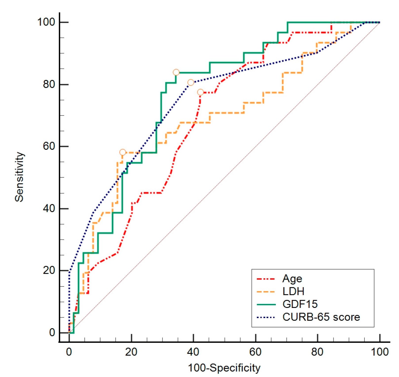
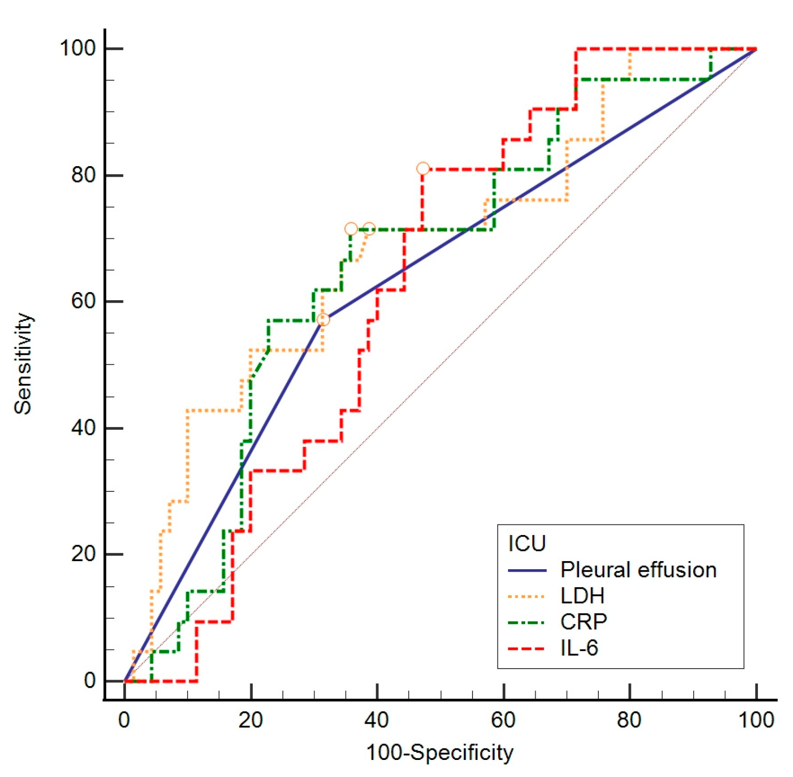
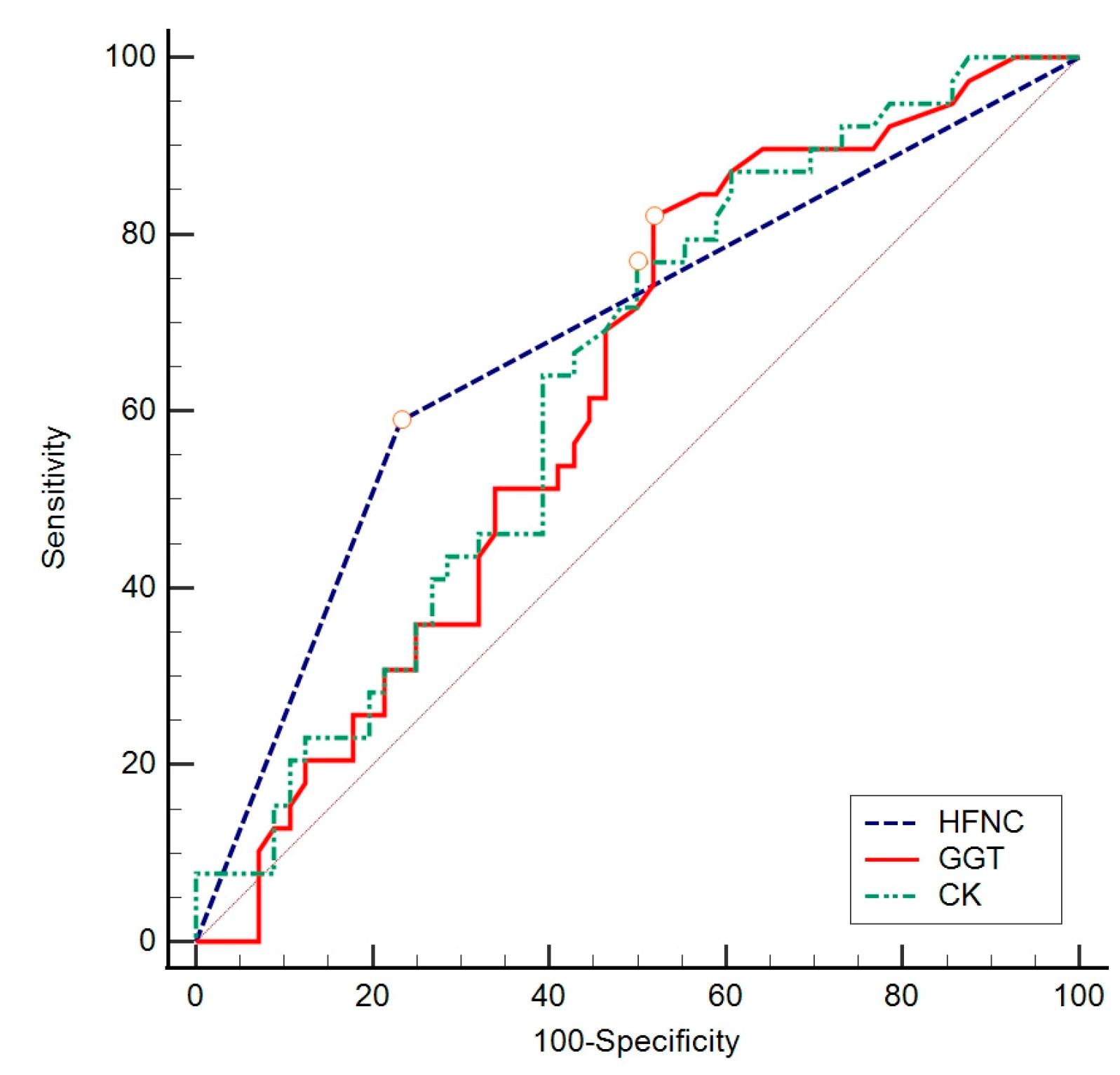
The ROC curve method was chosen to assess the difference in individual indicators between groups concerning a negative outcome, ICU admission, and length of hospitalization, based on specificity and sensitivity. To assess the value of individual parameters found to significantly contribute to previous analyses, the ROC curve calculation method was employed. This involved gradually changing the values that distinguished subjects concerning the outcome, creating the ROC curve objectively (Figure 2, Figure 3 and Figure 4) to determine which value best distinguished the compared groups. In these data, considering a negative outcome, significant diagnostic indicators include the age of the subjects, CURB-65 score, LDH, and GDF-15. The diagnostic indicator with the largest area under the curve is represented by GDF-15, although the other values presented in Table 7 also serve as significant diagnostic indicators.
For ICU admission, significant diagnostic indicators are LDH, CRP, and IL-6 (Table 8). Furthermore, considering the length of hospitalization, significant diagnostic indicators are CK, GGT, and oxygenation with an HFNC (Table 9).

4. Discussion

This study evaluated the predictive and diagnostic value of routine laboratory parameters, clinical scores (CURB-65 and qSOFA scores), and GDF-15 in hospitalized COVID-19 patients. It is worth noting that it showed an association between elevated levels of GDF-15 and negative outcome in COVID-19 patients. Furthermore, certain measured laboratory parameters, clinical scores, and interventions showed associations with mortality, admission to the ICU, and the prolonged hospitalization of the mentioned group of patients.
The observed patient cohort primarily consisted of elderly individuals who were unvaccinated and had not experienced COVID-19 before. Additionally, a significant proportion of the patients had at least one comorbidity, with nearly all of them complicated by pneumonia. It is worth noting that these findings align with expectations given that the study exclusively focused on hospitalized patients and are in line with the previous research, which has consistently identified an older age and the presence of comorbidities as significant risk factors for hospitalization among COVID-19 patients. Moreover, vaccination has been reported in prior studies to effectively reduce the risk of hospitalization in individuals afflicted with COVID-19 [27,28].

4.1. Findings Regarding GDF-15

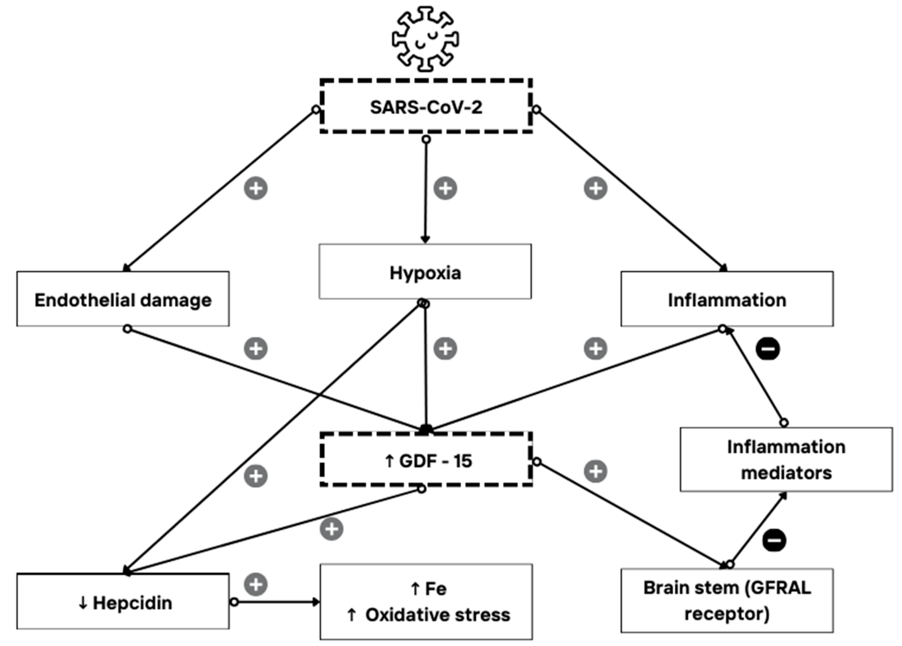
In this research, we assessed not only mortality but also ICU admission and the length of hospitalization, as all these findings can indicate the severity of COVID-19. The study aimed to introduce a possible novel biomarker for predicting COVID-19 severity, GDF-15. Generally speaking, the median values of GDF-15 in all the observed groups were higher than those expected in healthy individuals according to Doerstling et al. [29], implying the effect of SARS-CoV-2 on the GDF-15 levels in hospitalized patients. GDF-15 was more highly expressed in the non-survivor group compared to the survivor group, with a median value of 5762.0 pg/mL. The ROC analysis marked GDF-15 as a diagnostic indicator of a negative outcome with moderate specificity and sensitivity. Nevertheless, there was no statistically significant relationship between the GDF-15 levels and ICU admission and length of hospitalization. It has already been shown that GDF-15 levels rise in stress states, and its expression can be induced by hypoxia and endothelial damage [1,8,9,12], as well as aging [6,7]. SARS-CoV-2 has affinity for causing such states—tissue hypoxia as part of pneumonia-caused respiratory failure and endothelial damage due to direct impact of virus, coagulopathy, complement activation, and hypovolemia in severe forms of COVID-19 [12,30,31,32]. When considering the above, it is reasonable to anticipate elevated levels of GDF-15 in patients with severe manifestations of COVID-19, as detailed in the hypothesized mechanism explained in Figure 5. Our findings align with this expectation, supporting similar observations made by other authors [19,20,21]. However, this pilot study examines the associations between GDF-15 and clinical outcomes; it does not directly demonstrate causality. Furthermore, the specified cutoff value of 3528 pg/mL, determined through the ROC analysis, can assist clinicians in assessing the mortality risk in hospitalized COVID-19 patients when observing this parameter, thereby enabling timely and appropriate treatment to reduce negative outcomes.
This is not to neglect that higher GDF-15 levels were associated with a higher CURB-65 score, as well as a higher qSOFA score, older age, and elevated CRP, PCT, and IL-6 levels. Our results suggest that older age, a higher CURB-65 score, raised CRP values, and elevated IL-6 levels could predict a negative outcome. It is also recognized that an age greater than 74 years and a CURB-65 score higher than 2 serve as possible diagnostic indicators of a negative outcome. These findings make us believe that GDF-15 could potentially provide stronger evidence as a biomarker for severe COVID-19 in a larger cohort of patients.
The found association of GDF-15 with the PCT levels of non-survivors and ICU patients in this cohort also introduces the need for further research into GDF-15’s possible connections with other, more studied inflammatory markers. Considering the speculated mechanism of GDF-15’s interaction with SARS-CoV-2 and endothelial tissue, further studies should concentrate on the association between GDF-15 and other inflammatory markers elevated in endothelial dysfunction, such as platelet factor 4 (PF-4, CXCL4) [34,35], which has recently been studied in COVID-19 patients [36,37]. The findings of such studies could help us better understand the pathophysiological mechanisms of GDF-15 release in COVID-19 and in general.
Given that increases in GDF-15 values do not always align with those of other inflammatory markers, we should also consider research aimed at investigating how early in the course of infectious or inflammatory diseases GDF-15 levels rise. Understanding the precise dynamics of GDF-15 levels throughout disease progression could help us confirm or dismiss its role as an early diagnostic biomarker. It is plausible that its values peak earlier or later in the disease course compared to established acute-phase reactants.
Since there is a receptor for GDF-15 in the brainstem, it would be interesting to observe its levels in patients with COVID-19 and neurological comorbidity. An umbrella review of 9,228,588 COVID-19 patients by Jong Mi Park et al. showed an increased mortality risk in patients with certain neurological conditions [38]. Further exploration of the mechanisms behind such events, which could be linked to the inflammatory process and biomarkers like GDF-15, is warranted. Another interesting field for further exploration of GDF-15 is in patients with low seropositivity after COVID-19 vaccination, as described in a systematic review by Kyuyeon Cho et al. Low seropositivity is often found in patients with weakened immunity due to comorbidities, and an adequate biomarker to differentiate such patients in the general population has yet to be found [39]. Such a biomarker could be helpful in distinguishing these patients and providing the appropriate care.

4.2. Highlighted Findings on Other Laboratory Parameters

The study demonstrates that various routinely determined parameters can be beneficial during the initial evaluation of patients concerning each observed criterion—clinical outcomes, ICU admission, and length of stay.
When considering the population observed in this research, both higher CRP values and higher LDH values were expressed in the less favourable groups—non-survivors, ICU-admitted patients, and those hospitalized for 10 or more days. Additionally, LDH stood out as predictor of negative outcomes and of transfer to the ICU, as well as a diagnostic indicator of a negative outcome with a cutoff value of 395 U/L and of transfer to the ICU with a cutoff value of 404 U/L. C-reactive protein is a known inflammatory marker, and its levels are usually higher in acute inflammation, especially with a larger extent of inflammation [40]. The results obtained in this study lead to the conclusion that higher CRP levels are unfavorable in hospitalized COVID-19 patients. LDH is recognized as a marker of tissue damage and is elevated in a large number of diseases, but our findings also suggest that LDH levels should be considered when evaluating COVID-19 patients. Such results for CRP and LDH are also supported by findings from other authors [41,42].
Both of the aforementioned biomarkers, as well as others proven statistically significant in this study, are common in clinical use, widely available, and usually cost-effective.

4.3. The Study’s Limitations

This study is primarily a pilot study, as it involved a limited number of patients, with a high proportion having cardiovascular comorbidities. It is a single-centre study conducted in one country, and the generalizability of our results is unknown. Therefore, further research in this field is warranted. Nonetheless, our study can certainly serve as a foundation for such research. The main limitation of this study is the sample size, which may explain the absence of statistically significant differences in the GDF-15 concentrations between the groups of ICU and non-ICU patients and between those hospitalized for less than ten days and those hospitalized for ten or more days. Despite this limitation, the median values of GDF-15 were higher in the ICU group of patients and in those hospitalized for ten or more days. It is possible to presume that studies on a larger sample of patients could potentially strengthen GDF-15’s diagnostic significance in survivors and non-survivors, as well as possibly show its significance in ICU and non-ICU patients or patients with prolonged hospitalization. Such studies are yet to be conducted to either confirm or refute our findings.
This research also faces confounding bias, as some of the observed patients had comorbidities that could theoretically have affected their GDF-15 levels, such as adiposity or chronic diseases. Omitting comorbidities in the study was not feasible, as individuals with COVID-19 requiring hospitalization tended to have such medical conditions.
Another limitation of this study is the lack of observation of the patients after hospitalization. Our study lacks an assessment of downstream morbidity and mortality. More precisely, GDF-15 levels could also be measured after the clinical resolution of COVID-19, and it remains to be determined whether the levels of GDF-15 will decrease to their basal values. Such results could help better determine the impact of comorbidities (confounding bias) on GDF-15 levels and shed light on GDF-15 levels in long COVID-19 compared to patients who achieve full recovery.

5. Conclusions

Considering the obtained results, our hypothesis is partially accepted, and further research is necessary.
To our knowledge, the reports on GDF-15 expression during COVID-19 are limited and somewhat contradictory. Some studies have been conducted on specific patient groups, such as patients on hemodialysis or those with chronic kidney disease, where underlying diseases could affect the GDF-15 levels [14,43]. Most of the previously performed studies support evidence of higher GDF-15 levels in COVID-19 patients requiring hospitalization and those with a more severe form of the disease [15,16,17,18,19,20,21,22], while others have provided data supporting lower levels of GDF-15 in hospitalized COVID-19 patients or no significant difference between hospitalized and discharged patients, nor between SARS-CoV-2-positive and -negative patients [18,23,24]. Our findings align with the majority of studies conducted so far, indicating a higher expression of GDF-15 at the moment of hospitalization in patients with a negative outcome of COVID-19. As far as we are aware, this is the first study examining GDF-15 expression in relation to three key parameters in assessing disease severity: clinical outcomes, ICU admission, and the duration of hospitalization. Since studies are scarce, it is imperative to continue research on this topic with larger patient cohorts to fully understand the potential of GDF-15 as a biomarker of COVID-19 severity and to recognize its pathophysiological pathways in this disease, as well as in others. Questions regarding the dynamics of GDF-15 elevation and its time interval for chronic diseases, pregnancy, adiposity, and other prolonged clinical states, as well as the same questions for acute diseases such as COVID-19 and sepsis, remain to be pondered. Also, the expected levels of GDF-15 and cutoff values in those states require further study.
In conclusion, this study reaffirms the significance of commonly used laboratory parameters and clinical scores in the evaluation of COVID-19, but it also introduces the potential for a new diagnostic approach and research concerning GDF-15 levels in this widespread disease.

Author Contributions

Conceptualization: L.Š., D.L., K.K. and S.M.; methodology: L.Š., B.G., E.S., T.R. and S.M.; data curation: L.Š., B.G., M.Z., N.V.V., P.S. and D.S.; formal analysis and investigation: L.Š. and K.K.; writing—original draft preparation: L.Š., B.G., M.Z., E.S., N.V.V., P.S., T.R. and K.K.; writing—review and editing: D.L., D.S. and S.M.; funding acquisition: D.L.; resources: L.Š., T.R. and S.M.; supervision: D.L. and S.M. All authors have read and agreed to the published version of the manuscript.

Funding

The research received partial funding from the institutional projects of the Faculty of Medicine Osijek, located at J. Huttlera 4, 31000 Osijek, Croatia, with project number IP17 for the academic year 2023/24. The other measurements were conducted as part of routine clinical work.

Institutional Review Board Statement

This study was conducted in accordance with the Declaration of Helsinki and approved by the Institutional Ethics Committee of the University Hospital Centre Osijek (R1-4012/2023, 4 April 2023) and the Institutional Ethics Committee of the Faculty of Medicine Osijek (KLASA: 602-04/23-08/03, URBROJ: 2158-61-46-23-18, 23 February 2023).

Informed Consent Statement

Informed consent was obtained from all the subjects involved in the study.

Data Availability Statement

The data that support the findings of this study are available from the corresponding author, S.M., upon reasonable request.

Acknowledgments

Gratitude is extended to Ira Rešetar, mag. educ. philol. angl. et mag. educ. soc., for their meticulous proofreading, significantly enhancing the manuscript’s clarity and precision.

Conflicts of Interest

The authors declare no conflicts of interest.

References

  1. Wischhusen, J.; Melero, I.; Fridman, W.H. Growth/Differentiation Factor-15 (GDF-15): From Biomarker to Novel Targetable Immune Checkpoint. Front. Immunol. 2020, 11, 951. [Google Scholar] [CrossRef] [PubMed]
  2. Modi, A.; Dwivedi, S.; Roy, D.; Khokhar, M.; Purohit, P.; Vishnoi, J.; Pareek, P.; Sharma, S.; Sharma, P.; Misra, S. Growth differentiation factor 15 and its role in carcinogenesis: An update. Growth Factors 2019, 37, 190–207. [Google Scholar] [CrossRef]
  3. Moore, A.G.; Brown, D.A.; Fairlie, W.D.; Bauskin, A.R.; Brown, P.K.; Munier, M.L.C.; Russell, P.K.; Salamonsen, L.A.; Wallace, E.M.; Breit, S.N. The transforming growth factor-ss superfamily cytokine macrophage inhibitory cytokine-1 is present in high concentrations in the serum of pregnant women. J. Clin. Endocrinol. Metab. 2000, 85, 4781–4788. [Google Scholar] [CrossRef]
  4. Lockhart, S.M.; Saudek, V.; O’Rahilly, S. GDF15: A Hormone Conveying Somatic Distress to the Brain. Endocr. Rev. 2020, 41, 610–642. [Google Scholar] [CrossRef]
  5. Siddiqui, J.A.; Pothuraju, R.; Khan, P.; Sharma, G.; Muniyan, S.; Seshacharyulu, P.; Jain, M.; Nasser, M.W.; Batra, S.K. Pathophysiological role of growth differentiation factor 15 (GDF15) in obesity, cancer, and cachexia. Cytokine Growth Factor. Rev. 2022, 64, 71–83. [Google Scholar] [CrossRef] [PubMed]
  6. Chen, L.; Zheng, S. Understand variability of COVID-19 through population and tissue variations in expression of SARS-CoV-2 host genes. Inform. Med. Unlocked 2020, 21, 100443. [Google Scholar] [CrossRef] [PubMed]
  7. Conte, M.; Giuliani, C.; Chiariello, A.; Iannuzzi, V.; Franceschi, C.; Salvioli, S. GDF15, an emerging key player in human aging. Ageing Res. Rev. 2022, 75, 101569. [Google Scholar] [CrossRef] [PubMed]
  8. Rochette, L.; Dogon, G.; Zeller, M.; Cottin, Y.; Vergely, C. GDF15 and Cardiac Cells: Current Concepts and New Insights. Int. J. Mol. Sci. 2021, 22, 8889. [Google Scholar] [CrossRef] [PubMed]
  9. Adela, R.; Banerjee, S.K. GDF-15 as a Target and Biomarker for Diabetes and Cardiovascular Diseases: A Translational Prospective. J. Diabetes Res. 2015, 2015, 490842. [Google Scholar] [CrossRef] [PubMed]
  10. He, Y.W.; He, C.S. Association of Growth and Differentiation Factor 15 in Rheumatoid Arthritis. J. Inflamm. Res. 2022, 15, 1173–1181. [Google Scholar] [CrossRef]
  11. Sarıyıldız, M.A.; Yazmalar, L.; Batmaz, I.; Alpaycı, M.; Burkan, Y.K.; Sula, B.; Kaplan, I.; Yıldız, M.; Akar, Z.A.; Bozkurt, M. Serum GDF-15 level in Behçet’s disease: Relationships between disease activity and clinical parameters. Int. J. Dermatol. 2016, 55, 1289–1294. [Google Scholar] [CrossRef] [PubMed]
  12. Wan, Y.; Fu, J. GDF15 as a key disease target and biomarker: Linking chronic lung diseases and ageing. Mol. Cell Biochem. 2023, 1, 453–466. [Google Scholar] [CrossRef] [PubMed]
  13. Ebihara, T.; Matsumoto, H.; Matsubara, T.; Togami, Y.; Nakao, S.; Matsuura, H.; Kojima, T.; Sugihara, F.; Okuzaki, D.; Hirata, H.; et al. Cytokine Elevation in Severe COVID-19 From Longitudinal Proteomics Analysis: Comparison With Sepsis. Front. Immunol. 2022, 12, 798338. [Google Scholar] [CrossRef] [PubMed]
  14. Gisby, J.; Clarke, C.L.; Medjeral-Thomas, N.; Malik, T.H.; Papadaki, A.; Mortimer, P.M.; Buang, N.B.; Lewis, S.; Pereira, M.; Toulza, F.; et al. Longitudinal proteomic profiling of dialysis patients with COVID-19 reveals markers of severity and predictors of death. eLife 2021, 10, e64827. [Google Scholar] [CrossRef]
  15. Díaz-Troyano, N.; Gabriel-Medina, P.; Weber, S.; Klammer, M.; Barquín-DelPino, R.; Castillo-Ribelles, L.; Esteban, A.; Hernández-González, M.; Ferrer-Costa, R.; Pumarola, T.; et al. Soluble Angiotensin-Converting Enzyme 2 as a Prognostic Biomarker for Disease Progression in Patients Infected with SARS-CoV-2. Diagnostics 2022, 12, 886. [Google Scholar] [CrossRef]
  16. Batista, F.d.M.; Puga, M.A.M.; da Silva, P.V.; Oliveira, R.; dos Santos, P.C.P.; da Silva, B.O.; Tatara, M.B.; Tsuha, D.H.; Pires, M.A.d.S.; Gonçalves, C.C.M.; et al. Serum biomarkers associated with SARS-CoV-2 severity. Sci. Rep. 2022, 12, 15999. [Google Scholar] [CrossRef]
  17. Alserawan, L.; Peñacoba, P.; Echevarría, S.E.O.; Castillo, D.; Ortiz, E.; Martínez-Martínez, L.; Naranjo, E.M.; Domingo, P.; Castellví, I.; Juárez, C.; et al. Growth Differentiation Factor 15 (GDF-15): A Novel Biomarker Associated with Poorer Respiratory Function in COVID-19. Diagnostics 2021, 11, 1998. [Google Scholar] [CrossRef] [PubMed]
  18. Torrens-Mas, M.; Perelló-Reus, C.M.; Trias-Ferrer, N.; Ibargüen-González, L.; Crespí, C.; Galmes-Panades, A.M.; Navas-Enamorado, C.; Sanchez-Polo, A.; Piérola-Lopetegui, J.; Masmiquel, L.; et al. GDF15 and ACE2 stratify COVID-19 patients according to severity while ACE2 mutations increase infection susceptibility. Front. Cell. Infect. Microbiol. 2022, 12, 942951. [Google Scholar] [CrossRef] [PubMed]
  19. Romualdo, L.G.d.G.; Mulero, M.D.R.; Olivo, M.H.; Rojas, C.R.; Arenas, V.R.; Morales, M.G.; Abellán, A.B.; Conesa-Zamora, P.; García-García, J.; Hernández, A.C.; et al. Circulating levels of GDF-15 and calprotectin for prediction of in-hospital mortality in COVID-19 patients: A case series. J. Infect. 2021, 82, e40. [Google Scholar] [CrossRef]
  20. Teng, X.; Zhang, J.; Shi, Y.; Liu, Y.; Yang, Y.; He, J.; Luo, S.; Huang, Y.; Liu, Y.; Liu, D.; et al. Comprehensive Profiling of Inflammatory Factors Revealed That Growth Differentiation Factor-15 Is an Indicator of Disease Severity in COVID-19 Patients. Front. Immunol. 2021, 12, 662465. [Google Scholar] [CrossRef]
  21. Myhre, P.L.; Prebensen, C.; Strand, H.; Røysland, R.; Jonassen, C.M.; Rangberg, A.; Sørensen, V.; Søvik, S.; Røsjø, H.; Svensson, M.; et al. Growth Differentiation Factor 15 Provides Prognostic Information Superior to Established Cardiovascular and Inflammatory Biomarkers in Unselected Patients Hospitalized With COVID-19. Circulation 2020, 142, 2128–2137. [Google Scholar] [CrossRef] [PubMed]
  22. Ackermann, M.; Ackermann, M.; Kamp, J.C.; Kamp, J.C.; Werlein, C.; Werlein, C.; Walsh, C.L.; Walsh, C.L.; Stark, H.; Stark, H.; et al. The fatal trajectory of pulmonary COVID-19 is driven by lobular ischemia and fibrotic remodelling. EBioMedicine 2022, 85, 104296. [Google Scholar] [CrossRef]
  23. Delaye, J.-B.; Alarcan, H.; Vallet, N.; Veyrat-Durebex, C.; Bernard, L.; Hérault, O.; Ropert, M.; Marlet, J.; Gyan, E.; Andres, C.; et al. Specific changes of erythroid regulators and hepcidin in patients infected by SARS-CoV-2. J. Investig. Med. 2022, 70, 934–938. [Google Scholar] [CrossRef]
  24. Wang, Y.; Yao, P.; Li, K.; Qin, S. GDF-15 (a biomarker for metformin) and the risk of COVID-19: A two-sample Mendelian randomization study. Medicine 2023, 102, e34675. [Google Scholar] [CrossRef] [PubMed]
  25. Lim, W.S.; van der Eerden, M.M.; Laing, R.; Boersma, W.G.; Karalus, N.; I Town, G.; A Lewis, S.; Macfarlane, J.T. Defining community acquired pneumonia severity on presentation to hospital: An international derivation and validation study. Thorax 2003, 58, 377–382. [Google Scholar] [CrossRef] [PubMed]
  26. Evans, L.; Rhodes, A.; Alhazzani, W.; Antonelli, M.; Coopersmith, C.M.; French, C.; Machado, F.R.; Mcintyre, L.; Ostermann, M.; Prescott, H.C.; et al. Surviving sepsis campaign: International guidelines for management of sepsis and septic shock 2021. Intensive Care Med. 2021, 47, 1181–1247. [Google Scholar] [CrossRef] [PubMed]
  27. Vahey, G.M.; McDonald, E.; Marshall, K.; Martin, S.W.; Chun, H.; Herlihy, R.; Tate, J.E.; Kawasaki, B.; Midgley, C.M.; Alden, N.; et al. Risk factors for hospitalization among persons with COVID-19-Colorado. PLoS ONE 2021, 16, e0256917. [Google Scholar] [CrossRef] [PubMed]
  28. García, H.A.V.; Adu, P.A.; Harrigan, S.; Wilton, J.; Rasali, D.; Binka, M.; Sbihi, H.; Smolina, K.; Janjua, N.Z. Risk factors for COVID-19 hospitalization after COVID-19 vaccination: A population-based cohort study in Canada. Int. J. Infect. Dis. 2023, 127, 116–123. [Google Scholar] [CrossRef]
  29. Doerstling, S.; Hedberg, P.; Öhrvik, J.; Leppert, J.; Henriksen, E. Growth differentiation factor 15 in a community-based sample: Age-dependent reference limits and prognostic impact. Ups. J. Med. Sci. 2018, 123, 86–93. [Google Scholar] [CrossRef]
  30. Perico, L.; Benigni, A.; Casiraghi, F.; Ng, L.F.P.; Renia, L.; Remuzzi, G. Immunity, endothelial injury and complement-induced coagulopathy in COVID-19. Nat. Rev. Nephrol. 2021, 17, 46–64. [Google Scholar] [CrossRef]
  31. Hilton, J.; Boyer, N.; Nadim, M.K.; Forni, L.G.; Kellum, J.A. COVID-19 and Acute Kidney Injury. Crit. Care Clin. 2022, 38, 473–489. [Google Scholar] [CrossRef] [PubMed]
  32. Rochette, L.; Zeller, M.; Cottin, Y.; Vergely, C. GDF15: An emerging modulator of immunity and a strategy in COVID-19 in association with iron metabolism. Trends Endocrinol. Metab. 2021, 32, 875–889. [Google Scholar] [CrossRef]
  33. Chambers, K.; Ashraf, M.A.; Sharma, S. Physiology, Hepcidin. In StatPearls; StatPearls Publishing: Treasure Island, FL, USA, 2023. [Google Scholar]
  34. Liu, Z.; Li, L.; Zhang, H.; Pang, X.; Qiu, Z.; Xiang, Q.; Cui, Y. Platelet factor 4(PF4) and its multiple roles in diseases. Blood Rev. 2024, 64, 101155. [Google Scholar] [CrossRef] [PubMed]
  35. Bikfalvi, A. Platelet factor 4: An inhibitor of angiogenesis. Semin. Thromb. Hemost. 2004, 30, 379–385. [Google Scholar] [CrossRef] [PubMed]
  36. Iba, T.; Wada, H.; Levy, J.H. Platelet Activation and Thrombosis in COVID-19. Semin. Thromb. Hemost. 2023, 49, 55–61. [Google Scholar] [CrossRef] [PubMed]
  37. Favaloro, E.J.; Pasalic, L.; Lippi, G. Antibodies against Platelet Factor 4 and Their Associated Pathologies: From HIT/HITT to Spontaneous HIT-like Syndrome, to COVID-19, to VITT/TTS. Antibodies 2022, 11, 7. [Google Scholar] [CrossRef] [PubMed]
  38. Park, J.M.; Woo, W.; Lee, S.C.; Park, S.; Yon, D.K.; Lee, S.W.; Smith, L.; Koyanagi, A.; Shin, J.I.; Kim, Y.W. Prevalence and Mortality Risk of Neurological Disorders during the COVID-19 Pandemic: An Umbrella Review of the Current Evidence. Neuroepidemiology 2023, 57, 129–147. [Google Scholar] [CrossRef] [PubMed]
  39. Cho, K.; Park, S.; Kim, E.; Koyanagi, A.; Jacob, L.; Yon, D.K.; Lee, S.W.; Kim, M.S.; Radua, J.; Elena, D.; et al. Immunogenicity of COVID-19 vaccines in patients with diverse health conditions: A comprehensive systematic review. J. Med. Virol. 2022, 94, 4144–4155. [Google Scholar] [CrossRef] [PubMed]
  40. Black, S.; Kushner, I.; Samols, D. C-reactive Protein. J. Biol. Chem. 2004, 279, 48487–48490. [Google Scholar] [CrossRef]
  41. Izcovich, A.; Ragusa, M.A.; Tortosa, F.; Marzio, M.A.L.; Agnoletti, C.; Bengolea, A.; Ceirano, A.; Espinosa, F.; Saavedra, E.; Sanguine, V.; et al. Prognostic factors for severity and mortality in patients infected with COVID-19: A systematic review. PLoS ONE 2020, 15, e0241955. [Google Scholar] [CrossRef]
  42. Malik, P.; Patel, U.; Mehta, D.; Patel, N.; Kelkar, R.; Akrmah, M.; Gabrilove, J.L.; Sacks, H. Biomarkers and outcomes of COVID-19 hospitalisations: Systematic review and meta-analysis. BMJ Evid. Based Med. 2021, 26, 107–108. [Google Scholar] [CrossRef] [PubMed]
  43. Galassi, A.; Ciceri, P.; Bono, V.; Magagnoli, L.; Sala, M.; Artioli, L.; Rovito, R.; Hadla, M.; Yellenki, V.; Monforte, A.D.; et al. Growth Differentiation Factor 15 (GDF-15) Levels Associate with Lower Survival in Chronic Kidney Disease Patients with COVID-19. Biomedicines 2022, 10, 3251. [Google Scholar] [CrossRef] [PubMed]
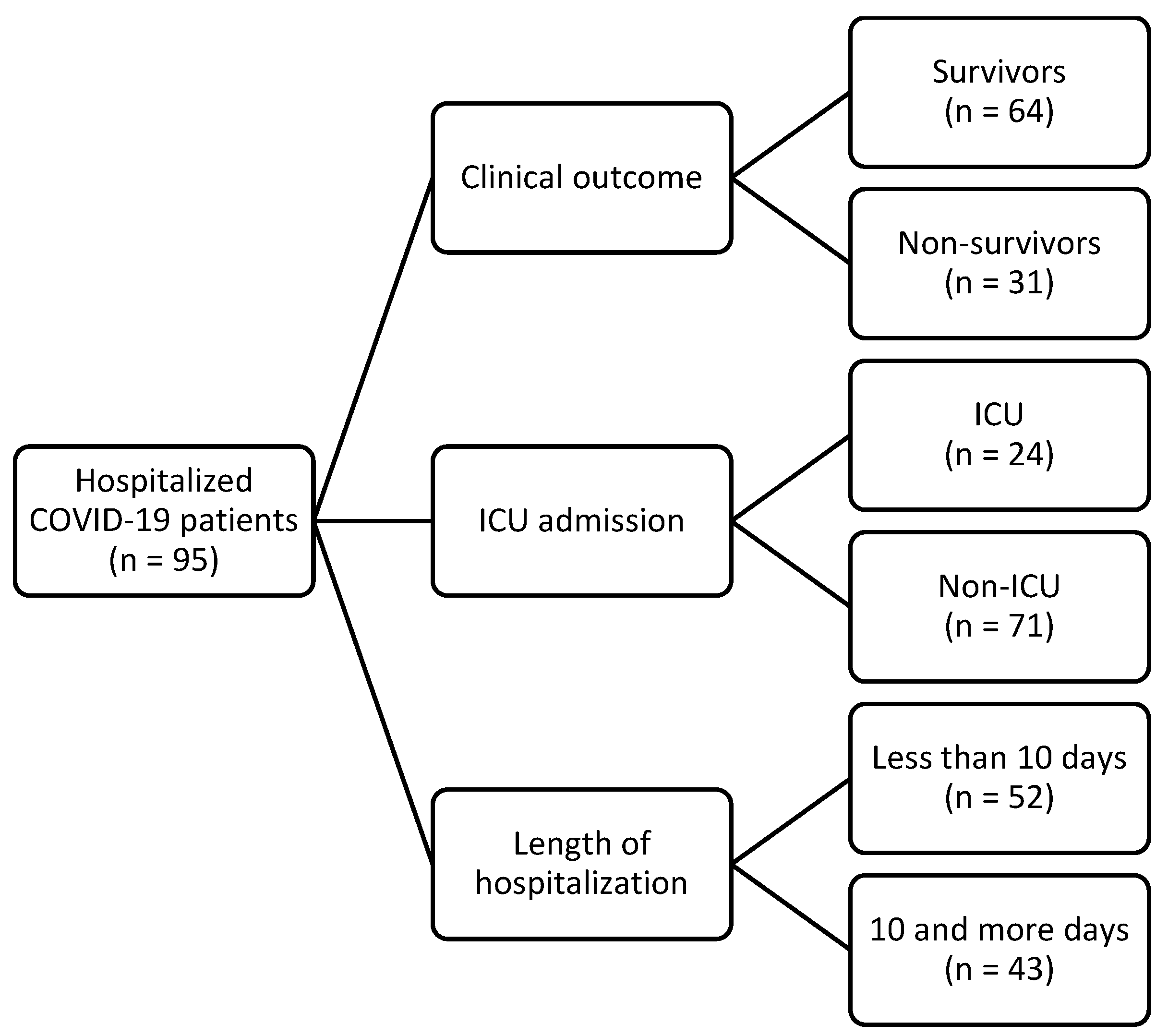
Figure 1. Flowchart of participants enrolled in this study. ICU—intensive care unit.
Biomedicines 12 00757 g001
Figure 2. ROC analysis of sensitivity, specificity, and threshold values for observed variables concerning negative outcomes of the disease. LDH—lactate dehydrogenase; GDF-15—growth differentiation factor 15.
Biomedicines 12 00757 g002
Figure 3. ROC analysis of sensitivity, specificity, and threshold values for observed values with respect to the ICU transfer. ICU—intensive care unit; LDH—lactate dehydrogenase; CRP—C-reactive protein; IL-6—interleukin 6.
Biomedicines 12 00757 g003
Figure 4. ROC analysis of sensitivity, specificity, and threshold values for observed values with respect to hospitalization longer than or equal to 10 days. HFNC—high-flow nasal cannula; GGT—gamma-glutamyltransferase; CK—creatine kinase.
Biomedicines 12 00757 g004
Figure 5. Proposed mechanism of SARS-CoV-2, GDF-15, and iron metabolism interaction. SARS-CoV-2 causes endothelial damage, tissue hypoxia, and inflammation due to the direct impact of the virus, coagulopathy, complement activation, and hypovolemia in severe forms of COVID-19. Endothelial damage, hypoxia, and inflammation have all been previously reported to result in higher expression of GDF-15 and an increase in its levels. Both GDF-15 and hypoxia have been shown to lower hepcidin levels, leading to the loss of the normal role of hepcidin—an acute-phase reactant—in reducing serum iron during an infection. This disruption leads to impaired iron homeostasis, elevated serum iron values, and oxidative stress. Additionally, GDF-15 directly reduces the release of various inflammation mediators by binding to the GDNF family receptor α–like (GFRAL) in the brainstem, thereby decreasing the extent of inflammation. These findings suggest a possible dual role of GDF-15 in acute inflammation: a protective one through GFRAL receptors and a non-protective role through hepcidin regulation [5,12,30,31,32,33]. However, further research is needed.
Biomedicines 12 00757 g005
Table 1. Baseline demographic and clinical characteristics of participants.
Sex [n (%)]
 Male41 (43)
 Female54 (57)
Age [Median (range)]76 (22–93)
Length of hospitalization [Median (IQR)]9 (6–13)
Mean arterial pressure—MAP (mmHg)83.3 (76.7–93.3)
Vaccinated [n (%)]34 (36)
Comorbidities [n (%)]
 Type 2 diabetes mellitus21 (22)
 Hypertension68 (72)
 Cardiomyopathy21 (22)
 Atrial fibrillation9 (10)
 Lung disease 10 (11)
 Chronic kidney disease4 (4)
Clinical symptoms [n (%)]
General symptoms
 Fever71 (75)
 Cephalea5 (5)
 Chills/shivering19 (20)
 General weakness63 (66)
 Nausea15 (16)
 Myalgia or arthralgia21 (22)
 Confusion15 (16)
Respiratory symptoms
 Cough81 (85)
 Sore throat5 (5)
 Chest pain28 (30)
 Hemoptysis3 (3)
 Dyspnea (anamnestic)60 (63)
 Dyspnea (at examination)73 (77)
Gastrointestinal symptoms
 Diarrhea26 (27)
 Vomiting6 (6)
Skin changes2 (2)
Complications [n(%)]
Pulmonary complications
 Pneumonia91 (96)
  Unilateral involvement10/91 (11)
  Bilateral involvement81/91 (89)
 Pulmonary embolism1 (1)
 Pleural effusion36 (38)
 Oxygenation (nasal catheter or mask)89 (94)
 Oxygenation (high-flow nasal cannula—HFNC) 36 (38)
 Invasive mechanical ventilation (IMV)20 (21)
IQR—interquartile range.
Table 2. Baseline characteristics of the participants according to the studied outcomes.
Clinical Outcomep 3ICU Admissionp 3Length of Hospitalizationp 3
Survivors
(n = 64)
Non-Survivors
(n = 31)
Non-ICU
(n = 71)
ICU
(n = 24)
Less than 10 Days (n = 52)10 and More Days (n = 43)
Demographic data
Female, n (%)36 (56)18 (58)0.870 141 (58)13 (54)0.760 131 (59)23 (54)0.550 1
Age in years, median (range)71 (22–90)79 (56–93)0.00374 (22–93)77 (56–89)0.40077 (22–93)76 (33–89)0.550
Vaccinated, n (%)25 (39)9 (29)0.340128 (39)65 (25)0.200 122 (42)12 (28)0.150 1
Length of hospitalization in days, median (IQR)8 (6–13)11 (7–15)0.0908 (6–12)13 (8–15)0.0027 (5–8)14 (12–16)<0.001
MAP in mmHg, median (IQR)83.3 (80–93.3)83.3 (73.3–86.7)0.12083.3 (73.3–90)85 (78.3–83.3)0.24083.3 (73.3–89.6)83.3 (80–93.3)0.340
Comorbidity, n (%)
Type 2 diabetes mellitus15 (23)6 (19)0.650 115 (21)6 (25)0.690 113 (25)8 (19)0.460 1
Hypertension42 (66)26 (84)0.070 150 (70)18 (75)0.670 136 (69)32 (74)0.580 1
Cardiomyopathy10 (16)11 (36)0.030 113 (18)8 (33)0.130 110 (19)11 (26)0.460 1
Atrial fibrillation4 (6)5 (16)0.15 27 (10)2 (8)>0.990 24 (8)5 (12)0.730 2
Lung disease6 (9)4 (13)0.720 17 (10)3 (13)0.710 15 (10)5 (12)0.750 1
Chronic kidney disease2 (3)2 (7)0.600 23 (4)1 (4)>0.990 23 (6)1 (2)0.620 2
Clinical scores, median (IQR)
qSOFA score1 (1–1)1 (1–2)0.0101 (1–2)1 (1–2)0.7601 (1–2)1 (1–1)0.670
CURB-65 score2 (2–3)3 (3–4)<0.0011 (2–3)2 (2–4)0.2003 (2–3)3 (2–3)0.580
Laboratory findings, median (IQR)
WBC (×109/L)6.6 (5.3–9.4)6.8 (4.6–11.9)0.9906.4 (5.1–10.1)7.5 (4.8–10.1)0.8707.1 (5.1–10.5)6.3 (5.1–9.3)0.570
RBC (×1012/L)4.5 (4–5)4.3 (4–4.9)0.3804.46 (4–5)4.48 (4–4.9)0.8404.39 (3.9–4.9)4.64 (4.1–5)0.060
Hb (g/L)136
(119–146)
131
(120–146)
0.850135
(119–144)
135
(122–1501)
0.470130
(116–143)
138
(125–150)
0.050
Hct (L/L)0.4 (0.4–0.4)0.4 (0.4–0.4)0.8100.4 (0.4–0.4)0.41 (0.4–0.4)0.3400.39 (0.4–0.4)0.41 (0.4–0.4)0.040
MCV fL)87.8
(84.6–91.8)
89.9
(87–93.7)
0.11088.2
(84.5–92.6)
89.05
(87–93.4)
0.33088.9 (86.1–92.9)87.4 (84.3–92.3)0.220
MCHC (g/L)334
(327–342)
331
(320–338)
0.040333
(326–342)
332
(321–342)
0.410334 (325–341)333 (326–342)0.920
RDW-CV (%)13.3
(12.9–14.1)
14.3
(13.4–15.9)
0.00213.4
(13.1–14.6)
13.95
(13–15.2)
0.33013.4
(13–14.7)
13.7
(13.1–14.6)
0.620
Platelets (×109/L)213
(155–273)
195
(138–224)
0.220210
(148–281)
197 (159–222)0.390204
(159–307)
195
(138–251)
0.130
Neutrophils (%)76 (68–86)79 (73–87)0.20076 (68–86)80 (74–88)0.11076 (68–87)78 (70–86)0.320
Lymphocytes (%)15 (8–22)12 (7–17)0.19015 (8–21)12 (6–16)0.12015 (8–23)12 (7–17)0.170
Band cells (%)0 (0–0)0 (0–0)0.0800 (0–0)0 (0–0)0.9300 (0–0)0 (0–0)0.230
Monocytes (%)8 (5–9)6 (4–9)0.1708 (5–9)6 (4–9)0.2708 (5–9)7 (5–10)0.590
Basophils (%)0 (0–0)0 (0–0)0.1100 (0–0)0 (0–0)0.1800 (0–0)0 (0–0)0.240
Eosinophils (%)0 (0–7)0 (0–0)0.0300 (0–0)0 (0–0)0.0700 (0–7)0 (0–1)0.410
PT (INR) 0.9 (0.9–1)1 (0.9–1)0.1600.94 (0.9–1)0.97 (0.9–1)0.9500.95 (0.9–1)0.96 (0.9–1)0.790
Fbg (g/L)6.4 (5.7–7.9)6.1 (5.4–8.6)0.8306.4 (5.4–7.9)6.4 (5.6–9.5)0.4506.35 (5.4–7.4)6.6 (5.6–9.5)0.140
APTT (1)0.8 (0.8–0.9)0.9 (0.8–1)0.1100.85 (0.8–0.9)0.82 (0.8–0.9)0.5600.85 (0.8–0.9)0.82 (0.8–0.9)0.400
D-dimers (μg/L FEU)1281
(799–2173)
1514
(1124–3117)
0.3301335
(808–2120)
1438
(1037–3356)
0.4301286
(816–2110)
1506
(840–2685)
0.710
AT-3 (1)1.1 (1–1.3)1.1 (1–1.2)0.0601.1 (1–1.3)1.1 (0.9–1.2)0.2001.1 (1–1.2)1.1 (1–1.3)0.650
AST (U/L)44 (31–66)57 (43–98)0.00344 (34–66)67 (44–98)0.00845 (29–68)50 (41–86)0.060
ALT (U/L)34 (19–51)36 (23–51)0.51031 (16–50)39 (30–53)0.10031 (17–52)36 (23–50)0.330
ALP (U/L)68 (56–89)57 (51–75)0.18063 (53–89)63 (50–75)0.24061 (55–86)64 (50–89)0.570
GGT (U/L)41 (24–101)37 (20–96)0.63039 (22–90)38 (23–111)0.72030 (17–86)50 (30–116)0.030
CK (U/L)82 (50–228)142 (74–386)0.03083 (55–223)112 (73–430)0.08076 (49–233)107 (78–288)0.030
LDH (U/L)309
(255–382)
405
(278–491)
0.003314
(253–392)
405
(298–492)
0.005296
(252–400)
365
(282–432)
0.030
Urea (mmol/L)6.9 (5.5–9.4)9.1 (6.9–16)0.0107.1 (5.6–10.3)7.5 (6.2–11.8)0.3907.1 (6–10.7)7.4 (5.5–10.7)0.850
Creatinine (μmol/L)80 (65–96)93 (79–152)0.00483 (66–107)90 (78–114)0.16082 (65–108)86 (72–116)0.340
Albumin (g/L)34.7
(31.2–36.8)
32.2
(30.2–37.2)
0.22034.45
(30.6–36.6)
33.8
(31.4–37.7)
0.51034.8
(31.1–37.1)
33.6
(30.5–36.3)
0.420
CRP (mg/L)64
(30.5–110.9)
116.6
(73.3–158.4)
0.00271.9
(30.9–114.2)
116.8
(60.9–152.8)
0.01072.6
(30.1–109.1)
101.7
(48.3–152.1)
0.020
PCT (μg/L)0.1 (0.1–0.2)0.2 (0.1–0.4)0.0020.13 (0.1–0.3)0.18 (0.1–0.3)0.1500.14 (0.1–0.3)0.17 (0.1–0.3)0.310
hsTnI (ng/L)16.8
(8.9–35.5)
29.8
(21.4–64.8)
0.02024.6
(9.9–57.7)
26.2
(12.6–53.1)
0.96021.4
(9.8–66)
26.7
(12.3–37.9)
0.550
IL-6 (ng/L)50.4
(24.5–115.6)
96.9
(58.6–179.5)
0.00355.6
(26.2–121.2)
76.3
(58.7–154)
0.08053
(20.5–124)
70.6
(36–136.3)
0.16
Ferritin (μg/I)582.8
(200–1036.6)
559.8
(234–1275.4)
0.700528.3
(199.5–1071.7)
594.2
(291.1–1185.9)
0.650422.8
(191.8–1101.3)
653.5
(361–1070.8)
0.260
Na (mmol/L)138 (136–140)138 (137–141)0.350138 (136–141)138 (137–141)0.750139 (136–141)138 (136–141)0.400
K (mmol/L)4.1 (3.7–4.4)4.2 (3.7–4.4)0.6404.1 (3.7–4.4)4.2 (3.8–4.4)0.4504.2 (3.7–4.4)4.1 (3.7–4.4)0.990
Cl (mmol/L)101 (96–103)101 (99–104)0.360100 (96–103)101 (99–104)0.450102 (98–104)100 (96–102)0.060
Glucose (mmol/L)7.1 (6.1–9)7.2 (6–9.8)0.9706.9 (6.1–8.6)7.4 (6.1–10.2)0.2806.8 (6–8.5)7.4 (6.2–9.9)0.150
GDF-15 (pg/mL)3078.0
(1671.5–5041.3)
5762.0
(4213.0–10,795)
<0.0013298
(2144–6282)
4845.5
(3039–6700)
0.0803517
(1957.3–6419)
4143
(2842–5762)
0.590
1 Chi-square test; 2 Fisher’s exact test; 3 Mann–Whitney U test, IQR—interquartile range; ICU—intensive care unit; WBC—white blood cells; RBC—red blood cells; Hb—hemoglobin; Hct—hematocrit, MCV—mean cell volume; MCHC—mean corpuscular hemoglobin concentration; RDW-CV—red blood cell distribution width—coefficient of variation; PT—prothrombin time; INR—international normalized ratio; Fbg—fibrinogen; APTT—activated partial thromboplastin time; AT-3—antithrombin 3; AST—aspartate aminotransferase; ALT—alanine aminotransferase; ALP—alkaline phosphatase; GGT—gamma-glutamyltransferase; CK—creatine kinase; LDH—lactate dehydrogenase; CRP—C-reactive protein; PCT—procalcitonin; hsTnI—high-sensitivity troponin I; IL-6—interleukin 6; Na—sodium; K—potassium; Cl—chloride; GDF-15—growth differentiation factor 15.
Table 3. The association of GDF-15 with age and qSOFA and CURB-65 scores, as well as inflammatory values upon admission, in relation to the outcome and transfer to the ICU in the group of all patients.
Spearman’s Rank Correlation Coefficient Rho (p Value) GDF-15
All PatientsClinical OutcomeICU Admission
SurvivorsNon-SurvivorsNon-ICUICU
Age0.442 (<0.001)0.479 (<0.001)0.152 (0.420)0.541 (<0.001)−0.011 (0.960)
qSOFA score0.307 (0.003)0.055 (0.660)0.644 (<0.001)0.248 (0.040)0.499 (0.010)
CURB-65 score0.537 (<0.001)0.427 (<0.001)0.541 (0.002)0.534 (<0.001)0.448 (0.030)
Inflammatory markers
CRP0.337 (0.001)0.319 (0.010)0.042 (0.820)0.380 (0.001)−0.012 (0.960)
PCT 0.586 (<0.001)0.482 (<0.001)0.619 (<0.001)0.541 (<0.001)0.656 (0.001)
IL-6 0.461 (<0.001)0.401 (0.001)0.424 (0.030)0.520 (<0.001)0.106 (0.650)
ICU—intensive care unit; CRP—C-reactive protein; PCT—procalcitonin; IL-6—interleukin 6.
Table 4. Prediction of the probability of a negative outcome (bivariate and multivariate regression).
Negative OutcomeβWaldp ValueOdds Ratio95% CI
Bivariate regression
Age 0.078.480.0041.071.02 to 1.12
Cardiomyopathy1.094.570.0302.971.09 to 8.6
CURB-65 score0.9914.6<0.0012.71.62 to 4.5
MCHC −0.056.320.0100.950.91 to 0.98
RDW-CV 0.254.40.0401.291.02 to 1.64
AST 0.026.950.0081.021.005 to 1.03
CK 0.0014.050.0401.0011.00 to 1.0003
LDH 0.0057.990.0051.0051.002 to 1.009
Urea 0.127.270.0071.131.03 to 1.23
Creatinine0.017.330.0071.011.004 to 1.03
CRP0.0086.540.0101.011.002 to 1.01
PCT 0.963.360.0702.600.94 to 7.22
hsTnI −0.0010.010.9100.990.99 to 1.001
IL-6 0.0023.870.0401.0021.00 to 1.005
GDF-15 0.00027.990.0051.00021.0001 to 1.0003
Multivariate regression
CURB-65 score0.9911.21<0.0012.551.48 to 4.42
LDH 0.0055.050.0201.011.001 to 1.009
Constant−5.3118.01<0.001
β—regression coefficient; MCHC—mean corpuscular hemoglobin concentration; RDW-CV—red blood cell distribution width—coefficient of variation; AST—aspartate aminotransferase; CK—creatine kinase; LDH—lactate dehydrogenase; CRP—C-reactive protein; PCT—procalcitonin; hsTnI—high-sensitivity troponin I; IL-6—interleukin 6; GDF-15—growth differentiation factor 15.
Table 5. Prediction of the probability of ICU admission (bivariate and multivariate regression).
ICU AdmissionβWaldp ValueOdds Ratio95% CI
Bivariate regression
Age 0.031.910.1701.030.98 to 1.07
Cardiomyopathy0.802.290.1302.230.79 to 6.31
CURB-65 score0.231.190.2701.260.84 to 1.89
Pleural effusion1.145.450.0203.121.20 to 8.10
AST 0.0051.470.2301.010.99 to 1.01
LDH 0.0044.650.0301.0041.0003 to 1.007
CRP0.0053.370.0701.0050.99 to 1.01
IL-6 −0.0010.390.5300.990.99 to 1.002
GDF-15 00.790.3701.001.00 to 1.0001
Multivariate regression
LDH 0.0044.330.0301.0041.0003 to 1.007
Constant−2.5512.8<0.001
β—regression coefficient; ICU—intensive care unit; AST—aspartate aminotransferase; LDH—lactate dehydrogenase; CRP—C-reactive protein; IL-6—interleukin 6; GDF-15—growth differentiation factor 15.
Table 6. Prediction of the probability of hospitalization for 10 days or more (bivariate and multivariate regression).
Length of Hospitalization ≥ 10 DaysβWaldp ValueOdds Ratio95% CI
Bivariate regression
Age 0.0070.200.6501.010.97 to 1.03
Bilateral pneumonia1.394.120.0404.01.05 to 15.3
Pleural effusion0.863.930.0402.351.01 to 5.49
High-flow nasal cannula (HFNC)1.4410.20.0014.211.74 to 10.17
IMV 1.628.070.0055.041.65 to 15.36
GGT 0.0020.320.5701.0020.99 to 1.01
CK 0.0012.090.1501.0010.99 to 1.002
LDH0.0032.520.1101.0030.99 to 1.01
CRP0.0075.250.0201.011.001 to 1.01
GDF-15−0.00030.490.4801.000.99 to 1.0001
Multivariate regression
High-flow nasal cannula (HFNC)1.5611.8<0.0014.751.95 to 11.6
Constant−0.9911.4<0.001
β—regression coefficient; IMV—invasive mechanical ventilation; GGT—gamma-glutamyl transferase; CK—creatine kinase; LDH—lactate dehydrogenase; CRP—C-reactive protein; GDF-15—growth differentiation factor 15.
Table 7. Diagnostic value of patient’s age, GDF-15, CURB-65, and LDH (ROC analysis) with respect to a negative outcome.
GDF-15
AUC95% CISensitivitySpecificityCut OffYouden Indexp Value
GDF-15 (pg/mL)0.7670.669–0.84883.965.6>35280.50<0.001
Age (years)0.6910.588–0.78277.457.8>740.35<0.001
CURB-65 score0.7520.653–0.83580.660.9>20.42<0.001
LDH (U/L)0.6910.587–0.78158.182.8>3950.410.002
AUC—area under the curve; GDF-15—growth differentiation factor 15; LDH—lactate dehydrogenase.
Table 8. Diagnostic value of patient’s LDH, CRP, and IL-6 (ROC analysis) with respect to an ICU admission.
AUC95% CISensitivitySpecificityCut OffYouden Indexp Value
Pleural effusion 0.6370.532–0.73358.369.0>00.270.020
LDH (U/L)0.6940.591–0.78454.280.3>4040.340.002
CRP (mg/L)0.6760.572–0.76870.864.8>95.90.360.004
IL-6 (ng/L)0.6290.521–0.72881.052.9>58.50.340.030
AUC—area under the curve: ICU—intensive care unit; LDH—lactate dehydrogenase; CRP—C-reactive protein; IL-6—interleukin 6.
Table 9. Diagnostic value of patient’s need for HFNC and levels of GGT and CK (ROC analysis) with respect to hospitalization for 10 days or more.
AUC95% CISensitivitySpecificityCut OffYouden Indexp Value
High-flow nasal cannula (HFNC)0.6790.575–0.77159.076.8>00.36<0.001
GGT (U/L)0.6210.516–0.71982.148.2>280.300.030
CK (U/L)0.6350.530–0.73176.950.0>760.270.020
AUC—area under the curve, GGT—gamma-glutamyltransferase; CK—creatine kinase.
Disclaimer/Publisher’s Note: The statements, opinions and data contained in all publications are solely those of the individual author(s) and contributor(s) and not of MDPI and/or the editor(s). MDPI and/or the editor(s) disclaim responsibility for any injury to people or property resulting from any ideas, methods, instructions or products referred to in the content.