Article

Thermal-Induced Autolysis Enzymes Inactivation, Protein Degradation and Physical Properties of Sea Cucumber, Cucumaria frondosa

1
Technology Innovation Centre of Marine Fishery Equipment in Liaoning, Dalian Ocean University, Dalian 116023, China
2
College of Food Science and Technology, Huazhong Agricultural University, Wuhan 430070, China
3
Key Laboratory of Environment Controlled Aquaculture, Ministry of Education, Dalian Ocean University, Dalian 116023, China
4
Matis ohf. Icelandic Food and Biotech R&D, IS-113 Reykjavík, Iceland
5
GRÓ Fisheries Training Programme, United Nations Education, Scientific and Cultural Organization, IS-113 Reykjavík, Iceland
6
Faculty of Food Science and Nutrition, University of Iceland, IS-102 Reykjavík, Iceland
*
Authors to whom correspondence should be addressed.
Processes 2022, 10(5), 847; https://doi.org/10.3390/pr10050847
Submission received: 14 March 2022 / Revised: 17 April 2022 / Accepted: 21 April 2022 / Published: 25 April 2022
(This article belongs to the Special Issue Method Optimization of Various Food Processing Technologies)

Abstract

:
The main objective is to effectively denature the autolysis enzymes of C. frondosa on the premise of avoiding the quality deterioration caused by overheating. The effects of the different thermal treatments (blanching at 40–80 °C for 45 min, boiling and steaming at 100 °C for 15–120 min) on the cooking yield, moisture content, protein degradation, texture, and enzyme inactivation were studied, and the inner relationship was investigated by multivariate analysis. The autolysis enzymes of C. frondosa were thermally stable and cannot be denatured completely by blanching. Boiling and steaming could efficiently inactivate the enzymes but overheating for 60–120 min reduced the cooking yield and texture quality. Boiling at 100 °C for 45 min was suitable for pre-treatment, with cooking yield of 70.3% and protein content of 78.5%. Steaming at 100 °C for at least 30 min was preferable for long-term storage and instant food, in which the relative activity was only 3.2% with better palatability.

1. Introduction

Sea cucumber is an invertebrate animal belonging to the phylum Echinodermata. There are about 1100 species of sea cucumbers around the world, but only 40 of them are edible [1], mainly including Stichipus japonicus [2], Apostichopus japonicus [3], Holothuria forskali [4], Holothuria polii, Holothuria tubulosa [5], etc. Sea cucumber is very popular among Asian countries. In 2020, the output value of the whole industry chain in China reached USD 9.5 billion [6]. Since the sea cucumber sulfated polysaccharide was proved to have strong inhibitory activity on SARS-CoV-2 [7], consumer demand surged.
Cucumaria frondosa is an inhabitant of the North Atlantic Ocean, not only a valuable food source consisting of high quantity of nutrients [8,9], but also has health care functions, such as anti-inflammatory, antimicrobial, antioxidant, and anti-cancer functions [10]. Fisheries for this species have been well-established since 1988 [11,12]. According to FAO statistics, the annual production quantity of C. frondosa has increased from 1411 tons to 2655 tons from 2008 and 2015. However, C. frondosa, similar to Stichipus japonicus, Apostichopus japonicus, and other species of sea cucumber, has autolysis particularity. The protein degradation caused by autolysis enzymes, mitochondrial apoptosis caused by ultraviolet light and temperature, and microbial degradation were the main reasons for quality deterioration of sea cucumber. Dong [13] monitored the activities of 13 endogenous enzymes, and found that leucine aminopeptidase, alkaline protease, phosphohydrolase, and glycosylhydrolase had higher activities during the sea cucumber heating process, which may play a leading role in body wall degradation. Even though producers have attempted a variety of treatment methods, including freezing quickly after capture, bloodletting, and freezing, etc., enzymatic autolysis is still a reoccurring problem during shipping and storage. The sea cucumber industry in the North Atlantic has been seriously hampered as a result of the huge financial losses.
Heating treatments are commonly used methods for enzyme deactivation. A few studies have investigated the enzyme thermal stability of sea cucumber. Hernandez-Samano [14] extracted metalloproteases from Isostichopus fuscus ventral muscle and found that more than 74% of residual activity was still retained after heating at 80 °C for 1 h. The enzymes showed higher heat-resistant properties than those in Holothurians [15], in which the enzymes could be inactivated at 60 °C for 55 min. High hydrostatic pressure and high-pressure steam were utilized for autolysis enzyme deactivation of Apostichopus japonicus, but quality deterioration took place during storage because the autolysis enzymes were not fully denatured [16,17]. This indicates that although the thermal stability of autolysis enzymes is different among species, autolysis is still a widespread issue in this field. Heating treatment not only inactivates enzymes, but also changes the physicochemical quality of sea cucumber. Gao et al. [2] studied the heating effects of different durations at 70 °C on the rheological properties of Stichipus japonicus sea cucumbers and observed that the collagen in the body wall was converted into gelation within 1 h and both the weight and rupture strength of sea cucumbers decreased continuously during thermal treatments. Moreover, Dong et al. [18] observed that inappropriate low-temperature heating caused decrease in hardness, as well as high drip loss.
Currently, studies on C. frondosa only focus on nutritional and bioactive compounds [10,19,20]. However, the effect of heating treatments on physicochemical quality and enzyme inactivation of C. frondosa is unknown. To be noticed, C. frondosa is extremely big and tough with a thick body wall and tendons, and the nutritional compounds are different from many other sea cucumber species [9]. It may result in tremendous differences compared with other species, not only in physicochemical quality properties, but also in heat transfer resistance. This research intends to provide a promising strategy for C. frondosa autolysis enzyme thermal inactivation while guaranteeing physicochemical quality. The effects of blanching, boiling, and steaming on the physicochemical quality and enzyme inactivation of C. frondosa were investigated. The results give insights into the physicochemical properties altering mechanism of C. frondosa from the perspective of protein degradation.

2. Materials and Methods

2.1. Raw Materials and Pre-Treatment

C. frondosa was caught on the east coast of Iceland, out of Stöðvarfjörður by dredge (2.4 m wide), which was dragged for a maximum of 25 min each time. The raw materials were stored at −25 °C within a month and thawed at 4 °C for 18 h before use. Individuals with an average weight of 490 ± 50 g (n = 140) were selected for the experiment. The guts and crown of the oral tentacles were meticulously removed. The body wall was cleaned with cold water and placed on ice until thermal treatments. The raw materials (n = 140) were divided into 14 groups of 10 sea cucumbers each.

2.2. Thermal Treatments

For the blanching and boiling cooking, an electro-thermostatic water bath (Julabo B33, JULABO GMBH, Seelbach, Germany) was used. The water bath was preheated to specific temperatures. The samples were immersed in water (sample:water = 1:2), and blanched at 40, 60, and 80 °C for 45 min (abbreviated to B40 °C, B60 °C, and B80 °C, respectively), and heated at 100 °C for 15, 30, 45, 60, and 120 min (abbreviated to B15, B30, B45, B60, and B120, respectively). For the steaming cooking, an electro-steam oven (Convostar, Convotherm Elektrogerate GmbH, Eglfing, Germany) was used and preheated to 100 °C. The samples were arranged on trays tidily with the entocoele downwards and steamed at 100 °C for 15, 30, 45, 60, and 120 min (abbreviated to S15, S30, S45, S60, and S120, respectively). After cooking, the samples were placed on ice with a plastic membrane until analyzed.

2.3. Cooking Yield

The raw and cooked samples were weighed accurately before (Wb) and after heating (Wa) at room temperature. The cooking yields were calculated according to the following equation (n = 5).
Cooking   yield = W a W b × 100 %

2.4. Water Content and Water Holding Capability (WHC)

Approximately 30 g of tissue was taken from every individual. These tissue samples (300 g per group) were collected and homogenized (8000 rpm) in an Ultra Turrax mixer (T25, IKA-Labortechnik, Staufen, Germany) for 10 min prior to the following tests.
Approximately 5 g of minced sea cucumber samples were dried in a ceramic bowl at 105 °C for 4 h. The water content of the sea cucumber tissue was calculated based on the weight changes according to ISO 6496 1999 (n = 3).
The WHC was measured according to Zhang et al. [21], with slight modification. Approximately 2 g of the sea cucumber samples were weighed precisely into a vial and centrifuged (TJ 25 centrifuge, Beckman coulter, Pasadena, CA, USA) at 210× g-force (1500 rpm) and at 2–5 °C for 5 min. The WHC (%) was calculated as the ratio of water in the sample after centrifugation to the water in the sample before centrifugation (n = 3).

2.5. Protein Characteristics

2.5.1. Crude Protein Content

The total nitrogen (TN) content of the C. frondosa sea cucumbers was measured by the micro-Kjeldahl method (Kjeltec 8400 Tecator Line, Foss Analytic Hilleroed, Hilleroed, Denmark) according to Mizuta et al. (n = 2).

2.5.2. Trichloroacetic Acid (TCA)-Soluble Nitrogen

Approximately 20 g of minced sea cucumber were weighed accurately into a 250 mL flask, and 50 mL of 10% (w/w) trichloroacetic acid (TCA) were added. It was mixed for 4 min in an Ultra Turrax mixer (T25, IKA-Labortechnik, Staufen, Germany) and centrifuged (TJ 25 centrifuge, Beckman coulter, CA, USA) at 4 °C for 30 min at the speed of 2300× g-force. Then, 10–12 g of the supernatant were used for the determination of TCA-soluble nitrogen by the Kjeldahl method (Kjeltec KT 200 Labtec Line, Foss Analytic, Hilleroed, Denmark). The ratio of TCA-soluble nitrogen (TCA-N) to total nitrogen on a dry basis was calculated to represent the protein degradation during the heating process. Due to the ease with which TCA-N dissolves into water, the TCA-N in water was also measured (n = 2).

2.5.3. Electrophoresis SDS-PAGE

Protein degradation was analyzed with sodium dodecyl sulphate–polyacrylamide gel electrophoresis (SDS–PAGE). The proteins were extracted by solution (20 mM Tris–HCl, pH 7.4, 150 mM NaCl, 2 mM ethylenediaminetetraacetic acid disodium salt, 10 mM dithiothreitol, 1% Triton X- 100, 0.1% SDS) on ice for 20 min [22] with a ratio of minced sample to extraction solution 1:3 (w/v). Extracts were centrifuged (Centrifuge 5415 D, Eppendorf, Hamburg, Germany) at 4 °C for 15 min at the speed of 12,000× g-force, 30 μL of supernatants were then added to 10 μL of sample buffer (277 mM Tris-HCl, pH 6.8, 44.4% glycerol, 4.4% Lithium Dodecyl Sulfate). A Bradford colorimetric assay (Colorimetric Protein Determination with Coomassie Blue, Bio-Rad Laboratories Ltd., Stockholm, Sweden) was adopted to obtain a protein concentration of 2 mg/mL. After blanching the diluted sample solution for 5 min, 20 μL of the blanched solution was analyzed with 10% polyacrylamide gel electrophoresis, and a running buffer of Tris/Tricine/SDS using a Mini Protein system (Bio-Rad Laboratories Ltd., Stockholm, Sweden). The cooking liquid was collected carefully and subjected to freeze drying (Alpha 2-4 LSC plus, Martin Christ, Osterode am Harz, Germany). The dried materials from cooking liquids were analyzed using the same procedure as described before.

2.6. Texture Profile Analysis (TPA)

The raw and cooked C. frondosa were cut into 3 × 3 × 1 cm portions and analyzed with a cylindrical probe P/50. (Texture analyzer TA.XT Plus, Stable MicroSystems, London, UK). The pre-test speed, test speed, and post-test speed were set as 1, 0.5, and 1 mm/s, respectively. The deformation rate was set to 60%, in agreement with the description of Bi et al. [23]. The hardness, springiness, adhesiveness, springiness, cohesiveness, and resilience of the samples were assessed from the obtained texture profiles (n = 5).

2.7. Relative Enzymes Activity

The sea cucumber body wall is mainly composed of protein, and proteases showed high activity in the thermal processing, so protease activity was measured to represent the autolysis enzymes inactivation [24]. Approximately 6 g of minced sample were added to 30 mL of phosphate buffer solution. The mixture was shaken for 40 s and left at 4 °C for 6 h. The solution was centrifuged (TJ 25 centrifuge, Beckman coulter, CA, USA) at 4000 rpm for 8 min. Then, 3.0 mL of supernatants were analyzed with 3.0 mL of distilled water taken as a blank control. Samples were placed in a water bath (Julabo 19, JULABO GMBH, Seelbach, Germany) at 30 °C, and then 2.0 mL of 0.5% casein solution were added to each sample. Mixtures were heated for 10 min accurately, and 2.0 mL of 10% trichloroacetic acid (TCA) solution were added to terminate the protein denaturation reaction. In a subsequent step, the solution was centrifuged at 4000 rpm for 8 min, and 1.0 mL of supernatant was added with 0.5 mL of 0.55 mol/L Na2CO3 solution and 0.5 mL of Folin-Phenol reagent. The solution was shaken and subjected to a water bath at 30 °C for 15 min. The absorbance was measured by a UV spectrophotometer (Ultrospec 3000 pro, Amersham pharmacia biotech Biochrom, Cambridge, UK) at 650 nm (n = 6).
The relative enzymes activity (REA) was calculated as follows,
REA   = A c t i v i t y s a m p l e / A c t i v i t y c o n t r o l P r o t e i n   c o n t e n t s a m p l e / P r o t e i n   c o n t e n t c o n t r o l × 100 %

2.8. Statistical Analysis and PCA

The data were processed by Microsoft Office Excel 2016 (Microsoft Inc., Redmond, Washington, DC, USA), the analysis of variance (ANOVA) was performed in the IBM SPSS Statistics 22 software. Principal component analysis (PCA) was completed in Origin pro 2019b (Origin Lab Corporation, Northampton, MA, USA).

3. Results

3.1. Effects of Thermal Treatments on Cooking Yield

The cooking yield of C. frondosa after various thermal treatments is illustrated in Figure 1. The cooking yield reduced significantly to 89.5 ± 0.5% when the temperature was up to 60 °C, and the drip loss was increased with the heating temperature and duration. When cooked at 100 °C, the first 15 min contributed most to the drip loss. In comparison, the cooking yield decreased steadily throughout the steaming treatment, but it was relatively steady in boiling groups. As a result, as high as 35.4 ± 1.5% and 28.6 ± 1.2% of the raw material was lost in the drip during the heating process by steaming and by boiling within 120 min. The average yield rate of the boiling cooking was 3.5% higher than that of the steaming cooking, which was in line with the finding on crab meat by Crapo et al. [25]. Therefore, the cooking yield could be improved by choosing boiling instead of steaming treatment when the heating time exceeded 30 min at 100 °C. Salomina et al. [26] considered that the cooking yield was mostly a function of moisture loss. In this study, all the cooking liquid turned from transparent to dark brown, indicating that the ash, saponin, chondroitin sulfate, and other soluble components may release from C. frondosa to the cooking liquid. Furthermore, the dry matter content of cooking liquid was measured to be 3.2 ± 0.1% in group S120 and 1.9 ± 0.2% in group B120. It confirmed that the nutrients of C. frondosa were also released to the ambient liquid lost as drip.

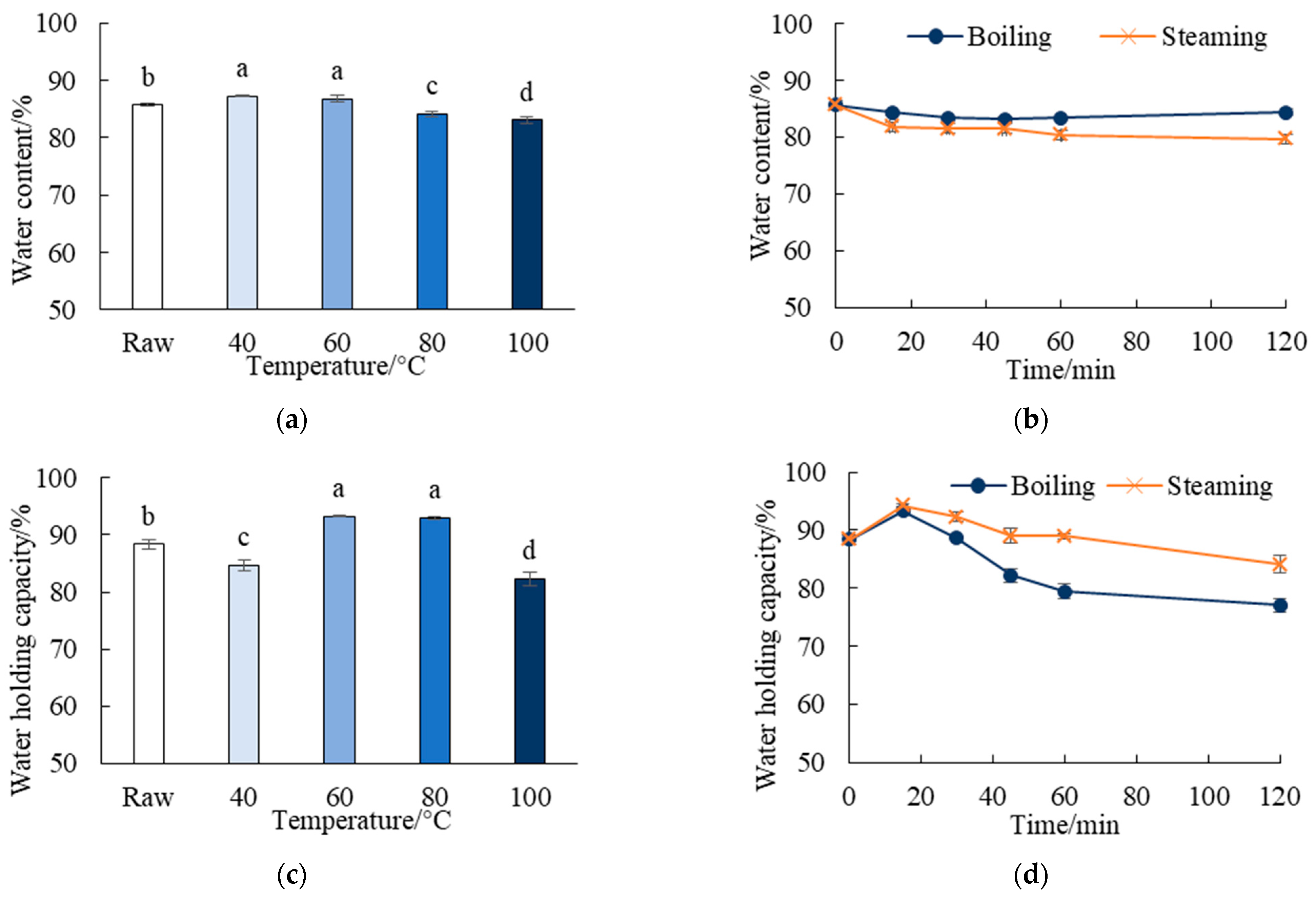
3.2. Effects of Thermal Treatments on the Water Content and WHC

The water content and WHC of the raw and cooked C. frondosa is shown in Figure 2. Water content is an important factor that affects economic efficiency, and the water inside collagenous tissue is crucial for the preservation of mechanical properties [27]. When the temperature exceeded 80 °C, the water content decreased significantly in comparison to the raw material (p < 0.05). There was a negative correlation between steaming time and water content at 100 °C, owing to the drip loss. It reduced significantly from 85.7% to 79.7% after steaming for 120 min. While the water content remained within the range of 83.1 ± 0.5% to 85.7 ± 0.2%, it did not decline continuously with boiling duration. As a consequence, the water content of the boiled sea cucumber was 2.3 ± 1.5% higher than that of the steamed group in average, which partially explained the yield advantages.
Interestingly, blanching of the C. frondosa at 40 and 60 °C resulted in higher moisture contents than in the raw sea cucumber. Xue [28] reported that an expansion of apertures among muscle fibrils in Stichopus japonicus was formed by hydrogen bond rupture during heating at 40 °C. Therefore, the rise in cooking yield and water content in group B40 °C may be attributed to the amplified space within the muscle fibrils which absorbed moisture from the blanching liquid. However, the absorbed water was easy to lose, leading to a low WHC in the B40 °C group. The cooked sea cucumber shrank obviously after blanching at 60 °C, resulting in smaller apertures between fibrils and it may explain the high WHC in the B60 °C group. In conjunction with the results of the cooking yield, it was obvious that the loss of nutrients surpassed the absorption of water during boiling at 60 °C, indicating the initiation of serious protein denaturation. Prolonged cooking time also exerted significant effects on the WHC (p < 0.05) of the C. frondosa dermis. When heated at 100 °C, the WHC was increased in the early 15 min, but declined successively to 84.1 ± 1.5% by steaming and 77.0 ± 1.2% by boiling 120 min later. WHC reflects the juiciness and tenderness of the product, and is significantly associated with quality loss, shrinkage, and compactness. The thermal-induced decrease in WHC also indicated that the microstructure of the C. frondosa dermis degraded during heating, which is consistent with Bi et al. [23].

3.3. Effects of Thermal Treatments on Protein Denaturation

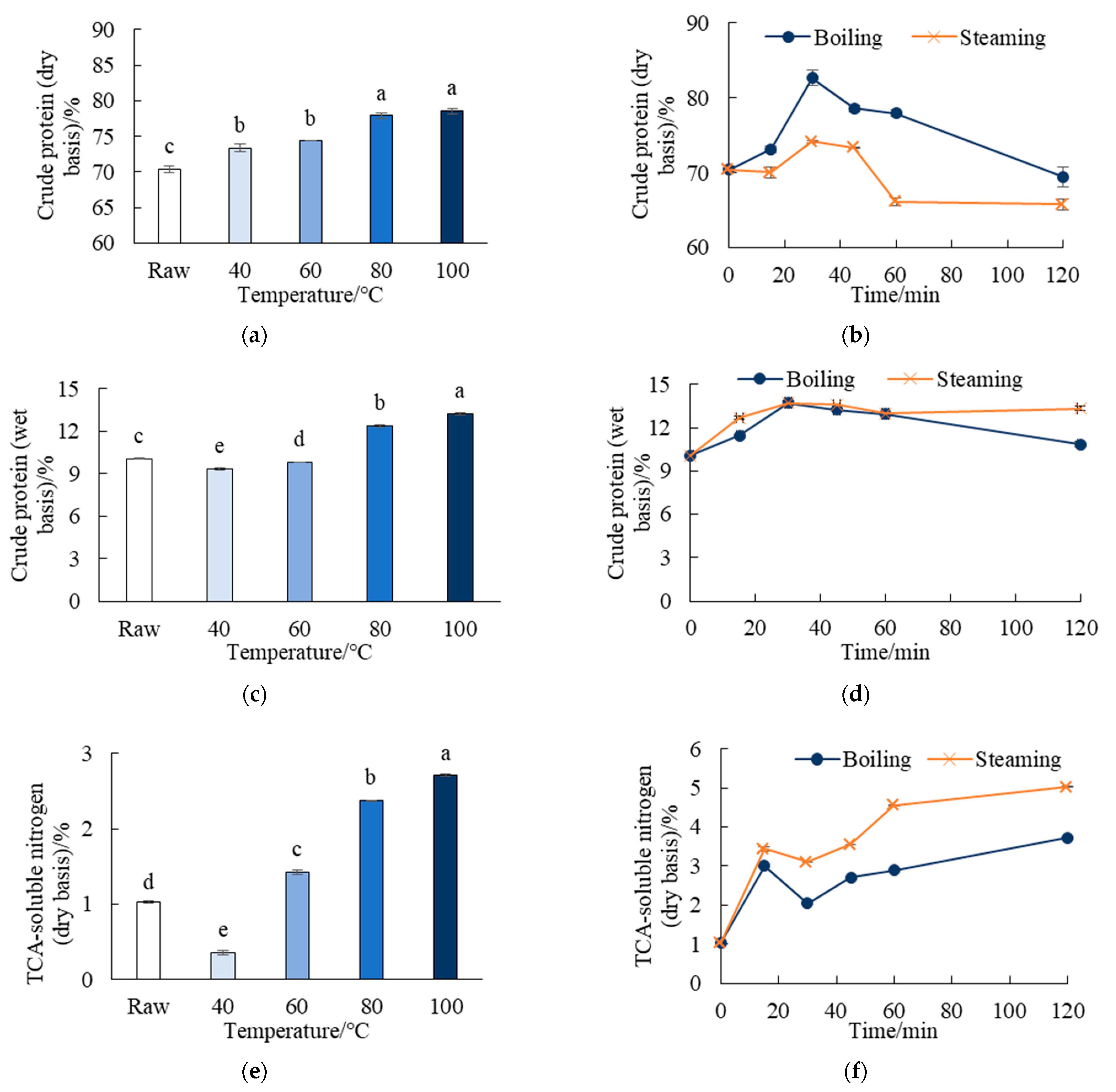
3.3.1. Crude Protein and TCA-Soluble Nitrogen

The contents of crude protein and TCA-soluble nitrogen are presented in Figure 3. Heating treatments under specific circumstances could increase the crude protein content per unit weight of sea cucumber, owing to the loss in moisture and water-soluble components during prolonged cooking. However, the crude protein content decreased when cooked for more than 30 min at 100 °C, implying that the thermal-induced protein denaturation took place during heating. It was consistent with Damodaran [29], who expounded that the collagen fibrils were degraded into small fractions and released into cooking liquid while being heated. Remarkably, the protein level on the dry basis of the boiled C. frondosa was consistently higher than that of the steamed sea cucumber, with a maximum difference of 11.8%. However, the content of crude protein on the wet basis in steaming groups was higher than that in boiling groups. It means that if the sea cucumber is dehydrated, boiling is more suitable than steaming in terms of protein content.
TCA-soluble nitrogen refers to amino acids and small peptides which can be released during protein degradation [30]. It was apparent that the TCA-soluble nitrogen was generated when temperatures exceeded 60 °C and increased both with the cooking temperature and duration. It is in accordance with previous reports, which states that the collagen denatured temperature of Stichopus japonicus to be in the range from 49.7 [23] to 57 °C [2].

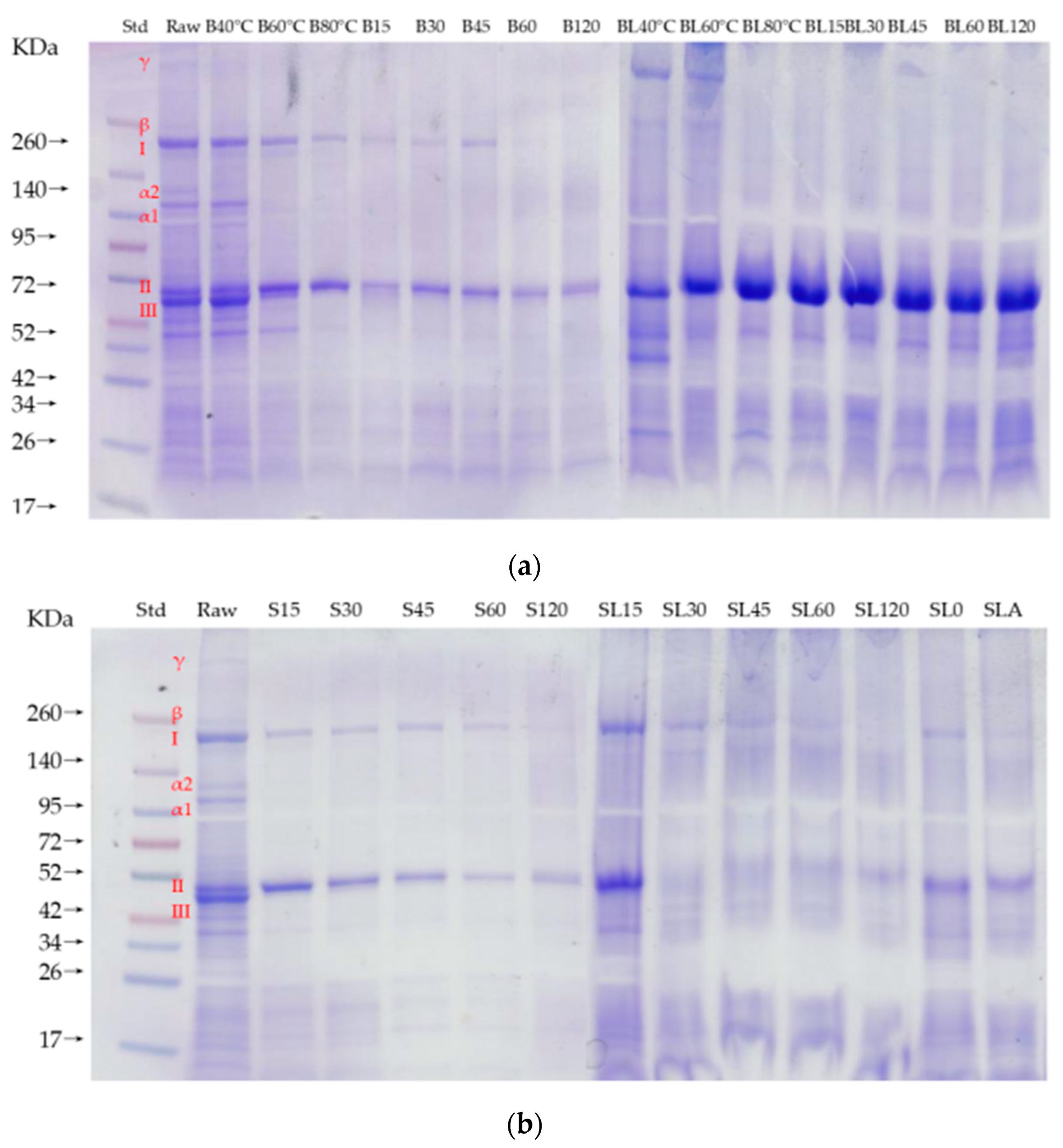
3.3.2. Protein Distribution Pattern

As a means of investigating the thermal-induced protein degradation of C. frondosa, protein distribution patterns were analyzed by electrophoresis SDS-PAGE (Figure 4). The SDS-PAGE of the raw sea cucumber samples revealed three major proteins with molecular weights of 200 (Band I), 50 (Band II), and 44 kDa (Band III). Bands I and III were assumed to be the two same structural proteins observed in Stichopus japonicus, i.e., major yolk protein (MYP, Band I) and actin (Band III) [22,31]. Band II was not familiar among other sea cucumber species, whereas the protein molecular weight approximated troponin in fishes [32]. Considering that troponin is unavailable in the majority of echinoderms, further studies are needed to identify the protein composition of Band II.
A variety of sea cucumber collagen proteins have been reported, including those of Stichopus japonicus [33,34,35], Parastichopus californicus [36], Stichopus monotuberculatus [37], and Cucumaria frondosa [38]. However, the collagen structure of sea cucumbers is still controversial. Hou et al. [33] and Cui et al. [34] reported the collagen structure of Stichopus japonicus sea cucumber as [α1(I)]3, which is consistent with Trotter [38], who concluded that the collagen of C. frondosa belonged to type I collagen with α1 trimers molecular composition. Saito et al. [35], on the other hand, classed the collagen structure of Stichopus japonicus as [α1(I)]22(I)). According to the current study, the collagen of C. frondosa contains two different α-chains (α1, α2), one β-chain and one γ-chain (Figure 4). The concentration of band α1 was close to double that of the α2, meaning that the collagen was more likely to be comprised of two α1-chain units, and one unit of α2-chain ([α1(I)]22(I)), which was in line with the observations of Saito et al. [35]. Similar patterns have been observed in other animals, including sea urchins [39], cephalopods [40], and fish [41].
The molecular weight of the α-chain in collagen of sea cucumber has earlier been reported in the range of 135–146 kDa [33,42]. The molecular weight of α-chains of the C. frondosa species were close to these reports, but slightly smaller than that of Stichopus japonicus and Apostichopus japonicus.
As seen in Figure 4a, the α2-chain began to degrade during cooking at 40 °C, and the β-chain and γ-chain vanished together with the α1-chain at 60 °C due to collagen fibrils unscrewing. Actin (Band III) also began to degrade at 60 °C, and completely disappeared at 80 °C. The MYP (Band I) of C. frondosa was relatively steady, and did not degrade until boiled at 100 °C for 120 min. Troponin (Band II) was highly thermostable during the boiling treatments, which is consistent with the studies by Chen et al. [43], who found the porcine troponin was stable during heat treatment up to 126 °C for 120 min. Considering that Band II has not been reported among other sea cucumber species yet, it indicates the differences not only in body wall protein composition, but also in the physicochemical properties during heating of C. frondosa from other reported sea cucumbers. In the boiling liquid, only Troponin could be clearly recognized for temperatures over 60 °C, and by then a new band appeared above it, presumably as an oxidized polymerization [44]. This was in line with a previous investigation of TCA-soluble nitrogen, which showed an increase in the fractions of small molecules during cooking.
In the raw materials, the collagen structural characteristics of the α1-chain, α2-chain, β-chain, and γ-chain were clear, but completely degraded in all steamed groups, indicating that the typical triple-helical structure of collagen was destroyed once steamed at 100 °C. Compared to earlier findings, these results were not unexpected. As reported by Wang et al. [45], the collagen fibrils in the body wall of the sea cucumber are sensitive to temperature change. The nonspecific bond strength of the collagen fibrils of the sea cucumber body wall was increased in the heating process of 60–90 °C, but the ionic bond, hydrogen bond, hydrophobic bond, and disulphide bond decreased. In consequence, the collagen fibrils degraded completely after heating at 90 °C for 60 min. Dong et al. [46] revealed that the thermal treatments resulted in conversion of α-helixes to β-sheets, and affected N–H bending, coupled with C–N stretching during heating. Moreover, the MYP was relatively intact during steaming until 120 min, which is consistent with the results of the boiling groups.

3.4. Effects of Thermal Treatments on Texture

As shown in Figure 5, the raw C. frondosa was thick, firm, and sticky. The texture characteristics (in Table 1) revealed an extremely high value of hardness and adhesiveness, and low springiness and chewiness. These results were similar to the TPA of Stichopus japonicus [23]. After heating, the dermis tightened and rolled inward, making the steamed sea cucumber crimpier.
After blanched at 40 °C, the hardness of the C. frondosa was increased sharply, while adhesiveness and springiness decreased significantly. It may be because of the aggregation and contraction of fibrous tissue induced by the degeneration of muscle fibrous protein, as documented by Xue [28]. When the temperature was increased to 60 °C, the hardness was reduced and the adhesiveness, springiness, and chewiness were significantly increased. It may be because the triple-helical structure of collagen and the actin deteriorated, so the body wall of C. frondosa was slightly loose and elastic. When the heating temperature exceeded 80 °C, the cohesiveness and resilience was improved, and the hardness and chewiness was reduced effectively; this was in line with Gao et al. [47], who reported that the collagen gelation occurred and the solubility of collagen fibrils increased. The dissolved collagen lubricated in muscle fibrils, Furthermore, with the increase in temperature and heating duration, collagen fibrils were dissolved continuously, and myofibrils were broken, causing the texture to be further tenderized.
Hardness and springiness are the main factors of texture evaluation of the sea cucumber. Considering that the collagen gelation contributes much to palatability [48], it is wise to blanch above 80 °C. The hardness decreased dramatically from 8895 ± 501 g to 3090 ± 257~3693 ± 558 g after boiling for 120 min or steaming for 45–120 min. The changing trend was consistent with previous reports [47], in which the rupture strength of Stichipus japonicus decreased from 130 ± 13 N (raw material) to 17 ± 3 N after heating for 2 h. By contrast, the toughness of the C. frondosa body wall was relatively high, and difficult to tenderize.

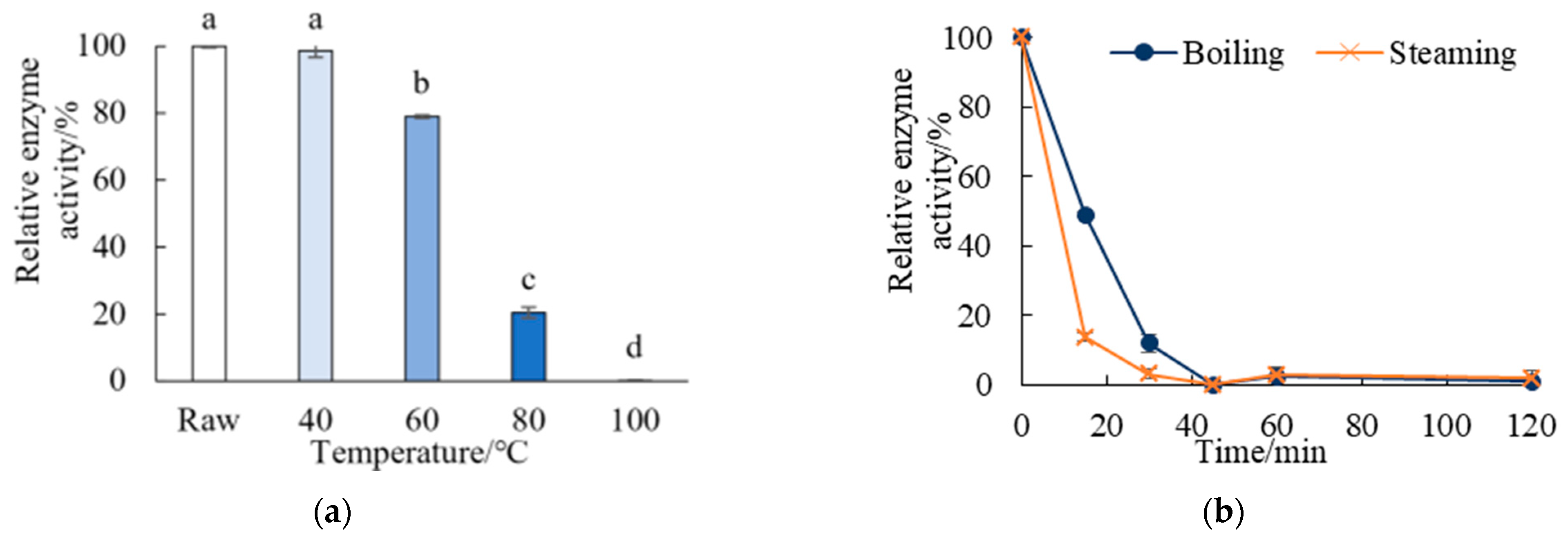
3.5. Thermal Inactivation Kinetics of Autolysis Enzymes

Figure 6 depicts the variations in relative enzyme activity (REA) caused by various heating temperatures and durations. The enzymes activity was significantly affected by the temperature and time. The autolysis enzymes were relatively stable at 40 °C. Similar findings were confirmed by Zeng et al. [16], who found that the autolysis enzyme of sea cucumber was even activated at 40 °C and proposed the processing temperature should be controlled below 10 °C or higher than 60 °C for enzyme inactivation. The REA was only decreased to 79.0 ± 0.4% when blanched at 60 °C. It is not congruent with the research on Holothurians [15], of which 46% REA was denatured at 40–60 °C for 10 min and the enzyme was almost fully inactivated after heating at 60 °C for 55 min [15], and the alkaline protease could be inactivated after heating at 80 °C for 1 h [13]. Even though 58.5% of REA were inactivated in the temperature range of 60–80 °C, the heat-resistant enzymes survived up to a temperature of 100 °C, and only when the cooking time was more than 45 min by boiling or 30 min by steaming could the REA reduce to below 10%, which was reported to be safe for storage as well as avoiding overheating [49]. The autolysis enzymes of C. frondosa are more thermally stable than previously reported species, which means the common processing methods of sea cucumber are not applicable for C. frondosa from an enzyme inactivation point of view.

3.6. Multivariate Analysis

In order to further clarify the relationship between the analyzed physicochemical properties of the C. frondosa and to improve its processing characteristics, a principal component analysis (PCA) of all variables was performed (Figure 7). The cumulative contribution of the first two principal components accounted for 84.4%, meaning that they explained 84.4% of the variation effects between the thermal treatments.
The body wall of the raw C. frondosa had high mechanical strength (hardness). During the blanching process, 60 °C was a key temperature for the physicochemical quality of C. frondosa. When the blanching temperature was below 60 °C, the water content and autolysis enzymes activity were comparable to that of the raw sea cucumber. When above 60 °C, the protein denatured but crude protein content increased because of drip loss. It resulted in the increase in chewiness, and the decrease in hardness, hence improving the palatability of C. frondosa. However, since the enzyme was not fully inactivated, the blanched sea cucumber was not stable for long-term preservation because the enzyme not fully inactivated.
When boiled and steamed at 100 °C, the collagen fibrils of the C. frondosa were fractured and dissolved, resulting in the formation of TCA-soluble nitrogen along with heating. However, the texture was further improved in consideration of the increased springiness and decreased hardness with increased heating duration. The autolysis enzymes were fully inactivated at 100 °C, considering that the cooking yield was reduced during long-time cooking, 30–45 min was sufficient for enzyme deactivation and benefitted texture enhancement. Additionally, the cooking yield, protein content, and moisture content of the boiled C. frondosa were greater than that of the steamed samples when heated for the same duration. However, the steamed sea cucumber was superior in terms of palatability because of the lower hardness and chewiness.

4. Conclusions

The study demonstrated that the C. frondosa’s enzyme activity and protein denaturation were significantly affected by various heating treatments, which, in turn, influenced the cooking yield, water holding capacity, and texture quality.
The autolysis enzyme of C. frondosa was thermally stable, which cannot be denatured completely by blanching. High temperature may deactivate enzymes but also leads to rapid product quality deterioration in WHC and protein content. Taking the physicochemical quality and enzyme inactivation into consideration, boiling at 100 °C for 45 min was more suitable for pre-treatment of subsequent processing (such as salting, drying, or freezing), with cooking yield of 70.3% and protein content of 78.5%. Steaming at 100 °C for at least 30 min is preferable for long-term preservation and instant food, in which the relative activity was only 3.2% with better palatability.

Author Contributions

Conceptualization, Q.Z. and T.T.; methodology, Q.Z. and M.G. (Margrét Geirsdóttir); software, Q.Z.; resources, M.G. (Margrét Geirsdóttir); writing—original draft preparation, Q.Z., R.L., M.G. (Margrét Geirsdóttir) and M.G. (María Gudjónsdóttir); writing—review and editing, S.L., S.X., M.G. (María Gudjónsdóttir) and X.L.; supervision, M.G. (María Gudjónsdóttir) and X.L.; project administration, T.T. and X.L. All authors have read and agreed to the published version of the manuscript.

Funding

This work was supported by the Fisheries Training Programme of United Nations University (UNU-FTP2017), Research and Demonstration of Efficient Clean Production Mode of Important Marine Fish in Liaoning Province (2020JH1/10200002), National Key R&D Program of China (2019YFD0901800), Project of Education Department of Liaoning Province (JL202011), Project of Ocean and Fisheries Department of Liaoning Province (201722), Key Laboratory of Environment Controlled Aquaculture (Dalian Ocean University) Ministry of Education (202203); National Key R&D Program of China (2020YFD0900600).

Institutional Review Board Statement

Not applicable.

Data Availability Statement

The article contains all the data necessary to substantiate the conclusions of this research.

Acknowledgments

The authors would like to express their heartfelt gratitude to United Nations University Fisheries Training Programme (now GRÓ Fisheries Training Programme, United Nations Education, Scientific and Cultural Organization) and Matis for supporting this research. Special thanks go to Tumi Tomasson, Thor Asgeirsson, Mary Frances Davidson, Julie Ingham, Stefan Ulfarsson, and Lilja Bjork Jonsdottir, for their enthusiastic guidance and assistance during the experiment.

Conflicts of Interest

The authors declare no conflict of interest.

References

  1. Zhu, Q.; Lin, L.; Zhao, M. Sulfated fucan/fucosylated chondroitin sulfate-dominated polysaccharide fraction from low-edible-value sea cucumber ameliorates type 2 diabetes in rats: New prospects for sea cucumber polysaccharide based-hypoglycemic functional food. Int. J. Biol. Macromol. 2020, 159, 34–45. [Google Scholar] [CrossRef] [PubMed]
  2. Gao, X.; Zhang, Z.; Liu, L.; Liu, Q. Rheological changes of sea cucumber Stichipus japonicus during different heated times. Int. J. Fish. Aquac. 2011, 2, 258–262. [Google Scholar]
  3. Mizuta, S.; Koizumi, Y.; Yokoyama, Y.; Yoshinaka, R. Purification and immunochemical detection of a quantitatively major collagen in the dermis of sea cucumber Apostichopus japonicus. Chem. Biochem. 2022, 88, 173–180. [Google Scholar] [CrossRef]
  4. David, F.; Hubas, C.; Laguerre, H.; Badou, A.; Herault, G.; Bordelet, T.; Ameziane, N. Food sources, digestive efficiency and resource allocation in the sea cucumber Holothuria forskali (Echinodermata: Holothuroidea): Insights from pigments and fatty acids. Aquac. Nutr. 2019, 26, 1568–1583. [Google Scholar] [CrossRef]
  5. Montero, N.; Marras, B.; Nurchis, P.; Bettoschi, A.; Nurchis, P.; Coroneo, V.; Sanna, C.; Schintu, M. Trace metal levels in the edible tissues of sea cucumbers (Holothuria tubulosaand Holothuria polii) from Sardinia (Western Mediterranean). Ital. J. Food Saf. 2021, 10, 9576. [Google Scholar]
  6. Fisheries, B.O.; Agriculture, M.O. China Fishery Statistics Yearbook; China Agriculture Press: Beijing, China, 2021. [Google Scholar]
  7. Song, S.; Peng, H.; Wang, Q.; Liu, Z.; Dong, X.; Wen, C.; Ai, C.; Zhang, Y.; Wang, Z.; Zhu, B. Inhibitory activities of marine sulfated polysaccharides against SARS-CoV-2. Food Funct. 2020, 9, 7415–7420. [Google Scholar] [CrossRef]
  8. Mamelona, J.; Pelletier, E.M.; Girard-Lalancette, K.; Legault, J.; Karboune, S.; Kermasha, S. Quantification of phenolic contents and antioxidant capacity of Atlantic sea cucumber, Cucumaria frondosa. Food Chem. 2007, 104, 1040–1047. [Google Scholar] [CrossRef]
  9. Song, Z.; Li, H.; Wen, J.; Zeng, Y.; Ye, X.; Zhao, W.; Xu, T.; Xu, N.; Zhang, D. Consumers’ attention on identification, nutritional compounds, and safety in heavy metals of Canadian sea cucumber in Chinese food market. Food Sci. Nutr. 2020, 8, 5962–5975. [Google Scholar] [CrossRef]
  10. Hu, S.; Xu, L.; Shi, D.; Wang, J.; Wang, Y.; Lou, Q.; Xue, C. Eicosapentaenoic acid-enriched phosphatidylcholine isolated from Cucumaria frondosa exhibits anti-hyperglycemic effects via activating phosphoinositide 3-kinase-protein kinase B signal pathway. J. Biosci. Bioeng. 2014, 117, 457–463. [Google Scholar] [CrossRef]
  11. Nelson, E.J.; Macdonald, B.A.; Robinson, S.M.C. A Review of the Northern Sea Cucumber Cucumaria frondosa (Gunnerus, 1767) as a Potential Aquaculture Species. Rev. Fish. Sci. 2012, 20, 212–219. [Google Scholar] [CrossRef]
  12. Hossain, A.; Dave, D.; Shahidi, A.F. Northern Sea Cucumber (Cucumaria frondosa): A Potential Candidate for Functional Food, Nutraceutical, and Pharmaceutical Sector. Mar. Drugs 2020, 18, 274. [Google Scholar] [CrossRef] [PubMed]
  13. Dong, J. Study on Quality and Nutrient Composition of Sea Cucumber during Thermal Processing; Ocean University of China: Qingdao, China, 2015. [Google Scholar]
  14. Hernández-Sámano, A.C.; Guzmán-García, X.; García-Barrientos, R.; Ascencio-Valle, F.; Sierra-Beltrán, A.; Vallejo-Córdoba, B.; González-Córdova, A.F.; Torres-Llanez, M.J.; Guerrero-Legarreta, I. Extraction and characterization of Sea Cucumber Isostichopus fuscus Proteases, Collected at the Gulf of California, Mexico. Rev. Mex. Ing. Química. 2015, 14, 35–47. [Google Scholar]
  15. Zhu, B.; Han, B. Characterization and Purification of Holothurians Autoenzyme. Food Ferment. Ind. 2004, 30, 132–137. [Google Scholar]
  16. Zeng, S.; Zheng, M.; Chen, L.; Guo, Z.; Zheng, B. Inactivation Kinetics Model of Sea Cucunber Processed by High Pressure and the Storage Quality of Soft Canned Sea Cucumber. J. Chin. Inst. Food Sci. Technol. 2017, 17, 173–180. [Google Scholar]
  17. Chen, T.; Peng, Z.; Lu, J.; Li, B.; Hou, H. Self-degradation of Sea Cucumber body wall under 4C storage condition. J. Food Process. Preserv. 2015, 40, 715–723. [Google Scholar] [CrossRef]
  18. Dong, X.; Li, Y.; Li, Y.; Song, L.; Cheng, S.; Li, D.; Zhu, B.; Zhou, D.; Tan, M. Combination of NMR and MRI Techniques for Non-invasive Assessment of Sea Cucumber (Stichopus japonicas) Tenderization During Low-Temperature Heating Process. Food Anal. Methods 2017, 10, 2207–2216. [Google Scholar] [CrossRef]
  19. Stefaniak-Vidarsson, M.M.; Varsha AKaleb, M.G.; Marteinsdottir, G.; Fridjonsson, O.; Hreggvidsson, G.O.; Sigurjonsson, O.E.; Omarsdottir, S.; Kristbergsson, K. Bioactive effect of sulphated polysaccharides derived from orange-footed sea cucumber (Cucumaria frondosa) toward THP-1 macrophage. Bioact. Carbohydr. Diet. Fibre 2017, 12, 14–19. [Google Scholar] [CrossRef]
  20. Duan, X.; Wang, H.; Ren, G.; Zhu, K. Research progress of dry-cure technology of sea cucumber. Sci. Technol. Food Ind. 2012, 33, 427–431. [Google Scholar]
  21. Zhang, L.; Huang, X.; Miao, S.; Zeng, S.; Zhang, Y.; Zheng, B. Influence of ultrasound on the rehydration of dried sea cucumber (Stichopus japonicus). J. Food Eng. 2016, 178, 203–211. [Google Scholar] [CrossRef]
  22. Qi, H.; Fu, H.; Dong, X.; Feng, D.; Li, N.; Wen, C.; Nakamura, Y.; Zhu, B. Apoptosis induction is involved in UVA-induced autolysis in sea cucumber Stichopus japonicus. J. Photochem. Photobiol. B Biol. 2016, 158, 130–135. [Google Scholar] [CrossRef]
  23. Bi, J.; Li, Y.; Cheng, S.; Dong, X.; Kamal, T.; Zhou, D.; Li, D.; Jiang, P.; Zhu, B.-W.; Tan, M. Changes in Body Wall of Sea Cucumber during a two-step heating process assessed by Rheology, LF-NMR, and Texture Profile Analysis. Food Biophys. 2016, 11, 257–265. [Google Scholar] [CrossRef]
  24. Xia, Y.; Liu, Z.; Deng, J.; Liu, X.; Li, N.; Chen, S.; Li, Z. Effects of High Hydrostatic Pressure Treatment on Holothurians Autoenzyme Activity. Chin. J. High Press. Phys. 2009, 23, 377–383. [Google Scholar]
  25. Crapo, C.A.; Crawford, D.L. Influence of Polyphosphate Soak and Cooking Procedures on Yield and Quality of Dungeness Crab Meat. J. Food Sci. 1991, 56, 657–659. [Google Scholar] [CrossRef]
  26. Heerden, S.M.V.; Strydom, P.E. Nutrient retention values and cooking yield factors for three South African lamb and mutton cuts. J. Sci. Food Agric. 2017, 97, 5037–5042. [Google Scholar] [CrossRef] [PubMed]
  27. Lewis, P.N.; Pinali, C.; Young, R.D.; Meek, K.M.; Quantock, A.J.; Knupp, C. Structural Interactions between Collagen and Proteoglycans Are Elucidated by Three-Dimensional Electron Tomography of Bovine Cornea. Structure 2010, 18, 239–245. [Google Scholar] [CrossRef] [PubMed] [Green Version]
  28. Xue, D. Study on Physical Properties and Processing Technology of Sea Cucumber; Ocean University of China: Qingdao, China, 2006. [Google Scholar]
  29. Parkin, K.; Damodaran, S. Amino Acids, Peptides, and Proteins; CRC Press: New York, NY, USA, 2017; pp. 272–273. [Google Scholar]
  30. Zhao, Y.; Xue, Y.; Dong, J.; Li, Z.; Wang, Y.; Xue, C. Effect of Two Plant Extracts on the Stability of the Body Wall Collagen in Instant Sea Cucumber. Mod. Food Sci. Technol. 2015, 31, 113–119. [Google Scholar]
  31. Wu, F.; Xue, Y.; Liu, X.; Xue, C.; Wang, J.; Du, L.; Koretaro, T.; Wang, Y. The protective effect of eicosapentaenoic acid-enriched phospholipids from sea cucumber Cucumaria frondosa on oxidative stress in PC12 cells and SAMP8 mice. Neurochem. Int. 2014, 64, 9–17. [Google Scholar] [CrossRef] [Green Version]
  32. Liu, S.; Junpeng, Z.; Wei, Z.; Hong, Y.; Lan, W. Effects of Extraction Methods on the Composition and Functional Properties of Fish Proteins. Mod. Agric. Equip. 2019, 40, 64–71. [Google Scholar]
  33. Hou, H.; Chen, T.; Peng, Z.; Zhang, Z.; Xue, C.; Li, B. Stability and degradation regulation of body wall gel of sea cucumber treated by high hydrostatic pressure. Trans. Chin. Soc. Agric. Eng. 2014, 30, 316–322. [Google Scholar]
  34. Cui, F.; Xue, C.; Li, Z.; Zhang, Y.; Dong, P.; Fu, X.; Gao, X. Characterization and subunit composition of collagen from the body wall of sea cucumber Stichopus japonicus. Food Chem. 2007, 100, 1120–1125. [Google Scholar] [CrossRef]
  35. Saito, M.; Kunisaki, N.; Urano, N.; KIMURA, S. Collagen as the Major Edible Component of Sea Cucumber (Stichopus japonicus). J. Food Sci. 2002, 67, 1319–1322. [Google Scholar] [CrossRef]
  36. Liu, Z.; Oliveira, A.C.M.; Su, Y. Purification and Characterization of Pepsin-Solubilized Collagen from Skin and Connective Tissue of Giant Red Sea Cucumber (Parastichopus californicus). J. Agric. Food Chem. 2010, 58, 1270–1274. [Google Scholar] [CrossRef] [PubMed]
  37. Zhong, M.; Chen, T.; Hu, C.; Ren, C. Isolation and Characterization of Collagen from the Body Wall of Sea Cucumber Stichopus monotuberculatus. J. Food Sci. 2015, 80, 671–679. [Google Scholar] [CrossRef] [PubMed]
  38. Trotter, J.A.; Lyons-Levy, G.; Thurmond, F.A.; Koobt, T.J. Covalent composition of collagen fibrils from the dermis of the sea cucumber, Cucumaria frondosa, a tissue with mutable mechanical properties. Camp. Biochem. Physiol. 1995, 112A, 463–478. [Google Scholar] [CrossRef]
  39. Cluzel, C.; Lethias, C.; Garrone, R.; Exposito, J.-Y. Sea urchin fibrillar collagen 2α chain participates in heterotrimeric molecules of (1α)22α stoichiometry. Matrix Biol. 2000, 19, 545–547. [Google Scholar] [CrossRef]
  40. Morales, J.; Montero, P.; Moral, A. Isolation and partial characterization of two types of muscle collagen in some cephalopods. J. Agric. Food Chem. 2000, 48, 2142–2148. [Google Scholar] [CrossRef] [Green Version]
  41. Zhang, W.; Zheng, J.; Tian, X.; Tang, Y.; Ding, G.; Yang, Z.; Jin, H. Pepsin-soluble collagen from the skin of Lophius litulo a preliminary study evaluating physicochemical, antioxidant, and wound healing properties. Mar. Drugs 2019, 17, 708. [Google Scholar] [CrossRef] [Green Version]
  42. Tian, M.; Xue, C.; Chang, Y.; Shen, J.; Zhang, Y.; Li, Z.; Wang, Y. Collagen fibrils of sea cucumber (Apostichopus japonicus) are heterotypic. Food Chem. 2020, 316, 126272. [Google Scholar] [CrossRef]
  43. Chen, F.-C.; Hsieh, Y.-H.P. Porcine troponin I: A thermostable species marker protei. Meat Sci. 2002, 61, 55–60. [Google Scholar] [CrossRef]
  44. Yan, B.; Jiao, X.; Zhu, H.; Wang, Q.; Huang, J.; Zhao, J.; Cao, H.; Zhou, W.; Zhang, W.; Ye, W.; et al. Chemical interactions involved in microwave heat-induced surimi gel fortified with fish oil and its formation mechanism. Food Hydrocoll. 2020, 106, 105779. [Google Scholar] [CrossRef]
  45. Wang, Y.; Wang, J.; Zhao, Y.; Hu, S.; Shi, D.; Xue, C. Fucoidan from sea cucumber Cucumaria frondosa exhibits anti-hyperglycemic effects in insulin resistant mice via activating the PI3K-PKB pathway and GLUT4. J. Biosci. Bioeng. 2016, 121, 36–42. [Google Scholar] [CrossRef] [PubMed]
  46. Liu, Z.; Zhou, D.; Liu, Y.; Yu, M.; Liu, B.; Song, L.; Dong, X.; Qi, H.; Shahidi, F. Inhibitory effect of natural metal ion chelators on the autolysis of sea cucumber (Stichopus japonicus) and its mechanism. Food Res. Int. 2020, 133, 109205. [Google Scholar] [CrossRef] [PubMed]
  47. Gao, F.; Xu, Q.; Yang, H. Seasonal biochemical changes in composition of body wall tissues of sea cucumber Apostichopus japonicus. Chin. J. Oceanol. Limnol. 2011, 29, 252–260. [Google Scholar] [CrossRef]
  48. Zhang, K.; Hou, H.; Bu, L.; Li, B.; Xue, C.; Peng, Z.; Su, S. Effects of heat treatment on the gel properties of the body wall of sea cucumber (Apostichopus japonicus). J. Food Sci. Technol. 2017, 54, 707–717. [Google Scholar] [CrossRef] [Green Version]
  49. Almeida, G.M.D.; Mendonça, C.M.N.O.; Converti, A.; Oliveira, R.P.d.S. Kinetic and thermodynamic parameters of nisin thermoinactivation. J. Food Eng. 2020, 280, 109986. [Google Scholar]
Figure 1. Cooking yield of C. frondosa after various heat treatments, (a) comparison of cooking yield during cooking for 45 min at different temperatures (40, 60, 80, and 100 °C), and (b) comparison of cooking yield after boiling and steaming at 100 °C for 0–120 min. Different letters (a, b, c, d) indicate significant differences between groups as determined via one-way ANOVA (p < 0.05).
Processes 10 00847 g001
Figure 2. Water content and WHC of C. frondosa as affected by various heat treatments, (a) comparison of water content (a) and WHC (c) during cooking for 45 min at different temperatures (40, 60, 80, and 100 °C), and comparison of water content (b) and WHC (d) after boiling and steaming at 100 °C for 0–120 min. Different letters (a, b, c, d) indicate significant differences between groups as determined via one-way ANOVA (p < 0.05).
Processes 10 00847 g002
Figure 3. Content of crude protein and TCA-soluble nitrogen of C. frondosa after various heat treatments, comparison of crude protein on dry basis (a) crude protein on wet basis (c) and TCA-soluble nitrogen (e) during cooking for 45 min at different temperatures (40, 60, 80, and 100 °C), and comparison of crude protein on dry basis (b) crude protein on wet basis (d) and TCA-soluble nitrogen (f) after boiling and steaming at 100 °C for 0–120 min. Different letters (a, b, c, d, e) indicate significant differences between groups as determined via one-way ANOVA (p < 0.05).
Processes 10 00847 g003
Figure 4. SDS-PAGE analysis of blanched and boiled samples (a) and steamed samples (b). BL samples correspond to the cooking liquid of the boiled sea cucumber samples (B), respectively. SL samples represent the steaming liquid from 0–15 min, 15–30 min, 30–45 min, 45–60 min, 60–120 min, 0–60 min, 0–120 min, respectively, of the steamed sea cucumber samples (S).
Processes 10 00847 g004
Figure 5. Dermis of C. frondosa ((IIII) represent the raw, boiled, and steamed samples on the ventral side, (IVVI) represent them on the dorsal side).
Processes 10 00847 g005
Figure 6. Relative enzyme activity of C. frondosa after various heat treatments, (a) comparison of relative enzyme activity during cooking for 45 min at different temperatures (40, 60, 80, and 100 °C), and (b) comparison of relative enzyme activity after boiling and steaming at 100 °C for 0–120 min. Different letters (a, b, c, d) indicate significant differences between groups as determined via one-way ANOVA (p < 0.05).
Processes 10 00847 g006
Figure 7. Principal component PC1 and PC2 scores plot (a) and component plot in rotated space from the principal component analysis of C. frondosa samples (b) assessing the effects of blanching, boiling, and steaming at different temperatures and cooking durations. Raw is sample without heating treatment.
Processes 10 00847 g007
Table 1. Texture of C. frondosa after various heating treatments. Different letters (a, b, c, d, e, f, g, h) indicate significant differences between groups as determined via one-way ANOVA (p < 0.05).
Group AbbreviationGroup DescriptionHardness/gAdhesiveness/g.sSpringinessCohesivenessChewiness/gResilience
RawRaw8895.19 ± 501.37 b−331.97 ± 10.97 d0.73 ± 0.05 d0.33 ± 0.03 e1679.57 ± 295.47 g0.13 ± 0.02 g
B40 °CBlanch 40 °C 45 min12,532.65 ± 288.70 a−668.44 ± 37.01 e0.54 ± 0.05 e0.28 ± 0.04 f2020.45 ± 249.92 f g0.15 ± 0.03 g
B60 °CBlanch 60 °C 45 min7416.39 ± 612.14 c−143.01 ± 68.51 c0.80 ± 0.02 c0.67 ± 0.04 d4453.01 ± 576.48 a0.36 ± 0.01 f
B80 °CBlanch 80 °C 45 min5955.04 ± 423.38 d−30.88 ± 7.38 a0.86 ± 0.02 a, b0.78 ± 0.01 b c3995.48 ± 930.18 a, b0.44 ± 0.01 e
B15Boil 100 °C 15 min6504.57 ± 405.68 d−41.72 ± 14.55 a, b0.84 ± 0.01 b c0.81 ± 0.01 a, b4176.96 ± 300.27 a0.47 ± 0.01 d
B30Boil 100 °C 30 min6394.92 ± 283.05 d−27.89 ± 10.46 a0.89 ± 0.02 a, b0.82 ± 0.02 a4030.32 ± 665.58 a, b0.50 ± 0.02 b, c
B45Boil 100 °C 45 min4958.39 ± 627.12 e, f−28.77 ± 12.12 a0.90 ± 0.03 a0.83 ± 0.01 a3301.81 ± 465.00 c0.51 ± 0.01 b
B60Boil 100 °C 60 min4542.24 ± 426.70 f−37.07 ± 6.38 a0.89 ± 0.01 a, b0.82 ± 0.01 a3024.04 ± 608.18 c, d0.53 ± 0.01 a, b
B120Boil 100 °C 120 min3693.17 ± 558.31 g−30.23 ± 8.47 a0.84 ± 0.06 b, c0.81 ± 0.03 a, b2788.71 ± 566.11 c, d, e0.55 ± 0.01 a
S15Steam 100 °C 15 min5162.88 ± 199.91 e−72.37 ± 8.58 b0.86 ± 0.03 a, b0.77 ± 0.02 c3415.58 ± 242.77 b, c0.43 ± 0.01 e
S30Steam 100 °C 30 min4739.95 ± 450.27 e, f−51.52 ± 12.91 a, b0.89 ± 0.03 a b0.81 ± 0.03 a b3398.32 ± 114.26 b, c0.48 ± 0.04 c, d
S45Steam 100 °C 45 min3459.53 ± 430.16 g, h−44.11 ± 5.17 a b0.88 ± 0.06 a, b0.81 ± 0.03 a, b2654.81 ± 254.46 d, e, f0.47 ± 0.03 d
S60Steam 100 °C 60 min3443.25 ± 215.68 g, h−46.29 ± 11.27 a b0.88 ± 0.04 a, b0.81 ± 0.01 a2514.34 ± 262.32 d, e, f0.48 ± 0.03 c d
S120Steam 100 °C 120 min3090.11 ± 256.76 h−20.27 ± 3.43 a0.89 ± 0.02 a, b0.82 ± 0.01 a2260.60 ± 238.41 e, f, g0.52 ± 0.01 b
Publisher’s Note: MDPI stays neutral with regard to jurisdictional claims in published maps and institutional affiliations.

