Article

Electrospun SF/PLGA/ICG Composite Nanofibrous Membranes for Potential Wound Healing and Tumor Therapy

1
State Key Laboratory of Silkworm Genome Biology, Southwest University, Chongqing 400715, China
2
Chongqing Engineering Research Center of Biomaterial Fiber and Modern Textile, College of Sericulture, Textile and Biomass Sciences, Southwest University, Chongqing 400715, China
3
Key Laboratory of Sericultural Biology and Genetic Breeding, Ministry of Agriculture and Rural Affairs, Southwest University, Chongqing 400715, China
*
Authors to whom correspondence should be addressed.
Processes 2022, 10(5), 850; https://doi.org/10.3390/pr10050850
Submission received: 16 March 2022 / Revised: 18 April 2022 / Accepted: 22 April 2022 / Published: 25 April 2022

Abstract

:
Indocyanine green (ICG) is a near-infrared (NIR) organic reagent for clinical bioimaging and phototherapy. It is a suitable photosensitizer for photodynamic antimicrobial chemotherapy (PACT). In this study, various ICG-loaded nanofibrous membranes were prepared. The water vapor transmission rate (WVTR) of SF/PLGA/20ICG was 3040.49 ± 157.11 g·m−2 day−1, which allowed the maintenance of a humid environment above the wound. The growth inhibition rates for S. aureus and E. coli were 91.53% and 87.95%, respectively. The nanofibrous membranes exhibited excellent antimicrobial performance. Cellular experiments showed that the nanofibrous membranes have good cytocompatibility and antitumor efficacy. SF/PLGA/20ICG showed good potential for application in wound healing and cancer therapy.

1. Introduction

Photodynamic antimicrobial chemotherapy (PACT) has received increasing attention because of its unequaled characteristics, such as low systemic toxicity, local application, and minimal invasiveness [1,2]. Under appropriate wavelengths of light, cancer cells and bacteria are killed by reactive oxygen species (ROS) and free radicals produced by photosensitizers under aerobic conditions [3]. As a key element of PACT, several photosensitizers have been reported in recent decades, including conjugated polyelectrolytes, porphyrin, phenothiazinium dyes, heme, cationic functionalized fullerene, phthalocyanine, methylene blue, as well as nanoparticles [4,5,6]. ICG is a near-infrared (NIR) organic reagent for clinical bioimaging and phototherapy [7] authorized by the U.S. Food and Drug Administration (FDA). ICG with intense NIR-absorbing and fluorescence properties is an ideal photosensitizer for PACT due to its excellent photothermal conversion performance and its low toxicity [8]. Gholibegloo et al. [9] synthesized various ICG-loaded nanocomposites including graphene oxide (GO), GO–carnosine (Car), and GO–Car/Hydroxyapatite (HAp). The inhibition rates of GO@ICG, GO–Car@ICG, GO–Car/HAp@ICG of S. mutans biofilm formation were 51.4%, 63.8%, and 56.8%, respectively. The characteristics of ICG endow these nanocomposites with excellent antimicrobial properties based on the generation of ROS.
However, the use of ICG is still limited in the clinic because it can easily bind non-specifically to plasma proteins, which causes its rapid body clearance (plasma half-life of 2–4 min) and marked in vivo instability, with lack of target specificity [10]. ICG is often loaded on nanoparticles, hydrogels, and nanofibrous membranes in the medical field. Nanofibrous membranes are currently used in tissue engineering, drug sustained-release systems, wastewater treatment, and other fields [11]. Nanofibers mimic the physical dimensions of the natural extracellular matrix (ECM) and can also release drugs in a controlled manner depending on various parameters, their morphology, polymer–drug interactions, their degradation, and chemical properties [12,13,14,15,16]. Preis et al. [17] used the needle-free electrospinning technique to produce an ICG-loaded poly(D, L-lactide) nanofiber web as a photoresponsive wound dressing. Antimicrobial tests were carried out on several bacteria, and this nanofibrous mesh showed excellent antimicrobial properties. ICG-loaded nanofibers represent a promising and innovative wound dressing for chronic wounds associated with skin infections.
Poly(lactic-co-glycolic acid) (PLGA) is a biodegradable amorphous polymer composed of lactic acid (LA) and glycolic acid (GA) [18]. PLGA is widely used in nanoparticles, hydrogels, electrospinning membranes, and other biomaterials [19]. PLGA nanocarriers are commonly used carriers for ICG. Saxena et al. [20] developed ICG-supported PLGA nanospheres using an emulsion solvent diffusion method and observed that increasing the amount of polymer resulted in high encapsulation efficiency of ICG. The results showed that the water stability, photostability, and thermal stability of the encapsulated dyes were significantly improved compared to those of free ICG.
As a material for nanofiber membranes, silk fibroin (SF) is a natural polymer material extracted from silk with unique mechanical properties and biological properties. SF-based biomaterials are highly biocompatible and capable of controlled drug release both in vitro and in vivo [21]. During the extraction process, the structure of SF is vulnerable to destruction. Therefore, nanofibrous membranes prepared from pure SF usually have poor mechanical properties and lack the necessary flexibility and elasticity [22,23]. The introduction of PLGA can increase the flexibility and elasticity necessary for nanofiber membranes. PLGA can meet the need for good mechanical properties. Peng et al. [24] designed an SF/PLGA nanofibrous membrane loaded with artemisinin (ART). Composite membranes prepared by using synthetic and natural polymers can compensate the disadvantages of the single polymers and have a better performance. Nanofibrous membranes have a lot of attractive features. Due to their large specific volume surface, high drug loading is achievable. They are easy to produce and cost-effective and can be mass-produced. In this work, SF and PLGA were used to prepare nanofibers by electrospinning. As a photosensitizer, ICG was loaded into the nanofibers. ICG was released controllably depending on different concentrations of SF and PLGA. This system can be implanted or overlaid on the margins of surgical resection at cancer sites or in solid tumors, enabling localized drug delivery. This approach can avoid the use of complex physicochemical processes to specifically target cancer cells, which is unavoidable for nanoparticle-based drug delivery systems. The preparation and application of nanofiber membrane is shown in Scheme 1. The prepared nanofibers showed excellent antimicrobial performance, in vitro anti-tumor efficacy, and cytocompatibility. These nanofibers have potential applications in wound healing and cancer therapy.

2. Materials and Methods

2.1. Materials

Silkworm cocoons were obtained from the State Key Laboratory of Silkworm Genome Biology, Southwest University. PLGA, average molecular weight 1.1 × 10⁵, was from Daigang Biotechnology Co., Ltd. (Shandong, China). Hexafluoroisopropanol (HFIP) was provided by Aladdin (Shanghai, China). CaCl2 and C2H5OH were supplied by Chuandong Chemical Co., Ltd. (Chongqing, China). L929 cells were obtained from the State Key Laboratory of Silkworm Genome Biology, Southwest University. Bacteria (S. aureus and E. coli) and HepG2 cells were obtained from the College of Pharmacy, Southwest University.

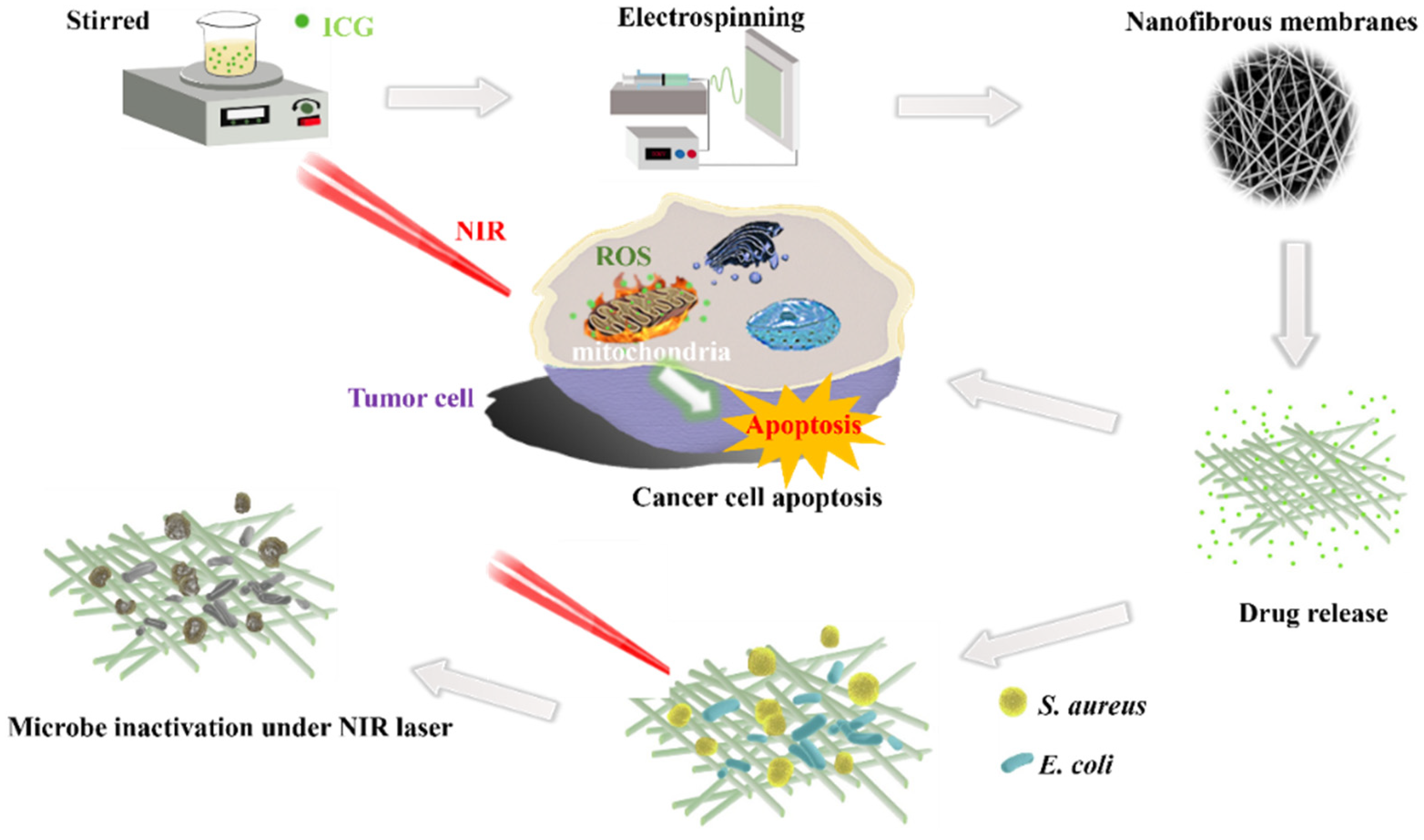
2.2. Preparation of Nanofibrous Membranes Loaded with ICG

The cocoons were degummed with a Na2CO3 solution and then dissolved in a ternary solution (nCaCl2:nH2O:nC2H5OH = 1:8:2). The solution from the previous step was dialyzed at 4 °C for 5 to 7 d. Water was removed by freeze-drying to produce SF. HFIP was used to dissolve different proportions of PLGA and SF, with stirring for several hours as a control group. The total mass fraction of PLGA and SF was 12%. Their proportions were 1:1, 1:3, and 1:5. To prepare three spinning liquids, we used HFIP to dissolve PLGA and SF and stirred them for several hours as a control group. After mixing with HFIP, we formulated the experimental group of SF/PLGA/ICG. The parameters of the nanofibrous membrane are shown in Table 1. The polymer solution was spun through an electrospinning machine (TL-Pro-BM, China) under high-pressure electrostatic force (−2–20 KV).

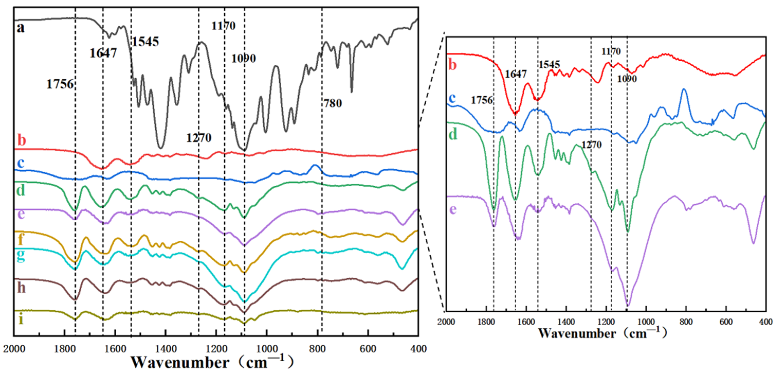
2.3. Characterization of the Nanofibrous Membranes

The nanofibrous membranes were characterized as follows. They were examined by a Scanning Electron Microscope (SEM, SU8020, Tokyo, Japan) for their morphology. The nanofibrous membranes were examined by Fourier Transmission infrared (FTIR, Bruker, Karlsruhe, Germany) to analyze their internal structure. They were fixed on the platform of water contact angle (WCA). The contact angle on the surface of the nanofiber membranes at different times was measured by a contact angle meter. The water vapor transmission rate of each group was tested using the standard ASTM E96. An amount of 3.0 g of CaCl2 was added to the sample bottle as an absorbent. The sample was then fixed in a CaCl2 vial with a diameter of 1.68 cm (denoted as D), and the total mass of the vial with the nanofiber sample fixed was weighed and indicated as M1. The vial was then placed in an incubator at constant temperature and humidity, i.e., at 50% humidity and 37 °C. The total mass of the vial was weighed after 24 h and denoted as M2. All tests were repeated three times (n = 3). The water vapor transmission rate of the nanofiber membranes was calculated by the following equation.
WVTR = 4 ( M 2   M 1 ) / ( π · D 2 ) × 100 %

2.4. Biodegradability of Nanofibrous Membranes

The biodegradability of the materials was tested using mass loss by impregnation in PBS (pH = 7.4, 0.1 M) at 37 °C. The membranes were cut into squares (4 cm × 4 cm), and the weighed mass was recorded as W0. An amount of 5 mL of PBS solution was added to a conical flask that was placed in a constant-temperature shaker set at 37 °C and 50 rpm. The PBS was renewed once a day, and the remaining dry mass of the material (Wt) was measured in a cycle of 2 weeks. The experiment was set up with three parallel controls (n = 3), and the biodegradation rate of the materials was calculated according to the following equation.
W r = W t / W 0 × 100 %

2.5. In Vitro Release of ICG

The sample was placed in a test tube with buffers and incubated at a speed of 120 r/min in a 37 °C incubator. After a certain period of time, the sample was removed and transferred to a new test tube, and the new buffers were added for sustained release. To collect the slow-release solution for each phase, this operation was repeated at regular intervals. A microplate reader was used to determine the drug content in the sustained-release solution.

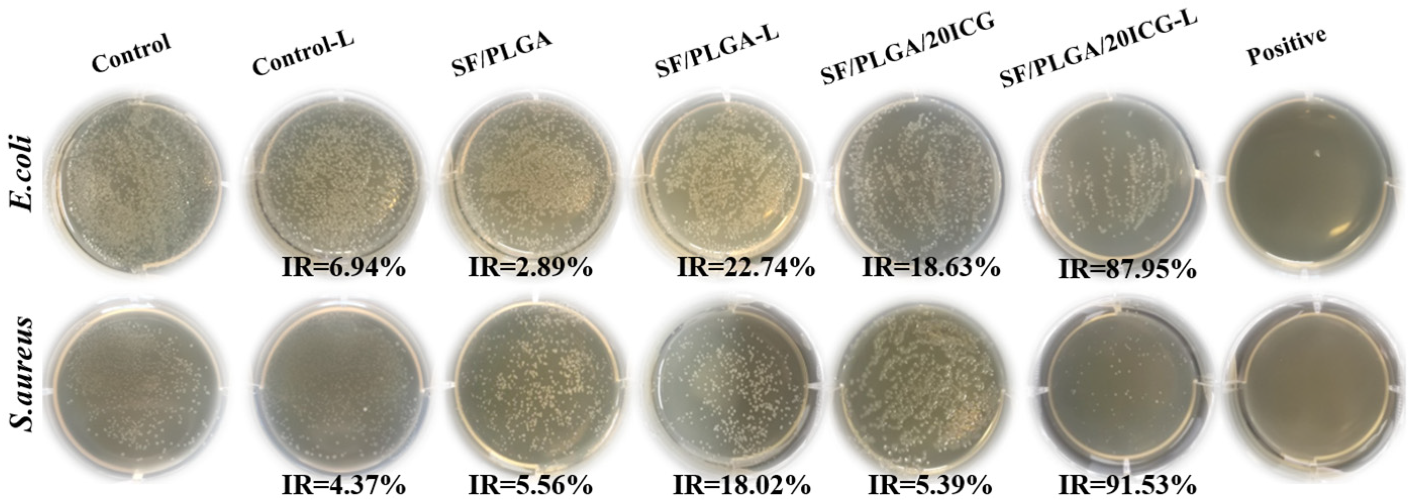
2.6. Antimicrobial Activity of the Nanofibrous Membranes

The antimicrobial effect was analyzed by the plate-counting method. E. coli and S. aureus were used to test the antimicrobial properties of the nanofibrous membranes. Firstly, a bacterial solution with a concentration of 108–109 CFU/mL was diluted to 1.5 × 10⁵–3.5 × 10⁵ CFU/mL by the four-step dilution method. After the sample was irradiated by ultraviolet light for 1 h to sterilize it, 1.5 g of sample was placed into a 250 mL conical flask. Then, 70 mL of PBS buffer and 5 mL of standard bacterial fluid were added to the conical flask at 37 °C and shaken for 18 h. Then, the bacterial solution was diluted with PBS buffer 100 times. After 24 h at 37 °C, the bacteria inhibition rate (IR) was calculated according to the following formula.
IR% = 100(C − X)/C
where C represents the number of bacteria (CFU) in the control group, and X represents the sample groups.

2.7. In Vitro Cytocompatibility of the Nanofibrous Membranes

The complete medium was Dulbecco’s modified Eagle medium (DMEM) with antibiotics and fetal bovine serum (FBS). The concentrations of the antibiotics (penicillin–streptomycin) and of FBS were 100 μg/mL and 10%, respectively. The nanofibrous membranes were trimmed into 0.6 cm-diameter wafers and sterilized by UV irradiation for 30 min. Mouse cells (L929) were cultured in complete medium. The cells were kept under aseptic conditions at 37 °C and 5% CO2. The nanofibrous membranes were placed in a 96-well plate. The cell suspension was added to each well at a concentration of 1 × 10⁴ cells/mL for 24 h, 48 h, and 72 h. The Cell Counting Kit-8 (CCK8) assay and cell staining assay were used to evaluate the cytocompatibility of the membranes. The medium was aspirated from each well. At this point, the well plate was added to the medium containing CCK8, which was 1/10 of the medium volume. Then, the 96-well plates were incubated in the incubator for 1 h. Finally, the well plates were removed and put into the microplate reader for detection, with the wavelength set to 450 nm.

2.8. In Vitro Anti-Tumor Efficacy

Tumor cell destruction induced by SF/PLGA/20ICG was evaluated using HepG2 cells. Initially, a cell suspension was added to each well at a concentration of 8 × 10³ cells/mL and incubated for 12 h. Afterward, the cells were incubated with fresh culture medium containing SF/PLGA/20ICG for 4 h, followed by irradiation using a NIR laser (808 nm, 0.6 W cm−2) for 20 min.
Then, the cells were further incubated for 24 and 48 h. Afterward, the cells were gently washed with PBS. The anti-tumor efficacy of the membranes was evaluated through the CCK8 assay and a cell staining assay. A Calcein/Propidium Iodide (PI) Cell Viability/Cytotoxicity Assay Kit was used for cell staining. The medium was aspirated from each well. A 100 µL volume of Calcein AM/PI solution was added to each well of the plate. Then, the plates were placed in the incubator for 30 min. After incubation, staining was observed under a confocal microscope (Calcein AM for green fluorescence, Excitation wavelength (Ex)/Emission wavelength (Em) = 494/517 nm; PI for red fluorescence, Ex/Em = 535/617 nm).

3. Results and Discussion

3.1. Characterization of the ICG-Loaded Nanofibrous Membranes

The morphology of the nanofibrous membranes was observed by SEM, and the fiber diameter distribution was analyzed by Nano Measurer Software, as shown in Figure 1. SEM observations revealed that the fiber structure of the six components was similar, and the fiber surface was smooth. There were no beads or crystals on the surface of the nanofibrous membranes. The fiber diameter distribution is presented in Figure 1 (Top). The average diameters for SF/PLGA, SF/PLGA3, and SF/PLGA5 were 437.8 ± 81.9 nm, 560.4 ± 124.2 nm, and 778.8 ± 198.0 nm, respectively. As the concentration of PLGA increased, the diameter of the nanofibrous membranes increased. At the same concentration of PLGA and SF, no significant differences between unloaded and ICG-loaded nanofibrous membranes were observed, because of the incorporation of ICG into the nanofibers [17].
The structural conformation of the nanofibrous membranes was examined by FTIR analysis (Figure 2). The visible peaks of C-O were at 1170 cm−1 and 1084 cm−1, and the peak of C=O was at 1756 cm−1; these are the characteristic peaks of PLGA [25]. All spectra showed these characterized PLGA peaks. Different secondary structures of silk fibroins are analyzed in the ranges of 1700–1600 cm−1 (amide I), 1600–1500 cm−1 (amide II), and 1350–1200 cm−1 (amide III) [26,27]. Peaks at 1647 cm−1 (amide I), 1545 cm−1 (amide II), and 1270 cm−1 (amide III) were observed in the FTIR spectra of unloaded and ICG-loaded nanofibrous membranes, which were assigned to random-coil structures [28]. The characteristic peak of ICG is at 780 cm−1. These data showed that there were no chemical interactions between each component and other components.
Good degradability is important for biomaterials. The biodegradability of the material was tested using mass loss by impregnation in PBS. SF/PLGA and SF/PLGA/20ICG showed the fastest degradation rates (Figure 3a). After 2 weeks, their residual weights were 72.16% and 70.83% of the original weights, respectively. SF/PLGA5 and SF/PLGA5/20ICG showed the slowest degradation rates. At week 8, the residual weights were still 70.28% and 74.89% of the original ones. The weights of the nanofibrous membranes continued to decrease as the time of immersion in PBS increased. This was attributed to the hydrolysis of the polymer. The main cause of weight loss was the breakage of the bonds of the polymer during hydrolysis, resulting in soluble oligomers or monomers. These soluble oligomers or monomers would further diffuse into the PBS solution, resulting in a reduction of the mass of the nanofibrous membranes. With the increase of SF content, the degradation rate of the nanofibrous membranes membrane increased (Figure 3). SF is a natural protein derived from silk, therefore has excellent degradation properties [29]. The Water Vapor Transmission Rate (WVTR) assesses the ability of a dressing to carry water away from a wound. The evaporative water loss [30] from injured skin generally ranges from 204 ± 12 to 5138 ± 202 g·m−2 day−1. A high WVTR value can cause rapid water loss, leading to the dehydration of the wound, possibly causing the dressing to stick to the broken skin. However, lower WVTR values can lead to fluid retention and back pressure that can damage the healthy tissue surrounding the wound [31]. Figure 3b shows that the WVTR of SF/PLGA/20ICG was 3040.49 ± 157.11 g·m−2 day−1, which would allow maintaining a humid environment above the wound.
The water wettability of the nanofibrous membranes was evaluated by an optical contact angle meter (Figure 3c). At the third second, the water contact angle of the SF/PLGA was 28.53°, while the water contact angle of the SF/PLGA/20ICG increased to 73.23°. This was because ICG is an amphiphilic molecule and is less hydrophilic than SF. This will increase the contact angle if ICG is uniformly distributed in the SF/PLGA composite. As the concentration of PLGA increased, the water contact angle of the nanofibrous membranes became larger. These results showed that SF/PLGA/20ICG membranes have good water vapor permeability and excellent hydrophilicity, which means that they can maintain permeability and moisture absorption.

3.2. In Vitro Release of ICG from the Nanofibrous Membranes

The release of drugs helps to ensure a lasting effect in wounds or cancer sites. The in vitro release profiles of ICG from SF/PLGA/20ICG, SF/PLGA3/20ICG, and SF/PLGA5/20ICG were examined via a Microplate Reader at specific time intervals. The time-dependent curves of ICG release are shown in Figure 2d. After 7 days, the cumulative release of ICG from SF/PLGA/20ICG, SF/PLGA3/20ICG, and SF/PLGA5/20ICG in PBS was 9.61, 15.90, and 54.97%, respectively. In contrast, the cumulative release of ICG from these three membranes was 7.11, 9.10, and 47.75% at pH 5.3, respectively. This interesting phenomenon was likely due to the low aqueous solubility of ICG in the acetic acid buffer. Moreover, the cumulative release of ICG from SF/PLGA5/20ICG was the lowest among the three groups. The hydrophilic and degradable properties of SF increased the release of the drug. Furthermore, the release rate decreased as the fiber diameter increased [32] (Figure 1). A large fiber diameter led to a low specific surface, thus the release rate was low.

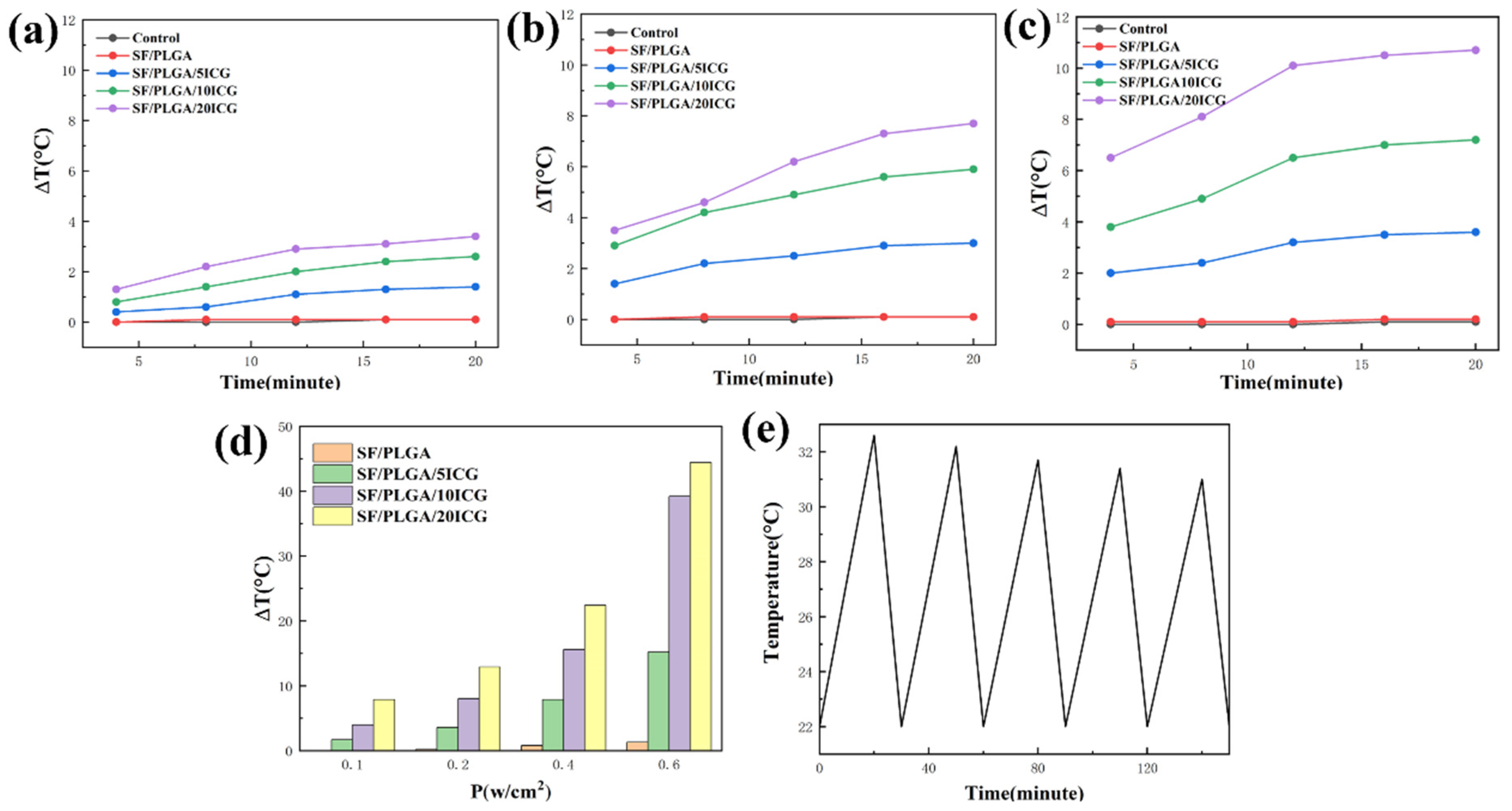
3.3. Photothermal Performance of the Nanofibrous Membranes

To demonstrate the photothermal capability of ICG-loaded nanofibrous membranes, the nanofibrous membranes were irradiated by a NIR laser, and the temperature variation was dynamically monitored with a thermal imaging camera. After NIR irradiation, (0.6 w/cm2), SF/PLGA/5ICG, SF/PLGA/10ICG, and SF/PLGA/20ICG showed a temperature increase of 3.6 °C, 7.2 °C, and 10.7 °C, respectively (Figure 4c). The temperature elevation of the ICG-loaded nanofibrous membranes exhibited a concentration-dependent profile (Figure 4a–c). In contrast, the final temperature elevation of SF/PLGA/5ICG, SF/PLGA/10ICG, and SF/PLGA/20ICG was of 15.2 °C, 39.2 °C, and 44.4 °C, considerably higher than that of SF/PLGA alone (Figure 4d). To investigate the photothermal stability of the ICG-loaded nanofibrous membranes, cyclic exposure to a NIR laser was used, with each cycle consisting of 20 min of effective irradiation (Figure 4e). Photothermal stability could be observed within a certain range owing to the light-induced decomposition of indocyanine green [33]. These results suggest the possibility of controlling the photothermal effect of ICG-loaded nanofibrous membranes by modulating laser power and agent concentration.

3.4. Antimicrobial Performance of the Nanofibrous Membranes Loaded with ICG

Wound healing is characterized by hemostasis, inflammation, cell proliferation, and tissue remodeling. In the early stage of wound healing, there are many bacteria on the wound surface [34]. This can lead to wound infection and stymie the healing process. Therefore, the antimicrobial properties of wound dressings represent a significant factor. To investigate the antimicrobial performance of the membranes against the Gram-positive bacterium S. aureus and the Gram-negative bacterium E. coli, the plate-counting method was implemented, and the results are shown in Figure 5. The ICG-loaded nanofibrous membranes exposed to NIR laser had an antimicrobial effect, compared with membranes not exposed to NIR. The growth inhibition rates for S. aureus and E. coli were 91.53% and 87.95%, respectively, demonstrating a good antimicrobial effect of ICG-loaded SF/PLGA/201CG. Furthermore, we observed that SF/PLGA/20ICG-L had a stronger effect on Gram-positive bacteria than on Gram-negative bacteria. Previous research has shown that Gram-positive bacteria and Gram-negative bacteria have different sensitivities to photosensitizers [35]. The cell wall of Gram-positive bacteria consists of porous peptidoglycan and phosphate, which allow the passage of photosensitizers. The cell wall of Gram-negative bacteria has an outer membrane composed of lipopolysaccharides, lipoproteins, and a lipid bilayer that form a permeable barrier that prevents photosensitizer penetration [36].

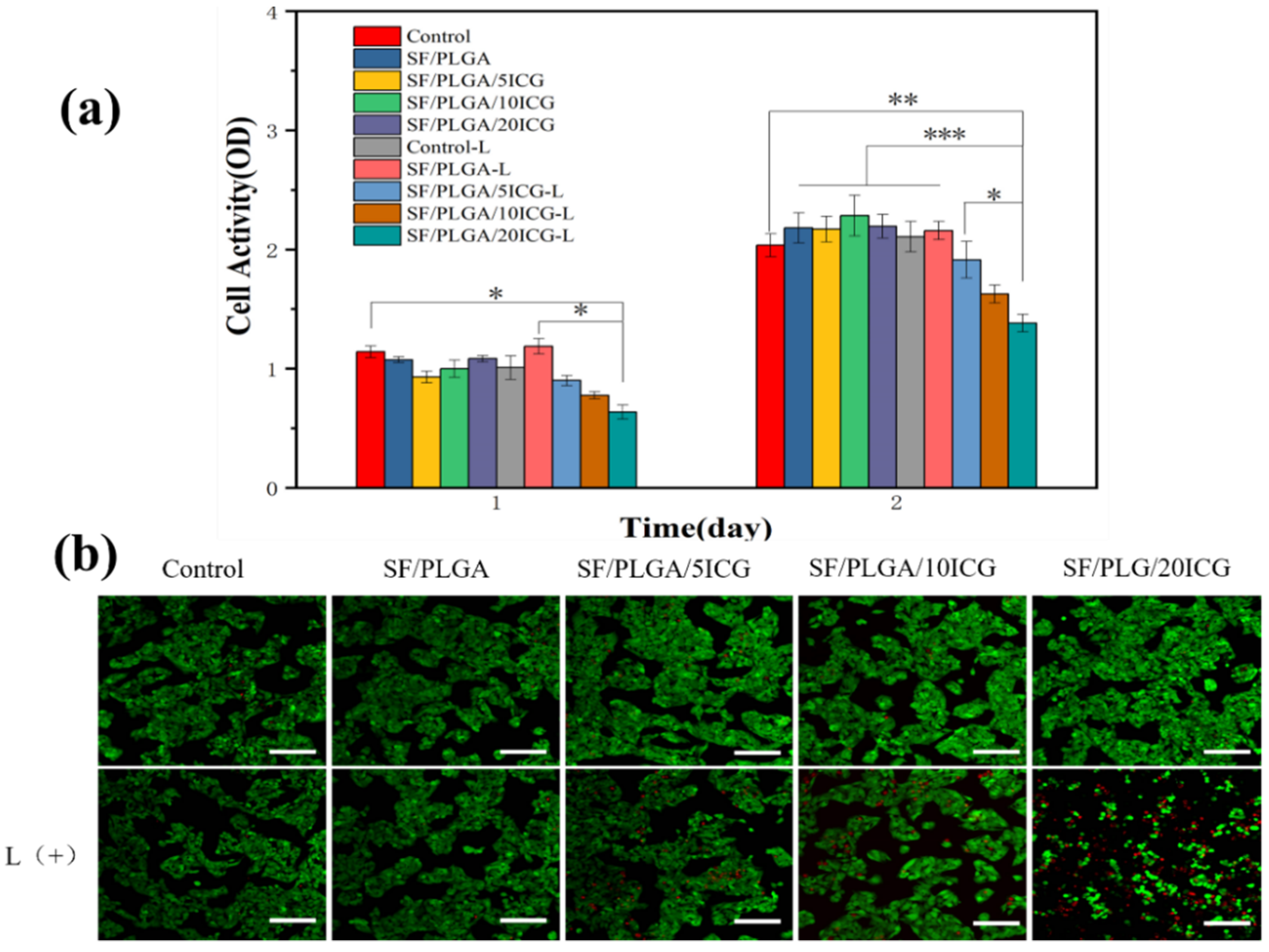
3.5. Biocompatibility of the Nanofibrous Membranes

Good biocompatibility of biomedical materials is necessary for tumor therapy and wound dressing [37]. To evaluate the biocompatibility of the nanofibrous membranes, L929 cells, which are usually used in toxicity assays, were seeded onto all membranes. All membranes appeared to be cytocompatible to levels comparable to those observed for the control group. (Figure 6a). Most significantly, the growth and proliferation of cells were unaffected by the ICG released from the ICG-loaded nanofibrous membranes, which is consistent with the results of CCK8. Therefore, these results revealed that the ICG-loaded nanofibrous membranes are biocompatible. In addition, the cell status was observed by live/dead cell staining (Figure 6b). Almost all cells in each group were alive, and nearly no dead cells were observed. SF has good cytocompatibility. SF can improve the hydrophilicity of the membranes and help promote cell adhesion, growth, and proliferation. Furthermore, ICG can be safely used as an amphiphilic cyanine dye with low toxicity [38]. According to these results, the ICG-loaded nanofibrous membranes have no significant toxicity to cells.

3.6. In Vitro Antitumor Efficacy

The liver cancer cells HepG2 were used to determine the antitumor efficacy of the nanofibrous membranes (Figure 7). Cells cultured in wells without membranes served as the control. The group with the “-L” symbol is the NIR-irradiated group. Compared with the control, the ICG-loaded nanofibrous membranes irradiated with a NIR laser showed distinct inhibition of HepG2. After NIR irradiation, as the concentration of ICG increased, the antitumor efficacy of the nanofibrous membranes increased significantly. This result is consistent with the confocal scan results. In the Figure, green fluorescence represents live cells and red fluorescence represents dead cells. SF/PLGA/20ICG-L presented less green-fluorescent cells and more red-fluorescent cells than the other groups. ICG is phagocytosed by cells and enters the cytoplasm. Mitochondria are particularly sensitive to heat shock and ROS, which lead to cell apoptosis [39]. ICG showed excellent photothermal conversion performance. Most importantly, heat and ROS produced by SF/PLGA/20ICG may have an excellent antitumor efficacy.

4. Conclusions

In this study, SF, PLGA, and ICG were used to prepare nanofibers by electrospinning. SF/PLGA/20ICG showed good water vapor permeability and excellent hydrophilicity, which are significant factors in wound healing. Furthermore, SF/PLGA/20ICG, irradiated by a NIR laser, also showed the ability to inhibit both Gram-positive and Gram-negative bacteria. The growth inhibition rates for S. aureus and E. coli were 91.53% and 87.95%, respectively. SF/PLGA/20ICG significantly inhibited tumor cell growth in in vitro experiments. These nanofibrous membranes have potential applications in wound healing and cancer therapy.

Author Contributions

Conceptualization, J.Z. and H.W. (Hai Wang); methodology, H.W. (Hai Wang); software, D.L.; validation, Y.P.; formal analysis, J.Z. and H.W. (Haiyan Wu); investigation, H.W. (Hai Wang); data curation, H.W. (Haiyan Wu); writing—original draft preparation, J.Z.; writing—review and editing, F.D.; visualization, Z.L.; supervision, Z.L.; funding acquisition, F.D. All authors have read and agreed to the published version of the manuscript.

Funding

This research was funded by grants from the National Natural Science Foundation of China (No. 31830094, No. U20A2058), Funds of China Agriculture Research System (No. CARS-18-ZJ0102), and the Innovation Program for Chongqing’s Overseas Returnees (No. cx2019090).

Institutional Review Board Statement

Not applicable.

Informed Consent Statement

Not applicable.

Data Availability Statement

Not applicable.

Conflicts of Interest

The authors declare no conflict of interest.

References

  1. Hu, H.; Chen, J.T.; Yang, H.; Huang, X.T.; Wu, H.L.; Wu, Y.X.; Li, F.F.; Yi, Y.F.; Xiao, C.; Li, Y.H.; et al. Potentiating photodynamic therapy of ICG-loaded nanoparticles by depleting GSH with PEITC. Nanoscale 2019, 11, 6384–6393. [Google Scholar] [CrossRef] [PubMed]
  2. Spring, B.Q.; Rizvi, I.; Xu, N.; Hasan, T. The role of photodynamic therapy in overcoming cancer drug resistance. Photochem. Photobiol. Sci. 2015, 14, 1476–1491. [Google Scholar] [CrossRef] [PubMed] [Green Version]
  3. Xia, D.L.; Xu, P.P.; Luo, X.Y.; Zhu, J.F.; Gu, H.Y.; Huo, D.; Hu, Y. Overcoming Hypoxia by Multistage Nanoparticle Delivery System to Inhibit Mitochondrial Respiration for Photodynamic Therapy. Adv. Funct. Mater. 2019, 29, 1807294. [Google Scholar] [CrossRef]
  4. Wainwright, M. ‘Safe’ photoantimicrobials for skin and soft-tissue infections. Int. J. Antimicrob. Agents 2010, 36, 14–18. [Google Scholar] [CrossRef] [PubMed] [Green Version]
  5. Fu, X.J.; Fang, Y.; Yao, M. Antimicrobial Photodynamic Therapy for Methicillin-Resistant Staphylococcus aureus Infection. BioMed Res. Int. 2013, 2013, 159157. [Google Scholar] [CrossRef] [PubMed] [Green Version]
  6. Sperandio, F.F.; Huang, Y.Y.; Hamblin, M.R. Antimicrobial Photodynamic Therapy to Kill Gram-negative Bacteria. Recent Pat. Anti-Infect. Drug Discov. 2013, 8, 108–120. [Google Scholar] [CrossRef] [PubMed] [Green Version]
  7. Chen, Q.; Xu, L.G.; Liang, C.; Wang, C.; Peng, R.; Liu, Z. Photothermal therapy with immune-adjuvant nanoparticles together with checkpoint blockade for effective cancer immunotherapy. Nat. Commun. 2016, 7, 13193. [Google Scholar] [CrossRef]
  8. Bhattarai, P.; Dai, Z.F. Cyanine based Nanoprobes for Cancer Theranostics. Adv. Healthc. Mater. 2017, 6, 1700262. [Google Scholar] [CrossRef] [PubMed]
  9. Gholibegloo, E.; Karbasi, A.; Pourhajibagher, M.; Chiniforush, N.; Ramazani, A.; Akbari, T.; Bahador, A.; Khoobi, M. Carnosine-graphene oxide conjugates decorated with hydroxyapatite as promising nanocarrier for ICG loading with enhanced antibacterial effects in photodynamic therapy against Streptococcus mutans. J. Photochem. Photobiol. B Biol. 2018, 181, 14–22. [Google Scholar] [CrossRef] [PubMed]
  10. Sheng, Z.H.; Hu, D.H.; Xue, M.M.; He, M.; Gong, P.; Cai, L.T. Indocyanine Green Nanoparticles for Theranostic Applications. Nano-Micro Lett. 2013, 5, 145–150. [Google Scholar] [CrossRef]
  11. Zhang, C.L.; Yu, S.H. Nanoparticles meet electrospinning: Recent advances and future prospects. Chem. Soc. Rev. 2014, 43, 4423–4448. [Google Scholar] [CrossRef] [PubMed]
  12. Boudriot, U.; Dersch, R.; Greiner, A.; Wendorff, J.H. Electrospinning approaches toward scaffold engineering—A brief overview. Artif. Organs 2006, 30, 785–792. [Google Scholar] [CrossRef] [PubMed]
  13. Grund, S.; Bauer, M.; Fischer, D. Polymers in Drug Delivery-State of the Art and Future Trends. Adv. Eng. Mater. 2011, 13, B61–B87. [Google Scholar] [CrossRef]
  14. Chou, S.F.; Carson, D.; Woodrow, K.A. Current strategies for sustaining drug release from electrospun nanofibers. J. Control. Release 2015, 220, 584–591. [Google Scholar] [CrossRef] [Green Version]
  15. Agarwal, S.; Wendorff, J.H.; Greiner, A. Use of electrospinning technique for biomedical applications. Polymer 2008, 49, 5603–5621. [Google Scholar] [CrossRef] [Green Version]
  16. Sridhar, R.; Lakshminarayanan, R.; Madhaiyan, K.; Barathi, V.A.; Limh, K.H.C.; Ramakrishna, S. Electrosprayed nanoparticles and electrospun nanofibers based on natural materials: Applications in tissue regeneration, drug delivery and pharmaceuticals. Chem. Soc. Rev. 2015, 44, 790–814. [Google Scholar] [CrossRef] [PubMed] [Green Version]
  17. Preis, E.; Anders, T.; Sirc, J.; Hobzova, R.; Cocarta, A.I.; Bakowsky, U.; Jedelska, J. Biocompatible indocyanine green loaded PLA nanofibers for in situ antimicrobial photodynamic therapy. Mater. Sci. Eng. C Mater. Biol. Appl. 2020, 115, 111068. [Google Scholar] [CrossRef] [PubMed]
  18. Martins, C.; Sousa, F.; Araujo, F.; Sarmento, B. Functionalizing PLGA and PLGA Derivatives for Drug Delivery and Tissue Regeneration Applications. Adv. Healthc. Mater. 2018, 7, 1701035. [Google Scholar] [CrossRef]
  19. Hsu, M.Y.; Hsieh, C.H.; Huang, Y.T.; Chu, S.Y.; Chen, C.M.; Lee, W.J.; Liu, S.J. Enhanced Paclitaxel Efficacy to Suppress Triple-Negative Breast Cancer Progression Using Metronomic Chemotherapy with a Controlled Release System of Electrospun Poly-d-l-Lactide-Co-Glycolide (PLGA) Nanofibers. Cancers 2021, 13, 3350. [Google Scholar] [CrossRef] [PubMed]
  20. Saxena, V.; Sadoqi, M.; Shao, J. Enhanced photo-stability, thermal-stability and aqueous-stability of indocyanine green in polymeric nanoparticulate systems. J. Photochem. Photobiol. B-Biol. 2004, 74, 29–38. [Google Scholar] [CrossRef] [PubMed]
  21. Tsang, V.L.; Bhatia, S.N. Three-dimensional tissue fabrication. Adv. Drug Deliv. Rev. 2004, 56, 1635–1647. [Google Scholar] [CrossRef]
  22. Bai, S.; Liu, S.; Zhang, C.; Xu, W.; Lu, Q.; Han, H.; Kaplan, D.L.; Zhu, H. Controllable transition of silk fibroin nanostructures: An insight into in vitro silk self-assembly process. Acta Biomater. 2013, 9, 7806–7813. [Google Scholar] [CrossRef]
  23. Siskova, A.O.; Kozma, E.; Opalek, A.; Kronekova, Z.; Kleinova, A.; Nagy, T.; Kronek, J.; Rydz, J.; Andicsova, A.E. Diclofenac Embedded in Silk Fibroin Fibers as a Drug Delivery System. Materials 2020, 13, 3580. [Google Scholar] [CrossRef]
  24. Peng, Y.; Ma, Y.; Bao, Y.; Liu, Z.L.; Chen, L.; Dai, F.Y.; Li, Z. Electrospun PLGA/SF/artemisinin composite nanofibrous membranes for wound dressing. Int. J. Biol. Macromol. 2021, 183, 68–78. [Google Scholar] [CrossRef]
  25. Stanic, V.; Pierre, A.C.; Etsell, T.H.; Mikula, R.J. Preparation and characterization of GeS2. J. Mater. Res. 1996, 11, 363–372. [Google Scholar] [CrossRef]
  26. Dionigi, C.; Posati, T.; Benfenati, V.; Sagnella, A.; Pistone, A.; Bonetti, S.; Ruani, G.; Dinelli, F.; Padeletti, G.; Zamboni, R.; et al. A nanostructured conductive bio-composite of silk fibroin-single walled carbon nanotubes. J. Mater. Chem. B 2014, 2, 1424–1431. [Google Scholar] [CrossRef]
  27. Zhang, C.C.; Song, D.W.; Lu, Q.; Hu, X.; Kaplan, D.L.; Zhu, H.S. Flexibility Regeneration of Silk Fibroin in Vitro. Biomacromolecules 2012, 13, 2148–2153. [Google Scholar] [CrossRef] [PubMed] [Green Version]
  28. Khan, M.M.I.R.; Morikawa, H.; Gotoh, Y.; Miura, M.; Ming, Z.; Sato, Y.; Iwasa, M. Structural characteristics and properties of Bombyx mori silk fiber obtained by different artificial forcibly silking speeds. Int. J. Biol. Macromol. 2008, 42, 264–270. [Google Scholar] [CrossRef] [PubMed]
  29. Lan, D.W.; Liu, Z.L.; Zhou, J.L.; Xu, M.T.; Li, Z.; Dai, F.Y. Preparation and characterization of silk fibroin/polyethylene oxide nanofiber membranes with antibacterial activity. J. Biomed. Mater. Res. Part A 2021, 110, 287–297. [Google Scholar] [CrossRef] [PubMed]
  30. RuizCardona, L.; Sanzgiri, Y.D.; Benedetti, L.M.; Stella, V.J.; Topp, E.M. Application of benzyl hyaluronate membranes as potential wound dressings: Evaluation of water vapour and gas permeabilities. Biomaterials 1996, 17, 1639–1643. [Google Scholar] [CrossRef]
  31. Morgado, P.I.; Aguiar-Ricardo, A.; Correia, I.J. Asymmetric membranes as ideal wound dressings: An overview on production methods, structure, properties and performance relationship. J. Membr. Sci. 2015, 490, 139–151. [Google Scholar] [CrossRef]
  32. Ege, Z.R.; Akan, A.; Oktar, F.N.; Lin, C.C.; Kuruca, D.S.; Karademir, B.; Sahin, Y.M.; Erdemir, G.; Gunduz, O. Indocyanine green based fluorescent polymeric nanoprobes for in vitro imaging. J. Biomed. Mater. Res. Part B Appl. Biomater. 2020, 108, 538–554. [Google Scholar] [CrossRef] [PubMed]
  33. Engel, E.; Schraml, R.; Maisch, T.; Kobuch, K.; Koenig, B.; Szeimies, R.M.; Hillenkamp, J.; Baumler, W.; Vasold, R. Light-induced decomposition of indocyanine green. Investig. Ophthalmol. Vis. Sci. 2008, 49, 1777–1783. [Google Scholar] [CrossRef] [PubMed]
  34. Simoes, D.; Miguel, S.P.; Ribeiro, M.P.; Coutinho, P.; Mendonca, A.G.; Correia, I.J. Recent advances on antimicrobial wound dressing: A review. Eur. J. Pharm. Biopharm. 2018, 127, 130–141. [Google Scholar] [CrossRef] [PubMed]
  35. Boluki, E.; Moradi, M.; Azar, P.S.; Fekrazad, R.; Pourhajibagher, M.; Bahador, A. The effect of antimicrobial photodynamic therapy against virulence genes expression in colistin-resistance Acinetobacter baumannii. Laser Ther. 2019, 28, 27–33. [Google Scholar] [CrossRef] [PubMed]
  36. de Freitas, M.T.M.; Soares, T.T.; Aragao, M.G.B.; Lima, R.A.; Duarte, S.; Zanin, I.C.J. Effect of Photodynamic Antimicrobial Chemotherapy on Mono- and Multi-Species Cariogenic Biofilms: A Literature Review. Photomed. Laser Surg. 2017, 35, 239–245. [Google Scholar] [CrossRef] [PubMed] [Green Version]
  37. Li, M.; Chen, J.; Shi, M.T.; Zhang, H.L.; Ma, P.X.; Guo, B.L. Electroactive anti-oxidant polyurethane elastomers with shape memory property as non-adherent wound dressing to enhance wound healing. Chem. Eng. J. 2019, 375, 121999. [Google Scholar] [CrossRef]
  38. Costa, R.A.; Farah, M.E.; Freymuller, E.; Morales, P.H.; Smith, R.; Cardillo, J.A. Choriocapillaris photodynamic therapy using indocyanine green. Am. J. Ophthalmol. 2001, 132, 557–565. [Google Scholar] [CrossRef]
  39. Xu, L.H.; Tong, G.H.; Song, Q.L.; Zhu, C.Y.; Zhang, H.L.; Shi, J.J.; Zhang, Z.Z. Enhanced Intracellular Ca2+ Nanogenerator for Tumor-Specific Synergistic Therapy via Disruption of Mitochondrial Ca2+ Homeostasis and Photothermal Therapy. Acs Nano 2018, 12, 6806–6818. [Google Scholar] [CrossRef]
Scheme 1. Schematic illustration of the preparation and SF/PLGA/20ICG for microbe inactivation and cancer treatment.
Processes 10 00850 sch001
Figure 1. SEM images and diameter distribution: (a) SF/PLGA, (b) SF/PLGA3, (c) SF/PLGA5, (d) SF/PLGA/20ICG, (e) SF/PLGA3/20ICG and (f) SF/PLGA5/20ICG.
Processes 10 00850 g001
Figure 2. FTIR spectra, (a): ICG, (b): SF, (c): PLGA, (d): SF/PLGA, (e): SF/PLGA/20ICG, (f): SF/PLGA3, (g): SF/PLGA3/20ICG, (h): SF/PLGA5 and (i): SF/PLGA5/20ICG.
Processes 10 00850 g002
Figure 3. (a) Degradation profile of the nanofibrous membranes; (b) WVTR; (c) contact angle of ICG-loaded nanofibrous membranes; (d) cumulative release of ICG in PBS and acetic acid buffer.
Processes 10 00850 g003
Figure 4. Photothermal performance. Temperature profiles of the nanofibrous membranes in PBS upon NIR irradiation of (a) 0.2 w/cm2, (b) 0.4 w/cm2, and (c) 0.6 w/cm2; (d) temperature elevation in the nanofibrous membranes using NIR with different laser powers; (e) temperature variations of SF/PLGA/20ICG exposed to NIR irradiation.
Processes 10 00850 g004
Figure 5. Antimicrobial effect of each group of membranes.
Processes 10 00850 g005
Figure 6. (a) Effect of the nanofibrous membranes on L929 cells; (b) live and dead cell staining on the third day.
Processes 10 00850 g006
Figure 7. In vitro antitumor efficacy. (a) Effects of the nanofibrous membranes on the viability of HepG2 cells; (b) live cell staining of HepG2 cells examined by confocal microscopy (bar = 100 μm). * p < 0.05, ** p < 0.01, *** p < 0.001, data represent means ± SD (n = 3).
Processes 10 00850 g007
Table 1. Parameters of the nanofibrous membranes.
Sample NameConcentration of SF wt%Concentration of PLGA wt%Concentration of ICG wt%
SF/PLGA6.06.0-
SF/PLGA/20ICG6.06.00.2
SF/PLGA33.09.0-
SF/PLGA3/20ICG3.09.00.2
SF/PLGA52.010.0-
SF/PLGA5/20ICG2.010.00.2
Publisher’s Note: MDPI stays neutral with regard to jurisdictional claims in published maps and institutional affiliations.

