Article

Untargeted LC-QTOF-MS Analysis of Metabolites Produced by Penicillium brevicompactum during the Bioconversion of Ganoderic Acid A

1
Department of Chemical Engineering, Faculty of Engineering and the Built Environment, University of Johannesburg, P.O. Box 17011, Doornfontein, Johannesburg 2028, South Africa
2
South African Medical Research Council Centre for Tuberculosis Research, Division of Molecular Biology and Human Genetics, Department of Medicine and Health Sciences, Stellenbosch University, Stellenbosch 7505, South Africa
*
Author to whom correspondence should be addressed.
Processes 2023, 11(10), 2963; https://doi.org/10.3390/pr11102963
Submission received: 17 September 2023 / Revised: 4 October 2023 / Accepted: 10 October 2023 / Published: 12 October 2023

Abstract

:
The repurposing of expired drugs through bioconversion remains one of the most crucial research milestones, as this practice reduces drug contamination while producing compounds of significance. The present study investigated the bioconversion of ganoderic acid A (GAA) using Penicillium brevicompactum over a period of 3, 6 and 9 days. The GAA intensity reduced from 22,099 cps on day 0 to 11,040, 4700 and 18,126 cps on day 3, 6 and 9, respectively, thus demonstrating the degradation of GAA over time. The produced metabolites that were recovered using ethyl acetate as a solvent were determined using LC-QTOF-MS. P. brevicompactum produced a variety of compounds in the absence of GAA, while in its presence, it was observed that P. brevicompactum was able to convert GAA and produced ganomastenol A/B/D, vitamin E succinate, and aminopregnane on day 3, while on day 6, armillaripin and ganolucidic acid A were produced. After 9 days of operation, vitamin E succinate, ganolucidic acid A and lucilactaene were produced. The present study is the first report on the ability of P. brevicompactum to bioconvert GAA. The identified metabolites have been established to possess bioactivity against various ailments, thus contributing to the discovery of new drugs.

1. Introduction

Ganoderma lucidum, which is commonly known as “Lingzhi” (Chinese) and “Reishi” (Japanese), is a basidiomycete mushroom with a diverse range of medicinal properties and has been used in Asian medicine for centuries [1]. The medicinal properties of this plant are primarily due to the high polysaccharide and triterpenoid content of these mushrooms, namely ganoderic, lucidenic and ganolucidic acids, ganoderiols and lucidones, all of which are G. lucidum derivatives and differ from one another based on their oxidation states [2]. Terpenoids, in general, act as allelochemicals in nature to allure pollinators and to attract or repel herbivores through the aromas and colours of their plant leaves, flowers and fruits [3]. As such, humanity utilises terpenoids for their fragrances in repellents and apply them in numerous medicinal applications [4]. Terpenoids occur as more complex molecules, which are categorised by the number of carbon atoms present in the molecule [4]. Triterpenoids are one of the sub-classes of terpenoid compounds where their cyclic skeletal arrangement has six isoprene (C5H8) units, making them C30 compounds that can be functionalised as alcohols, aldehydes and carboxylic acids [5]. Triterpenoids represent one of the largest phytochemical groups with thousands of known compounds to date [4]. This subcategory of specialised metabolites plays an important role in plant defense in nature and holds significant progress potential for applications in the food and pharmaceutical fields [6].
Ganoderic acid A (GAA) was the first ganoderic acid isolated from Ganoderma mushrooms in 1982 [7]. GAA has been reported to possess exceptionally potent pharmacological properties, which include hepatoprotective, cytotoxic, anti-cancer, anti-tumor and antioxidant capabilities to name a few [8]. GAA is currently in phase 1 clinical trials [8] and could potentially play an important role in the treatment of cancer and other diseases in the foreseeable future. Studies have indicated that GAA can significantly decrease cancer apoptosis in breast, hepatocellular, lymphoma, glioblastoma multiforme and osteosarcoma carcinomas [9,10,11,12].
Although GAA exhibits numerous biological activities, this compound may also possess hemolytic and cytostatic properties that may restrict its widespread pharmaceutical usage. In addition to this, there is currently several ways in which researchers are searching for new drugs that may possess properties that are needed in the medical field and microbial biotransformation and/or conversion has been determined to be one of these techniques. Microorganisms have the ability to shift the functional groups within precursor molecules, thus changing their properties, including their application.
Previous studies have demonstrated that biotransformation can culminate in compounds that are not found in nature. For example, Aspergillus oryzae hydroxylated soy isoflavone daidzein to form 6-hyroxydaidzein or 8-hydroxydaidzein, which displayed 10-fold higher anti-tyrosinase activity compared to the precursor daidzen [13], while Chang [14] biotransformed GAA to 3-O-Acetyl ganoderic acid A using Streptomyces sp., which was a new triterpenoid in microorganisms. The authors also biotransformed GAA using intestinal bacterium from zebrafish to GAA-15-O-glucoside. The biotransformation of various drugs using Penicillium sp. has been investigated before. As an example, Penicillium crustosum 2T01Y01 was used to biotransform gentiopicroside and yield seven deglycosylated metabolites, which were detected by ultraperformance liquid chromatography/quadrupole time of flight mass spectrometry (UPLC/Q-TOF MS) [15], while Tian [16] biotransformed artemisinic acid to various bioactive derivatives of Penicillium oxalicum B4. However, there are no studies that utilised P. brevicompactum in drug biotransformation studies with specific reference to GAA. As a result, this study aimed to bioconvert GAA using Penicillium brevicompactum over a 9-day period and to determine the metabolites produced from the bioconversion process using liquid chromatography/quadrupole time of flight mass spectrometry (LC-QTOF-MS). This is the first report that has investigated the bioconversion of GAA using Penicillium brevicompactum and to determine the metabolites that this fungal organism produces using LC-QTOF-MS.

2. Materials and Methods

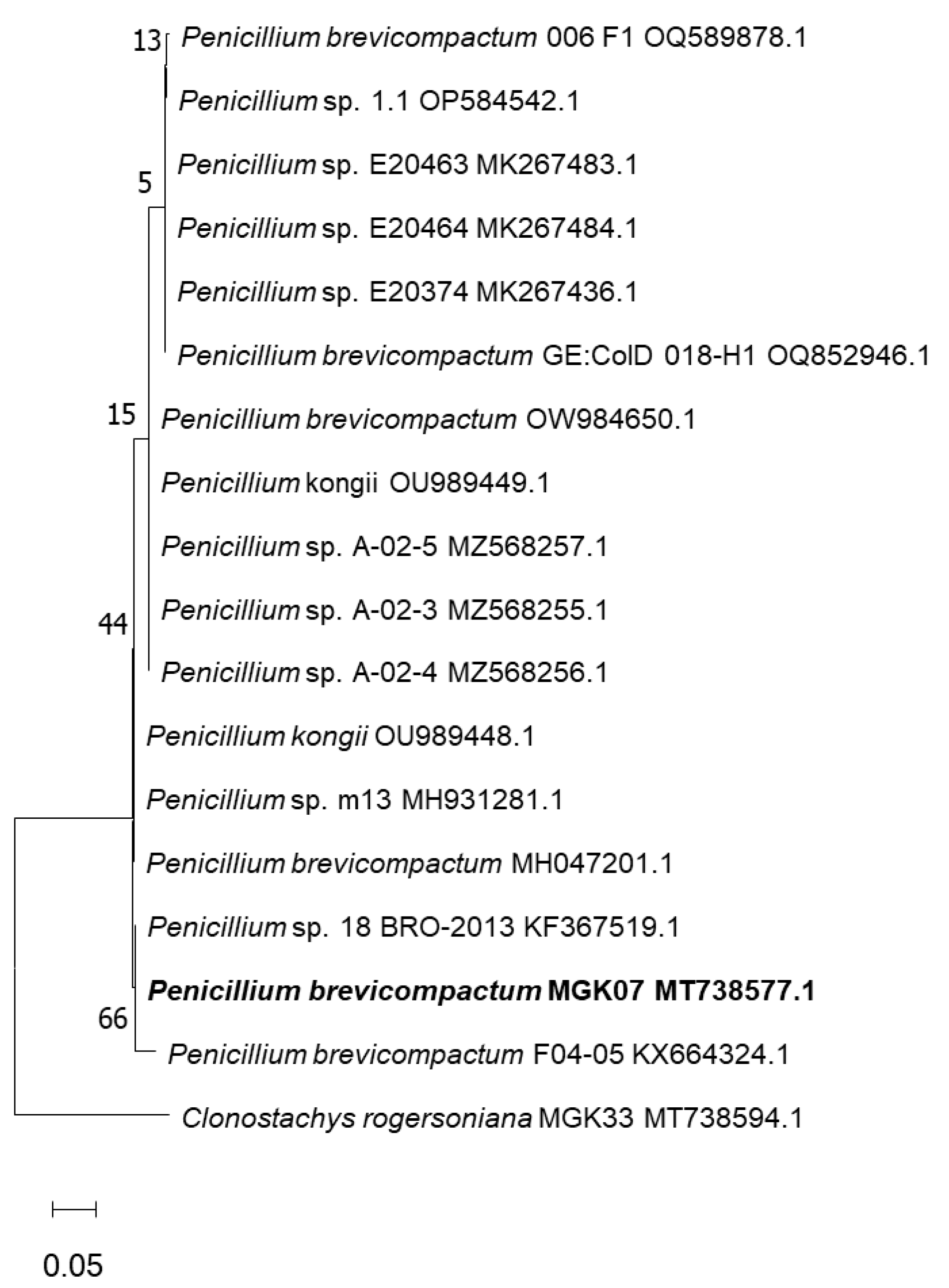
2.1. Isolation and Identification of the Fungal Organism

Penicillium brevicompactum was isolated from an unidentified brown mushroom in a forest that was dominated by Celtis africana according to the method described in the work of Tapfuma [17]. The isolate was revived by carefully slicing a small portion of the fungi that was previously grown on a potato dextrose agar (PDA) plate using a sterile scalpel under a laminar flow hood, where the cutting was transferred to a fresh PDA plate. The plate was incubated for 14 days at 30 °C to facilitate fungal growth. On day 14 of incubation, the microbes had grown sufficiently and were ready to serve as an inoculum for the subsequent experiments. Fresh cultures of P. brevicompactum were prepared by cutting a portion of the grown culture in PDA and placing it on fresh PDA plates and incubated at 30 °C for 14 days. The ribosomal DNA of the organism was extracted and the internal transcriber spacer 1 and 2 (ITS 1 and 2) was amplified using the primer pairs reported in the study by Tapfuma et al. [17] including the phylogenetic analysis approach. However, the outlier organism was Clonostachys rogersoniana MGK33. Fungal nucleotide sequences were then submitted to GenBank for curation and accession numbers were assigned (MT738577.1).
The evolutionary history was determined using the maximum likelihood method and Jukes–Cantor model [18]. The tree depicting the highest log likelihood (−6928.01) is presented, with the percentage of trees supporting each taxon grouping displayed next to their respective branches. To initiate the heuristic search process, initial trees were generated automatically by applying Neighbor-Join and BioNJ algorithms to a pairwise distance matrix calculated using the Jukes–Cantor model. From these initial trees, the topology yielding superior log likelihood value was chosen. The drawing of this tree adheres to scale as the branch lengths represent substitutions per site. This analysis involved 18 nucleotide sequences encompassing a total of 4375 positions in its final dataset. MEGA11 software version 11 facilitated these evolutionary analyses [19].

2.2. GAA Bioconversion

First, 1 L of MGN medium was prepared by mixing 5 g/L of peptone, 5 g/L dipotassium phosphate, 5 g/L yeast extract and 5 g/L sodium chloride [14]. The medium was autoclaved at 121 °C for 90 min; once cooled, 5 mg/L of GAA was added to the media. Next, 100 mL of the MGN medium containing 5 mg/L of GAA was decanted into sterile baffled Erlenmeyer flasks. The fungal plate containing the microorganism was covered with sterile distilled water and thereafter, the fungi was carefully scrapped from the agar such that the spores could be recovered. The mycelia were separated from the liquid solution that contained the fungal spores and 1000 µL of the separated spore solution was deposited into the MGN. The mixture was placed in an incubator set at 250 rpm and 30 °C for 3, 6 and 9 days, respectively. After the respective incubation periods, the mixture was centrifuged at 4000 rpm for 15 min to remove the cell debris. Thereafter, equal volumes of the supernatant and ethyl acetate were then mixed in a separating funnel and allowed to stand for approximately 30 min [17]. The solvent phase was then concentrated in a rotary evaporator set at 60 °C and the resulting mixture was dried for a period of 7 days in an oven (22–25 °C) to remove any remaining solvent. The resultant extract was analysed using liquid chromatography–quadrupole time-of-flight-mass-spectroscopy (LC-QTOF-MS) [14]. The control sample was incubated under the same conditions as described but in the absence of GAA. This was carried out to assess the metabolites that the fungal organism produces in the absence of GAA so that the metabolites that are produced from GAA bioconversion can be determined accurately.

2.3. Untargeted Analysis of Metabolites Using LC-QTOF-MS

The analysis of the samples was performed on a Bruker Compact Q-TOF high-resolution mass spectrometer (Bruker Daltonics, Bremen, Germany) using the positive ionisation mode. The samples were analysed using a binary solvent system (solvent A consists of H2O and 0.1% formic acid; solvent B consists of acetonitrile and 0.1% formic acid (v/v)). Next, 10 µL of the sample was injected into the Dionex Ultimate 3000 UHPLC system (Thermo Scientific, Dionex, Sunnyvale, CA, USA) and eluted using a gradient elution technique for 14 min at a gradient of 5–95% solvent B at a flow rate of 0.3 mL/min. The Raptor ARC-18 (2.7 μm; 100 × 2.1 mm) analytical column was used for the separation. The mass spectrum analysis was processed using the DataAnalysis version 4.0 (Bruker Daltonics, Bremen, Germany).

3. Results

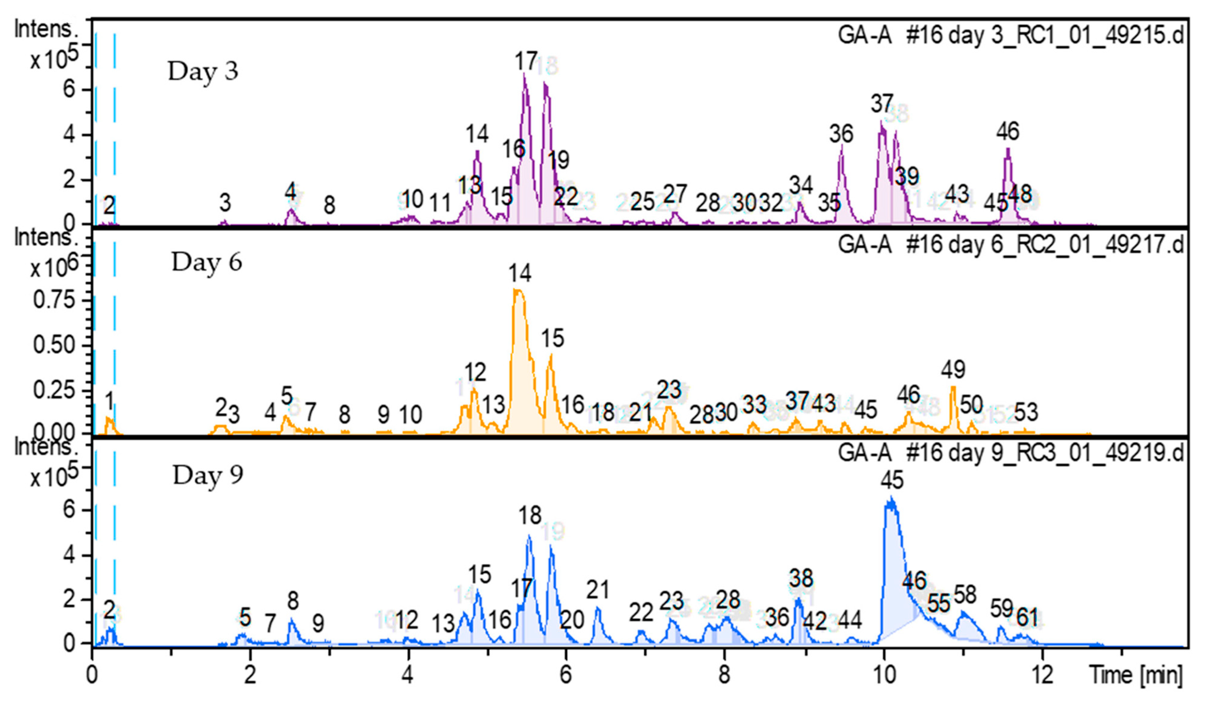
3.1. Bioconversion of Ganoderic Acid A and Detected Metabolites Using Penicillium brevicompactum MGK07

Figure 1 demonstrates the phylogenetic tree of the isolate, which was determined to be Penicillium brevicompactum. The organism was used in the degradation of GAA and to detect the metabolites that this organism produces in the absence and presence of GAA. Figure 2 shows the degradation profile of GAA over the 9-day period in contrast to the GAA standard. In order to understand the degree to which GAA was degraded, the intensity of the GAA peaks was studied and compared over the 9-day period (Figure 2).
The initial intensity of GAA in the medium was 22,099 cps and P. brevicompactum degraded GAA, resulting in an intensity of 18,126, 4700 and 11,040 cps for day 3, 6 and 9, respectively. The detected chromatograms that represent the metabolites from the conversion of GAA are shown in Figure 3.

3.2. Metabolites Produced from GAA Bioconversion of Penicillium brevicompactum MGK07: Day 3

The metabolites of GAA bioconversion using P. brevicompactum were determined. However, the metabolites that P. brevicompactum produced in the absence of GAA were determined and these metabolites allowed for this study to ascertain the accurate GAA bioconversion metabolites while using the data in Figure S1 and Table S1 as a basis for comparison. Table S1 shows that the metabolites that P. brevicompactum produced included 17 compounds in the absence of GAA and these compounds have various bioactive properties, which are shown in Table S2. It is worth noting that these compounds were not detected in the metabolites that were produced in the presence of GAA. The compounds produced from day 3 of biotransformed GAA are shown in Table 1 and Figure S2.
There were 51 compounds that were isolated from the day 3 sample (see Figure S2), and the main compounds that were produced from GAA transformation were cyclo (L-Pro-L-Val), cyclo (-Leu-Pro), cyclo (L-Phe-L-Pro), 4-deoxovermiculic acid, ganomastenol A, B or D, aminopregnane and vitamin E succinate. The main GAA derivative compound was identified to be either ganomastenol A/B/D, since all three isomers have the same molecular formula and m/z [H]+ of C15H24O3 and 253.18, respectively. It was observed that cyclo (L-Pro-L-Val) and cyclo (L-Phe-L-Pro) were produced in all the sampling periods of this study.

3.3. Metabolites Produced from GAA Bioconversion of Penicillium brevicompactum MGK07: Day 6

On day 6, there were 10 compounds that were detected after the screening process from a total of 48 compounds (see Figure S3) that were detected, in which 10 were regarded as the most abundant compounds, as shown in Table 2. Of the 10 detected compounds, 7 were newly isolated metabolites with the two major focuses of these 7 being ganoderic acid β/ganolucidic acid A/D and methylprednisone acetate/armillaripin. It is important to note that the three main compounds from day 3 (i.e., ganomastenol A, B/D, aminopregnane and vitamin E succinate) were not detected in the day 6 sample. This accounted for the large reduction and change that was viewed between the 10 and 12 min retention time (see Figure 2).
The major bioconversion product found from the day 6 sample is closely related to GAA with only one-oxygen atom difference between the two molecular formulas (i.e., GAA being C30H44O7 and M13 being C30H44O6) and was proposed to be either GAA isomers such as ganoderic acid beta, ganolucidic acid A or ganolucidic acid. These are all metabolites from G. lucidum, like GAA.

3.4. Metabolites Produced from GAA Bioconversion of Penicillium brevicompactum MGK07: Day 9

On day 9, 62 compounds were detected, where 10 compounds (see Figure S4) were regarded as the most abundant compounds in the sample, as shown in Table 3.
Table 3 shows that there were two new compounds that were isolated on day 9 and these were lucilactaene and an unknown amine derivative with a molecular formula of C10H13NO. Armillaripin was not detected in the day 9 sample; however, ganoderic Acid β/ganolucidic acid A/B showed a slight increase from day 6 to day 9. Vitamin E succinate resurfaced in the day 9 sample like the day 3 sample, after being undetected on day 6.
From the observed data, after the 9-day period, it can be said that the compounds with the highest intensities were the main compounds that were produced, while the other compounds were detected at low levels. These two compounds were detected at peaks 18 and 45, translating to cyclo (L-Phe-L-Pro) and an unknown compound with a molecular weight of C18H30N2O.

4. Discussion

4.1. Bioconversion of Ganoderic Acid A and Detected Metabolites Using Penicillium brevicompactum

The bioconversion of GAA was studied over a 9-day period where the intensities of the GAA profile were monitored during this period. Since this is the first report on the conversion of GAA over time where the intensities were monitored, the findings of this study cannot be compared to previous studies. During the bioconversion of GAA, metabolites were produced that were analysed using LC-QTOF-MS to detect their identities and potential applications. Figure 3 demonstrated that compounds 17, 14 and 18 for day 3, 6 and 9, respectively, were produced throughout the experimental period. This compound was identified as cyclo-(L-Phe-L-Pro), which is an antifungal peptide [20] and has demonstrated to possess free-radical scavenging activity with an IC50 value of 24 µM [21].

4.2. Metabolites Produced from GAA Bioconversion of Penicillium brevicompactum: Day 3

On day 3, various metabolites were produced during the bioconversion of GAA. Some of the metabolites that were produced include cyclo-L-prolyglycine, cyclo (L-Phe-L-Pro), ganomastenol A/B/C, aminopregnane and vitamin E succinate. Cyclo-L-Prolyglycine is a neuropeptide that has been described as a neuroprotective and memory agent with potent anti-bacterial and therapeutic properties [22], while cyclo (L-Phe-L-Pro) has been previously reported to possess antifungal properties and acts as a signaling molecule for the expression of the hydroperoxidase enzyme [20,23]. Ganomastenol A/B/D were first isolated from Ganoderma mastoporum in 1995 [24]. Ganoderma mastoporum, like Ganoderma lucidium, is housed under the same Ganodermataceae family [25]. G. mastoporum, however, is not well documented compared to G. lucidum and there is limited information about Ganomastenol A, B, and D. Given that these are both Ganoderma terpenes and the molecular formulas of GAA and the Ganomastenol isomers are C30H44O7 and C15H24O3, respectively, M8 is approximately half the molecular size of GAA. Therefore, it is possible that Ganomastenol A/B/D was synthesised via the oxidative cleavage of GAA.
Aminopregnane is a metabolite from Holarrhena antidysenterica, a medicinal plant abundantly found in Asia (more specifically, India) [26]. Holarrhena antidysenterica has been known for its anti-bacterial, anti-diarrheal, anti-malarial, anti-diabetic, anti-inflammatory, anti-oxidant properties, among many others [27]. To date, the bioactivity of aminopregnane has not been studied in detail and thus, the properties of this compound are unknown. M26 was proposed to be vitamin E succinate, a succinic acid ester derivative of vitamin E. Vitamin E succinate has been described as one of the strongest anti-cancer vitamin E derivatives, with its cancer properties being documented since the early 2000s [28,29,30]. Recent studies have shown that vitamin E succinate aids in apoptosis induction and demonstrates the ability to prohibit tumor growth via several pathways [30,31,32]. More recent applications include intestinal absorption of paclitaxel with the aid of vitamin E succinate for tumor treatment [33].

4.3. Metabolites Produced from GAA Bioconversion of Penicillium brevicompactum: Day 6

On day 6, the major compounds that were detected were ganoderic acid β, ganolucidic acid and Armillaripin. Ganoderic acid beta is part of the ganoderic acids that were isolated from Ganoderma lucidum with GAA. There is limited information about the bioactive properties of GA-β; however, as all the G. lucidum isolates have shown, it can be assumed that GA-β possesses similar pharmacological properties [34,35,36,37]. Ganolucidic acids are also Ganoderma derivatives. Ganolucidic acids A/D are the triterpenoid isolates that were first isolated from G. lucidum in 1985 [2,35]. In contrast to ganoderic acids, ganolucidic acids are not as well documented but have been found to possess significant anti-HIV properties [36,37]. Armillaripin has been described as a sesquiterpenoid aromatic ester that was first isolated from petroleum ether extracts of Armillaria mellea mycelium in 1990 [38]. Armillaria mellea has been known to possess anti-oxidant, anti-ageing, anti-vertigo and immunopotentiation properties; therefore, the sesquiterpene ester derivates and isolates are also biologically active and have shown anti-biotic and anti-fungal properties [38]. An unknown compound with a molecular formula C9H11N5O6 was detected on day 6 and 9, while the other unknown compounds were only detected on day 6.

4.4. Metabolites Produced from GAA Bioconversion of Penicillium brevicompactum: Day 9

Lucilactaene is a metabolite that is not well-documented; however, it has been reported to have anti-malarial and anti-cancer properties, with the possibility of anti-inflammatory properties [39,40]. Currently, more research is being conducted on this compound to gain a better understanding on its properties and cytotoxic effects [41,42].
The main compounds that were formed on day 3 disappeared on day 6. There is a possibility that these compounds were produced as a process step for Penicillium brevicompactum to produce armillaripin and ganoderic acid β or ganolucidic A/D or the other new compounds that were observed on day 6. However, from all the compounds isolated on day 6, these two compounds are mostly likely to be produced as a by-product of GAA bioconversion, as they are of a terpenoid nature and produced by Ganoderma, respectively. The disappearance of some of the detected compounds from day 3 to 9 may be due to their utilisation by P. brevicompactum. However, it is worth noting that two compounds were not identified in this study, and these have the following molecular formulas: C10H13NO and C18H30N2O. This is because nuclear magnetic resonance (NMR) spectroscopy was not utilised in this study to identify these compounds. This therefore calls for future research to utilise NMR to identify these compounds and evaluate their bioactivities.

5. Conclusions

This study showed that Penicillium brevicompactum can produce a multitude of compounds from the bioconversion of ganoderic Acid A over the different sampling periods. The bioconversion of ganoderic acid A yielded ganomastenol A/B/D, vitamin E succinate and aminopregnane on day 3, while on day 6, a triterpenoid compound determined to be either ganoderic acid β or ganolucidic acid A/D and a sesquiterpenoid known as armillaripin were produced. On day 9, P. brevicompactum produced ganoderic acid β or ganolucidic acid A/D, along with vitamin E succinate and lucilactaene. It was further observed that the most abundant compounds (high intensities) after the experimental run were cyclo (L-Phe-L-Pro) and an unknown compound with a molecular weight of C18H30N2O. It can, therefore, be concluded that P. brevicompactum has the ability to bioconvert ganoderic acid A to a variety of compounds with different properties, which can be utilised for several applications in both the medical and pharmaceutical industries, due to their vast and diverse range of pharmacological properties. It is worth noting that some of the produced compounds might not have emanated directly from the bioconversion of GAA but as compounds secreted by the organism in the presence of GAA. However, from the observed data of this study, future studies should conduct targeted analysis of the unknown compound that was detected after the 9-day period, including the determination of the structure of the compound using tools such as nuclear magnetic resonance spectroscopy. It is also recommended that future studies should have 24 h sampling intervals in order to better understand the changes that occur daily to explain some of the witnessed changes that were seen in this study.

Supplementary Materials

The following supporting information can be downloaded at: https://www.mdpi.com/article/10.3390/pr11102963/s1, Figure S1: Base Peak Chromatogram of P. brevicompactum in MM; Figure S2: Chromatogram expressing the metabolites produced by Penicillium brevicompactum from Ganoderic Acid on day 3; Figure S3: Chromatogram expressing the metabolites produced by Penicillium brevicompactum from Ganoderic Acid on day 6; Figure S4: Chromatogram expressing the metabolites produced by Penicillium brevicompactum from Ganoderic Acid on day 9; Table S1: Metabolites of Penicillium brevicompactum in MM as determined by LC-QTOF-MS/MS; Table S2: The functions/uses including the microbial sources of P. brevicompactum metabolites in MM.

Author Contributions

V.N.: Investigation, Data curation, Original draft preparation. V.M.: Data curation, Conceptualization, Writing—Reviewing and editing. K.T.: Writing—Reviewing and editing, Formal analysis, Validation. N.F.R.: Data processing, phylogenetic analysis, Writing—Reviewing and editing. L.M.: Supervision, Conceptualization, Writing—Reviewing and editing, Funding, Project administration. All authors have read and agreed to the published version of the manuscript.

Funding

The authors would like to acknowledge the financial support from the South African National Research Foundation through the Black Academic Advancement Programme (Grant number: 138245).

Data Availability Statement

The data associated with this work are embedded within the manuscript and in the Supplementary Materials.

Conflicts of Interest

The authors declare no conflict of interest.

References

  1. Wachtel-Galor, S.J.; Yuen, J.A.; Buswell, J.A.; Benzie, I.F. Ganoderma lucidum (lingzhi or reishi): A medicinal mushroom. In Herbal Medicine: Biomolecular and Clinical Aspects, 2nd ed.; Benzie, I.F.F., Wachtel-Galor, S., Eds.; CRC Press: Boca Raton, FL, USA, 2011. [Google Scholar]
  2. Hennicke, F.; Cheikh-Ali, Z.; Liebisch, T.; Maciá-Vicente, J.G.; Bode, H.B.; Piepenbring, M. Distinguishing commercially grown Ganoderma lucidum from ganoderma lingzhi from europe and east asia on the basis of morphology, molecular phylogeny, and triterpenic acid profiles. Phytochemistry 2016, 127, 29–37. [Google Scholar] [PubMed]
  3. Nahata, A. Ganoderma lucidum: A potent medicinal mushroom with numerous health benefits. Pharm. Anal. Acta 2013, 4, 1000e159. [Google Scholar]
  4. Bergman, M.E.; Davis, B.; Phillips, M.A. Medically useful plant terpenoids: Biosynthesis, occurrence, and mechanism of action. Molecules 2019, 24, 3961. [Google Scholar] [CrossRef]
  5. Perveen, S.; Al-Taweel, A. Introductory chapter: Terpenes and terpenoids. In Terpenes and Terpenoids; IntechOpen: London, UK, 2018; Volume 1, pp. 1–12. [Google Scholar]
  6. Thurman, E.M. Chapter seven—Analysis of terpenes in hemp (Cannabis sativa) by gas chromatography/mass spectrometry: Isomer identification analysis. In Comprehensive Analytical Chemistry; Ferrer, I., Thurman, E.M., Eds.; Elsevier: Amsterdam, The Netherlands, 2020; pp. 197–233. [Google Scholar]
  7. Cárdenas, P.D.; Almeida, A.; Bak, S. Evolution of structural diversity of triterpenoids. Front. Plant Sci. 2019, 10, 1523. [Google Scholar] [PubMed]
  8. Kubota, T.; Asaka, Y.; Miura, I.; Mori, H. Structures of ganoderic acid a and b, two new lanostane type bitter triterpenes from Ganoderma lucidum (fr.) karst. Helv. Chim. Acta 1982, 65, 611–619. [Google Scholar]
  9. Liang, C.; Tian, D.; Liu, Y.; Li, H.; Zhu, J.; Li, M.; Xin, M.; Xia, J. Review of the molecular mechanisms of Ganoderma lucidum triterpenoids: Ganoderic acids a, c2, d, f, dm, x and y. Eur. J. Med. Chem. 2019, 174, 130–141. [Google Scholar]
  10. Yang, Y.; Zhou, H.; Liu, W.; Wu, J.; Yue, X.; Wang, J.; Quan, L.; Liu, H.; Guo, L.; Wang, Z.; et al. Ganoderic acid a exerts antitumor activity against mda-mb-231 human breast cancer cells by inhibiting the janus kinase 2/signal transducer and activator of transcription 3 signaling pathway. Oncol. Lett. 2018, 16, 6515–6521. [Google Scholar]
  11. Wang, M.L.; Lu, C.H.; Xu, Q.Y.; Song, S.Y.; Hu, Z.Y.; Zheng, Z.H. Four new citrinin derivatives from a marine-derived penicillium sp. Fungal strain. Molecules 2013, 18, 5723–5735. [Google Scholar] [CrossRef]
  12. Wang, X.; Sun, D.; Tai, J.; Wang, L. Ganoderic acid a inhibits proliferation and invasion, and promotes apoptosis in human hepatocellular carcinoma cells. Mol. Med. Rep. 2017, 16, 3894–3900. [Google Scholar] [CrossRef]
  13. Chang, T.-S. Isolation, bioactivity, and production of ortho-hydroxydaidzein and ortho-hydroxygenistein. Int. J. Mol. Sci. 2014, 15, 5699–5716. [Google Scholar] [CrossRef]
  14. Chang, T.-S.; Chiang, C.-M.; Wang, T.-Y.; Lee, C.-H.; Lee, Y.-W.; Wu, J.-Y. New triterpenoid from novel triterpenoid 15-o-glycosylation on ganoderic acid a by intestinal bacteria of zebrafish. Molecules 2018, 23, 2345. [Google Scholar] [CrossRef]
  15. Zeng, W.-L.; Li, W.-K.; Han, H.; Tao, Y.-Y.; Yang, L.; Wang, Z.-T.; Chen, K.-X. Microbial biotransformation of gentiopicroside by the endophytic fungus penicillium crustosum 2t01y01. Appl. Environ. Microbiol. 2014, 80, 184–192. [Google Scholar] [CrossRef] [PubMed]
  16. Tian, H.; Li, X.P.; Zhao, J.; Gao, H.W.; Xu, Q.M.; Wang, J.W. Biotransformation of artemisinic acid to bioactive derivatives by endophytic penicillium oxalicum b4 from artemisia annual. Phytochemistry 2021, 185, 112682. [Google Scholar] [CrossRef] [PubMed]
  17. Tapfuma, K.I.; Nyambo, K.; Adu-Amankwaah, F.; Baatjies, L.; Smith, L.; Allie, N.; Keyster, M.; Loxton, A.G.; Ngxande, M.; Malgas-Enus, R.; et al. Antimycobacterial activity and molecular docking of methanolic extracts and compounds of marine fungi from saldanha and false bays, south africa. Heliyon 2022, 8, e12406. [Google Scholar] [CrossRef] [PubMed]
  18. Jukes, T.H.; Cantor, C.R. Evolution of protein molecules. In Mammalian Protein Metabolism; Munro, H.N., Ed.; Academic Press: New York, NY, USA, 1969; pp. 21–132. [Google Scholar]
  19. Tamura, K.; Stecher, G.; Kumar, S. Mega11: Molecular evolutionary genetics analysis version 11. Mol. Biol. Evol. 2021, 38, 3022–3027. [Google Scholar] [CrossRef] [PubMed]
  20. Ström, K.; Sjögren, J.; Broberg, A.; Schnürer, J. Lactobacillus plantarum milab 393 produces the antifungal cyclic dipeptides cyclo(l-phe-l-pro) and cyclo(l-phe-trans-4-oh-l-pro) and 3-phenyllactic acid. Appl. Environ. Microbiol. 2002, 68, 4322–4327. [Google Scholar] [CrossRef] [PubMed]
  21. Lee, K.H.; Rhee, K.H. Radioprotective effect of cyclo (l-phenylalanyl-l-prolyl) on irradiated rat lung. J. Microbiol. Biotechnol. 2008, 18, 369–376. [Google Scholar]
  22. Yousuf, M.; Jamil, W.; Mammadova, K. Microbial bioconversion: A regio-specific method for novel drug design and toxicological study of metabolites. Curr. Pharm. Biotechnol. 2019, 20, 1156–1162. [Google Scholar] [CrossRef]
  23. Kim, I.H.; Kim, S.-Y.; Park, N.-Y.; Wen, Y.; Lee, K.-W.; Yoon, S.-Y.; Jie, H.; Lee, K.-H.; Kim, K.-S. Cyclo-(l-phe-l-pro), a quorum-sensing signal of vibrio vulnificus, induces expression of hydroperoxidase through a toxr-leuo-hu-rpos signaling pathway to confer resistance against oxidative stress. Infect. Immun. 2018, 86, e00932-17. [Google Scholar] [CrossRef]
  24. Hirotani, M.; Ino, C.; Hatano, A.; Takayanagi, H.; Furuya, T. Ganomastenols a, b, c and d, cadinene sesquiterpenes, from ganoderma mastoporum. Phytochemistry 1995, 40, 161–165. [Google Scholar] [CrossRef]
  25. Baby, S.; Johnson, A.J.; Govindan, B. Secondary metabolites from ganoderma. Phytochemistry 2015, 114, 66–101. [Google Scholar] [PubMed]
  26. Jamadagni, P.S.; Pawar, S.D.; Jamadagni, S.B.; Chougule, S.; Gaidhani, S.N.; Murthy, S. Review of Holarrhena antidysenterica (L.) wall. Ex a. Dc.: Pharmacognostic, pharmacological, and toxicological perspective. Pharmacogn. Rev. 2017, 11, 141–144. [Google Scholar] [CrossRef]
  27. Sinha, S.; Sharma, A.; Reddy, P.H.; Rathi, B.; Prasad, N.; Vashishtha, A. Evaluation of phytochemical and pharmacological aspects of Holarrhena antidysenterica (wall.): A comprehensive review. J. Pharm. Res. 2013, 6, 488–492. [Google Scholar] [CrossRef]
  28. Neuzil, J. Vitamin e succinate and cancer treatment: A vitamin e prototype for selective antitumour activity. Br. J. Cancer 2003, 89, 1822–1826. [Google Scholar] [CrossRef] [PubMed]
  29. Malafa, M.P.; Neitzel, L.T. Vitamin e succinate promotes breast cancer tumor dormancy. J. Surg. Res. 2000, 93, 163–170. [Google Scholar] [CrossRef]
  30. Liang, L.; Qiu, L. Vitamin e succinate with multiple functions: A versatile agent in nanomedicine-based cancer therapy and its delivery strategies. Int. J. Pharm. 2021, 600, 120457. [Google Scholar]
  31. Huang, X.; Neckenig, M.; Sun, J.; Jia, D.; Dou, Y.; Ai, D.; Nan, Z.; Qu, X. Vitamin e succinate exerts anti-tumour effects on human cervical cancer cells via the cd47-sirpɑ pathway both in vivo and in vitro. J. Cancer 2021, 12, 3877–3886. [Google Scholar] [CrossRef] [PubMed]
  32. Lan, T.; Guan, X. Vitamin e succinate inhibits the growth of human gastric cancer cells. Indian J. Pharm. Sci. 2021, 83, 346–353. [Google Scholar] [CrossRef]
  33. Qu, X.; Zou, Y.; He, C.; Zhou, Y.; Jin, Y.; Deng, Y.; Wang, Z.; Li, X.; Zhou, Y.; Liu, Y. Improved intestinal absorption of paclitaxel by mixed micelles self-assembled from vitamin e succinate-based amphiphilic polymers and their transcellular transport mechanism and intracellular trafficking routes. Drug Deliv. 2018, 25, 210–225. [Google Scholar] [CrossRef]
  34. Hsu, C.-L.; Yen, G.-C. Chapter three—Ganoderic acid and lucidenic acid (triterpenoid). In The Enzymes; Bathaie, S.Z., Tamanoi, F., Eds.; Academic Press: Cambridge, MA, USA, 2014; pp. 33–56. [Google Scholar]
  35. Kikuchi, T.; Matsuda, S.; Murai, Y.; Ogita, Z. Ganoderic acid g and i and ganolucidic acid a and b, new triterpenoids from Ganoderma lucidum. Chem. Pharm. Bull. 1985, 33, 2628–2631. [Google Scholar] [CrossRef]
  36. Nishitoba, T.; Sato, H.; Sakamura, S. New terpenoids, ganolucidic acid d, ganoderic acid l, lucidone c and lucidenic acid g, from the fungus Ganoderma lucidum. Agric. Biol. Chem. 1986, 50, 809–811. [Google Scholar] [CrossRef]
  37. Huie, C.W.; Di, X. Chromatographic and electrophoretic methods for lingzhi pharmacologically active components. J. Chromatogr. B 2004, 812, 241–257. [Google Scholar] [CrossRef]
  38. Gao, L.W.; Li, W.Y.; Zhao, Y.L.; Wang, J.W. The cultivation, bioactive components and pharmacological effects of armillaria mellea. Afr. J. Biotechnol. 2009, 8, 7383–7390. [Google Scholar]
  39. Kato, S.; Motoyama, T.; Futamura, Y.; Uramoto, M.; Nogawa, T.; Hayashi, T.; Hirota, H.; Tanaka, A.; Takahashi-Ando, N.; Kamakura, T.; et al. Biosynthetic gene cluster identification and biological activity of lucilactaene from Fusarium sp. Rk97-94. Biosci. Biotechnol. Biochem. 2020, 84, 1303–1307. [Google Scholar] [CrossRef]
  40. Maharjan, S.; Lee, S.B.; Kim, G.J.; Cho, S.J.; Nam, J.-W.; Chin, J.; Choi, H. Isolation of unstable isomers of lucilactaene and evaluation of anti-inflammatory activity of secondary metabolites produced by the endophytic fungus Fusarium sp. Qf001 from the roots of scutellaria baicalensis. Molecules 2020, 25, 923. [Google Scholar] [CrossRef]
  41. Campbell, E.L. The total synthesis of lucilactaene and efforts towards the total synthesis of ceratamines a and b. ASSAY Drug Dev. Technol. 2009, 7, 325. [Google Scholar]
  42. Fukuda, T.; Uchida, R.; Inoue, H.; Ohte, S.; Yamazaki, H.; Matsuda, D.; Katagiri, T.; Tomoda, H. Fungal pyrrolidine-containing metabolites inhibit alkaline phosphatase activity in bone morphogenetic protein-stimulated myoblastoma cells. Acta Pharm. Sin. B 2012, 2, 23–27. [Google Scholar] [CrossRef]
Figure 1. The phylogenetic tree of Penicillium brevicompactum that was inferred by using the maximum likelihood method and Kimura 2-parameter model. The tree with the highest log likelihood (−6928.01) is shown. Clonostachys rogersoniana was used as an outgroup.
Processes 11 02963 g001
Figure 2. GAA degradation by Penicillium brevicompactum over a period of 3, 6 and 9 days.
Processes 11 02963 g002
Figure 3. The chromatograms of the detected metabolites from the Penicillium brevicompactum culture, which demonstrates the actual number of the metabolites produced on day 3 (purple), day 6 (orange) and day 9 (blue).
Processes 11 02963 g003
Table 1. Main chemical compounds isolated from Penicillium brevicompactum on day 3.
RT (s)m/z
[M + H]
Molecular FormulaErr (ppm)Possible IdentitiesSourceCAS ID
4.8197.129C10H16N2O22.770Cyclo(L-Pro-L-Val)Aspergillus fumigatus, Chromocleista sp.2854-40-2
5.3211.144C11H18N2O2−0.493Cyclo(-Leu-Pro)Aspergillus fumigatus2873-36-1
5.3–6.0245.128C14H16N2O2−1.852Cyclo-(L-Phe-L-Pro)Penicillium bilaii3705-26-8
8.97183.101C10H14O3−3.1184-deoxovermiculic acidTalaromyces flavus
9.48253.181C15H24O34.957Ganomastenol A/B/DGanoderma mastoproum168986-51-4
169107-02-2
10291.244C18H30N2O -
10.17304.299C21H37N−2.882Aminopregnane
11.7332.331C23H41N -
11.8531.407C33H54O54.891Vitamin E succinate
- Unknown compound.
Table 2. Main chemical compounds isolated from Penicillium brevicompactum day 6.
RT (s)m/z
[M + H]
Err (ppm)Molecular FormulaPossible IdentitiesSourceCAS ID
4.8197.128−2.303C10H16N2O2Cyclo(L-Pro-L-Val)Aspergillus fumigatus,
Chromocleista sp.
2854-40-2
5.4212.118−1.056C13H13N37,8,9,10-Tetrahydro-6,10-methano-6H-pyrazino(2,3-h)(3)benzazepine
5.8245.128−1.852C14H16N2O2Cyclo-(L-Phe-L-Pro)Penicillium bilaii3705-26-8
6.47105.0701.171C8H8-
6.7–6.9501.319−4.121C30H44O6Ganoderic acid beta
Ganolucidic acid A
Ganolucidic acid D
Ganoderma lucidum98665-21-5
102607-22-7
217476-76-
8.3194.1182.292C11H15NO2-
8.6415.2133.576C24H30O6ArmillaripinArmillaria mellea,
Caesalpinia crista
129741-56-6
8.9286.075−11.221C9H11N5O6-
10.9190.112 --
- Unknown compound.
Table 3. Main chemical compounds isolated from Penicillium brevicompactum on day 9.
Retention Time (s)Mass
[M − H]+
Err (ppm)Molecular FormulaPossible NamesSourceCAS ID
4.9197.128−2.303C10H16N2O2Cyclo(L-Pro-L-Val)Aspergillus fumigatus, Chromocleista sp.2854-40-2
5.5211.144−0.493C11H18N2O2Cyclo(-Leu-Pro)Aspergillus fumigatus2873-36-1
5.8245.128−1.852C14H16N2O2Cyclo-(L-Phe-L-Pro)Penicillium bilaii3705-26-8
6.4164.1070.061C10H13NO-
6.9501.320−2.126C30H44O6Ganoderic acid beta
Ganolucidic acid A
Ganolucidic acid D
Ganoderma lucidum98665-21-5
102607-22-7
217476-76-1
7.8–8.5402.190−2.770C22H27NO6LucilactaeneFusarium sp. RK97-94386278-77-9
9.0286.072 C9H11N5O6-
10.5291.245 C18H30N2O-
11.9531.404−0.754C33H54O5Vitamin E succinate
- Unknown compound.
Disclaimer/Publisher’s Note: The statements, opinions and data contained in all publications are solely those of the individual author(s) and contributor(s) and not of MDPI and/or the editor(s). MDPI and/or the editor(s) disclaim responsibility for any injury to people or property resulting from any ideas, methods, instructions or products referred to in the content.

