Article

In Vitro Cytotoxicity Assessment of Abutilon pannosum Chloroform Fraction and Its Phytoconstituents Analysis

1
Biology Department, College of Science, Imam Mohammad Ibn Saud Islamic University (IMSIU), Riyadh 11623, Saudi Arabia
2
Department of Pharmacognosy, College of Pharmacy, King Saud University, Riyadh 11451, Saudi Arabia
3
Department of Biological Sciences, College of Science, Taibah University, Al-Madinah Al-Munawwarah 41321, Saudi Arabia
*
Author to whom correspondence should be addressed.
Processes 2023, 11(5), 1306; https://doi.org/10.3390/pr11051306
Submission received: 20 March 2023 / Revised: 17 April 2023 / Accepted: 21 April 2023 / Published: 23 April 2023
(This article belongs to the Special Issue Bioactive Compounds from Natural Plants)

Abstract

:
Plants continue to play a critical role in the discovery of effective compounds, especially anticancer drugs. Abutilon pannosum has been historically used as a therapeutic plant in the medicinal system. In this study, an ethanolic crude extract was prepared from the dried powder of A. pannosum and subsequently fractionated to produce chloroform, butanol, and water fractions. The crude extract and fractions were tested for their cytotoxic action against various cancer cells using the MTT assay. Additionally, the effect of the most promising fraction on the cell cycle arrest and apoptosis induction was studied using flow cytometry and RT-PCR. Western blotting was employed to confirm the expression of apoptosis-related proteins. The chemical constituents of the most promising fraction were further analyzed by GC-MS. Among all the tested extracts, the A. pannosum chloroform fraction (APCF) exhibited the most potent activity against MCF-7 breast cancer cells, with an IC50 value of 50 µg/mL. The growth inhibition of the MCF-7 cells was found to be linked with cell cycle arrest at the G1 phase. Moreover, apoptosis was confirmed as a cell death mode using the FITC-annexin/PI assay, as well as the upregulation of proapoptotic genes, including Bax and caspase-7, and downregulation of the antiapoptotic Bcl-2 gene. The most abundant phytoconstituents revealed by the GC-MS analysis were palmitic acid (50.46%), quinic acid (11.84%), alpha-d-glucopyranoside (11.15%), parthenolide (9.65%), and phytol (6.65%). Our in vitro assessment indicates that A. pannosum could be a potential source of anticancer agents.

1. Introduction

Cancer remains one of the main issues affecting public health globally, resulting in several deaths every year. According to the available data, more than 19.3 million new cancer cases were documented in 2020, which was linked with almost 10 million deaths [1].
Over 50% of the compounds authorized to treat cancer between 1981 and 2014 were either natural products or their derivatives, with most of these molecules serving as antitumor agents. Natural products have been recognized in this context as plentiful and effective sources for discovering novel anticancer agents [2]. Among all natural products, medicinal plants have been an enormous pool of biologically active compounds with a variety of chemical structures and disease-preventing properties [3]. With nearly 244 genera and 4225 species, the Malvaceae family is considered one of the largest families that have been utilized in the traditional system since ancient times [4,5]. Among this genus, the Abutilon genus is widespread in tropical and subtropical districts of America, Africa, and Australia with approximately 150 annual plants, shrubs, and small trees [6]. Different Abutilon species have historically been credited with a variety of pharmaceutical and therapeutic properties. Furthermore, various phytoconstituents found in different parts of these species have been documented for several biological activities, such as laxative, diuretic, rheumatism, as well as demulcent [7]. Moreover, numerous phytoconstituents from Abutilon species, including steroid, ester glycoside, flavonoid, and triterpene, have been characterized [8,9].
This genus has five species in Saudi Arabia, which are primarily found in the Hijaz, Southern, and Eastern regions. They inhabit sandy, disturbed areas and can withstand drought. These five species are A. bidentatum Hoch, A. figarium Webb, A. fruticosum Guill and Perr., A. hirtum Don, and A. pannosum Schlecht [10].
Kanghi or khapat are popular names for the Abutilon pannosum species [11], which is traditionally utilized for wound and urinary tract cleaning, as well as for treating hemorrhoids, diabetes, and anemia [12]. The leaves of A. pannosum have been utilized against trachoma, while the roots have been employed to treat pulmonary infections, jaundice, and asthma.
Antibacterial, antioxidant, and antifungal activities have been documented for A. pannosum leaves [13]. The protective effects of A. pannosum for kidney and lung injuries have also been documented [14]. Several phytoconstituents including alkaloids, fatty acids, sterol lipids, and heterocyclic compounds have been detected in extracts of A. pannosum leaves. Flavonoids, quercetinm and kaempferol are also available in this species. Moreover, GC-MS analysis confirmed the presence of a number of medicinally significant compounds, including lupeol, n-hexadicanoic acid, vitamin E, and phytol [14,15].
Regarding its cytotoxic effects, only a few studies are available for some species of Abutilon genus, such as A. indicum [16] and A. fruticosum [17].
In this study, we attempt to investigate the cytotoxic activity and chemical components of A. pannosum as part of our ongoing research on the chemical and biological characterization of various plants that grow in rocky terrain.

2. Materials and Methods

2.1. Plant Collection, Crude Extracts, and Fractions Preparation

The aerial branches of A. pannosum were gathered from Al-Madinah Al-Munawara, Saudi Arabia, in March 2019. Professor Sami Zalat of the Biology Department in the College of Science at Taibah University (Saudi Arabia) verified the plant samples. After being carefully cleaned with distilled H2O, the aerial parts of A. pannosum were preserved for 15 days in the dark before being powdered. Then, a Soxhlet apparatus was utilized to extract 300 g of powdered plant material with 1.5 L of ethanol-water (70/30 v/v) for 48 h. Next, the mixture was centrifuged, and a rotary evaporator was used to concentrate the supernatant under low pressure. The ethanolic crude extract was then fractionated using solvents exhibiting different polarities such as chloroform (CHCl3), butanol (BuOH), and methanol (MeOH). Finally, the fractions were dissolved in DMSO to obtain a final concentration of 20 mg/mL.

2.2. Cell Culture and Antiproliferative Assay

The A549 (ACC 107), HepG2 (ACC 180), and MCF-7 (ACC 115) cancer cell lines used in this study were obtained from the German Collection of Microorganisms and Cell Cultures (DSMZ) (Braunschweig, Germany). The antiproliferative effect of A. pannosum against cancer cells was determined in lung (A549), liver (HepG2), and breast (MCF-7) cancer cells. DMEM media (Gibco, Grand Island, NY, USA) supplemented with 10% FBS (Gibco, Grand Island, NY, USA) and 100 U/mL penicillin/streptomycin was used to grow the cells. The cells were seeded at a concentration of 5 × 10⁴ (cells/mL) in a 96-well tissue culture plate. After 24 h, the cells were treated with A. pannosum ethanolic extract and fractioned at concentrations of 0–200 µg/mL for 48 h, as well DMSO (0.1%) which was served as a vehicle. Next, 10 µL of MTT reagent (5 mg/mL) was added to each well, and the cells were incubated for 2–4 h. The absorbance of the formazan product was then determined at a wavelength of 570 nm using a microplate reader (BioTek, Winooski, VT, USA). The percentage survival of cells was calculated from the dose–response curve using OriginPro software.

2.3. Flow Cytometric Analysis of DNA Content

Cell cycle analysis was conducted using propidium iodide staining, as previously described [15]. Briefly, both untreated and treated MCF-7 cells were harvested and washed with phosphate buffered saline (PBS). The supernatant was discarded, and the pellet was dispersed and fixed by adding 70% ethanol for 4 h. After removing the ethanol, the cells were resuspended in staining solution (propidium iodide/RNase buffer) and further incubated for 1 h in the dark at room temperature. Sample analysis was performed using a flow cytometer machine (Cytomics FC 500; Beckman Coulter, Brea, CA, USA).

2.4. Annexin V/Propidium Iodide Assay

The assay was carried out according to the manufacturer’s instructions (Biolegend, San Diego, CA, USA). Briefly, MCF-7 cells were seeded in a 6-well plate and incubated overnight. The cells were then treated with the chloroform fraction of A. pannosum at concentrations of 50 and 100 µg/mL for 24 h. Next, the cells were harvested, resuspended in annexin binding buffer (100 µL), and stained with both PI and FTIC-annexin V for 15 min. Finally, 400 µL of annexin binding buffer was added to the stained cells, which then analyzed on a Beckman Coulter flow cytometer machine.

2.5. RT-PCR

Total RNA was prepared from treated and untreated samples using TRIzol reagent. Briefly, the cells were washed with PBS, TRIzol was added to the adhered cells, and the mixture was thoroughly mixed by pipetting. The Homogenized samples were kept at room temperature for 5 min to permit complete lysis. For the first-strand cDNA synthesis, 1 µg of total RNA was used and reverse transcription was performed using the First-Strand cDNA Synthesis Kit (Invitrogen, Waltham, MA, USA) in a thermocycler (Applied Biosystem, Waltham, MA, USA) according with the company instructions. An internal control (GAPDH) and specific primers for each gene of interest were used for PCR amplification. The primer sequences used were as follows: Bax F: 5′-TTTGCTTCAGGGTTTCATCC-3′; R 5′-ATCCTCTGCAGCTCCATGTT-3′; Bcl-2 F: 5′-TGATGCCTTCTGTGAAGCAC-3′; R: 5′-ACAGGCGGAGCTTCTTGTAA-3′; caspase-7 F: 5′-AGTGACAGGTATGGGCGTTC-3′; R: 5′-TCCATGGCTTAAGAGGATGC-3′; and GAPDH F:5′-GTGATGGGATTTCCATTGAT-3′; R: 5′-GGAGTCAACGGATTTGGT. The PCR products were separated on a 1.5% agarose gel and images were captured using LICOR gel documentation. ImageJ software was utilized to quantify the gel bands.

2.6. Western Blot Analysis

MCF-7 cells were seeded in 6-well plate and treated with APCF at 50 and 100 µg/mL. RIPA buffer was used to extract proteins from the harvested cells. The extracted proteins were stored at −20 °C, the Bradford assay was used to measure the protein concentration, and an ELISA reader was used to measure the absorbance. Western blots were performed as previously described [18]. Briefly, equal amounts of proteins were loaded and separated by SDS-PAGE, then transferred to a nitrocellulose membrane. The membrane was washed in TBST before being blocked for an hour at room temperature with 5% skimmed milk in TBST. The primary antibodies (anti-Bax, anti-Bcl-2, anti-caspase-7, and anti-β-actin) were incubated with the membrane at 4 °C overnight on a slow shaker. The membranes were then rinsed three times in TBST for ten minutes each time. The membranes were then incubated for 1 h at room temperature on a slow shaker with the appropriate secondary antibody in 5% skimmed milk with TBST, then washed three more times. Chemiluminescent (ECL; Amersham, UK) and X-ray films (Amersham, UK) were used to identify the binding of immunoproteins.

2.7. GC-MS Analysis of Phytochemical Compounds

GC-MS analysis of A. pannosum chloroform fraction (APCF) was performed using a PerkinElmer Clarus GC System (PerkinElmer, Inc., Waltham, MA, USA). The temperature program was set as previously described [19]. Briefly, program began at 40 °C, was held for 2 min, then increased to 200 °C at a rate of 5 °C per minute, and held for an additional 2 min. The temperature was then raised from 200 °C to 300 °C at a rate of 5 °C per min, then maintained for an additional 2 min. The APCF constituents were determined by matching the obtained mass spectra with the mass spectra in the National Institute of Standards and Technology and WILEY spectral libraries. The mass spectra of APCF compounds were also matched with those of similar compounds in the Adams Library [20] and the Wiley GC/MS Library [21].

2.8. Statistical Analysis

Software (OriginPro 8.5) was used to perform the statistical analysis. Unpaired t-tests were used to evaluate all the data, which were all presented as mean ± SD. At p 0.05, differences were deemed statistically significant.

3. Results

3.1. Antiproliferative Activity of A. pannosum Extracts

The effects of A. pannosum crude extract as well as its three fractions were explored on the proliferation of A549, HepG2, and MCF-7 utilizing the MTT assay. Our initial screening for the cytotoxic action of the ethanolic crude extract revealed promising activity, especially towards MCF-7 breast cancer cells (IC50 = 99.5 µg/mL). Therefore, we assumed that fractionation could help us to find the active fractions that may have strong cytotoxicity. Table 1 display the IC50 values of the crude A. pannosum and fractions against various cancer cells. Interestingly, we found that the A. pannosum chloroform fraction (APCF) had lower IC50 values (highest cytotoxicity) against all tested cells compared to the crude extract and other fractions. In particular, the APCF exerted the highest toxicity against MCF-7 breast cancer, with an IC50 value of 50.1 µg/mL after 48 h of treatment (Figure 1). Thus, MCF-7 cells treated with APCF were selected for further analysis.

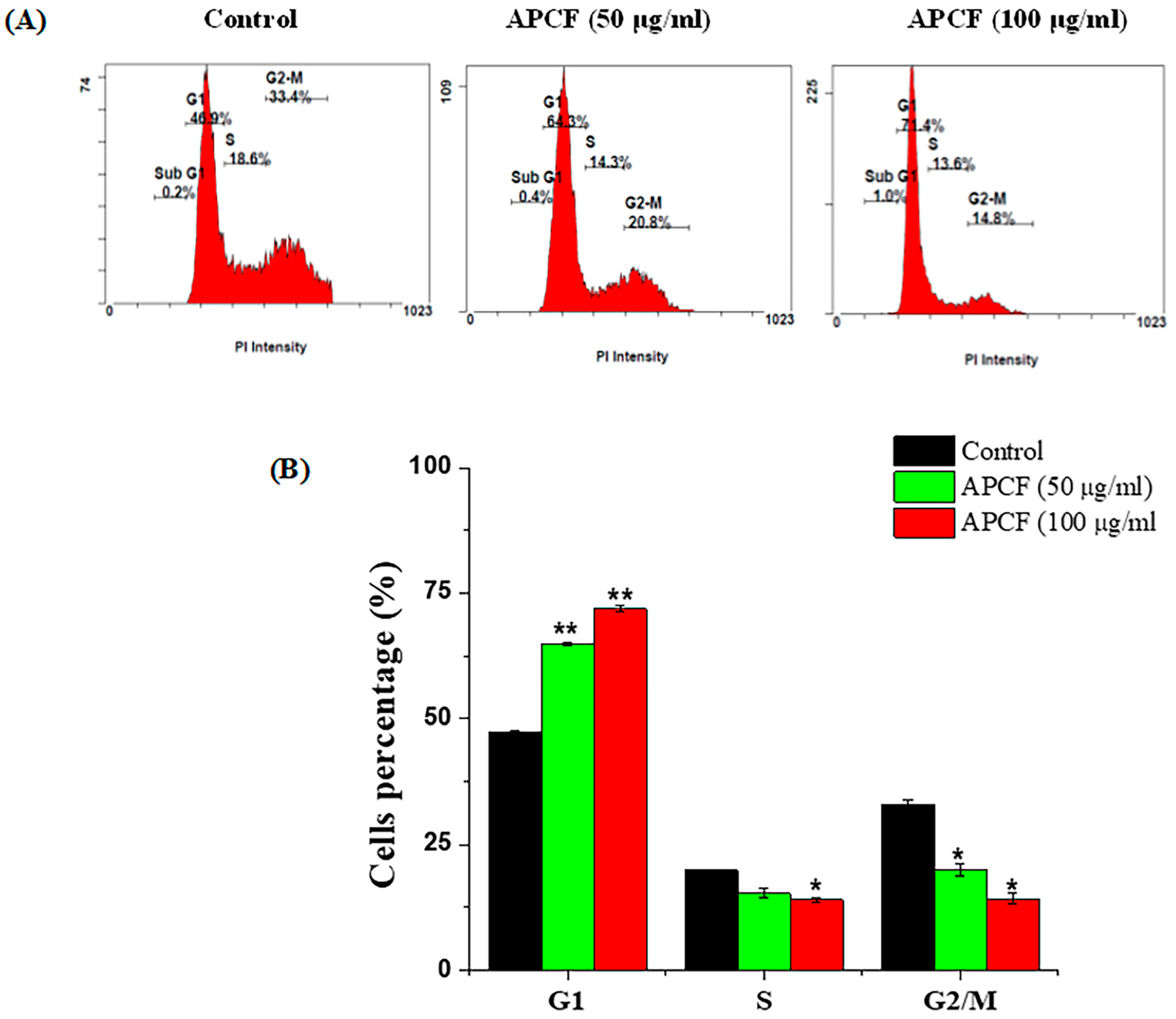
3.2. Cell Cycle Distribution after Treatment with APCF

Flow cytometry analysis was utilized to explore cell cycle phases after exposure to APCF in MCF-7 cells. The cells were treated for 48 h with two concentrations of APCF (50 and 100 μg/mL) or DMSO (0.1%) for controls. We found that exposure to APCF at concentrations of 50 and 100 µg/mL caused an enhancement in the proportion of MCF-7 cells in G1 phases (increased to 64.8 ± 0.42% and 71.8 ± 0.56%, respectively) compared to the controls (47.2 ± 0.42%). This increase in the G1 phase was associated with a reduction in the proportion of cells in the S and G2/M phases (Figure 2). These results clearly indicate that the growth inhibition of MCF-7 cancer cells mediated by APCF is associated with G1 phase cell cycle arrest.

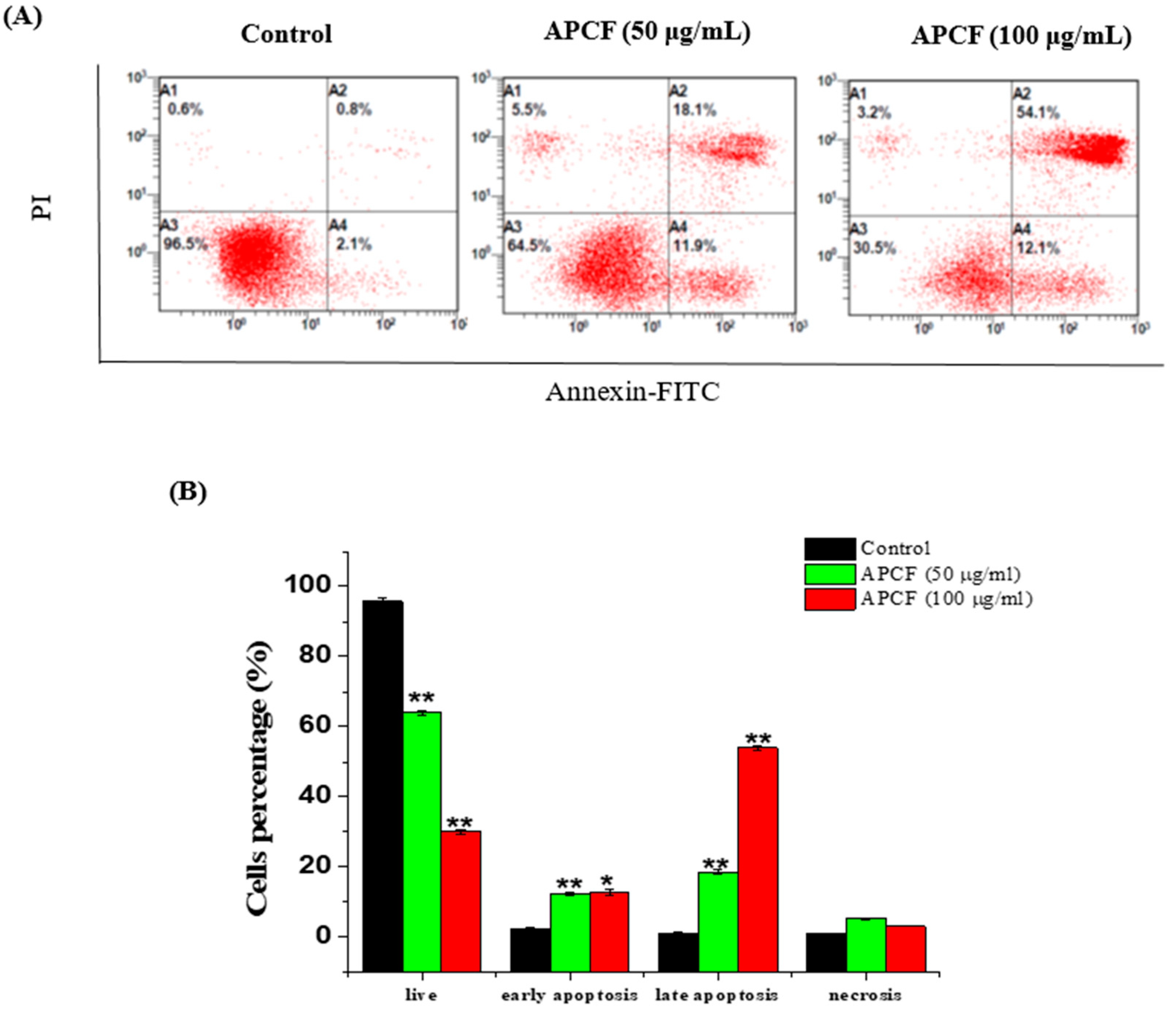
3.3. Apoptosis Evaluation of by Flow Cytometric Analysis

The induction of apoptosis was investigated in MCF-7 cells treated with APCF using the FITC-Annexin V/PI assay. Flow cytometry analysis showed that the proportion of apoptotic cells population (early and late) increased in APCF-treated MCF-7 cells compared to untreated control cells. After exposure to 50 µg/mL of APCF, MCF-7 cells displayed 12.2 ± 0.42% early apoptosis, 18.55 ± 0.63% late apoptosis, and 5.3 ± 0.28% necrosis (Figure 3). The proportion of cells in early and late apoptosis increased to 12.7 ± 0.84% and 54.1 ± 0.7%, respectively, upon increasing the concentration of APCF to 100 µg/mL. Our data clearly show that the percentage of apoptotic cells increased dose-dependently as the concentration of APCF increased.

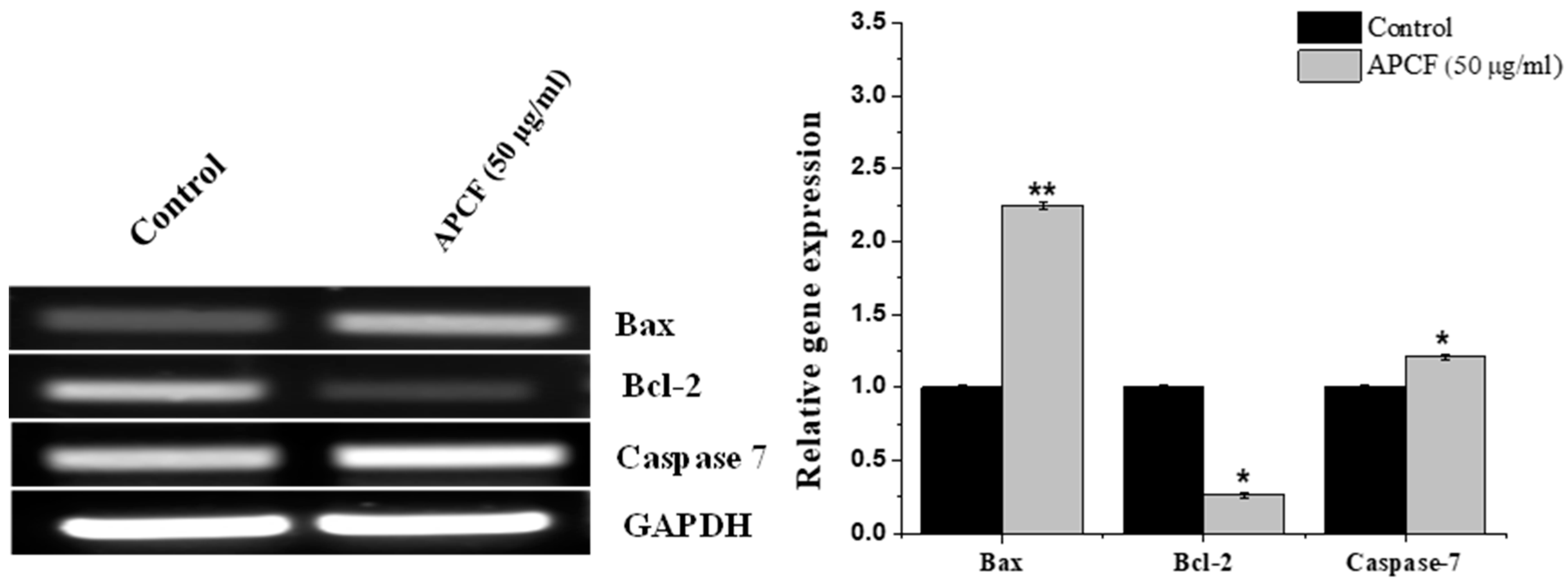
3.4. RT-PCR Analysis

MCF-7 cells were incubated with the IC50 concentration of the APCF fraction for 48 h, as well as DMSO as a vehicle in order to study the mechanism of apoptosis induction. RT-PCR was utilized to assess the expression of several apoptosis genes, including Bcl-2, Bax, and caspase-7. The RT-PCR analysis revealed an increase of 2.25-fold increase in Bax and a 1.21-fold increase in caspase-7 gene expression at the IC50 concentration of APCF compared to untreated control cells, while showing a 0.25-fold decrease in Bcl-2 gene expression at the same IC50 concentration of APCF compared to untreated control MCF-7 cells (Figure 4).

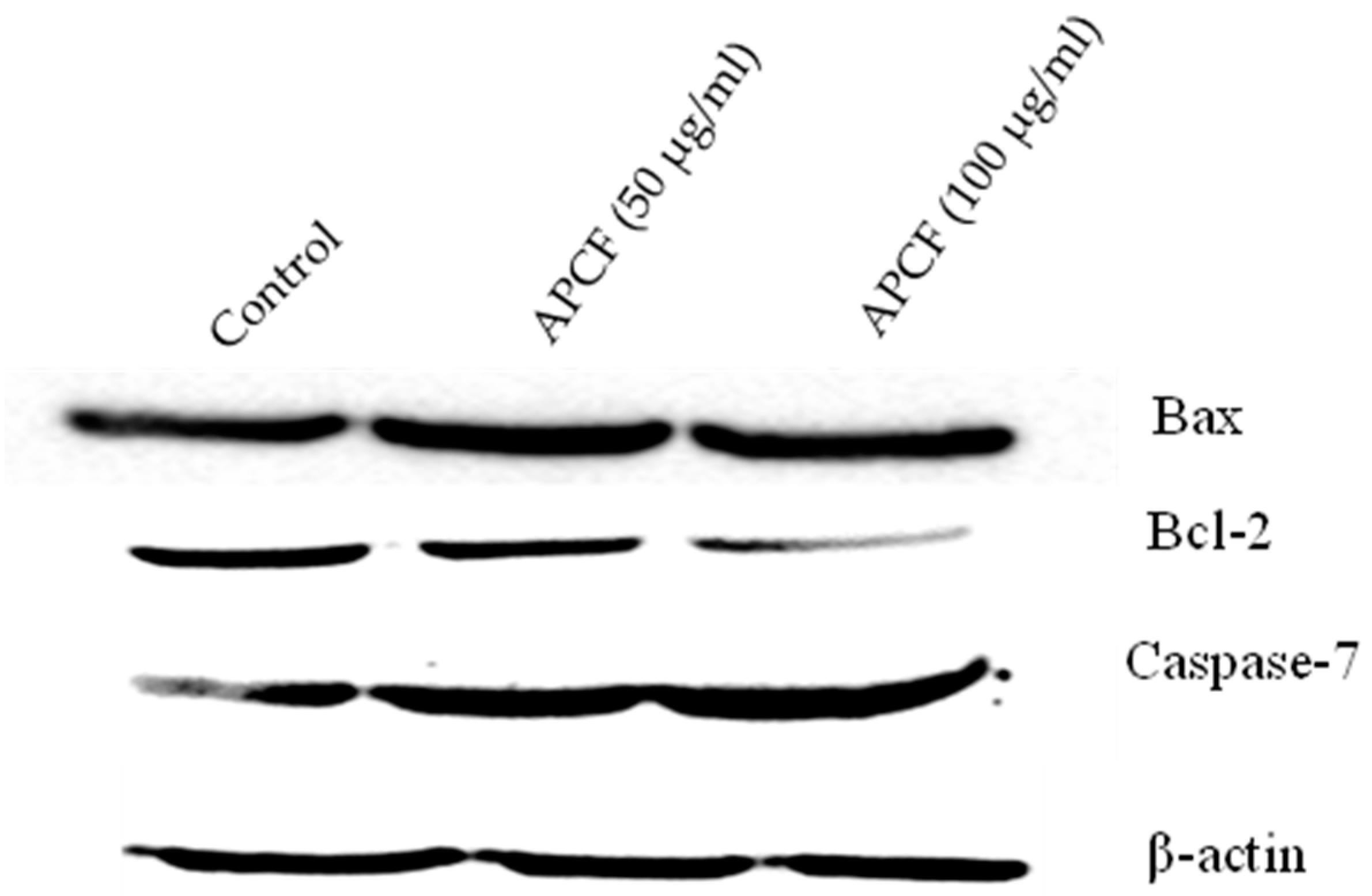
3.5. Alteration of Apoptosis Related Proteins Expression

Immunoblotting analysis was employed to measure the protein levels of Bcl-2, Bax, and caspase-7. The cells were treated for 48 h with two concentrations of APCF (50 and 100 μg/mL), or DMSO (0.1%) for controls. As shown in Figure 5, APCF significantly increased the expression of Bax, a protein that promotes apoptosis, while Bcl-2 (anti-apoptotic) was downregulated. The treatments with 50 and 100 μg/mL of APCF also led to the activation of caspase-7 in a dose-dependent manner (Figure 5). This data clearly indicates that APCF modulated the expression of apoptosis-related genes at the mRNA and protein level.

3.6. Phytochemical Analysis of APCF by GC/MS

Given the potential efficacy of the APCF fraction, GC-MS analysis was used to profile its chemical content (Figure 6). Based on their elution on the HP Innowax column, the chemical content, retention durations, and area percentages of APCF are displayed in Table 2. GC-MS analysis identified approximately 15 phytoconstituents. The major constituents identified in the extract were palmitic acid (50.46%), quinic acid (11.84%), alpha-d-glucopyranoside (11.15%), parthenolide (9.65%), and phytol (6.65%), and many other compounds were identified at low levels.

4. Discussion

Plants continue to be an important source of chemical compounds with diverse biological properties that provide safer and more potent drugs [22]. However, many medicinal plants still need extensive investigation to document their phytoconstituents, biological activities, toxicological studies, as well as their safety [23]. Among these medicinal plants, the Abutilon genus is considered an important medicinal plant that has been used for centuries to cure a variety of illnesses and diseases [24].
There are only a few reports available on the cytotoxic effect of the Abutilon genus. Consistent with our study, Khan et al. reported that A. indicum leaf extracts exhibited a cytotoxic effect against U87MG human glioblastoma cells, with IC50 values ranging from 42.6 to 64.5 g/mL [16]. Elshikh et al. studied the toxic effect of A. pannosum ethanoic extract on brine shrimp and the Vero normal cell line. They found that ethanolic extracts of A. pannosum were highly toxic to brine shrimp but had a weak cytotoxicity against Vero cells (IC50 = 279.94) [25]. In our study, we investigated the effects of ethanolic crude extract and different fractions of A. pannosum on various cancer cell lines for the first time and we found that the chloroform fraction exerted a potent cytotoxic effect in MCF-7 breast cancer cells. Moreover, our findings highlight the mode of cell death induced by A. pannosum.
Cell cycle progression is considered a crucial process for controlling cell division and proliferation. Various anticancer agents are being developed with the purpose of disrupting the progression of cell cycle [26]. Several plant phytochemicals are also known to induce cancer cell death by mediating cell cycle arrest and consequent cell death [27]. In light of this, the cell cycle progression following APCF treatment was studied. Consistent with the anti-proliferation observed using the MTT assay, we found that APCF arrested the cell cycle progression significantly at the G1 phase. A similar observation was described in HT-29 colon cancer cells, where polyphenol-stabilized nanoparticles prepared from Abutilon indicum leaf extract caused halting in the G1/S phase [28].
Apoptosis is an essential physiological process that balances cell survival and death without triggering an inflammatory response. Therefore, apoptosis stimulation is considered a key therapeutic approach in the management of cancer [29]. Phosphatidylserine, which is ordinarily only present in the inner leaflet of the plasma membrane, translocates to the cell surface during early apoptosis and can be detected using FITC (fluorochrome)-conjugated Annexin V. In contrast, cells lose their integrity at the final stages of apoptosis, allowing the staining of DNA with the fluorescent dye propidium iodide (PI). Both early (annexin V-positive, PI-negative) and late (annexin V-positive, PI-positive) apoptotic cells can be recognized and counted by utilizing FACS analysis with FITC-Annexin V and PI combined [30]. In this study, we performed annexin-FITC/PI staining to detect apoptosis activated by APCF in MCF-7 cells. Notably, the early and late apoptotic cells were dose-dependently increased following APCF treatment, as documented by flow cytometry analysis. Furthermore, to investigate the underlying mechanism, the gene expression levels of pro- and anti-apoptotic biomarkers were studied using RT-PCR. In fact, the modulation of Bcl-2 family members such as Bax and Bcl-2 have been reported to be associated with apoptosis [31]. Our results showed that APCF possessed an apoptotic effect by suppressing an anti-apoptotic marker (Bcl2) and enhancing a proapoptotic marker level (Bax). Moreover, APCF also upregulated caspase-7 expression, which has been implicated in activating apoptotic processes [32]. These data clearly demonstrate that APFC contains active ingredients with therapeutic potential. Therefore, GC-MS was applied to allow the identification of main constituents. GC-MS analysis has become a reliable phytochemical method for analyzing bioactive chemicals present in plant extracts [33]. In line with this study, previous GC-MS analysis revealed the presence of palmitic acid and phytol in methanolic leaf extracts of A. pannosum grown in Indian Thar desert [34]. However, different constituents with different proportions of other compound were observed, which could be attributed to geographic and genetics factors [35].
Palmitic acid, which has been reported to have antitumor effects [36,37], was one of the major components found in APCF (Table 2). Previous studies have also revealed that palmitic acid can induce apoptosis and cell cycle arrest in various cell types, including MCF-7 breast cancer cells [38,39,40,41]. Another important phytochemical found in APCF was quinic acid, which also has been reported to have anticancer action in breast cancer cells through apoptosis induction [42]. APCF also contains a considerable amount (9.65%) of the sesquiterpene lactone parthenolide. In numerous tumor models, parthenolide has been thoroughly investigated for its anticancer effects [43]. GC-MS analysis also revealed the presence of the phytol compound in a substantial amount (Table 2). Phytol’s inhibitory effects towards several cancer cells have been studied [44]. The cytotoxicity of phytol was assessed against seven tumor cells, and it was found that phytol exerted a concentration-dependent cytotoxic action in all cell lines, with the breast adenocarcinoma MCF-7 proving to be the most affected [45]. Moreover, several investigations have suggested a relationship between phytol and apoptosis and necrosis cells death in cancer, but no definitive findings have been reached [44]. It has been documented that synergistic effects play an important role in the interaction between plant compounds. In addition, plant-derived substances have a significant medicinal effect when used alone or in conjunction with traditional medications [46]. Thus, the observed antiproliferative activity of APCF may be explained by the synergistic effects of these compounds.

5. Conclusions

Our data revealed that the chloroform fraction of A. pannosum inhibited the proliferation of different cancer cells. In particular, the chloroform fraction of A. pannosum had a clear effect on MCF-7 breast cancer cells. Furthermore, we found that the chloroform fraction of A. pannosum induced cell arrest at the G1 phase and caused dose-dependent increases in early and late apoptotic cell populations of treated MCF-7 cells. Additionally, apoptosis initiation by the chloroform fraction of A. pannosum was supported by the elevation of proapoptotic gene markers in MCF-7 cells. In our study, we have also shown that the chloroform fraction of A. pannosum induced apoptosis by modulating BCl-2 proteins and upregulating caspase-7, which is critical for the induction of apoptosis. The major constituents found in A. pannosum chloroform fraction of were identified in the extract were palmitic acid (50.46%), quinic acid (11.84%), alpha-d-glucopyranoside (11.15%), parthenolide (9.65%), and phytol (6.65%), according GC-MS analysis. Thus, A. pannosum may represent a promising natural source of anticancer compounds that can cause apoptotic cell death. Further mechanistic studies as well as in vivo experiments are required to determine the potential cellular and molecular mechanisms behind the anticancer activity of A. pannosum and to confirm its efficiency.

Author Contributions

Conceptualization, M.A.-z. and A.A.Q.; methodology, I.O.B., F.A.N., O.M.N. and R.N.H.; writing—original draft preparation, M.A.-z. and F.A.N.; writing—review and editing, A.S.A., M.S.A. and H.A.R.; Resources, S.A.A.; funding acquisition, M.A.-z. All authors have read and agreed to the published version of the manuscript.

Funding

This research was funded by the Deanship of Scientific Research, Imam Mohammad Ibn Saud Islamic University (IMSIU), Saudi Arabia through Grant No. (221412019).

Data Availability Statement

Not applicable.

Acknowledgments

The authors extend their appreciation to the Deanship of Scientific Research, Imam Mohammad Ibn Saud Islamic University (IMSIU), Saudi Arabia, for funding this research work through Grant No. (221412019).

Conflicts of Interest

The authors declare no conflict of interest.

References

  1. Chhikara, B.S.; Parang, K. Global Cancer Statistics 2022: The trends projection analysis. Chem. Biol. Lett. 2023, 10, 451. [Google Scholar]
  2. Majolo, F.; de Oliveira Becker Delwing, L.K.; Marmitt, D.J.; Bustamante-Filho, I.C.; Goettert, M.I. Medicinal plants and bioactive natural compounds for cancer treatment: Important advances for drug discovery. Phytochem. Lett. 2019, 31, 196–207. [Google Scholar] [CrossRef]
  3. Petrovska, B.B. Historical review of medicinal plants’ usage. Pharm. Rev. 2012, 6, 1–5. [Google Scholar] [CrossRef] [PubMed]
  4. Christenhusz, M.J.; Byng, J.W. The number of known plants species in the world and its annual increase. Phytotaxa 2016, 261, 201–217. [Google Scholar] [CrossRef]
  5. Pearman, N.; Moxon, S.; Carnachan, S.M.; Cooke, M.E.; Nep, E.I.; Sims, I.M.; Morris, G.; Smith, A. Investigating potential wound healing properties of polysaccharides extracted from Grewia mollis Juss. and Hoheria populnea A. Cunn. (Malvaceae). Bioact. Carbohydr. Diet. Fibre 2019, 20, 100201. [Google Scholar] [CrossRef]
  6. Sikorska, M.; Matlawska, I. Polyphenolic compounds from Abutilon grandiflorum leaves. Acta Pol. Pharm. Drug Res. 2008, 65, 467–471. [Google Scholar]
  7. Baquar, S.R. Medicinal and Poisonous Plants of Pakistan. In Medicinal and Poisonous Plants of Pakistan; Printas: Karachi, Pakistan, 1989. [Google Scholar]
  8. Ahmed, Z.; Kazmi, S.N.-U.-H.; Malik, A. A new pentacyclic triterpene from Abutilon pakistanicum. J. Nat. Prod. 1990, 53, 1342–1344. [Google Scholar] [CrossRef]
  9. Hussain, M.; Zahra, D.N.; Hussain, S.S.; Ahmed, E.; Ahmad, I.; Malik, A.; Ahmed, Z. Structure determination of new steroids from Abutilon pakistanicum by NMR techniques. Magn. Reson. Chem. 2008, 46, 274–277. [Google Scholar] [CrossRef]
  10. Migahid, A. Flora of Saudi Arabia, 4th ed.; Routledge: London, UK, 1996; Volume 1, pp. 103–105. [Google Scholar]
  11. Abedin, S. Abutilon-muticum and Abutilon-pannosum complex. Pak. J. Bot. 1980, 12, 43–48. [Google Scholar]
  12. Ali, B.; Fatima, I.; Malik, A.; Ahmed, Z. New glycosidic constituents of Abutilon pakistanicum. Helv. Chim. Acta 2010, 93, 2245–2250. [Google Scholar] [CrossRef]
  13. Aadesariya, M.K.; Ram, V.R.; Dave, P.N. Extraction, Isolation and Identification of Useful Phyto Constituents from Dichloromethane Leave Extract of Abutilon pannosum and Grewia tenax Using Q-TOF LC/MS. Int. J. Adv. Res. Chem. Sci. 2017, 4, 1–14. [Google Scholar]
  14. Khalil, I.; Ghani, M.; Khan, M.R.; Akbar, F. Evaluation of biological activities and in vivo amelioration of CCl4 induced toxicity in lung and kidney with Abutilon pannosum (G. Forst.) Schltdl. in rat. J. Ethnopharmacol. 2020, 249, 112395. [Google Scholar] [CrossRef]
  15. Bano, I.; Deora, G. Studies on micro morphological taxonomic variations in Abutilon species of Indian Thar Desert. IOSR J. Pharm. Biol. Sci. 2017, 12, 60–68. [Google Scholar]
  16. Khan, R.S.; Senthi, M.; Rao, P.C.; Basha, A.; Alvala, M.; Tummuri, D.; Masubuti, H.; Fujimoto, Y.; Begum, A.S. Cytotoxic constituents of Abutilon indicum leaves against U87MG human glioblastoma cells. Nat. Prod. Res. 2015, 29, 1069–1073. [Google Scholar] [CrossRef] [PubMed]
  17. Gouda, H.M.; Morsy, A.A.; Youssef, A.K.; Tolba, I.A.E.-M.; Selim, A.A.M.A. The ethyl acetate extract from Abutilon fruticosum Guill and Perr. as a potential diabetes–cancer prophylactic: A cytotoxic, α-glucosidase, and in-silico study. S. Afr. J. Bot. 2023, 156, 110–114. [Google Scholar] [CrossRef]
  18. Alqahtani, A.S.; Ghorab, M.M.; Nasr, F.A.; Ahmed, M.Z.; Al Mishari, A.A.; Attia, S.M.; Farooq Khan, M. Cytotoxicity of Newly Synthesized Quinazoline–Sulfonamide Derivatives in Human Leukemia Cell Lines and Their Effect on Hematopoiesis in Zebrafish Embryos. Int. J. Mol. Sci. 2022, 23, 4720. [Google Scholar] [CrossRef]
  19. Nasr, F.A.; Noman, O.M.; Alqahtani, A.S.; Qamar, W.; Ahamad, S.R.; Al-Mishari, A.A.; Alyhya, N.; Farooq, M. Phytochemical constituents and anticancer activities of Tarchonanthus camphoratus essential oils grown in Saudi Arabia. Saudi Pharm. J. 2020, 28, 1474–1480. [Google Scholar] [CrossRef] [PubMed]
  20. Adams, R.P. Identification of Essential Oil Components by Gas Chromatography/Mass Spectrometry; Allured Publishing Corporation: Carol Stream, IL, USA, 2007; Volume 456. [Google Scholar]
  21. McLafferty, F.W.; Stauffer, D.B. The Wiley/NBS Registry of Mass Spectral Data; Wiley: New York, NY, USA, 1989; Volume 1. [Google Scholar]
  22. Talib, W.H.; Daoud, S.; Mahmod, A.I.; Hamed, R.A.; Awajan, D.; Abuarab, S.F.; Odeh, L.H.; Khater, S.; Al Kury, L.T. Plants as a Source of Anticancer Agents: From Bench to Bedside. Molecules 2022, 27, 4818. [Google Scholar] [CrossRef] [PubMed]
  23. Willis, K. State of the World’s Plants 2017; Royal Botanics Gardens Kew: Richmond, UK, 2017. [Google Scholar]
  24. Agrawal, T. Abutilon a vulnerable genus: A review. Res. Rev. J. Pharmacogn. Phytochem. 2017, 5, 44–46. [Google Scholar]
  25. Elshikh, A.A.; ElNour, M.E.; Elkamali, H.H.; Kabbashi, A.S. In-vitro Testing of Antioxidant, Anti-Parasite Activities, Cytotoxicity, and Chemical Evaluation of Abutilon pannosum and Cassia occidentalis Ethanolic Extracts. Annu. Res. Rev. Biol. 2021, 36, 112–121. [Google Scholar] [CrossRef]
  26. Suski, J.M.; Braun, M.; Strmiska, V.; Sicinski, P. Targeting cell-cycle machinery in cancer. Cancer Cell 2021, 39, 759–778. [Google Scholar] [CrossRef] [PubMed]
  27. Singh, R.P.; Dhanalakshmi, S.; Agarwal, R. Phytochemicals as cell cycle modulators—A less toxic approach in halting human cancers. Cell Cycle 2002, 1, 156–161. [Google Scholar] [CrossRef]
  28. Mata, R.; Nakkala, J.R.; Sadras, S.R. Polyphenol stabilized colloidal gold nanoparticles from Abutilon indicum leaf extract induce apoptosis in HT-29 colon cancer cells. Colloids Surf. B Biointerfaces 2016, 143, 499–510. [Google Scholar] [CrossRef]
  29. Carneiro, B.A.; El-Deiry, W.S. Targeting apoptosis in cancer therapy. Nat. Rev. Clin. Oncol. 2020, 17, 395–417. [Google Scholar] [CrossRef] [PubMed]
  30. van Engeland, M.; Nieland, L.J.; Ramaekers, F.C.; Schutte, B.; Reutelingsperger, C.P. Annexin V-affinity assay: A review on an apoptosis detection system based on phosphatidylserine exposure. Cytometry 1998, 31, 1–9. [Google Scholar] [CrossRef]
  31. Levine, B.; Sinha, S.C.; Kroemer, G. Bcl-2 family members: Dual regulators of apoptosis and autophagy. Autophagy 2008, 4, 600–606. [Google Scholar] [CrossRef]
  32. McComb, S.; Chan, P.K.; Guinot, A.; Hartmannsdottir, H.; Jenni, S.; Dobay, M.P.; Bourquin, J.P.; Bornhauser, B.C. Efficient apoptosis requires feedback amplification of upstream apoptotic signals by effector caspase-3 or -7. Sci. Adv. 2019, 5, eaau9433. [Google Scholar] [CrossRef]
  33. Konappa, N.; Udayashankar, A.C.; Krishnamurthy, S.; Pradeep, C.K.; Chowdappa, S.; Jogaiah, S. GC-MS analysis of phytoconstituents from Amomum nilgiricum and molecular docking interactions of bioactive serverogenin acetate with target proteins. Sci. Rep. 2020, 10, 16438. [Google Scholar] [CrossRef]
  34. Bano, I.; Deora, G. Preliminary phytochemical screening and GC-MS analysis of methanolic leaf extract of Abutilon pannosum (Forst. F.) Schlect. from Indian Thar desert. J. Pharmacogn. Phytochem. 2019, 8, 894–899. [Google Scholar]
  35. Shams, M.; Esfahan, S.Z.; Esfahan, E.Z.; Dashtaki, H.N.; Dursun, A.; Yildirim, E. Effects of climatic factors on the quantity of essential oil and dry matter yield of coriander (Coriandrum sativum L.). Indian J. Sci. Technol. 2016, 9, 1–4. [Google Scholar] [CrossRef]
  36. Harada, H.; Yamashita, U.; Kurihara, H.; Fukushi, E.; Kawabata, J.; Kamei, Y. Antitumor activity of palmitic acid found as a selective cytotoxic substance in a marine red alga. Anticancer Res. 2002, 22, 2587–2590. [Google Scholar] [PubMed]
  37. Zhu, S.; Jiao, W.; Xu, Y.; Hou, L.; Li, H.; Shao, J.; Zhang, X.; Wang, R.; Kong, D. Palmitic acid inhibits prostate cancer cell proliferation and metastasis by suppressing the PI3K/Akt pathway. Life Sci. 2021, 286, 120046. [Google Scholar] [CrossRef] [PubMed]
  38. Pereira, D.M.; Correia-da-Silva, G.; Valentão, P.; Teixeira, N.; Andrade, P.B. Palmitic acid and ergosta-7, 22-dien-3-ol contribute to the apoptotic effect and cell cycle arrest of an extract from Marthasterias glacialis L. in neuroblastoma cells. Mar. Drugs 2013, 12, 54–68. [Google Scholar] [CrossRef]
  39. Zafaryab, M.; Fakhri, K.U.; Khan, M.A.; Hajela, K.; Rizvi, M.M.A. In vitro assessment of cytotoxic and apoptotic potential of palmitic acid for breast cancer treatment. Int. J. Life Sci. Res. 2019, 7, 166–174. [Google Scholar]
  40. Liang, H.; Zhong, Y.; Zhou, S.; Li, Q.Q. Palmitic acid-induced apoptosis in pancreatic β-cells is increased by liver X receptor agonist and attenuated by eicosapentaenoate. In Vivo 2011, 25, 711–718. [Google Scholar] [PubMed]
  41. Yu, G.; Luo, H.; Zhang, N.; Wang, Y.; Li, Y.; Huang, H.; Liu, Y.; Hu, Y.; Liu, H.; Zhang, J.; et al. Loss of p53 Sensitizes Cells to Palmitic Acid-Induced Apoptosis by Reactive Oxygen Species Accumulation. Int. J. Mol. Sci. 2019, 20, 6268. [Google Scholar] [CrossRef] [PubMed]
  42. Samimi, S.; Ardestani, M.S.; Dorkoosh, F.A. Preparation of carbon quantum dots-quinic acid for drug delivery of gemcitabine to breast cancer cells. J. Drug Deliv. Sci. Technol. 2021, 61, 102287. [Google Scholar] [CrossRef]
  43. Karam, L.; Abou Staiteieh, S.; Chaaban, R.; Hayar, B.; Ismail, B.; Neipel, F.; Darwiche, N.; Abou Merhi, R. Anticancer activities of parthenolide in primary effusion lymphoma preclinical models. Mol. Carcinog. 2021, 60, 567–581. [Google Scholar] [CrossRef]
  44. Alencar, M.; Islam, M.T.; Ali, E.S.; Santos, J.V.O.; Paz, M.; Sousa, J.M.C.; Dantas, S.; Mishra, S.K.; Cavalcante, A. Association of Phytol with Toxic and Cytotoxic Activities in an Antitumoral Perspective: A Meta-Analysis and Systemic Review. Anticancer Agents Med. Chem. 2018, 18, 1828–1837. [Google Scholar] [CrossRef]
  45. Pejin, B.; Kojic, V.; Bogdanovic, G. An insight into the cytotoxic activity of phytol at in vitro conditions. Nat. Prod. Res. 2014, 28, 2053–2056. [Google Scholar] [CrossRef]
  46. Pezzani, R.; Salehi, B.; Vitalini, S.; Iriti, M.; Zuñiga, F.A.; Sharifi-Rad, J.; Martorell, M.; Martins, N. Synergistic effects of plant derivatives and conventional chemotherapeutic agents: An update on the cancer perspective. Medicina 2019, 55, 110. [Google Scholar] [CrossRef] [PubMed]
Figure 1. Antiproliferative effects of the A. pannosum chloroform fraction (APCF) on different cancer cells. The cells were exposed to increasing concentrations APCF for 48 h and cell viability was assessed by MTT assay. All values are given as means ± SDs from three independent experiments.
Processes 11 01306 g001
Figure 2. Effect of APCF on MCF-7 cell cycle phase distributions. (A) Cycle phase distribution of control cells and cells exposed to 50 and 100 μg concentrations of APCF analyzed using flow cytometry. (B) Bar graph representing the average percentage of cells in each phase for the treated and control groups. Data represent the average ± SD (standard deviation) of three independent experiments. Significance: * p ≤ 0.05 and ** p ≤ 0.01.
Processes 11 01306 g002
Figure 3. Effect of APCF on cell apoptosis as revealed by Annexin V-FITC/PI. (A) Dot plots of control and APCF-treated cells at concentrations of 50 and 100 µg/mL. (B) The percentage (%) of early apoptosis, late apoptosis, and necrotic MCF-7 cells after treatment. Data represent the average ± SD (standard deviation) of three independent experiments. Significance: * p ≤ 0.05 and ** p ≤ 0.01.
Processes 11 01306 g003
Figure 4. Effect of APCF on the gene expression of Bax, Bcl-2, and caspase-7 genes in MCF-7 cells detected using RT-PCR. Total RNA was prepared from untreated and treated cells, and RT-PCR were performed with specific primers for corresponding genes. Values are expressed as mean ± SD. *, ** indicates statistically significant difference at (p ≤ 0.05 and p ≤ 0.01 respectively).
Processes 11 01306 g004
Figure 5. APCF modulated Bax, Bcl-2, and caspase-7 expression in human MCF-7 cells. The cells were exposed to various concentrations of APCF (50 and 100 µg/mL), and the expression of Bcl-2, Bax, and caspase-7 was examined using western blots. Antibodies against each target gene were included to elucidate apoptosis induction, and a β-actin antibody was used as a loading control.
Processes 11 01306 g005
Figure 6. GC-MS chromatogram for some constituents of APCF.
Processes 11 01306 g006
Table 1. IC50 values (µg/mL) for A. pannosum fractions against various cancer cells.
Cell TypeIC50 Values (μg/mL)
CrudeCHCl3BuOHMeOHDoxorubicin
Lung (A549)174.2 ± 5.686.2 ± 2.9169.1 ± 2.6167.9 ± 2.90.98 ± 0.02
liver (HepG2)147.9 ± 3.278.5 ± 2.1112.5 ± 3.5174.7 ± 4.51.3 ± 0.4
Breast (MCF-7)99.5 ± 1.550.1 ± 1.2147.6 ± 4.2181.5 ± 4.91.1 ± 0.3
Table 2. GC-MS analysis of phytoconstituents identified in APCF.
Compound NameChemical FormulaMolecular Weight (g/mol)RT (min)%Area
p-Menthan-8-yl acetateC12H22O2198.3013.352.690
Alpha-d-glucopyranosideC6H11O6179.1516.1611.150
Quinic acidC7H12O6192.1719.0511.840
D-Glycero-d-ido-heptoseC7H14O7210.1819.681.780
Dihydro-alpha-iononeC13H22O194.3121.770.280
Methyl palmitateC17H34O2270.521.840.700
Ethyl palmitateC18H36O2284.522.530.490
Stearic acidC18H36O2284.523.381.740
alpha-Linolenic acidC18H30O2278.423.620.610
PhytolC20H40O296.523.746.650
Caryophyllene epoxideC15H24O220.3523.980.420
ParthenolideC15H20O3248.3224.489.650
gamma-elemeneC15H24204.3524.640.960
palmitic acidC16H32O2256.4225.7750.460
14-Pentadecenoic acid C15H28O2240.3828.010.320
Disclaimer/Publisher’s Note: The statements, opinions and data contained in all publications are solely those of the individual author(s) and contributor(s) and not of MDPI and/or the editor(s). MDPI and/or the editor(s) disclaim responsibility for any injury to people or property resulting from any ideas, methods, instructions or products referred to in the content.

