Article

MALDI-TOF Mass Spectrometry-Based Identification of Aerobic Mesophilic Bacteria in Raw Unpreserved and Preserved Milk

by
Nataša Mikulec
1,
Jasminka Špoljarić
1,
Dijana Plavljanić
1,
Nina Lovrić
1,
Fabijan Oštarić
1,
Jasenka Gajdoš Kljusurić
2,
Khan Mohd. Sarim
3,
Nevijo Zdolec
4 and
Snježana Kazazić
3,*
1
Faculty of Agriculture, University of Zagreb, 10000 Zagreb, Croatia
2
Faculty of Food Technology and Biotechnology, University of Zagreb, 10000 Zagreb, Croatia
3
Ruđer Bošković Institute, Bijenička cesta 54, 10000 Zagreb, Croatia
4
Faculty of Veterinary Medicine, University of Zagreb, 10000 Zagreb, Croatia
*
Author to whom correspondence should be addressed.
Processes 2024, 12(4), 731; https://doi.org/10.3390/pr12040731
Submission received: 21 March 2024 / Revised: 29 March 2024 / Accepted: 2 April 2024 / Published: 4 April 2024
(This article belongs to the Special Issue Microbial Cultures in Food Production)

Abstract

:
The number of aerobic mesophilic bacteria in milk is one of the indicators of the hygienic quality of milk. The aim of this work was to determine such aerobic mesophilic bacteria and their number in raw unpreserved milk and milk preserved with sodium azide. In 40 collected samples, the total number of aerobic mesophilic bacteria was determined using the classical method of counting colonies on a nutrient medium according to the international standard HRN EN ISO 4833-1:2013. The results showed a trend of decreasing the number of grown colonies in milk preserved with sodium azide. MALDI-TOF mass spectrometry also successfully identified 392 bacterial colonies in raw unpreserved milk samples and 330 colonies in preserved milk samples. Of these, 30 genera and 54 bacterial species were identified in the raw unpreserved milk samples, while 27 genera and 41 bacterial species were identified in the preserved samples. By using a collective approach, the present study provided a more detailed insight into milk’s hygienic quality and the presence of certain species before and after the preservation with sodium azide.

1. Introduction

Due to its rich chemical composition and physical properties that are favorable for most microorganisms, milk is an ideal medium for the growth and reproduction of various types of bacteria. The basic components of milk, including proteins, lactose and milk fat, have great biological and nutritional value. In addition to its chemical composition, the physical properties of milk also contribute to the rapid growth and reproduction of a large number of bacterial species [1]. Moreover, biological activity is an integral aspect of the quality standard and the taste of milk. In this context, specifically, aerobic mesophilic bacteria are those that grow only in the presence of oxygen between 20–45 °C with an optimum temperature range of 30–37 °C. This group of bacteria is one of the most general microbiological indicators of food quality that indicates the adequacy of temperature control and sanitation procedures during processing, transport and storage and reveals sources of contamination during milk production and subsequent processing [2]. Many aerobically mesophilic species of bacteria have an optimal growth temperature of around 37 °C [3]. These bacteria are frequent causes of milk spoilage and belong to the group of pathogenic microorganisms. The overall quality of raw milk is determined by its chemical and physical properties and hygienic quality. As stated by Antunac and Havranek [1], the hygienic quality of milk is determined by the total number of aerobic mesophilic bacteria and the total number of somatic cells present in milk.
Aerobic mesophilic bacteria found in raw milk include the following genera: Micrococcus, Enterococcus, Staphylococcus, Lactococcus, Serratia, Acinetobacter, Flavobacterium, Lactobacillus, Escherichia, Bacillus, Mycobacterium, Pseudomonas and others [4]. The main types of aerobic mesophilic bacteria found in raw milk are micrococci, streptococci, sporogenous Gram-positive bacteria from the genus Mycobacterium and Corynebacterium and sporogenous bacteria from the genus Bacillus [1]. They are considered universal indicators of hygiene in milk processing. Their presence in raw milk is inevitable because most of these genera are present in the udder, on the hands of the milker, on the surface of the equipment and in the air. The number of mesophilic bacteria in milk is directly affected by the conditions in which the milk is kept after milking. Raw milk with an aerobic mesophilic bacteria count higher than 5.0 log10 CFU/mL indicates poor hygiene during milking and production, while a count lower than 3.0 log10 CFU/mL indicates good production practices. Appropriate cooling (+4 °C ± 2 °C), good production practices and hygiene during milking are effective measures that successfully prevent subsequent contamination of raw milk with this group of bacteria [5].
Control of the hygienic quality of raw milk is necessary to ensure the health safety of milk and milk products. According to the Rulebook regarding the quality of fresh raw milk of the Ministry of Agriculture, Croatia (OG 136/20) [6], which is in accordance with the EU legislation, raw milk is classified depending on the number of microorganisms. Because of that, it is necessary to provide a cold chain that enables adequate transport of milk samples from the milk producer to the milk-testing laboratories where the hygienic quality and the physico-chemical composition of milk are analyzed. However, it is not always possible to provide an adequate cold chain, so substances that prevent microbial growth are often used. A milk preservative can be defined as any chemical compound that, when used in milk, prevents changes caused by contamination, growth and reproduction of microorganisms [7].
An ideal preservative protects against mechanical damage to fat globules. Today, various preservatives are used in laboratories for milk quality control, and their application is imperative in modern milk analysis. Preservatives are used to prevent contamination of raw milk from the farm to the laboratory and its rapid spoilage, which can compromise the results of the analysis. Milk samples very often come from rural areas where hygiene is not satisfactory, and subsequent contamination of milk samples is common, especially in the summer months when higher temperatures favor the growth of various types of bacteria. Because milk is a suitable medium for microbial growth and reproduction, preservatives have been introduced in milk-testing laboratories to ensure that milk composition remains unchanged and constant from the farm to the testing laboratory. In the past, the most commonly used preservatives to reduce microbial growth in milk were hydrogen peroxide, formaldehyde, potassium dichromate, mercuric chloride, boric acid or their combinations. Today, the more commonly used agents are bronopol, azidiol and sodium azide (NaN3), while K-dichromate is used less often [8].
The antimicrobial action of preservatives is achieved through the following processes: inhibition of protein synthesis, inhibition of enzymatic activity and damage to the cell membrane. Samaržija et al. [9] state that using sodium azide does not change the chemical composition or the microbial population in milk samples but prevents growth as a bacteriostatic, so the number of bacteria is reduced when analyzed after 2–3 days. Cabrol et al. [10] examined the effective concentration and sodium azide role in inhibition and concluded that 50 mM of sodium azide strongly inhibited microbial growth. Many research studies confirmed the inhibitory role of sodium azide on cell division [11] and the growth of bacteria through inhibiting the SecA protein required for protein translocation [12,13]. Suresh et al. [14] also noticed the inhibitory role of sodium azide on sludge destabilization. Sodium azide’s effect in inhibiting the biofilm formed by Escherichia coli was evaluated alongside silver nanoparticles and flashlight, and it was concluded that sodium azide was more effective because it blocked the ATPase chain and respiratory chain that lead to cell growth inhibition [15]. Few lactic acid bacteria (LAB) species are resistant to sodium azide. It is an inhibitor of cytochrome oxidase, which causes impairment of respiration; thus, aerobic bacteria are most affected by sodium azide. It is noteworthy that sodium azide strongly inhibits iron porphyrin and, therefore, inhibits most non-starter lactic acid bacteria (NSLAB), and a few fungi species as well, but LAB lack synthesizing iron porphyrin; thus, they can grow in the presence of sodium azide [16]. Adding sodium azide to milk does not generally change the milk’s physico-chemical structure, such as casein micelles, during milk storage at 4 °C, but it prevents microbial growth [17,18]. Sodium azide is the commonly used preservative in all milk testing laboratories for chemical and bacteriological tests.
Since preservatives with a bacteriostatic effect are mainly used to determine the hygienic quality of milk in official laboratories, the aim of this work was to determine whether there are differences in the bacterial population regarding the species and genus of bacteria between preserved and raw milk. The above-mentioned research has shown that preservatives affect the reduction of the total number of bacteria in milk, but they were not aimed at determining differences in bacterial species when a preservative was added to milk before analysis.
Using the classical method of counting colonies on a nutrient medium according to the international standard HRN EN ISO 4833-1:2013 [19] and matrix-assisted laser desorption/ionization time of flight mass spectrometry (MALDI-TOF MS), the influence of preservatives on the number and representation of aerobic mesophilic bacteria in raw unpreserved and preserved milk can be evaluated. Today, MALDI-TOF MS is an increasingly well-known, economical method that finds application in the rapid identification of microorganisms (bacteria, fungi (yeasts and molds)).
To evaluate the impact of sodium azide on microbial population in milk preservation, the present study aimed to enumerate the aerobic mesophilic bacteria in unpreserved raw milk and milk with added sodium azide using the classical plate method along with microbial typing using MALDI-TOF mass spectrometry.

2. Materials and Methods

2.1. Milk Sampling

A total of 40 raw cow’s milk samples in four batches (10 samples in each) were collected during the spring months from local milk producer (45.832554, 15.927526), Zagreb County, Croatia. The milk samples were collected into sterile bottles of 30 mL (in parallel) and transported to a laboratory in a portable refrigerator at a temperature of +4 °C (±2 °C). All samples were stored in refrigerator at +4 °C (±2 °C) until analysis. All analyses were started within 12 h of sampling.
One bottle of 30 mL with raw milk was used for determination of the number of mesophilic bacteria in unpreserved raw milk, along with microbial typing using potential MALDI-TOF mass spectrometry. For the evaluation of the impact of sodium azide on microbial population in raw milk, another sterile bottle of 30 mL of milk was preserved with the addition of 8 mg sodium azide tablet (Merck, Darmstadt, Germany). The final concentration of sodium azide in milk sample was 0.027% (w/v). Due to the minimal influence of the addition of sodium azide on the physico-chemical properties of the milk samples, especially for the determination of the somatic cell count, sodium azide was added in a maximum concentration of 0.024 g/100 mL of the sample [20].

2.2. Classical Method of Determining the Aerobic Mesophilic Bacteria Count in Raw Milk

The aerobic mesophilic bacteria count in preserved/unpreserved raw milk was determined in the nutrient medium according to the international norm HRN EN ISO 4833-1:2003 [19].
Milk samples were mixed in a vibrating mixer before analysis. To prepare a milk sample with a dilution of 10−1, 1 mL of milk was pipetted into a test tube with 9 mL of sterile peptone solution and prepared serial dilutions of milk samples in peptone buffer up to dilution of 10−6. Subsequently, 1 mL from each dilution was poured on the appropriate Petri dish, mixed with 12–15 mL of nutrient medium and incubated at 30 °C for 72 h. Colony counting was performed according to the prescribed criteria in the international standard HRN EN ISO 4833-1:2003 [19]. Petri dishes that gave a colony count of between 10 and 300 grown colonies per plate were taken for counting and further identification.

2.3. MALDI-TOF Mass Spectrometry

A single bacterial colony was taken with a sterile toothpick and smeared onto a 96-spot MALDI target plate, and 1 µL of 70% formic acid (Fisher Chemical, Alcobendas, Spain) was added. After drying at room temperature, each spot overlaid with 1 μL of 10 mg/mL alpha-4-cyano-4-hydroxycinnamic acid (CHCA, Bruker Daltonik, Bremen, Germany) in 50% acetonitrile and 2.5% trifluoroacetic acid and allowed to dry.
Measurements were performed using a Microflex LT mass spectrometer (Bruker Daltonik, Bremen, Germany), and spectra were acquired in positive linear mode within a mass range of 2000 to 20,000 Da. MBT Compass HT version 5.1 software (Bruker Daltonik, Bremen, Germany) was used for spectra matching to a reference database, version 11. External calibration was performed using the Bacterial Test Standard (Bruker Daltonik, Bremen, Germany). Identification criteria were as follows: a log score of 2.00–3.00 indicated high-confidence species identification, a log score of 1.70–1.99 indicated low-confidence species identification, while a score of 0–1.69 was considered unreliable identification.

2.4. Data Analysis

To determine the differences in the mean values of observed milk types and milks from different batches, an analysis of variance (ANOVA) was performed, and the Mann–Whitney U test was used (with the significance level of 0.05). Box–Whisker diagrams were used for the comparison of raw unpreserved and preserved milk from different batches. As the multivariate tool for quantitative data, the principal component analysis was used to explore the shares of different identification scores (high, low, none) in relation to total number of bacteria. As a multivariate tool for analyzing qualitative data, exploratory data analysis and heat map were used to investigate the grouping of bacteria species in different batches for the milk in two forms: (i) raw unpreserved and (ii) preserved. The scale ranged from red (impossible identification of the microorganism), through yellow (low-confidence identification), to green (high-confidence identification). Statistical Software for Excel (XLStat, 2007) was used in the qualitative analysis.

3. Results and Discussion

The study was framed to enumerate and identify the aerobic mesophilic bacteria in raw unpreserved and preserved milk with sodium azide. All the milk samples collected in four different batches were analyzed using the classical method of counting colonies on a nutrient medium, according to the HRN EN ISO 4833-1:2013 [19]. In addition, the MALDI-TOF MS approach was applied to identify microbes and the impact of azide preservation on microbial populations in different samples.

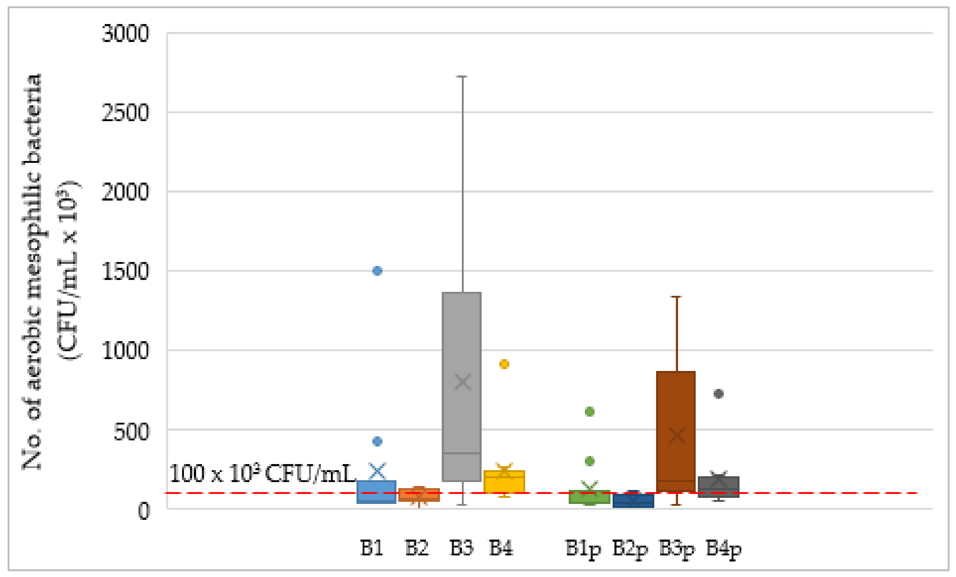
3.1. Classical Method of Determining the Number of Aerobic Mesophilic Bacteria in Raw and Preserved Milk

Table 1 shows comparative data of the number of CFU/mL in raw unpreserved and preserved milk in four batches (Batch 1 to Batch 4), 10 samples each.
Of the total number of samples (40) of raw unpreserved milk collected in four batches during the early spring months, 22 samples (55%) were not compliant with the prescribed requirement for the total bacterial count of less than 100,000 CFU/mL for milk of the first class, according to the Rulebook (OG 136/20) [6] regarding the quality of fresh raw milk of the Ministry of Agriculture, Croatia (Table 1).
The requirements for raw milk (Class 1; ≤100,000 CFU/mL) were met by 80% and 70% of the raw milk samples in Batches 1 and 2, respectively. Subsequently, comparatively higher CFU/mL counts were observed in Batches 3 and 4, where the hygienic quality of the raw milk was poor. Only 10% and 20% of the raw milk samples were compliant with the prescribed requirement, in Batches 3 and 4, respectively. Such a high number of non-compliant raw milk samples suggested that the poor hygienic quality of raw milk is probably due to the low hygiene during milking and the manipulation of milk after milking (Table 1).
According to the Croatian Agency for Agriculture and Food (HAPIH) in the Republic of Croatia in 2022 [21], 96% of milk belongs to the first class, which is why this requirement for the total number of microorganisms, ≤100,000 CFU/mL, is used as part of milk control and for the classification and determination of milk price.
It is also necessary to keep milk composition unchanged for a long period for chemical and bacteriological tests. For this purpose, various preservatives have been used [7,22,23]. In the present study, aerobic mesophilic bacteria in raw unpreserved and preserved milk samples with the addition of sodium azide were examined, and the impact microbial growth was evaluated.
Lichstein and Soule [24] published the first results on the influence of sodium azide on the growth and metabolism of the bacteria Bacillus subtilis and Pseudomonas aeruginosa, discovering the inhibition of oxygen consumption and reduced bacterial growth at very low concentrations of sodium azide. Sodium azide is used in bacteriological laboratories to prevent microbial growth in milk [22]. Therefore, the evaluation of raw unpreserved and sodium azide-preserved milk is important for subsequent milk studies. In addition to this, the observation of microbial populations in preserved milk is also an important aspect of monitoring the biological activity and keeping quality of milk.
The aerobic mesophilic bacteria in milk indicate overall raw milk quality because the majority of these bacteria might be present on the udder of the animal or the hands of a milker and give clear indication of unhygienic practice in pre- and post-milking processes.
The addition of sodium azide resulted in a reduction of the microbial population in all the analyzed milk samples in all four batches. The average number of bacteria in Batch 1 of raw unpreserved milk was reduced by adding sodium azide, which is an average reduction of 47% of the microbial population. Significant differences in the aerobic mesophilic bacteria count in raw unpreserved vs. preserved milk were not confirmed only in Batch 1 (p = 0.114). In Batch 2, the average count of raw unpreserved and preserved milk was reduced by 36%. After the addition of sodium azide, the reduction of the average bacterial count in Batch 3 and Batch 4 was 41% and 24%, respectively.
Figure 1 depicts a declining trend of aerobic mesophilic microbe counts in preserved milk compared to raw unpreserved milk. The red dashed line shows the requirement of 100,000 CFU/mL for milk of the first class, as per the Rulebook (OG 136/20) [6].
As presented, the Box–Whisker chart is a useful choice of graphic display because, in addition to indicating the range (from minimum to maximum), the average value, the median and the lower and upper quartiles (25th and 75th percentile), it also shows outliers (points below the minimum or above the maximum). Milk samples from Batch 3 differ significantly from other average values, which confirms the average value for raw unpreserved milk (B3 = 8.02 × 10⁵ ± 8.96 × 10⁵ CFU/mL) and preserved milk (B3p = 4.71 × 10⁵ ± 4.68 × 10⁵ CFU/mL).
The usefulness of such presentation of the counted aerobic mesophilic bacteria is evident in the comparison of raw unpreserved milk (B1, B2, B3 and B4) with milks with added sodium azide (B1p, B2p, B3p and B4p), where the medians are marked with a line inside the box, and the average value is marked with an X. So, the median for raw unpreserved milk in the first batch is 5.1 × 10⁴ CFU/mL, while the mean value is outside the boxed part (marked as a blue X) and is 2.39 × 10⁵. For this, the milk sample (B1) highlighted two outliers as corresponding dots over the box, i.e., the values 4.3 × 10⁵ and 1.5 × 10⁶. The milk samples from the third batch (B3 and B3p) did not indicate any outliers; however, the box parts of the diagrams are wider, thereby indicating a large range of values that fall between the 25th and the 75th percentile of the values observed for this batch. For the samples of Batch 3 (B3 and B3p), the trend also remained, according to which the median and the mean value (in the form of the arithmetic mean) differ significantly. E.g., for milk with added sodium azide (B3p), a box diagram is dominant, with a minimum of 2.5 × 10⁴, a maximum of 1.34 × 10⁶ and a median that also significantly differs from the mean value (1.81 × 10⁵ vs. 4.71 × 10⁵ CFU/mL). Outliers were identified in half of the investigated samples, regardless of whether the preservative (sodium azide) was used (B1, B1p, B4, B4p).

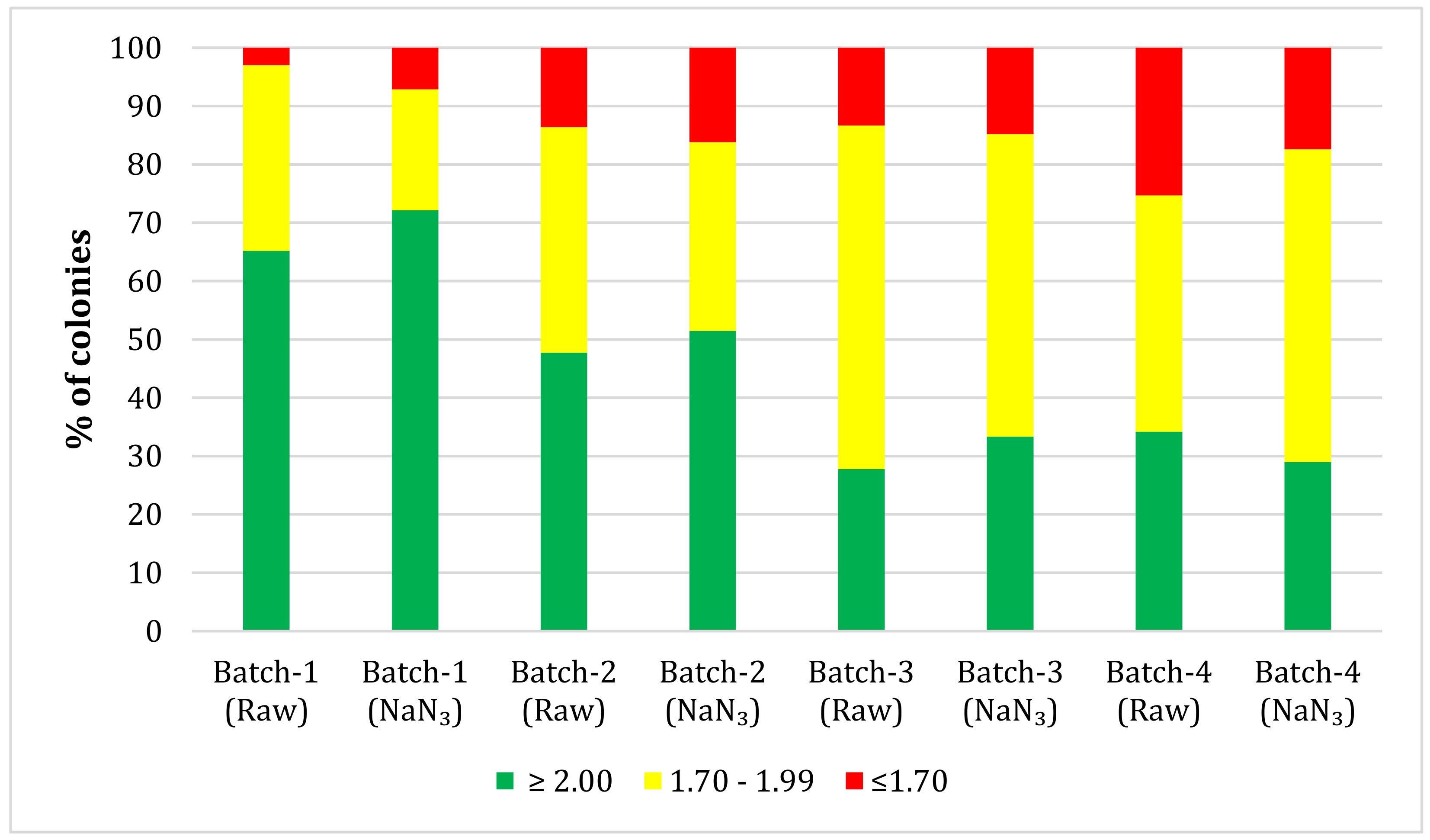
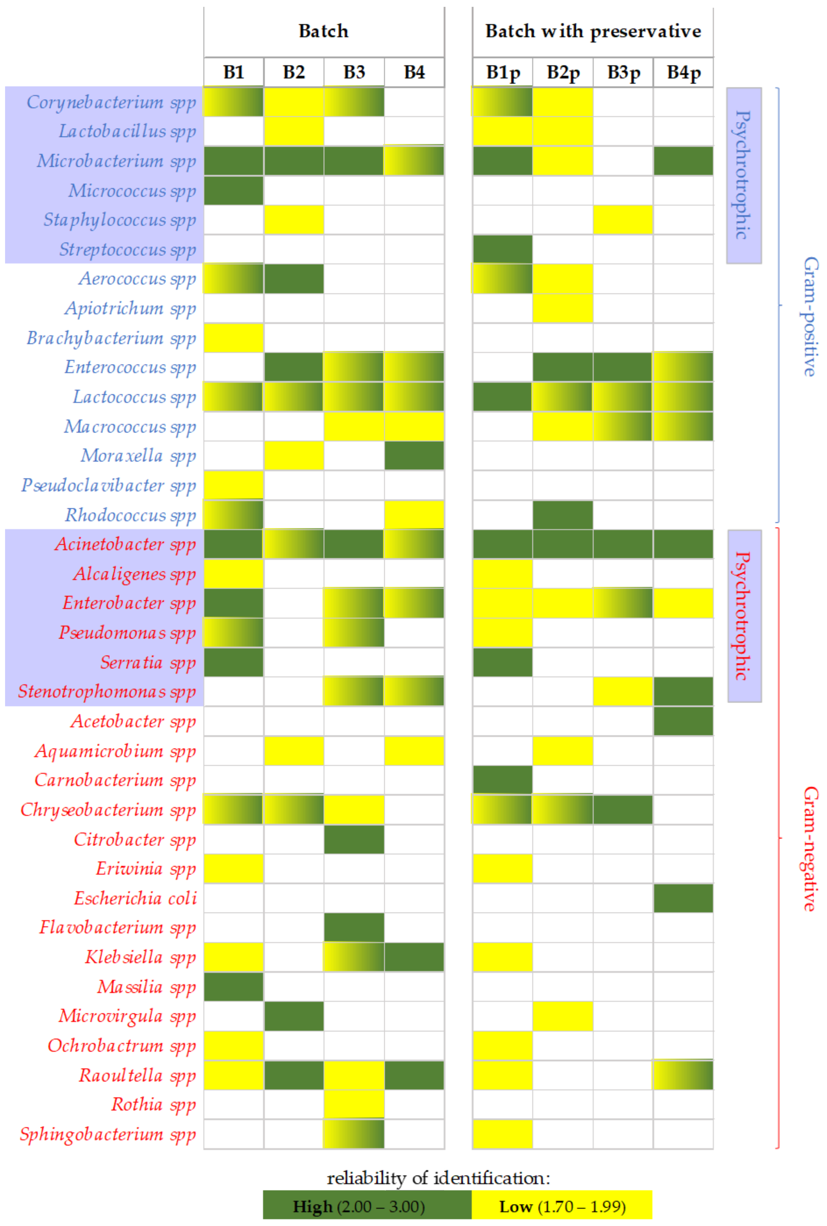
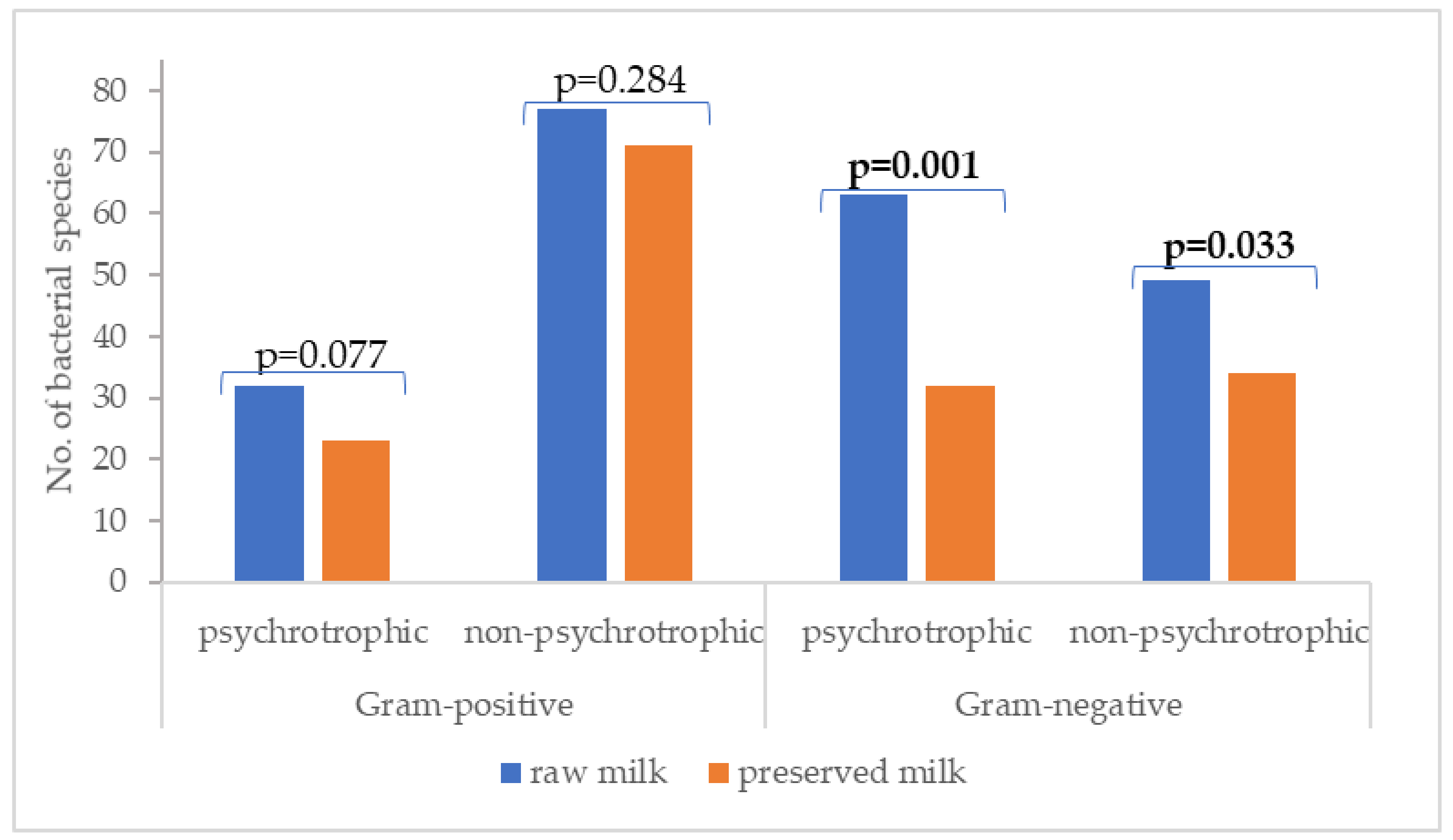
3.2. MALDI-TOF Mass Spectrometry Identification of Aerobic Mesophilic Bacteria

The bacterial colonies obtained from all the collected samples were cultured on a nutrient medium, and different morphotypes were selected and analyzed using MALDI-TOF MS. An instrument software MBT Compass HT version 5.1 using a match score identified the colonies based on a reference database version 11, and, depending on the match score, bacterial colonies were identified up to the genus and species level. To calculate the score values of each sample, raw data obtained from the MALDI-TOF mass spectrometer was first converted into a peak list, and a further list was compared to the reference database. A total of 392 bacterial colonies were identified in the raw unpreserved milk samples, representing 30 genera and 54 bacterial species. In the preserved milk samples, 330 colonies were identified, representing 27 genera and 41 bacterial species. All species identified in the raw unpreserved and preserved milk samples are shown in Supplementary Materials Tables S1 and S2, respectively. The confidence score was mentioned on a scale of ≥2.00, 1.70 to 1.99 and <1.70 (Figure 2).
In the raw unpreserved milk, 65%, 48%, 28% and 34% bacteria were identified with a confidence score of 2.00 and above in Batches 1, 2, 3 and 4, respectively. In preserved milk, these are 72%, 51%, 33% and 29% in the respective batch samples. In general, in all 392 bacterial colonies in raw unpreserved milk samples (Batches 1, 2, 3 and 4), the most predominant bacterial species were Lactococcus lactis (18%), Chryseobacterium vrystaatense (10%) and Acinetobacter johnsonii (8%). In all 330 colonies in the preserved milk samples of all four batches, a slight population shift was noticed, which was as follows: Lactococcus lactis (26%), Chryseobacterium vrystaatense (17%) and Lactococcus garvieae (8%).
In Batch 1, the results indicated that, in the raw unpreserved milk samples, out of a total of 135 bacterial species identified, the largest share is occupied by the bacteria Chryseobacterium vrystaatense (21%), followed by Serratia liquefaciens (13%) and Lactococcus lactis and Rhodococcus baikonurensis (7%). On the other hand, in the preserved milk samples, among 140 bacterial species, the most abundant species was re-recorded as Chryseobacterium vrystaatense (33%), followed by Lactococcus lactis (15%) and Serratia liquefaciens (12%). The least abundant species were Carnobacterium maltaromaticum, Corynebacterium frankenforstense and others, with a total occupancy of 1%.
In Batch 2, out of 88, the most abundant bacteria in the raw unpreserved milk samples were identified as Lactococcus lactis (28%), followed by Acinetobacter johnsonii and Enterococcus faecalis (14%). In preserved milk, among 68 bacterial species with a total abundance of 29%, Lactococcus lactis was the most abundant species. Besides this, Enterococcus faecalis and Acinetobacter johnsonii, with 16% and 6%, were the subsequently populated species.
Similarly, out of a total of 90 bacterial species in Batch 3, the major species in the raw unpreserved milk samples were Lactococcus garvieae (18%) followed by Lactococcus lactis (12%) and Chryseobacterium joostei and Enterobacter cloacae (8%). While in preserved milk with sodium azide, Lactococcus lactis, with a total percentage of 35%, Lactococcus garvieae (22%) and Chryseobacterium joostei and Macrococcus caseolyticus (5%) were the three most abundant species among the 54 bacterial species.
In Batch 4 of raw unpreserved milk, out of 79 species, the highest detected bacteria were Lactococcus lactis (18%), along with Lactococcus garvieae (13%), Acinetobacter johnsonii and Enterococcus faecalis (11%), while, in preserved milk, out of 69, the most abundant species were Lactococcus lactis (22%), Lactococcus garvieae and Macrococcus caseolyticus (14%), and Enterococcus faecalis (12%). It is noteworthy that the total number of populations of different bacteria is comparatively lower in the preserved than the unpreserved raw milk samples.
Bacterial identification using MALDI-TOF MS based on charged biomolecules, including proteins, to generate spectra—which are compared to the reference database—is a promising approach for rapid and reliable identification of the microbial population of milk. In this context, Dobranić et al. [25] analyzed raw cow’s milk originating from untreated and antibiotic-treated cows, with an emphasis on enterococci opportunistic pathogens and also psychotropic bacteria, staphylococci, E. coli, enterococci, Enterobacteria, Listeria spp. and sulfite-reducing clostridia. The results were determined using the classical method. MALDI-TOF MS-analyzed samples showed 100% concordance with simultaneously used API 20 Strep. The results endorsed MALDI-TOF MS as a potential tool for the identification of pathogens including Enterococcus faecalis. In our results, in the raw unpreserved and preserved milk samples of Batches 2, 3 and 4, Enterococcus faecalis was identified by MALDI-TOF MS. Besides this, coliform bacteria are sensitive to sodium azide [7]. Similarly, Elizondo et al. [22] stated that sodium azide is most effective against mesophilic microorganisms. Different microorganisms are present in milk, and LAB is a common species. Nacef et al. [26] successfully identified 197 colonies of lactic acid bacteria from cheese made from raw and pasteurized milk using the MALDI-TOF technology and concluded that Lactobacillus was the most predominant genus in raw and preserved milk. Our results also follow previous research outcomes [25,27,28].
In the present study, after microbial analysis using MALDI-TOF MS, it was noticed that a few bacterial species were present in the raw unpreserved milk samples but not detected in the respective preserved milk samples. The first of such bacteria is Acinetobacter lwoffii, an aerobic Gram-positive bacillus, generally found on the skin and considered an opportunistic pathogen [29]. Reports suggest that it is among the predominant deteriorating bacteria of milk [30]. Another organism observed was Corynebacterium xerosis; Hahne et al. [31] isolated C. xerosis, a coagulase-positive strain, from raw unpreserved milk. Its presence is also associated with the abundance of somatic cells in milk. Woudstra et al. [32] also identified C. xerosis in raw milk using MALDI-TOF MS. It is generally introduced into milk through the milking process as it is found on milking gloves and cow’s skin and in the environment [33].
In this list, the next organism is Brachybacterium nesterenkovii, a thermoduric bacteria and potential spoilage microbe [34]. This bacterium changes its shape from coccus/oval/rod during the logarithmic phase to coccus during the stationary phase, and, possibly because of non-viability of the strain under preservation, it could not be detected in the preserved samples. In addition, Chryseobacterium rhizosphaerae, found in raw unpreserved milk but not preserved, was first reported in coastal sand dune plant rhizosphere [35].
Serratia liquefaciens is a psychrotrophic bacteria equipped with features such as biofilm formation and proteolytic and lipolytic activity [36]. In addition to this, it also possesses antibiotic resistance; thus, sodium azide might not affect its population in preserved milk. Another, Sphingobacterium multivorum, is commonly found in soil and water but not in milk, although it can appear in milk during processing or preservation and can facilitate the environment for Sphingobacterium multivorum growth. Pukančíková et al. [27] also detected this bacterium in raw milk using MALDI-TOF MS, with a confidence score of 1.987. Preservation may alter the microbial composition of raw milk, and that can favor the growth of certain microorganisms. Rhodococcus baikonurensis, initially isolated from a space laboratory in Russia, a boron-tolerant bacterium belonging to actinobacteria, can be detected in raw milk and cause spoilage. It also produces extracellular enzymes and rhamnolipids. The organism is capable of bioremediation of various pollutants, including diesel, Hg, and other heavy metals [37,38]. These bacteria were found in our raw unpreserved milk sample but not in milk with added sodium azide.
Cantoni et al. [39] isolated a pigmented bacterium, Pseudoclavibacter helvolus, in raw milk, and, in the present study, we also detected it in raw unpreserved milk. However, MALDI-TOF MS could not find it in preserved milk. In addition, Micrococcus luteus was identified in raw unpreserved milk but not in preserved milk. M. luteus is a milk and milk product spoilage bacteria. Sodium azide in preserved milk successfully inhibited growth of M. luteus and advocated its efficiency in reducing spoilage organisms and improving the quality and storage of milk samples intended for physico-chemical analyses and analyses of the hygienic quality of milk control.
Carnobacterium maltaromaticum was first isolated from a milk sample with a different flavor due to the presence of aldehydes. It is an opportunistic lactic acid bacterium that can grow in milk in low/no competition with other LAB, which is why it probably could not grow to a threshold to be detected in raw milk, but as soon as sodium azide was reduced, the growth of the Carnobacterium maltaromaticum pathogenic bacteria in preserved milk grew well. A non-fermentative rod-shaped Gram-negative bacterium, Massilia timonae, was also not detected in preserved milk. Moreover, Streptococcus parauberis was detected in preserved milk, possibly because sodium azide has been used as a selective medium component that suppresses Gram-negative bacteria while promoting the growth of streptococci in medium (Hartmann, 1936 [40]).
Interestingly, in our MALDI-TOF MS identification, various genera were identified in raw unpreserved milk that have generally not been reported in previous studies. This indicates poor hygienic practices during milking and also points out the source introducing such microorganisms in milk, among which many are opportunistic pathogens, that can deteriorate the milk quality. The study demonstrates that sodium azide undoubtedly poses a great impact on reducing the opportunistic pathogen population in milk and reducing the risk of contamination of preserved milk samples intended for laboratory analyses of milk quality control.
To investigate the influence of the preservative on the microbiological composition of milk and isolated Gram-positive and Gram-negative bacterial species, the heatmap was applied (Figure 3). The reliability of the identification is highlighted with colors, where green represents the high reliability of identification, while the yellow field represents low reliability. The gradient with two colors, the yellow-green fields, points out that for the same bacterial species was detected more times but with low and high reliability (e.g., Corynebacterium spp. raw unpreserved milk of the first batch (B1, B3 and B1p; Figure 3)).
Figure 3 depicts the abundance of Lactococcus lactis in raw unpreserved and preserved milk and the reduction of a few species, such as Corynebacterium, Rhodococcus, etc., in preserved milk with sodium azide. In assessing the change, i.e., the number of sampled bacterial species in batches of raw unpreserved milk compared to preserved milk, a one-sided t-test was used, with a significance level of p < 0.05. The results are shown in Figure 4.
Finally, to relate the aerobic mesophilic bacteria count found in raw unpreserved and preserved milk to the proportion of bacteria identified using MALDI-TOF MS, principal component analysis was applied. The mentioned tool of multivariate analysis enabled a qualitative clarification of everything mentioned above (Figure 5), and the distribution of the analyzed milk from different batches in all four quadrants was clearly visible.
The study of Milanović et al. [41] used a qualitative multivariate tool in observing similarities and differences between used starter cultures; therefore, we applied it in the bacterial observation. We used a heatmap to qualitatively identify the similarities/differences of the milk samples based on the detected Gram-positive, Gram-negative and psychrotrophic bacteria. The presented trends in Figure 4 show the successful identification of the species in raw unpreserved and preserved milk samples per batch. Nevertheless, based on the fact that psychrotrophic bacteria have a negative impact on the quality of milk and milk products [42,43], the identifications were summarized per bacteria group and presented in Figure 4.
As Fernández et al. [44] indicated in their review of the impact of microorganisms present in fermented dairy products on human health, a few Gram-negative bacteria (species as E. coli, Pseudomonas aeruginosa, Klebsiella pneumoniae and Citrobacter freundii and the genera Enterobacter, Proteus, Psychrobacter, Halomonas and Serratia) are identified as indicators of potential health risk related to poor hygiene. Thus, it seems that sodium azide significantly contributed to the change of Gram-negative bacteria, regardless of whether they were psychrotrophs (p = 0.001) or not (p = 0.033). In the case of Gram-positive bacteria, no statistically significant difference was found in the raw unpreserved milk samples vs. milk to which a preservative, sodium azide, was added.
Total aerobic mesophilic bacteria count (Figure 5, marked as “CFU”) is negatively correlated (Pearson’s correlation coefficient) with “High” identification reliability (−0.357), which is an indication that regardless of the number of bacteria that are high, the effectiveness on MALDI-TOF MS identification will be high, and, with the increase of the bacteria number, the increase of “Low” identification will be proportional. However, it should be pointed out here that the representation in Figure 5 results from the mean values for CFU and the degrees of bacterial identification. To incorporate the experimental variance for each batch and improve the reliability of the analysis, PCA analysis was performed for each batch separately, with the corresponding ten observations for the raw and preserved milk samples (Figure S1). The trend of the relationship between CFU and the degree of identification changes depending on the observed batch of milk. But what all biplot presentations (Figure S1) have in common is the positioning of the degree of success of bacterial identification in different quadrants, depending on whether the milk is raw or preserved (B or Bp). The previously identified trend (CFU negatively correlates with high identification reliability) was confirmed for all observed batches of milk through the positioning of CFU values in opposite quadrants (for preserved milk), except for the first one (Figure S1). However, from the Pearson correlation matrix (which is an integral part of the PCA analysis), it can be seen that the value of the correlation coefficient (for B1p_CFU vs. B1p_High) is −0.3805, confirming the negative correlation. It should certainly be emphasized what was confirmed by the research of Wenning et al. [45]: the suitability and applicability of MALDI-TOF mass spectrometry evaluated for routine microbial diagnostics of microorganisms associated with food analysis. They additionally pointed out that an extremely important factor in the accuracy of identification and their ability to differentiate isolates is precisely the resistance of bacteria to changes in incubation time and/or media. It should also be noted that the appearance of new pathogens requires a revision of the bacteria database and the addition of new spectra to the database itself, which makes MALDI-TOF MS more effective in the diagnosis of microorganisms and thus in monitoring the quality of milk [46].

4. Conclusions

The present study concluded that MALDI-TOF MS is a promising approach that has the potential to identify bacteria up to the genus and even species levels. The preservative sodium azide was used to show trends in reducing aerobic mesophilic microorganisms in different milk samples.
In general, the most dominant bacterial species in the raw unpreserved milk samples were Lactococcus lactis, Chryseobacterium vrystaatense and Acinetobacter johnsonii, while, in preserved milk samples, the predominate populations were Lactococcus lactis, Chryseobacterium vrystaatense and Lactococcus garvieae.
Sodium azide definitely reduced the pathogenic microorganisms and also supported growth of various other bacteria by reducing the competition. Also, the results showed satisfactory quality and stability of the preserved milk samples intended for laboratory analyses of milk quality control. Many species present in raw unpreserved milk were not detected in preserved milk. Conclusively, MALDI-TOF MS analysis provided us a detailed insight into the hygienic quality of milk and showed the impact of the preservative on microbial species before and after preservation.

Supplementary Materials

The following supporting information can be downloaded at: https://www.mdpi.com/article/10.3390/pr12040731/s1, Table S1: Total species identified in raw unpreserved milk sample using MALDI-TOF MS. Table S2: Total species identified in preserved milk sample using MALDI-TOF MS. Figure S1: Biplots of the principal component analysis conducted on each milk batch separately (Batch 1 to Batch 4) for the CFU values (total number of aerobic mesophilic bacteria) and the identification levels of bacteria (High, Medium, Low and Total) in ten observations (Obs1 to Obs10), for analyzed raw milk (B1, B2, B3 and B4) and preserved milk (Bp1, Bp2, Bp3 and Bp4).

Author Contributions

Conceptualization, N.M. and S.K.; methodology, N.M., J.G.K., N.Z. and S.K.; software, J.G.K. and S.K.; validation, N.M., J.Š., D.P., F.O., J.G.K., K.M.S. and S.K.; formal analysis, D.P., N.L., F.O., J.G.K. and S.K.; investigation, N.M., J.Š., J.G.K., K.M.S. and S.K.; resources, N.M., J.G.K. and S.K.; data curation, N.M., J.Š., D.P., F.O., J.G.K., N.Z., K.M.S. and S.K.; writing—original draft preparation, N.M., J.Š., J.G.K., K.M.S. and S.K.; writing—review and editing, N.M., J.Š., J.G.K., N.Z., K.M.S. and S.K.; visualization, J.G.K. and K.M.S.; supervision, N.M.; project administration, N.M.; funding acquisition, N.M. All authors have read and agreed to the published version of the manuscript.

Funding

This research was funded by the EU Operational Program Competitiveness and Cohesion 2014–2020 project “Potential of microencapsulation in cheese production” K.K.01.1.1.04.0058.

Data Availability Statement

The data presented in this study are available on request from the corresponding author.

Conflicts of Interest

The authors declare no conflicts of interest. The funders had no role in the design of the study; in the collection, analyses, or interpretation of data; in the writing of the manuscript; or in the decision to publish the results.

References

  1. Antunac, N.; Havranek, J. Milk. Chemistry, Physics and Microbiology; Internal Script; University of Zagreb, Faculty of Agriculture: Zagreb, Croatia, 2013. [Google Scholar]
  2. Herrera, A.G. Mesophilic Aerobic Microorganisms. In Food Microbiology Protocols, Methods in Biotechnology; Spencer, J.F.T., de Ragout Spencer, A.L., Eds.; Humana Press: Totowa, NJ, USA, 2001; Volume 14. [Google Scholar] [CrossRef]
  3. Samaržija, D. Dairy Microbiology—General Properties of Bacteria, Yeasts, Molds and Viruses with Basics of Taxonomy; Internal Script; University of Zagreb, Faculty of Agriculture: Zagreb, Croatia, 2018. [Google Scholar]
  4. Weisglass, H. Bakterije i Bolesti Čovjeka: Specijalna Bakteriologija; Školska Knjiga: Zagreb, Croatia, 1983. [Google Scholar]
  5. Perin, L.M.; Pereira, J.G.; Bersot, L.S.; Nero, L.A. Chapter 3—The Microbiology of Raw Milk. In Raw Milk: Balance between Hazards and Benefits; Nero, L.A., De Carvalho, A.F., Eds.; Academic Press: Oxford, UK, 2019; pp. 45–64. [Google Scholar] [CrossRef]
  6. Ministarstvo Poljoprivrede. Pravilnik o Utvrđivanju Sastava Sirovog Mlijeka. Available online: https://narodne-novine.nn.hr/clanci/sluzbeni/2020_12_136_2605.html (accessed on 22 February 2024).
  7. Upadhyay, N.; Goyal, A.; Kumar, A.; Ghai, D.L.; Singh, R. Preservation of milk and milk products for analytical purposes. Food Rev. Int. 2014, 30, 203–224. [Google Scholar] [CrossRef]
  8. Antunac, N.; Mikulec, N.; Zamberlin, Š.; Sović, J.Š.; Blažek, A.; Horvat, I. Influence of various methods of milk sample preservation on the number of somatic cells. Milchwissenschaft 2010, 65, 313–316. [Google Scholar]
  9. Samaržija, D.; Antunac, N.; Pogačić, T.; Sikora, S. Determination of the total number of bacteria in raw milk by flow cytometry. Dairy 2004, 54, 39–51. Available online: https://hrcak.srce.hr/clanak/4022 (accessed on 22 February 2024).
  10. Cabrol, L.; Quemeneur, M.; Misson, B. Inhibitory effects of sodium azide on microbial growth in experimental resuspension of marine sediment. J. Microbiol. Methods 2017, 133, 62–65. [Google Scholar] [CrossRef]
  11. Cieśla, Z.; Mardarowicz, K.; Klopotowski, T. Inhibition of DNA synthesis and cell division in Salmonella typhimurium by azide. Mol. Gen. Genet. 1974, 135, 339–348. [Google Scholar] [CrossRef] [PubMed]
  12. Oliver, D.B.; Cabelli, R.J.; Dolan, K.M.; Jarosik, G.P. Azide-resistant mutants of Escherichia coli alter the SecA protein, an azide-sensitive component of the protein export machinery. Proc. Natl. Acad. Sci. USA 1990, 87, 8227–8231. [Google Scholar] [CrossRef] [PubMed]
  13. Chandler, R.; Jamshad, M.; Yule, J.; Robinson, A.; Alam, F.; Dunne, K.A.; Nabi, N.; Henderson, I.; Huber, D. Genetic screen suggests an alternative mechanism for azide-mediated inhibition of SecA. bioRxiv 2018, 173039. [Google Scholar] [CrossRef]
  14. Suresh, A.; Pan, C.; Ng, W.J. Sodium azide inhibition of microbial activities and impact on sludge floc destabilization. Chemosphere 2020, 244, 125452. [Google Scholar] [CrossRef] [PubMed]
  15. Darvishi, S.; Girault, H.H. Revealing the effects of three different antimicrobial agents on E. coli biofilms by using Soft-Probe Scanning Electrochemical Microscopy. Appl. Nano 2023, 4, 260–279. [Google Scholar] [CrossRef]
  16. Sobrun, Y.; Bhaw-Luximon, A.; Jhurry, D.; Puchooa, D. Isolation of lactic acid bacteria from sugar cane juice and production of lactic acid from selected improved strains. Adv. Biosci. Biotechnol. 2012, 3, 398–407. [Google Scholar] [CrossRef]
  17. Sešķēna, R.J.; Jankevica, L. Influence of chemical preservatives on the quality and composition indices of raw milk samples. Acta Univ. Latv. 2007, 723, 171–180. [Google Scholar]
  18. Sinaga, H.; Deeth, H.; Bhandari, B. Effect of sodium azide addition and aging storage on casein micelle size. IOP Conf. Ser. Earth Environ. Sci. 2018, 122, 012083. [Google Scholar] [CrossRef]
  19. ISO 4833-1:2013; Microbiology of the Food Chain—Horizontal Method for the Enumeration of Microorganisms—Part 1: Colony Count at 30 °C by the Pour Plate Technique. International Organization for Standardization: Geneva, Switzerland, 2013.
  20. Gonzalo, C.; Martínez, J.R.; Carriedo, J.A.; San Primitivo, F. Fossomatic Cell-Counting on Ewe Milk: Comparison with Direct Microscopy and Study of Variation Factors. J. Dairy Sci. 2003, 86, 138–145. [Google Scholar] [CrossRef] [PubMed]
  21. Croatian Agency for Agriculture and Food (HAPIH). Annual Report for 2022; Editorial Center for Quality Control of Livestock Products: Osijek, Croatia, 2023. [Google Scholar]
  22. Elizondo, J.; Aldunate, A.; Ezcurra, P.; Gallego, I.; Saigos, E.; Ulayar, E.; Izco, J.M. Efficiency of the proportion of azidiol on preservation in ewe’s milk samples for analysis. Food Control 2007, 18, 185–190. [Google Scholar] [CrossRef]
  23. Zajác, P.; Zubrická, S.; Čapla, J.; Zeleňáková, L.; Židek, R.; Čurle, J. Effect of preservatives on milk composition determination. Int. Dairy J. 2016, 61, 239–244. [Google Scholar] [CrossRef]
  24. Lichstein, H.C.; Soule, M.H. Studies of the effect of sodium azide on microbic growth and respiration: I. The action of sodium azide on microbic growth. J. Bacteriol. 1944, 47, 221–230. [Google Scholar] [CrossRef]
  25. Dobranić, V.; Kazazić, S.; Filipović, I.; Mikulec, N.; Zdolec, N. Composition of raw cow’s milk microbiota and identification of enterococci by MALDI-TOF MS—Short communication. Vet. Arh. 2016, 86, 581–590. Available online: https://hrcak.srce.hr/166060 (accessed on 5 March 2024).
  26. Nacef, M.; Chevalier, M.; Chollet, S.; Drider, D.; Flahaut, C. MALDI-TOF mass spectrometry for the identification of lactic acid bacteria isolated from a French cheese: The Maroilles. Int. J. Food Microbiol. 2017, 247, 2–8. [Google Scholar] [CrossRef]
  27. Pukančíková, L.; Lipničanová, S.; Kačániová, M.; Chmelová, D.; Ondrejovič, M. Natural microflora of raw cow milk and their enzymatic spoilage potential. Nova Biotechnol. Chim. 2016, 15, 142–155. [Google Scholar] [CrossRef]
  28. Kačániová, M.; Terentjeva, M.; Godočíková, L.; Puchalski, C.; Kunová, S.; Kluz, M.; Kordiaka, R.; Haščík, P. Identification of lactic acid bacteria in milk and milk products with MALDI-TOF mass spectrometry. Sci. Pap. Anim. Sci. Biotechnol. 2017, 50, 115–120. [Google Scholar]
  29. Regalado, N.G.; Martin, G.; Antony, S.J. Acinetobacter lwoffii: Bacteremia associated with acute gastroenteritis. Travel Med. Infect. Dis. 2009, 7, 316–317. [Google Scholar] [CrossRef] [PubMed]
  30. Ribeiro Júnior, J.C.; Tamanini, R.; de Oliveira, A.L.M.; Alfieri, A.A.; Beloti, V. Genetic diversity of thermoduric spoilage microorganisms of milk from Brazilian dairy farms. J. Dairy Sci. 2018, 101, 6927–6936. [Google Scholar] [CrossRef]
  31. Hahne, J.; Kloster, T.; Rathmann, S.; Weber, M.; Lipski, A. Isolation and characterization of Corynebacterium spp. from bulk tank raw cow’s milk of different dairy farms in Germany. PLoS ONE 2018, 13, e0194365. [Google Scholar] [CrossRef] [PubMed]
  32. Woudstra, S.; Lücken, A.; Wente, N.; Zhang, Y.; Leimbach, S.; Gussmann, M.K.; Kirkeby, C.; Krömker, V. Reservoirs of Corynebacterium spp. in the Environment of Dairy Cows. Pathogens 2023, 12, 139. [Google Scholar] [CrossRef] [PubMed]
  33. Dean, C.J.; Slizovskiy, I.B.; Crone, K.K.; Pfennig, A.X.; Heins, B.J.; Caixeta, L.S.; Noyes, N.R. Investigating the cow skin and teat canal microbiomes of the bovine udder using different sampling and sequencing approaches. J. Dairy Sci. 2021, 104, 644–661. [Google Scholar] [CrossRef] [PubMed]
  34. Gvozdyak, O.R.; Nogina, T.M.; Schumann, P. Taxonomic study of the genus Brachybacterium: Brachybacterium nesterenkovii sp. nov. Int. J. Syst. Microbiol. 1992, 42, 74–78. [Google Scholar] [CrossRef] [PubMed]
  35. Cho, S.H.; Lee, K.S.; Shin, D.S.; Han, J.H.; Park, K.S.; Lee, C.H.; Park, K.H.; Kim, S.B. Four new species of Chryseobacterium from the rhizosphere of coastal sand dune plants, Chryseobacterium elymi sp. nov., Chryseobacterium hagamense sp. nov., Chryseobacterium lathyri sp. nov. and Chryseobacterium rhizosphaerae sp. nov. Syst. Appl. Microbiol. 2010, 33, 122–127. [Google Scholar] [CrossRef] [PubMed]
  36. Machado, S.G.; da Silva, F.L.; Bazzolli, D.M.S.; Heyndrickx, M.; Costa, P.M.d.A.; Vanetti, M.C.D. Pseudomonas spp. and Serratia liquefaciens as predominant spoilers in cold raw milk. J. Food Sci. 2015, 80, M1842–M1849. [Google Scholar] [CrossRef] [PubMed]
  37. Lee, M.; Kim, M.K.; Singleton, I.; Goodfellow, M.; Lee, S.-T. Enhanced biodegradation of diesel oil by a newly identified Rhodococcus baikonurensis EN3 in the presence of mycolic acid. J. Appl. Microbiol. 2006, 100, 325–333. [Google Scholar] [CrossRef] [PubMed]
  38. Yoon, J.; Miwa, H.; Ahmed, I.; Yokota, A.; Fujiwara, T. Rhodococcus baikonurensis BTM4c, a boron-tolerant actinobacterial strain isolated from soil. Biosci. Biotechnol. Biochem. 2010, 74, 178–181. [Google Scholar] [CrossRef] [PubMed]
  39. Cantoni, C.; Milesi, S.; Iacumin, L. Description of new pigmented bacteria in raw milk. Ind. Aliment. 2006, 45, 1133–1136. [Google Scholar]
  40. Hartmann, G. Ein Beitrag zur Reinzüchtung von Mastitisstreptokokken aus verunreinigtem Material. Milchw. Forsch. 1936, 18, 116–122. [Google Scholar]
  41. Milanović, V.; Cardinali, F.; Boban, A.; Gajdoš Kljusurić, J.; Osimani, A.; Aquilanti, L.; Garofalo, C.; Budić-Leto, I. White grape variety Maraština as a promising source of non-Saccharomyces yeasts intended as starter cultures. Food Biosci. 2023, 55, 103033. [Google Scholar] [CrossRef]
  42. Samaržija, D.; Zamberlin, Š.; Pogačić, T. Psychrotrophic bacteria and milk and dairy products quality. Mljekarstvo/Dairy 2012, 62, 77–95. Available online: https://hrcak.srce.hr/83325 (accessed on 15 February 2024).
  43. Yuan, H.; Han, S.; Zhang, S.; Xue, Y.; Zhang, Y.; Lu, H.; Wang, S. Microbial Properties of Raw Milk throughout the Year and their Relationships to Quality Parameters. Foods 2022, 11, 3077. [Google Scholar] [CrossRef] [PubMed]
  44. Fernández, M.; Hudson, J.A.; Korpela, R.; de los Reyes-Gavilán, C.G. Impact on human health of microorganisms present in fermented dairy products: An overview. BioMed Res. Int. 2015, 2015, 412714. [Google Scholar] [CrossRef] [PubMed]
  45. Wenning, M.; Breitenwieser, F.; Konrad, R.; Huber, I.; Busch, U.; Scherer, S. Identification and differentiation of food-related bacteria: A comparison of FTIR spectroscopy and MALDI-TOF mass spectrometry. J. Microbiol. Methods 2014, 103, 44–52. [Google Scholar] [CrossRef] [PubMed]
  46. Barberis, C.; Almuzara, M.; Join-Lambert, O.; Ramírez, M.S.; Famiglietti, A.; Vay, C. Comparison of the Bruker MALDI-TOF mass spectrometry system and conventional phenotypic methods for identification of Gram-positive rods. PLoS ONE 2014, 9, e106303. [Google Scholar] [CrossRef] [PubMed]
Figure 1. Box–Whisker diagram for the total number of aerobic mesophilic bacteria in raw unpreserved (B1, B2, B3 and B4) and preserved milk (Bp1, Bp2, Bp3 and Bp4) with indicated outliers (corresponding points over the box). Each batch and type of milk is shown in a different color.
Processes 12 00731 g001
Figure 2. Percentage of reliability of identified colonies using MALDI-TOF MS.
Processes 12 00731 g002
Figure 3. Heatmap representing the results of semi-quantitative evaluation of identification of Gram-positive and Gram-negative bacteria with highlighted psychrotrophic bacteria in raw unpreserved milk (four batches, B1–B4) and in preserved milk (four batches, B1p–B4p).
Processes 12 00731 g003
Figure 4. Changes in the number of sampled bacterial species in raw unpreserved milk compared to preserved milk. The values of the parameter “p” are bold for those observations that are statistically significantly different (p < 0.05).
Processes 12 00731 g004
Figure 5. Biplot of the principal component analysis of different identification levels (reliability of identification) of bacteria and the total number of aerobic mesophilic bacteria, for analyzed milk samples in raw (blue dots: B1, B2, B3 and B4) and preserved (green dots: Bp1, Bp2, Bp3 and Bp4) form.
Processes 12 00731 g005
Table 1. Total number of aerobic mesophilic bacteria in raw unpreserved and preserved milk (CFU/mL).
SampleBatch 1Batch 2Batch 3Batch 4
Raw MilkPreserved MilkRaw MilkPreserved MilkRaw MilkPreserved MilkRaw MilkPreserved Milk
144,00043,000141,000105,00029,00025,00077,00074,000
250,00046,00067,00035,000201,000170,000188,000142,000
343,00036,00082,00079,000232,000151,00090,00050,000
448,00043,000145,000111,000920,000830,000120,000117,000
546,00026,00058,00019,0001,900,000830,000116,00076,000
680,00056,00074,00057,0002,720,0001,340,000910,000730,000
752,00052,000123,00063,000248,000191,000234,00093,000
8430,000300,00080006000460,000127,000229,000156,000
91,500,000620,00059,00020,000120,000100,000225,000221,000
1093,00048,00036,00012,0001,190,000950,000263,000204,000
Average ± Standard deviation2.39 × 105 ± 4.59 × 10⁵ A1.27 × 105 ± 1.91 × 10⁵ a7.93 × 104 ± 4.48 × 10⁴ B5.07 × 104 ± 3.84 × 10⁴ a,*8.02 × 10⁵ ± 8.96 × 10⁵ A4.71 × 105 ± 4.68 × 10⁵ b,*2.45 × 10⁵ ± 2.43 × 10⁵ A1.86 × 105 ± 1.99 × 10⁵ a,*
Significant differences in the total number of aerobic mesophilic bacteria are indicated with capital letters (A and B) for raw unpreserved milk from different batches and with small letters (a and b) for preserved milk samples. The significant differences for samples of the same batch (raw unpreserved vs. preserved milk) are indicated with an *. Significance level is p < 0.05.
Disclaimer/Publisher’s Note: The statements, opinions and data contained in all publications are solely those of the individual author(s) and contributor(s) and not of MDPI and/or the editor(s). MDPI and/or the editor(s) disclaim responsibility for any injury to people or property resulting from any ideas, methods, instructions or products referred to in the content.

