Article

Retention and Antimicrobial Activity of Alginate-Encapsulated Bioactive Compounds from Leaves and Fruits of Myrtle (Myrtus communis L.)

by
Daniela Cvitković
,
Jasna Mrvčić
,
Erika Dobroslavić
,
Verica Dragović-Uzelac
and
Sandra Balbino
*
Faculty of Food Technology and Biotechnology, University of Zagreb Pierottijeva 6, 10000 Zagreb, Croatia
*
Author to whom correspondence should be addressed.
Processes 2025, 13(4), 1220; https://doi.org/10.3390/pr13041220
Submission received: 12 March 2025 / Revised: 12 April 2025 / Accepted: 14 April 2025 / Published: 17 April 2025
(This article belongs to the Section Biological Processes and Systems)

Abstract

:
Myrtle (Myrtus communis L.) is a rich source of bioactive compounds and nutraceuticals of different polarities obtained from different parts of the plant, whose synergistic effect could be harnessed through the formulation of capsules. The aim of this work was to investigate the antimicrobial activity of three myrtle formulations: essential oil and aqueous leaf extract and supercritical fruit extract, as well as the retention of volatile, phenolic and lipid compounds in low-viscosity alginate capsules obtained by the electrostatic extrusion microencapsulation of these formulations combined. At a temperature of 48 °C, 1.27% emulsifier and 3% CaCl2, 72.86% of the volatiles, 61.13% of the phenolics and 62.80% of the lipids were retained. All tested extracts showed good antibacterial activity, especially against Staphylococcus aureus and Listeria monocytogenes, which were the most sensitive. An exceptionally high inhibitory effect was achieved by aqueous leaf extract against L. monocytogenes (35.25 mm) and essential oil against Escherichia coli (28 mm). Supercritical extract and essential oil showed good antifungal activity, and none of the extracts showed inhibitory activity against lactic acid bacteria.

1. Introduction

Myrtle (Myrtus communis L., Myrtaceae) is an aromatic medicinal evergreen shrub of the Mediterranean region that is used in folk medicine for therapeutic purposes. Its leaves are traditionally used to produce essential oil in the perfume industry and as fruits in liqueur production, and it is used as a spice [1]. Myrtle leaf contains a wide range of bioactive molecules with different polarities, i.e., flavonols (myricetin 3-O-galactoside and myricetin 3-O-rhamnoside), phenolic acids, and essential oil components such as terpenoids (myrtenyl acetate and 1,8-cineole) as well as carotenoids (lutein and β-carotene) [2], while unsaturated fatty acids (linoleic acid), tocopherols and sterols are found in the lipid fraction of the berry [3]. These compounds have a bioactive effect on human health, such as antioxidant, antimicrobial, antiseptic, hypoglycemic [1] and prebiotic [4].
Due to the complexity of plant composition and the interactions among bioactive compounds, it is well established that their combined synergistic effect often exceeds the sum of their individual effects. A better understanding of these interactions could, therefore, contribute to the development of novel therapeutic applications, e.g., new antimicrobial agents [5,6,7]. Furthermore, combining these extracts with conventional antibiotics may produce a synergistic antibacterial response, offering a promising strategy to mitigate the excessive use of antibiotics [8,9].
Considering the diverse chemical properties of plant-derived and their potential synergistic effects, optimizing their recovery and therapeutic properties requires the investigation of various solvent systems and extraction techniques. In addition, the stabilization and efficient delivery of these chemically incompatible compounds in the form of multi-component mixtures can be achieved through the co-encapsulation method [10]. The use of co-encapsulation of bioactives has a high potential for application in industry as it yields nutraceuticals with enhanced desired properties and bioactivity [11,12]. It can also be used in cosmetics for preserving essential oils and fragrances [13,14] and in the food industry for the preparation of additives in the production of functional foods [15]. Numerous studies have been conducted on the co-encapsulation of probiotic bacteria with prebiotics [16,17,18], fatty acids and probiotic bacteria [19], vitamins B12 and D3 [20], various herbal extracts [21], vegetable oils with phenolic antioxidants [22], fish oil, phytosterols and limonene [23]. In the polymer matrix, capsules protect bioactives from external physico-chemical influences, such as temperature and oxygen during food processing and storage or different pH values in the gastrointestinal tract [24], and enable their incorporation into different functional products. The most common encapsulation methods used to protect sensitive compounds are spray drying, spray cooling/chilling, extrusion, lyophilization, coacervation, etc., the choice depending on the type of core and wall material [25]. Electrostatic extrusion is the most common choice for the protection of volatile and unstable compounds [26]. It can be applied to the encapsulation of viscous emulsions [27] and for the production of extremely dense capsules where the core and wall material are immiscible [28]. It is based on the formation of capsules by mixing the desired components with an alginate solution that passes through the nozzle of the extruder and falls into the ionic solution, usually calcium chloride, where external gelation takes place in which the sodium ion from alginate is displaced by the divalent cation, i.e., calcium [11]. Alginate is the most commonly used carrier due to its many advantages, such as good adaptability, low toxicity and wide industrial application, as well as its beneficial effects, especially the prebiotic effect of low molecular weight alginates on the human body. In addition to the type of polymer, the encapsulation efficiency depends on many parameters, such as concentration of polymer and calcium chloride, pressure, frequency, valve position, amplitude or nozzle size [27].
The goal of this study is to investigate the antimicrobial activity of the essential oil and hydroethanolic extract obtained from myrtle leaves, as well as the supercritical lipid extract obtained from myrtle berries, against Gram-positive bacteria, Gram-negative bacteria, yeasts and lactic acid bacteria. Additionally, since the application of electrostatic extrusion to produce myrtle extract capsules has not yet been investigated, its efficiency for encapsulations of these formulations will be evaluated. Specifically, the effects of varying key encapsulation parameters, including temperature, emulsifier amount (g), and CaCl2 amount (%), on the efficiency of the encapsulation process will be determined.

2. Materials and Methods

2.1. Chemicals

Ethanol 96% and petrolether were procured from J. T. Baker (Phillipsburg, NJ, USA). Anhydrous sodium carbonate (99.5%), anhydrous sodium sulfate and trisodium citrate dihydrate were obtained from Kemika (Zagreb, Croatia). Low-viscosity sodium alginate, Tween 20, calcium chloride, Folin–Ciocalteu reagent and cycloheximide (actidione) solution were purchased from Sigma Aldrich (St. Louis, MO, USA); DMSO (Dimethyl sulfoxide) was obtained from Lach-Ner Ltd. (Továrni, Czech Republic); Mueller–Hinton agar was purchased from Biolife (Milan, Italy); and kanamycin and nystatin were purchased from Biolab Inc. (Budapest, Hungary).

2.2. Plant Material

Myrtle (M. communis L.) leaves and fruits were harvested on the island of Mljet, Croatia (Babino Polje, 42°43′54.8″ N; 17°35′02.9″ E), in February 2021, when the plant was in the ripening stage. Leaves and fruits were dried at room temperature until a constant weight was reached. Prior to extraction, fruits were ground using an electric laboratory mill (WSG30, Waring Commercial, Torrington, CT, USA), while leaves were cryogenically ground using a cryomill (Spex 6875D Freezer/Mill, Metuchen, NJ, USA) following the best results of a previous study [29]. Namely, to maximize the yield of phenolic compounds and essential oil, the myrtle leaf was cryogenically ground on a cryomill for 9 min before extraction and for 3 min before distillation.

2.3. Supercritical Fluid Extraction

The lipophilic extract of myrtle fruit was prepared by a laboratory-scale supercritical fluid extraction system (SFE 100 mL, Extratex, Pont-Saint-Vincent, France) using carbon dioxide as a nonpolar solvent. The extraction cell was filled with 30 ± 0.5 g of ground fruit and placed in the extractor, and the previously determined optimal parameters of flow rate of 40 g min−1, pressure of 400 bar and temperature of 60 °C were set. The extraction time was 2 h. The obtained extract was stored at −18 °C until further analysis.

2.4. Hydro-Alcoholic Extraction

The aqueous extract of myrtle leaves was prepared in a vacuum extractor (Pilotech, YC-05 5 L, Shanghai, China) at 60 °C for 30 min. A total of 300 g of the dried and cryogrounded plant material was extracted with 3 L of 30% ethanol. The obtained extract was filtered and centrifuged at 5000 rpm for 10 min. The extract was evaporated on a rotary evaporator to a dry matter content of 5%. The obtained extract was stored at −18 °C until further analysis.

2.5. Hydrodistillation

The hydrodistillation of cryoground myrtle leaves (20 g) was performed with 200 mL of distilled water on a Clevenger-type apparatus for 30 min. The essential oil was collected in vials, dried with anhydrous sodium sulfate and stored at −18 °C until further analysis.

2.6. Preparation of the Microcapsules

An emulsion of 1 g lipophilic myrtle fruit extract, 3 g aqueous myrtle leaf extract and 1 g myrtle leaf essential oil was prepared and homogenized with a T-25 Ultra Turrax Homogenizer (IKA-Werke GmbH & Co., Staufen, Germany) with different proportions (0.5, 1 and 1.5 g) of Tween 20 emulsifier in 100 mL of 1% low-density sodium alginate solution. The alginate solution was prepared by weighing 1 g of low viscosity alginate, dissolving it in 100 mL of distilled water and stirring it with a magnetic stirrer for 24 h. Subsequently, electrostatic extrusion was performed on a B-390 laboratory encapsulator (Buchi, Flawil, Switzerland) through a 1 mm nozzle at different temperatures (28, 38 and 48 °C) at a constant frequency value (80 Hz) and an electrode voltage (500 V). The emulsion was collected in a CaCl2 solution of different concentrations (3, 6 and 9%) on a magnetic stirrer at room temperature and medium speed, forming capsules. The obtained capsules were well filtered and weighed for the determination of the retention of phenolic compounds, essential oil and lipid fraction.

2.7. Antimicrobial Activity

2.7.1. Determination of Antimicrobial Activity Using the Disc Diffusion Method

The antimicrobial activity of the myrtle extracts was determined by the disc diffusion method using the following microorganisms: Gram-positive bacteria (S. aureus, B. subtilis, E. faecium and L. monocytogenes), Gram-negative bacteria (P. aeruginosa, E. coli and S. enterica s. Typhimurium), lactic acid bacteria (L. brevis, L. plantarum and L. kimchi) and yeasts (C. albicans, S. cerevisiae, C. utilis and Rhodotorula sp.). The bacterial and yeast cultures used belong to the microorganism collection of the Laboratory of Fermentation and Yeast Technology and Laboratory of General Microbiology and Food Microbiology, Faculty of Food and Biotechnology, University of Zagreb (Croatia). Prepared homogenized suspensions of pure microbial cultures whose cell density was adjusted to 106 cell/mL are inoculated onto a previously applied layer of Mueller–Hinton agar or De Man–Rogosa–Sharpe agar (thickness 4 mm) in a Petri dish within 15 min of preparing the bacterial suspension. After the inoculum has been evenly applied to the agar surface, filter discs (diameter 6 mm) are applied. These must adhere evenly to the agar, and their arrangement must ensure clearly visible inhibition zones. Then, 10 µL of one of the prepared myrtle extracts and a positive control (kanamycin (10 mg L−1) or nystatin (5 mg mL−1)) are added to the discs. The Petri dish is then placed in the refrigerator for 20 min and incubated in a thermostat at 37 °C (Gram-positive and Gram-negative bacteria), 32 °C (lactic acid bacteria) or 28 °C (yeast). At the end of the incubation, the results are read by measuring the zone of inhibition (in mm) around each disc. The analyses were performed in parallel for each formulation and each test microorganism.

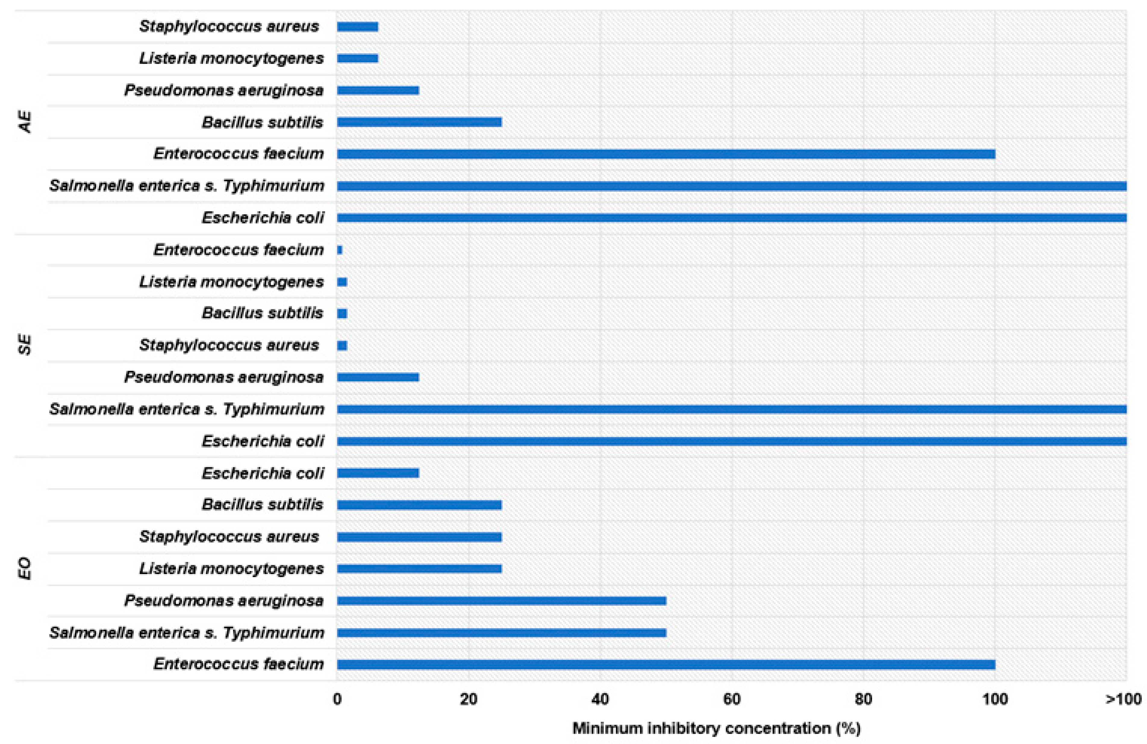
2.7.2. Determination of the Minimum Inhibitory Concentration (MIC)

The test is performed by serially diluting an extract with antimicrobial activity in a nutrient medium and then inoculating with the test microorganism. After incubation, the presence of turbidity (broth) or the growth of colonies on the substrate is checked. The antimicrobial activity of a compound with antimicrobial activity can be determined by the growth of microorganisms in samples where natural ingredients and microorganisms are in contact. Since the problem in determining the MIC of an essential oil is that the essential oil is not mixed with the nutrient medium, and there is no standardized method to overcome this problem, this paper proposes a modified disc diffusion method for determining the MIC in which a series of dilutions of the essential oil in DMSO are applied to the discs. The concentration of the compound with antimicrobial activity found on the last disc (in the series) around which there is no growth of the test microorganism is called the minimum inhibitory concentration and is expressed in %.

2.8. Retention of Lipid Fraction

Wet capsules (20 g) were dried at 105 °C to a constant mass, and the dry matter was determined using a moisture analyzer (Ohaus MB27, Parsippany, NJ, USA). For determining the oil content in ground capsules, the standard ISO Soxhlet extraction method was used [30] with petroleum ether as solvent. The sample is extracted in three cycles (4 h + 2 h + 2 h) with intermediate grinding to improve efficiency. After extraction, the solvent is evaporated, and the lipid fraction is dried at 103 °C to constant mass. The lipid content is then calculated based on the extracted oil weight.

2.9. Retention of Phenolic Compounds

Wet capsules (3 g) were dissolved in 20 mL of 5% sodium citrate solution with stirring on a magnetic stirrer for 30 min. The dissolved capsules were filtered, and the spectrophotometric determination (UV-1600PC, VWR, Radnor, PA, USA) of phenolic compounds was performed by the Folin–Ciocalteu method by mixing 100 µL of the sample with 200 µL of Folin–Ciocalteu reagent and 2 mL of distilled water. After 3 min, 1 mL of a 20% sodium carbonate solution was added, the mixture was vortexed and thermostatted at 50 °C for 25 min and absorbance was then measured at 765 nm. Sodium citrate was used as a blank sample.

2.10. Retention of Essential Oil

Wet capsules (20 g) were dissolved in 200 mL of 5% sodium citrate solution with stirring on a magnetic stirrer for 30 min. The retention of essential oil in the mixture was determined by distillation on a Clevenger apparatus for 90 min.

2.11. Statistical Analysis

One-way analysis of variance (ANOVA) coupled with the Tukey post hoc test was used to evaluate differences between means of antimicrobial activity determined as inhibition zones of extracts. The assumptions of normality and homoscedasticity were confirmed by analyzing the residuals using the Shapiro–Wilk test and Levene’s test, respectively. The software system Design-Expert 10.0 (Stat-Ease Inc., Minneapolis, MN, USA) was used for the experimental design and statistical processing of the data. Response Surface Methodology (RSM) was used to optimize the encapsulation conditions, where the fixed variables were 1% low-density sodium alginate solution, a frequency value of 80 Hz and an electrode voltage of 500 V. The effect of temperature (28, 38 and 48 °C), emulsifier (0.5, 1 and 1.5 g) and CaCl2 (3, 6 and 9%) was evaluated using a 17-point Box–Behnken experimental design (Table 1). The retention of total phenols, essential oil and lipids were determined as dependent factors. An analysis of variance (ANOVA) was performed to determine the significance of the model and the influence of each factor at a 95% confidence level, and the fit of the model was tested and verified by determining the coefficient of determination (R2) and the lack of fit.

3. Results and Discussion

Given the rich composition of M. communis L. and the diverse polarity of its bioactive compounds, this study aimed to assess the potential of encapsulating multiple formulations (aqueous leaf extract, supercritical fruit extract and leaf essential oil) using electrostatic extrusion. The focus was on evaluating both the retention of key compounds (volatile, phenolic and lipid) in the resulting capsules and the antimicrobial activity of the individual extracts and essential oils against a range of microorganisms.

3.1. Antimicrobial Activity of Extracts

Table 2 shows the results of the myrtle antimicrobial activity in the tested aqueous leaf extract (AE), supercritical fruit extract (SE) and leaf essential oil (EO) against tested Gram-positive (Staphylococcus aureus, Bacillus subtilis, Enterococcus faecium and Listeria monocytogenes) and Gram-negative bacteria (Pseudomonas aeruginosa, Escherichia coli and Salmonella enterica s. Typhimurium), yeasts (Candida albicans, Saccharomyces cerevisiae, Candida utilis and Rhodotorula sp.) and lactic acid bacteria (Lactobacillus brevis, Lactobacillus plantarum and Lactobacillus kimchi).
The zones of inhibition of bacteria for AE of myrtle leaves ranged from 6.5 ± 0.71 mm to 35.25 ± 0.35 mm. Among the bacteria tested, the highest zone of inhibition was observed against L. monocytogenes (35.25 mm), followed by E. faecium (25 mm) and S. aureus (19.75 mm). The smallest diameters of the inhibition zone observed with the SE were in a narrow range from 7.75 ± 1.77 mm to 12.25 ± 0.35 mm. Inhibition zones in the range of 8.5 ± 0.71 mm to 28 ± 2.83 mm were observed when EO was used. The highest inhibition zone from the group of Gram-positive bacteria was measured for L. monocytogenes (20.75 mm) and from Gram-negative bacteria for E. coli (28 mm). The positive control for bacteria (kanamycin) showed an inhibition zone between 21.5 ± 2.83 mm and 40.5 ± 0.71 mm. The negative control (DMSO) showed no inhibitory effect. Similar results on antibacterial activity were obtained by Amensour et al. [31], who tested methanol, ethanol and ethyl acetate extracts of M. communis, and most of them showed good antibacterial activity against most bacteria, while there was no inhibitory effect for E. coli. In the work of Taheri et al. [12], among the bacterial species tested, the highest antibacterial activity was obtained at a concentration of 80 mg/mL of the hydroalcoholic extract for S. aureus and Vibrio cholera, while no effect was obtained on P. aeruginosa and a very weak effect on E. coli. Similar to their results, the methanol extract of myrtle leaves showed the highest inhibition for S. aureus [32]. To our knowledge, Pereira et al. [33] and Pereira et al. [34] are the only authors who have investigated the antimicrobial effect of SE of myrtle, however, in leaf extract. The most sensitive bacterium to myrtle EO is E. coli, with an inhibition zone of 28 mm, followed by L. monocytogenes (20.75 mm) and S. aureus (18.75 mm), which was similar to the work of Hsouna et al. [35] that showed good antibacterial activity of EO on L. monocytogenes, S. aureus, B. subtilis, E. coli and P. aeruginosa. Cherrat et al. [36] demonstrated the successful antibacterial effect of EO on L. monocytogenes and S. aureus, while it was weak on E. coli and significant on E. faecium and B. subtilis. The differences in the inhibition zones of the EO for individual bacteria in this study and in the studies of other authors are most likely the result of their different chemotypes, i.e., the different chemical composition of the EO. The oil used in this work is the myrtenyl acetate chemotype, which contains significant amounts of myrtenyl acetate and α-pinene [29].
AE had no inhibitory effect on the tested yeast. As for AE, 30% ethanol was used as the solvent, and it can be assumed that the results differ from those where other extraction solvents were used. Alyousef [37] tested the antifungal activity of different parts of M. communis against five Candida species (Candida albicans, Candida glabrata, Candida kefyr, Candida parapsilosis and Candida tropicalis), and only the methanol extract of the root achieved an inhibitory effect against C. glabrata, with an inhibition zone diameter of 23.5 mm, while the other Candida species showed resistance to all tested extracts. Bonjar et al. [38] tested the antimicrobial effect of methanolic extracts from 221 plant species, including myrtle, on C. albicans (24 mm) and C. utilis (22 mm), while inhibition on S. cerevisiae was not achieved. In the work by Hassan and Abd-Elaziz [39], an inhibition zone of 18 mm was achieved in Rhodotorula spp. with an ethanolic extract of M. communis leaves at a concentration of 30 mg/mL. Nejad et al. [40] tested the antifungal effect of ethanolic extracts of M. communis against 24 Candida species and 3 Aspergillus, with the strongest inhibition against C. glabrata and the weakest inhibitory effect against Aspergillus spp. SE had a similar effect on the inhibition of C. albicans, C. utilis and R. sp. and was between 19.25 and 21.25 mm. The SE from myrtle fruit showed stronger antifungal than antibacterial activity, and the zones of inhibition ranged from 11.25 to 21.25 mm. As with the antibacterial effect of SE from myrtle fruits, the antifungal effect of SE has not yet been investigated; Pereira et al. [34] only investigated the antifungal effect of SE from myrtle leaves. When using EO, the highest zone of inhibition was measured in the yeast C. utilis (20.5 mm). Ghasemi et al. [41] achieved the same inhibition diameter of 15 mm with myrtle EO in C. albicans, as in our study. Rasooli et al. [42] showed that EO exhibited better antifungal activity for C. albicans and S. cerevisiae than the antibacterial activity tested. In the work by Mahboubi and Bidgoli [43], EO showed good antifungal activity against C. albicans and various species of Aspergillus sp., while in the work by Curini et al. [44], significant activity was only shown against Rhodotorula solani among the Rhizoctonia solani, Fusarium solani and Colletotrichum lindemuthianum that were examined. A high content of oxygenated monoterpenes in the composition of the EO [29] has been shown to have a strong antifungal effect [45].
The antibacterial activity of the extracts was not observed for lactic acid bacteria, with the exception of the SE, which caused a slight inhibition of Lactobacillus brevis (9 mm). This is highly desirable because lactic acid bacteria are “good” inhabitants of the intestinal microbiota, and research by Berendika et al. [46] has shown that the AEs of myrtle have a positive effect on the growth of Lactobacillus and Bifidobacterium thanks to their polyphenol composition while reducing the growth of pathogenic bacteria, which has an impact on the health of the organism. Öztürk et al. [4] investigated the prebiotic influence of white and dark blue myrtle fruit pulp on the growth of the probiotic bacterium Lactobacillus casei in goat’s milk ice cream and demonstrated its prebiotic activity. Ghazanfari et al. [47] showed an increase in Lactobacillus and a decrease in the number of Escherichia coli in the intestinal microbiology of chickens as a result of dietary supplementation with myrtle EO. Further studies on the prebiotic and probiotic effects of myrtle extracts are needed.
All three extracts tested inhibited the growth of two extremely pathogenic Gram-positive bacteria that are among the major foodborne pathogens and are highly resistant and difficult to treat with conventional antibiotics, namely S. aureus and L. monocytogenes [35], as well as B. subtilis, E. faecium and Gram-negative P. aeruginosa, for which the SE showed the strongest antimicrobial activity (Figure 1). Only the EO inhibited the growth of all tested bacterial species, while the concentration of the AE and SE was not sufficient to inhibit S. Typhimurium and E. coli.
In general, Gram-positive bacteria are more sensitive to the antimicrobial effect of essential oils and herbal extracts than Gram-negative bacteria. The cell wall of Gram-positive bacteria is thinner and consists of several layers of peptidoglycans containing teichoic acids. The cell wall of Gram-negative bacteria is multilayered and consists of a peptidoglycan layer, then a lipoprotein layer and lipopolysaccharide, and is, therefore, more resistant to external influences [12,31,35,48]. For the inhibition of S. aureus and L. monocytogenes, a concentration of 6.25% of the AE was sufficient, whereas for E. faecium, the highest tested concentration, i.e., 100%, was required, and for S. Typhimurium and E. coli, the tested concentration was not sufficient. This agrees with the results of Amensour et al. [31], who, as in our work, tested all bacterial species except S. Typhimurium, and only E. coli was not inhibited by any of the extracts tested. Compared to the other extracts, the SE of myrtle fruit was the most successful in inhibiting the growth of all Gram-positive bacteria, which is reflected in several times lower MIC values. The use of an EO or AE of myrtle certainly makes sense, considering that the SE also contains EO, phenolic compounds, lipid components such as phytosterols, and fatty acids that synergistically inhibit bacteria [33]. The EO successfully inhibited the growth of all bacteria tested and was the only one that inhibited the growth of E. coli and S. Typhimurium compared to the other two extracts tested, and even an oil concentration of 12.5% was sufficient for E. coli. This is in line with the literature [35,49,50,51]. The main constituents of myrtle EO, like 1,8-cineole, linalool, α-terpineol and eugenol, are thought to be responsible for its antimicrobial activity [1]. They act synergistically with other groups of compounds present in varying amounts [35].

3.2. Efficiency of Microencapsulation of Volatiles, Phenolics and Lipids

The retention efficiency of encapsulated myrtle bioactives, including essential oil, phenolic compounds, and lipids, was evaluated as an indicator of microencapsulation performance. The results of analytical determinations are shown in Table 3. The encapsulation efficiency (EE) results are consistent with the work of Volić et al. [52] and Natrajan et al. [53], but with the addition of a coating agent such as chitosan, there is a significant increase in the EE of the EO [53]. However, one of the factors influencing the efficiency of encapsulation is the concentration of alginate used, and a higher concentration of alginate showed better retention of the EO within the capsule, as a denser structure is formed that traps the oil. In the experiment, 1% alginate was used as a fixed variable, according to the study of Dobroslavić et al. [54], as their experiment showed this concentration of alginate was optimal. At higher alginate concentrations, which means better retention, encapsulation by electrostatic extrusion is not possible due to excessive viscosity [27].
The response surface methodology based on the Box–Behnken experimental design led to appropriate prediction models for the co-encapsulation of essential oil and extract of myrtle leaves and the supercritical extract of myrtle fruit. Models developed through statistical analysis (Table 4 and Figure 2) confirmed that both the amount of emulsifier and CaCl2 concentration significantly affected the retention of all three compound groups. The retention of volatiles was influenced by linear and quadratic components of all independent factors, and the interaction of CaCl2 amount with temperature and emulsifier amount was favored by intermediate emulsifier amounts and lower temperatures in combination with a higher CaCl2 amount. Emulsifiers play a crucial role in stabilizing oil-in-water emulsions by reducing interfacial tension and forming protective barriers around oil droplets; therefore, they must be included in formulations composed of polar and non-polar components. In the present study, intermediate emulsifier concentrations likely facilitated the formation of a stable emulsion, minimizing the diffusion and volatilization of aromatic compounds. Similarly, a study from Teo [52] observed that increasing Tween 20 concentrations up to 2.5% (w/w) resulted in decreased droplet sizes in emulsions. However, at concentrations above 2.5%, droplet size increased from 183 nm to 505 nm. This increase was attributed to the formation of surfactant micelles leading to droplet flocculation due to depletion effects. In a study reported by Shen et al. [53], the stability of heavy crude oil-in-water emulsions stabilized by Tween 20 was investigated. They observed that increasing the emulsifier concentration from 1.1 to 1.6% reduced the creaming index, leading to the formation of a more stable emulsion. However, with a further increase in emulsifier concentration, droplet size increased.
Calcium chloride contributes to emulsion stability through ionic cross-linking, which enhances the mechanical strength of the interfacial film. This cross-linking can lead to the formation of a more rigid and cohesive network, effectively entrapping volatile compounds [54]. Research has shown that incorporating CaCl2 in the microencapsulation process can improve the encapsulation efficiency of essential oils by promoting the formation of a robust matrix [55]. In the present study, higher CaCl2 concentrations at lower temperatures enhanced the retention of volatiles, likely due to the formation of a more structured and less permeable encapsulating matrix.
Temperature is another critical factor influencing the retention of volatile compounds. Up to a certain point, an increase in temperature can lower the viscosity of the sodium alginate at higher temperatures [27,56]. During the passage through the encapsulator, the encapsulation process was carried out faster, i.e., the capsules were produced faster so that the essential oil was efficiently trapped in the capsules. On the other hand, elevated temperatures can increase molecular mobility and evaporation rates, leading to greater losses of volatile constituents. Studies on the encapsulation of essential oils have reported that lower processing temperatures help preserve the integrity of volatile compounds by reducing thermal degradation and evaporation [57,58]. In this study, lower temperatures combined with higher CaCl2 concentrations resulted in the improved retention of volatile compounds, suggesting a synergistic effect between these variables.
In contrast, the retention of phenols was only influenced by linear components and the interaction of the factors, with the exception of the interaction between temperature and CaCl2 amount. Higher amounts of emulsifier led to higher retention of phenols, while a higher amount of CaCl2 caused their decrease at lower amounts of emulsifier.
Similar values for the efficiency of the microencapsulation of polyphenolic compounds were obtained by other authors; Stojanovic et al. [56] achieved between 50% and 80% for the encapsulation of an AE of Thymus serpyllum L. and Belščak-Cvitanović et al. [59] achieved between 60.78% and 80.17% for the encapsulation of polyphenols from green tea extracts. One way to achieve a higher EE of the polyphenolic compounds is to use a polymer filler/coating particle/thickener [60], whereby a combination of alginate and calcium caseinate as a polymer filler retained up to 80% of the polyphenols [59].
Finally, the retention of lipids was also favored by higher amounts of emulsifier and lower amounts of CaCl2. As mentioned, a higher proportion of emulsifiers is expected to have a better effect on compound retention since the main property of emulsifiers is to enable emulsion stability by reducing the tension between two immiscible liquids, and the Tween 20 used is a suitable emulsifier for this oil-in-water (O/W) emulsion due to its HLB value (16.7) [27]. In addition to the physical properties, such as size, shape and porosity of the capsules, the efficiency of the encapsulation of lipid droplets is also influenced by the stability of the emulsion and the degree of alginate cross-linking, and the release of free fatty acids is significantly reduced by the encapsulation of lipids in alginate droplets [61]. The study by Lupo et al. [62] showed that the influence of emulsifier concentration on emulsion stability depends on the type of emulsifier used. In a study by Won et al. [63], different CaCl2 concentrations showed a very weak effect on the loading efficiency and immobilization yield of lipase. Furthermore, the influence of different CaCl2 concentrations from 0.125% to 6% on the EE of proliposomal granules was tested, and the concentration of 1% had the strongest effect, while higher concentrations did not lead to higher EE [64]. In our case, the highest EE of the lipids was achieved by the lowest tested concentration of 3%.
Considering all results, overall, optimal co-encapsulation was achieved at 48 °C, 1.27 g emulsifier and 3% CaCl2 in the receiver solution, resulting in the retention of 72.86% of volatiles, 61.13% of phenolic compounds and 62.80% of lipids.

4. Conclusions

In general, Gram-positive bacteria, S. aureus and L. monocytogenes, were the most sensitive bacteria to the tested myrtle extracts, while Gram-negative S. Typhimurium bacteria were the most resistant. Only EO inactivated S. Typhimurium and E. coli. SE and EO showed antifungal activity against all yeasts tested, while AE showed no antifungal activity. SE showed stronger antifungal than antibacterial activity. Since the tested extracts had no antimicrobial effect on lactic acid bacteria, they have a great potential prebiotic effect. Due to the different antimicrobial effects of the investigated extracts on individual microorganisms, a combination of bioactive molecule groups in suitable proportions or a suitable formulation could achieve the desired inhibitory effects against a variety of microorganisms. The obtained results clearly show that electrostatic extrusion was successfully used for the co-encapsulation of hydrophilic and lipophilic extracts from myrtle leaves and fruit. The amount of emulsifier and CaCl2 significantly influenced the retention of all three groups of compounds, while the temperature additionally influenced the retention of the volatiles. Under optimal encapsulation conditions, a high level of retention was achieved for all major groups of bioactive compounds, including volatiles, phenolics and lipids. Particularly in the form of capsules, the formulations of the extracts could have a cumulative effect on the target group of microorganisms, which must be tested further. Extracts of myrtle leaves and fruit have shown great potential for use as an antimicrobial agent against pathogens in the food industry. Future developments shall include studies on potential synergistic interactions between different extracts, and studies on the stability of bioactive compounds and their cytotoxicity and safety should also be conducted to optimize the integration of these encapsulated systems into food preservation and pharmaceutical applications.

Author Contributions

Conceptualization, D.C. and S.B.; methodology, D.C., J.M. and E.D.; investigation, D.C., J.M. and S.B.; writing—original draft preparation, D.C.; writing—review and editing, S.B.; supervision, S.B.; funding acquisition, V.D.-U. All authors have read and agreed to the published version of the manuscript.

Funding

This research was funded by the project “Bioactive molecules of medical plants as natural antioxidants, microbicides and preservatives” (KK.01.1.1.04.0093), co-financed by the Croatian Government and the European Union through the European Regional Development Fund—Operational Programme Competitiveness and Cohesion, grant number KK.01.1.1.04.

Data Availability Statement

The original contributions presented in this study are included in the article. Further inquiries can be directed to the corresponding author.

Conflicts of Interest

The authors declare no conflicts of interest.

References

  1. Aleksic, V.; Knezevic, P. Antimicrobial and Antioxidative Activity of Extracts and Essential Oils of Myrtus communis L. Microbiol. Res. 2014, 169, 240–254. [Google Scholar] [CrossRef] [PubMed]
  2. Cvitković, D.; Lisica, P.; Zorić, Z.; Repajić, M.; Pedisić, S.; Dragović-Uzelac, V.; Balbino, S. Composition and Antioxidant Properties of Pigments of Mediterranean Herbs and Spices as Affected by Different Extraction Methods. Foods 2021, 10, 2477. [Google Scholar] [CrossRef] [PubMed]
  3. Özcan, M.M.; Al Juhaimi, F.; Ahmed, I.A.M.; Babiker, E.E.; Ghafoor, K. Antioxidant Activity, Fatty Acid Composition, Phenolic Compounds and Mineral Contents of Stem, Leave and Fruits of Two Morphs of Wild Myrtle Plants. J. Food Meas. Charact. 2020, 14, 1376–1382. [Google Scholar] [CrossRef]
  4. Öztürk, H.İ.; Demirci, T.; Akın, N. Production of Functional Probiotic Ice Creams with White and Dark Blue Fruits of Myrtus communis: The Comparison of the Prebiotic Potentials on Lactobacillus Casei 431 and Functional Characteristics. LWT Food Sci. Technol. 2018, 90, 339–345. [Google Scholar] [CrossRef]
  5. Vaou, N.; Stavropoulou, E.; Voidarou, C.; Tsakris, Z.; Rozos, G.; Tsigalou, C.; Bezirtzoglou, E. Interactions between Medical Plant-Derived Bioactive Compounds: Focus on Antimicrobial Combination Effects. Antibiotics 2022, 11, 1014. [Google Scholar] [CrossRef]
  6. Abreu, A.C.; McBain, A.J.; Simões, M. Plants as Sources of New Antimicrobials and Resistance-Modifying Agents. Nat. Prod. Rep. 2012, 29, 1007–1021. [Google Scholar] [CrossRef]
  7. Wagner, H. Synergy Research: Approaching a New Generation of Phytopharmaceuticals. Fitoterapia 2009, 82, 34–37. [Google Scholar] [CrossRef]
  8. Shariati, A.; Arshadi, M.; Khosrojerdi, M.A.; Abedinzadeh, M.; Ganjalishahi, M.; Maleki, A.; Heidary, M.; Khoshnood, S. The Resistance Mechanisms of Bacteria against Ciprofloxacin and New Approaches for Enhancing the Efficacy of This Antibiotic. Front. Public Health 2022, 10, 1025633. [Google Scholar] [CrossRef]
  9. Aqil, F.; Ahmad, I.; Owais, M. Evaluation of Anti-Methicillin-Resistant Staphylococcus Aureus (MRSA) Activity and Synergy of Some Bioactive Plant Extracts. Biotechnol. J. 2006, 1, 1093–1102. [Google Scholar] [CrossRef]
  10. Long, F.; Yang, H.; Xu, Y.; Hao, H.; Li, P. A Strategy for the Identification of Combinatorial Bioactive Compounds Contributing to the Holistic Effect of Herbal Medicines. Sci. Rep. 2015, 5, srep12361. [Google Scholar] [CrossRef]
  11. Chawda, P.J.; Shi, J.; Xue, S.; Young Quek, S. Co-Encapsulation of Bioactives for Food Applications. Food Qual. Saf. 2017, 1, 302–309. [Google Scholar] [CrossRef]
  12. Taheri, A.; Seyfan, A.; Jalalinezhad, S.; Nasery, F. Antibacterial Effect of Myrtus communis Hydro-Alcoholic Extract on Pathogenic Bacteria. Zahedan J. Res. Med. Sci. 2013, 15, 19–24. [Google Scholar]
  13. Carvalho, I.T.; Estevinho, B.N.; Santos, L. Application of Microencapsulated Essential Oils in Cosmetic and Personal Healthcare Products—A Review. Int. J. Cosmet. Sci. 2016, 38, 109–119. [Google Scholar] [CrossRef] [PubMed]
  14. Martins, I.M.; Barreiro, M.F.; Coelho, M.; Rodrigues, A.E. Microencapsulation of Essential Oils with Biodegradable Polymeric Carriers for Cosmetic Applications. Chem. Eng. J. 2014, 245, 191–200. [Google Scholar] [CrossRef]
  15. Champagne, C.P.; Fustier, P. Microencapsulation for the Improved Delivery of Bioactive Compounds into Foods. Curr. Opin. Biotechnol. 2007, 18, 184–190. [Google Scholar] [CrossRef]
  16. Okuro, P.K.; Thomazini, M.; Balieiro, J.C.C.; Liberal, R.D.C.O.; Fávaro-Trindade, C.S. Co- Encapsulation of Lactobacillus Acidophilus with Inulin or Polydextrose in Solid Lipid Microparticles Provides Protection and Improves Stability. Food Res. Int. 2013, 53, 96–103. [Google Scholar] [CrossRef]
  17. Iyer, C.; Kailasapathy, K. Effect of Co-Encapsulation of Probiotics with Prebiotics on Increasing the Viability of Encapsulated Bacteria under In Vitro Acidic and Bile Salt Conditions and in Yogurt. J. Food Sci. 2005, 70, M18–M23. [Google Scholar] [CrossRef]
  18. Sathyabama, S.; Ranjith kumar, M.; Bruntha devi, P.; Vijayabharathi, R. Brindha priyadharisini Co-Encapsulation of Probiotics with Prebiotics on Alginate Matrix and Its Effect on Viability in Simulated Gastric Environment. LWT Food Sci. Technol. 2014, 57, 419–425. [Google Scholar] [CrossRef]
  19. Eratte, D.; McKnight, S.; Gengenbach, T.R.; Dowling, K.; Barrow, C.J.; Adhikari, B.P. Co-Encapsulation and Characterisation of Omega-3 Fatty Acids and Probiotic Bacteria in Whey Protein Isolate-Gum Arabic Complex Coacervates. J. Funct. Foods 2015, 19, 882–892. [Google Scholar] [CrossRef]
  20. Bajaj, S.R.; Marathe, S.J.; Singhal, R.S. Co-Encapsulation of Vitamins B12 and D3 Using Spray Drying: Wall Material Optimization, Product Characterization, and Release Kinetics. Food Chem. 2021, 335, 127642. [Google Scholar] [CrossRef]
  21. Koushki, V.; Babaei, A.; Mehraban Sangatash, M.; Safari, O. The Efficacy of Co-Encapsulation with Herbal Extracts on Viability of Probiotic Bacteria During Storage in Fruit Juices. J. Innov. Food Sci. Technol. 2021, 13, 1. [Google Scholar]
  22. Le Priol, L.; Gmur, J.; Dagmey, A.; Morandat, S.; El Kirat, K.; Saleh, K.; Nesterenko, A. Co-Encapsulation of Vegetable Oils with Phenolic Antioxidants and Evaluation of Their Oxidative Stability under Long-Term Storage Conditions. LWT 2021, 142, 111033. [Google Scholar] [CrossRef]
  23. Chen, Q.; McGillivray, D.; Wen, J.; Zhong, F.; Quek, S.Y. Co-Encapsulation of Fish Oil with Phytosterol Esters and Limonene by Milk Proteins. J. Food Eng. 2013, 50, 505–512. [Google Scholar] [CrossRef]
  24. Bamidele, O.P.; Emmambux, M.N. Encapsulation of Bioactive Compounds by “Extrusion” Technologies: A Review. Crit. Rev. Food Sci. Nutr. 2021, 61, 3100–3118. [Google Scholar] [CrossRef]
  25. Poshadri, A.; Kuna, A. Microencapsulation Technology: A Review. J. Res. ANGRAU 2010, 38, 86–102. [Google Scholar] [CrossRef]
  26. Manojlovic, V.; Rajic, N.; Djonlagic, J.; Obradovic, B.; Nedovic, V.; Bugarski, B. Application of Electrostatic Extrusion—Flavour Encapsulation and Controlled Release. Sensors 2008, 8, 1488–1496. [Google Scholar] [CrossRef]
  27. Aguilar, N.N. Study of the Influence of Buchi Encapsulator Input Variables on Properties of Formed Alginate Beads. Ph.D. Thesis, Universitat de Barcelona, Barcelona, Spain, 2018. [Google Scholar]
  28. Choudhury, N.; Meghwal, M.; Das, K. Microencapsulation: An Overview on Concepts, Methods, Properties and Applications in Foods. Food Front. 2021, 2, 426–442. [Google Scholar] [CrossRef]
  29. Cvitković, D.; Lisica, P.; Zorić, Z.; Pedisić, S.; Repajić, M.; Dragović-Uzelac, V.; Balbino, S. The Influence of Cryogrinding on Essential Oil, Phenolic Compounds and Pigments Extraction from Myrtle (Myrtus communis L.) Leaves. Processes 2022, 10, 2716. [Google Scholar] [CrossRef]
  30. HRN EN ISO 659; Oilseeds—Determination of Oil Content (Reference Method). Croatian Standards Institute: Zagreb, Croatia, 2010.
  31. Amensour, M.; Bouhdid, S.; Fernández-López, J.; Idaomar, M.; Senhaji, N.S.; Abrini, J. Antibacterial Activity of Extracts of Myrtus communis against Food-Borne Pathogenic and Spoilage Bacteria. Int. J. Food Prop. 2010, 13, 1215–1224. [Google Scholar] [CrossRef]
  32. Özcan, M.M.; Uyar, B.; Ünver, A. Antibacterial Effect of Myrtle (Myrtus communis L.) Leaves Extract on Microorganisms. J. Food Saf. Food Qual. 2015, 66, 18–21. [Google Scholar] [CrossRef]
  33. Pereira, P.; Bernardo-Gil, M.G.; Cebola, M.J.; Mauricio, E.; Romano, A. Supercritical Fluid Extracts with Antioxidant and Antimicrobial Activities from Myrtle (Myrtus communis L.) Leaves. Response Surface Optimization. J. Supercrit. Fluids 2013, 83, 57–64. [Google Scholar] [CrossRef]
  34. Pereira, P.; Mauricio, E.M.; Duarte, M.P.; Lima, K.; Fernandes, A.S.; Bernardo-Gil, G.; Cebola, M.-J. Potential of Supercritical Fluid Myrtle Extracts as an Active Ingredient and Co-Preservative for Cosmetic and Topical Pharmaceutical Applications. Sustain. Chem. Pharm. 2022, 28, 100739. [Google Scholar] [CrossRef]
  35. Hsouna, A.; Ben; Hamdi, N.; Miladid, R.; Abdelkafid, S. Myrtus communis Essential Oil: Chemical Composition and Antimicrobial Activities against Food Spoilage Pathogens. Chem. Biodivers. 2014, 11, 571–580. [Google Scholar] [CrossRef] [PubMed]
  36. Cherrat, L.; Espina, L.; Bakkali, M.; García-Gonzalo, D.; Pagán, R.; Laglaoui, A. Chemical Composition and Antioxidant Properties of Laurus nobilis L. and Myrtus communis L. Essential Oils from Morocco and Evaluation of Their Antimicrobial Activity Acting Alone or in Combined Processes for Food Preservation. J. Sci. Food Agric. 2014, 94, 1197–1204. [Google Scholar] [CrossRef]
  37. Alyousef, A.A. Antifungal Activity and Mechanism of Action of Different Parts of Myrtus communis Growing in Saudi Arabia against Candida Spp. J. Nanomater. 2021, 2021, 3484125. [Google Scholar] [CrossRef]
  38. Bonjar, G.H.S.; Aghighi, S.; Nik, A.K. Antibacterial and Antifungal Survey in Plants Used in Indigenous Herbal-Medicine of South East Regions of Iran. J. Biol. Sci. 2004, 4, 405–412. [Google Scholar] [CrossRef]
  39. Hassan, A.A.; Abd-Elazis, G.O. Genotoxicity and Antimicrobial Activity of Myrtus communis L., Ziziphus spina-Christi (L.) Willd and Cassia Angustifolia Vahl Extracts. Bangladesh J. Bot. 2020, 49, 557–566. [Google Scholar] [CrossRef]
  40. Nejad, B.S.; Nejad, M.E.; Naanaie, S.Y.; Zarrin, M. Antifungal Efficacy of Myrtus communis Linn. Jentashapir J. Health Res. 2014, 5, e21879. [Google Scholar] [CrossRef]
  41. Ghasemi, P.A.; Jahanbazi, P.; Enteshari, S.; Malekpoor, F.; Hamedi, B. Antimicrobial Activity of Some Iranian Medicinal Plants. Arch. Biol. Sci. 2010, 62, 633–641. [Google Scholar] [CrossRef]
  42. Rasooli, I.; Moosavi, M.L.; Rezaee, M.B.; Jaimand, K. Susceptibility of Microorganisms to Myrtus communis L. Essential Oil and Its Chemical Composition. J. Agric. Sci. Technol. 2002, 4, 127–133. [Google Scholar]
  43. Mahboubi, M.; Bidgoli, F.G. In Vitro Synergistic Efficacy of Combination of Amphotericin B with Myrtus communis Essential Oil against Clinical Isolates of Candida albicans. Phytomedicine 2010, 17, 771–774. [Google Scholar] [CrossRef] [PubMed]
  44. Curini, M.; Bianchi, A.; Epifano, F.; Bruni, R.; Torta, L.; Zambonelli, A. Composition and in Vitro Antifungal Activity of Essential Oils of Erigeron Canadensis and Myrtus communis from France. Chem. Nat. Compd. 2003, 39, 191–194. [Google Scholar] [CrossRef]
  45. Kordali, S.; Kotan, R.; Cakir, A. Screening of Antifungal Activities of 21 Oxygenated Monoterpenes In-Vitro as Plant Disease Control Agents. Allelopath. J. 2007, 19, 373–392. [Google Scholar]
  46. Berendika, M.; Domjanić Drozdek, S.; Odeh, D.; Oršolić, N.; Dragičević, P.; Sokolović, M.; Garofulić, I.E.; Đikić, D.; Jurčević, I.L. Beneficial Effects of Laurel (Laurus nobilis L.) and Myrtle (Myrtus communis L.) Extract on Rat Health. Molecules 2022, 27, 581. [Google Scholar] [CrossRef]
  47. Ghazanfari, S.; Moradi, M.A.; Bardzardi, M.M. Intestinal Morphology and Microbiology of Broiler Chicken Fed Diets Containing Myrtle (Myrtus communis) Essential Oil Supplementation. Iran. J. Appl. Anim. Sci. 2014, 4, 549–554. [Google Scholar]
  48. Mir, M.A.; Bashir, N.; Alfaify, A.; Oteef, M.D.Y. GC-MS Analysis of Myrtus communis Extract and Its Antibacterial Activity against Gram-Positive Bacteria. BMC Complement. Med. Ther. 2020, 20, 86. [Google Scholar] [CrossRef]
  49. Mohamadi, Y.; Lograda, T.; Ramdani, M.; Figueredo, G.; Chalard, P. Chemical Composition and Antimicrobial Activity of Myrtus communis Essential Oils from Algeria. Biodiversitas J. Biol. Divers. 2021, 22, 933–946. [Google Scholar] [CrossRef]
  50. Pirbalouti, A.G.; Hamedi, B.; Mehravar, L.; Firouznejhad, M. Diversity in Chemical Composition and Antibacterial Activity of the Essential Oils of Wild Populations of Myrtle from Natural Habitats in Southwestern Iran. Indian. J. Tradit. Knowl. 2014, 13, 484–489. [Google Scholar]
  51. Yadegarinia, D.; Gachkar, L.; Rezaei, M.B.; Taghizadeh, M.; Astaneh, S.A.; Rasooli, I. Biochemical Activities of Iranian Mentha piperita L. and Myrtus communis L. Essential Oils. Phytochemistry 2006, 67, 1249–1255. [Google Scholar] [CrossRef]
  52. Anges, H.M. Development of Novel Nanoemulsions as Delivery Systems. Ph.D. Thesis, Massey University, Palmerston North, New Zealand, 2016. [Google Scholar]
  53. Shen, W.; Koirala, N.; Mukherjee, D.; Lee, K.; Zhao, M.; Li, J. Tween 20 Stabilized Conventional Heavy Crude Oil-In-Water Emulsions Formed by Mechanical Homogenization. Front. Environ. Sci. 2022, 10, 1–11. [Google Scholar] [CrossRef]
  54. Noppakundilograt, S.; Piboon, P.; Graisuwan, W.; Nuisin, R.; Kiatkamjornwong, S. Encapsulated Eucalyptus Oil in Ionically Cross-Linked Alginate Microcapsules and Its Controlled Release. Carbohydr. Polym. 2015, 131, 23–33. [Google Scholar] [CrossRef] [PubMed]
  55. Pratiwi, L.; Eddy, D.R.; Al Anshori, J.; Harja, A.; Wahyudi, T.; Mulyawan, A.S.; Julaeha, E. Microencapsulation of Citrus Aurantifolia Essential Oil with the Optimized CaCl2 Crosslinker and Its Antibacterial Study for Cosmetic Textiles. RSC Adv. 2022, 12, 30682–30690. [Google Scholar] [CrossRef] [PubMed]
  56. Stojanovic, R.; Belscak-Cvitanovic, A.; Manojlovic, V.; Komes, D.; Nedovic, V.; Bugarski, B. Encapsulation of Thyme (Thymus Serpyllum L.) Aqueous Extract in Calcium Alginate Beads. J. Sci. Food Agric. 2012, 92, 685–696. [Google Scholar] [CrossRef] [PubMed]
  57. Dobroslavić, E.; Cegledi, E.; Robić, K.; Elez Garofulić, I.; Dragović-Uzelac, V.; Repajić, M. Encapsulation of Fennel Essential Oil in Calcium Alginate Microbeads via Electrostatic Extrusion. Appl. Sci. 2024, 14, 3522. [Google Scholar] [CrossRef]
  58. Napiórkowska, A.; Szpicer, A.; Wojtasik-Kalinowska, I.; Perez, M.D.T.; González, H.D.; Kurek, M.A. Microencapsulation of Juniper and Black Pepper Essential Oil Using the Coacervation Method and Its Properties after Freeze-Drying. Foods 2023, 12, 4345. [Google Scholar] [CrossRef]
  59. Belščak-Cvitanović, A.; Đorđević, V.; Karlović, S.; Pavlović, V.; Komes, D.; Ježek, D.; Bugarski, B.; Nedović, V. Protein-Reinforced and Chitosan-Pectin Coated Alginate Microparticles for Delivery of Flavan-3-Ol Antioxidants and Caffeine from Green Tea Extract. Food Hydrocoll. 2015, 51, 361–374. [Google Scholar] [CrossRef]
  60. Kurozawa, L.E.; Hubinger, M.D. Hydrophilic Food Compounds Encapsulation by Ionic Gelation. Curr. Opin. Food Sci. 2017, 15, 50–55. [Google Scholar] [CrossRef]
  61. Zeeb, B.; Saberi, A.H.; Weiss, J.; McClements, D.J. Retention and Release of Oil-in-Water Emulsions from Filled Hydrogel Beads Composed of Calcium Alginate: Impact of Emulsifier Type and PH. Soft Matter 2015, 11, 2228–2236. [Google Scholar] [CrossRef]
  62. Lupo, B.; Maestro, A.; Porras, M.; Gutiérrez, J.M.; González, C. Preparation of Alginate Microspheres by Emulsification/Internal Gelation to Encapsulate Cocoa Polyphenols. Food Hydrocoll. 2014, 38, 56–65. [Google Scholar] [CrossRef]
  63. Won, K.; Kim, S.; Kim, K.-J.; Park, H.W.; Moon, S.-J. Optimization of Lipase Entrapment in Ca-Alginate Gel Beads. Process Biochem. 2005, 40, 2149–2154. [Google Scholar] [CrossRef]
  64. Tunsirikongkon, A.; Pyo, Y.-C.; Kim, D.-H.; Tran, P.; Park, J.-S. Effect of Calcium Chloride on the Protein Encapsulation and Stability of Proliposomal Granules. J. Drug Deliv. Sci. Technol. 2020, 57, 101672. [Google Scholar] [CrossRef]
Figure 1. Minimum inhibitory concentrations of aqueous extract (AE), supercritical extract (SE) and essential oil (EO) in %.
Processes 13 01220 g001
Figure 2. Response surface plots of volatiles (A), phenolics (B) and lipids (C) retention as a function of emulsifier and CaCl2 amount at a constant temperature of 38 °C. The color gradient represents the response value from lowest (blue) to highest (red) while dots represent actual experimental data points.
Processes 13 01220 g002
Table 1. Box–Behnken experimental design of microencapsulation parameters.
Exp.1234567891011121314151617
Temperature (°C)2838484848382838383838283838384828
Emulsifier (g)11110.50.511111.50.51.510.51.51.5
CaCl2 (%)36936996669636366
Table 2. Antimicrobial activity as inhibition zones of AE, SE and EO (mm, mean ± standard deviation, n = 3, nd—not determined) against Gram-positive and Gram-negative bacteria, yeast and lactic acid bacteria.
MOAESEEODMSO
10 µL
Kanamycin
(10 mg L−1)/5 µL
Staphylococcus aureus *19.75 b ± 3.1812.25 a ± 0.3518.75 b ± 0.35nd30.25 c ± 1.77
Bacillus subtilis *11.50 b ± 4.948.75 a ± 0.3512.00 b ± 2.12nd22.25 c ± 5.67
Enterococcus faecium *25.00 c ± 2.1210.00 b ± 4.247.75 a ± 0.35nd21.50 c ± 2.83
Listeria monocytogenes *35.25 c ± 0.357.75 a ± 1.7720.75 b ± 4.60nd32.50 c ± 4.95
Pseudomonas aeruginosa *8.75 a ± 3.188.00 a ± 2.8316.00 b ± 5.65nd32.00 c ± 4.24
Escherichia coli *6.50 a ± 1.136.50 a ± 0.7128.00 b ± 2.83nd40.50 c ± 0.71
Salmonella enterica s.
Typhimurium *
6.50 a ± 0.716.00 a ± 1.418.50 b ± 0.71nd23.25 c ± 2.47
MOAESEEODMSO
10 µL
Nystatin
(5 mg mL−1)/10 µL
Candida albicans *nd19.5 c ± 3.5415.25 a,b ± 4.60nd17.00 b ± 0.00
Saccharomyces cerevisiae *nd11.25 a ± 3.1818.00 b ± 5.31nd25.00 c ± 0.00
Candida utilis *nd20.00 a ± 0.0020.5 a ± 2.12nd25.00 b ± 0.00
Rhodotorula sp. *nd21.25 b ± 3.8910.25 a ± 6.01nd21.00 b ± 0.00
MOAESEEODMSO
10 µL
Kanamycin
(50 mg L−1)/5 µL
Lactobacillus brevis *nd9.00 a ± 1.50ndnd17.00 b ± 2.10
Lactobacillus plantarumndndndnd10.00 ± 2.34
Lactobacillus kimchindndndndnd
* ANOVA results show significant differences between the extracts (p ≤ 0.001). Groups with different letters are significantly different from each other. MO—microorganisms; AE—aqueous leaf extract; SE—supercritical fruit extract; EO—essential oil; DMSO—Dimethyl sulfoxide.
Table 3. Analytical determinations of co-encapsulation efficiency measured as volatiles, phenolics and lipids retention.
Exp.Temperature
(°C)
Emulsifier (g)CaCl2
(%)
Volatiles
Retention (%)
Phenolics Retention (%)Lipids
Retention (%)
1281377.0737.6548.73
2381674.3642.7522.21
3481977.2246.9927.86
4481377.2547.6540.64
5480.5669.0323.6616.21
6380.5969.1547.2414.69
7281983.6655.1625.15
8381672.5647.4920.17
9381674.3440.0323.73
10381672.5845.5117.79
11381.5971.2761.4129.79
12280.5675.4533.1423.68
13381.5362.8962.6662.80
14381671.9748.5922.62
15380.5371.4612.8926.16
16481.5664.9662.9636.17
17281.5666.2443.0137.32
Table 4. Analysis of variance (ANOVA) of co-encapsulation efficiency parameter models (A—Temperature; B—Emulsifier; C—CaCl2).
Source of VariationVolatiles Retention (%)Phenolics Retention (%)Lipids Retention (%)
F Valuep ValueF Valuep ValueF Valuep Value
A15.370.006 *0.900.3642.360.168
B30.760.001 *76.56<0.001 *87.47<0.001 *
C12.570.009 *14.920.003 *78.51<0.001 *
AB4.160.08110.370.009 *0.960.360
AC6.920.034 *3.950.0752.800.138
BC18.030.004 *15.170.003 *11.140.012
A222.860.002 *--8.710.021
B2136.85<0.001 *--2.340.170
C219.440.003 *--37.70<0.001 *
Lack of fit0.3130.2360.154
R20.9740.9240.971
ModelVolatiles retention (%) = 113.90 − 2.33 × A + 32.02 × B − 2.76 × C + 0.26 × A × B − 0.06 × A × C + 1.78 × B × C + 0.03 × A2− 28.70 × B2 + 0.30 × C2Phenolics retention (%) = −16.17 − 0.41 × A + 7.95 × B + 13.77 × C + 1.47 × A × B − 0.15 × A × C − 5.93 × B × CLipids retention (%) = 153.03 − 4.56 × A + 11.65 × B − 16.07 × C + 0.32 × A × B + 0.09 × A × C − 3.59 × B × C + 0.05 × A2 + 9.61 × B2+ 1.07 × C2
* Significant influence of factor.
Disclaimer/Publisher’s Note: The statements, opinions and data contained in all publications are solely those of the individual author(s) and contributor(s) and not of MDPI and/or the editor(s). MDPI and/or the editor(s) disclaim responsibility for any injury to people or property resulting from any ideas, methods, instructions or products referred to in the content.

