Article

The Preparation of Experimental Resin-Based Dental Composites Using Different Mixing Methods for the Filler and Matrix †

1
Department of General Dentistry, Medical University of Lodz, 92-213 Lodz, Poland
2
University Laboratory of Materials Research, Medical University of Lodz, 92-213 Lodz, Poland
3
Student Science Club “Materials in Dentistry”, Medical University of Lodz, 92-213 Lodz, Poland
*
Author to whom correspondence should be addressed.
This paper is an extended version of abstract published in programme and materials of the XXII Conference of “Biomaterials and mechanics in dentistry”, Wisła, Poland, 10–13 October 2024.
Processes 2025, 13(5), 1332; https://doi.org/10.3390/pr13051332
Submission received: 1 April 2025 / Revised: 18 April 2025 / Accepted: 21 April 2025 / Published: 27 April 2025

Abstract

:
Resin-based composites are common and widely used materials in dentistry in direct and indirect applications. Their mechanical properties depend on the composition and homogeneity of the resulting structure. This study aims to optimize the mixing process to obtain the most homogeneous mixture possible, which will allow for the better mechanical properties of the composite. A mixture of bis-GMA/UDMA/HEMA/TEGDMA monomers forming a polymer matrix was filled with silanized silica (45 wt%) using different mixing methods. This study analyzed five manufacturing methods—hand mixing (agate mortar), mixing in a centrifugal Hauschild SpeedMixer, and the hybrid method—combined with the abovementioned methods. The effect of the mixing method on the Vickers hardness (HV), flexural strength (FS), compressive strength (CS), and diametral tensile strength (DTS) of the produced composites was investigated, and the stresses generated during composite polymerization were determined. Mechanically prepared composites have the highest flexural strength and hardness. The lowest shrinkage stress was achieved by the composite, which was prepared partially manually. The results showed that the mixing method affects the morphology of the filler and, hence, the strength properties of the resulting material.

1. Introduction

Resin-based composites, also known as dental composite or dental composite resin, are commonly used in dentistry for direct esthetic restorations that mimic the appearance of teeth [1,2]. Generally, composites are specific materials made of at least two components (phases) with different properties from each other, so the new material produced has properties that are better and/or new, in additional to the material’s individual components. By observation, a composite is a monolithic material, but it has visible boundaries between the components. The basic classification of composites is its type of matrix material—which can be metallic, ceramic, or polymer. One of the representatives of composites with a polymer matrix in the dentistry area is, as mentioned above, the resin-based dental composite (RBC). Dental composites have undergone significant evolution over the past 50 years, with recent advancements primarily focusing on enhancing the polymer matrix [3,4]. Alongside these improvements, the choice of reinforcements plays a crucial role, with options including grains, nanoparticles, and chopped fibers [5]. These inorganic structures, when added to the matrix, influence various properties, such as the strength, stiffness, abrasion resistance [6], and modulus of elasticity [3,6,7]. The organic phase of dental composites constitutes about 20–30% of the weight of the material and mainly consists of resins, which can be polymers, oligomers, or monomers [8]. These resins typically contain both linear and branched polymers, with the latter being more common in dental composites [9]. The reinforcing fillers, comprising the majority of resin composites in terms of weight and volume, typically make up 50 to 80% of the blend. This phase consists mainly of silica-based (SiO2) fillers in various forms, such as crystalline quartz and glass fillers [3,7]. Moreover, fillers play a crucial role in reducing polymerization shrinkage, the coefficient of thermal expansion, and water absorption, thereby enhancing the material durability and stability [10,11]. The polymerization process in dental composites, facilitated by initiators and inhibitors added to the resins, is essential for curing the composite. These compounds control the polymerization process, affecting its speed, which is critical to the material’s working time and final properties. In addition to initiators and inhibitors, substances such as dyes and UV absorbers are added to the resins to affect the material’s esthetics. Other components include salts containing aluminum, silicon, and sodium, as well as boron, lithium, heavy metal oxides, and synthetic materials like ceramics, hydroxyapatite, or silicates [7,12]. Furthermore, bonding agents have been developed to ensure the tightest possible bond between the composite material and the enamel and dentin. This helps prevent the formation of marginal gaps and microleakages, ultimately enhancing the longevity and effectiveness of dental restorations [13].
It is also important to use the right production method for these composites, as they must have the best possible properties. In the case of the polymer matrix, there are several possible technologies for manufacturing composites, which are hand molding, continuous pressing, forming composites with liquid resins (LMP—liquid molding process), resin transfer molding (RTM), and structural reaction injection molding (SRIM) [14,15].
Various articles show different possibilities for preparing dental resin-based composites. Magnetic stirrers [16] and ultrasonic homogenizers [17] have been used, but they have also been manually made with only a stainless steel spatula and then sonicated with an ultrasonic tip (ultrasonic homogenizer) [18]. A magnetic stirrer is a commonly used laboratory device that consists of a rotating magnet or stationary electromagnet. This magnet generates a rotating magnetic field that allows for the stirring, rapid rotation, mixing, or blending of solutions [19]. In our work, we used it to blend monomers and make the matrix. There is also a method that uses ultrasonic homogenizers using cavitation to process liquids, generating intense shear forces for mixing, dispersion, particle size reduction, extraction, and sonochemical reactions. These devices are also called sonifiers or sonicators [20]. Many articles point to the use of a centrifugal mixer in various research projects, as was performed in this study [20,21,22,23,24].
Considering that different methods and equipment can be used to prepare resin-based composites, the question becomes the following: Does the manufacturing method of resin-based dental composites influence the basic properties of materials? Hence, in the presented research, we compare the properties of composites made manually with an agate mortar and a centrifugal mixer in different configurations [25]. The null hypothesis is that there is no significant difference in the properties of composites made in the mixer than those made in the mortar manually.

2. Materials and Methods

Firstly, an appropriate amount of bisphenol A-glycerolate dimethacrylate (bis-GMA) and diurethane dimethacrylate (UDMA) resins was mixed. 2-hydroxyethyl methacrylate (HEMA) and triethylene glycol dimethacrylate (TEGDMA) were then added to the resulting mixture (ratio of 40/40/10/10 wt%). The mixture was placed in a dark glass container and heated on a hotplate at 70 degrees Celsius. After heating, preliminary homogenization was carried out manually with a glass rod until a clear solution was obtained. Then, the polymerization process stabilizer butylated hydroxytoluene (BHT) at 0.1 wt%, co-initiator 2-(dimethylamine)ethyl methacrylate (DMAEMA) at 0.9 wt%, and photoinitiator camphorquinone (CQ) at 0.4 wt% were added. All the above chemical compounds were purchased from Sigma-Aldrich (St. Louis, MO, USA). The mixing process was then started using a magnetic stirrer. The vessel was covered with an opaque lid to limit light and avoid premature polymerization. The entire mixing process took about 4 h until a homogeneous mixture was obtained. After one week, the matrix was filled with silica Arsil at 45 wt% (Zakłady Chemiczne Rudniki S.A., Rudniki, Poland). Silica with more than 85% purity and a specific surface area of 120 m2/g was used. It was functionalized by silanization using methacryloxypropyl-trimethoxysilane (γ-MPTS). Five mixing methods were used to incorporate the filler into the matrix (Table 1). Each composite contained 3.3 g of resin combined with 2.7 g of silica. After each composite was made, it was transferred to nontransparent containers to prevent it from becoming light-cured.
The prepared materials were tested to evaluate Vickers hardness (HV), bending strength (FS), compressive strength (CS), and diametral tensile (DTS) strength. During the study, the values of shrinkage stresses formed during the photopolymerization of the material were also evaluated. Depending on the test method, samples were made using silicone molds. The material was placed in the appropriate molds (Figure 1) and exposed for 20 s using THE CURE TC-01 polymerization lamp (SPRING; Norristown, PA, USA) with a power of 1200 mW/cm2 at a material thickness of 1.5 mm.
A semi-automatic ZHμ-2 hardness tester (Zwick/Roell, Ulm, Germany) with a load of 10 N was used to test Vickers hardness (HV). For each material, nine specimens were prepared in a cylindrical shape with a height of 3 mm and a diameter of 6 mm. The formula needed for the test results is as follows:
HV = F/A
where
  • F—force;
  • A—lateral area of imprint A [mm2].
Six specimens with dimensions of 25 mm × 2 mm × 2 mm were used for the three-point bending test, which was conducted following ISO 4049 [26] using a Zwick/Roell Z020 universal testing machine (Ulm, Germany). The speed of sliding during the test was 1 mm/min. The formula needed for the test results is as follows:
FS = 3FL/(2bh2)
where
  • L—support spacing, 20 mm [mm].
  • F—load [N];
  • b—width of the specimen [mm];
  • h—height of the specimen [mm].
Nine specimens of the same shape and dimensions as those used for hardness testing were used in the diametral tensile test (DTS). The tests were conducted using a Zwick/Roell Z020 universal testing machine (Ulm, Germany). The machine’s crosshead moved at a speed of 2 mm/min. The formula needed for the test results is
DTS = 2P/πDT
where
  • P—compressive force [N];
  • D—diameter of the specimen [mm];
  • T—thickness of the specimen [mm].
Six cylinders with a diameter of 3 mm and a height of 6 mm were used to test the compressive strength (CS) of the specimens. The test was conducted using the same as for DTS and FS universal testing devices at a transverse beam travel speed of 2 mm/min. All types of samples used are illustrated in Figure 1. Compressive strength was calculated using the formula strength divided by the cross-sectional area of the sample.
During the curing of the composite material, shrinkage stresses are generated. An elasto-optical method was used to evaluate the stresses, using a Gunt FL200 circular polariscope (Gunt Gerätebau GmbH, Barsbüttel, Germany). The stresses generated during the polymerization of the materials were determined based on transformed Timoshenko formulas and elasticity theory formulas. An elaborate description of the method used was presented in earlier work [27,28]. The value that represents the stress state on the border of dental composite and teeth tissue is reduced stress (σint, MPa). The elastic theory formulas from the transformed Timoshenko formulas were used to calculate stress:
σ r = a 2 ·   p s b 2 a 2 · b 2 r 2 1
σ θ = a 2 ·   p s b 2 a 2 · b 2 r 2 + 1
where
  • σr—radial stress;
  • σθ—circumferential stress;
  • ps—shrinkage stress at the periphery of the hole;
  • a—inner radius of the hole in the plate;
  • b—radius of the largest isochrome;
  • r—radius in the ab region.
Microscopic investigations were performed using a BX51 optical microscope (Olympus Corporation, Tokyo, Japan). The tested composite material was placed on an uncolored, cleaned microscope slide, 26 × 72 mm (Mar-Four, Konstantynów Łódzki, Poland). Then, it was covered with the same microscope slide, on which the end edge was ¾ of the length of the first one. The top microscope slides were pressed and moved so that both glasses coincided in dimensions. The prepared samples were placed in a microscope and photos were taken using LC20 camera with dedicated software LCmicro 5.0 (Olympus Soft Imaging Solution GmbH, Münster, Germany) at 50× magnification.
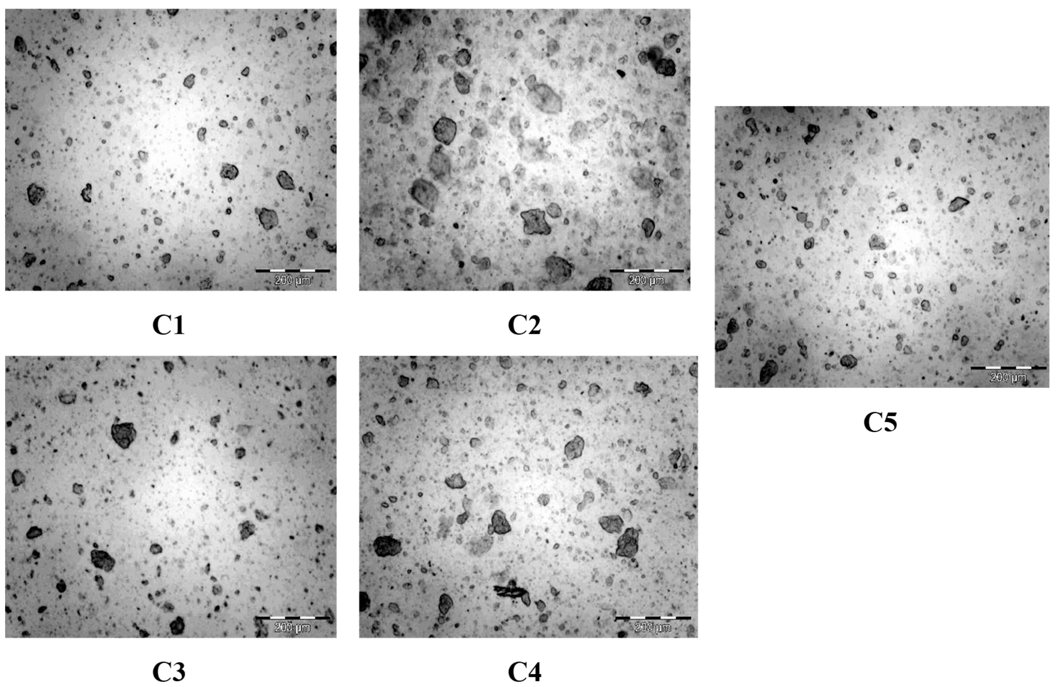
The Met-Ilo program (version 12.5 ©Janusz Szala 2012 Łódź, Poland) was used to describe microscopic images and summarize the grain size distribution. The tool allows performing quantitative descriptions of images. Images (Figure 2) were imported into Met-Ilo software. They were converted automatically to grayscale images. Then, the Sobel filter was used (it helps reveal edges in gray images), and automatic measurement was performed.
The statistical analysis was conducted using the Statistica v.13 computer program (Soft stat, Software Inc., Krakow, Poland). The Shapiro–Wilk test was used to verify that the distribution conformed to the normal distribution. The next step in hypothesis verification was the Kruskal–Wallis test (for independent samples). We determined which groups were statistically different from the others. Multiple comparisons of mean ranks for all groups were used for this. The significance level was 0.05.

3. Results

The resulting composite materials produced by different mixing methods were studied for their mechanical properties. The stresses developed during curing were also investigated. The results are summarized in Table 2.
  • Hardness test
Composite C2 obtained the highest values during the hardness test (34.1 ± 2.3) and C4 the lowest (30.9 ± 1.5). The Shapiro–Wilk test showed no inconsistencies with the normal distribution. The Scheffe test showed no significant statistical differences between the hardness results of the materials. Only the performance of the Tukey test showed statistical differences between the hardness of the C2 and C4 composites.
  • Flexural strength
In the case of FS, the highest values were achieved by composite C2 (75.47 ± 10.4 MPa) and the lowest by C5 (58.2 ± 15.2 MPa). The use of the Shapiro–Wilk test showed the presence of inconsistencies with a normal distribution (p < 0.05). The Kruskal–Wallis test also showed no statistically significant differences; only applying the test of the multiple comparisons of mean ranks for all samples showed statistically significant differences between C1 and C2.
  • Flexural modulus
The C3 (3738.3 ± 291.2 MPa) composite achieved the highest Ef values, while the C3 (3076.7 ± 229.8 MPa) composite achieved the highest Ef values. The Shapiro–Wilk test showed no inconsistency with the normal distribution. The Scheffe test was performed, but only the Tukey test showed statistically significant differences between the C1 and C3 materials.
  • Diametral tensile strength
The results collected in Table 2 show that the highest values were achieved by the C1 (33.5 ± 4.5 MPa) composite and the lowest by the C5 (27.1 ± 4.8 MPa) composite. In the DTS study, the Shapiro–Wilk test showed no inconsistency with a normal distribution. The Scheffe test showed statistically significant differences between the C2 and C5 composites.
  • Compressive strength
The highest CS is obtained by the C1 composite (301.2 ± 63.1 MPa) and the lowest by C3 material (195.2 ± 66.5 MPa). Based on the Shapiro–Wilk test, an inconsistency with the normal distribution was demonstrated (p < 0.05). The Kruskal–Wallis test showed no statistically significant differences.
  • Shrinkage stress
The highest reduced shrinkage stress is observed when the C2 composite is polymerized, the lowest characterized composites manually prepared (C3, C5 and C1, Table 3).
In the case of hand-mixed samples (C1), a decrease in the presence of large grains and an increase in the proportion of smaller grains can be observed. In the photos of mechanically mixed materials (C2, C4), small and large grains of filler are observed. Hybrid-mixed materials show clusters of fine grains, but they are not as large as in the C2 and C4 composites (Figure 2).
The results of the grain distribution analysis obtained using the Met-Ilo program are summarized in Table 4. It was found that the largest number of objects occurred in C5 (1913) and C3 (1753). For most of the samples studied—C1, C2, C3, and C5—the minimum grain value was 0.557 μm. As for the maximum grain sizes, they were obtained by C2 (136 μm) and C4 (114 μm). The largest grain size dispersion was shown for C2 (183%) and the smallest for C4 (130%).

4. Discussion

This article presents the effect of mixing the resin matrix with filler on selected mechanical properties. Because the literature provided limited information in this area, especially in terms of resin-based dental composites, it is necessary to determine the most favorable mixing method [25,29,30]. The results obtained (Table 2 and Table 3) indicate that different mixing methods influence the mechanical properties of the tested materials and stresses during curing.
To perform each dental reconstruction and/or hard tissue replacement, it is necessary to use appropriate materials. Restorative materials must have specific functional properties; they must have an adequate strength, hardness, stiffness, and abrasion resistance. The method of mixing the resin with the filler can affect the properties of the final composite material [31]. ISO 4049 specifies the precise requirements for composite materials to create fillings, restorations, or cementation. In the above-mentioned standard, only one mechanical property is specified—flexural strength. However, to determine all the utilitarian mechanical properties of the resin-based composite further tests, such as hardness, flexural strength, diametral tensile strength, abrasion resistance, fatigue flexural limit, and fracture toughness [31,32,33,34] should be performed.
The hardness test is used to determine the resistance of a material to forces acting on the restoration during chewing. During the Vickers hardness test, the materials are subjected to a standardized indentation of the indenter at a specified force. The maximum hardness value of commercial dental resin composites is about 114 HV [35,36,37]; for experimental composites the hardness is in the range of 8–35 HV [38,39,40]. The hardness of RBCs is affected by several factors. First, there is a correlation between the filler amount and material hardness [33,41,42,43]. Beyond that, hardness depends on the filler type, particle size, morphology, shape, and distribution [6,44,45,46,47]. The higher hardness observed when the composite is prepared by mixing may be an effect of the greater share of large grains and the more uniform distribution of filler particles. Studies conducted by Henry A. St. and Germain Jr. have shown that the increase in the size of filler particles can affect the hardness of the material [48]. Sneha Samal, in his article, argues that the aggregation of filler particles, occurring during composite mixing, causes their inhomogeneous distribution. As a result, this causes deterioration in the mechanical properties of the material [26]. In the case of this manufacturing method (centrifugal mixer), this process occurs to the least extent. Worth reference is the work of Wang R. et al. [49], in as filler was added silica with different sizes of particles (S-920, S-920+195, S-920+360, and S-920+360+195, nm). It was observed that the composite formulated with all types of particles was characterized by the best mechanical performance and lowest polymerization shrinkage. So, it can be concluded that the different sizes of filler particles in our composite could be beneficial for the material hardness and flexural strength (Figure 2, C2–C4). Also, following the above relationship are conclusions of the study of Ornaghi B. et al. [50]—the composite with a larger granulometric distribution showed a higher resistance to subcritical crack growth, which could translate to longer durability. It is also important to note the disadvantages of hardness testing. They include the surface nature of the measurement, and the presence of inhomogeneities in the material can affect the result of the test. Operator error, if semi-automatic tester is used —the subjective evaluation of the imprint—should also be taken into account.
Composite fillings are exposed to bending forces. Therefore, flexural strength tests according to ISO 4049 are necessary [51]. Nevertheless sample production process requires high precision, especially in terms of appropriate irradiation; minor variations can also result from operator errors (Figure 2).
The performed tests showed that regardless of the manufacturing method used, all strength values obtained meet the requirements of the standard. The C2 and C4 composites achieve FS values close to 80 MPa, which enables their use on loaded (chewing) surfaces. Other materials exceed 50 MPa—the minimum value for less loaded areas [38,52]. According to research, to increase the bending strength of composite fillings, the matrix can be filled with filler in larger quantities or with larger particles [33,53]. Shao-Yun Fu, in his research, showed that there was a relationship between the fracture toughness and the size of the filler grains [54]. The strength increased with the increase in the average grain diameter. It was assumed that large grains block the propagation of micro-cracks. While evaluating test results, it is important to take into account possible internal defects that can weaken the material and affect its strength. It is possible that the same mechanism occurred during the three-point bending test of our composites. In the case of the samples prepared in this study using the mixer, a significantly higher strength was obtained than those mixed manually or in a hybrid method. There is a greater share of large filler grains in the structures for materials C2 and C4 (Figure 2). Hongquan Zhang obtained similar results. He tested a resin based on Bis-GMA filled with a nanofiller and hydroxyapatite whiskers with dimensions of the order of 100 μm. These studies showed that for materials with a larger filler, higher parameters of fracture toughness, flexural strength, and Vickers hardness were obtained [55]. It was assumed that this results from the fact that nano-precipitates are more easily aggregated, and their full dispersion is more difficult to obtain. Kumar Naresh [56] examined the bi-axial flexure strength (BFS) of hand-spatulated and mechanically mixed composites. They noticed the superiority of mechanical mixing over manual mixing considering BFS and the value of the Weibull modulus of BFS [56]. In microscopic pictures consistently larger and more numerous microscopic defects in handmade RBCs were visible compared with mechanically mixed and commercial RBCs, which could translate to the deterioration in the mechanical parameters. This is consistent with our results, as the highest hardness and flexural strength were obtained for samples prepared using a mixer. This method provided a visibly better dispersion of the smaller filler particles and did not significantly affect the fragmentation of the large ones.
DTS determines the ability of a filling to resist the tensile stresses that take place during chewing [57,58]. For direct restorations, the American Dental Association has established a norm no. 27 that specifies a minimum tensile strength—not less than 24 MPa [31,59]. For all composite manufacturing methods, average values meeting this requirement were obtained.
Regardless of the manufacturing method used, the statistical CS values did not differ significantly. Taira Miyasaka obtained similar results. His research showed no significant change in the compressive strength for composites with fillers with dimensions above 6 µm [60].
The occurrence of high shrinkage stresses adversely affects the mechanical properties of the material. At the interface between the composite material and the patient’s own tissues, microleakage can appear, which is an ideal place for the formation of bacterial biofilms, which in the long run leads to the mechanical and chemical degradation of the reconstruction material [38,59]. The shrinkage stress limits that can cause the failure of the tooth–composite junction are in the range of 17–20 [51,61]. In our study, the highest values of shrinkage stresses were shown for the composite produced with a mixer (C2). The C3 composite generated the lowest shrinkage stresses during polymerization. The obtained results are summarized in Table 3. Studies by Domarecka et al. and Szczesio-Wlodarczyk et al. have shown that dental composites are characterized by varying magnitudes of shrinkage stresses, approx. 4–18 MPa [62,63]. Our studies have shown that the use of manual mixing (regardless of if it was completely manual or hybrid mixing) results in obtaining lower values of shrinkage stresses. This may be due to the greater grain refinement and the creation of samples with voids. This process probably promotes the compensation/relaxation of contraction stresses for C1, C3, and C5. The differentiated grain sizes increase the interface filler/matrix and provide the opportunity for inner flows that influence stress relaxation. Additionally, all the handmade samples probably gain air porosity in comparison with composites prepared by mechanical mixing [56]. The occurrence of porosity in RBCs rather negatively influences the mechanical strength of the material [56,64,65], but the air bubbles increase the free volume, and unbounded spaces enable resin flow and some relaxation of the contraction stress [37]. Also, in the work of Buelvas et al., one can find a reverse dependency between a high polymerization shrinkage stress and the concentration of pores [66].

5. Conclusions

Taking into account the limitations of the research carried out, it is possible to highlight the following:
  • The method of the filler and matrix mixing affects the flexural strength and hardness of experimental composites. Mechanically prepared composites have the highest flexural strength parameters and hardness.
  • The method of the filler and matrix mixing does not influence the compressive strength of the resulting composites.
  • The mixing method influences contraction stress. The lowest contraction stress is observed when the composite is partially manually prepared.
  • The method of the filler and matrix mixing affects the distribution and grain size of the silanized silica.
Hence, production methods should also, alongside composition, be considered to ensure the optimal properties of the material. The long-term clinical durability of resin fillings is the sum of the functional properties (including mechanical ones) and stresses generated at the filling–tooth interface.
Considering all the above, there is still a necessity to strive for a compromise between durability properties and contraction stress. The presented study should be continued to assess the porosity of the material and morphology and distribution of the filler in solid form.

Author Contributions

Conceptualization, M.Z. and K.B.; methodology, M.Z., validation, K.B.; formal analysis, M.Z.; investigation, M.K., O.D., A.J., A.Ż. and P.K.; data curation, M.Z.; writing—original draft preparation, M.Z., K.B., O.D., A.J., A.Ż. and P.K; writing—review and editing, K.B., M.Z. and M.K.; supervision, K.B. and M.K. All authors have read and agreed to the published version of the manuscript.

Funding

This research received no external funding.

Data Availability Statement

The raw data supporting the conclusions of this article are available in public repository, https://doi.org/10.60941/d5gw-4686, accessed on 24 April 2025.

Conflicts of Interest

The authors declare no conflicts of interest. The funders had no role in the design of the study; in the collection, analysis, or interpretation of data; in the writing of the manuscript; or in the decision to publish the results.

References

  1. Aminoroaya, A.; Neisiany, R.E.; Khorasani, S.N.; Panahi, P.; Das, O.; Madry, H.; Cucchiarini, M.; Ramakrishna, S. A Review of Dental Composites: Challenges, Chemistry Aspects, Filler Influences, and Future Insights. Compos. B Eng. 2021, 216, 108852. [Google Scholar] [CrossRef]
  2. Cadenaro, M.; Josic, U.; Maravić, T.; Mazzitelli, C.; Marchesi, G.; Mancuso, E.; Breschi, L.; Mazzoni, A. Progress in Dental Adhesive Materials. J. Dent. Res. 2023, 102, 254–262. [Google Scholar] [CrossRef]
  3. Ilie, N.; Hilton, T.J.; Heintze, S.D.; Hickel, R.; Watts, D.C.; Silikas, N.; Stansbury, J.W.; Cadenaro, M.; Ferracane, J.L. Academy of Dental Materials Guidance—Resin Composites: Part I—Mechanical Properties. Dent. Mater. 2017, 33, 880–894. [Google Scholar] [CrossRef] [PubMed]
  4. Paolone, G.; Diana, C.; Cantatore, G. 2023 State-of-the-Art in Resin-Based Composites and Future Trends. Compend. Contin. Educ. Dent. 2023, 44, 98–100. [Google Scholar] [PubMed]
  5. Bourbia, M.; Finer, Y. Biochemical Stability and Interactions of Dental Resin Composites and Adhesives with Host and Bacteria in the Oral Cavity: A Review. J. Can. Dent. Assoc. 2018, 84, i1. [Google Scholar]
  6. Liu, J.; Zhang, H.; Sun, H.; Liu, Y.; Liu, W.; Su, B.; Li, S. The Development of Filler Morphology in Dental Resin Composites: A Review. Materials 2021, 14, 5612. [Google Scholar] [CrossRef]
  7. Rizzante, F.A.P.; Duque, J.A.; Duarte, M.A.H.; Mondelli, R.F.L.; Mendonca, G.; Ishikiriama, S.K. Polymerization Shrinkage, Microhardness and Depth of Cure of Bulk Fill Resin Composites. Dent. Mater. J. 2019, 38, 403–410. [Google Scholar] [CrossRef]
  8. Ferracane, J.L. Resin Composite—State of the Art. Dent. Mater. 2011, 27, 29–38. [Google Scholar] [CrossRef]
  9. Hata, K.; Ikeda, H.; Nagamatsu, Y.; Masaki, C.; Hosokawa, R.; Shimizu, H. Development of Dental Poly(Methyl Methacrylate)-Based Resin for Stereolithography Additive Manufacturing. Polymers 2021, 13, 4435. [Google Scholar] [CrossRef]
  10. Topa-Skwarczyńska, M.; Ortyl, J. Photopolymerization Shrinkage: Strategies for Reduction, Measurement Methods and Future Insights. Polym. Chem. 2023, 14, 2145–2158. [Google Scholar] [CrossRef]
  11. Wang, K.; Wang, Z. Shrinkage Stress Evolution during Photopolymerization: Theory and Experiments. J. Mech. Phys. Solids 2023, 178, 105350. [Google Scholar] [CrossRef]
  12. Alsharif, S.; Alhareb, A.; Abudalazez, A. Components of Dental Resin Composites: A Literature Review. AlQalam J. Med. Appl. Sci. 2024, 7, 427–440. [Google Scholar] [CrossRef]
  13. Czech, Z.; Minciel, E. Światłoutwardzalne Kompozyty Zawierające Akrylowane Żywice Wielofunkcyjne—Skrócony Przegląd Literaturowy. Apar. Badaw. I Dydaktyczna 2015, 20, 270–275. [Google Scholar]
  14. Sozer, E.M.; Simacek, P.; Advani, S.G. Resin Transfer Molding (RTM) in Polymer Matrix Composites. In Manufacturing Techniques for Polymer Matrix Composites (PMCs); Elsevier: Amsterdam, The Netherlands, 2012; pp. 245–309. [Google Scholar]
  15. Terekhov, I.V.; Chistyakov, E.M. Binders Used for the Manufacturing of Composite Materials by Liquid Composite Molding. Polymers 2021, 14, 87. [Google Scholar] [CrossRef] [PubMed]
  16. Winnier, J.; Hambire, C.; Hambire, U. Effect of Nanohydroxyapatite, Zirconia and Glass Filler Particles on the Wear and Microhardness of Experimental Dental Composite Resin. Int. J. Clin. Pediatr. Dent. 2023, 16, S81–S84. [Google Scholar] [CrossRef]
  17. Al-Odayni, A.-B.; Alotaibi, D.H.; Saeed, W.S.; Al-Kahtani, A.; Assiri, A.; Alkhtani, F.M.; Alrahlah, A. Eugenyl-2-Hydroxypropyl Methacrylate-Incorporated Experimental Dental Composite: Degree of Polymerization and In Vitro Cytotoxicity Evaluation. Polymers 2022, 14, 277. [Google Scholar] [CrossRef]
  18. Pontons-Melo, J.C.; Balbinot, G.d.S.; Sauro, S.; Collares, F.M. Experimental Composite Resin with Myristyltrimethylammonium Bromide (MYTAB) and Alpha-Tricalcium Phosphate (α-TCP): Antibacterial and Remineralizing Effect. J. Funct. Biomater. 2023, 14, 303. [Google Scholar] [CrossRef]
  19. Loos, M. Processing of Polymer Matrix Composites Containing CNTs. In Carbon Nanotube Reinforced Composites; Elsevier: Amsterdam, The Netherlands, 2015; pp. 171–188. [Google Scholar]
  20. Fu, Y.-C.; Chen, C.-H.; Wang, C.-Z.; Wang, Y.-H.; Chang, J.-K.; Wang, G.-J.; Ho, M.-L.; Wang, C.-K. Preparation of Porous Bioceramics Using Reverse Thermo-Responsive Hydrogels in Combination with RhBMP-2 Carriers: In Vitro and in Vivo Evaluation. J. Mech. Behav. Biomed. Mater. 2013, 27, 64–76. [Google Scholar] [CrossRef]
  21. McDaniel, T.F.; Kramer, R.T.; Im, F.; Snow, D. Effects of Mixing Technique on Bubble Formation in Alginate Impression Material. Gen. Dent. 2013, 61, 35–39. [Google Scholar]
  22. Chambers, C.; Stewart, S.B.; Su, B.; Jenkinson, H.F.; Sandy, J.R.; Ireland, A.J. Silver Doped Titanium Dioxide Nanoparticles as Antimicrobial Additives to Dental Polymers. Dent. Mater. 2017, 33, e115–e123. [Google Scholar] [CrossRef]
  23. Um, J.G.; Jun, Y.-S.; Alhumade, H.; Krithivasan, H.; Lui, G.; Yu, A. Investigation of the Size Effect of Graphene Nano-Platelets (GnPs) on the Anti-Corrosion Performance of Polyurethane/GnP Composites. RSC Adv. 2018, 8, 17091–17100. [Google Scholar] [CrossRef] [PubMed]
  24. Lim, J.-M.; Cai, T.; Mandaric, S.; Chopra, S.; Han, H.; Jang, S.; Il Choi, W.; Langer, R.; Farokhzad, O.C.; Karnik, R. Drug Loading Augmentation in Polymeric Nanoparticles Using a Coaxial Turbulent Jet Mixer: Yong Investigator Perspective. J. Colloid. Interface Sci. 2019, 538, 45–50. [Google Scholar] [CrossRef] [PubMed]
  25. Bociong, K.; Zalega, M.; Dawicka, O.; Jasińska, A.; Żabecka, A.; Kałuża, P. Wpływ metody wytwarzania światłoutwardzalnego kompozytu do zastosowań w stomatologii na wybrane właściwości wytrzymałościowe in programme and materials of the XXII Conference of XXII Konferencji BIOMATERIAŁY I MECHANIKA W STOMATOLOGII, Wisła, Poland, 10–13 October 2024.
  26. ISO 4049; Dentistry—Polymer-Based Restorative Materials. ISO: Geneva, Switzerland, 2019; pp. 1–29.
  27. Bociong, K.; Krasowski, M.; Domarecka, M.; Sokolowski, J. Effect of the Method of Photopolymerization of Dental Composites Based on Dimethacrylate Resin on the Shrinkage Stresses and Selected Properties of the Cured Material. Polimery 2016, 61, 499–508. [Google Scholar] [CrossRef]
  28. Krasowski, M.; Ciesielska, S.; Śmielak, B.; Kopacz, K.; Bociong, K. Preparation of an Experimental Dental Composite with Different Bis-GMA/UDMA Proportions. Mater. Manuf. Process. 2024, 39, 1044–1051. [Google Scholar] [CrossRef]
  29. Ferracane, J.L. Resin-Based Composite Performance: Are There Some Things We Can’t Predict? Dent. Mater. 2013, 29, 51–58. [Google Scholar] [CrossRef]
  30. Ferracane, J.L. Current Trends in Dental Composites. Crit. Rev. Oral Biol. Med. 1995, 6, 302–318. [Google Scholar] [CrossRef]
  31. He, X.; Zhang, D.; Deng, S. Effect of Duration and Infection Control Barriers of Light Curing Unit on Hardness of Bulk Fill Composite Resin. PeerJ 2024, 12, e18021. [Google Scholar] [CrossRef]
  32. Ren, Z.; Chen, H.; Wang, R.; Zhu, M. Comparative Assessments of Dental Resin Composites: A Focus on Dense Microhybrid Materials. ACS Biomater. Sci. Eng. 2024, 10, 3718–3726. [Google Scholar] [CrossRef]
  33. Chen, F.; Sun, L.; Luo, H.; Yu, P.; Lin, J. Influence of Filler Types on Wear and Surface Hardness of Composite Resin Restorations. J. Appl. Biomater. Funct. Mater. 2023, 21, 22808000231193524. [Google Scholar] [CrossRef]
  34. Zalega, M.; Nowak, J.; Bociong, K. The Influence of Quaternary Ammonium Salts on Mechanical Properties of Light-Cured Resin Dental Composites. Polimery 2023, 68, 195–205. [Google Scholar] [CrossRef]
  35. Szczesio-Wlodarczyk, A.; Kopacz, K.; Szynkowska-Jozwik, M.I.; Sokolowski, J.; Bociong, K. An Evaluation of the Hydrolytic Stability of Selected Experimental Dental Matrices and Composites. Materials 2022, 15, 5055. [Google Scholar] [CrossRef]
  36. Szczesio-Wlodarczyk, A.; Barszczewska-Rybarek, I.M.; Chrószcz-Porębska, M.W.; Kopacz, K.; Sokolowski, J.; Bociong, K. Can Modification with Urethane Derivatives or the Addition of an Anti-Hydrolysis Agent Influence the Hydrolytic Stability of Resin Dental Composite? Int. J. Mol. Sci. 2023, 24, 4336. [Google Scholar] [CrossRef] [PubMed]
  37. Jun, S.-K.; Kim, D.-A.; Goo, H.-J.; Lee, H.-H. Investigation of the Correlation between the Different Mechanical Properties of Resin Composites. Dent. Mater. J. 2013, 32, 48–57. [Google Scholar] [CrossRef]
  38. Wysokińska-Miszczuk, J.; Piotrowska, K.; Paulo, M.; Madej, M. Composite Materials Used for Dental Fillings. Materials 2024, 17, 4936. [Google Scholar] [CrossRef] [PubMed]
  39. Bociong, K.; Szczesio, A.; Krasowski, M.; Sokolowski, J. The Influence of Filler Amount on Selected Properties of New Experimental Resin Dental Composite. Open Chem. 2018, 16, 905–911. [Google Scholar] [CrossRef]
  40. Shekofteh, K.; Kashi, T.J.; Behroozibakhsh, M.; Sadr, A.; Najafi, F.; Bagheri, H. Evaluation of Physical/Mechanical Properties of an Experimental Dental Composite Modified with a Zirconium-Based Metal-Organic Framework (MOF) as an Innovative Dental Filler. Dent. Mater. 2023, 39, 790–799. [Google Scholar] [CrossRef]
  41. Yadav, R.; Lee, H.; Lee, J.-H.; Singh, R.K.; Lee, H.-H. A Comprehensive Review: Physical, Mechanical, and Tribological Characterization of Dental Resin Composite Materials. Tribol. Int. 2023, 179, 108102. [Google Scholar] [CrossRef]
  42. Kundie, F.; Azhari, C.H.; Muchtar, A.; Ahmad, Z.A. Effects of Filler Size on the Mechanical Properties of Polymer-Filled Dental Composites: A Review of Recent Developments. J. Phys. Sci. 2018, 29, 141–165. [Google Scholar] [CrossRef]
  43. Elfakhri, F.; Alkahtani, R.; Li, C.; Khaliq, J. Influence of Filler Characteristics on the Performance of Dental Composites: A Comprehensive Review. Ceram. Int. 2022, 48, 27280–27294. [Google Scholar] [CrossRef]
  44. Germain, H.S.; Swartz, M.L.; Phillips, R.W.; Moore, B.K.; Roberts, T.A. Properties of Microfilled Composite Resins as Influenced by Filler Content. J. Dent. Res. 1985, 64, 155–160. [Google Scholar] [CrossRef]
  45. Samal, S. Effect of Shape and Size of Filler Particle on the Aggregation and Sedimentation Behavior of the Polymer Composite. Powder Technol. 2020, 366, 43–51. [Google Scholar] [CrossRef]
  46. Wang, R.; Habib, E.; Zhu, X.X. Evaluation of the Filler Packing Structures in Dental Resin Composites: From Theory to Practice. Dent. Mater. 2018, 34, 1014–1023. [Google Scholar] [CrossRef] [PubMed]
  47. Ornaghi, B.P.; Meier, M.M.; Rosa, V.; Cesar, P.F.; Lohbauer, U.; Braga, R.R. Subcritical Crack Growth and in Vitro Lifetime Prediction of Resin Composites with Different Filler Distributions. Dent. Mater. 2012, 28, 985–995. [Google Scholar] [CrossRef]
  48. Domarecka, M.; Szczesio-Wlodarczyk, A.; Krasowski, M.; Fronczek, M.; Gozdek, T.; Sokolowski, J.; Bociong, K. A Comparative Study of the Mechanical Properties of Selected Dental Composites with a Dual-Curing System with Light-Curing Composites. Coatings 2021, 11, 1255. [Google Scholar] [CrossRef]
  49. Yesudhas Jayakumari, B.; Nattanmai Swaminathan, E.; Partheeban, P. A Review on Characteristics Studies on Carbon Nanotubes-Based Cement Concrete. Constr. Build. Mater. 2023, 367, 130344. [Google Scholar] [CrossRef]
  50. Fu, S.-Y.; Feng, X.-Q.; Lauke, B.; Mai, Y.-W. Effects of Particle Size, Particle/Matrix Interface Adhesion and Particle Loading on Mechanical Properties of Particulate–Polymer Composites. Compos. B Eng. 2008, 39, 933–961. [Google Scholar] [CrossRef]
  51. Zhang, H.; Darvell, B.W. Mechanical Properties of Hydroxyapatite Whisker-Reinforced Bis-GMA-Based Resin Composites. Dent. Mater. 2012, 28, 824–830. [Google Scholar] [CrossRef]
  52. Kumar, N.; Shortall, A. Standardisation of mixing method for experimental resin-based composite. J. Pak. Dent. Assoc. 2011, 20, 131–134. [Google Scholar]
  53. Craig, R.G. (Ed.) Restorative Dental Materials; Elsevier: Amsterdam, The Netherlands, 2019; ISBN 9780323478212. [Google Scholar]
  54. Okulus, Z. Otrzymywanie Oraz Charakterystyka Dentystycznych Materiałów Kompozytowych. Ph.D. Thesis, Poznan University of Technology, Faculty of Chemical Technology, Poznań, Poland, 2016. [Google Scholar]
  55. Al-Shekhli, A.A.; Aubi, I. Al Composite Diametral Tensile Strength. World J. Dent. 2018, 9, 457–461. [Google Scholar] [CrossRef]
  56. Cho, G.C.; Kaneko, L.M.; Donovan, T.E.; White, S.N. Diametral and Compressive Strength of Dental Core Materials. J. Prosthet. Dent. 1999, 82, 272–276. [Google Scholar] [CrossRef]
  57. MIYASAKA, T. Effect of Shape and Size of Silanated Fillers on Mechanical Properties of Experimental Photo Cure Composite Resins. Dent. Mater. J. 1996, 15, 98–110,249. [Google Scholar] [CrossRef] [PubMed]
  58. Zalega, M.; Bociong, K. Antibacterial Agents Used in Modifications of Dental Resin Composites: A Systematic Review. Appl. Sci. 2024, 14, 3710. [Google Scholar] [CrossRef]
  59. Domarecka, M.; Sokołowski, K.; Krasowski, M.; Łukomska-Szymańska, M.; Sokołowski, J. The Shrinkage Stress of Modified Flowable Dental Composites. Dent. Med. Probl. 2015, 52, 424–433. [Google Scholar] [CrossRef]
  60. Szczesio-Wlodarczyk, A.; Garoushi, S.; Vallittu, P.; Bociong, K.; Lassila, L. Polymerization Shrinkage Stress of Contemporary Dental Composites: Comparison of Two Measurement Methods. Dent. Mater. J. 2024, 43, 2023–2192. [Google Scholar] [CrossRef]
  61. Baudin, C.; Osorio, R.; Toledano, M.; de Aza, S. Work of Fracture of a Composite Resin: Fracture-toughening Mechanisms. J. Biomed. Mater. Res. A 2009, 89A, 751–758. [Google Scholar] [CrossRef]
  62. Sarna-Boś, K.; Skic, K.; Sobieszczański, J.; Boguta, P.; Chałas, R. Contemporary Approach to the Porosity of Dental Materials and Methods of Its Measurement. Int. J. Mol. Sci. 2021, 22, 8903. [Google Scholar] [CrossRef]
  63. Linden, L.-A.; Jakubiak, J. Contraction (Shrinkage) in Polymerization. Part II. Dental Resin Composites. Polimery 2001, 46, 590–595. [Google Scholar] [CrossRef]
  64. Buelvas, D.D.A.; Besegato, J.F.; Vicentin, B.L.S.; Jussiani, E.I.; Hoeppner, M.G.; Andrello, A.C.; Di Mauro, E. Impact of Light-Cure Protocols on the Porosity and Shrinkage of Commercial Bulk Fill Dental Resin Composites with Different Flowability. J. Polym. Res. 2020, 27, 292. [Google Scholar] [CrossRef]
  65. Tyas, M.J. Correlation between Fracture Properties and Clinical Performance of Composite Resins in Class IV Cavities. Aust. Dent. J. 1990, 35, 46–49. [Google Scholar] [CrossRef]
  66. Heintze, S.D.; Ilie, N.; Hickel, R.; Reis, A.; Loguercio, A.; Rousson, V. Laboratory Mechanical Parameters of Composite Resins and Their Relation to Fractures and Wear in Clinical Trials—A Systematic Review. Dent. Mater. 2017, 33, e101–e114. [Google Scholar] [CrossRef]
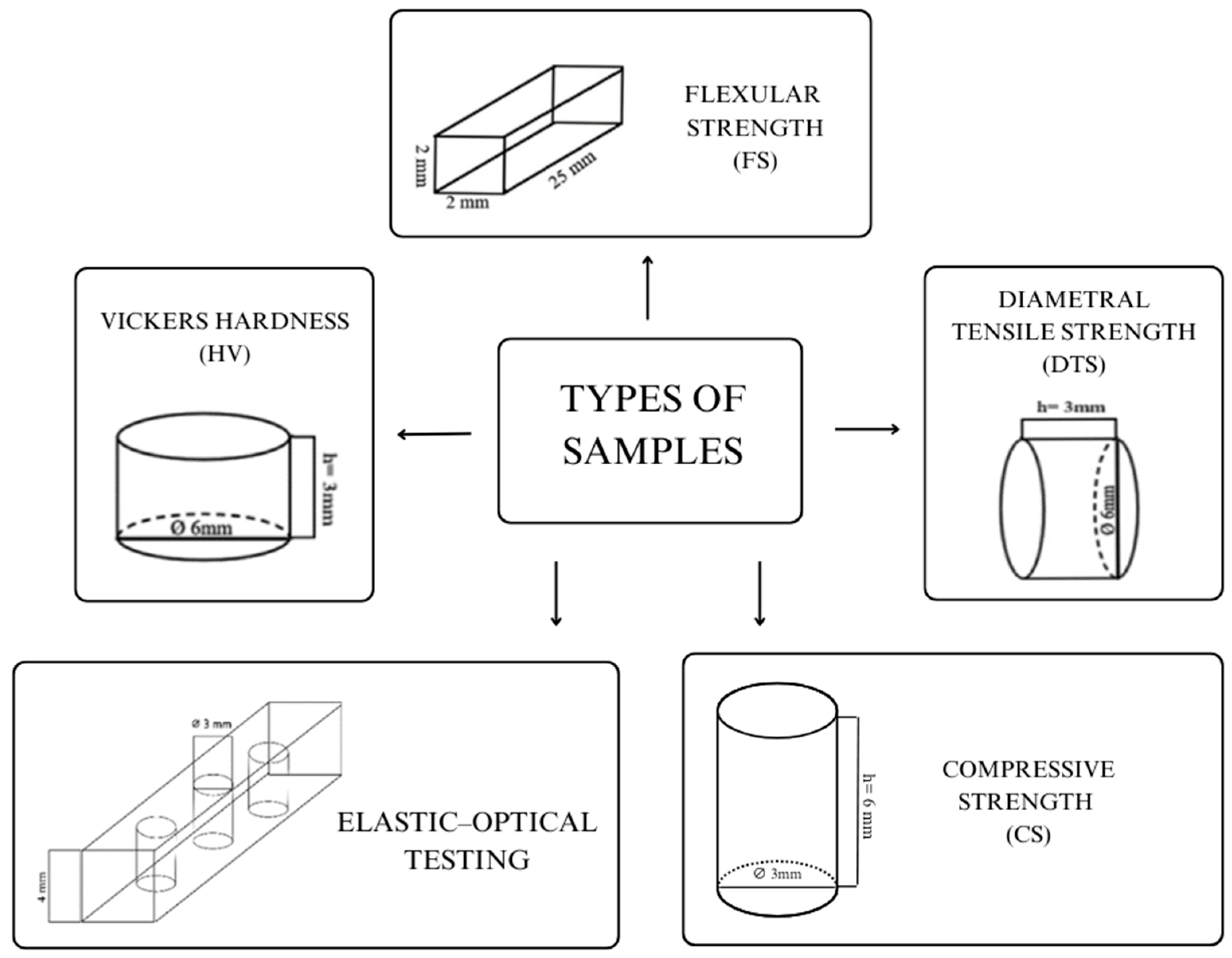
Figure 1. Shapes of the samples.
Processes 13 01332 g001
Figure 2. Microscopic images of composites prepared in five mixing methods.
Processes 13 01332 g002
Table 1. Five mixing methods were used during composites manufacturing.
Composite C1Handmade with the use of agate mortar. Small portions of silica were added every 2–5 min when it was noticed that the two components had already been properly combined and were becoming one. It took approximately 50 min to make this type of composite.
Composite C2Mixed with the use of the Hauschild SpeedMixer device (TM DAC 150 FVZ, Hauschild and Co., Hamm, Germany). Silica was progressively added to a precisely measured amount of resin. The mixing process in the device was initiated at the speed of 1500 RPM. Over approximately one hour, the speed was gradually increased until reaching a value of 3000 RPM, along with the complete filling of the measured resin with silica.
Composite C3Hybrid method. The process started with the use of a Hauschild SpeedMixer, (TM DAC 150 FVZ, Hauschild Engineering, Hamm, Germany) in which half of the silica was mixed with the resin. Then, this mixture was transferred to a mortar, where the remaining silica was further combined with the previously mixed material. Manual mixing of the material took about 30 min to achieve a complete integration of the components.
Composite C4Made with a Hauschild SpeedMixer (TM DAC 150 FVZ, Hauschild Engineering, Hamm, Germany) mixing device, with some modifications. For about an hour, silica was added to the resin in small portions, and the speed on the machine did not exceed 1500 RPM (every 7 min or so, starting at 1000 RPM, the speed was increased by 50 RPM). However, the last 1% of silica was added by hand in the mortar due to its failure to combine with the mixing machine.
Composite C5Modified hybrid method—mixed in the Hauschild SpeedMixer (TM DAC 150 FVZ, Hauschild Engineering, Hamm, Germany) device, then grated in a species mortar for 15 min.
Table 2. Effect of type of mixing method on selected mechanical properties of cured composites (with CURE lamp with outputs power 1250 mW/cm2 for 20 s per 1.5 mm of material).
HV [-]FS [MPa]Ef [MPa]CS [MPa]DTS [MPa]
C1
31.9 ± 2.858.2 ± 8.0 *3156.7 ± 507.0 *301.2 ± 63.133.5 ± 4.5
C2
34.1 ± 2.3 *75.5 ± 10.4 *3738.3 ± 291.2221.6 ± 50.930.2 ± 3.1 *
C3
33.1 ± 1.865.9 ± 5.23076.7 ± 229.8 *195.2 ± 66.532.0 ± 3.9
C4
30.9 ± 1.5 *72.3 ± 5.63426.7 ± 394.2281.2 ± 55.429.4 ± 4.1
C5
31.9 ± 3.158.2 ± 15.23596.7 ± 402.7232.2 ± 55.627.1 ± 4.8 *
HV—hardness; FS—flexural strength; Ef—flexural modulus; CS—compressive strength; and DTS—diametral tensile strength; * within a single characteristic there is a statistical difference.
Table 3. Effect of type of mixing of composite on shrinkage stress values formed during polymerization (with CURE lamp with output power 1250 mW/cm2 for 20 s per 1.5 mm of material).
Type of Compositeσr [MPa]σϴ [MPa]σint [MPa]
C18.4 ± 0.6−9.8 ± 0.718.3 ± 1.3
C211.4 ± 0.3−13.4 ± 0.624.8 ± 0.9
C37.6 ± 1.1−9.1 ± 1.116.8 ± 2.2
C49.6 ± 0.6−11.2 ± 0.620.8 ± 1.1
C57.9 ± 1.1−9.5 ± 1.217.4 ± 2.3
σr—radial stress, σθ—circumferential stress, and σint—reduced shrinkage stress.
Table 4. Summary of grain size distribution.
C1C2C3C4C5
Total number of objects134633217533911913
Minimum value [μm]0.5570.5570.5570.640.557
Maximum value [μm]50.713665.111452.9
Average value [μm]2.538.212.099.112.39
Variability index [%]159183169130146
Disclaimer/Publisher’s Note: The statements, opinions and data contained in all publications are solely those of the individual author(s) and contributor(s) and not of MDPI and/or the editor(s). MDPI and/or the editor(s) disclaim responsibility for any injury to people or property resulting from any ideas, methods, instructions or products referred to in the content.