Article

Bioactive Compounds Produced by the Accompanying Microflora in Bulgarian Yoghurt

1
Institute of Microbiology, Bulgarian Academy of Sciences, Acad. G. Bontchev Str., bl. 26, 1113 Sofia, Bulgaria
2
Institute of Chemical Engineering, Bulgarian Academy of Sciences, Acad. G. Bontchev Str., bl. 103, 1113 Sofia, Bulgaria
3
Department of Medical Chemistry and Biochemistry, Faculty of Medicine, Medical University of Sofia, 1000 Sofia, Bulgaria
4
State Key Laboratory of Dairy Biotechnology, Dairy Research Institute, Bright Dairy & Food Co., Ltd., Shanghai 201103, China
*
Author to whom correspondence should be addressed.
Processes 2021, 9(1), 114; https://doi.org/10.3390/pr9010114
Submission received: 14 December 2020 / Revised: 4 January 2021 / Accepted: 5 January 2021 / Published: 7 January 2021
(This article belongs to the Special Issue Novel Processes for Development of Fermentation-Based Products)

Abstract

:
Bulgarian yoghurt is associated with health benefits and longevity of consumers. The specific microflora producing bioactive metabolites is responsible for this effect. The present study examines the biodiversity in four homemade yoghurts from regions containing endemic microflora. Metagenome sequencing indicated Lactobacillus delbrueckii subsp. bulgaricus and Streptococcus thermophilus were predominant in all samples. In addition, yoghurts contained accompanying lactic acid bacteria (LAB) including Lacticaseibacillus paracasei, Lb. helveticus, Limosilactobacillus fermentum, Lb. rhamnosus, Lactococcus lactis, Pediococcus acidilactici, Leuconostoc mesenteroides, and Leuc. pseudomesenteroides. A negligible amount of pollutant strains was found. Twenty-four LAB strains were isolated from the yoghurts and identified. Lb. delbrueckii subsp. bulgaricus strains were genotyped by randomly amplified polymorphic DNA–PCR (RAPD), multi-locus sequence typing (MLST), and pulse field gel electrophoresis (PFGE), which demonstrated their uniqueness and non-commercial origin. To estimate the bioactive metabolites produced by the accompanying microflora, yoghurts fermented by single LAB strains were analyzed using liquid chromatography and mass spectrometry (LC-MS). The fermented samples contained large amounts of free essential amino acids (arginine, citrulline, tryptophan, lysine, and histidine), the neuroprotector indole-3-propionic acid (IPA), and significant quantities of the cyclic antimicrobial peptides cyclo(phenylalanyl-prolyl) and cyclo(leucyloprolyl). The disclosure of these special qualities draws attention to the accompanying microflora as a source of potential probiotic strains that can fortify the yoghurts’ content with bioactive compounds.

1. Introduction

For centuries, conventional yoghurt consumption has been associated with beneficial effects on human health. Recent studies show cardio-metabolic benefits and ameliorative effects on obesity, diabetes, and biomarkers of chronic diseases, including inflammation [1]. The regular intake of yoghurt lactic acid bacteria (LAB) and their metabolites also yields anti-carcinogenic, anti-oxidative, and immunomodulatory effects and reduces blood cholesterol [2]. LAB in yoghurt can regulate the microbiome supporting the gut barrier [3] and improve the immune system response [4,5].
The remarkable properties of Bulgarian yoghurt are due exclusively to its microbial content. The microbial flora of yoghurt is not a random set of species, but a unique microecological niche [6]. The most studied yoghurt starters rely on the protocooperation between Lactobacillus delbrueckii subsp. bulgaricus and Streptococcus thermophilus [7,8]. According to the FAO/WHO definition, yoghurt must contain no less than 107 viable colony-forming units (CFU) per gram of product. Starter cultures produce volatiles, which are responsible for the typical yoghurt aroma (acetaldehyde, diacetyl, acetoin, acetone, ethanol, and 2-butanone) [9], organic acids, prebiotics such as galactooligosaccharides (GOS) [10], and bioactive compounds. Bioactive peptides, such as bacteriocins, are connected with the antimicrobial effects against pathogens in the gastrointestinal tract, such as clostridia, Enterobacteriaceae, pseudomonads, and Helicobacter pylori [11,12,13]. Other peptides produced by Lb. delbrueckii subsp. bulgaricus are known to harmonize the blood pressure by angiotensin-converting enzyme (ACE) inhibitory activity [14].
Contemporary culture-independent techniques, such as metagenomics, allow the comprehensive investigation of the yoghurt consortia and reveal that they contain a wide range of accompanying microflora with no less useful properties [15]. These microbial communities are an object of increasing scientific interest, at least in three different directions: (i) investigation of the symbiosis and other internal interactions between LAB to select starters with better quality; (ii) elucidation of the specific dietetic properties of yoghurt; and (iii) determination of the specific compounds produced by lactic acid bacteria (LAB), which can resolve health problems without drug use. A study engaging healthy volunteers revealed that the daily intake of yoghurt fortified with Lb. paracasei (L. casei 431®) improves immune response by enhancing NK cell activity and increasing interleukin (IL)-12 and immunoglobulin (Ig) G1 levels, and IFN-γ concentration [16]. Another strain of this species, Lb. paracasei CNCM I-1518, decreases bacterial translocation, gut dysbiosis, and ileal oxidative damage, and increases ileal β-defensin-1 expression in rats, suggesting an improvement in the intestinal barrier integrity [17]. Lb. helveticus H9 yoghurt possesses excellent ACE-inhibitory (ACE I) activity with high levels of antihypertensive peptides, such as Val-Pro-Pro and Ile-Pro-Pro [18,19]. Lb. helveticus LH-B02 is known to favor the formation of bioactive peptides, such as αS1-CN f(24–32) and β-CN f(193–209) [20]. The patent-protected Lb. rhamnosus GG (ATCC 53103) is the world’s most studied probiotic bacterium with more than 800 scientific studies. The authors claim that it is bile-stable, has a great avidity for human intestinal mucosal cells, and possesses various health benefits, such as the alleviation of gastroenteritis, diarrhea, eczema, and many other disorders [21,22,23].
Bulgarian yoghurt (kiselo mlyako) is the hallmark of the country, being one of the most popular types of yoghurt worldwide. The present study adds novel comprehensive information about Bulgarian yoghurt microflora to complement the discovery of Lb. delbrueckii subsp. bulgaricus discovery one hundred years ago [24]. A collection of homemade Bulgarian yoghurts from remote locations revealed unique microflora content. Considering the significant influence of both starter cultures and the accompanying LAB on yoghurt’s beneficial properties, the present work aims to reveal the biodiversity of Bulgarian yoghurts using metagenomics, to isolate and identify new starters and accompanying LAB strains by engaging modern genetic approaches, and to study the bioactive metabolites produced from concomitant species using precise analytical methods.

2. Materials and Methods

2.1. Yogurt Sample Collection

Four samples were collected from remote territories known with endemic yoghurt microflora: Rila, Pirin, and Rhodope Mountains, and the Thracian valley. The samples derived from cow (Samples 1 and 3), goat (Sample 2), and buffalo milk (Sample 4). They were prepared according to Bulgarian national recipes passed on by generations of local people from the respective villages. By this technology, the milk for yoghurt production is boiled and then fermented from an artisanal starter culture. The owners of the samples declared that they did not use commercial starters. The samples were collected in the period October–December 2015 in sterile containers and were stored frozen at −20 °C until their analysis in 2020.

2.2. Bacterial Strains, Media and Cultivation Conditions

The strains were isolated by the following procedure: 10–20 μL of the yoghurt was inoculated in 5 mL MRS broth selective medium (Roth, Germany) and cultivated 24 h either at 37 °C or 44 °C. Decimal dilutions (up to 10−9) of these cultures were mixed with MRS and M17 agar selective for lactobacilli, and lactic acid cocci, respectively, and then loaded in Petri dishes. The agar was stored with a temperature below 50 °C, the colonies appeared in the agar after 48 h of cultivation. The selection was performed at two different temperatures: at 37 °C and 44 °C. At 37 °C Pediococcus and Lactococcus strains appeared, at 44 °C—Lb. delbrueckii subsp. bulgaricus and Str. thermophilus. Lactic acid rod-shaped bacteria were cultivated in MRS medium; lactic acid cocci in M17 medium, at 42 °C, or 37 °C, respectively; at anaerobic conditions, using an Anaerocult® A mini (Merck KGaA, Darmstadt, Germany). The cultivation of strains was done in semi-skimmed milk, containing 1.2% fat and 4.1% lactose. For cell propagation aimed at DNA isolation, the strains were cultured in MRS for 24 h, at optimal temperature for each strain. Single colonies were picked and inoculated again in MRS or M17 broth (3 mL).

2.3. Total DNA Isolation from Yoghurt

Total DNA from yoghurt samples was extracted using a new method that combines the first steps of the method of Lick et al. (1996) [25], followed by biomass lysis and DNA purification by the use of PureLink™ Genomic DNA Mini Kit (InvitrogenTM, Thermo Fisher Scientific, Waltham, MA, USA). Briefly, a yoghurt sample with volume 8 mL was mixed with 2.4 mL of 0.4 M NaOH and 1.2 mL of 0.25 M Trisodium citrate dihydrate. The tubes were vigorously shaken (at 2500 rpm/min) for 10 s and incubated at ambient temperature for 5 min. The following centrifugation (5000× g, 10 min) separated three fractions: upper white layer (fat), yellowish supernatant, and grainy pellet (microbial cells). The biomass was re-suspended in 500 μL of the remaining supernatant, and after addition of 150 μL 0.4 M NaOH and 75 μL 40% Trisodium citrate dihydrate, the mixture was vigorously shaken again, incubated at room temperature for 5 min, and centrifuged at 12,000× g for 3 min. The obtained pellet was resuspended in 200 μL of Lysozyme Digestion Buffer of the PureLink™ Genomic DNA Mini Kit. Then, the procedure as described by the manufacturer InvitrogenTM was followed. DNA concentration and quality were measured using a QB 3000 spectrophotometer (Quawell Ltd., Hong Kong, China).

2.4. Molecular Approaches in Strains’ Identification

2.4.1. Isolation of Chromosomal DNA, PCR Amplification of the 16S rRNA Gene and Sequencing

Chromosomal DNA from LAB isolates was isolated from 24 h old cultures, grown in MRS-starch medium, using a GeneJET Genomic DNA Purification Kit (Thermo Fisher Scientific Inc., Waltham, MA, USA), and following the manufacturer’s recommendations. PCR amplifications were prepared with Prime Taq premix (Genet Bio, Daejeon, Korea), in a total volume of 50 μL and final concentrations of primers 0.5 pmol/μL (Macrogen Inc., Amsterdam, The Netherlands) in a QB-96 Satellite Gradient Thermal Cycler (LKB Vertriebs GmbH, Vienna, Austria). The amplification of the 16S rRNA gene was performed with universal eubacterial primer pair: 27F: 5′ AGAGTTTGATCCTGGCTCAG 3′ and 1492R: 5′ AAGGAGGTGATCCAGCC 3′. The final concentration of the template DNA was 2 ng/μL, the temperature profile was: 95 °C for 5 min, 35 cycles consisting of 94 °C for 1 min, 55 °C for 1 min, 72 °C for 2 min, followed by final elongation at 72 °C for 5 min. All obtained PCR amplification products were purified using the GFX PCR DNA and gel band purification kit (Amersham Biosciences) and then sequenced by Macrogen Inc. (Amsterdam, The Netherlands). Sequence comparison with the GenBank data was performed using BLAST and ClustalW programs.

2.4.2. Randomly Amplified Polymorphic DNA–PCR (RAPD) Analysis

RAPD analysis was performed using the modified procedure of Cebeci et al. (2011) [26]. The primers used were 1254: 5′CCGCAGCCAA3′ (10-mer), rpd1: 5′CCCGACTGA3′ (9-mer), rpd2: 5′CCG AGACAC3′ (9-mer), rpd3: 5′GCCTGTTCTT3′ (10-mer), and rpd4: 5′-GCCAGAACAA-3′ (10-mer). The PCR mix contained: DNA—1 µL (100 ng), 100 pmol primers—3 µL, 2× Phusion Master Mix (Thermo Scientific Inc., Waltham, MA, USA)—10 µL, 25 mmol MgCl2—2 µL, H2O—1 µL. The program followed the temperature profile: 98 °C—30 s, 4 cycles: denaturation 98 °C 10 s, annealing 36 °C—20 s, elongation 72 °C—15 s, 30 cycles 98 °C—10 s, 20 °C—20 s, 68 °C—15 s, 72 °C—5 min. The obtained products were separated in 2% agarose, for 2.5 h, at 50 V (5 v/cm). A molecular size standard of 100 bp was used (Thermo Scientific Inc., Waltham, MA, USA).

2.4.3. Multi-Locus Sequence Typing (MLST)

This analysis was conducted as described by Naser et al. (2005) [27] using the primers listed in Table 1. The primer pairs targeted the genes rpoB encoding the β-subunit of bacterial RNA polymerase, groEL encoding the protein of the chaperonin family, pheS for the α-subunit of phenylalanine tRNA synthetase, and pyrG encoding CTP synthase. The sequencing was performed with the reverse primers. Divergence data were obtained by combined similarities in 4 housekeeping genes analyzed by MEGA6 software.

2.4.4. Pulse Field Gel Electrophoresis (PFGE)

The cells for PFGE were prepared using a partially modified procedure of Michaylova et al. (2007) [28]. The cells were grown overnight, to OD600 0.6–0.8, harvested, washed in 1 mL ET buffer (1 M TRIS/HCl, 100 mM EDTA, pH 8), and resuspended in 50 μL ET buffer. After incubation for 1 min at 45 °C, the suspension was mixed with 60 µL 1.5% low melting agarose (Sigma-Aldrich, St. Louis, MO, USA) in LMP buffer (10 mM TRIS/HCl, 100 mM EDTA, 20 mM NaCl, pH 8), and was pipetted in molds (Bio-Rad Laboratories, Hercules, CA, USA). The molds were treated with 500 µl Buffer A: 1 mM TRIS/HCl, 100 mM EDTA, 1 M NaCl, 0.5% (v/v) TRITON X 100, pH 8 + 106 U/mL Lysozyme (Merck KGaA, Darmstadt, Germany ) + 75 U/mL mutanolysin (Sigma-Aldrich, St. Louis, MO, USA). Incubation for 18 h at 37 °C was followed by washing for 5 h at 55 °C with 500 mL Lysis solution, which contained 1mM TRIS/HCl, 250 mM EDTA, 1% SDS, pH 8, and 30 mU/mL (1 mg/mL) Proteinase K (Merck KGaA, Darmstadt, Germany). Blocks were washed with TE Buffer (10 mM TRIS/HCl, 1 mM EDTA, pH 7.5) and 1 mM phenyl methyl sulfonyl fluoride (PMSF) for protease inactivation followed by washing 3 times with TE. Aliquots of inserts were subjected to digestion for 24 h at 37 °C in 80 µL buffer containing 10× restriction buffer and 20 U XhoI.—2 µL enzyme. After washing with TE, the blocks were inserted in the slots of 1% PFGE agarose (Bio-Rad Laboratories, Hercules, CA, USA).
The apparatus used was a CHEF—DRII (Bio-Rad Laboratories, Hercules, CA, USA), the electrophoretic buffer contained 0.5× TBB (45 mM TRIS, 45 mM Boric acid, 1 mM EDTA, pH 8.3). The electrophoresis parameters were: initial pulse—3 s, final pulse—25 s, 5 v/cm, and running time—24 h. The gels were stained with 200 µg/mL SYBR Green (Thermo Fisher Scientific, Waltham, MA, USA) and examined by UV transillumination.

2.5. Metagenome Library Construction, Sequencing and Bioinformatics Analysis

Metagenome library construction and sequencing was done by Macrogen Inc., Republic of Korea. Preparation of the 16S metagenomic sequencing library was performed using a Herculase II Fusion DNA Polymerase Nextera XT Index Kit V2. The sequencing (Illumina platform) was conducted with reading length of 301 bp and FastQC quality control. The assembly results showed that the quality-filtered data contained between 73,209,822 and 82,104,372 total bases, and around 243,222 read counts for each sample. The percentage of Q20 quality reads was 94.52%.

2.6. Data Availability

Raw sequencing data were deposited in GenBank of NCBI with the following accession numbers: BioProject ID PRJNA681359, BioSample: SAMN16961356, Sequence Read Archive (SRA) SUB8652830. The partial 16S rRNA gene sequences of strains used in this study are deposited in GenBank with the following accession numbers: MG437344, MG437345, MG437346, MG437357, MG437359, MG437363, MG437367, MG437372, MG437380, MG437386, MG437389, MG437390, MG437352, MG437355, MG438473, MG438475, MG438468, MG438466, MG437360, MG437368, MG437358, MG437375, MG437361, and MG438467.

2.7. Liquid Chromatography and Mass Spectrometry (LC-MS) Analysis

Analyses were carried out on a Q Exactive® mass analyzer equipped with TurboFlow® LC system and APCI atmospheric pressure electrospray ionization module (Thermo Scientific Inc., Waltham, MA, USA) using an iHILIC®-Fusion 1.8 μm, 100 A, 100 × 2.1 mm analytical column (HILICON AB, Sweden) in gradient elution mode. The following mobile phases were used: A—20 mM ammonium acetate in water; B—buffer A/acetonitrile (1/9 v/v) at a flow rate of 300 μL/min and gradient elution. Full-scan mass spectra over the m/z range 67–1000 were acquired in negative ion mode at resolution settings of 140,000. Qualification of the compounds was achieved using top 5N mode of operation of the mass spectrometer at resolution settings of 17,500 and isolation window of 0.8 over the m/z range 67–1000. The used value of the collision energy was HCD 30%. All MS parameters were optimized for sensitivity to the target analytes using the instrument control software program. Data acquisition and processing were carried out with the Xcalibur 2.4® software package (Thermo Scientific Inc., Waltham, MA, USA). The raw data files were processed by Compound Discoverer® 2.0 software (Thermo Scientific Co., USA).

3. Results and Discussion

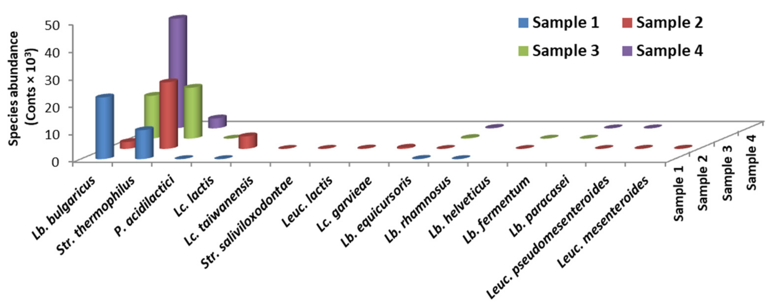
3.1. Metagenomes Analysis

Several regions in Bulgaria are known to contain endemic microflora: the high mountains of Rila, Pirin, and Rhodope, and the Thracian valley. The species biodiversity of yoghurt samples collected from these locations was observed after metagenome sequencing (Figure 1, Table S1). As expected, between 68.4% and 99.6% of the total microflora in the examined samples consisted of two species: Lb. delbrueckii subsp. bulgaricus and Str. thermophilus, and the ratio between them varied widely. In Sample 2 (Rila), Str. thermophilus was the most abundant species, whereas in Sample 4 (Rhodope), Lb. bulgaricus strongly prevailed. Considering the distribution by region, the proto-cooperation between Lb. bulgaricus and Str. thermophilus existed in all samples, however, the recommended ratio of ~1:1 between strains was observed only in Sample 3 (from the Thracian valley). This ratio in the starter strongly affects yoghurt qualities, such as texture, color, taste, and post-acidification [29,30]. Hence, the artisanal yoghurts are usually characterized by a more pronounced sour taste [10]. Rhodope’s yoghurt was the richest source of Lb. bulgaricus, and this species is typical for Bulgarian yoghurt and is not as widespread in the neighboring countries. For instance, in a Greek study, the isolation of only one Lb. delbrueckii subsp. bulgaricus strain from nine analyzed yoghurts was reported [31]. Comparatively low counts of Lb. bulgaricus in Sample 2 could be explained by the repeated transfers of the same starter culture in an unsterile home environment. Under these conditions, bacteriophages could destroy part of the starter [32,33], thus giving rise to the microbial counts of the accompanying microflora.
According to Sieuwerts et al. (2008), the traditional yoghurt culture is composed of Lb. delbrueckii subsp. bulgaricus and Str. thermophilus, and the name “yoghurt” is only allowed for those products that contain starters of these species [34]. Recent metagenome studies devoted to the juxtaposition of yoghurt microbiota and gut microbiome revealed the presence of Lacticaseibacillus paracasei, Lacticaseibacillus rhamnosus, Lb. parabuchneri, Lb. parafarraginis, Lactiplantibacillus plantarum, Lactococcus lactis, Leuconostoc mesenteroides, and Pediococcus acidilactici in both niches [35,36]. Our results partially confirmed these observations, because among autochthonic lactobacilli in Bulgarian artisanal yoghurts Lb. rhamnosus and Lb. paracasei were found. In addition, Lb. helveticus, Limosilactobacillus fermentum, and Lb. equicursoris were detected (Table S1). However, whereas Lb. helveticus was frequently observed in Bulgarian yoghurts and identified using genetic techniques [37,38], the presence of Lb. fermentum, Lb. paracasei, and Lb. rhamnosus is highly unusual. Lb. plantarum, typical of Greek yoghurt, was not found in Bulgarian yoghurt. Although Lb. equicursoris belongs to the Lb. delbrueckii phylogenetic branch, our study is the first that reports its presence in yoghurts. This species was initially isolated from the feces of a thoroughbred racehorse by Morita et al. (2010) [39] and recently found in kefir [40]. Other accompanying LAB species were Lc. lactis, Lc. garvieae, Lc. taiwanensis, P. acidilactici, Leuc. lactis, Leuc. mesenteroides, and Leuc. paramesenteroides. A portion of these species is known to contribute to the sweet, fruity, or vanilla aroma via the production of ethanol, 2-butanone, 2-pentanone, and 2,3-pentanedione [41], and is currently preferred by consumers. Other desirable additives to the starters are also the strains producing extracellular exopolysaccharides and, importantly, those with high proteolytic activity.
Figure 2 and Table S2 present the spoilage species found in the metagenomes. Acinetobacter spp. and Chryseobacterium spp. were identified as the main spoilage species. These Gram-negative, post-process contaminant bacteria limit the shelf life of milk produced by microfiltration and subsequent pasteurization, and have also been found in yoghurts in Germany, Austria, and Switzerland [42]. The presence of the Gram-negative, obligate anaerobe Bacteroides graminisolvens in two cow samples from different regions (Samples 1 and 3) is indicative of soil contamination during milking and/or the transportation of the raw milk. This species is ubiquitous in barns and rural areas.
Other microbial species, such as Moraxella osloensis, Alcaligenes aquatilis, Str. ruminantium, and Str. saliviloxodontae, were found in counts between 0.1% and 0.01%, or less. The low microbial counts of contaminating species (Table S2) indicate that single cells from the environment contaminated the milk, but these species did not multiply due to the antagonistic activity of LAB [43]. Our observations are consistent with the research of other authors reporting the presence of contaminant Gram-negative psychrotrophs in yoghurts, such as Pseudomonas sp. and Klebsiella sp. [44]. Similarly, Bulgarian yoghurts were contaminated, but with insignificant amounts of Ps. japonica and Citrobacter europaeus. However, dangerous zoonotic pathogens, such as coliforms, Salmonella sp., Listeria monocytogenes, Campylobacter jejuni, Yersinia enterocolitica, Brucella sp., Mycobacterium bovis, or Enterobacter sakazakii, were not found in any of the samples, although their presence in milk has been previously reported [45,46,47].

3.2. LAB Strains Isolation and Identification

Twenty-four LAB strains belonging either to the starter or to the accompanying microflora were isolated and identified by 16S rRNA gene sequencing: Lb. delbrueckii subsp. bulgaricus (12 strains), Str. thermophilus (two strains), Lb. helveticus (two strains), Lb. paracasei (two strains), Lb. fermentum (two strains), Lb. rhamnosus, P. acidilactici, Leuc. mesenteroides, and Leuc. pseudomesenteroides. Although present in the sample, some species were not isolated as pure cultures.
To examine the diversity and specificity of Lb. delbrueckii subsp. bulgaricus strains, three different genotyping approaches were applied: RAPD, MLST, and PFGE.

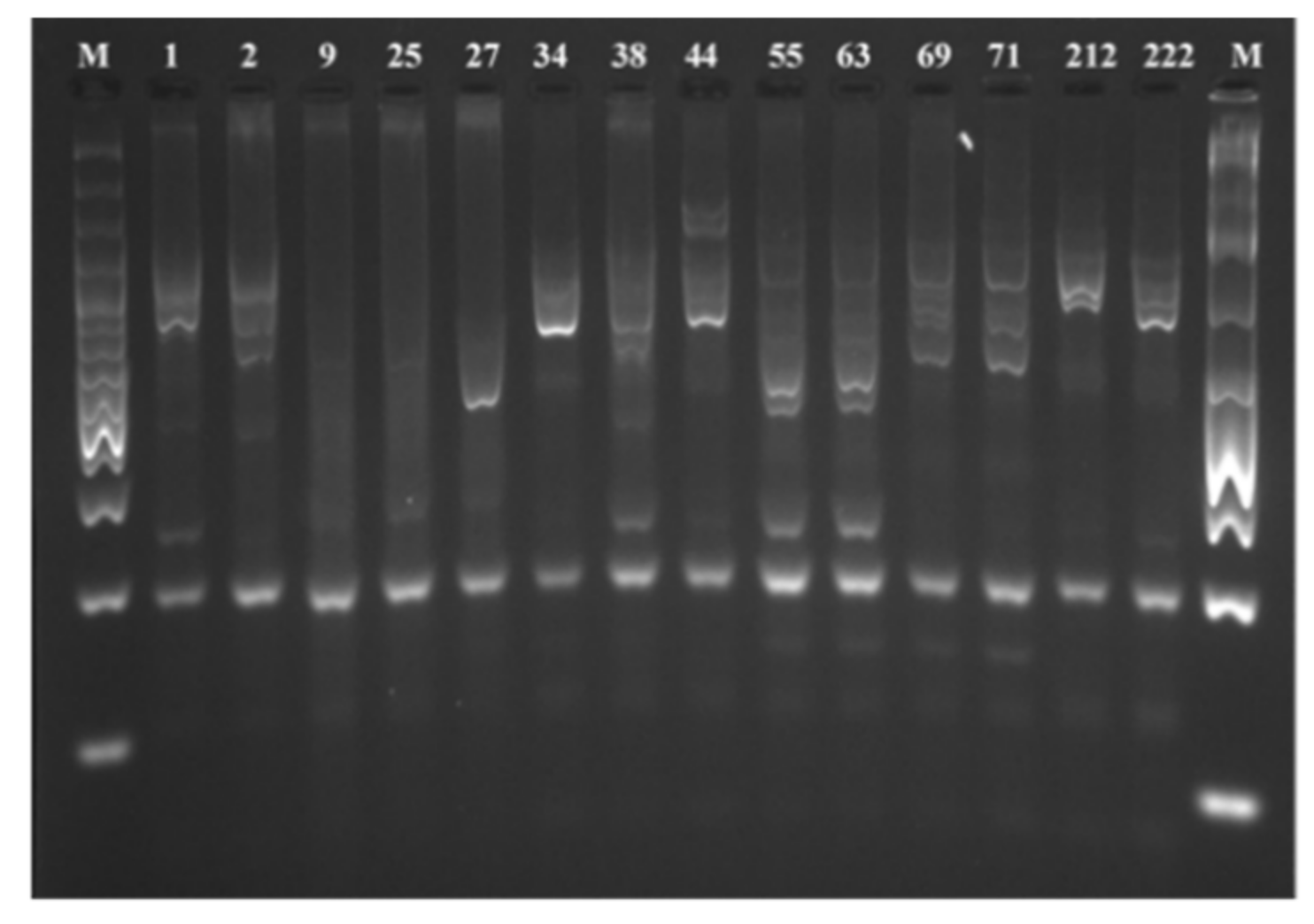
3.2.1. RAPD Analysis

The analysis of the profile of PCR amplified DNA fragments is shown in Figure 3. A set of five different primers was checked, and the most discriminative profiles were obtained using the combination of primers 1254: 5′CCGCAGCCAA3′ (10-mer) and rpd1: 5′CCCGACTGA3′ (9-mer).
The results showed that the newly isolated Lb. delbrueckii subsp. bulgaricus strains are genetically different and also that they differ from the starter cultures of the most popular Bulgarian market yoghurts, i.e., those of the companies Domlyan and Group Danone. However, RAPD was not the method that was able to distinguish all genetically close strains. Two pairs of the isolates did not provide sufficiently good profiles (9, 25, 55, and 63). To prove their genetic uniqueness, the use of other molecular techniques was needed.

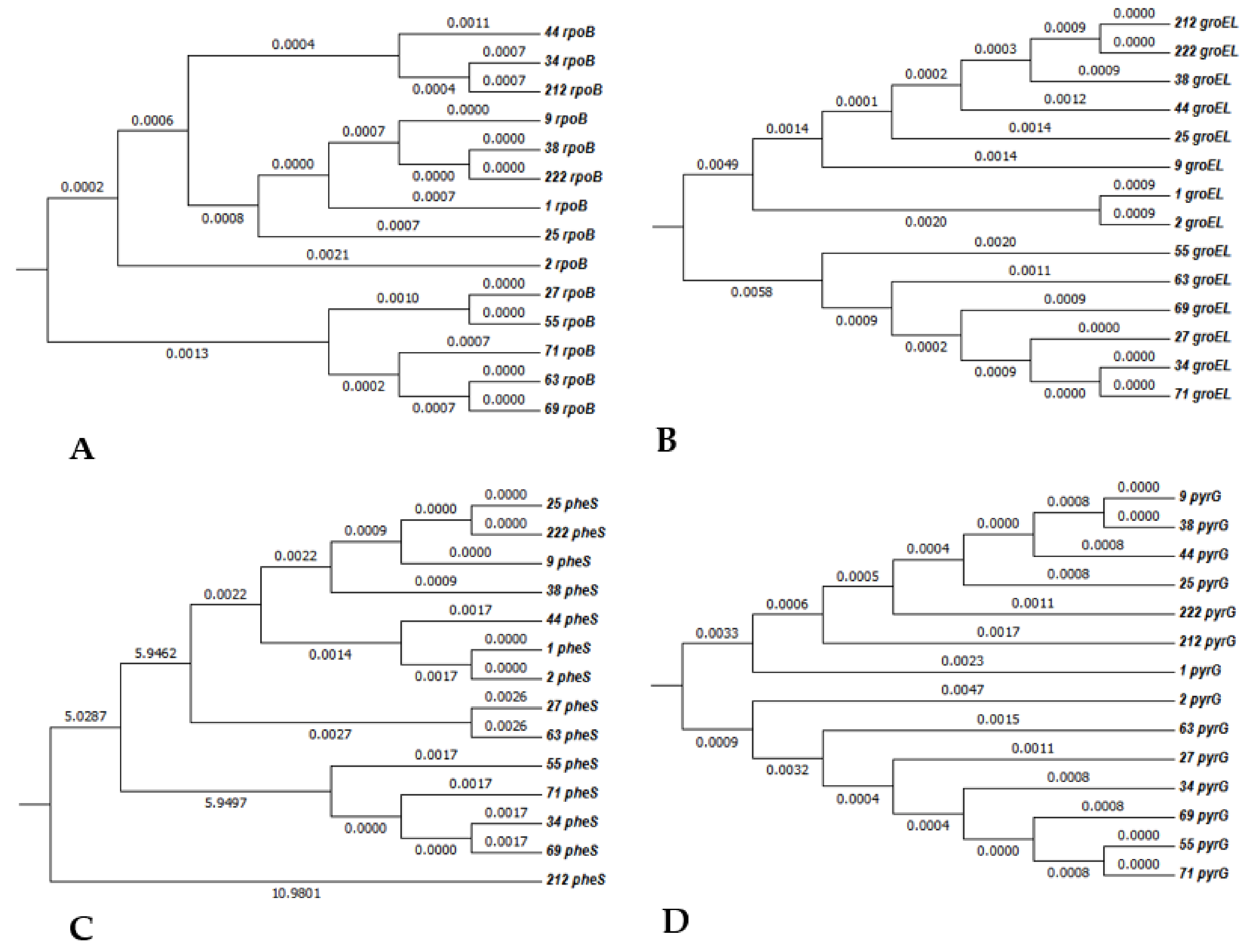
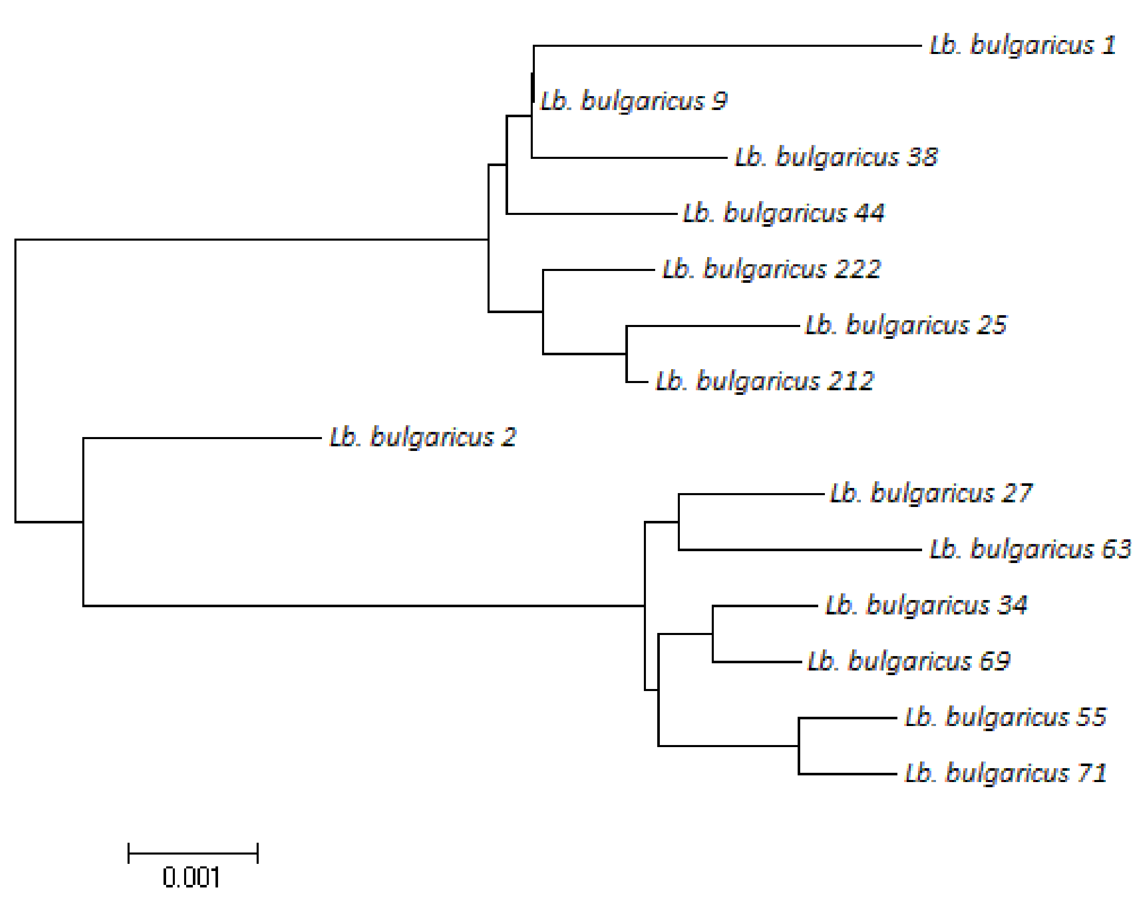
3.2.2. MLST Analysis

MLST is generally regarded as a bacterial identification method that depends on the genomic properties or experimental design (such as the kinds of genes analyzed), and provides a higher resolution than RAPD. MLST uses automated DNA sequencing to characterize the alleles present at different housekeeping gene loci. Because it is based on nucleotide sequences, it is highly discriminatory and provides unambiguous results. MLST analyses were performed with four housekeeping genes, shown at Figure 4: rpoB, groEL, pheS, and pyrG, encoding the β-subunit of bacterial RNA polymerase, the chaperonin family of molecular chaperones, the α-subunit of phenylalanine tRNA synthetase, and CTP synthase, respectively. For the strains examined, parts of these genes were amplified and sequenced.
By sequencing of the genes and sequence comparison using ClustalW, Lb. delbrueckii subsp. bulgaricus strains were clustered into different branches according to the used housekeeping gene. There were ten different alleles for rpoB, eleven for groEL, eleven for pheS, and twelve for pyrG. Table 2 estimates the evolutionary divergence of combined sequences, and the number of base substitutions per site from between sequences is shown. It could be deduced that all strains are diverse and each strain is genetically different from the others. Figure 5 shows the strains’ evolutionary relationships.

3.2.3. PFGE

The PFGE method is strain-specific and provides precise insight into the isolated strains (Figure 6).
Although several enzymes were engaged (NotI, ApaI), the most highly discriminative profiles were obtained by the digest of chromosomal DNA using the restriction enzyme XhoI. The results presented in Figure 6 display the affiliation of the strains to 14 different groups, i.e., they are clustered separately because they are genetically different, indicating the large genetic diversity among Lb. delbrueckii subsp. bulgaricus strains isolated from home-made yoghurt.

3.3. Bioactive Metabolites Produced by the Accompanying Microflora of Bulgarian Yoghurts

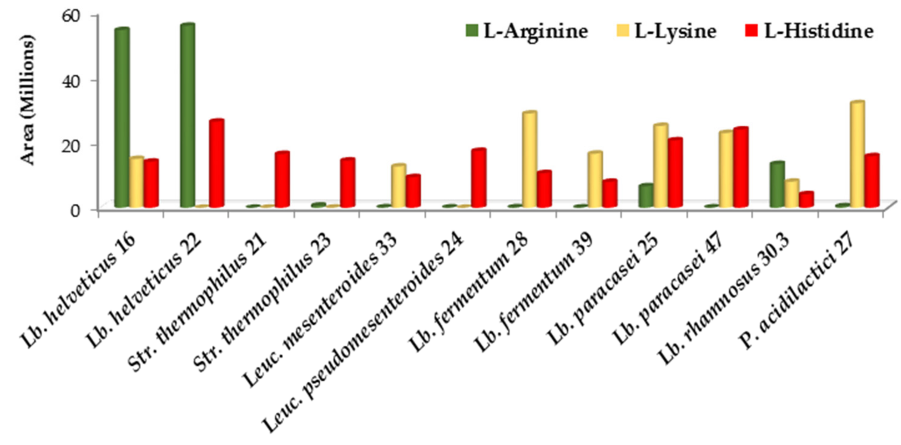
3.3.1. Amino Acids and Their Derivatives

Aiming to elucidate the ability of the accompanying LAB strains to produce valuable metabolites, single-strains fermented yoghurts were subjected to LC-MS analysis (Table S3). Interestingly, Lb. delbrueckii subsp. bulgaricus is an auxotroph for several amino acids (for instance, branched-chained amino acids) and the means of obtaining them is to use those formed by the accompanying microflora in the course of biosynthetic processes [48]. Considering the available data, reports of free amino acids accumulation by LAB are scarce. However, a Lb. helveticus strain isolated from kefir reached 53.38 mg free amino acids per 100 g product [49]. In our study, the amino acid that was accumulated in the largest amount was arginine, and Lb. helveticus 16 and 22 produced it the most compared to the other LAB strains (Figure 7). Arginine is an essential amino acid and has been suggested as a possible prebiotic [50]. L-arginine and its precursor L-citrulline are widely used in the treatment of cardiovascular diseases associated with endothelial dysfunction, such as hypertension, heart failure, atherosclerosis, diabetic vascular disease, and ischemia-reperfusion injury [51]. The ability of the studied LAB to accumulate L-citrulline is highly significant. Citrulline is known as a laxative and as a blood-plasma marker for human irritable bowel syndrome. Supplementation of citrulline with Lb. helveticus can improve intestinal barrier functions [52]. L-arginine and L-citrulline oral intake improve erection hardness in men; both amino acids are used as a treatment of erectile dysfunction [53,54].
Lb. fermentum 28, Lb. paracasei 25 and 47, and P. acidilactici 27 accumulated lysine and tryptophan (Figure 8), and LAB enzymes involved in these pathways may have future industrial applications [55,56]. Almost all strains produced histidine, but the potentially toxic biogenic amines deriving from its degradation were not detected.

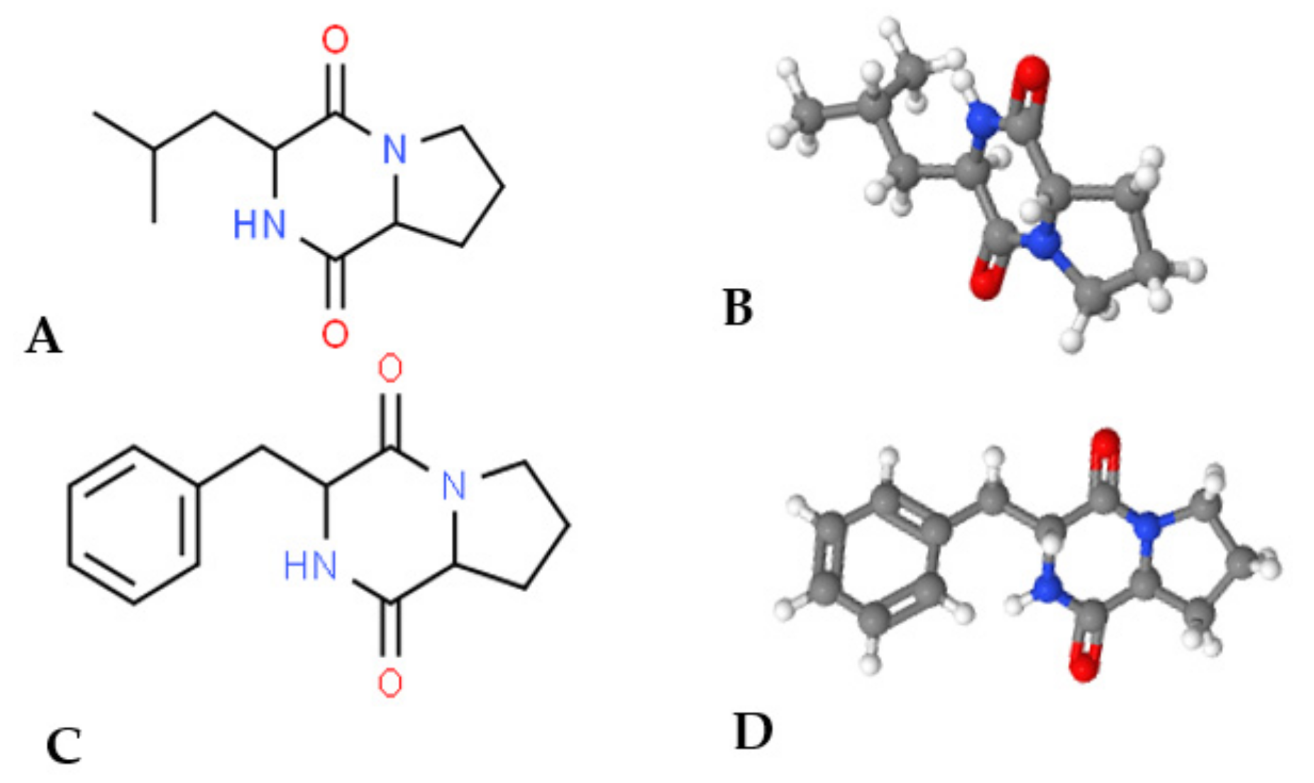
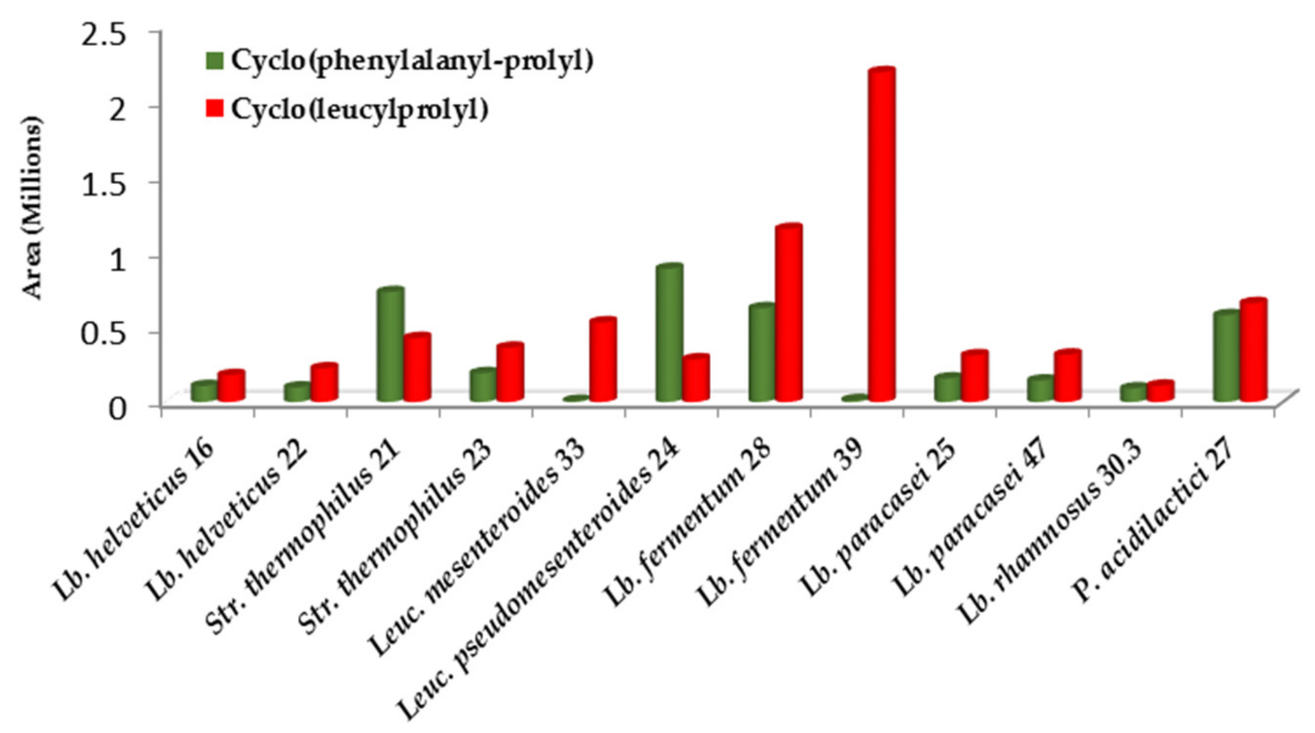
3.3.2. Antimicrobial Peptides

One of the most important bioactive metabolites produced by the accompanying microflora is indole-3-propionic acid (IPA) because it is being studied as a therapeutic in the treatment of many neurodegenerative disorders. According to Mimori et al. (2019), IPA acts as “chemical chaperone”, suppressing stress-induced cell death of neurons and ceasing the aggregation of the denatured proteins in vitro [57]. IPA, and its esters or salts, prevent the cytotoxic effects of amyloid beta-protein on cells and can be used as a treatment for fibrillogenic diseases. This inhibition of beta-amyloid fibril formation implies novel therapy for Alzheimer’s disease [58,59]. IPA is an even more potent scavenger of hydroxyl radicals than melatonin, moreover, it does so without generating subsequent reactive and pro-oxidant intermediate compounds. In 2017, elevated concentrations of IPA in human blood plasma were found to correlate with a lower risk of type 2 diabetes [60]. Because the phenomenon was also connected with the higher consumption of fiber-rich foods (prebiotics), this effect was assigned to IPA, which is endogenously produced by the human microbiota. Other beneficial metabolites produced by LAB of the accompanying microflora were two cyclic peptides: cyclo(phenylalanyl-prolyl) and cyclo(leucyloprolyl) (Figure 9 and Figure 10).
Cyclo(phenylalanyl-prolyl) has broad-spectrum antimicrobial activity, including antibacterial, antifungal, antiviral, anti-settlement, antiprotozoal, antiparasitic, and antitumor properties, and has a radioprotective effect [62]. It is produced in significant amounts by Leuc. pseudomesenteroides 24, Str. thermophilus 21, Lb. fermentum 28, and P. acidilactici 27.
Cyclo(leucyloprolyl), also known as the antibiotic Gancidin W, is another antimicrobial compound produced by the concomitant LAB. It is synthesized in higher amounts by Lb. fermentum, Leuc. mesenteroides, and P. acidilactici. It is a bioactive natural product, and an alkaloid that provides the bitter component of sake and contributes to the flavor of the beer. Recently, the anticancer effect of Gandicin W against breast cancer was demonstrated [63]. Until now, both antimicrobial compounds have been found only in marine bacteria (named drugs from the ocean) [64]. Our investigation reports for the first time their presence in yoghurt.

4. Conclusions

Metagenomic study of authentic Bulgarian yoghurts revealed the presence of “classical” starter strains Lb. delbrueckii subsp. bulgaricus and Str. thermophilus, accompanied by many LAB and spoilage genera. RAPD, MLST, and PFGE analyses showed that the Lb. delbrueckii subsp. bulgaricus strains in homemade yoghurts are different, authentic, and do not participate in commercial starters. The formation of unique metabolites belonging to the class of amino acids is a significant novelty of the present work. The accumulation of arginine, lysine, and histidine by the biosynthetic properties of the accompanying microflora is beneficial for the propagation of the starter cultures Lb. bulgaricus, but also for the consumer of the yoghurt. Metabolites that are valuable for human health, such as indole-3-propionic acid and L-citrulline, were detected. This identification could address the issue of the “magic” effects of Bulgarian yoghurt on the elderly, who retain clear minds and sexual activity to an advanced age. The strains producing the antimicrobial peptides cyclo(phenylalanyl-prolyl) and cyclo(leucyloprolyl) may be included in future starter formulations. In summary, the specific natural and climatic conditions in Bulgaria contributed to the evolution of yoghurt starter cultures with unique features. The comprehensive examination of the current status of the authentic Bulgarian yoghurt microbial diversity showed that homemade yoghurts, produced in small farms in Bulgarian mountainous areas, are the source of LAB with health-promoting effects.

Supplementary Materials

The following are available online at https://www.mdpi.com/2227-9717/9/1/114/s1, Table S1: Metagenome study of Lactic acid bacteria—species abundance in four samples of Bulgarian home-made yoghurt prepared using an artisanal starter. Yoghurt origin: Sample 1 and 2, from villages in Rila and Pirin mountains, Sample 3, from the Thracian Plain, Sample 4, from Rhodope mountain; Table S2: Metagenome study of contaminant microflora species abundance in four samples of Bulgarian home-made yoghurt prepared using an artisanal starter. Yoghurt origin: Sample 1 and 2, from villages in Rila and Pirin mountains, Sample 3, from the Thracian Plain, Sample 4, from Rhodope mountain; Table S3: LC-MS (Liquid chromatography/Mass spectrometry) analysis of the valuable metabolites produced by the accompanying LAB strains in single-strain fermented yoghurts. LC-MS analysis was carried out on Q Exactive® mass analyzer equipped with TurboFlow® LC system and APCI atmospheric pressure electrospray ionization module. Data acquisition and processing were carried out with Xcalibur 2.4® software package.

Author Contributions

Conceptualization, P.P. and K.P.; methodology, I.I., V.L., I.H.; writing—original draft preparation, I.I.; writing—review and editing, P.P., Z.W., Z.L. All authors have read and agreed to the published version of the manuscript.

Funding

Metagenomics experiments were funded by the Bulgarian Ministry of Education and Science with the financial support by the “Healthy Foods for a Strong Bio-Economy and Quality of Life” National Research Program approved by DCM # 577/17.08.2018. The study was financially supported also by the company Bright Dairy & Food Co. Ltd, China, grant number 1/18.06.2015.

Institutional Review Board Statement

Not applicable.

Informed Consent Statement

Not applicable.

Data Availability Statement

Conflicts of Interest

The authors declare no conflict of interest. The funders had no role in the design of the study; in the collection, analyses, or interpretation of data; in the writing of the manuscript, or in the decision to publish the results.

References

  1. Fernandez, M.A.; Panahi, S.; Daniel, N.; Tremblay, A.; Marette, A. Yogurt and cardiometabolic diseases: A critical review of potential mechanisms. Adv. Nutr. 2017, 8, 812–829. [Google Scholar] [CrossRef] [PubMed] [Green Version]
  2. Berthold-Pluta, A.M.; Pluta, A.S.; Garbowska, M.; Stasiak-Różańska, L. Exopolysaccharide-producing Lactic Acid Bacteria—Health-promoting properties and application in the dairy industry. Adv. Microbiol. 2019, 58, 191–204. [Google Scholar] [CrossRef] [Green Version]
  3. Popović, N.; Brdarić, E.; Đokić, J.; Dinić, M.; Veljović, K.; Golić, N.; Terzić-Vidojević, A. Yogurt produced by novel natural starter cultures improves gut epithelial barrier in vitro. Microorganisms 2020, 8, 1586. [Google Scholar] [CrossRef] [PubMed]
  4. Julio, P.D.; Carolina, G.L.; Luis, F.; Angel, G. Modulation of immunity and inflammatory gene expression in the gut, in inflammatory diseases of the gut and in the liver by probiotics. World J. Gastroenterol. 2014, 20, 15632–15649. [Google Scholar]
  5. Mousavi, S.N.; Somayeh, S.; Asbaghi, O. Effect of daily probiotic yogurt consumption on inflammation: A systematic review and meta-analysis of randomized Controlled Clinical trials. Obes. Med. 2020, 18, 100221. [Google Scholar] [CrossRef]
  6. De Filippis, F.; Parente, E.; Ercolini, D. Metagenomics insights into food fermentations. Microb. Biotechnol. 2016, 10, 91–102. [Google Scholar] [CrossRef] [PubMed]
  7. Hao, P.; Zheng, H.; Yu, Y.; Ding, G.; Gu, W.; Chen, S.; Yu, Z.; Ren, S.; Oda, M.; Konno, T.; et al. Complete sequencing and pan-genomic analysis of Lactobacillus delbrueckii subsp. bulgaricus reveal its genetic basis for industrial yogurt production. PLoS ONE 2011, 6, e15964. [Google Scholar] [CrossRef] [Green Version]
  8. Liu, M.; Siezen, R.J.; Nauta, A. In Silico prediction of horizontal gene transfer events in Lactobacillus bulgaricus and Streptococcus thermophilus reveals protocooperation in yogurt manufacturing. Appl. Environ. Microbiol. 2009, 75, 4120–4129. [Google Scholar] [CrossRef] [PubMed] [Green Version]
  9. Beshkova, D.; Simova, E.; Frengova, G.; Simov, Z. Production of flavour compounds by yogurt starter cultures. J. Ind. Microbiol. Biotechnol. 1998, 20, 180–186. [Google Scholar] [CrossRef]
  10. Velikova, P.; Petrov, K.; Lozanov, V.; Tsvetanova, F.; Stoyanov, A.; Wu, Z.; Liu, Z.; Petrova, P. Microbial diversity and health-promoting properties of the traditional Bulgarian yogurt. Biotechnol. Biotechnol. Equip. 2018, 32, 1205–1217. [Google Scholar] [CrossRef] [Green Version]
  11. Suzuki, Y.; Ikeda, K.; Sakuma, K.; Kawai, S.; Sawaki, K.; Asahara, T.; Takahashi, T.; Tsuji, H.; Nomoto, K.; Nagpal, R.; et al. Association between yogurt consumption and intestinal microbiota in healthy young adults differs by host gender. Front. Microbiol. 2017, 8, 847. [Google Scholar] [CrossRef] [PubMed] [Green Version]
  12. Boyanova, L.; Stephanova-Kondratenko, M.; Mitov, I. Anti-Helicobacter pylori activity of Lactobacillus delbrueckii subsp. bulgaricus strains: Preliminary report. Lett. Appl. Microbiol. 2009, 48, 579–584. [Google Scholar] [CrossRef] [PubMed]
  13. Boyanova, L.; Gergova, G.; Markovska, R.; Yordanov, D.; Mitov, I. Bacteriocin-like inhibitory activities of seven Lactobacillus delbrueckii subsp. bulgaricus strains against antibiotic susceptible and resistant Helicobacter pylori strains. Lett. Appl. Microbiol. 2017, 65, 469–474. [Google Scholar] [CrossRef] [PubMed]
  14. Stefanova, T.; Urshev, Z.; Dimitrov, Z.; Fatchikova, N.; Minkova, S. Species affiliation of dairy lactobacilli with angiotensin converting enzyme inhibitory activity. Biotechnol. Biotechnol. Equip. 2009, 23, 1250–1254. [Google Scholar] [CrossRef] [Green Version]
  15. Cocolin, L.; Ercolini, D. Zooming into food-associated microbial consortia: A “cultural” evolution. Curr. Opin. Food Sci. 2015, 2, 43–50. [Google Scholar] [CrossRef]
  16. Lee, A.; Lee, Y.J.; Yoo, H.J.; Kim, M.; Chang, Y.; Lee, D.S.; Lee, J.H. Consumption of dairy yogurt containing Lactobacillus paracasei ssp. paracasei, Bifidobacterium animalis ssp. lactis and heat-treated Lactobacillus plantarum improves immune function including natural killer cell activity. Nutrients 2017, 9, 558. [Google Scholar] [CrossRef]
  17. Sánchez, E.; Nieto, J.; Vidal, S.; Santiago, A.; Martinez, X.; Sancho, F.J.; Sancho-Bru, P.; Mirelis, B.; Corominola, H.; Juárez, C.; et al. Fermented milk containing Lactobacillus paracasei subsp. paracasei CNCM I-1518 reduces bacterial translocation in rats treated with carbon tetrachloride. Sci. Rep. 2017, 7, 45712. [Google Scholar]
  18. Wang, J.; Li, C.; Xue, J.; Yang, J.; Zhang, Q.; Zhang, H.; Chen, Y. Fermentation characteristics and angiotensin I-converting enzyme-inhibitory activity of Lactobacillus helveticus isolate H9 in cow milk, soy milk, and mare milk. J. Dairy Sci. 2015, 98, 3655–3664. [Google Scholar] [CrossRef] [Green Version]
  19. Zhou, T.; Huo, R.; Kwok, L.Y.; Li, C.; Ma, Y.; Mi, Z.; Chen, Y. Effects of applying Lactobacillus helveticus H9 as adjunct starter culture in yogurt fermentation and storage. J. Dairy Sci. 2019, 102, 223–235. [Google Scholar] [CrossRef] [Green Version]
  20. Cavalheiro, G.F.; Baptista, D.P.; Galli, B.D.; Negrão, F.; Eberlin, M.N.; Gigante, M.L. High protein yogurt with addition of Lactobacillus helveticus: Peptide profile and angiotensin-converting enzyme ACE-inhibitory activity. Food Chem. 2020, 333, 127482. [Google Scholar] [CrossRef]
  21. Cameron, D.; Hock, Q.S.; Kadim, M.; Mohan, N.; Ryoo, E.; Sandhu, B.; Yamashiro, Y.; Jie, C.; Hoekstra, H.; Guarino, A. Probiotics for gastrointestinal disorders: Proposed recommendations for children of the Asia-Pacific region. World J. Gastroenterol. 2017, 23, 7952–7964. [Google Scholar] [CrossRef] [PubMed]
  22. Szajewska, H.; Guarino, A.; Hojsak, I.; Indrio, F.; Kolacek, S.; Shamir, R.; Vandenplas, Y.; Weizman, Z. Use of probiotics for management of acute gastroenteritis: A position paper by the ESPGHAN Working Group for Probiotics and Prebiotics. J. Ped. Gastroenterol. Nutr. 2014, 58, 531–539. [Google Scholar] [CrossRef] [PubMed] [Green Version]
  23. Szajewska, H.; Horvath, A. Lactobacillus rhamnosus GG in the primary prevention of eczema in children: A systematic review and meta-analysis. Nutrients 2018, 10, 1319. [Google Scholar] [CrossRef] [PubMed] [Green Version]
  24. Grigoroff, S. Étude sur une lait fermentée comestible. Le “Kissélo mléko” de Bulgarie. Rev. Méd. Suisse Romande 1905, 25, 714–720. [Google Scholar]
  25. Lick, S.; Keller, M.; Bockelmann, M.; Heller, K.J. Optimized DNA extraction method for starter cultures from yoghurt. Milchwissenschaft 1996, 51, 183–186. [Google Scholar]
  26. Cebeci, A.; Gurakan, C. Comparative typing of L. delbrueckii subsp. bulgaricus strains using multilocus sequence typing and RAPD–PCR. Eur. Food. Res. Technol. 2011, 233, 377–385. [Google Scholar] [CrossRef]
  27. Naser, S.M.; Thompson, F.L.; Hoste, B.; Gevers, D.; Dawyndt, P.; Vancanneyt, M.; Swings, J. Application of multilocus sequence analysis (MLSA) for rapid identification of Enterococcus species based on rpoA and pheS genes. Microbiology 2005, 151, 2141–2150. [Google Scholar] [CrossRef] [Green Version]
  28. Michaylova, M.; Minkova, S.; Kimura, K.; Sasaki, T.; Isawa, K. Isolation and characterization of Lactobacillus delbrueckii ssp. bulgaricus and Streptococcus thermophilus from plants in Bulgaria. FEMS Microbiol. Lett. 2007, 269, 160–169. [Google Scholar] [CrossRef] [Green Version]
  29. Undugoda, L.J.S.; Nilmini, A.H.L. Effect of lactic acid microbial ratio of yoghurt starter culture in yoghurt fermentation and reduction of post acidification. J. Food Ind. Microbiol. 2019, 5, 1. [Google Scholar]
  30. Surono, I.S.; Hosono, A. Fermented milks. Types and Standards of Identity. In Encyclopedia of Dairy Sciences, 2nd ed.; Fuquay, J.W., Fox, P.F., McSweeney, P.L.H., Eds.; Academic Press: New York, NY, USA, 2011; pp. 470–476. [Google Scholar]
  31. Georgalaki, M.; Zoumpopoulou, G.; Mavrogonatou, E.; Van Driessche, G.; Alexandraki, V.; Anastasiou, R.; Papadelli, M.; Kazou, M.; Manolopoulou, E.; Kletsas, D.; et al. Evaluation of the antihypertensive angiotensin-converting enzyme inhibitory (ACE-I) activity and other probiotic properties of lactic acid bacteria isolated from traditional Greek dairy products. Int. Dairy J. 2017, 75, 10–21. [Google Scholar] [CrossRef]
  32. Urshev, Z.L.; Pashova-Baltova, K.N.; Dimitrov, Z.P. Tracing Streptococcus thermophilus strains in three-component yogurt starters. World J. Microbiol. Biotechnol. 2006, 22, 1223–1228. [Google Scholar] [CrossRef]
  33. Ma, C.; Chen, Z.; Gong, G.; Huang, L.; Li, S.; Ma, A. Starter culture design to overcome phage infection during yogurt fermentation. Food Sci. Biotechnol. 2015, 24, 521–527. [Google Scholar] [CrossRef]
  34. Sieuwerts, S.; De Bok, F.A.; Hugenholtz, J.; Van Hylckama Vlieg, J.E. Unraveling microbial interactions in food fermentations: From classical to genomics approaches. Appl. Environ. Microbiol. 2008, 74, 4997–5007. [Google Scholar] [CrossRef] [Green Version]
  35. Alvarez, A.S.; Tap, J.; Chambaud, I.; Cools-Portier, S.; Quinquis, L.; Bourlioux, P.; Marteau, P.; Guillemard, E.; Schrezenmeir, J.; Derrien, M. Safety and functional enrichment of gut microbiome in healthy subjects consuming a multi-strain fermented milk product: A randomised controlled trial. Sci. Rep. 2020, 10, 15974. [Google Scholar] [CrossRef] [PubMed]
  36. Roder, T.; Wüthrich, D.; Bär, C.; Sattari, Z.; von Ah, U.; Ronchi, F.; Macpherson, A.J.; Ganal-Vonarburg, S.C.; Bruggmann, R.; Vergères, G. In Silico Comparison Shows that the Pan-Genome of a Dairy-Related Bacterial Culture Collection Covers Most Reactions Annotated to Human Microbiomes. Microorganisms 2020, 8, 966. [Google Scholar] [CrossRef]
  37. Miteva, V.I.; Abadjieva, A.N.; Stefanova, T.T. M13 DNA fingerprinting, a new tool for classification and identification of Lactobacillus spp. J. Appl. Bacteriol. 1992, 73, 349–354. [Google Scholar] [CrossRef]
  38. Dimitrov, Z.; Michaylova, M.; Mincova, S. Characterization of Lactobacillus helveticus strains isolated from Bulgarian yogurt, cheese, plants and human faecal samples by sodium dodecyl sulfate polyacrylamide gel electrophoresis of cell-wall proteins, ribotyping, and pulsed-field gel fingerprinting. Int. Dairy J. 2005, 15, 998–1005. [Google Scholar] [CrossRef]
  39. Morita, H.; Shimazu, M.; Shiono, H.; Toh, H.; Nakajima, F.; Akita, H.; Takagi, M.; Takami, H.; Murakami, M.; Masaoka, T.; et al. Lactobacillus equicursoris sp. nov., isolated from the faeces of a thoroughbred racehorse. Int. J. Syst. Evol. Microbiol. 2010, 60, 109–112. [Google Scholar] [CrossRef]
  40. Beleza do Campo, K. Available online: https://www.belezadocampo.com.br (accessed on 10 December 2020).
  41. Chen, C.; Zhao, S.; Hao, G.; Yu, H.; Tian, H.; Zhao, G. Role of lactic acid bacteria on the yogurt flavour: A review. Int. J. Food Prop. 2017, 20, S316–S330. [Google Scholar] [CrossRef] [Green Version]
  42. Verena, S.J.; Schmidt, V.K.; Kulozik, U.; Scherer, S.; Wenning, M. Microbial biodiversity, quality and shelf life of microfiltered and pasteurized extended shelf life (ESL) milk from Germany, Austria and Switzerland. Int. J. Food Microbiol. 2012, 154, 1–9. [Google Scholar]
  43. Kagkli, D.M.; Vancanneyt, M.; Hill, C.; Vandamme, P.; Cogan, T.M. Enterococcus and Lactobacillus contamination of raw milk in a farm dairy environment. Int. J. Food Microbiol. 2007, 114, 243–251. [Google Scholar] [CrossRef] [PubMed]
  44. Agu, K.; Archibong, E.; Anekwe, D.; Ago, C.; Okafor, A.; Awah, N. Assessment of bacteria present in yoghurt sold on Awka Metropolis. Sch. J. Appl. Med. Sci. 2014, 2, 3071–3075. [Google Scholar]
  45. Pal, M.; Awel, H. Public health significance of Listeria monocytogenes in milk and milk products. J. Vet. Public Health 2014, 12, 1–5. [Google Scholar]
  46. Addis, M.F.; Tanca, A.; Uzzau, S.; Oikonomou, G.; Bicalho, R.C.; Moroni, P. The bovine milk microbiota: Insights and perspectives from -omics studies. Mol. Biosyst. 2016, 12, 2359–2372. [Google Scholar] [CrossRef] [PubMed] [Green Version]
  47. Lal, M.; Kaur, H.; Gupta, L.K.; Sood, N.K. Isolation of Yersinia enterocolitica from raw milk and pork in Ludhiana. Ind. J. Pathol. Microbiol. 2005, 48, 286–287. [Google Scholar]
  48. Lee, K.; Lee, J.; Kim, Y.H.; Moon, S.H.; Park, Y.H. Unique properties of four Lactobacilli in amino acid production and symbiotic mixed culture for lactic acid biosynthesis. Curr. Microbiol. 2001, 43, 383–390. [Google Scholar] [CrossRef] [PubMed]
  49. Simova, E.; Simov, Z.; Beshkova, D.; Frengova, G.; Dimitrov, Z.; Spasov, Z. Amino acid profiles of lactic acid bacteria, isolated from kefir grains and kefir starter made from them. Int. J. Food Microbiol. 2006, 107, 112–123. [Google Scholar] [CrossRef]
  50. Nascimento, M.M. Potential uses of arginine in dentistry. Adv. Dent. Res. 2018, 29, 98–103. [Google Scholar] [CrossRef] [Green Version]
  51. Romero, M.J.; Platt, D.H.; Caldwell, R.B.; Caldwell, R.W. Therapeutic use of citrulline in cardiovascular disease. Cardiovasc. Drug Rev. 2006, 24, 275–290. [Google Scholar] [CrossRef]
  52. Ho, S.W.; El-Nezami, H.; Shah, N.P. Effects of supplementation of citrulline and Lactobacillus helveticus ASCC 511 on intestinal epithelial cell integrity. J. Funct. Foods 2020, 64, 103571. [Google Scholar] [CrossRef]
  53. Stanislavov, R.; Nikolova, V. Treatment of erectile dysfunction with pycnogenol and L-arginine. J. Sex Marital Ther. 2003, 29, 207–213. [Google Scholar] [CrossRef] [PubMed]
  54. Cormio, L.; De Siati, M.; Lorusso, F.; Selvaggio, O.; Mirabella, L.; Sanguedolce, F.; Carrieri, G. Oral L-citrulline supplementation improves erection hardness in men with mild erectile dysfunction. Urology 2011, 77, 119–122. [Google Scholar] [CrossRef] [PubMed]
  55. Wang, X.; Yang, L.; Cao, W.; Ying, H.; Chen, K.; Ouyang, P. Efficient production of enantiopure d-lysine from l-lysine by a two-enzyme cascade system. Catalysts 2016, 6, 168. [Google Scholar] [CrossRef]
  56. Lim, Y.H.; Foo, H.L.; Loh, T.C.; Mohamad, R.; Abdul-Rahim, R. Rapid evaluation and optimization of medium components governing tryptophan production by Pediococcus acidilactici tp-6 isolated from Malaysian food via statistical approaches. Molecules 2020, 25, 779. [Google Scholar] [CrossRef] [Green Version]
  57. Mimori, S.; Kawada, K.; Saito, R.; Takahashi, M.; Mizoi, K.; Okuma, Y.; Hosokawa, M.; Kanzaki, T. Indole-3-propionic acid has chemical chaperone activity and suppresses endoplasmic reticulum stress-induced neuronal cell death. Biochem. Biophys. Res. Commun. 2019, 517, 623–628. [Google Scholar] [CrossRef]
  58. Pappolla, M. Indole-3-propionic Acids, Salts and Esters Thereof Used as Medicaments. U.S. Patent WO1999042102A1, 26 August 1999. [Google Scholar]
  59. Zhang, L.S.; Davies, S.S. Microbial metabolism of dietary components to bioactive metabolites: Opportunities for new therapeutic interventions. Genome Med. 2016, 8, 46. [Google Scholar] [CrossRef] [Green Version]
  60. De Mello, V.D.; Paananen, J.; Lindström, J.; Lankinen, M.A.; Shi, L.; Kuusisto, J.; Bergdahl, I.A. Indolepropionic acid and novel lipid metabolites are associated with a lower risk of type 2 diabetes in the Finnish diabetes prevention study. Sci. Rep. 2017, 7, 46337. [Google Scholar] [CrossRef]
  61. Chemspider: Search and Share Chemistry, a Site of Royal Society of Chemistry. Available online: http://www.chemspider.com (accessed on 6 December 2020).
  62. Lee, K.H.; Rhee, K.H. Radioprotective effect of cyclo(l-phenylalanyl-l-prolyl) on irradiated rat lung. J. Microbiol. Biotechnol. 2008, 18, 369–376. [Google Scholar]
  63. Ravi, L.; Ragunathan, A.; Krishnan, K. Marine Streptomyces paradoxus VITALK03 derived gancidin W mediated cytotoxicity through Ras-Raf-MEK-ERK signalling pathway. Ind. J. Biotechnol. 2017, 16, 164–175. [Google Scholar]
  64. Nweze, J.A.; Mbaoji, F.N.; Huang, G.; Li, Y.; Yang, L.; Zhang, Y.; Huang, S.; Pan, L.; Yang, D. Antibiotics development and the potentials of marine-derived compounds to stem the tide of multidrug-resistant pathogenic bacteria, fungi, and protozoa. Mar. Drugs 2020, 18, 145. [Google Scholar] [CrossRef] [Green Version]
Figure 1. Metagenome study of species abundance in four samples of Bulgarian home-made yoghurt prepared using an artisanal starter. Yoghurt origin: Sample 1 and 2, from villages in Rila and Pirin mountains; Sample 3, from the Thracian Plain; Sample 4, from Rhodope mountain.
Processes 09 00114 g001
Figure 2. Metagenome study of species abundance of the contaminant microflora in four samples of Bulgarian home-made yoghurt.
Processes 09 00114 g002
Figure 3. Randomly amplified polymorphic DNA–PCR (RAPD) analysis of 12 Lb. delbrueckii subsp. bulgaricus isolates and two commercial Lb. delbrueckii subsp. bulgaricus strains. The strain 212 derives from the yoghurt “Domlyan”, and 222 from the yoghurt “Na baba” of Danone.
Processes 09 00114 g003
Figure 4. Phylogenetic trees of Lb. delbrueckii subsp. bulgaricus isolates based on the sequences of four housekeeping genes: rpoB (A), groEL (B), pheS (C), and pyrG (D). Evolutionary analyses were conducted using MEGA6 software. The evolutionary history was inferred using the neighbor-joining method. The evolutionary distances were computed using the maximum composite likelihood method and are in the units of the number of base substitutions per site. The analysis involved 14 nucleotide sequences. Codon positions included were 1st + 2nd + 3rd + Noncoding. All positions containing gaps and missing data were eliminated.
Processes 09 00114 g004
Figure 5. Evolutionary relationships between newly isolated Lb. delbrueckii subsp. bulgaricus strains. The evolutionary history was inferred using the minimum evolution method. The optimal tree with the sum of branch length = 0.01438984 is shown. The evolutionary distances were computed using the maximum composite likelihood method and are in the units of the number of base substitutions per site. The ME tree was searched using the Close-Neighbor-Interchange (CNI) algorithm at a search level of 1. All positions containing gaps and missing data were eliminated by MEGA6.
Processes 09 00114 g005
Figure 6. Macro-restriction profiles of Xho I-digested DNA of Lb. delbrueckii subsp. bulgaricus strains.
Processes 09 00114 g006
Figure 7. Production of amino acids arginine, lysine, and histidine by analyzed LAB. The LC-MS analyses were carried out using a Q Exactive® mass analyzer equipped with TurboFlow® LC system and APCI atmospheric pressure electrospray ionization module. Data acquisition and processing were carried out with the Xcalibur 2.4® software package.
Processes 09 00114 g007
Figure 8. Production of indole-3-propionic acid, L-tryptophan, and L-citrulline by analyzed LAB. The LC-MS analyses were carried out using a Q Exactive® mass analyzer equipped with a TurboFlow® LC system and APCI atmospheric pressure electrospray ionization module. Data acquisition and processing were carried out with the Xcalibur 2.4® software package.
Processes 09 00114 g008
Figure 9. Two- (2D), and three-dimensional (3D) structural formulas of antimicrobial compounds produced by accompanying LAB in yoghurt. (A,B) Cyclo(phenylalanyl-prolyl), Mw 244 Da; (C,D) Cyclo(leucyloprolyl), 210 Da. The models were obtained from the free chemical structure database of the Royal Society of Chemistry ChemSpider [61].
Processes 09 00114 g009
Figure 10. Production of antimicrobial peptides by LAB strains. The LC-MS analyses were carried out using a Q Exactive® mass analyzer equipped with a TurboFlow® LC system and APCI atmospheric pressure electrospray ionization module. Data acquisition and processing were carried out with the Xcalibur 2.4® software package.
Processes 09 00114 g010
Table 1. Primers used for multi-locus sequence typing (MLST) amplification and sequencing.
GenePrimer NameSequence (5′→3′)Annealing
T °C
Product Position in GenePosition in Genome 1Template Size (bp)
rpoBrpoB_F
rpoB_R
GGCGGAAAGAGTTATCGT GATGTCGGCTGGAGTGAT58399–1170334519–338184772
groELgroEL_F
groEL_R
TCGGCAAGGACGGTGTT
GTGGATTACGGCTACGC
57500–11341392354–1393967635
pheSpheS_F
pheS_R
CATCGGCATGAGCTACCA
CCTCCTGACGGAATTGTTG
55369–10421283032–1284081674
pyrGpyrG_F
pyrG_R
AAGCCGACCCAGCAATC
AGCCCAGACGCAAGGTG
57568–1309301679–303298742
1 Note: Position in the genome of Lb. delbrueckii subsp. bulgaricus ATCC11842T (GenBank Accession No: NC_008054).
Table 2. Estimates of evolutionary divergence between combined sequences. The number of base substitutions per site between sequences is shown. Analyses were conducted using the maximum composite likelihood model. The analysis involved 14 combined nucleotide sequences. There were a total of 2474 positions in the final dataset.
1129252734384455636971212
20.004
90.0030.004
250.0030.0040.001
270.0080.0090.0100.010
340.0070.0080.0090.0090.002
380.0030.0040.0010.0010.0090.008
440.0020.0040.0020.0020.0090.0070.002
550.0080.0080.0090.0090.0010.0010.0090.009
630.0080.0090.0100.0100.0020.0010.0090.0080.001
690.0090.0090.0100.0100.0020.0010.0090.0090.0020.001
710.0080.0090.0100.0100.0020.0010.0090.0080.0010.0010.001
2120.0030.0040.0010.0010.0100.0080.0010.0010.0090.0090.0090.009
2220.0030.0040.0010.0010.0090.0090.0010.0010.0090.0090.0100.0090.001
Publisher’s Note: MDPI stays neutral with regard to jurisdictional claims in published maps and institutional affiliations.

