Article

Folate-Receptor-Targeted Gold Nanoparticles Bearing a DNA-Binding Anthraquinone

by
Ana B. Caballero
1,†,‡,
Nikolas J. Hodges
2,* and
Michael J. Hannon
1,*
1
School of Chemistry, University of Birmingham, Edgbaston, Birmingham B15 2TT, UK
2
School of Biosciences, University of Birmingham, Edgbaston, Birmingham B15 2TT, UK
*
Authors to whom correspondence should be addressed.
Present address: Department of Inorganic and Organic Chemistry, Faculty of Chemistry, University of Barcelona, Martí i Franquès 1-11, 08028 Barcelona, Spain; ana.caballero@ub.edu.
Present address: Institute of Nanoscience and Nanotechnology (IN2UB), University of Barcelona, 08028 Barcelona, Spain.
Inorganics 2025, 13(3), 87; https://doi.org/10.3390/inorganics13030087
Submission received: 6 February 2025 / Revised: 2 March 2025 / Accepted: 11 March 2025 / Published: 17 March 2025
(This article belongs to the Section Bioinorganic Chemistry)

Abstract

:
In recent years, anthraquinones have been widening their therapeutic opportunities given their numerous health benefits. The search for adequate delivery platforms to improve their pharmacokinetics leads us to propose herein folate-capped gold nanoparticles with an anthraquinone derivative attached onto their surface. Through a straightforward, two-step procedure, we obtained stable nanoparticles that can deliver anthraquinones selectively to cells overexpressing folate receptors. The new conjugates were highly toxic against two tumour cell lines, lung carcinoma A549 and cervical carcinoma HeLa, and showed significant in vitro targeting effects for FR+ HeLa cells. We anticipate that the convenience of this synthetic procedure could enable the future development of folate-targeted conjugates bearing highly active anthraquinone-derived drugs.

1. Introduction

Anthraquinones are antioxidant molecules that have found many applications not only in industry (dyes, antioxidant, polymerization inhibitors, etc.) but also in medicine [1,2,3]. They possess numerous beneficial effects, such as anti-bacterial [4], anti-trypanosomal [5,6], anti-inflammatory [7] and anti-neoplastic activities [8,9]. Anthraquinone derivatives can intercalate DNA and are cytotoxic to human cancer cells, such as lung adenocarcinoma A549, myelogenous leukaemia HL-60 and cervical carcinoma HeLa cell lines. They act mainly by generating reactive oxygen species (ROS) and the induction of apoptosis [10]. Interestingly, anthraquinone-tethered platinum complexes have been shown to better penetrate and accumulate in cancer cell spheroids compared to cisplatin, suggesting they may have improved bioavailability in vivo [11]. Moreover, some natural anthraquinones, such as emodin, mitoxantrone and pixantrone, prevent aggregation of toxic Aβ(1–40) in Alzheimer’s disease by intercalation, with possible indications for the treatment of neurodegenerative diseases [12]. Very recently, the anthraquinone derivative parietin exhibited light-enhanced fungicidal activity, following the search for urgently needed novel antimicrobials in the global fight against spreading resistance and offering new options for antimicrobial photodynamic therapy [13]. Such a wide therapeutic potential is prompting scientists to explore new and effective delivery mechanisms for this family of drugs.
Despite their exciting promise, the rapid metabolism and poor water solubility of anthraquinone derivatives have hindered their clinical development [14]. In this sense, the design of biocompatible nanocarriers can improve the bioavailability of this family of drugs. For example, nanoparticles made of magnetite or poly(lactic acid) have been studied to deliver anthraquinone-derived drugs such as rhein and parietin to increase both their bioavailability (in vitro and in vivo) and their selectivity [15,16,17].
In this regard, gold nanoparticles (AuNPs) are interesting nanoplatforms in anticancer drug delivery. AuNPs are non-toxic, biocompatible and inert, allow easy control of particle size and, most importantly, they possess a tuneable surface functionality that makes it possible to conjugate multiple entities on the surface, e.g., active drug, tumour-targeting ligands, optical probes, etc. [18,19,20,21]. Moreover, AuNPs can act intrinsically as optical probes for cellular imaging due to their unique optical response of surface plasmon resonance (SPR) [22,23]. These features render them ideal targeted carriers for poorly soluble anticancer drugs, such as anthraquinone derivatives. However, they have scarcely been explored as carriers for anthraquinone derivatives. For example, Porta et al. reported on the use of 50 nm diameter AuNPs to deliver the anti-bacterial and anti-inflammatory drugs Aloin A and aloesin inside cells [24].
On the other hand, several studies have pointed to the co-attachment of targeting vectors on the surface of nanoparticles to improve cancer cell targeting and minimize the toxicity on healthy tissues. A variety of tumour-targeting ligands have been used to facilitate the binding/uptake of carriers to target cells. Folate-mediated targeting holds substantial promise and has been one of the major strategies assimilated by the nanomedical field for targeted drug delivery and also imaging of tumours [25]. Folate receptors (FRs), particularly FRα, are overexpressed in various types of cancers, e.g., over 90% of ovarian carcinomas overexpress FRα. However, folate receptors are not expressed or are present in low levels in most normal tissues, which makes folate a highly selective tumour marker [26]. Folic acid has an extremely high affinity towards its receptors (KD of approx. 0.1 nM for FRα). This enables the nanoconjugate to bind robustly to the folate receptors before becoming internalised via an endocytic process, yielding a highly selective uptake [26].
A number of folate-functionalized gold nanoparticles have been reported using different linkers and conjugation procedures [27,28,29,30,31,32,33]. Folate-capped AuNPs have been studied mostly for targeted drug delivery [27,34,35], but they have also found uses as radiosensitizers for chemotherapy [36], as optical probes for cellular imaging [22], and for image contrast enhancement in X-ray computed tomography [37].
The present work describes an easy two-step protocol to obtain 18 nm AuNPs bearing the anthraquinone derivative 1-aminoanthraquinone using folic acid as a surface-modifying agent to improve the uptake and the selectivity of the delivery into cancer cells. In these conjugates (AA-FA-AuNP), folic acid acts as a reducing and stabilizing agent and as a cancer-targeting vector molecule. In vitro preliminary studies have been carried out to evaluate the cytotoxicity and selectivity of these conjugates towards folate-expressing (FR+) and non-expressing (FR−) cancer cells, namely HeLa cervical carcinoma and A549 lung carcinoma cell lines, respectively [38].

2. Results and Discussion

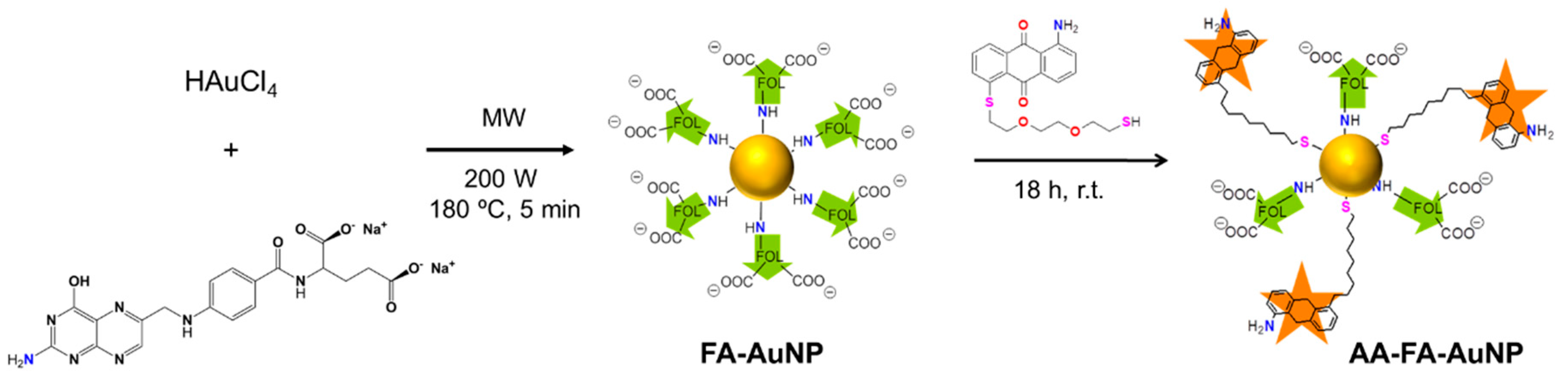
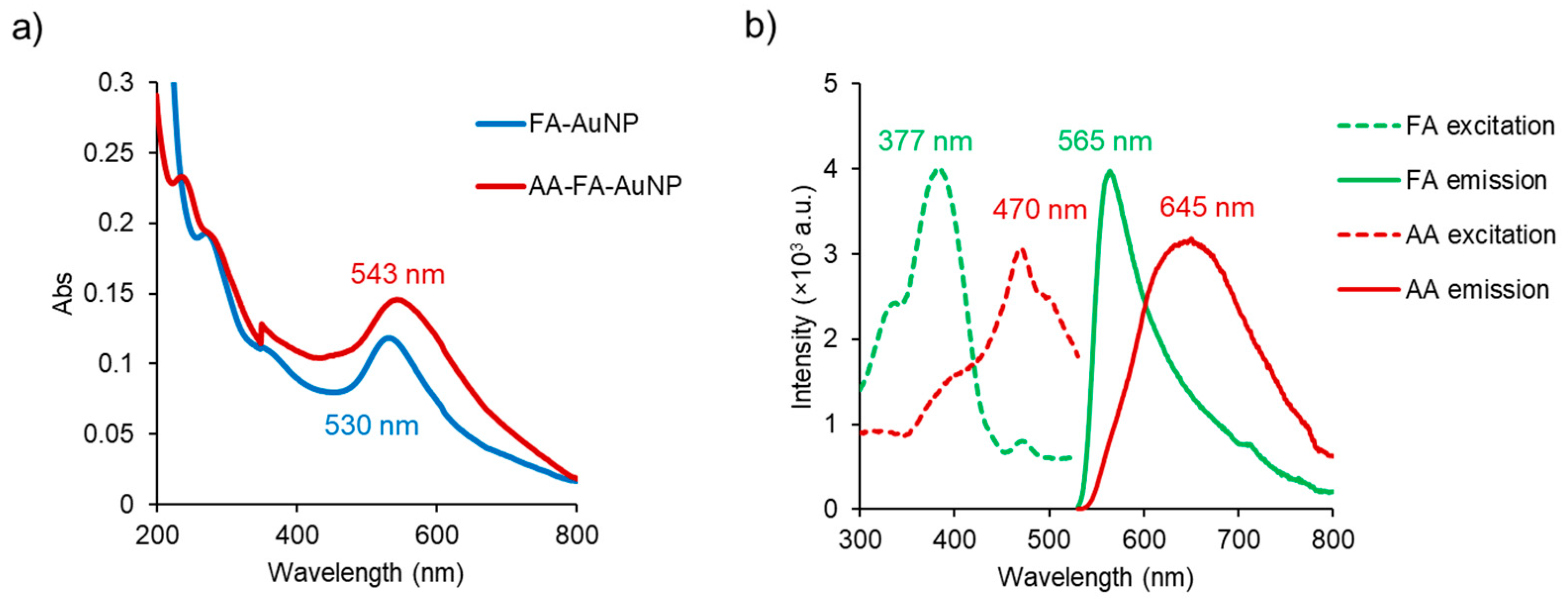
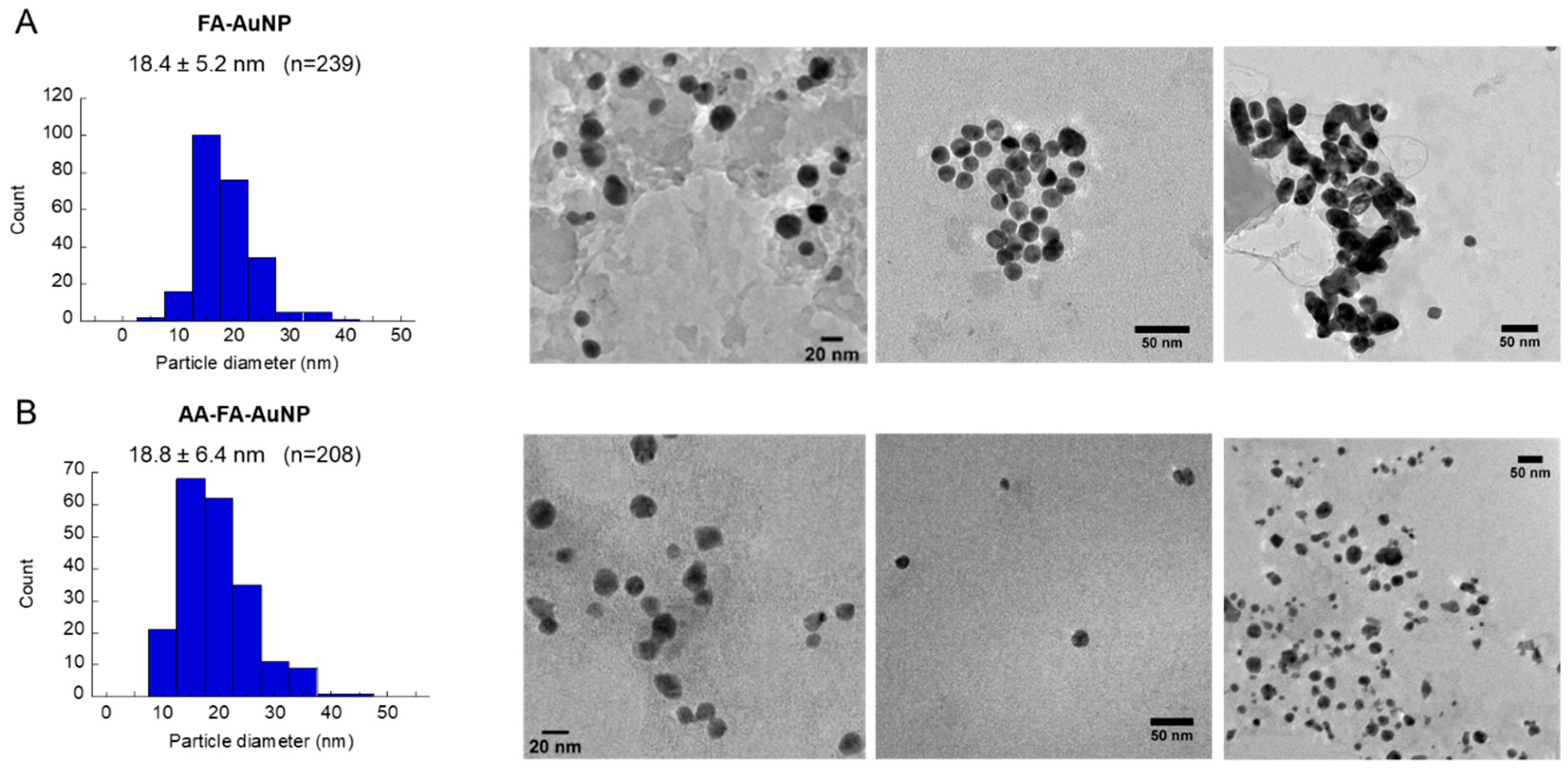
2.1. Synthesis and Characterization

The conjugates, AA-FA-AuNP, were obtained through a straightforward, two-step procedure as follows. In the first step of the synthesis, folate-coated gold nanoparticles (FA-AuNP) were prepared by the one-step reduction of HAuCl4 by folic acid under microwave irradiation (see Section 3). After purification, the FA-AuNP were reacted with 1-amine-5-(4,7-dioxa-1,10-dithiadecyl)anthracene-9,10-dione (AA) at room temperature for 18 h (Scheme 1). The resulting AA-FA-AuNP were subjected to several steps of purification, including washing with chloroform, dialysis and centrifugation. AA is a 1-amine-anthraquinone molecule with a pending short thiol-terminated ethylene glycol chain that acts as anchoring linker with the AuNPs.
The absence of unbound anthraquinone molecules was confirmed by UV–Vis. Moreover, an AA-FA-AuNP suspension was checked periodically for the possible release of any weakly bound AA for one month. To achieve this, aliquots were washed with chloroform three times, and the AA content in the organic phase (previously concentrated up to 2 mL) was measured by UV–Vis. Negative results were obtained in all batches.
The characterization of the as-synthesized FA-AuNP and AA-FA-AuNP was carried out by UV–Vis spectroscopy, infrared spectroscopy (FTIR), fluorescence spectroscopy, dynamic light scattering (DLS), and transmission electron microscopy (TEM). The UV–Vis spectrum of the AA-FA-AuNP showed a surface plasmon band at 543 nm, which has a redshift of 13 nm with respect to the SPR band of the precursor FA-AuNP (Figure 1a) and confirms the attachment of the anthraquinone. The FTIR spectra (Figure S1) of the FA-AuNP and AA-FA-AuNP display the two characteristic intense bands of folic acid at 1693 cm−1 (C=O stretching) and 1606 cm−1 (NH- bending) as a single broader band at 1638 cm−1, which confirms the folate attachment onto the nanoparticle surface. In a reported microwave-assisted synthesis of folate-capped AuNPs, the authors suggested that the conjugation of the folate possibly takes place via the primary amino group in the pteridine ring by forming a N-Au bond [39]. This is also evidenced in the FTIR spectra of the FA-AuNP and AA-FA-AuNP by the disappearance of the –NH2 bands of the pteridine ring at 3530 and 3415 cm−1. The FTIR spectrum of the AA-FA-AuNP also shows a band at 1266 cm−1, which is the strongest band of the anthraquinone derivative AA.
The conjugation of AA to FA-AuNP was further confirmed by the strong fluorescence at 645 nm (Figure 1b). The presence of FA was also confirmed by its emission at 565 nm. Given the partial overlap between the excitation spectra of FA and AA, we recorded the emission spectrum of the FA-AuNP (Figure S2) under excitation at 470 nm and confirmed that the emission at 645 nm is comparatively negligible compared to that of the FA-AA-AuNP. It should be noted that the emission of AA was relatively strong despite the potential for quenching upon binding to the AuNP surface.
According to size distribution measurements by dynamic light scattering (DLS), the attachment of AA onto the FA-AuNP dramatically reduced their polydispersity index below 0.5, and aggregates of average sizes of 20 and 180 nm were detected (Figure S3) [40]. Transmission electron microscopy (TEM) images of the FA-AuNP and AA-FA-AuNP (Figure 2) confirmed the formation of well-dispersed spherical nanoparticles with a size range of 18.4 ± 5.2 nm and 18.8 ± 6.4 nm, respectively. The surface charge in both cases was negative, and the respective zeta-potential values in a moderately basic media (pH 9) indicated that they form stable suspensions under the following storage conditions: −41.2 ± 12.7 mV for the FA-AuNP and −35.0 ± 8.0 mV for the AA-FA-AuNP (Figure S4).

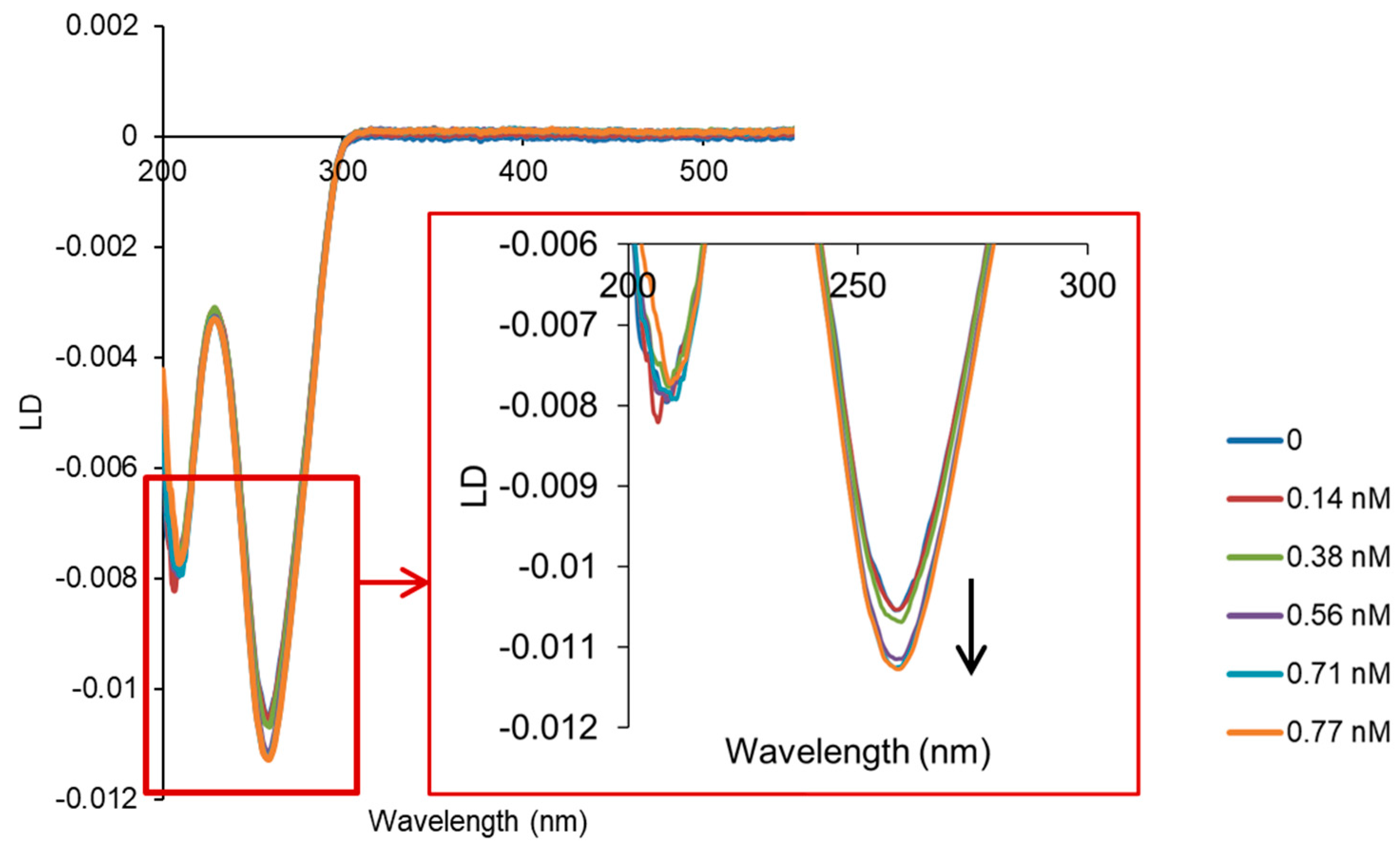
2.2. Interaction with ct-DNA

Given the capability of AA to intercalate between the nucleobase pairs of nucleic acids, we investigated whether this capability can be translated into the AA-FA-AuNP conjugates using flow linear dichroism (LD, Figure 3). The experiments were performed with calf thymus DNA (double stranded) in a pH 7.5 buffer containing 1 mM Tris-HCl and 10 mM NaCl. No particle aggregation and/or ct-DNA precipitation was observed in these conditions. As shown in Figure 3, the characteristic LD negative band of ct-DNA at 260 nm increases in magnitude upon the addition of an increasing concentration of AA-FA-AuNP. A similar behaviour was previously reported for the free AA in the same conditions, and it is typically observed when DNA stiffening is caused by the intercalation of a molecule, leading to greater orientation in the flow [19]. However, the behaviour is opposite to that of previously reported AuNPs of similar size bearing AA but coated with lipoic acid instead of FA, for which the band at 260 nm decreases in magnitude at increasing concentrations of nanoparticles because of a DNA wrapping effect [19]. In the case of the AA-FA-AuNP, we hypothesise that the much lower loading of AA on the surface of the AA-FA-AuNP (compared to the lipoic-containing analogue—see Figure S5) makes the AA-FA-AuNP behave as a single intercalating molecule.

2.3. In Vitro Cytotoxicity

The antiproliferative activity of the AA-FA-AuNP was tested in vitro for two cancer cell lines: HeLa, a folate receptor overexpressing cell line (FR+), and A549 cells, a FR− cell line [41,42,43]. The stability of the AA-FA-AuNP in the cell culture media (DMEM) was confirmed by UV–Vis spectra (Figure S6).
To assess the toxicity of the AA-FA-AuNP, the MTT assay was used, which is an assay of cell viability based on quantifying the ability of viable cells to reduce the tetrazolium dye MTT, 3-(4,5-dimethylthiazol-2-yl)-2,5-diphenyltetrazolium bromide, to its insoluble purple formazan crystals. Experiments showed a relatively high toxicity of AA-FA-AuNP in both cell lines upon a 72 h treatment compared to cisplatin, which was used as a positive control. The AA-FA-AuNP gave half maximal inhibitory concentration values (IC50) of 1.7 nM for FR− A549 and as low as 0.03 nM for FR+ HeLa cells, referring to nanoparticle concentration, while the cisplatin IC50 values were 5.1 and 0.5 μM, respectively (Figure S7). The high toxicity of the AuNPs observed could be due to an enhanced uptake and the generation of ROS inside the FR+ cells. In the FR− A549 cells, the AA-FA-AuNP exhibited a similar toxicity to that of GNP-lip-AA, which we have previously reported [19]. The cytotoxicity of the AA-FA-AuNP might be attributed to the capacity of the anthraquinone moiety to generate ROS and/or its DNA intercalating ability. Further in vitro cell studies would allow to elucidate the mechanism of action of these nanoparticles.
The cytotoxicity of these conjugates is in sharp contrast with reported folate-functionalized AuNPs bearing another antitumoral anthraquinone derivative, doxorubicin. These particles showed IC50 values of 125 and 75 nM, respectively [44].

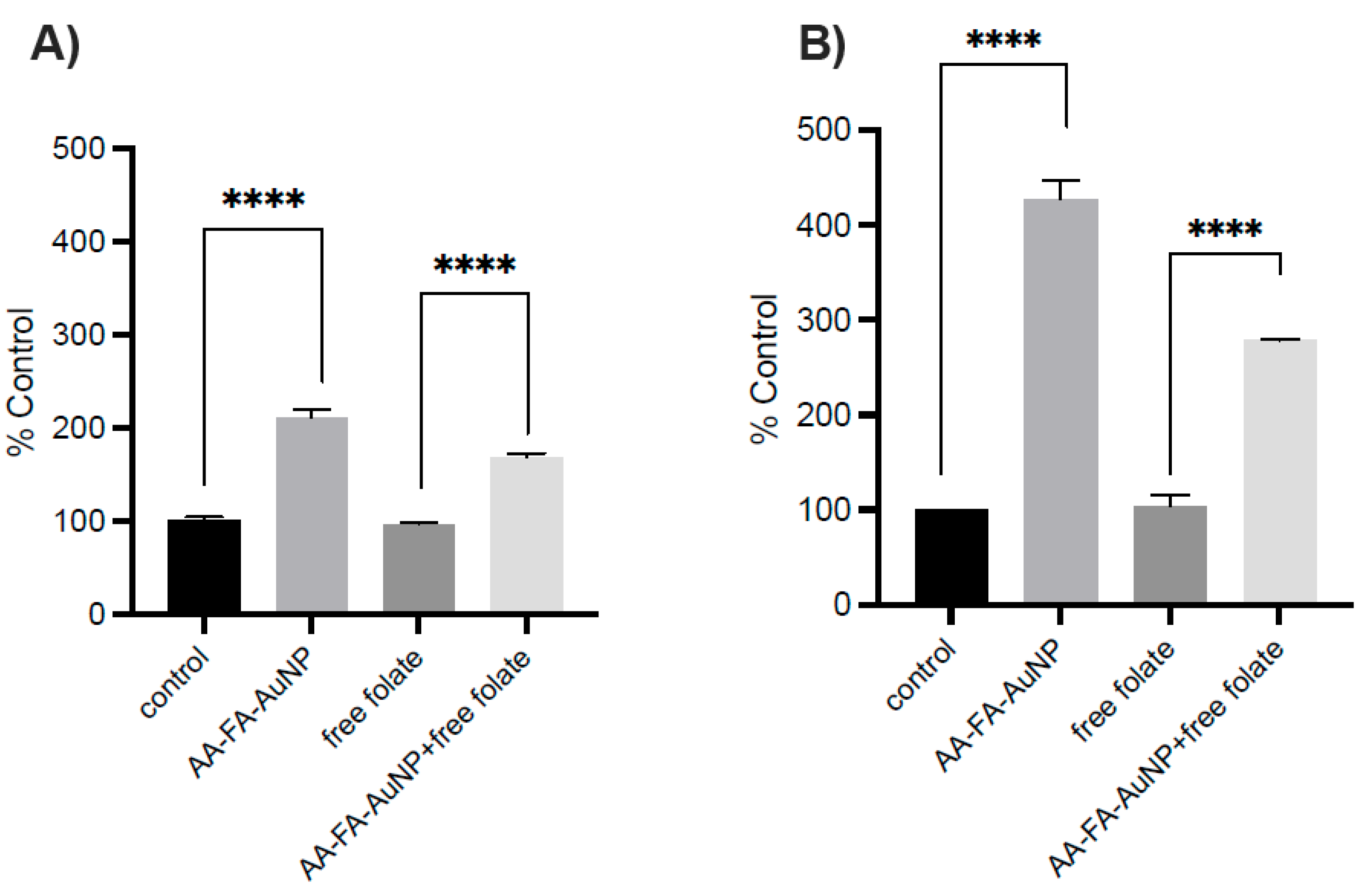
2.4. Cellular Uptake

Flow cytometry experiments upon incubation for 5 h were performed to quantify the potential for selective cell uptake of AA-FA-AuNP in FR+ cells compared with FR− cells. Uptake of the AA-FA-AuNP was analysed as mean AA fluorescence. Figure 4 shows that the HeLa cells internalised the AA-FA-AuNP better than A549. In the A549 cells, the mean increase in fluorescence was 210.7% compared to untreated controls; in contrast, in the HeLa cells, the mean increase in fluorescence was almost double at 427.6%. Since HeLa cells are about 40% bigger in size than A549 and their different size may affect the uptake, free folate (1 mM) was added to confirm a folate-receptor-mediated mechanism of uptake by competitive binding. Uptake of AA-FA-AuNP was significantly inhibited in both cell lines, but such inhibition was greater in HeLa cells (34.7%) compared to A549 cells (19.9%). These data further support the role of folate receptors in uptake but also indicate that other pathways may be involved in the uptake of particles in both cell lines. Cell viability experiments (5 h) confirmed no direct cytotoxic effect of particles with viability over 98% in all flow cytometry experiments, as confirmed by bioluminescent detection of any released adenylate kinase (Figure S8). In vitro studies with a larger number and variety of cell lines would confirm the therapeutic selectivity of AA-FA-AuNP.

3. Materials and Methods

Common chemicals and solvents (HPLC-grade or reagent-grade quality) were purchased from commercial sources and used without further purification. Deuterated solvents were purchased from Eurisotop (Saint-Aubin, France). 1H and 13C{1H} NMR spectra were carried out on a Bruker AV 400 or Bruker AV 600 NMR spectrometer (Billerica, MA, USA), and chemical shifts are reported in ppm and cited relative to SiMe4 and using the residual proton impurities in the solvents for 1H and 13C{1H} NMR spectroscopy. Peak multiplicities are abbreviated as follows: s = singlet, m = multiplet, d = doublet, t = triplet and dd = doublet of doublet.
The C, H and N analyses were performed with a Carlo Erba model EA 1108 microanalyzer (Milano, Italy), with EAGER 200 software. The combustion of the samples was carried out in the presence of V2O5 and MgO as additives. The duration of the analysis was 15 min.
Direct insertion probe–mass spectrometry (MS-DIP): Mass spectra were recorded on an Agilent Q-TOF 6550 hybrid spectrometer (Santa Clara, CA, USA) with JetStream electrospray + i-Funnel ionization source. Mass spectrometry data were acquired in the positive ionization mode. The isotopic distribution of the heaviest set of signals matched very closely that of the calculated ones for the formulation of the complex cation in every case.
The UV–Vis spectra were registered using a Perkin-Elmer Lambda 750 S spectrometer. Fluorescence spectra were registered at room temperature with a HORIBA Jobin–Yvon iHR320 spectrofluorometer (Kyoto, Japan). The instrument excitation and emission slits were both set at 5 nm.
All AuNP suspensions were stored in darkness at 4 °C and filtered (glass fibre syringe filter 1 µm) before being used/characterized. Deionised water was used in all syntheses. FT-IR spectra were recorded on a Thermo Nicolet Avatar 320 spectrophotometer (Thermo Scientific, Waltham, MA, USA) with an ATR attachment. UV–Vis measurements were carried out at room temperature in a Varian Cary 5000 UV–Vis spectrophotometer (Palo Alto, CA, USA) using 10 mm cuvettes. Steady-state spectra were recorded on a Photon Technology International (PTI, Edison, NJ, USA) QM-1 emission spectrometer. The hydrodynamic diameter and surface charge (ξ-potential) of the nanoparticles were measured using a Zetasizer Nano ZS (Malvern Instruments Ltd., Malvern, UK). Measurements were performed (12 cycles/run) in triplicate.
For AuNP size determination by transmission electron microscopy, a JEOL2100 TEM (Tokyo, Japan), 200 keV, and Gatan (Pleasanton, CA, USA) multiscan camera was used. Samples were mounted onto copper electron microscope grids (Formvar film). TEM images were acquired using Digital Micrograph 1.8 (Gatan, Pleasanton, CA, USA), and post-imaging analysis was performed using ImageJ (https://imagej.net/ij/).
Concentrations of particle solutions were first determined by ICP-MS, and their molar extinction coefficients were calculated: ε(FA-AuNP, 530 nm) = 7.4 × 10⁸ M−1 cm−1, ε(AA-FA-AuNP, 540 nm) = 12.7 × 10⁸ M−1 cm−1. For the ICP-MS analysis, samples were digested with aqua regia at 80 °C overnight then diluted to 2% aqua regia solutions and filtered. Gold standards (Fluka, Buchs, Switzerland) were prepared containing 2% aqua regia. Gold concentration was determined in an Agilent 7500CX ICP-MS (Pt simple cone in no-gas mode, Santa Clara, CA, USA). Analysis conditions were as follows: Ar flow 15 L/min, auxiliar gas 0.9 L/min, make-up gas 0.15 L/min, RF power 1550 W, RF matching 1.8 V, analogue HV 1750 V, pulse HV 1130 V, spray chamber temperature 15 °C and nebuliser pump 0.08 rps. An internal standard (50 ppb Er solution) was introduced into the sample flow through a T-piece. All readings were conducted in triplicate. Samples were also prepared in triplicate. Gold nanoparticle concentration was calculated using the size determined by TEM.

3.1. Synthesis

3.1.1. 1-Amine-5-(4,7-dioxa-1,10-dithiadecyl)anthracene-9,10-dione (AA)

A previously reported procedure was followed [19]. Briefly, 1-amino-5-chloroanthraquinone (1.43 g, 5 mmol) and 3,6-dioxy-2,2′-(Ethylenedioxy)diethanethiol (1.92 g, 10 mmol) were mixed with NaH (60% oil dispersion, 0.80 g, 20 mmol) in anhydrous THF (100 mL), and the mixture was stirred for 3 h at room temperature under Ar atmosphere. Then, the mixture was poured into ice-water (200 mL) and acidified carefully to pH 5 with HCl 1 M. The solution was extracted with CH2Cl2 (4 × 50 mL), and the organic solution was washed with water (2 × 50 mL) and dried over Na2SO4. The solvent was evaporated in vacuo, and the dark red residue was purified by column chromatography (silica gel, CH2Cl2: ethyl acetate, 98:2, v/v). Rf 0.3. Yield 50%. 1 NMR δ6.84 (NH2), 6.96 (a2, dd, 1H), 7.50 (a3, t, 1 H), 7.67–7.70 (a4, dd, 1 H; a7, t, 1H; a8, dd, 1H), 8.15 (a6, dd, 1 H). Elem. Anal. % calc. for C20H21NO4S2 (% found): N, 3.47 (2.96); C, 59.53 (59.61); H, 5.25 (6.19); S, 15.89 (17.25). ESI-MS(+), m/z: 426.3 (M + Na)+. IR (KBr pellet, cm−1): 3460 (O-H), 3344 (S-H), 2890m (N-H). Completely soluble in acetone, ethanol, methanol, CHCl3, CH2Cl2, THF and DMSO. Non-soluble in water. Luminescence in EtOH. λexc 480 nm, λem 590 nm.

3.1.2. FA-AuNP

The synthetic procedure described by Sun et al. [39] was modified as follows. A mixture containing 18 mL of a 0.8 mM solution of HAuCl4, 90 µL NaOH 1 M and 0.72 mL of sodium folate 1 mM (prepared by adding 21 μL NaOH 1 M to a 5 mL folic acid 1 mM solution) was stirred at room temperature for 30 min. Afterwards the mixture was made to react in a single-mode microwave reactor (CEM Instruments, Matthews, NC, USA) at 200 W for 5 min at 180 °C and left to cool slowly until room temperature. The solution was purified by dialysis (Spectra-Por® Float-A-Lyzer® G2, 5 mL, MWCO 8–10 kDa, Spectrum Laboratories, Inc., Rancho Dominguez, CA, USA) for two days against deionized water with NaOH (pH 10) and further centrifugation at 8000 rpm for 30 min and resuspension in NaOH 50 µM (pH 10). The final concentration (ICP-MS) was 0.14 nM. The SPR band was 530 nm.

3.1.3. AA-FA-AuNP

An ethanolic solution of AA (200 µM, 30 mL) was added to a 100 mL FA-AuNP suspension (0.14 nM) and left stirring overnight in darkness at r.t. The resulting clear solution was washed with chloroform to remove unbound AA molecules and afterwards dialysed against deionized water with NaOH (pH 10). Finally, it was centrifuged at 8000 rpm for 30 min and resuspended in NaOH 50 µM (pH 10). The final concentration (ICP-MS) was 0.1 nM. The SPR band was 543 nm.

3.2. DNA Binding Studies

Linear dichroism (LD) experiments were performed at 20 °C using a Chirascan-plus instrument by Applied Photophysics (Leatherhead, UK), a High Shear Couette Cell Accessory (CCA). LD titration was performed by adding increasing concentrations of AA-FA-AuNP from a stock solution in water to calf thymus (ct) DNA (100 µM in NaCl 10 mM—Tris HCl 1 mM, pH 7.5), keeping the DNA concentration constant. ct-DNA (Sigma-Aldrich, Burlington, MA, USA) concentration was determined by UV–Vis using the molar extinction coefficient ε258 = 6600 M−1 cm−1.

3.3. Cell Culture

A549 lung carcinoma cells (86012804) and HeLa cervix carcinoma cells (93021013) were maintained in Dulbecco’s modified Eagle’s medium (DMEM) supplemented with 10% v/v foetal calf serum, L-glutamine (2 mM), penicillin (100 μg/mL) and streptomycin (100 μg/mL). Cells were grown in a humidified incubator at 37 °C, 5% CO2 and 95% relative humidity to approximately 80% confluence and passaged using trypsin–EDTA either into new T75 flasks or well culture plates for experimentation as appropriate.
The source for both cell lines was the Health Protection Agency General Cell Culture Collection: https://www.culturecollections.org.uk/products/cell-cultures/general-cell-collection/ (accessed on 13 March 2025).

3.3.1. Flow Cytometry

Both cell lines were seeded in a 24-well culture plate at a density of 1 × 10⁵ cells/well and incubated overnight to allow attachment. Cell media were removed followed by washing with PBS and then replaced with fresh cell media, which contained 1 mM folic acid for the FA(+) samples. After one hour of incubation, cells were treated with 0.14 nM AA-FA-AuNP and incubated for further 5 h. Controls with and without folic acid were also assayed. Once the incubation period was finished, the cells were washed with PBS, trypsinised and incubated for 10 min at 37 °C. Next, 1 mL PBS was added, and cells were transferred to microcentrifuge tubes. Cells were pelleted by centrifugation at 1000× g (5 min), the supernatant was removed, and the cell pellet was re-suspended in PBS and transferred to flow cytometry tubes for flow cytometry analysis (FACScalibur, BD Biosciences, Franklin Lakes, NJ, USA). For each treatment, the green fluorescence (530/30 nm bandpass filter) of 10,000 cells was quantified with untreated cells used as a blank to control for background fluorescence. Floreada.io software (https://floreada.io) was used to analyse the results and calculate the mean fluorescence of each population of cells. Note: The nanoparticle suspension was brought to the assay concentrations with DMEM medium.

3.3.2. Cell Viability (Release of Adenylate Kinase)

Once the treatment period of samples used for flow cytometry measurements was over, aliquots of 200 µL of their cell media were taken and stored at −20 °C for less than one week to perform an adenylase kinase assay. These aliquots were incubated with lysis reagent (ToxiLight™ Non-destructive Cytotoxicity BioAssay Kit, Lonza, Basel, Switzerland) for 20 min, according to the kit protocol. Afterwards, bioluminescence was recorded in a plate reader at 20 °C (Tecan Infinite F200 Pro (Tecan, Männedorf, Switzerland)). Three replicates were carried out per cell line.

3.3.3. MTT Cell Proliferation Assay

A549 and HeLa cells at the logarithmic growth phase were seeded in a 96-well culture plate at densities of 5 × 10³ cells/mL, respectively, and incubated overnight to allow attachment. Afterwards, 100 μL of medium containing sample was added at different concentrations (four replicates per sample), and the plates were incubated at 37 °C in 5% CO2 and 95% relative humidity for 72 h. The cells were thoroughly washed with PBS before adding twenty microliters of thiazolyl blue tetrazolium bromide (5 mg/mL). The cells were further incubated for 2 h. The medium was carefully removed by aspiration and 200 µL of DMSO added to dissolve the purple crystals. Absorbance was measured using a 96-well plate reader set at 590 nm. Non-treated cells and cisplatin-treated cells were used as negative and positive controls, respectively. Four independent measurements were carried out.

4. Conclusions

An easy two-step procedure has been developed to obtain stable and monodisperse folate-capped AuNPs that are able to deliver anthraquinones selectively to cells overexpressing folate receptors. The new AA-FA-AuNP conjugates were highly toxic against two tumour cell lines, lung carcinoma A549 and cervical carcinoma HeLa, and showed significant in vitro targeting effects for FR+ HeLa cells. Moreover, they exhibited fluorescence and thus could be potentially used as tumour markers. This work further demonstrates, as reported earlier, that the inclusion of an anthraquinone derivative on a nanoparticle’s surface translates the nucleobase-intercalating behaviour of the anthraquinone to the nanoparticle. Acting as DNA binders, the resulting nanoparticle is no longer an inert carrier but an active component that may add to the overall bioactivity of the conjugate.
In line with other reported nanoconjugates bearing another anthraquinone derivative (viz. doxorubicin), this work highlights the potential of folate-receptor-targeted nanoparticles in enhancing the delivery and efficacy of chemotherapeutic agents in FR-overexpressing cancer cells [45,46,47]. Hence, we anticipate that this straightforward synthetic procedure will accelerate future development of folate-targeted conjugates bearing highly active anthraquinone-derived drugs. Likewise, metal-binding substituents such as the amino group present in the herein-described anthraquinone are potential donor atoms for further attachment of cytotoxic metal complexes to enhance their anticancer activity or add new properties.

Supplementary Materials

The following supporting information can be downloaded at: https://www.mdpi.com/article/10.3390/inorganics13030087/s1, Figure S1: FTIR spectra of AA, FA, FA-AuNP and AA-FA-AuNP; Figure S2: Excitation spectrum and emission spectrum of AA-FA-AuNP. Comparison with the emission of FA-AuNP at the same excitation wavelength and concentration; Figure S3: Intensity-weighted distributions of the hydrodynamic size of non-filtered FA-AuNP (a) and AA-FA-AuNP (b); Figure S4: Zeta-potential curves of FA-AuNP (a) and AA-FA-AuNP (b); Figure S5: Excitation and emission spectra of the previously reported GNP-lip-AA and the herein described AA-FA-AuNP, at 0.3 nM nanoparticle concentration in both cases. GNP-lip-AA display 20-fold lower intense emission of AA; Figure S6: UV-Vis spectra of AA-FA-AuNP (0.5 nM) over time in cell media DMEM. The nanoparticles were stable for the maximum period used in cell studies (72 h) at 0.5 nM concentration, which is higher than the concentration used in tests; Figure S7: Growth vs. concentration curves of cisplatin and AA-FA-AuNP incubated with A549 (a) and HeLa cells (b) for 72 h; Figure S8: Cell viability (adenylate kinase assay) in A549 and HeLa cells after treatment with 0.14 nM AA-FA-AuNP in absence and in presence of 1 mM free folic acid, and the corresponding controls.

Author Contributions

Conceptualization, A.B.C. and M.J.H.; methodology and investigation, A.B.C.; formal analysis, A.B.C., M.J.H. and N.J.H.; writing—original draft preparation, A.B.C.; writing—review and editing, A.B.C., N.J.H. and M.J.H.; supervision, N.J.H. and M.J.H. All authors have read and agreed to the published version of the manuscript.

Funding

This research was funded by the University of Birmingham, grant number EP/L016346/1. The postdoctoral fellowship of A.B.C. was funded by the University of Granada (Plan Propio Programme).

Informed Consent Statement

Not applicable.

Data Availability Statement

Data is contained within the article or supplementary material.

Acknowledgments

We thank Zoe Pikramenou for assistance and advice with gold nanoparticles and the technical support of Ashleigh Freer.

Conflicts of Interest

The authors declare no conflicts of interest.

References

  1. Khan, A.; Ezati, P.; Rhim, J.W. Alizarin: Prospects and sustainability for food safety and quality monitoring applications. Colloids Surf. B Biointerfaces 2023, 223, 113169. [Google Scholar] [CrossRef] [PubMed]
  2. Malik, E.M.; Muller, C.E. Anthraquinones As Pharmacological Tools and Drugs. Med. Res. Rev. 2016, 36, 705–748. [Google Scholar] [CrossRef]
  3. Ruan, W.; Mao, J.; Chen, Q. Redox flow batteries toward more soluble anthraquinone derivatives. Curr. Opin. Electrochem. 2021, 29, 100748. [Google Scholar] [CrossRef]
  4. Qun, T.; Zhou, T.; Hao, J.; Wang, C.; Zhang, K.; Xu, J.; Wang, X.; Zhou, W. Antibacterial activities of anthraquinones: Structure-activity relationships and action mechanisms. RSC Med. Chem. 2023, 14, 1446–1471. [Google Scholar] [CrossRef] [PubMed]
  5. Winter, R.W.; Cornell, K.A.; Johnson, L.L.; Ignatushchenko, M.; Hinrichs, D.J.; Riscoe, M.K. Potentiation of the antimalarial agent rufigallol. Antimicrob. Agents Chemother. 1996, 40, 1408–1411. [Google Scholar] [CrossRef]
  6. Patel, O.P.S.; Beteck, R.M.; Legoabe, L.J. Antimalarial application of quinones: A recent update. Eur. J. Med. Chem. 2021, 210, 113084. [Google Scholar] [CrossRef] [PubMed]
  7. Choi, R.J.; Ngoc, T.M.; Bae, K.; Cho, H.J.; Kim, D.D.; Chun, J.; Khan, S.; Kim, Y.S. Anti-inflammatory properties of anthraquinones and their relationship with the regulation of P-glycoprotein function and expression. Eur. J. Pharm. Sci. 2013, 48, 272–281. [Google Scholar] [CrossRef]
  8. Garcia-Vilas, J.A.; Pino-Angeles, A.; Martinez-Poveda, B.; Quesada, A.R.; Medina, M.A. The noni anthraquinone damnacanthal is a multi-kinase inhibitor with potent anti-angiogenic effects. Cancer Lett. 2017, 385, 1–11. [Google Scholar] [CrossRef]
  9. Mugas, M.L.; Calvo, G.; Marioni, J.; Cespedes, M.; Martinez, F.; Vanzulli, S.; Saenz, D.; Di Venosa, G.; Nunez Montoya, S.; Casas, A. Photosensitization of a subcutaneous tumour by the natural anthraquinone parietin and blue light. Sci. Rep. 2021, 11, 23820. [Google Scholar] [CrossRef]
  10. Huang, Q.; Lu, G.; Shen, H.M.; Chung, M.C.; Ong, C.N. Anti-cancer properties of anthraquinones from rhubarb. Med. Res. Rev. 2007, 27, 609–630. [Google Scholar] [CrossRef]
  11. Bryce, N.S.; Zhang, J.Z.; Whan, R.M.; Yamamoto, N.; Hambley, T.W. Accumulation of an anthraquinone and its platinum complexes in cancer cell spheroids: The effect of charge on drug distribution in solid tumour models. Chem. Commun. 2009, 2673–2675. [Google Scholar] [CrossRef] [PubMed]
  12. Campora, M.; Canale, C.; Gatta, E.; Tasso, B.; Laurini, E.; Relini, A.; Pricl, S.; Catto, M.; Tonelli, M. Multitarget Biological Profiling of New Naphthoquinone and Anthraquinone-Based Derivatives for the Treatment of Alzheimer’s Disease. ACS Chem. Neurosci. 2021, 12, 447–461. [Google Scholar] [CrossRef]
  13. Fiala, J.; Roach, T.; Holzinger, A.; Husiev, Y.; Delueg, L.; Hammerle, F.; Armengol, E.S.; Schobel, H.; Bonnet, S.; Laffleur, F.; et al. The Light-activated Effect of Natural Anthraquinone Parietin against Candida auris and Other Fungal Priority Pathogens. Planta Medica 2024, 90, 588–594. [Google Scholar] [CrossRef] [PubMed]
  14. Malik, M.S.; Alsantali, R.I.; Jassas, R.S.; Alsimaree, A.A.; Syed, R.; Alsharif, M.A.; Kalpana, K.; Morad, M.; Althagafi, I.I.; Ahmed, S.A. Journey of anthraquinones as anticancer agents—A systematic review of recent literature. RSC Adv. 2021, 11, 35806–35827. [Google Scholar] [CrossRef] [PubMed]
  15. Cheng, L.; Chen, Q.; Pi, R.; Chen, J. A research update on the therapeutic potential of rhein and its derivatives. Eur. J. Pharmacol. 2021, 899, 173908. [Google Scholar] [CrossRef]
  16. Jeevitha, D.; Amarnath, K. Chitosan/PLA nanoparticles as a novel carrier for the delivery of anthraquinone: Synthesis, characterization and in vitro cytotoxicity evaluation. Colloids Surf. B Biointerfaces 2013, 101, 126–134. [Google Scholar] [CrossRef]
  17. Angheluta, A.; Bacaita, E.; Raduv, V.; Agop, M.; Ignat, L.; Uritu, C.; Maier, S.; Pinteala, M. Mathematical modelling of the release profile of anthraquinone-derived drugs encapsulated on magnetite nanoparticles. Rev. Roum. De Chim. 2013, 58, 217–221. [Google Scholar]
  18. Dreaden, E.C.; Alkilany, A.M.; Huang, X.; Murphy, C.J.; El-Sayed, M.A. The golden age: Gold nanoparticles for biomedicine. Chem. Soc. Rev. 2012, 41, 2740–2779. [Google Scholar] [CrossRef]
  19. Caballero, A.B.; Cardo, L.; Claire, S.; Craig, J.S.; Hodges, N.J.; Vladyka, A.; Albrecht, T.; Rochford, L.A.; Pikramenou, Z.; Hannon, M.J. Assisted delivery of anti-tumour platinum drugs using DNA-coiling gold nanoparticles bearing lumophores and intercalators: Towards a new generation of multimodal nanocarriers with enhanced action. Chem. Sci. 2019, 10, 9244–9256. [Google Scholar] [CrossRef]
  20. King, S.M.; Claire, S.; Teixeira, R.I.; Dosumu, A.N.; Carrod, A.J.; Dehghani, H.; Hannon, M.J.; Ward, A.D.; Bicknell, R.; Botchway, S.W.; et al. Iridium Nanoparticles for Multichannel Luminescence Lifetime Imaging, Mapping Localization in Live Cancer Cells. J. Am. Chem. Soc. 2018, 140, 10242–10249. [Google Scholar] [CrossRef]
  21. Dosumu, A.N.; Claire, S.; Watson, L.S.; Girio, P.M.; Osborne, S.A.M.; Pikramenou, Z.; Hodges, N.J. Quantification by Luminescence Tracking of Red Emissive Gold Nanoparticles in Cells. JACS Au 2021, 1, 174–186. [Google Scholar] [CrossRef] [PubMed]
  22. Tsai, S.W.; Liaw, J.W.; Hsu, F.Y.; Chen, Y.Y.; Lyu, M.J.; Yeh, M.H. Surface-Modified Gold Nanoparticles with Folic Acid as Optical Probes for Cellular Imaging. Sensors 2008, 8, 6660–6673. [Google Scholar] [CrossRef]
  23. Rosyidah, A.l.; Kerdtoob, S.; Yudhistyra, W.I.; Munfadlila, A.W. Gold nanoparticle-based drug nanocarriers as a targeted drug delivery system platform for cancer therapeutics: A systematic review. Gold Bull. 2023, 56, 121–134. [Google Scholar] [CrossRef]
  24. Krpetic, Z.; Scari, G.; Caneva, E.; Speranza, G.; Porta, F. Gold nanoparticles prepared using cape aloe active components. Langmuir 2009, 25, 7217–7221. [Google Scholar] [CrossRef]
  25. Fernandez, M.; Javaid, F.; Chudasama, V. Advances in targeting the folate receptor in the treatment/imaging of cancers. Chem. Sci. 2018, 9, 790–810. [Google Scholar] [CrossRef] [PubMed]
  26. Scaranti, M.; Cojocaru, E.; Banerjee, S.; Banerji, U. Exploiting the folate receptor alpha in oncology. Nat. Rev. Clin. Oncol. 2020, 17, 349–359. [Google Scholar] [CrossRef]
  27. Samanta, S.; Arivarasan, V.K. A multifunctional drug delivery cargo: Folate-driven gold nanoparticle-vitamin B complex for next-generation drug delivery. Mater. Lett. 2024, 355, 135425. [Google Scholar] [CrossRef]
  28. Li, G.; Li, D.; Zhang, L.; Zhai, J.; Wang, E. One-step synthesis of folic acid protected gold nanoparticles and their receptor-mediated intracellular uptake. Chemistry 2009, 15, 9868–9873. [Google Scholar] [CrossRef]
  29. Guerrero-Florez, V.; Mendez-Sanchez, S.C.; Patron-Soberano, O.A.; Rodriguez-Gonzalez, V.; Blach, D.; Martinez, O.F. Gold nanoparticle-mediated generation of reactive oxygen species during plasmonic photothermal therapy: A comparative study for different particle sizes, shapes, and surface conjugations. J. Mater. Chem. B 2020, 8, 2862–2875. [Google Scholar] [CrossRef]
  30. Castillo, J.; Bertel, L.; Páez-Mozo, E.; Martínez, F. Photochemical Synthesis of the Bioconjugate Folic Acid-Gold Nanoparticles. Nanomater. Nanotechnol. 2013, 3, 18. [Google Scholar] [CrossRef]
  31. Li, X.; Zhou, H.; Yang, L.; Du, G.; Pai-Panandiker, A.S.; Huang, X.; Yan, B. Enhancement of cell recognition in vitro by dual-ligand cancer targeting gold nanoparticles. Biomaterials 2011, 32, 2540–2545. [Google Scholar] [CrossRef] [PubMed]
  32. Zhang, Z.; Jia, J.; Lai, Y.; Ma, Y.; Weng, J.; Sun, L. Conjugating folic acid to gold nanoparticles through glutathione for targeting and detecting cancer cells. Bioorg. Med. Chem. 2010, 18, 5528–5534. [Google Scholar] [CrossRef] [PubMed]
  33. Mansoori, G.A.; Brandenburg, K.S.; Shakeri-Zadeh, A. A comparative study of two folate-conjugated gold nanoparticles for cancer nanotechnology applications. Cancers 2010, 2, 1911–1928. [Google Scholar] [CrossRef]
  34. Yucel, O.; Sengelen, A.; Emik, S.; Onay-Ucar, E.; Arda, N.; Gurdag, G. Folic acid-modified methotrexate-conjugated gold nanoparticles as nano-sized trojans for drug delivery to folate receptor-positive cancer cells. Nanotechnology 2020, 31, 355101. [Google Scholar] [CrossRef]
  35. Daniele, R.; Brazzale, C.; Arpac, B.; Tognetti, F.; Pesce, C.; Malfanti, A.; Sayers, E.; Mastrotto, F.; Jones, A.T.; Salmaso, S.; et al. Influence of Folate-Targeted Gold Nanoparticles on Subcellular Localization and Distribution into Lysosomes. Pharmaceutics 2023, 15, 864. [Google Scholar] [CrossRef]
  36. Khoshgard, K.; Hashemi, B.; Arbabi, A.; Rasaee, M.J.; Soleimani, M. Radiosensitization effect of folate-conjugated gold nanoparticles on HeLa cancer cells under orthovoltage superficial radiotherapy techniques. Phys. Med. Biol. 2014, 59, 2249–2263. [Google Scholar] [CrossRef]
  37. Beik, J.; Jafariyan, M.; Montazerabadi, A.; Ghadimi-Daresajini, A.; Tarighi, P.; Mahmoudabadi, A.; Ghaznavi, H.; Shakeri-Zadeh, A. The benefits of folic acid-modified gold nanoparticles in CT-based molecular imaging: Radiation dose reduction and image contrast enhancement. Artif. Cells Nanomed. Biotechnol. 2018, 46, 1993–2001. [Google Scholar] [CrossRef]
  38. Uhlen, M.; Fagerberg, L.; Hallstrom, B.M.; Lindskog, C.; Oksvold, P.; Mardinoglu, A.; Sivertsson, A.; Kampf, C.; Sjostedt, E.; Asplund, A.; et al. Proteomics. Tissue-based map of the human proteome. Science 2015, 347, 1260419. [Google Scholar] [CrossRef] [PubMed]
  39. Zhang, Z.; Jia, J.; Ma, Y.; Weng, J.; Sun, Y.; Sun, L. Microwave-assisted one-step rapid synthesis of folic acid modified gold nanoparticles for cancer cell targeting and detection. MedChemComm 2011, 2, 1079. [Google Scholar] [CrossRef]
  40. Mudalige, T.; Qu, H.; Van Haute, D.; Ansar, S.M.; Paredes, A.; Ingle, T. Chapter 11—Characterization of Nanomaterials: Tools and Challenges. In Nanomaterials for Food Applications; López Rubio, A., Fabra Rovira, M.J., Martínez Sanz, M., Gómez-Mascaraque, L.G., Eds.; Elsevier: Amsterdam, The Netherlands, 2019; pp. 313–353. [Google Scholar]
  41. Hwa Kim, S.; Hoon Jeong, J.; Chul Cho, K.; Wan Kim, S.; Gwan Park, T. Target-specific gene silencing by siRNA plasmid DNA complexed with folate-modified poly(ethylenimine). J. Control Release 2005, 104, 223–232. [Google Scholar] [CrossRef]
  42. Stepanov, A.V.; Xie, J.; Zhu, Q.; Shen, Z.; Su, W.; Kuai, L.; Soll, R.; Rader, C.; Shaver, G.; Douthit, L.; et al. Control of the antitumour activity and specificity of CAR T cells via organic adapters covalently tethering the CAR to tumour cells. Nat. Biomed. Eng. 2024, 8, 529–543. [Google Scholar] [CrossRef] [PubMed]
  43. Ren, D.; Kratz, F.; Wang, S.W. Engineered drug-protein nanoparticle complexes for folate receptor targeting. Biochem. Eng. J. 2014, 89, 33–41. [Google Scholar] [CrossRef] [PubMed]
  44. Pandey, S.; Oza, G.; Mewada, A.; Shah, R.; Thakur, M.; Sharon, M. Folic acid mediated synaphic delivery of doxorubicin using biogenic gold nanoparticles anchored to biological linkers. J. Mater. Chem. B 2013, 1, 1361. [Google Scholar] [CrossRef] [PubMed]
  45. Vinothini, K.; Dhilip Kumar, S.S.; Abrahamse, H.; Rajan, M. Enhanced Doxorubicin Delivery in Folate-Overexpressed Breast Cancer Cells Using Mesoporous Carbon Nanospheres. ACS Omega 2021, 6, 34532–34545. [Google Scholar] [CrossRef]
  46. Fernandes, R.S.; Arribada, R.G.; Silva, J.O.; Silva-Cunha, A.; Townsend, D.M.; Ferreira, L.A.M.; Barros, A.L.B. In Vitro and In Vivo Effect of pH-Sensitive PLGA-TPGS-Based Hybrid Nanoparticles Loaded with Doxorubicin for Breast Cancer Therapy. Pharmaceutics 2022, 14, 2394. [Google Scholar] [CrossRef]
  47. Chen, H.; Li, L.; Cui, S.; Mahounga, D.; Zhang, J.; Gu, Y. Folate conjugated CdHgTe quantum dots with high targeting affinity and sensitivity for in vivo early tumor diagnosis. J. Fluoresc. 2011, 21, 793–801. [Google Scholar] [CrossRef]
Scheme 1. Synthesis of folate-coated gold nanoparticles (FA-AuNP) and conjugation of 1-amino-anthraquinone (AA) to their surface to obtain folate-coated gold nanoparticles (AA-FA-AuNP).
Inorganics 13 00087 sch001
Figure 1. (a) UV–Vis spectra of FA-AuNP and AA-FA-AuNP at 0.1 nM; (b) excitation (dashed lines) and emission (solid lines) spectra of AA and FA moieties in AA-FA-AuNP. NaOH 10 μM (pH 10).
Inorganics 13 00087 g001
Figure 2. Size distribution histograms (left) and transmission electron micrographs (right) of FA-AuNP (A) and AA-FA-AuNP (B).
Inorganics 13 00087 g002
Figure 3. Linear dichroism spectra of ct-DNA (100 μM) with increasing concentrations of AA-FA-AuNP. Tris–HCl 1 mM, NaCl 10 mM, pH 7.5.
Inorganics 13 00087 g003
Figure 4. Cellular uptake of AA-FA-AuNP (0.14 nM) in the presence and absence of free folate (1 mM) quantified by flow cytometry, as described in the Materials and Methods. (A) FR− A549 cells and (B) FR+ HeLa cells. **** Significantly different from appropriate control as assessed by a 1-way ANOVA and a post hoc Tukey T-test. The results represent the mean of three independent experiments +/− SEM.
Inorganics 13 00087 g004
Disclaimer/Publisher’s Note: The statements, opinions and data contained in all publications are solely those of the individual author(s) and contributor(s) and not of MDPI and/or the editor(s). MDPI and/or the editor(s) disclaim responsibility for any injury to people or property resulting from any ideas, methods, instructions or products referred to in the content.

