Article

Comparison of the Probiotic Potential between Lactiplantibacillus plantarum Isolated from Kimchi and Standard Probiotic Strains Isolated from Different Sources

1
Microbiology and Functionality Research Group, World Institute of Kimchi, Gwangju 61755, Korea
2
Department of Food Science and Biotechnology of Animal Resources, Konkuk University, Seoul 05029, Korea
3
Research Institute, Korea Prime Pharm. Co., Ltd., Gwangju 61473, Korea
4
Department of Oral Biochemistry, College of Dentistry, Chosun University, Gwangju 61452, Korea
*
Author to whom correspondence should be addressed.
Foods 2021, 10(9), 2125; https://doi.org/10.3390/foods10092125
Submission received: 23 June 2021 / Revised: 6 September 2021 / Accepted: 6 September 2021 / Published: 8 September 2021

Abstract

:
In the present study, the properties of the Lactiplantibacillus (Lpb.) plantarum WiKim0112 isolated from kimchi were evaluated by comparing its probiotic properties to those of Lpb. plantarum WCFS1 and KACC 11451 isolated from different sources. In both pH 2 and 3, media containing pepsin, Wikim0112, and WCFS1 showed higher cell viability than KACC11451. Viability of all Lpb. plantarum strains in a medium containing pancreatin and bile salt oxgall was significantly decreased compared to the control. WCFS1 showed the highest thermotolerance, followed by Wikim0112 and KACC11451. Wikim0112 showed a similar level of antibacterial activity to WCFS1 and exhibited an overall higher antibacterial activity than KACC11451 against six pathogens. All Lpb. plantatum strains showed high antioxidant activities in SOD, DPPH, and ABTS assays, especially Wikim0112 and WCFS1 exhibited a higher antioxidant activity than KACC11451. All Lpb. plantarum strains showed approximately 60–62% adhesion rates to Caco-2 cells. Moreover, in LPS-stimulated Caco-2 cells, all Lpb. plantarum strains significantly decreased the mRNA expression of pro-inflammatory cytokines (i.e., IL-1β, IL-6, and TNF-α); Wikim0112 significantly increased the mRNA expression of IL-4 and IFN-γ. Wikim0112 was resistant to streptomycin and vancomycin, whereas WCFS1 and KACC11451 were resistant to four (clindamycin, ciprofloxacin, tetracycline, and vancomycin) and three (ciprofloxacin, tetracycline, and vancomycin) antibiotics, respectively. These results, taken together, indicated that compared to Lpb. plantarum strains isolated from different sources, Wikim0112 showed desirable probiotic properties, suggesting its potential applications in the food and pharmaceutical industries.

1. Introduction

Kimchi is a traditional Korean fermented vegetable food that is prepared by fermenting salted vegetables and various ingredients, including red pepper powder, garlic, and ginger using lactic acid bacteria (LAB). Among the LAB used in kimchi production, members of the genera Leuconostoc, Weissella, and Lactobacillus are known to play a critical role in kimchi fermentation [1]. Kimchi-derived LAB have been reported to have beneficial effects on human health. According to a previous study, Lactobacillus acidophilus KFRI342 isolated from kimchi exhibited probiotic properties suppressing human colon cancer cell growth [2]. Levilactobacillus brevis KU15147 isolated from kimchi showed higher antioxidant activity than that of Lactobacillus rhamnosus GG used as a reference strain; the KU15147 strain also showed immune-enhancing effects via nitric oxide (NO) production and cytokine induction (i.e., inducible NO synthase, and tumor necrosis factor-α (TNF- α)) [3].
Lactiplantibacillus (Lpb.) plantarum, which is abundant in kimchi, represents an ideal candidate for developing probiotics owing to remarkable adhesion and adaptation capabilities in the gastrointestinal (GI) tract and beneficial effects such as antimicrobial, anti-inflammatory, and antioxidant activities [4]. Lpb. plantarum strains have been widely used in the food industry as starter cultures in the fermentation of foods and beverages as they provide organoleptic properties, texture, and flavor [5]. Thus, they are isolated from various sources such as fermented vegetables [6,7,8], milk products [9,10], and meat [11,12], and their functional characteristics, including probiotic potential, have been evaluated.
However, single species have been found to show differences in characteristics based on the isolation source [13,14]. Therefore, in this study Lpb. plantarum WiKim0112 isolated from Korean radish water kimchi was evaluated by comparing its probiotic properties to two Lpb. plantarum strains isolated from different sources.

2. Materials and Methods

2.1. Bacterial Strains and Growth Conditions

Two Lpb. plantarum strains showing desirable probiotic features included Lpb. plantarum WCFS1 ATCC BAA-793 isolated from human saliva and Lpb. plantarum KACC11451 isolated from pickles that were obtained from the American Type Culture Collection (ATCC) and Korean Agricultural Culture Collection (KACC), respectively. Lpb. plantarum 8 species (WiKim0112, DK4, YB5, JC8, YP2, PC2, TS3, LB10), Lacticaseibacillus casei 5 species (KCKM0991, DO1, T18, PC2, LB2), Lacticaseibacillus paracasei 5 species (KCKM1011, L01, LB17, FS31, TS6), and Limosilactobacillus fermentum 2 species (KCKM0729, PC5) isolated from kimchi and obtained from the Korean Collection for Kimchi Microorganisms (KCMC) were cultured anaerobically in de Man, Rogosa, and Sharpe (MRS) broth for 48 h at 30 °C. Six bacterial pathogens obtained from ATCC and the Korean Culture Center of Microorganisms (KCCM) included: Escherichia coli O157:H7 (ATCC 35150), Listeria monocytogenes (ATCC 15313), Salmonella choleraesuis (KCCM40763), Shigella boydii (KCCM41649), Staphylococcus aureus (ATCC25923), and Yersinia enterocolitica (KCCM41657). All strains were cultured in tryptic soy broth (TSB; Difco, Sparks, MD, USA) for 24 h at 30 °C and stored in a TSB medium containing 20% glycerol at −70 °C.

2.2. Cell Culture

The monocyte/macrophage cell line RAW264.7 and the human colorectal adenocarcinoma cell line Caco-2 were obtained from the Korean Cell Line Bank (Jongno, Seoul, Korea). The cells were maintained in Dulbecco’s modified Eagle’s medium (DMEM; Welgene Inc., Gyeongsan, Korea) containing 10% fetal bovine serum (Welgene Inc., Gyeongsan, Korea) and antibiotics at 37 °C in a humidified atmosphere containing 5% CO2. The medium was replaced with fresh medium every 2–3 d. The cells were digested using 0.05% trypsin/0.53 mM ethylenediaminetetraacetic acid (Welgene Inc., Gyeongsan, Korea) when they reached 80% confluency in T-25 flasks. The cells were then seeded in 6-well plates.

2.3. Selection of Bacteria Showing Anti-Inflammatory Activity

RAW264.7 cells (1 × 10⁵ cells/mL) were seeded in 6-well plates, incubated overnight, and then exposed to lipopolysaccharide (LPS; 100 ng/mL) for 24 h. Culture supernatants of isolates were collected via centrifugation at 3800× g (4 °C) for 15 min. The levels of TNF-α, interleukin (IL)-6, and IL-1β secreted into the culture supernatants were determined in duplicate using respective Quantikine enzyme-linked immunosorbent assay (ELISA) kits (R&D Systems Inc., Minneapolis, MN, USA) according to the manufacturer’s instructions. The concentration of NO in the cell culture supernatant was determined using the Griess reagent (Sigma-Aldrich, St. Louis, MO, USA). The culture supernatant (50 μL) was mixed with an equal volume of Griess reagent and incubated for 15 min at room temperature (25–28 °C). Absorbance (540 nm) was measured using an ELISA microplate reader Infinite 200 pro (Tecan Austria GmbH, Grödig, Austria).

2.4. Identification of the Strains

Chromosomal DNA was extracted from bacteria using a commercial QIAamp DNA Mini Kit (Qiagen, Hilden, Germany) according to the manufacturer’s instructions. The conserved region of the 16S rRNA gene was amplified with the universal primer pairs (FP: (27F) 5′-AGAGTTTGATCCTGGCTCAG-3′ and RP: (1492R) 5′-GGTTACCTTGTTACGACTT-3′) using an Applied biosystems thermal cycler (Thermo Fisher Scientific, Pittsburgh, PA, USA). Polymerase chain reaction (PCR) amplification was performed in 20 μL of final reaction volume containing 18 μL of PCR master mix and 2 μL of DNA sample. The PCR reaction conditions were as follows: initial denaturation for 4 min at 94 °C, followed by 35 cycles of denaturation for 1 min at 94 °C, primer annealing for 1 min at 57 °C, extension for 1 min at 72 °C, and a final elongation for 10 min at 72 °C. The amplified PCR products were sequenced using ABI 3730XL Genetic Analyzer (Applied Biosystems, Waltham, MA, USA). The assembled 16S rRNA gene sequence was identified using the EZBioCloud database (www.ezbiocloud.net/eztaxon, accessed on 10 May 2021) and compared to closely related strains.

2.5. Tolerance of GI Environment and Heat

Tolerance of the Lpb. plantarum strains to the GI environment was determined as previously described by Ranadheera et al. (2012) [15], with certain modifications. MRS broth was added with or without 0.3% (w/v) pepsin (Sigma Aldrich, St. Louis, MO, USA), and the pH was adjusted to 2, 3, or 6.5 (control) using 1 N HCl to evaluate gastric juice tolerance. MRS broth was supplemented with or without (control) 0.1% (w/v) pancreatin (Sigma Aldrich, St. Louis, MO, USA) and 0.3% (w/v) bile salts (Sigma Aldrich, St. Louis, MO, USA), and the pH was adjusted to 7.5 using1N NaOH to evaluate small intestinal juice tolerance. Overnight cultured Lpb. plantarum strains were inoculated into simulated gastric and small intestinal juice solutions at a density of 1 × 10⁷ colony forming units (CFU)/mL and then incubated at 37 °C for 4 h and 24 h, respectively. To assess the thermotolerance of the Lpb. plantarum strains, the inoculated MRS broths (1 × 10⁷ CFU/mL) were incubated at 50 °C for 1 h. After incubation, the MRS broths containing bacteria were serially diluted and spread onto MRS agar plates, followed by incubation at 30 °C for 48 h. The number of colonies formed on the MRS agar plates was counted using the plate counting method. For all tolerance tests, the bacterial survival was calculated using the following formula:
Survival (%) = (treatment group CFU/mL)/(control group CFU/mL) × 100

2.6. Antibacterial Activity

The antibacterial activity was evaluated using the well diffusion method. Soft tryptic soy agar (TSA) was seeded with pathogenic bacteria (1 × 10⁵ CFU/mL). Wells of 8 mm in diameter were created on the TSA agar plates using a sterilized cork borer. Culture supernatants were prepared via centrifugation (3800× g, 4 °C, 15 min) and used as an antagonistic substance. Culture supernatants 100 μL were poured into the wells and subsequently incubated at 30 °C for 24 h. The diameter of the zone of growth inhibition was measured in millimeters to evaluate the antagonistic effects. MRS broth was used as the negative control.

2.7. Antioxidant Activity

The antioxidative activity of the Lpb. plantarum strains was estimated by superoxide dismutase (SOD), 2,2-diphenyl-1-picryl-hydrazyl (DPPH), and 2,2′-azino-bis(3-ethylbenzothiazoline-6-sulphonic acid) (ABTS) assays. The Lpb. plantarum strains were cultured in a minimal medium (1% (w/v) glucose containing 0.5% (w/v) yeast extract) at 30 °C for 48 h. After centrifugation (8000× g, 15 min), the supernatant was used as a sample to measure the antioxidant activity.
The SOD assay was performed according to the manual of the Amplite Colorimetric SOD Assay Kit (AAT Bioquest, Sunnyvale, CA, USA). In the assay, xanthine is converted to hydrogen peroxide (H2O2) radical ions, uric acid, and H2O2 by xanthine oxidase. The degree of oxidation of xanthine, which catalyzes the conversion to H2O2, was measured and interpreted as SOD-like activity. The sample (50 μL) was mixed with a reagent (50 μL) containing xanthine and xanthine oxidase and reacted at room temperature for 30 min. Subsequently, the absorbance was measured at 450 nm using an ELISA microplate reader Infinite 200 pro (Tecan Austria GmbH, Grödig, Austria), and SOD-like activity was expressed as follows:
SOD-like activity (%) = [1 − (ODsample/ODblank) × 100]
The DPPH assay was conducted as described previously with certain modifications [16]. Briefly, 100 μL of Lpb. plantarum supernatant was mixed with 100 μL of the DPPH reagent in a 96-well plate. After incubation in the dark at room temperature for 30 min, the absorbance was measured at 517 nm using an ELISA microplate reader Infinite 200 pro (Tecan Austria GmbH, Grödig, Austria). As a positive control, 0.5% L-ascorbic acid was added in the same amount as that of the sample.
The ABTS assay was performed as previously described with certain modifications [17]. ABTS was dissolved in 50% ethanol at a concentration of 70.0 mM and mixed with 24.5 mM potassium persulfate; the reaction occurred in the dark for 12 h. After the reaction, the solution was diluted to obtain an absorbance value of 0.7–0.8 ± 0.02 at 734 nm. The supernatant of the Lpb. plantarum strains (100 μL) was mixed with 100 μL of ABTS+ solution in a 96-well plate. After incubation in the dark at room temperature for 15 min, the absorbance was measured at 734 nm using an ELISA microplate reader Infinite 200 pro (Tecan Austria GmbH, Grödig, Austria). As a positive control, 0.5% L-ascorbic acid was added in the same amount as that of the sample. DPPH and ABTS radical scavenging activities (%) were calculated as follows:
Radical scavenging activity (%) = [1 − (ODsample/ODcontrol) × 100]

2.8. Adhesion Ability to Intestinal Epithelial Cells

The adhesion ability of the Lpb. plantarum strains to intestinal epithelial cells was evaluated using Caco-2 cells as described in a previous study [18]. Briefly, overnight cultures of the Lpb. plantarum strains were centrifuged at 4 °C (6000× g, 10 min). The pellet was washed with phosphate-buffered saline (PBS) and resuspended in antibiotic-free DMEM. A monolayer of Caco-2 cells (80–90% confluency) was treated with bacterial suspensions (1 × 10⁷ CFU/mL), followed by incubation at 37 °C for 2 h. The cells were then rinsed with PBS thrice to remove non-adherent bacteria. Next, Caco-2 cells were detached using trypsin (Welgene Inc., Gyeongsan, Korea) and lysed with 0.1% Triton X-100 (Sigma Aldrich, St. Louis, MO, USA). The serially diluted cell lysates were spread onto MRS agar plates and incubated at 37 °C for 24 h. After incubation, the number of colonies formed on MRS agar plates was counted by the plate counting method.

2.9. Anti-Inflammatory Activity

To evaluate the effects of Lpb. plantarum strains on the gene expression of inflammatory cytokines [TNF-α, IL-6, IL-1β, IL-4, IL-10, and interferon-gamma (IFN-γ)], quantitative real-time PCR was performed. Caco-2 cells were treated with 1 × 10⁷ CFU/mL of Lpb. plantarum strains in antibiotic-free DMEM for 6 h. The medium containing bacteria was removed from the well plates, and the cells were exposed to LPS (1 μL/mL) for 2 h to induce an inflammatory response. Total RNA was extracted from the cells using the TRIzol reagent (Ambion, Austin, TX, USA). Reverse transcription was then performed using the TOPscript RT DryMIX kit (Enzynomics, Daejeon, Korea) according to the manufacturer’s protocol. The mRNA expression levels were measured by real-time PCR using the Roche LightCycler® 96 System (Roche, Basel, Switzerland) and a real-time PCR mix (SolGent, Daejeon, Korea). The thermal cycling conditions for PCR were as follows: initial denaturation at 95 °C for 15 min (enzyme activation), followed by 40 cycles at 95 °C for 10 s, 60 °C for 10 s, and 72 °C for 10 s. The mRNA expression was relatively quantified using the ΔΔCq method; glyceraldehyde 3-phosphate dehydrogenase (GAPDH) was used as the housekeeping gene. The primers designed are shown in Table 1.

2.10. Antibiotic Susceptibility

Antibiotic susceptibility of the Lpb. plantarum strains was investigated as described previously [19]. A total of 13 antibiotics were used to evaluate antibiotic resistance: ampicillin (AMP; 10 μg), chloramphenicol (C; 30 μg), clindamycin (CD; 10 μg), ciprofloxacin (CIP; 5 μg), gentamicin (CN; 10 μg), doxycycline (DXT; 30 μg), erythromycin (E; 15 μg), kanamycin (K; 30 μg), penicillin G (P; 10 IU), streptomycin (S; 10 μg), trimethoprim-sulfamethoxazole (SXT; 25 μg), tetracycline (TE; 30 μg), and vancomycin (VA; 30 μg) (KisanBio, Seoul, Korea). Each Lpb. plantarum strain (1 × 10⁷ CFU/mL; 100 μL) was spread onto MRS agar plates, and antibiotic discs were placed on the plates. After incubation at 37 °C for 24 h, the diameter of the inhibition zone was measured. The results were presented according to the Clinical and Laboratory Standards Institute 2012 standards [20].

2.11. Statistical Analysis

Data are presented as the mean ± standard deviation (SD) or standard error of the mean (SEM) (n = 3 or 4). Statistical significance was determined using the independent two-sample t-test and Tukey’s post hoc test. The tests were performed using the Predictive Analytics Software statistics for Windows (version 19.0; SPSS, Chicago, IL, USA). A value of p < 0.05 was considered significant.

3. Results and Discussion

3.1. Selection of Bacteria with Anti-Inflammatory Activity

Twenty kimchi-fermenting microorganisms, including Lpb. plantarum, Lacticaseibacillus casei, Lacticaseibacillus paracasei, and Limosilactobacillus fermentum were screened for potential probiotic strains. Among them, strain Wikim0112 decreased the levels of pro-inflammatory cytokines including TNF-α, IL-6, and IL-1β, and NO in LPS-stimulated RAW264.7 cells (data not shown).

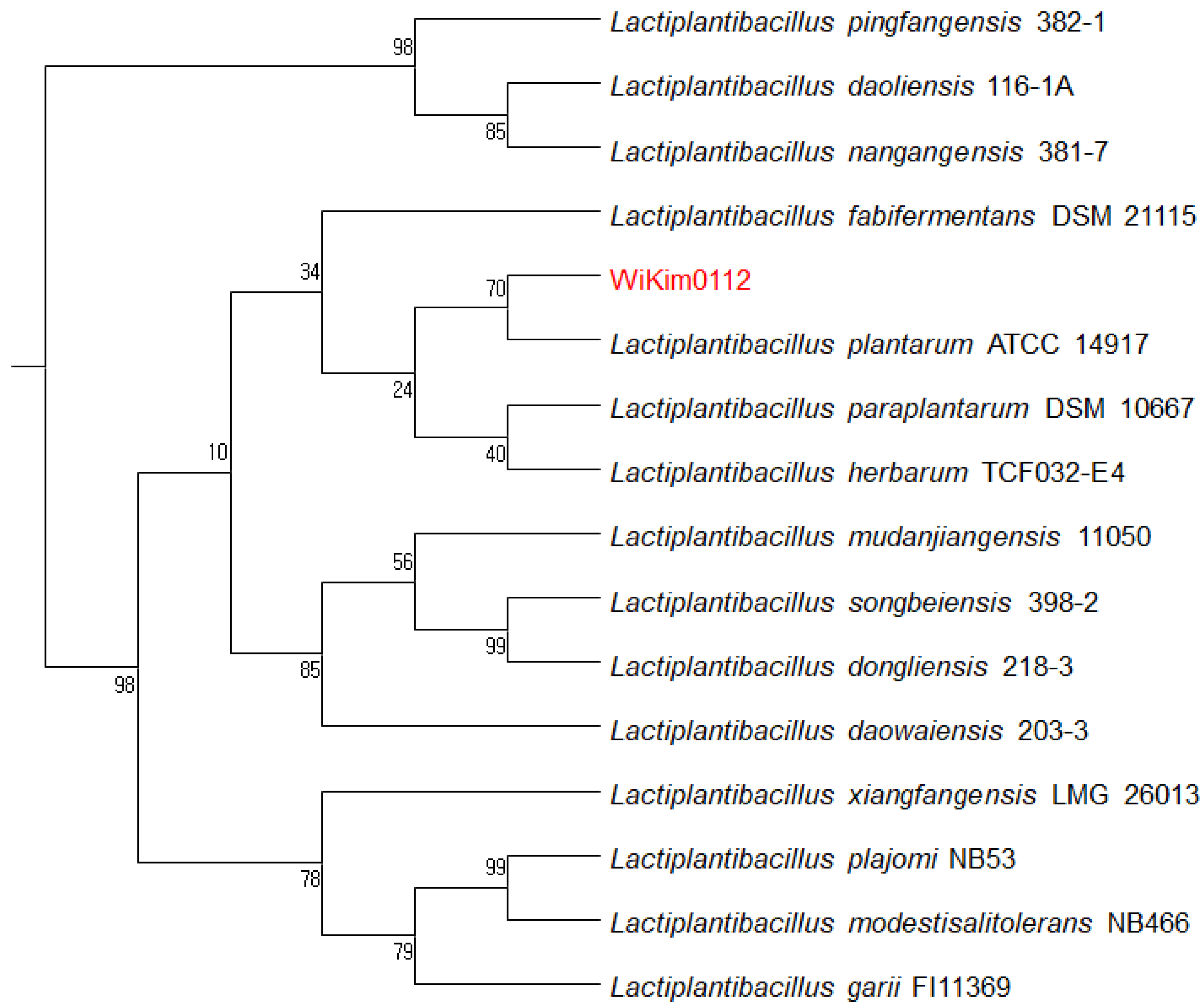
3.2. Identification of WiKim0112

A phylogenetic tree was constructed based on the 16S rRNA sequences to identify strain Wikim0112 and investigate the evolutionary relationships. The 16S rRNA sequence of WiKim0112 was compared to similar sequences using the EZBioCloud database. The sequence showed high similarity to Lpb. plantarum (16S rRNA sequence similarity, >99%). According to the phylogenetic tree, the isolated WiKim0112 was closely related to Lpb. plantarum ATCC 14917, with bootstrap values of 70% (Figure 1).

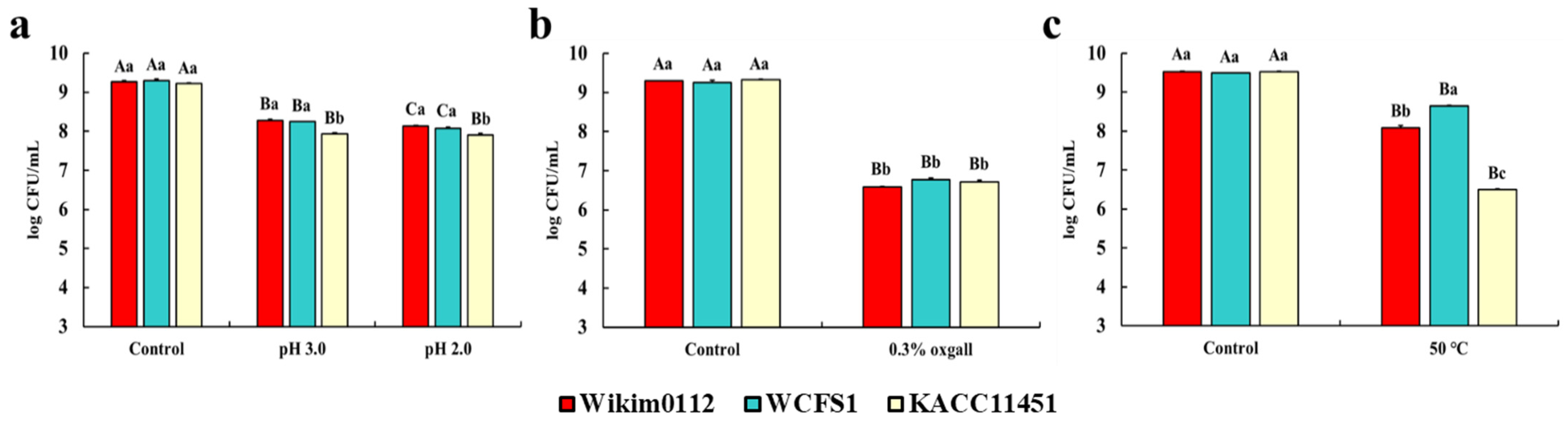
3.3. Viability of Lpb. plantarum Strains to GI Environment and Heat Treatment

Tolerance to the GI environment, including acidic juice containing pepsin, bile, and pancreatin, is a valuable property of probiotics that is associated with beneficial effects on host health [21]. In particular, acid tolerance is important in bacteria not only for enduring gastric stress but also for extending the duration of survival of the bacteria in acidic foods such as yogurt [22]. Since the pH of gastric juice after food ingestion is known to be approximately 3, we used pH 2 and 3 media containing pepsin in our study to mimic gastric juice [23]. In addition, considering the general bile salt concentration of the human GI tract (0.03–0.3%), 0.3% bile salt oxgall and pancreatin were incorporated in the culture media [24]. The tolerance of Lpb. plantarum Wikim0112, Lpb. plantarum WCFS1, and Lpb. plantarum KACC11451 to the GI environment is presented in Figure 2. All Lpb. plantarum strains showed relatively high cell viabilities of 8.28 log CFU/mL for Wikim0112, 8.25 log CFU/mL for WCFS1, and 7.93 log CFU/mL for KACC11451 in pH 3 medium containing 0.3% pepsin, compared to the control (9.22–9.29 log CFU/mL, pH 6.5, without pepsin) (Figure 2a). In particular, Wikim0112 and WCFS1 showed significantly higher viability than that of KACC11451 (Figure 2a). Moreover, in a pH 2 medium containing pepsin, Wikim0112 (8.14 log CFU/mL), WCFS1 (8.01 log CFU/mL), and KACC11451 (7.91 log CFU/mL) showed relatively high cell viability, and KACC11451 showed significantly lower viability than that of the other two strains (Figure 2a). This difference in acid resistance in the same species may be attributed to the relative amount of H+-ATPase present in the cell membranes [25]. H+-ATPase controls proton permeability, allowing cells to withstand large differences in pH between the bacterial cytoplasm and the medium [26]. The bile tolerance of Lpb. plantarum strains were investigated using a medium containing 0.1% pancreatin and 0.3% bile salt oxgall. The cell viability of all Lpb. plantarum strains was significantly decreased to 6.58–6.77 log CFU/mL, compared to 9.26–9.32 log CFU/mL of the control (without pancreatin and bile salt) (Figure 2b). However, there were no significant differences between the strains.
The thermotolerance of probiotics may facilitate their survival during thermal food processing. Therefore, we analyzed the survival of the strains at 50 °C to evaluate their thermotolerance. We found that WCFS1 (8.65 log CFU/mL) showed the highest thermotolerance, followed by Wikim0112 (8.14 log CFU/mL) and KACC (6.45 log CFU/mL) compared to the initial cell density (9.50–9.53 log CFU/mL) (Figure 2c). This result may be attributed to the fact that WCFS1 was isolated from human saliva, the temperature of which is relatively higher than that of the isolation sources of the other two strains. Similarly, a previous study reported that LAB isolated from cooked meat products showed thermotolerance [27].

3.4. Antibacterial Activity of Lpb. plantarum Strains

Lpb. plantarum strains are known to produce a wide range of antibacterial compounds, including bacteriocins, organic acids, ethanol, diacetyl, and H2O2 [28,29,30]. Furthermore, antibacterial substances produced by LAB are generally recognized as non-toxic and non-irritant components because they are naturally degraded in the body or the environment [31,32]. Therefore, antibacterial activity is an important property of probiotics. The antibacterial activity of cell-free culture supernatants of Lpb. plantarum strains against six strains of foodborne bacterial pathogens are shown in Table 2. Wikim0112 showed a similar level of antibacterial activity to that of the reference strain WCFS1 and exhibited an overall higher antibacterial activity than that of KACC11451 against six pathogens. Our results suggest that Wikim0112 may show an antibacterial activity that is not inferior to that of the two commercial Lpb. plantarum strains, particularly against bacterial pathogens associated with foodborne diseases.

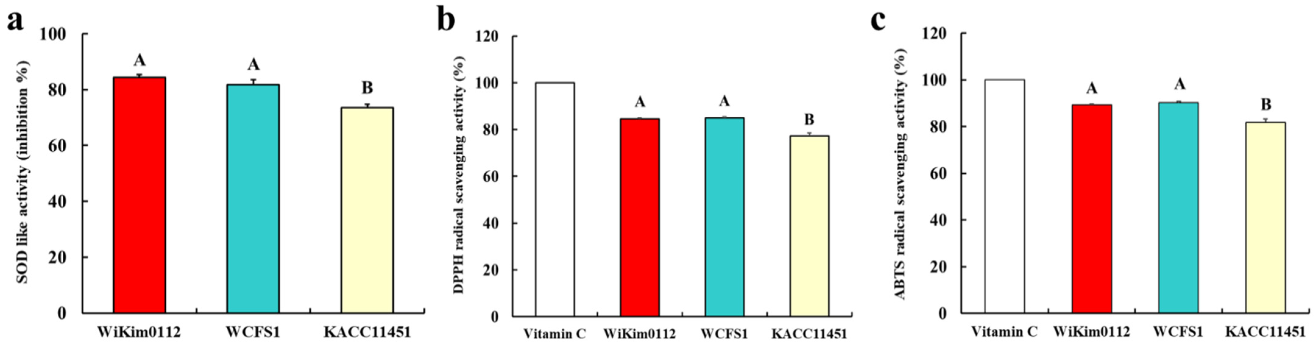
3.5. Antioxidant Activity of Lpb. plantarum Strains

A previous study showed that the antioxidant effects of probiotics might be mediated via the chelation of metal ions, expression of anti-oxidases, generation of antioxidant metabolites, modulation of anti-oxidase activity in the host, and regulation of the gut microbiota [33]. In the present study, we determined the antioxidant activity of the Lpb. plantarum strains via SOD, DPPH, and ABTS assays. All Lpb. plantatum strains showed more than 70% of SOD-like activity, especially Wikim0112 (84.3%) and WCFS1 (81.7%) had higher SOD-like activity than KACC11451(73.5%) (Figure 3a). Moreover, in DPPH and ABTS assay, Wikim0112 and WCFS1 showed significantly higher radical scavenging activities than KACC11451 (Figure 3b,c). A previous study reported that Lpb. plantarum C88 isolated from traditional Chinese fermented foods demonstrated DPPH scavenging activity of 44.3% [34]. In particular, heat-killed Lpb. plantarum Ln1 isolated from kimchi showed a high ABTS radical scavenging activity of 70.2% [35]. Our results indicate that Wikim0112 had similar or stronger antioxidant activity than standard probiotic strains.

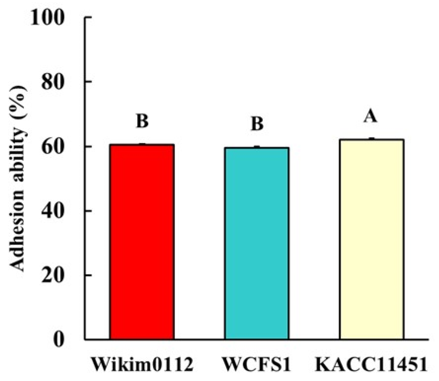
3.6. Adhesion Ability of Lpb. plantarum Strains to Intestinal Epithelial Cells

Probiotics may provide beneficial health effects by inhibiting the colonization of pathogens and enhancing mucosal healing during adhesion to the intestinal epithelium [36]. Therefore, the ability of probiotics to adhere to the intestinal epithelium has been evaluated. In general, the Caco-2 cell line has been commonly used in assays to estimate the adhesion ability of potential probiotic microorganisms due to its functional differentiation in vitro and morphological characteristics similar to those of human enterocytes [37,38]. The capability of the selected Lpb. plantarum strains to adhere to the intestinal epithelium was estimated by incubating the cultures with Caco-2 cells for 2 h. All Lpb. plantarum strains showed adhesion rates of approximately 60–62% (Figure 4). In particular, KACC11451 showed a higher adhesion ability than that of Wikim0112 and WCFS1 (Figure 4). The adhesion ability of probiotics may be affected by several factors, such as non-specific binding, including electrostatic and hydrophobic interactions, and specific binding to molecules, such as the mannose-specific binding of strain WCFS1 [39,40]. Therefore, isolates of the same microbial species may differ in their capability to adhere to the intestinal epithelium.

3.7. Anti-Inflammatory Activity of Lpb. plantarum Strains in Intestinal Epithelial Cells

Probiotics adhered to the intestinal epithelium may exhibit immunomodulatory activity by regulating pro- and anti-inflammatory cytokine production and inducing peripheral immunoglobulin production and immunoglobulin A secretion [41]. We found that all Lpb. plantarum strains significantly decreased the mRNA expression levels of pro-inflammatory cytokines (IL-1β, IL-6, and TNF-α) in LPS-stimulated Caco-2 cells (Figure 5a–c). The WCFS1 and KACC11451 strains did not affect the mRNA expression of anti-inflammatory cytokines (Figure 5d–f), whereas Wikim0112 significantly increased the mRNA expression of IL-4 and IFN-γ in LPS-stimulated Caco-2 cells (Figure 5d,f). Another study similarly reported anti-inflammatory activity in LAB isolated from kimchi. Park et al. (2017) found that administration of Lactobacillus paracasei LS2 isolated from kimchi significantly decreased the mRNA levels of pro-inflammatory cytokines (TNF-α, IL-1β, and IL-6) in dextran sulfate sodium-induced colitis mice [42]. Our results indicated that the Wikim0112 strain isolated from kimchi showed higher anti-inflammatory activity than that of the other two strains.

3.8. Antibiotic Susceptibility of Lpb. plantarum Strains

Antibiotic resistance of bacteria is largely classified into intrinsic and acquired resistance (for example, by spontaneous mutations in the bacterial genome or acquisition of additional genes) [43,44]. According to previous reports, the intrinsic resistance of bacteria was not transferred horizontally and did not present a risk in non-pathogenic bacteria, whereas acquired resistance can be horizontally transferred between bacteria [43,44]. Thus, it is essential to evaluate the antibiotic resistance of LAB. Antibiotic susceptibility of the Lpb. plantarum strains was determined using 13 antibiotics (AMP, C, CD, CIP, CN, DXT, E, K, P, S, SXT, TE, and VA). Wikim0112 was resistant to two antibiotics (S and VA) (Table 3). WCFS1 and KACC11451 were resistant to four (CD, CIP, TE, and VA) and three (CIP, TE, and VA) antibiotics, respectively (Table 3). The Lpb. plantarum strains are generally susceptible to β-lactam antibiotics, which inhibit the cell wall synthesis, such as ampicillin and penicillin, except for cephalosporins and oxacillin [44]. However, they are usually resistant to glycopeptides, such as VA [44]. Similarly, we found that Wikim0112 showed resistance to glycopeptides (VA) and aminoglycoside antibiotics (S), which are known to be intrinsic to Lactiplantibacilli [45,46]. These results indicated that Wikim0112 might not present a risk to the host compared to the other two standard probiotic strains regarding antibiotic resistance.

4. Conclusions

Compared to the probiotic Lpb. plantarum strains isolated from human saliva and pickle, kimchi-derived Lpb. plantarum Wikim0112 exhibited desirable probiotic properties, such as heat tolerance, resistance to the GI environment, and adhesion to the intestinal epithelium, along with antibacterial, antioxidant, and anti-inflammatory activities. Although further studies on the safety of Lpb. plantarum Wikim0112 are warranted, Wikim0112 represented an ideal candidate for probiotic development in the food and pharmaceutical industries.

Author Contributions

Conceptualization, S.-W.H. and S.-G.H.; formal analysis, D.-S.K. and C.-S.K.; investigation, C.-H.J., H.S., H.H., and H.-J.L.; writing—original draft preparation, C.-H.J.; writing—review and editing T.-W.K. and S.-G.H.; supervision, S.-W.H. All authors have read and agreed to the published version of the manuscript.

Funding

This work was supported by the Korea Institute of Planning and Evaluation for Technology in Food, Agriculture and Forestry (IPET) through the Innovative Food Product and Natural Food Materials Development Program, funded by the Ministry of Agriculture, Food and Rural Affairs (MAFRA) (119028-03-3-HD030), and a part of the project titled ‘Bioactive material for algae-based bio-health care substantiation’, funded by the Ministry of Oceans and Fisheries (20210656), and a grant from the World Institute of Kimchi (KE2101-2) funded by the Ministry of Science and ICT, Korea.

Institutional Review Board Statement

Not applicable.

Informed Consent Statement

Not applicable.

Conflicts of Interest

The authors declare no conflict of interest.

References

  1. Jung, J.Y.; Lee, S.H.; Jeon, C.O. Kimchi microflora: History, current status, and perspectives for industrial kimchi production. Appl. Microbiol. Biotechnol. 2014, 98, 2385–2393. [Google Scholar] [CrossRef]
  2. Chang, J.H.; Shim, Y.; Cha, S.K.; Chee, K. Probiotic characteristics of lactic acid bacteria isolated from kimchi. J. Appl. Microbiol. 2010, 109, 220–230. [Google Scholar] [CrossRef] [PubMed]
  3. Kim, K.-T.; Yang, S.J.; Paik, H.-D. Probiotic properties of novel probiotic Levilactobacillus brevis KU15147 isolated from radish kimchi and its antioxidant and immune-enhancing activities. Food Sci. Biotechnol. 2021, 30, 257–265. [Google Scholar] [CrossRef] [PubMed]
  4. Garcia-Gonzalez, N.; Battista, N.; Prete, R.; Corsetti, A. Health-promoting role of lactiplantibacillus plantarum isolated from fermented foods. Microorganisms 2021, 9, 349. [Google Scholar] [CrossRef] [PubMed]
  5. Behera, S.S.; Ray, R.C.; Zdolec, N. Lactobacillus plantarum with functional properties: An approach to increase safety and shelf-life of fermented foods. BioMed Res. Int. 2018, 2018, 18. [Google Scholar] [CrossRef] [PubMed] [Green Version]
  6. Atrih, A.; Rekhif, N.; Milliere, J.; Lefebvre, G. Detection and characterization of a bacteriocin produced by Lactobacillus plantarum C19. Can. J. Microbiol. 1993, 39, 1173–1179. [Google Scholar] [CrossRef]
  7. Wen, L.S.; Philip, K.; Ajam, N. Purification, characterization and mode of action of plantaricin K25 produced by Lactobacillus plantarum. Food Control 2016, 60, 430–439. [Google Scholar] [CrossRef]
  8. Hu, M.; Zhao, H.; Zhang, C.; Yu, J.; Lu, Z. Purification and characterization of plantaricin 163, a novel bacteriocin produced by Lactobacillus plantarum 163 isolated from traditional Chinese fermented vegetables. J. Agric. Food Chem. 2013, 61, 11676–11682. [Google Scholar] [CrossRef]
  9. Gonzalez, B.; Arca, P.; Mayo, B.; Suárez, J.E. Detection, purification, and partial characterization of plantaricin C, a bacteriocin produced by a Lactobacillus plantarum strain of dairy origin. Appl. Environ. Microbiol. 1994, 60, 2158–2163. [Google Scholar] [CrossRef] [Green Version]
  10. Zhu, X.; Zhao, Y.; Sun, Y.; Gu, Q. Purification and characterisation of plantaricin ZJ008, a novel bacteriocin against Staphylococcus spp. from Lactobacillus plantarum ZJ008. Food Chem. 2014, 165, 216–223. [Google Scholar] [CrossRef]
  11. Todorov, S.D.; Perin, L.M.; Carneiro, B.M.; Rahal, P.; Holzapfel, W.; Nero, L.A. Safety of Lactobacillus plantarum ST8Sh and its bacteriocin. Probiotics Antimicrob. Proteins 2017, 9, 334–344. [Google Scholar] [CrossRef] [Green Version]
  12. Rekhif, N.; Atrih, A.; Lefebvrexy, G. Activity of plantaricin SA6, a bacteriocin produced by Lactobacillus plantarum SA6 isolated from fermented sausage. J. Appl. Bacteriol. 1995, 78, 349–358. [Google Scholar] [CrossRef] [PubMed]
  13. Dixit, G.; Samarth, D.; Tale, V.; Bhadekar, R. Comparative studies on potential probiotic characteristics of Lactobacillus acidophilus strains. EurAsian J. BioSci. 2013, 7, 1–9. [Google Scholar] [CrossRef] [Green Version]
  14. Guo, Z.; Wang, J.; Yan, L.; Chen, W.; Liu, X.-M.; Zhang, H.-P. In vitro comparison of probiotic properties of Lactobacillus casei Zhang, a potential new probiotic, with selected probiotic strains. LWT Food Sci. Technol. 2009, 42, 1640–1646. [Google Scholar] [CrossRef]
  15. Ranadheera, C.S.; Evans, C.; Adams, M.; Baines, S. In vitro analysis of gastrointestinal tolerance and intestinal cell adhesion of probiotics in goat’s milk ice cream and yogurt. Food Res. Int. 2012, 49, 619–625. [Google Scholar] [CrossRef]
  16. Jeong, C.H.; Ryu, H.; Zhang, T.; Lee, C.H.; Seo, H.G.; Han, S.G. Green tea powder supplementation enhances fermentation and antioxidant activity of set-type yogurt. Food Sci. Biotechnol. 2018, 27, 1419–1427. [Google Scholar] [CrossRef] [PubMed]
  17. Zhang, T.; Jeong, C.H.; Cheng, W.N.; Bae, H.; Seo, H.G.; Petriello, M.C.; Han, S.G. Moringa extract enhances the fermentative, textural, and bioactive properties of yogurt. LWT 2019, 101, 276–284. [Google Scholar] [CrossRef]
  18. Cheng, W.; Jeong, C.; Kim, D.; Han, S. Effects of moringa extract on adhesion and invasion of Escherichia coli O55 in bovine mammary epithelial cells. J. Dairy Sci. 2020, 103, 7416–7424. [Google Scholar] [CrossRef]
  19. Won, S.-M.; Chen, S.; Park, K.W.; Yoon, J.-H. Isolation of lactic acid bacteria from kimchi and screening of Lactobacillus sakei ADM14 with anti-adipogenic effect and potential probiotic properties. LWT 2020, 126, 109296. [Google Scholar] [CrossRef]
  20. Cockerill, F.R.; Wikler, M.A.; Alder, J.; Dudley, M.; Eliopoulos, G.; Ferraro, M.; Hardy, D.; Hecht, D.; Hindler, J.; Patel, J. Performance standards for antimicrobial susceptibility testing: Twenty-second informational supplement. Clin. Lab. Stand. Inst. 2012, 32, M100–S122. [Google Scholar]
  21. Angmo, K.; Kumari, A.; Bhalla, T.C. Probiotic characterization of lactic acid bacteria isolated from fermented foods and beverage of Ladakh. LW Food Sci. Technol. 2016, 66, 428–435. [Google Scholar] [CrossRef]
  22. Wang, C.-Y.; Lin, P.-R.; Ng, C.-C.; Shyu, Y.-T. Probiotic properties of Lactobacillus strains isolated from the feces of breast-fed infants and Taiwanese pickled cabbage. Anaerobe 2010, 16, 578–585. [Google Scholar] [CrossRef]
  23. Conway, P.; Gorbach, S.; Goldin, B. Survival of lactic acid bacteria in the human stomach and adhesion to intestinal cells. J. Dairy Sci. 1987, 70, 1–12. [Google Scholar] [CrossRef]
  24. Huang, R.; Tao, X.; Wan, C.; Li, S.; Xu, H.; Xu, F.; Shah, N.P.; Wei, H. In vitro probiotic characteristics of Lactobacillus plantarum ZDY 2013 and its modulatory effect on gut microbiota of mice. J. Dairy Sci. 2015, 98, 5850–5861. [Google Scholar] [CrossRef]
  25. Hong, S.-I.; Kim, Y.-J.; Pyun, Y.-R. Acid Tolerance ofLactobacillus plantarumfromKimchi. LWT Food Sci. Technol. 1999, 32, 142–148. [Google Scholar] [CrossRef]
  26. Nannen, N.L.; Hutkins, R.W. Proton-translocating adenosine triphosphatase activity in lactic acid bacterial. J. Dairy Sci. 1991, 74, 747–751. [Google Scholar] [CrossRef]
  27. Ramirez-Chavarin, M.; Wacher, C.; Eslava-Campos, C.; Perez-Chabela, M. Probiotic potential of thermotolerant lactic acid bacteria strains isolated from cooked meat products. Int. Food Res. J. 2013, 20, 991–1000. [Google Scholar]
  28. Vataščinová, T.; Pipova, M.; Fraqueza, M.; Maľa, P.; Dudrikova, E.; Drážovská, M.; Laukova, A. Antimicrobial potential of Lactobacillus plantarum strains isolated from Slovak raw sheep milk cheeses. J. Dairy Sci. 2020, 103, 6900–6903. [Google Scholar] [CrossRef]
  29. Lin, T.-H.; Pan, T.-M. Characterization of an antimicrobial substance produced by Lactobacillus plantarum NTU 102. J. Microbiol. Immunol. Infect. 2019, 52, 409–417. [Google Scholar] [CrossRef] [PubMed]
  30. Kothari, D.; Lee, W.-D.; Jung, E.S.; Niu, K.-M.; Lee, C.H.; Kim, S.-K. Controlled fermentation using autochthonous Lactobacillus plantarum improves antimicrobial potential of Chinese chives against poultry pathogens. Antibiotics 2020, 9, 386. [Google Scholar] [CrossRef]
  31. Piard, J.; Desmazeaud, M. Inhibiting factors produced by lactic acid bacteria. 2. Bacteriocins and other antibacterial substances. Lait 1992, 72, 113–142. [Google Scholar] [CrossRef] [Green Version]
  32. Ouwehand, A.C.; Vesterlund, S. Antimicrobial components from lactic acid bacteria. Food Sci. Technol. N. Y. Marcel Dekker 2004, 139, 375–396. [Google Scholar]
  33. Wang, Y.; Wu, Y.; Wang, Y.; Xu, H.; Mei, X.; Yu, D.; Wang, Y.; Li, W. Antioxidant properties of probiotic bacteria. Nutrients 2017, 9, 521. [Google Scholar] [CrossRef] [PubMed]
  34. Li, S.; Zhao, Y.; Zhang, L.; Zhang, X.; Huang, L.; Li, D.; Niu, C.; Yang, Z.; Wang, Q. Antioxidant activity of Lactobacillus plantarum strains isolated from traditional Chinese fermented foods. Food Chem. 2012, 135, 1914–1919. [Google Scholar] [CrossRef] [PubMed]
  35. Jang, H.J.; Song, M.W.; Lee, N.-K.; Paik, H.-D. Antioxidant effects of live and heat-killed probiotic Lactobacillus plantarum Ln1 isolated from kimchi. J. Food Sci. Technol. 2018, 55, 3174–3180. [Google Scholar] [CrossRef] [PubMed]
  36. Ouwehand, A.C.; Salminen, S.; Isolauri, E. Probiotics: An overview of beneficial effects. In Lactic Acid Bacteria: Genetics, Metabolism and Applications; Siezen, R.J., Kok, J., Abeem, T., Schaafsma, G., Eds.; Springer: Amsterdam, The Netherlands, 2002; pp. 279–289. [Google Scholar]
  37. De Palencia, P.F.; López, P.; Corbí, A.L.; Peláez, C.; Requena, T. Probiotic strains: Survival under simulated gastrointestinal conditions, in vitro adhesion to Caco-2 cells and effect on cytokine secretion. Eur. Food Res. Technol. 2008, 227, 1475–1484. [Google Scholar] [CrossRef] [Green Version]
  38. Sohn, H.; Chang, Y.H.; Yune, J.H.; Jeong, C.H.; Shin, D.-M.; Kwon, H.C.; Kim, D.H.; Hong, S.W.; Hwang, H.; Jeong, J.Y. Probiotic Properties of Lactiplantibacillus plantarum LB5 Isolated from Kimchi Based on Nitrate Reducing Capability. Foods 2020, 9, 1777. [Google Scholar] [CrossRef] [PubMed]
  39. Muñoz-Provencio, D.; Llopis, M.; Antolín, M.; De Torres, I.; Guarner, F.; Pérez-Martínez, G.; Monedero, V. Adhesion properties of Lactobacillus casei strains to resected intestinal fragments and components of the extracellular matrix. Arch. Microbiol. 2009, 191, 153–161. [Google Scholar] [CrossRef]
  40. Pretzer, G.; Snel, J.; Molenaar, D.; Wiersma, A.; Bron, P.A.; Lambert, J.; de Vos, W.M.; van der Meer, R.; Smits, M.A.; Kleerebezem, M. Biodiversity-based identification and functional characterization of the mannose-specific adhesin of Lactobacillus plantarum. J. Bacteriol. 2005, 187, 6128. [Google Scholar] [CrossRef] [PubMed] [Green Version]
  41. Yahfoufi, N.; Mallet, J.; Graham, E.; Matar, C. Role of probiotics and prebiotics in immunomodulation. Curr. Opin. Food Sci. 2018, 20, 82–91. [Google Scholar] [CrossRef]
  42. Park, J.-S.; Joe, I.; Rhee, P.D.; Jeong, C.-S.; Jeong, G. A lactic acid bacterium isolated from kimchi ameliorates intestinal inflammation in DSS-induced colitis. J. Microbiol. 2017, 55, 304–310. [Google Scholar] [CrossRef] [PubMed]
  43. Hollenbeck, B.L.; Rice, L.B. Intrinsic and acquired resistance mechanisms in enterococcus. Virulence 2012, 3, 421–569. [Google Scholar] [CrossRef] [PubMed] [Green Version]
  44. Zhou, J.; Pillidge, C.; Gopal, P.; Gill, H. Antibiotic susceptibility profiles of new probiotic Lactobacillus and Bifidobacterium strains. Int. J. Food Microbiol. 2005, 98, 211–217. [Google Scholar] [CrossRef]
  45. Mathur, S.; Singh, R. Antibiotic resistance in food lactic acid bacteria—A review. Int. J. Food Microbiol. 2005, 105, 281–295. [Google Scholar] [CrossRef] [PubMed]
  46. Gueimonde, M.; Sánchez, B.; de Los Reyes-Gavilán, C.G.; Margolles, A. Antibiotic resistance in probiotic bacteria. Front. Microbiol. 2013, 4, 202. [Google Scholar] [CrossRef] [PubMed] [Green Version]
Figure 1. Phylogenetic tree constructed using neighbor-joining based on 16S rRNA gene sequences of WiKim0112. Bootstrap percentages for 1000 re-sampling are given.
Foods 10 02125 g001
Figure 2. Gastrointestinal environment and heat tolerance of Lpb. plantarum Wikim0112, WCFS1, and KACC11451. All Lpb. plantarum strains were exposed (a) without (control) or with 0.3% (w/v) pepsin at pH 6.5 (control), 3.0, or 2.0 for 4 h, (b) without (control) or with 0.1% (w/v) pancreatin and 0.3% (w/v) bile salt (oxgall) for 24 h, and (c) 30 (control) or 50 °C for 1 h. The values represent the mean ± SEM (n = 3). Different uppercase letters represent significant difference between the same strains (p < 0.5), while different lowercase letters represent significant difference between the same treatment groups (p < 0.5).
Foods 10 02125 g002
Figure 3. Antioxidative activities of Lpb. plantarum Wikim0112, WCFS1, and KACC11451. (a) Superoxide dismutase (SOD)-like activity, (b) 2,2-diphenyl-1-picryl-hydrazyl (DPPH), and (c) 2-azino-bis(3-Ethylbenzothiazoline-6-Sulfonic Acid) (ABTS) scavenging activities were estimated. As a positive control, 0.5% L-ascorbic acid was used with the same amount as the sample. The values represent the mean ± SD (n = 3). Different letters represent significant difference (p < 0.5).
Foods 10 02125 g003
Figure 4. The adhesion ability of Lpb. plantarum Wikim0112, WCFS1, and KACC11451 in Caco-2 cells. The cells were exposed to Lpb. plantarum strain at 37 °C for 2 h. The adhesion rates were calculated by counting the number of bacteria before and after incubation. The values represent the mean ± SEM (n = 4). Different letters represent significant difference (p < 0.5).
Foods 10 02125 g004
Figure 5. Effects of Lpb. plantarum Wikim0112, WCFS1, and KACC1451 on the mRNA expression levels of (ac) pro-inflammatory cytokines (IL-1β, IL-6, and TNF-α) and (df) anti-inflammatory cytokines (IL-4, IL-10, and IFN-γ) in Caco-2 cells. The cells were pretreated with each Lpb. plantarum strain (106 and 107 CFU/mL) for 6 h followed by treatment with LPS (1 μL/mL) for 2 h. The values represent the mean ± SEM (n = 3); ** p < 0.01 indicates a significant difference vs. the control and # p < 0.05, ## p < 0.01, ### p < 0.001 indicates a significant difference vs. the LPS-treated group.
Foods 10 02125 g005
Table 1. Primers used for real-time PCR.
GenesPrimer Sequences (5′–3′)
TNF-α a(F) AAG CCC TGG TAT GAG CCC ATC TAT(R) AGG GCA ATG ATC CCA AAG TAG ACC
IL-6(F) ACA GCC ACT CAC CTC TTC AGA AC(R) TTT TCT GCC AGT GCC TCT TTG C
IL-1β(F) TGT ACC TGT CCT GCG TGT TGA AAG(R) CTG GGC AGA CTC AAA TTC CAG CTT
IL-4(F) TCA TTT TCC CTC GGT TTC AG(R) AGA ACA GAG GGG GAA GCA GT
IL-10(F) TCA GGG TGG CGA CTC TAT(R) TGG GCT TCT TCT AAA TCG TTC
IFN-γ(F) ATA TCT TGG CTT TTC AGC TC(R) CTC CTT TTT CGC TTC CCT GT
GAPDH(F) GAC CCC TTC ATT GAC CTC AAC TAC(R) ATG ACA AGC TTC CCG TTC TCA G
a TNF-α, tumor necrosis factor-alpha; IL-6, interleukin-6; IL-1β, interleukin-1 beta; IL-4, interleukin-4; IL-10, interleukin-10; IFN-γ, interferon-gamma; and GAPDH, glyceraldehyde 3-phosphate dehydrogenase.
Table 2. Antibacterial activity of Lpb. plantarum Wikim0112, WCFS1, and KACC11451 against foodborne bacterial pathogens.
PathogensInhibition Zone (mm) ± SD a
WiKim0112WCFS1KACC11451
E. coli O157:H7
(ATCC 35150)
18.0 ± 0.519.0 ± 0.516.0 ± 0.1
Listeria monocytogenes
(ATCC 15313)
20.0 ± 0.418.0 ± 0.215.0 ± 0.5
Salmonella choleraesuis
(KCCM40763)
20.0 ± 0.320.0 ± 0.116.0 ± 0.2
Shigella boydii
(KCCM41649)
23.0 ± 0.424.0 ± 0.221.0 ± 0.2
Staphylococcus aureus
(ATCC25923)
23.0 ± 0.323.0 ± 0.120.0 ± 0.3
Yersinia enterocolitica
(KCCM41657)
23.0 ± 0.224.0 ± 0.522.0 ± 0.5
a Standard deviation.
Table 3. Antibiotic susceptibility of Lpb. plantarum Wikim0112, WCFS1, and KACC11451.
AntibioticsWikim0112WCFS1KACC11451
Zone Diameter (mm) ± SD aSensitivityZone Diameter (mm) ± SDSensitivityZone Diameter (mm) ± SDSensitivity
Ampicillin (AMP)29.0 ± 1.4S39.3 ± 1.3S29.7 ± 1.3S
Chloramphenicol (C)31.0 ± 0.8S30.7 ± 0.9S30.7 ± 0.9S
Clindamycin (CD)26.3 ± 1.3S31.7 ± 1.3S12.0 ± 1.6R
Ciprofloxacin (CIP)14.3 ± 0.9I13.0 ± 1.4R13.7 ± 0.5R
Gentamicin (CN)18.7 ± 1.3I28.7 ± 0.9S28.3 ± 1.3S
Doxycycline (DXT)28.7 ± 0.9S16.3 ± 1.3I18.7 ± 0.9I
Erythromycin (E)37.0 ± 0.8S38.0 ± 1.6S33.7 ± 1.3S
Kanamycin (K)12.3 ± 1.3S31.3 ± 0.5S28.3 ± 0.5S
Penicillin G (P)29.3 ± 0.9S31.3 ± 1.3S24.7 ± 0.5S
Streptomycin (S)13.3 ± 1.3R25.3 ± 1.3S23.0 ± 1.4S
Trimethoprim-sulfamethoxazole (SXT)29.3 ± 1.6S26.7 ± 0.5S29.7 ± 1.4S
Tetracycline (TE)18.7 ± 1.3I12.0 ± 1.3R-R
Vancomycin (VA)- bR-R-R
a Standard deviation; b Indicates no inhibition zone. The inhibition zones were evaluated according to the Clinical and Laboratory Standards Institute (CLSI) 2012 standards and presented as follows: Susceptible (S) ≥ 20 mm; Intermediate (I) 15–19 mm; Resistant (R) ≤ 14 mm.
Publisher’s Note: MDPI stays neutral with regard to jurisdictional claims in published maps and institutional affiliations.

