Article

Vibrio parahaemolyticus Isolates from Asian Green Mussel: Molecular Characteristics, Virulence and Their Inhibition by Chitooligosaccharide-Tea Polyphenol Conjugates

1
International Center of Excellence in Seafood Science and Innovation, Faculty of Agro-Industry, Prince of Songkla University, Hat Yai, Songkhla 90110, Thailand
2
Department of Pathology, Faculty of Medicine, Prince of Songkla University, Hat Yai, Songkhla 90110, Thailand
3
College of Fisheries, Guru Angad Dev Veterinary and Animal Sciences University, Ludhiana 141004, India
*
Author to whom correspondence should be addressed.
Foods 2022, 11(24), 4048; https://doi.org/10.3390/foods11244048
Submission received: 14 November 2022 / Revised: 6 December 2022 / Accepted: 12 December 2022 / Published: 14 December 2022
(This article belongs to the Section Foods of Marine Origin)

Abstract

:
Fifty isolates of Vibrio parahaemolyticus were tested for pathogenicity, biofilm formation, motility, and antibiotic resistance. Antimicrobial activity of chitooligosaccharide (COS)-tea polyphenol conjugates against all isolates was also studied. Forty-three isolates were randomly selected from 520 isolates from Asian green mussel (Perna viridis) grown on CHROMagarTM Vibrio agar plate. Six isolates were acquired from stool specimens of diarrhea patients. One laboratory strain was V. parahaemolyticus PSU.SCB.16S.14. Among all isolates tested, 12% of V. parahaemolyticus carried the tdh+trh gene and were positive toward Kanagawa phenomenon test. All of V. parahaemolyticus isolates could produce biofilm and showed relatively strong motile ability. When COS-catechin conjugate (COS-CAT) and COS-epigallocatechin-3-gallate conjugate (COS-EGCG) were examined for their inhibitory effect against V. parahaemolyticus, the former showed the higher bactericidal activity with the MBC value of 1.024 mg/mL against both pathogenic and non-pathogenic strains. Most of the representative Asian green mussel V. parahaemolyticus isolates exhibited high sensitivity to all antibiotics, whereas one isolate showed the intermediate resistance to cefuroxime. However, the representative clinical isolates were highly resistant to nine types of antibiotics and had multiple antibiotic resistance (MAR) index of 0.64. Thus, COS-CAT could be used as potential antimicrobial agent for controlling V. parahaemolyticus-causing disease in Asian green mussel.

Graphical Abstract

1. Introduction

Vibrio parahaemolyticus has become a serious foodborne pathogen and raised public health concern in Thailand, China, Japan, and other Asian countries [1]. V. parahaemolyticus present in aquatic products contributes to significant economic losses across the entire supply chain [2]. It is a member of the genus Vibrio from family Vibrionaceae, a Gram-negative, marine halophilic bacterium that naturally inhabits in global coastal waters, sediment and various types of marine animals [3] such as fish, shrimp, crab, clam, oyster [4,5,6,7], and mussel [8]. V. parahaemolyticus can also be transmitted to humans when consuming contaminated raw or poorly cooked seafood products [9,10]. This bacterium contributes to an acute gastroenteritis, which includes nausea, diarrhea, vomiting, fever, and chills. It can also cause severe symptoms in children, the elderly, and immunocompromised patients [11,12]. The pathogenic V. parahaemolyticus often infects and causes disease by using several virulence factors. Adhesions, thermostable direct hemolysin (TDH), and TDH-related hemolysin (TRH) are the most important virulence factors in this bacterium [13]. The hemolysis-associated genes, tdh, were found in the most multidrug-resistant (MDR) isolate as well as those having certain virulence characteristics and biofilm capacity. It is able to attach to several surfaces and subsequently proliferate, in which a multicellular consortium with a three-dimensional structure can be formed. Such a biofilm can protect the cells toward environmental stress [14]. In addition, the augmented resistance to antimicrobials is achieved. Moreover, biofilms can release viable V. parahaemolyticus cells into the environment as well as foods [14]. The aforementioned factors augment the pathogenicity of V. parahaemolyticus, causing the severe public health problem globally.
Recently, seafood has gained popularity internationally due to its health benefits, resulting in the augmented production and domestic consumption in Southeast Asia. Asian green mussel, Perna viridis, is abundant along the coasts and estuaries of Asian-Pacific regions. It is also a widespread species found from the Persian Gulf to the Southwest Pacific and from Southern Japan to Papua New Guinea [15]. It is an important commercial aquaculture bivalve in Southeast Asian countries [16]. The prevalence and outbreaks of V. parahaemolyticus infection in mussels were documented [17,18].
Antibiotic therapy is the treatment commonly used for bacterial infections; however antibiotic-resistant bacteria have been a major concern worldwide [19]. Natural alternative approaches to control foodborne pathogens have been researched. In recent years, many antimicrobial compounds from seafood processing leftover have been reported to prevent the proliferation of several pathogenic bacteria [20,21]. Those included low-molecular-weight (MW) chitooligosaccharide (COS), a deacetylated form of chitosan. It shows non-toxicity, biodegradable, and biocompatible properties. COS has been modified through several processes, particularly via polyphenol grafting [20]. Several polyphenols have been utilized for the modification of COS to augment their bioactivities involving antioxidant, antimicrobial, anticancer, antihypertension, etc., [22,23,24]. Singh et al. [20] prepared COS-epigallocatechin-3-gallate conjugates (COS-EGCG) encompassing high antioxidant and antimicrobial activities. Recently, Mittal et al. [21] documented that COS-catechin conjugate (COS-CAT) showed superior antioxidant and antimicrobial activities to COS and other COS-polyphenol conjugates. COS-CAT possessed the greatest antimicrobial activity toward both Gram-negative and Gram-positive bacteria. In general, antimicrobial activity of COS and polyphenols was linked to bacterial cell wall disintegration via electrostatic interaction with their -OH and amino groups. Furthermore, alterations in microbial mRNA, DNA, and protein synthesis as induced by the diffused COS and polyphenols can cause cell death [20,21]. COS polyphenol conjugates have been reported to be effective against a variety of microorganisms. Nevertheless, there are no reports on its antimicrobial activity against V. parahaemolyticus isolated from Asian green mussel and clinical sample. In addition, drug sensitivity, virulence, and molecular characteristics of V. parahaemolyticus isolated from Asian green mussel collected from the south of Thailand have not been investigated.

2. Materials and Methods

2.1. Sample Collection and Bacterial Isolation

Asian Green mussels (Perna viridis) (Figure 1) were collected randomly from the different Asian green mussel farms, natural habitat and fresh markets located in the southern provinces of Thailand (Suratthani, Trang and Songkhla provinces). Provinces, types of places, and number of collected samples are given in Table 1. The samples were brought to the laboratory in polyethylene bags containing ice within 1 h. The samples were washed with sterilized distilled water for surface sterilization and shucked by aseptic technique. Briefly, 25 g of Asian green mussel samples were transferred into 225 mL of alkaline peptone water (APW) (polypeptone, 10 g/L; NaCl, 20 g/L; pH 8.6) and mixed with the aid of stomacher (Stomacher 400 Seaward medicals, Worthing, UK) at 230 rpm for 1 min. The mixture was incubated at 41.5 °C for 6–8 h. The APW-enriched culture was diluted from 104 to 107–fold with the APW. Subsequently, 100 μL of the diluted samples were spread on thiosulphate citrate bile salts sucrose agar plate (TCBS Agar; Oxoid, Thermo Fischer Scientific, Waltham, MA, USA), and CHROMagarTM Vibrio agar plate (CHROMagarTM, Paris, France) was adopted for selection of V. parahaemolyticus isolates. These two agar plates were used for confirmation. The result from CHROMagarTM Vibrio plates was mainly used for further experiments. Forty-three isolates with different colony colors from the total 520 of isolates on CHROMagarTM Vibrio agar plate were randomly selected and then characterized and specified by the MALDI-Biotyper® system (microflex LT; Bruker Daltonik GmbH, Bremen, Germany) [25]. Halophilism was also performed using NaCl-tryptone broth (T1N0, T1N3, T1N6, T1N8, and T1N10).
Fifty isolates used in this study were molecularly identified and confirmed for V. parahaemolyticus. Forty-three isolates were retrieved from the Asian green mussel, while the remaining six isolates were isolated from the stool specimens of diarrhea patients from Songklanagarind Hospital, Faculty of Medicine and one laboratory strain of V. parahaemolyticus PSU.SCB.16S.14 was gifted by the Food Safety Laboratory, Prince of Songkla University, Hat Yai, Thailand.

2.2. Polymerase Chain Reaction (PCR) Assay

Twenty microliters of glycerol stock of V. parahaemolyticus isolates (n = 50) was inoculated into 5 mL of Luria-Bertani (LB) broth (Merck, Burlington, MA, USA) containing 3% NaCl (w/v) and incubated at 37 °C for 16–18 h, followed by centrifugation (8000 × g for 5 min). The genomic DNA was isolated using a PureLinkTM Genomic DNA Mini Kit (Invitrogen, Thermo Fisher Scientific, Waltham, MA, USA). DNA concentration was measured with the aid of a NanoDrop spectrophotometry (Thermo Fisher Scientific, Waltham, MA, USA). PCR primers were synthesized via Integrated DNA Technologies (Singapore city, Singapore) as shown in Table 2. PCR reaction mixture comprised 5 μL of 4 × Taq PCR Mastermix (QIAGEN, Germantown, MD, USA), 2 μL of genomic DNA (50 ng/μL), 0.5 μL of primer pair solution (10 μM each), and 12 μL of Rnase free water. PCR was amplified under the selected conditions: pre-denaturation at 95 °C for 2 min, 30 cycles for denaturation at 95 °C for 5 s, annealing at 58 °C for 15 s, and extension at 72 °C for 10 s, and ending extension at 72 °C for 5 min [26]. PCR products were finally determined using 2% agarose gel electrophoresis.

2.3. Preparation of COS-Tea Polyphenol Conjugates Using Free Radical Grafting Method

COS-CAT and COS-EGCG conjugates were prepared using free radical grafting method as tailored by Mittal et al. [21]. First, pH of COS solution (1%, w/v) was adjusted to 5.0 using acetic acid (1 M). Simultaneously, 1 M H2O2 (4 mL) containing 0.10 g ascorbic acid were incubated (40 °C, 10 min) to generate hydroxyl radicals. Both solutions were then mixed and the mixture was incubated at room temperature for 1 h with continuous stirring. CAT and EGCG (10%, w/w of COS) were then added into the mixture and incubated for 24 h in dark, at room temperature. With dialysis against distilled water, the unbound CAT and EGCG were removed. COS-CAT and COS-EGCG conjugate powders were obtained after lyophilization of dialysates.

2.4. Determination of Minimum Inhibitory Concentration (MIC) and Minimum Bactericidal Concentration (MBC)

MIC and MBC of COS-CAT and COS-EGCG toward V. parahaemolyticus isolates were measured following the guidelines of Clinical and Laboratory Standards Institute (CLSI). Overnight culture (18–24 h) of V. parahaemolyticus isolate was adjusted to a final concentration of 108 CFU/mL (corresponding to approximately 0.5 McFarland standard). The standardized suspension was then diluted by 200-fold with Mueller Hilton Broth (MHB) (DifcoTM, Baltimore, MD, USA) supplemented with 3% NaCl (w/v), namely “diluent” to obtain the working concentration of 106 CFU/mL. The COS-CAT and COS-EGCG powders were dissolved and diluted with deionized water [28]. Stock solution was subjected to two-fold dilution to attain the highest concentrations of 2.048 mg/mL and the lowest concentration of 0.004 mg/mL. One-hundred microliters of bacterial suspension and 100 μL of COS-CAT/COS-EGCG working solutions were mixed in each well, and incubated for 24 h at 37 °C. Thereafter, 20 μL of resazurin (0.09%) solutions was added for each well, and further incubated for 3 h at 37 °C. Subsequently, the wells with no color change were scored as “above the MIC value”. MBC was determined by plating directly the content of wells with concentration higher than the MIC value. Culture solution (10 μL) was pipetted from each well with no bacterial growth and dropped uniformly on a sterile MHA medium [29], followed by incubation (37 °C for 24 h). MBC was defined as the minimum concentration of COS-tea polyphenol conjugated solutions without colony formation. Positive control consisted of bacterial suspension and diluent, while negative control comprised MHB and diluent.

2.5. Biofilm Crystal Violet (CV) Staining

CV staining method was adopted [30]. Overnight cultures were diluted 50-fold using 200 μL of Oxoid TSB broth (Oxoid Ltd., Hampshire, England) containing 3% NaCl (w/v) (TSB-N) in 96-well plates (Corning Inc., Corning, NY, USA). Culture was allowed to proliferate at 37 °C for 48 h. The cultures were removed and the well with the adherent biofilm was gently washed with 200 μL of sterile phosphate buffered saline (PBS) for three times. Then, 200 μL of 0.1% crystal violet was used to stain the surface-attached cells for 15 min. After solution removal, the well was thoroughly washed with sterile H2O for three times. Bound dye in each well was solubilized using 200 μL of ethanol (Analytical grade ≥ 99.9% in pure, RCI Labscan™, Bangkok, Thailand). Absorbance at 570 nm (A570) was measured.

2.6. Analysis of Swimming and Swarming Motility

Swimming and swarming abilities of V. parahaemolyticus isolates were examined on semi-solid swimming plated (TSB-N in the presence of 0.2% agar) and solid swarming plates (TSB-N containing 0.5% agar), respectively [30,31]. The overnight cultures were diluted 50-fold using 5 mL of TSB-N broth and cultured at 37 °C with continuous shaking (200 rpm) until A570 reached 1.2–1.4. Those cultures were used for testing.

2.7. Kanagawa Phenomenon (KP) Test

KP test was performed as detailed by Zhang et al. [32]. First, 2 μL of the third-round cell cultures were inoculated onto Wagatsuma agar medium consisting of 5% rabbit red blood cells (RBCs). The radius from the inoculation place to the edge of β-hemolysin zone was detected after static incubation (37 °C for 24 h).

2.8. Antibiotic Susceptibility Testing

Antimicrobial susceptibility testing of eight V. parahaemolyticus isolated from clinical sample, Asian green mussel samples from different origins and laboratory strain were performed using the SensititreTM microbroth dilution system (Trek Diagnostic Systems, Cleveland, OH, USA) [33]. Cultures were grown overnight on TSA supplemented with 2.5% NaCl (w/v) plates at 37 °C. The cultures were transferred to sterile demineralized 2.5% saline solution to obtain the turbidity, equivalent to that of 0.5 McFarland standard. One-hundred milliliters of each suspension were transferred into sterile cation-adjusted MHB, and broth solution (50 mL) was dispersed onto CML1FMAR custom MIC plates (Trek Diagnostic Systems Inc., Cleveland, OH, USA) containing 21 different antibiotics with varying ranges of concentrations (μg/mL): amikacin (8–32), ampicillin (8–16), ampicillin/sulbactam (4/2–16/8), amikacin/clavulanic acid (4/2–16/8), cefepime (1–32), cefotaxime (1–32), cefoxitin (4–16), ceftazidime (1–32), ceftriaxone (0.5–32), cepfuroxime (8–16), ciprofloxacin (0.06–2), colistin (1–8), doripenem (0.5–16), ertapenem (0.5–4), gentamicin (2–8), imipenem (0.5–16), levofloxacim (0.06–8), meropenem (0.05–16), netilmicin (8–16), piperacillin-tazobactam (8/4–64/4), and sulfamethoxazole (1/19–4/76). MIC was the lowest concentration of the tested antibiotic, which totally inhibited bacterial growth [34]. Resistance breakpoints were also used [34]. Multiple antibiotic resistance (MAR) index was calculated as tailored by Krumperman [35], in which the following equation was used:
MAR index = a/b
where “a” is the number of antibiotics, to which the particular isolate was resistant and “b” is the total number of antibiotics tested.

2.9. Statistical Analyses

Completely randomized design (CRD) was used for the entire study. The experiments and analyses were conducted in triplicate. Data were subjected to one-way analysis of variance (ANOVA) and a least significant difference test was used. p < 0.05 was considered a significant difference.

3. Results and Discussion

3.1. Characteristics of V. parahaemolyticus Isolates

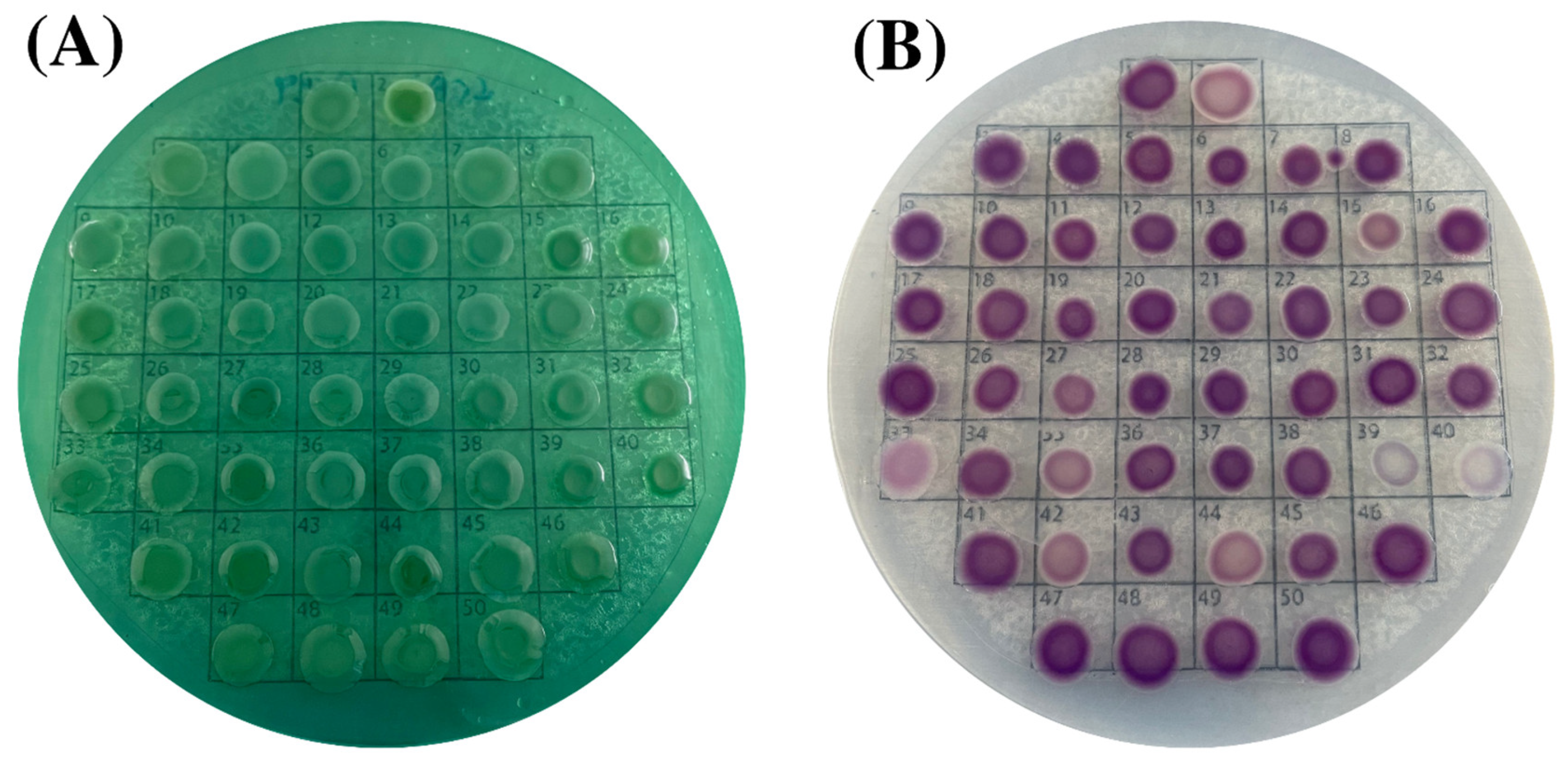
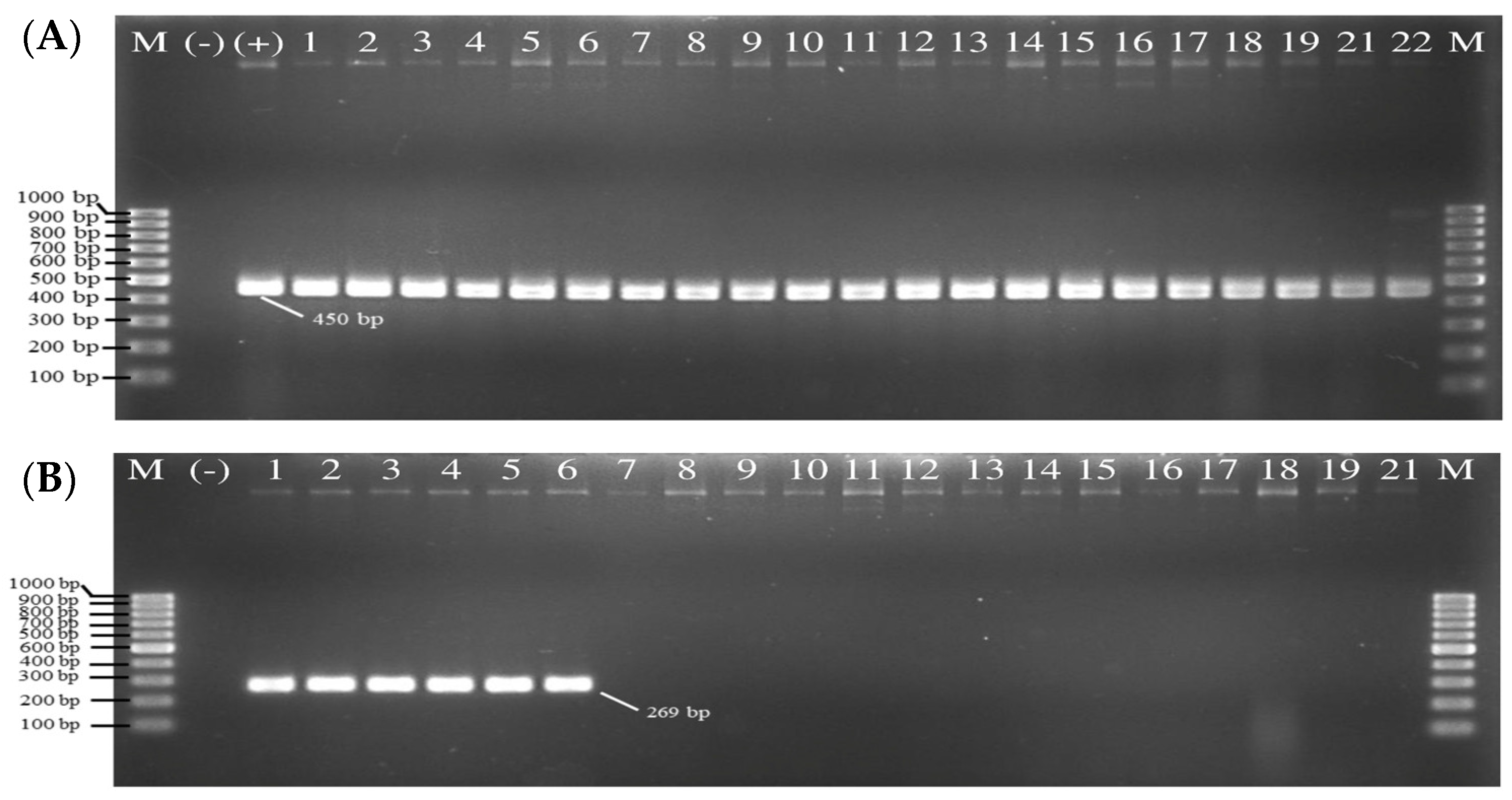
All fifty collected isolates from Asian green mussel samples, clinical and laboratory strains, were identified as V. parahaemolyticus based on their morphological and biochemical characteristics. Double-plating method was used to identify species involving TCBS and CHROMagar™ Vibrio agars, the selective media providing the direct colony-color-based identification of V. parahaemolyticus by specific color development of the particular colonies. Out of 26 Asian green mussel collected samples, V. parahaemolyticus was detected in all the samples on TCBS agar (Figure 2A) and CHROMagarTM Vibrio agar (Figure 2B). Colonies of fifty V. parahaemolyticus isolates appeared. All V. parahaemolyticus colonies were spherical, transparent, and bluish-green or green color on TCBS plates. On CHROMagarTM plates, the colonies of fifty isolates were round, smooth, flat, mauve or purple red or purplish cream colony in color (positive colony = mauve color). On CHROMagarTM Vibrio agar, the colony colors of 50 V. parahaemolyticus were varied. No.1 was a laboratory strain (PSU.SCB.16S.14); No. 2–44 were V. parahaemolyticus isolated from Asian green mussel; and No. 45–50 were V. parahaemolyticus isolated from clinical samples. Lee et al. [36] found that 4 (10.5%) of the 38 V. parahaemolyticus strains had white colonies on ChromoVP agar. Su et al. [37] documented that 5% of V. parahaemolyticus strains appeared as white colonies on Bio-Chrome Vibrio medium. High variability and differential colony colors were observed when culture-based techniques were used for seafood, clinical, and environmental samples. Hence, molecular confirmation must be conducted to ensure the accurate detection of V. parahaemolyticus. All the isolates were also confirmed by MALDI Biotyper® analysis and thermolabile hemolysin encoded by the tlh gene as species marker. As shown in Figure 3A, 100% of the 49 isolates including a positive laboratory strain (PSU.SCB.16S.14) were tlh-positive. The salt tolerance test also showed that all the recovered strains required sodium ions for their growth in media supplemented with 1% NaCl up to 8%. The result was in agreement with that reported by Beleneva et al. [38]. However, the isolates should be collected from other provinces or different geographic locations to acquire more data, in which a variety and abundance of strains can be gained.

3.2. Virulence, Molecular and Biochemical Characteristics of V. parahaemolyticus Isolates

3.2.1. Virulence Genes

All fifty isolates identified as V. parahaemolyticus by biochemical, MALDI-Biotyper® system tests and confirmed by PCR were detected for the tdh and trh genes. DNA fragments of 269 and 500 bp in size were produced from the amplification of V. parahaemolyticus pathogenic tdh and trh genes, respectively (Table 2). Six out of fifty (12%) samples were positive for the tdh gene (tdh+trh) (Figure 3B). However, the isolates of V. parahaemolyticus having both tdh+trh+ and tdhtrh+ were not detected in this study. Most V. parahaemolyticus clinical isolates had positive result for KP test (Figure 4), thus confirming the presence of hemolysin tdh and/or trh genes [39]. Most V. parahaemolyticus isolated from food and environment do not carry tdh and/or trh genes [39]. V. parahaemolyticus strains having tdh gene and strains possessing both tdh and trh genes were found at very low level in mussel [40,41]. Vibrio species was isolated from bivalves and the culture environments along the Gyeongnam coast in Korea [42]. One hundred and ninety isolates of V. parahaemolyticus from oyster, mussel, and ark shell were negative for the tdh virulence genes, while 18 (9.5%) isolates were positive for trh virulence genes. All strains were positive for the trh gene when isolated from only oyster samples [42]. No trh+ V. parahaemolyticus strains was detected in warm climate, including Thailand. Rodriguez-Castro et al. [43] found that trh+ strains were dominant in the cold water, whereas tdh+ V. parahaemolyticus disseminated in warm water. Only clinical V. parahaemolyticus strains carried the trh+ genes. Bhoopong et al. [44] documented that only 0.5% (3/629) of the clinical V. parahaemolyticus isolates from the 63 patients in Thailand carried the trh gene alone, whereas 87.4% (550/629) and 7% (44/629) of the isolates possessed the tdh gene and both genes, respectively. Chen et al. [45] found that 93% and 1% of the 501 clinical V. parahaemolyticus isolates from southeastern China carried tdh and trh genes, respectively. Nevertheless, distributions of tdh+ and/or trh+ strains may vary, depending on detection method, sample sources and geographical origin [46].

3.2.2. Hemolytic Activity

KP test was used to determine hemolytic activity of the isolates on the Wagatsuma agar containing 5% RBCs as depicted in Figure 4. Based on KP, the pathogenic isolates of V. parahaemolyticus could be differentiated from non-pathogenic counterpart. When bacterium lyses human erythrocytes, a pore-forming toxin known as the thermostable direct hemolysin (TDH) was produced. As shown in Figure 4, no Asian green mussel isolates showed β-hemolysis, whereas all of clinical isolates exhibited β-hemolysis; the latter were isolated from the stool of patients. All the tdh+trh isolates displayed a positive reaction as evidenced by a β-hemolysis zone surrounding the growth spot, whereas all the tdhtrh isolates showed the negative reaction. Although four isolates namely M6, M13, M48 and M58 from Asian green mussel exhibited weak hemolysis (Figure 4), none of them exhibited strong β-hemolysis. This weak hemolysis might be related with other virulence factors, apart from TDH orTRH. Strains, which produce few extracellular enzymes, could have the weak hemolysis [47]. Although no isolates showed β-hemolysis activity, the potential risk involved in consuming Asian green mussel must be taken into consideration because of its short generation time.

3.2.3. Motility Ability

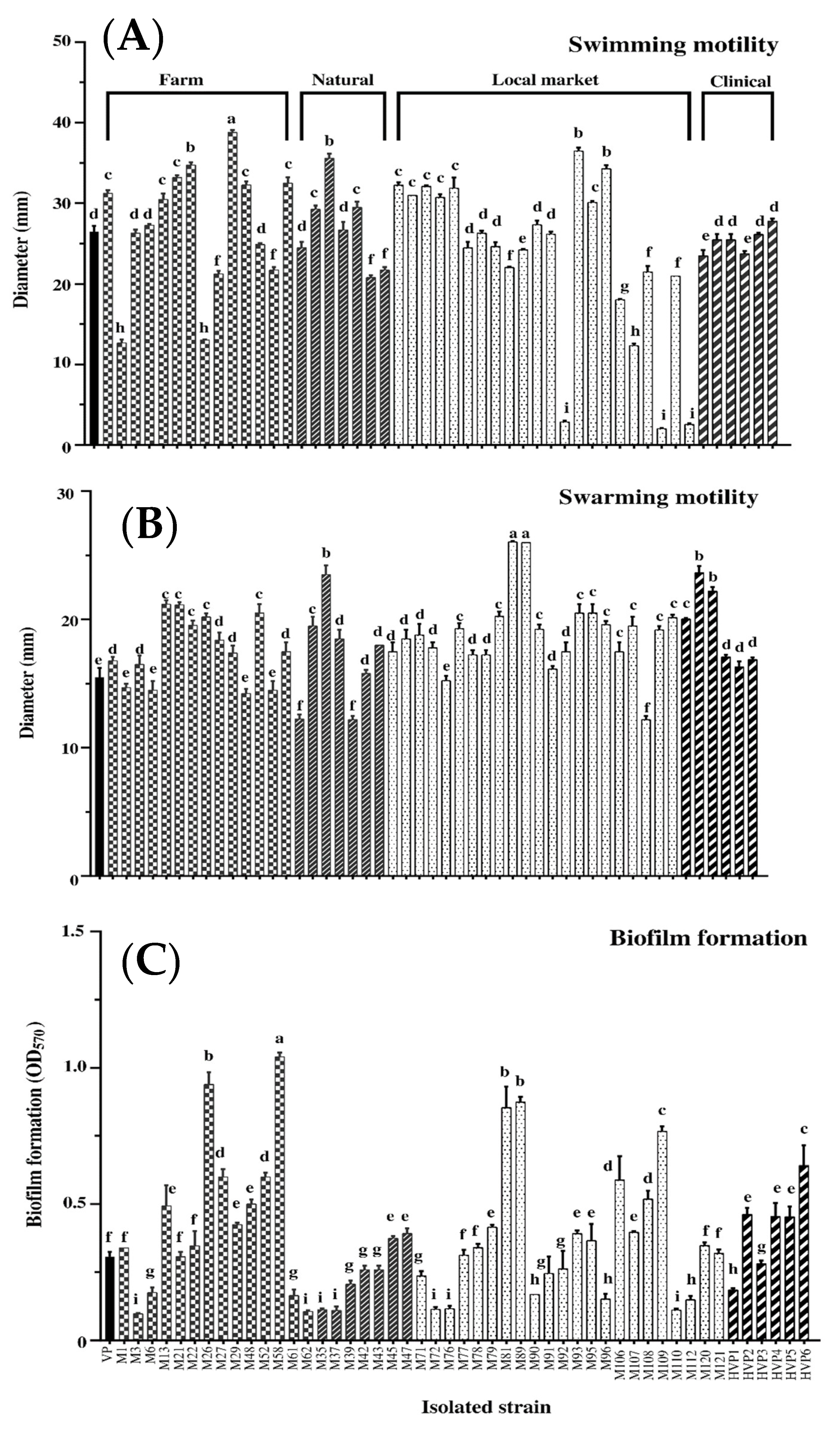
V. parahaemolyticus has dual flagellar systems, i.e., a single polar flagellum for swimming in liquid and peritrichous lateral flagella for swarming on surfaces [48]. In the present study, swimming and swarming of clinical and Asian green mussel isolates were compared. Mobility abilities of 50 isolates could be classified into three levels: weak, medium, and strong, which respectively indicated that their mobilities were much lower, similar to and significantly higher than those of laboratory strains of V. parahaemolyticus PSU.SCB.16S.14. As shown in Figure 5A, all the 50 isolates were swimmers; 7 isolates were weak swimmers (< 15 mm); 26 isolates were moderate swimmers (< 30 mm); and 17 isolates were strong swimmers (> 30 mm). Similarly, all isolates were swarmers (Figure 5B). Among all isolates, 43 isolates were moderates swarm cells; and 7 isolates were strong swarm cells. Thus, all isolates showed a relatively strong mobility. V. parahaemolyticus could move via propelling with the aid of flagella. Swimming and swarming behaviors are initial requirement for biofilm formation [49]. All V. parahaemolyticus isolates had relatively strong mobility, associated with their biofilm formation.

3.2.4. Biofilm Formation Capacity

The bacterial biofilm protects pathogens from environmental stress such as antimicrobial and increases disease severity in infected host [50,51,52]. The biofilm was formed by 50 isolates when tested using the CV staining (Figure 5C). V. parahaemolyticus was able to form biofilms and attached to the surfaces of seafood [53]. Sun et al. [54] found that V. parahaemolyticus isolated from stool specimens of diarrhea patients exhibited biofilm formation. All clinical V. parahaemolyticus isolates were biofilm producers. Biofilm formation is governed by the source of isolates and cultural temperature. In general, pathogenic isolates produced more biofilms than non-pathogenic counterpart [55,56]. Optimum temperature for biofilm formation by V. parahaemolyticus was 37 °C [57]. In general, bacterial cells entrapped in biofilms are more resistant to harsh conditions [53].

3.3. Antimicrobial Activity of COS-Tea Polyphenol Conjugates toward V. parahaemolyticus Isolates

Antimicrobial effects of COS-tea polyphenol conjugates on clinical and Asian green mussel V. parahaemolyticus isolates were examined. Antimicrobial activity was expressed as MIC and MBC of COS-tea polyphenol conjugates against 50 V. parahaemolyticus isolates. COS-tea polyphenol conjugates showed the adverse effect on the growth of V. parahaemolyticus isolated from clinical and Asian green mussel samples. COS–CAT had MIC (0.128–1.024 mg/mL) and MBC (0.256–2.048 mg/mL), whereas COS–EGCG possessed MIC (0.032–0.128 mg/mL) and MBC (0.256–2.048 mg/mL). The MBC/MIC ratio has been used to determine the antibacterial potential of substances. MBC lower than 1.024 mg/mL was observed for both COS-CAT and COS-EGCG. COS-CAT showed a MBC/MIC ratio of ≤ 4 against 19 tested isolates including M1, M3, M21, M26, M42, M45, M47, M71, M72, M77, M89, M91, M92, M95, M106, M112, M121, HVP1, and HVP 2. COS-EGCG had a MBC/MIC ratio of ≤ 4 toward seven tested isolates involving M3, M21, M26, M89, M109, M121, and HVP2. The COS-CAT showed a MBC/MIC ratio of ≤ 8 against eight tested isolates, which included M6, M37, M43, M52, M58, M95, HVP3, and HVP5, whereas COS- EGCG had a MBC/MIC ratio of ≤ 8 against 14 tested isolates, which consisted of M1, M42, M77, M78, M79, M90, M91, M92, M95, M106, M112, M120, HVP1 and HVP5. COS-polyphenol conjugates have the potential to control foodborne pathogens [20,21]. Recently, Mittal et al. [21] reported that COS-CAT conjugate showed higher antioxidant and antimicrobial activities than COS and other COS-polyphenol conjugates. COS-CAT showed antimicrobial activity against both Gram-negative and Gram-positive bacteria. Antimicrobial activity of COS and polyphenols was linked to bacterial cell wall disintegration. Furthermore, changes in microbial DNA, mRNA, and protein synthesis via diffused COS and polyphenols could bring about the cell death [20,21]. Blueberry extract showed stronger antimicrobial effect on V. parahaemolyticus, which had no virulence genes than V. parahaemolyticus ATCC17802 (tdh/trh+) and ATCC 33847 (tdh+/trh), which had virulence genes. Increased virulence was associated with augmented antibiotic resistance [41]. COS-tea polyphenol conjugates as a bactericidal/bacteriostatic substance might have stronger antibacterial activity against a virulent strain. Antimicrobial activity of COS polyphenol conjugates against V. parahaemolyticus PSU.SCB.16S, MIC and MBC were 32 μg/mL and 64 μg/mL for the COS-CAT, respectively; and MIC and MBC of 64 μg/mL and 128 μg/mL were recorded for the COS-EGCG, respectively [21]. Gram-negative bacteria generally have hydrophilic thin outer membrane, comprising lipopolysaccharides. Therefore, they are susceptible to cellular lysis via COS and its polyphenol conjugates [58]. COS-tea polyphenol, especially COS-CAT, was a promising antimicrobial agent toward both spoilage and pathogenic bacteria. Sun et al. [59] reported that tolC gene expression was downregulated in V. parahaemolyticus F13. Although MIC and MBC of COS-CAT were higher than those of some antibiotics, it had the efficacy in inhibiting both pathogenic and non-pathogenic V. parahaemolyticus. Overall, non-pathogenic V. parahaemolyticus generally had more sensitivity to antibiotics than pathogenic V. parahaemolyticus. However, pathogenic and non-pathogenic V. parahaemolyticus were similarly susceptible to COS-CAT in the present study.

3.4. Antibiotic Susceptibility Profile of Different V. parahaemolyticus Isolates

Eight V. parahaemolyticus isolates represented V. parahaemolyticus PSU.SCB.16S.14 (Laboratory strain, VP), Asian green mussel from farm (M1 isolate), Asian green mussel from natural habitat (M42 isolate), Asian green mussel from local markets (M77, M91, M92, and M106 isolates), and clinical sample of stool specimens of diarrhea patients (HVP1 isolate) were used for testing. MBCs of COS-tea polyphenol conjugates against all V. parahaemolyticus (8 strains) were 1.024 mg/mL as shown in Table 3. Varying antibiotic susceptibility profiles with 21 antibiotics toward those eight isolates were noticeable (Table 4). Seven antibiotics namely ampicillin, cefoxitin, ceftriaxone, colistin, doripenem, ertapenem, and netilmicin did not show susceptible, intermediate, and resistant results because CLSI breakpoints of these antibiotics did not exist for V. parahaemolyticus [60]. Isolates tested were highly susceptible to antibiotics such as Amikacin (100%), ciprofloxacin (100%), gentamicin (100%), imipenem (100%), and levofloxacin (100%). However, HPV1 clinical isolate showed high resistance to amoxicillin/clavulanic acid, ampicillin/sulbactam, cefepime, cefotaxime, ceftazidime, cefuroxime, meropenem, piperacillin/tazobactam, and trimethoprim/sulfamethoxazole with MAR index of 0.64. This isolate was resistance to 9 antibiotics of 14 antibiotics tested. Most of the six Asian green mussel V. parahaemolyticus isolates in this study exhibited high sensitivity to all antibiotics, but M42 isolate exhibited intermediate resistance to cefuroxime. Elexson et al. [57] found that all V. parahaemolyticus isolates from cultured seafood products were resistant to penicillin and ampicillin. However, it has been discovered that the Asian green mussel cultivated in Thailand is frequently an open system culture in coasts and estuaries, in which antibiotics are not required. As a result, no drug resistant V. parahaemolyticus isolated from natural Asian green mussel farms and Asian green mussels sold in the local market was found in this study. Another health risk may arise with cross-contamination by shellfish to other seafoods in the market. To address the potential consequences of pathogenic V. parahaemolyticus in seafood, continuous monitoring of environmental and seafood samples, including mussel as well as tracking the source of clinical and environmental strains are still needed.

4. Conclusions

Fifty collected isolates including Asian green mussel samples, clinical and laboratory strains were identified as V. parahaemolyticus based on their morphological, biochemical, and molecular characteristics. They were all biofilm producers with strong motile ability. Only six isolates (12%) from the clinical sample were positive for the virulence tdh gene (tdh+trh) and had a positive result for the KP test. COS-CAT demonstrated the greatest bactericidal action against V. parahaemolyticus isolated from Asian green mussels and clinical samples with an MBC value of 1.024 mg/mL. In addition, V. parahaemolyticus isolated from Asian green mussel farms, natural habitat, and local markets showed no antibiotic resistance. Only the sample clinical isolates had a MAR value of 0.64 and were extremely resistant to nine kinds of antibiotics. Hence, to address the potential consequences of pathogenic V. parahaemolyticus in seafood, constant monitoring of environmental and seafood samples is still essential for food safety assurance.

Author Contributions

Conceptualization, S.P., M.Y., J.S., J.B. and S.B.; methodology— review and editing, S.P., A.M., M.Y., J.S. and J.B.; validation, S.B.; formal analysis, S.P. and M.Y.; investigation, S.P. and S.B.; data curation, S.P.; resources, A.M., M.Y. and S.B.; writing—original draft, S.P.; writing—review and editing, A.T., P.S. and S.B.; supervision, S.B.; funding acquisition, S.B. All authors have read and agreed to the published version of the manuscript.

Funding

This study was funded by the National Research Council of Thailand (NRCT), Thailand under the International Research Network program (IRN) and Prachayacharn Grant awarded by Prince of Songkla University, Hat Yai, Thailand (Grant no. AGR6502111N).

Data Availability Statement

Data is contained within the article or supplementary material.

Acknowledgments

The authors would like to thank the International Center of Excellence in Seafood Science and Innovation, Faculty of Agro-Industry, Prince of Songkla University for the use of all facilities.

Conflicts of Interest

The authors declare no conflict of interest.

References

  1. Eli, J.; Exue, F.; Eyang, Z.; Ezhang, X.; Ezeng, D.; Echao, G.; Ejiang, Y.; Eli, B. Vibrio parahaemolyticus Strains of Pandemic Serotypes Identified from Clinical and Environmental Samples from Jiangsu, China. Front. Microbiol. 2016, 7, 787. [Google Scholar] [CrossRef]
  2. Liu, H.; Wang, Y.; Cao, J.; Jiang, H.; Yao, J.; Gong, G.; Chen, X.; Xu, W.; He, X. Antimicrobial activity and virulence attenuation of citral against the fish pathogen Vibrio alginolyticus. Aquaculture 2020, 515, 734578. [Google Scholar] [CrossRef]
  3. Kaneko, T.; Colwell, R.R. Ecology of Vibrio parahaemolyticus in Chesapeake Bay. J. Bacteriol. 1973, 113, 24–32. [Google Scholar] [CrossRef] [PubMed] [Green Version]
  4. Givens, C.E.; Bowers, J.C.; DePaola, A.; Hollibaugh, J.T.; Jones, J.L. Occurrence and distribution of Vibrio vulnificus and Vibrio parahaemolyticus—Potential roles for fish, oyster, sediment and water. Lett. Appl. Microbiol. 2014, 58, 503–510. [Google Scholar] [CrossRef]
  5. Mahmoud, B.S. The efficacy of grape seed extract, citric acid and lactic acid on the inactivation of Vibrio parahaemolyticus in shucked oysters. Food Control 2014, 41, 13–16. [Google Scholar] [CrossRef]
  6. Odeyemi, O.A. Incidence and prevalence of Vibrio parahaemolyticus in seafood: A systematic review and meta-analysis. Springerplus 2016, 5, 464. [Google Scholar] [CrossRef] [Green Version]
  7. Froelich, B.A.; Phippen, B.; Fowler, P.; Noble, R.T.; Oliver, J.D. Differences in Abundances of Total Vibrio spp., V. vulnificus, and V. parahaemolyticus in Clams and Oysters in North Carolina. Appl. Environ. Microbiol. 2017, 83, e02265-16. [Google Scholar] [CrossRef] [Green Version]
  8. Paranjpye, R.N.; Nilsson, W.B.; Liermann, M.; Hilborn, E.D.; George, B.J.; Li, Q.; Bill, B.D.; Trainer, V.L.; Strom, M.S.; Sandifer, P.A. Environmental influences on the seasonal distribution of Vibrio parahaemolyticus in the Pacific Northwest of the USA. FEMS Microbiol. Ecol. 2015, 91, fiv121. [Google Scholar] [CrossRef] [Green Version]
  9. Letchumanan, V.; Pusparajah, P.; Tan, T.H.; Yin, W.F.; Lee, L.H.; Chan, K.G. Occurrence and Antibiotic Resistance of V. para-haemolyticus from Shellfish in Selangor, Malaysia. Front. Microbiol. 2015, 6, 1417. [Google Scholar] [CrossRef] [Green Version]
  10. Eschbach, E.; Martin, A.; Huhn, J.; Seidel, C.; Heuer, R.; Schumacher, J.-H.; Ulrich, S.; Axe, J.-O.; Konietzny, A.; Strauch, E.; et al. Detection of enteropathogenic Vibrio parahaemolyticus, Vibrio cholerae and Vibrio vulnificus: Performance of real-time PCR kits in an interlaboratory study. Eur. Food Res. Technol. 2017, 243, 1335–1342. [Google Scholar] [CrossRef]
  11. Altekruse, S.F.; Bishop, R.D.; Baldy, L.M.; Thompson, S.G.; Wilson, S.A.; Ray, B.J.; Griffin, P.M. Vibrio gastroenteritis in the US Gulf of Mexico region: The role of raw oysters. Epidemiol. Infect. 2000, 124, 489–495. [Google Scholar] [CrossRef]
  12. Baker-Austin, C.; Oliver, J.D.; Alam, M.; Ali, A.; Waldor, M.K.; Qadri, F.; Martinez-Urtaza, J. Vibrio spp. infections. Nat. Rev. Dis. Primers 2018, 4, 1–19. [Google Scholar] [CrossRef]
  13. West, C.K.G.; Klein, S.L.; Lovell, C.R. High Frequency of Virulence Factor Genes tdh, trh, and tlh in Vibrio parahaemolyticus Strains Isolated from a Pristine Estuary. Appl. Environ. Microbiol. 2013, 79, 2247–2252. [Google Scholar] [CrossRef] [Green Version]
  14. Yan, J.; Nadell, C.D.; Stone, H.A.; Wingreen, N.S.; Bassler, B.L. Extracellular-matrix-mediated osmotic pressure drives Vibrio cholerae biofilm expansion and cheater exclusion. Nat. Commun. 2017, 8, 327. [Google Scholar] [CrossRef] [Green Version]
  15. Siddall, S.E. A Clarification of the Genus Perna (Mytilidae). Bull. Mar. Sci. 1980, 30, 858–870. [Google Scholar]
  16. Rajagopal, S.; Venugopalan, V.P.; van der Velde, G.; Jenner, H.A. Greening of the coasts: A review of the Perna viridis success story. Aquat. Ecol. 2006, 40, 273–297. [Google Scholar] [CrossRef]
  17. Nakaguchi, Y. Contamination by Vibrio parahaemolyticus and Its Virulent Strains in Seafood Marketed in Thailand, Vietnam, Malaysia, and Indonesia. Trop. Med. Health 2013, 41, 95–102. [Google Scholar] [CrossRef] [Green Version]
  18. Cruz, C.D.; Hedderley, D.; Fletcher, G.C. Long-Term Study of Vibrio parahaemolyticus Prevalence and Distribution in New Zealand Shellfish. Appl. Environ. Microbiol. 2015, 81, 2320–2327. [Google Scholar] [CrossRef] [Green Version]
  19. World Health Organization (WHO). Antimicrobial Resistance: Global Report on Surveillance. 2014. Available online: http://apps.who.int/iris/bitstream/handle/10665/112642/9789241564748_eng.pdf (accessed on 15 January 2019).
  20. Singh, A.; Benjakul, S.; Huda, N.; Xu, C.; Wu, P. Preparation and characterization of squid pen chitooligosaccharide–epigallocatechin gallate conjugates and their antioxidant and antimicrobial activities. RSC Adv. 2020, 10, 33196–33204. [Google Scholar] [CrossRef]
  21. Mittal, A.; Singh, A.; Zhang, B.; Visessanguan, W.; Benjakul, S. Chitooligosaccharide Conjugates Prepared Using Several Phenolic Compounds via Ascorbic Acid/H2O2 Free Radical Grafting: Characteristics, Antioxidant, Antidiabetic, and Antimicrobial Activities. Foods 2022, 11, 920. [Google Scholar] [CrossRef]
  22. Eom, T.-K.; Senevirathne, M.; Kim, S.-K. Synthesis of phenolic acid conjugated chitooligosaccharides and evaluation of their antioxidant activity. Environ. Toxicol. Pharmacol. 2012, 34, 519–527. [Google Scholar] [CrossRef] [PubMed]
  23. Chatterjee, N.S.; Panda, S.K.; Navitha, M.; Asha, K.; Anandan, R.; Mathew, S. Vanillic Acid and Coumaric Acid Grafted Chitosan Derivatives: Improved Grafting Ratio and Potential Application in Functional Food. J. Food Sci. Technol. 2015, 52, 7153–7162. [Google Scholar] [CrossRef]
  24. Park, H.-H.; Ko, S.-C.; Oh, G.-W.; Jang, Y.-M.; Kim, Y.-M.; Park, W.S.; Choi, I.-W.; Jung, W.-K. Characterization and biological activity of PVA hydrogel containing chitooligosaccharides conjugated with gallic acid. Carbohydr. Polym. 2018, 198, 197–205. [Google Scholar] [CrossRef] [PubMed]
  25. Moussa, M.; Cauvin, E.; Le Piouffle, A.; Lucas, O.; Bidault, A.; Paillard, C.; Benoit, F.; Thuillier, B.; Treilles, M.; Travers, M.A.; et al. A MALDI-TOF MS database for fast identification of Vibrio spp. potentially pathogenic to marine mollusks. Appl. Microbiol. Biotechnol. 2021, 105, 2527–2539. [Google Scholar] [CrossRef] [PubMed]
  26. ISO 21872-1:2017; Microbiology of the Food Chain—Horizontal Method for the Determination of Vibrio Spp.—Part 1, Detection of Potentially Enteropathogenic V. parahaemolyticus, Vibrio cholerae and Vibrio vulnificus. International Organization for Standardization: Geneva, Switzerland, 2017.
  27. Siddique, A.B.; Moniruzzaman, M.; Ali, S.; Dewan, N.; Islam, M.R.; Islam, S.; Amin, M.B.; Mondal, D.; Parvez, A.K.; Mahmud, Z.H. Characterization of Pathogenic Vibrio parahaemolyticus Isolated From Fish Aquaculture of the Southwest Coastal Area of Bangladesh. Front. Microbiol. 2021, 12, 635539. [Google Scholar] [CrossRef]
  28. Singh, A.; Mittal, A.; Benjakul, S. Chitosan, Chitooligosaccharides and Their Polyphenol Conjugates: Preparation, Bioactivities, Functionalities and Applications in Food Systems. Food Rev. Int. 2021, 1–23. [Google Scholar] [CrossRef]
  29. Herigstad, B.; Hamilton, M.; Heersink, J. How to optimize the drop plate method for enumerating bacteria. J. Microbiol. Methods 2001, 44, 121–129. [Google Scholar] [CrossRef]
  30. Wang, L.; Ling, Y.; Jiang, H.; Qiu, Y.; Qiu, J.; Chen, H.; Yang, R.; Zhou, D. AphA is required for biofilm formation, motility, and virulence in pandemic Vibrio parahaemolyticus. Int. J. Food Microbiol. 2013, 160, 245–251. [Google Scholar] [CrossRef]
  31. Fang, M.; Wang, R.; Agyekumwaa, A.K.; Yu, Y.; Xiao, X. Antibacterial effect of phenyllactic acid against Vibrio parahaemolyticus and its application on raw salmon fillets. LWT 2022, 154, 112586. [Google Scholar] [CrossRef]
  32. Zhang, Y.; Hu, L.; Osei-Adjei, G.; Zhang, Y.; Yang, W.; Yin, Z.; Lu, R.; Sheng, X.; Yang, R.; Huang, X.; et al. Autoregulation of ToxR and Its Regulatory Actions on Major Virulence Gene Loci in Vibrio parahaemolyticus. Front. Cell. Infect. Microbiol. 2018, 8, 291. [Google Scholar] [CrossRef]
  33. Shaw, K.S.; Goldstein, R.E.R.; He, X.; Jacobs, J.M.; Crump, B.C.; Sapkota, A.R. Antimicrobial Susceptibility of Vibrio vulnificus and Vibrio parahaemolyticus Recovered from Recreational and Commercial Areas of Chesapeake Bay and Maryland Coastal Bays. PLoS ONE 2014, 9, e89616. [Google Scholar] [CrossRef]
  34. CLSI. Methods for Antimicrobial Dilution and Disk Susceptibility Testing of Infrequently Isolated or Fastidious Bacteria; Approved Guideline-Second Edition (M45-A2); Clinical and Laboratory Standards Institute: Wayne, PA, USA, 2010. [Google Scholar]
  35. Krumperman, P.H. Multiple antibiotic resistance indexing of Escherichia coli to identify high-risk sources of fecal contamination of foods. Appl. Environ. Microbiol. 1983, 46, 165–170. [Google Scholar] [CrossRef] [Green Version]
  36. Lee, J.-M.; Azizah, R.N.; Kim, K.-S. Comparative evaluation of three agar media-based methods for presumptive identification of seafood-originated Vibrio parahaemolyticus strains. Food Control 2020, 116, 107308. [Google Scholar] [CrossRef]
  37. Su, Y.-C.; Duan, J.; Wu, W.-H. Selectivity and Specificity of a Chromogenic Medium for Detecting Vibrio parahaemolyticus. J. Food Prot. 2005, 68, 1454–1456. [Google Scholar] [CrossRef]
  38. Beleneva, I.A.; Maslennikova, E.F.; Magarlamov, T.Y. Physiological and Biochemical Characteristics of the Halophilic Bacteria Vibrio parahaemolyticus and V. alginolyticus Isolated from Marine Invertebrates of Peter the Great Bay, Sea of Japan. Russ. J. Mar. Biol. 2004, 30, 96–100. [Google Scholar] [CrossRef]
  39. Tan, C.W.; Malcolm, T.T.H.; Kuan, C.H.; Thung, T.Y.; Chang, W.S.; Loo, Y.Y.; Premarathne, J.M.K.J.K.; Ramzi, O.B.; Norshafawatie, M.F.S.; Yusralimuna, N.; et al. Prevalence and Antimicrobial Susceptibility of Vibrio parahaemolyticus Isolated from Short Mackerels (Rastrelliger brachysoma) in Malaysia. Front. Microbiol. 2017, 8, 1087. [Google Scholar] [CrossRef] [Green Version]
  40. Kang, C.-H.; Shin, Y.; Jang, S.; Yu, H.; Kim, S.; An, S.; Park, K.; So, J.-S. Characterization of Vibrio parahaemolyticus isolated from oysters in Korea: Resistance to various antibiotics and prevalence of virulence genes. Mar. Pollut. Bull. 2017, 118, 261–266. [Google Scholar] [CrossRef]
  41. Jiang, Y.; Chu, Y.; Xie, G.; Li, F.; Wang, L.; Huang, J.; Zhai, Y.; Yao, L. Antimicrobial resistance, virulence and genetic relationship of Vibrio parahaemolyticus in seafood from coasts of Bohai Sea and Yellow Sea, China. Int. J. Food Microbiol. 2019, 290, 116–124. [Google Scholar] [CrossRef]
  42. Mok, J.S.; Ryu, A.; Kwon, J.Y.; Kim, B.; Park, K. Distribution of Vibrio species isolated from bivalves and bivalve culture environments along the Gyeongnam coast in Korea: Virulence and antimicrobial resistance of Vibrio parahaemolyticus isolates. Food Control 2019, 106, 106697. [Google Scholar] [CrossRef]
  43. Rodriguez-Castro, A.; Ansede-Bermejo, J.; Blanco-Abad, V.; Varela-Pet, J.; Garcia-Martin, O.; Martinez-Urtaza, J. Prevalence and Genetic Diversity of Pathogenic Populations of Vibrio parahaemolyticus in Coastal Waters of Galicia, Spain. Environ. Microbiol. Rep. 2010, 2, 58–66. [Google Scholar] [CrossRef]
  44. Bhoopong, P.; Palittapongarnpim, P.; Pomwised, R.; Kiatkittipong, A.; Kamruzzaman, M.; Nakaguchi, Y.; Nishibuchi, M.; Ishibashi, M.; Vuddhakul, V. Variability of Properties of Vibrio parahaemolyticus Strains Isolated from Individual Patients. J. Clin. Microbiol. 2007, 45, 1544–1550. [Google Scholar] [CrossRef] [PubMed]
  45. Chen, Y.; Chen, X.; Yu, F.; Wu, M.; Wang, R.; Zheng, S.; Han, D.; Yang, Q.; Kong, H.; Zhou, F.; et al. Serology, virulence, antimicrobial susceptibility and molecular characteristics of clinical Vibrio parahaemolyticus strains circulating in southeastern China from 2009 to 2013. Clin. Microbiol. Infect. 2016, 22, 258.e9–258.e16. [Google Scholar] [CrossRef] [PubMed] [Green Version]
  46. Raghunath, P. Roles of Thermostable Direct Hemolysin (TDH) and TDH-Related Hemolysin (TRH) in V. parahaemolyticus. Front. Microbiol. 2015, 5, 805. [Google Scholar] [CrossRef] [PubMed]
  47. Lee, C.-Y.; Cheng, M.-F.; Yu, M.-S.; Pan, M.-J. Purification and characterization of a putative virulence factor, serine protease, from Vibrio parahaemolyticus. FEMS Microbiol. Lett. 2002, 209, 31–37. [Google Scholar] [CrossRef] [PubMed] [Green Version]
  48. McCarter, L.L. Dual Flagellar Systems Enable Motility under Different Circumstances. Microb. Physiol. 2004, 7, 18–29. [Google Scholar] [CrossRef]
  49. Yildiz, F.H.; Visick, K.L. Vibrio biofilms: So much the same yet so different. Trends Microbiol. 2009, 17, 109–118. [Google Scholar] [CrossRef] [Green Version]
  50. Simões, M.; Cleto, S.; Pereira, M.O.; Vieira, M.J. Influence of Biofilm Composition on the Resistance to Detachment. Water Sci. Technol. 2007, 55, 473–480. [Google Scholar] [CrossRef]
  51. Flemming, H.C.; Wingender, J. The Biofilm Matrix. Nat. Rev. Microbiol. 2010, 8, 623–633. [Google Scholar] [CrossRef]
  52. Mizan, M.F.R.; Jahid, I.K.; Ha, S.-D. Microbial Biofilms in Seafood: A Food-Hygiene Challenge. Food Microbiol. 2015, 49, 41–55. [Google Scholar] [CrossRef]
  53. Ashrafudoulla, M.; Mizan, F.R.; Park, S.H.; Ha, S.-D. Current and future perspectives for controlling Vibrio biofilms in the seafood industry: A comprehensive review. Crit. Rev. Food Sci. Nutr. 2021, 61, 1827–1851. [Google Scholar] [CrossRef]
  54. Sun, J.; Li, X.; Hu, Z.; Xue, X.; Zhang, M.; Wu, Q.; Zhang, W.; Zhang, Y.; Lu, R. Characterization of Vibrio parahaemolyticus isolated from stool specimens of diarrhea patients in Nantong, Jiangsu, China during 2018–2020. PLoS ONE 2022, 17, e0273700. [Google Scholar] [CrossRef]
  55. Song, X.; Ma, Y.; Fu, J.; Zhao, A.; Guo, Z.; Malakar, P.K.; Pan, Y.; Zhao, Y. Effect of temperature on pathogenic and non-pathogenic Vibrio parahaemolyticus biofilm formation. Food Control 2017, 73, 485–491. [Google Scholar] [CrossRef]
  56. Ahmed, H.A.; El Bayomi, R.M.; Hussein, M.A.; Khedr, M.H.; Remela, E.M.A.; El-Ashram, A.M. Molecular characterization, antibiotic resistance pattern and biofilm formation of Vibrio parahaemolyticus and V. cholerae isolated from crustaceans and humans. Int. J. Food Microbiol. 2018, 274, 31–37. [Google Scholar] [CrossRef]
  57. Elexson, N.; Yaya, R.; Nor, A.M.; Kantilal, H.K.; Son, R. Biofilm Assessment of V. parahaemolyticus from Seafood Using Random Amplified Polymorphism DNA-PCR. Int. Food Res. J. 2014, 21, 59–65. [Google Scholar]
  58. Goy, R.C.; Morais, S.T.; Assis, O.B. Evaluation of the antimicrobial activity of chitosan and its quaternized derivative on E. coli and S. aureus growth. Rev. Bras. Farm. 2016, 26, 122–127. [Google Scholar] [CrossRef] [Green Version]
  59. Sun, X.-H.; Hao, L.-R.; Xie, Q.-C.; Lan, W.-Q.; Zhao, Y.; Pan, Y.-J.; Wu, V.C. Antimicrobial effects and membrane damage mechanism of blueberry (Vaccinium corymbosum L.) extract against Vibrio parahaemolyticus. Food Control 2020, 111, 107020. [Google Scholar] [CrossRef]
  60. CLSI. M100: Performance Standards for Antimicrobial Susceptibility Testing, 32nd ed.; Clinical and Laboratory Standards Institute: Wayne, PA, USA, 2022. [Google Scholar]
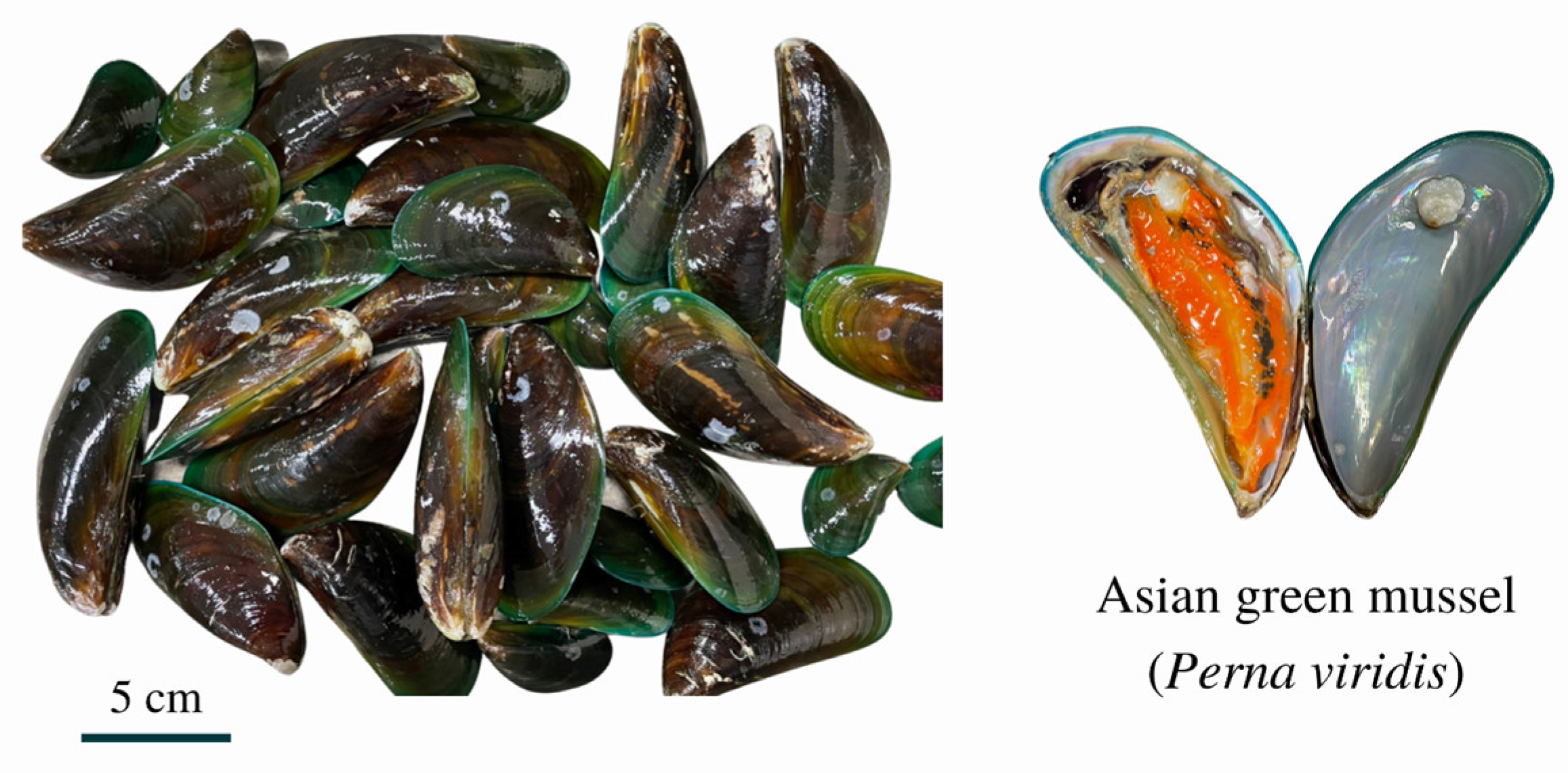
Figure 1. Asian green mussel (Perna viridis) collected from the south of Thailand.
Foods 11 04048 g001
Figure 2. Colony morphology of fifty V. parahaemolyticus isolates on thiosulphate citrate bile salts sucrose agar plate (A) and CHROMagarTM Vibrio agar plate (B).
Foods 11 04048 g002
Figure 3. Gel electrophoresis of products of tlh primer (A) (Lane M: DNA marker, Lanes 1–22: representative samples, Lane (−): negative-control (DNA free template), Lane (+): positive-control (V. parahaemolyticus PSU.SCB.16S.14) and products of tdh primer (B) Lane M: DNA marker, Lanes 1–6: representative isolates of pathogenic V. parahaemolyticus, Lanes 7–21: representative isolates of non-pathogenic V. parahaemolyticus, Lane (−): negative-control (DNA free template), Lane (+).
Foods 11 04048 g003
Figure 4. The hemolytic activity of V. parahaemolyticus isolates against RBCs. β-hemolysis zone surrounding the spot of growth on the Wagatsuma agar plate was measured. V. parahaemolyticus cells were grown on Wagatsuma agar for 24 h. * Isolates with weak hemolysis.
Foods 11 04048 g004
Figure 5. Swimming motility (A), swarming motility (B), and biofilm formation ability (C) of 50 isolates of V. parahaemolyticus. Different lowercase letters on the bars denote the significant differences (p < 0.05).
Foods 11 04048 g005
Table 1. Provinces, types of places, and number of V. parahaemolyticus isolates collected from the south of Thailand.
ProvincesTypes of Places CollectedNumber of Samples
SuratthaniLocal market2
Asian green mussel farm6
Natural habitat4
Trang Local market2
Asian green mussel farm4
Natural habitat2
SongkhlaLocal market8
Table 2. Primers selected for the detection of virulence genes of V. parahaemolyticus.
Primer Sequence (5′-3′)Amplicon Size (bp)Reference
tlhFAAA GCG GAT TAT GCA GAA GCA CTG450Siddique et al. [27]
RGCT ACT TTC TAG CAT TTT CTC TGC
tdhFCCA TCT GTC CCT TTT CCT GCC269Siddique et al. [27]
RCCA CTA CCA CTC TCA TAT GC
trhFTTG GCT TCG ATA TTT TCA GTA TCT500Siddique et al. [27]
RCAT AAC AAA CAT ATG CCC ATT TCC G
Table 3. Minimum inhibitory concentration (MIC) and minimum bactericidal concentration (MBC) of COS-tea polyphenol conjugates against eight isolates of V. parahaemolyticus.
Bacterial StrainsCOS-CAT (mg/mL)COS-EGCG (mg/mL)
MICMBCMICMBC
VP PSU.SCB.16S.140.2561.0240.0641.024
M10.2561.0240.1281.024
M420.2561.0240.1281.024
M770.2561.0240.1281.024
M910.2561.0240.1281.024
M920.2561.0240.1281.024
M1060.2561.0240.1281.024
HVP10.2561.0240.1281.024
Table 4. Twenty-one antibiotics resistance profiles of V. parahaemolyticus isolates from Asian green mussel, clinical, and laboratory samples.
AntibioticsConcentration
(μg/mL)
VPM1M42M77M91M92M106HVP1
Amikacin8–32SSSSSSSS
Amoxicillin/Clavulanic acid4/2–16/8SSSSSSSR
Ampicillin8–16NINININININININI
Ampicillin/Sulbactam4/2–16/8SSSSSSSR
Cefepime1–32SSSSSSSR
Cefotaxime1–32SSSSSSSR
Cefoxitin4–16NINININININININI
Ceftazidime1–32SSSSSSSR
Ceftriaxone0.5–32NINININININININI
Cefuroxime8–16SSISSSSR
Ciprofloxacin0.06–2SSSSSSSS
Colistin1–8NINININININININI
Doripenem0.5–16NINININININININI
Ertapenem0.5–4NINININININININI
Gentamicin2–8SSSSSSSS
Imipenem0.5–16SSSSSSSS
Levofloxacin0.06–8SSSSSSSS
Meropenem0.5–16SSSSSSSR
Netilmicin8–16NINININININININI
Piperacillin/Tazobactam8/4–64/4SSSSSSSR
Trimethoprim/Sulfamethoxazole1/19–4/76SSSSSSSR
S: Susceptible; I: Intermediate; R: Resistant; NI: No interpretation. Note: Eight V. parahaemolyticus isolates included V. parahaemolyticus PSU.SCB.16S.14 (VP), isolates from Asian green mussel from farm (M1 isolate), Asian green mussel from natural habitat (M42 isolate), Asian green mussel from local markets (M77, M91, M92, and M106 isolates) and clinical isolate from stool specimens of diarrhea patients (HVP1 isolate).
Publisher’s Note: MDPI stays neutral with regard to jurisdictional claims in published maps and institutional affiliations.