Article

The Galloyl Group Enhances the Inhibitory Activity of Catechins against LPS-Triggered Inflammation in RAW264.7 Cells

1
Guangdong Key Laboratory of Science and Technology of Lingnan Specialty Food, Zhongkai University of Agriculture and Engineering, Guangzhou 510225, China
2
Key Laboratory of Green Processing and Intelligent Manufacturing of Lingnan Specialty Food, Ministry of Agriculture, Zhongkai University of Agriculture and Engineering, Guangzhou 510225, China
*
Author to whom correspondence should be addressed.
Foods 2024, 13(16), 2616; https://doi.org/10.3390/foods13162616
Submission received: 22 July 2024 / Revised: 4 August 2024 / Accepted: 14 August 2024 / Published: 21 August 2024
(This article belongs to the Section Food Nutrition)

Abstract

:
The galloyl group in catechins was confirmed to be crucial for their health benefits. However, whether the catechins’ galloyl group had a contribution to their anti-inflammation remains unclear. This study investigated the anti-inflammation properties and mechanisms of catechins in RAW264.7 cells by using ELISA, fluorometry, flow cytometer, Western blot, and molecular docking. Results showed that the galloyl group enhanced the inhibitory abilities of catechins on inflammatory cytokines (NO, PGE2, IL-1β, and TNF-α) and ROS release in LPS-induced cells. This suppression was likely mediated by delaying cells from the G0/G1 to the S phase, blocking COX-2 and iNOS via the TLR4/MAPK/NF-κB pathway with PU.1 as an upstream target. The research proved that the existence of galloyl groups in catechins was indispensable for their anti-inflammatory capacities and offered a theoretical basis for the anti-inflammatory mechanism of galloylated catechins. Future research is needed to verify the anti-inflammatory effects of catechins in various sources of macrophages or the Caco-2/RAW264.7 cell co-culture system.

1. Introduction

Normally, inflammation facilitates an automatic defense response [1]. Nevertheless, exaggerated inflammation can cause pathological alterations, such as oxidative stress, extracellular matrix remodeling, angiogenesis, and fibrosis [2]. These changes will result in many chronic illnesses, such as type 2 diabetes, angiocardiopathy, and neurodegenerative disorders [3,4]. Pharmacological interference is deemed an effective strategy to control inflammation-associated diseases. Traditional treatments depended mainly on steroid and non-steroidal anti-inflammatory medicines; however, the side effects of both drugs are becoming increasingly commonplace [5]. At present, natural anti-inflammatory ingredients, including polyphenols and polysaccharides, draw broad attention because of their potential applications [6,7].
Lipopolysaccharide (LPS), a principal constituent of the outer membrane of Gram-negative bacteria, robustly triggers inflammatory signaling pathways in macrophages [8]. Inflammation in LPS-induced macrophages is initiated by the interaction of LPS with LPS-binding protein (LBP), and then cluster of differentiation 14 (CD14) transports LPS to the toll-like receptor 4 (TLR4)/myeloid differential protein 2 (MD2) complex [9]. TLR4/MD2 further activates the inflammatory cascade through both the myeloid differentiation primary response 88 (MyD88) and TIR-domain-containing adapter-inducing interferon-β (TRIF) [10]. This event triggers the downstream signaling cascades, especially the nuclear factor-kappa-B (NF-κB) and mitogen-activated protein kinases (MAPKs) pathways, subsequently accelerating the release of inflammatory cytokines (e.g., IL-1β and TNF-α) [11,12].
Green tea, made from the Camelia sinensis leaves, is a globally cherished natural beverage due to its diverse health-promoting benefits [13]. Green tea is particularly rich in polyphenols, including phenolic acids, flavonoids, procyanidins, etc. [14]. The catechins represent 80–90% of the green tea flavonoids, making up approximately 30% of tea leaves (dry weight) [15]. Tea catechins mainly contain epicatechin (EC) and epigallocatechin (EGC), which have no galloyl group, and epicatechin gallate (ECG) and epigallocatechin gallate (EGCG), which have one galloyl group at their 3-positions (Figure 1). Numerous studies have proved that the galloyl group in catechins played a decisive role in their biological activities, such as antioxidant [16], anti-obesity [17], antiviral [18], and anti-cancer effect [19]. Nevertheless, it has not yet been documented whether the galloyl group augmented the anti-inflammatory properties of catechins. Thus, this research aimed to evaluate the effects of the galloyl group on enhancing the anti-inflammatory capabilities of catechins in LPS-triggered RAW264.7 macrophages. Additionally, the anti-inflammation mechanisms of catechins were investigated by using ELISA, fluorometry, flow cytometer, Western blot, and molecular docking.

2. Materials and Methods

2.1. Chemicals and Reagents

Catechins were sourced from Yuanye Bio-Technology (Shanghai, China), with a purity of over 98%. Moreover, 3-(4,5-dimethylthiazole-2-yl)-2 and 5-diphenyltetrazolium bromide (MTT) were from Solarbio Science and Technology (Beijing, China). Antibodies were obtained from Cell Signaling Technology (Cambridge, MA, USA). Mouse PGE2, IL-1β, and TNF-α ELISA kits and reactive oxygen species (ROSs) detection kits were from Beyotime Biotechnology (Shanghai, China).

2.2. Cell Culture and Treatment

RAW264.7 cells were cultivated in 10% fetal bovine serum (FBS)-supplemented DMEM (Gibco, Miami, FL, USA), 100 U/mL of penicillin, and streptomycin at 37 °C with 5% CO2. After confluence, the cells were incubated in 10% FBS-supplemented DMEM with 1 μg/mL LPS (Sigma-Aldrich, St. Louis, MO, USA) for 24 h.

2.3. Cell Viability Assay

Cells were seeded at a density of 5 × 10³ cells/well into a 96-well plate. Upon reaching confluence, the cells were exposed to fresh medium containing 50–150 μM of catechins for 24 h. After that, cells were colored by MTT dye (0.5 mg/mL) for 4 h, and then the absorbance of DMSO-dissolved cells was read at OD570 nm.

2.4. Determination of Pro-Inflammatory Cytokines

As stated in the earlier reference [20], the measurement of NO in the cell culture medium was conducted by using the Griess method. The medium supernatant was combined with isopyknic Griess reagent, and then measured the absorbance at OD540 nm. The level of PGE2, IL-1β, and TNF-α in cell culture medium was analyzed by using the corresponding ELISA kit based on the standard procedure.

2.5. ROS Assay

Intracellular ROSs were detected as previously mentioned with a slight change [21]. RAW264.7 cells were treated with or without catechins (100 μM) and then marked by DCFH-DA probe (2.5 μM) for 15 min at 37 °C. The fluorescent signal was visualized by an OLYMPUS IX73 microscope (OLYMPUS, Tokyo, Japan), and the fluorescent strength was recorded by emission at 485 nm and excitation at 535 nm.

2.6. Cell Cycle Analysis

Quantitative analysis of the cell cycle was performed as previously mentioned [22]. RAW264.7 cells were intervened with or without catechins (100 μM) containing LPS (1 μg/mL) for 24 h. Next, macrophages were collected with PBS and fixed overnight in cold 75% EtOH, and then treated with RNase A (50 μg/mL) and PI (50 μg/mL). Cell cycle analysis was carried out using FACSVerse (BD Biosciences, Franklin Lakes, NJ, USA).

2.7. Immunoblot Analysis

Protein expression levels were assessed through immunoblot analysis as published [23]. The antibody details are as follows: rabbit anti-β-actin (GB15003, Servicebio), rabbit anti-MAPKs (#9926, CST), rabbit anti-NF-κB (#8242, CST), rabbit anti-Phospho-MAPKs (#9910, CST), rabbit anti-Phospho-NF-κB (#3033, CST), rabbit anti-COX-2 (GB115672, Servicebio), rabbit anti-iNOS (#2982, CST), rabbit anti-CD14 (#93882, CST), rabbit anti-TLR4 (#14358, CST), rabbit anti-MD-2 (ab24182, Abcam), rabbit anti-MyD88 (#4283, CST), rabbit anti-TRAF6 (#8028, CST), and anti-rabbit IgG (#7074, CST).

2.8. Molecular Docking

The molecular docking study was conducted to predict the interactions between PU.1-DNA (PDB ID: 1PUE) and catechins. The protein structure was prepared using Discovery Studio v.4.5, including water molecules deletion, hydrogen atom addition, and energy minimization. Ligands were prepared by using ChemBioDraw v.14.0. Docking was executed using AutoDock Vina v.1.2.0; the top-ranked poses were selected based on the lowest binding energy.

2.9. Statistical Analysis

GraphPad Prism 9.0 and SPSS 27.0 were adopted for data visualization and significance analysis. Results were shown as mean ± SD from no less than three experimental replications. Difference significance was analyzed using one-way ANOVA with Tukey’s post-test, with p < 0.05 indicating significance.

3. Results and Discussion

Prior studies have established that the catechins’ galloyl group augmented their biological activities, for example, anti-oxidative, anti-obesity, and anti-angiogenesis [24]. Nevertheless, the anti-inflammatory properties of galloylated catechins have yet to be elucidated. This study aimed to define the importance of the galloyl moiety for catechins’ anti-inflammatory efficacies. At first, the influences of four catechins on cell viability were measured by MTT colorimetry. Figure 2 presents that all the catechins were found to be non-cytotoxic within 50–150 μM. Therefore, these concentrations were selected for the follow-up study.
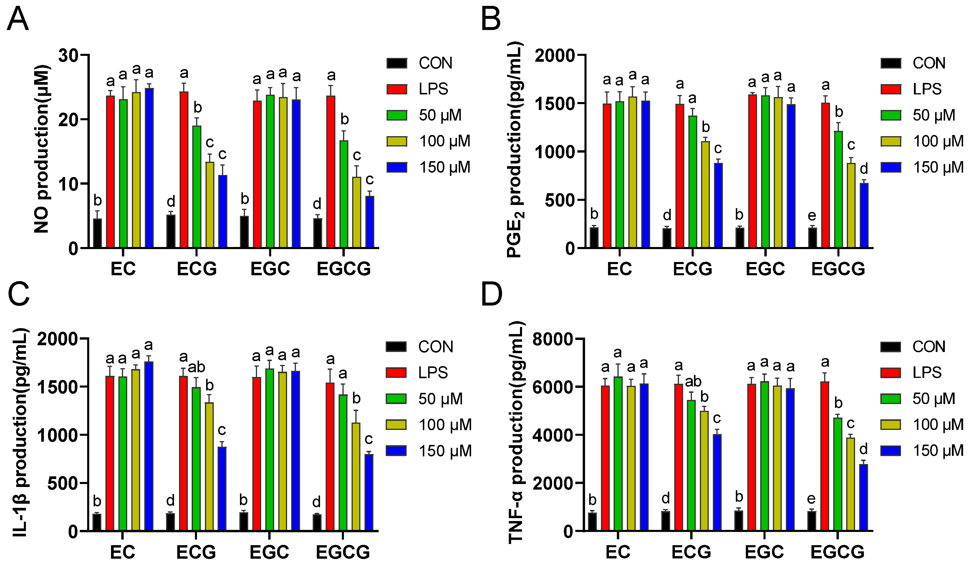
NO, PGE2, IL-1β, and TNF-α have been established as pivotal biomarkers of inflammation. The substantial secretion of these cytokines by macrophages can precipitate a myriad of diseases associated with inflammation [25,26]. Decreasing the pro-inflammatory agent levels is recognized as an efficacious strategy for mitigating diverse forms of inflammatory responses [27]. Therefore, the inhibitory activities of different catechins on NO, PGE2, IL-1β, and TNF-α release were evaluated in LPS-stimulated macrophages. Figure 3 shows that the levels of these pro-inflammatory factors were significantly (p < 0.05) improved after LPS treatment. However, the observed increases were concentration-dependently inhibited by galloylated catechins (ECG and EGCG) but not by non-galloylated catechins (EC and EGC). Yu et al. reported that the inflammatory agents (NO, IL-1β, TNF-α, and IL-6) scavenging abilities of procyanidin B2-3′-O-gallate (with one galloyl moiety) were superior to that of procyanidin B2 (without galloyl moiety) [28]. Thus, these results proved that the catechins’ galloyl group was instrumental in their capacity to exert anti-inflammatory effects.
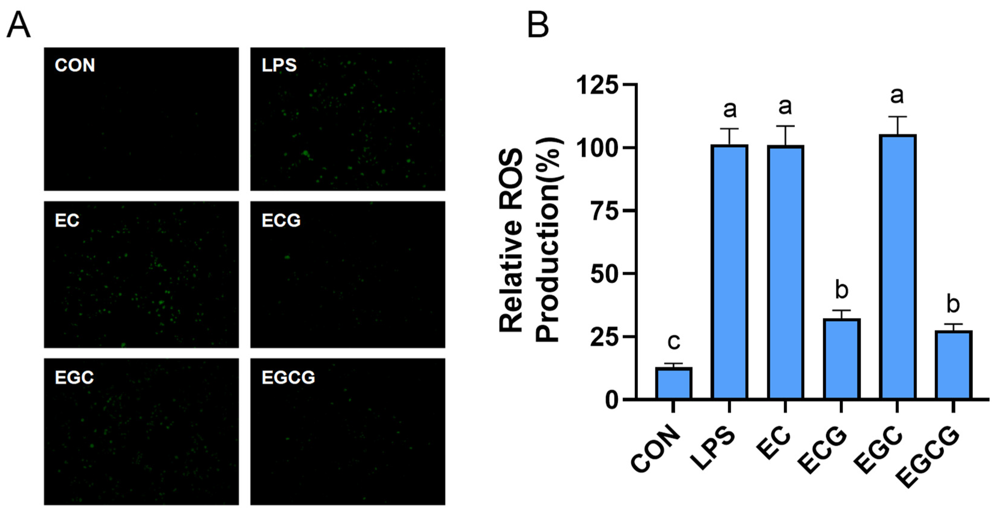
Once stimulated, inflammatory cells will release a variety of ROS, which can cause cell and tissue damage [29]. Excess ROS can also activate inflammation-related signaling pathways; thus, eliminating ROS is an effective way to control inflammatory diseases and their complications [30]. This research assessed the inhibition activities of catechins on the generation of ROS in LPS-triggered RAW264.7 cells. Figure 4 shows the ROS fluorescent intensity in LPS-induced cells was obviously higher than that in the non-treated cells (CON). By comparison, ECG and EGCG markedly (p < 0.05) reduced the fluorescence intensity, whereas EC and EGC did not exhibit any noticeable quenching effects. The findings indicated that the catechins’ galloyl group was essential for their ROS inhibition abilities. A previous study has confirmed that the free radical scavenging abilities of the galloylated catechins (ECG and EGCG) were superior to those of the non-galloylated molecules [16]. Thus, the elimination ability of free radicals might be a potential mechanism for ECG and EGCG in decreasing ROS.
Inflammation usually causes cell growth inhibition and even apoptosis [31]. The cell cycle involves several critical checkpoints, notably the G0/G1 to S and G2 to M phases. The G0/G1 to S transition is recognized as the most critical juncture in the process of DNA replication and mitosis [32]. In the present study, the impacts of four catechins on the cell cycle progression were examined in LPS-stimulated cells. Figure 5 shows that the cells increase in the G0/G1 phase (from 57.28% to 69.33%) and the cells decrease in the S phase (from 37.13% to 23.20%) were observed after LPS induction, suggesting that LPS prevented cells from entering the S phase. However, ECG and EGCG released the LPS-induced G0/G1 phase block, which was aligned with Li et al. (2022), who indicated that the anti-inflammatory activities of dandelion were achieved by relieving macrophage G0/G1 phase [33]. Compared to ECG and EGCG, EC and EGC exhibited minimal impacts. This proved that the galloyl group within catechins was pivotal in mitigating cell cycle arrest in LPS-induced RAW264.7 macrophages.
Inducible nitric oxide synthase (iNOS) serves as the primary regulator of NO production under pro-inflammatory conditions [34]. Cyclooxygenase-2 (COX-2) is the pro-inflammatory enzyme that mediates the conversion of arachidonic acid to prostaglandin H2, which functions as a precursor for the synthesis of PGE2, particularly in activated macrophages [35]. The excessive expression of iNOS and COX-2 can result in elevated levels of pro-inflammatory factors [36]. Therefore, this study further examined the effects of four catechins on the expression levels of iNOS and COX-2. Figure 6 shows that the iNOS and COX-2 expression levels in cells were prominently (p < 0.05) increased after LPS treatment. In contrast, ECG and EGCG markedly (p < 0.05) restrained these two markers, while no inhibition was found when intervened with EC and EGC. A previous study also proved that prodelphinidin B2 3,3′-di-O-gallate, having two galloyl groups, exhibited the strongest inhibitory ability against COX-2 expression in LPS-activated RAW264 cells, whereas prodelphinidin B2, having no galloyl group, failed to display an inhibition activity [37]. This suggested that the catechins’ galloyl group was vital for their inhibitory abilities on iNOS and COX-2.
NF-κB, a pivotal transcription factor, which modulates the expression of cell cycle regulatory proteins and pro-inflammatory mediators (iNOS and COX-2) [38]. In normal macrophages, NF-κB exists in the cytoplasm as an NF-κB/IκB complex. Once stimulated by LPS, NF-κB transcribes into the nucleus and triggers the excessive release of inflammation cytokines [39]. To ascertain the necessity of the catechins’ galloyl moiety for their anti-inflammatory activities via the NF-κB pathway, the NF-κB phosphorylation level was examined by using immunoblot. Figure 7A presents that the phosphorylated NF-κB level was prominently (p < 0.05) up-regulated after LPS stimulation. It was found that ECG and EGCG could dramatically (p < 0.05) inhibit NF-κB phosphorylation, while EC and EGC showed no inhibitory action. Wang et al. also found that ECG and EGCG reduced the secretion of inflammatory factors (IL-6, IL-1β, and TNF-α) in LPS-stimulated dental pulp cells by inhibiting activation of the NF-κB [40]. MAPKs, as a key regulator in the communication between cell surface receptors and nucleus DNA, which can transmit extracellular signals through continuous phosphorylation events in response to extracellular stimuli [41]. Previous research has shown that MAPKs activation can further phosphorylate NF-κB, which regulates inflammation-related downstream signaling [42]. Thus, this study further investigated the influences of four catechins on the phosphorylation of ERK, JNK, and p38 in LPS-induced macrophages. Figure 7B shows that LPS prominently (p < 0.05) activated the phosphorylation of ERK, JNK, and p38. However, ECG and EGCG markedly (p < 0.05) blocked the activation of ERK, JNK, and p38, whereas EC and EGC exhibited no suppressive effects. Liang et al. reported that EGCG blocked cigarette smoke medium-stimulated ROS and IL-8 production by suppressing ERK, p38 MAPK, and NF-κB signaling pathways in human AC16 cardiomyocytes [43]. Therefore, the above findings elucidated that the catechins’ galloyl moiety was indispensable for blocking MAPK/NF-κB activation in LPS-triggered cells, and the anti-inflammation action of galloylated catechins (ECG and EGCG) was achieved by restraining the MAPK/NF-κB pathway, consequently curbing the secretion of inflammatory cytokines.
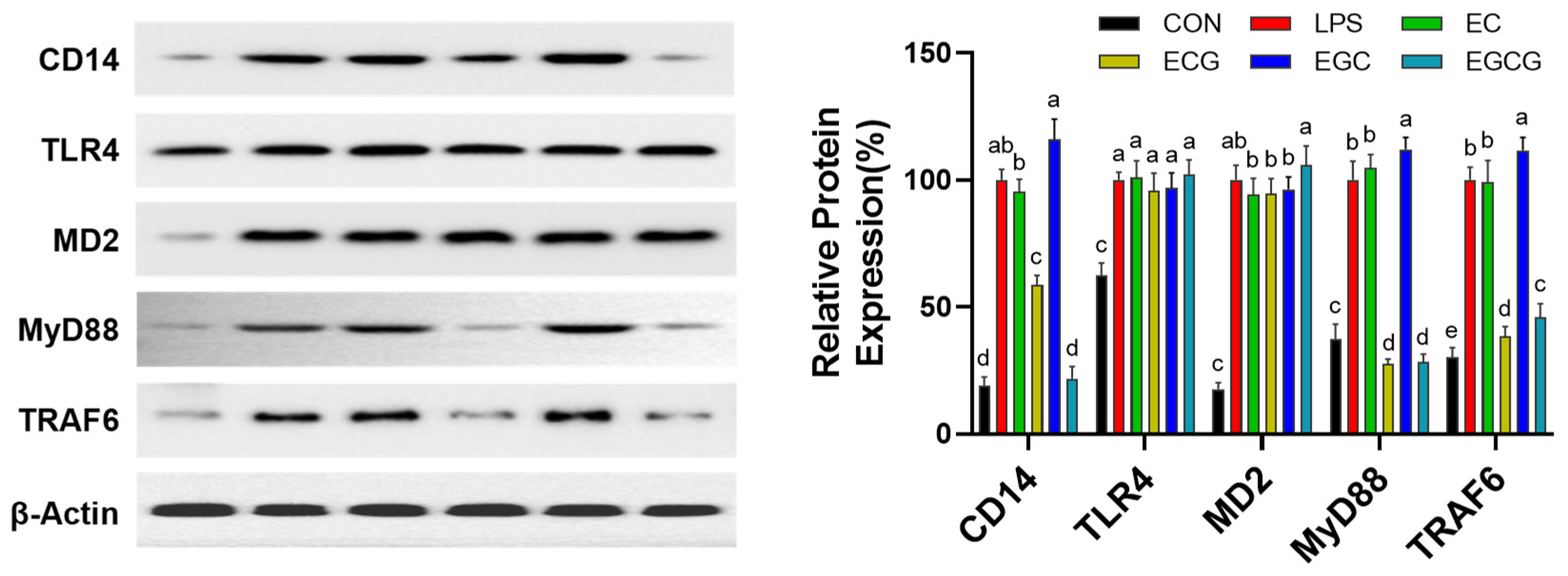
Toll-like receptor 4 (TLR4), an important signaling receptor of LPS, exerts a pivotal influence on the modulation of inflammatory pathways [44]. LPS is conveyed to the TLR4/MD2 complex by CD14; subsequently, TLR4 itself forms a dimer to recruit MyD88 and/or TRIF [45]. For the MyD88-dependent signaling cascade, activated TRAF6 results in the transport, synthesis, and release of inflammatory factors via MAPK/NF-κB signaling cascades [46]. Therefore, this research further evaluated the influences of catechins on the TLR4/CD14 signaling pathway. Figure 8 presents that LPS treatment results in a significant (p < 0.05) up-regulation of CD14, TLR4, MD-2, MyD88, and TRAF6 expression levels compared to the untreated cells (CON). ECG and EGCG inhibited CD14 expression without affecting TLR4 and MD2, while EC and EGC showed no inhibitory effects on the three protein expressions. Moreover, ECG and EGCG were found to significantly (p < 0.05) attenuate the expression levels of the downstream signaling proteins MyD88 and TRAF6. While EC and EGC had no prominent (p > 0.05) inhibiting effects on MyD88 and TRAF6 expressions, and EGC even prominently (p < 0.05) up-regulated their expressions. Xue et al. also found that EGCG reduced the levels of inflammatory factors (IL-2, IFN-γ, IL-4, and IL-1) by regulating the TLR4/MyD88/NF-κB signaling pathway to alleviate intestinal inflammation in Sprague–Dawley rats [47]. These results suggested that galloylated catechins (ECG and EGCG) restrain the MAPK/NF-κB signaling cascade by blocking the upstream TLR4/CD14 signaling pathway.
The growth and maturation of macrophages are heavily dependent on PU.1, a transcription factor in the E-twenty-six (ETS) family [48]. Previous studies have demonstrated that PU.1 is crucial for regulating TLR1, TLR2, TLR4, and CD14 and the adjustment of TLR signaling by targeting PU.1 [49]. The aforementioned study has demonstrated that ECG and EGCG inhibit the TLR signaling pathway through the down-regulation of CD14 expression. We hypothesized that galloylated catechins (ECG and EGCG) might regulate TLR signaling through interactions with the transcription factor PU.1. Docking analysis was conducted to investigate the binding affinity between catechins and the PU.1-DNA complex. Figure 9A,C shows that ECG and EGCG exhibited different binding poses in the PU.1-DNA complex. The binding free energy of ECG and EGCG with PU.1 was −8.4 and −8.9 kcal/mol, separately. This suggested that EGCG possessed stronger binding capacity with PU.1 than ECG, which was consistent with their anti-inflammatory activity. Figure 9B,D presents that ECG interacted with eight amino acid residues (LYS E:208, TYR E:227, GLU E:228, ARG E:235, DA A:3, DA A:4, DC B:27, and DC B:28) of the PU.1-DNA complex by forming Van der Waals forces; five residues (GLU E:209, DA A:4, DA A:5, DG A:6, and DG A:7) of the PU.1-DNA complex by forming hydrogen bonds; two residues (ALA E:231 and DA A:5) of the PU.1-DNA complex by forming π interactions. EGCG bound to five amino acid residues (LYS E:208, THR E:226, ARG E:235, LEU E:250, and DC B:27) of the PU.1-DNA complex by forming Van der Waals forces; nine residues (DA A:4, DA A:5, DG A:6, DG A:7, DC B:28, GLU: E:209, THR: E:226, TYR: E:227, and GLU: E:228) of the PU.1-DNA complex by forming hydrogen bonds; two residues (GLU E:228 and ALA E:231) of the PU.1-DNA complex by forming π interactions. Xia et al. uncovered that the mechanism of anti-neuroinflammatory activities of sophotokin involved suppression of the TLR4 signaling pathway at the sites of NF-κB and MAPK with PU.1 as a likely upstream target [46]. Therefore, ECG and EGCG might restrain the inflammation-related TLR4 pathway by disturbing the interaction of PU.1 with the DNA.

4. Conclusions

To summarize, this study confirmed that the catechins’ galloyl group was indispensable for their anti-inflammatory capacities. This strong anti-inflammatory activity is achieved by disturbing cells from the G0/G1 to the S phase, as well as downstreaming iNOS and COX-2 suppression by blocking the TLR4/MAPK/NF-κB signaling cascade and further restraining the generation of inflammatory agents (NO, PGE2, IL-1β, and TNF-α) and ROS. Docking analysis suggested that ECG and EGCG could bind with the PU.1-DNA complex by forming hydrogen bonds, Van der Waals forces, and π interactions, preventing the DNA binding. Therefore, galloylated catechins (ECG and EGCG) could serve as a dietary functional factor for alleviating inflammation and inflammation-associated chronic diseases. Considering various inflammatory parts involve different types of cells, future studies are needed to evaluate galloylated catechins’ anti-inflammatory activities in other sources of macrophages or the Caco-2/RAW264.7 cell co-culture model.

Author Contributions

Conceptualization, methodology, data curation, writing—original draft preparation, and funding, J.P.; conceptualization, investigation, and validation, G.C., S.G., Z.L., J.L. and W.Y.; writing—review and editing, supervision, and funding, Q.W. and G.X. All authors have read and agreed to the published version of the manuscript.

Funding

This work was funded by the Guangdong Basic and Applied Basic Research Foundation (2021A1515110660), the Key Area Research and Development Program of Guangdong Province (2021B0707060001), the Characteristic Innovation Project of the Ordinary University of Guangdong Province (2023KTSCX051), and the Key Laboratory of Processing and Nutritional Health of Tropical and Subtropical Aquatic Specialty Foods (2023KSYS006).

Institutional Review Board Statement

Not applicable.

Informed Consent Statement

Not applicable.

Data Availability Statement

The original contributions presented in the study are included in the article, further inquiries can be directed to the corresponding author.

Acknowledgments

We gratefully acknowledge all of those who contributed to this study.

Conflicts of Interest

The authors declare no conflicts of interest.

References

  1. Tasneem, S.; Liu, B.; Li, B.; Choudhary, M.I.; Wang, W. Molecular pharmacology of inflammation: Medicinal plants as anti-inflammatory agents. Pharmacol. Res. 2019, 139, 126–140. [Google Scholar] [CrossRef]
  2. Libby, P. Inflammatory Mechanisms: The Molecular Basis of Inflammation and Disease. Nutr. Rev. 2007, 65, S140–S146. [Google Scholar] [CrossRef] [PubMed]
  3. Laveti, D.; Kumar, M.; Hemalatha, R.; Sistla, R.; Naidu, V.G.M.; Talla, V.; Verma, V.; Kaur, N.; Nagpal, R. Anti-inflammatory treatments for chronic diseases: A review. Inflamm. Allergy Drug Targets 2013, 12, 349–361. [Google Scholar] [CrossRef]
  4. Goswami, S.K.; Ranjan, P.; Dutta, R.K.; Verma, S.K. Management of inflammation in cardiovascular diseases. Pharmacol. Res. 2021, 173, 105912. [Google Scholar] [CrossRef] [PubMed]
  5. Song, D.; Niu, J.; Zhang, Z.; Sun, Z.; Wang, D.; Li, J.; Yang, B.; Ling, N.; Ji, C. Purple Sweet Potato Polysaccharide Exerting an Anti-inflammatory Effect via a TLR-Mediated Pathway by Regulating Polarization and Inhibiting the Inflammasome Activation. J. Agric. Food Chem. 2024, 72, 2165–2177. [Google Scholar] [CrossRef] [PubMed]
  6. Tang, C.; Wang, Y.; Chen, D.; Zhang, M.; Xu, J.; Xu, C.; Liu, J.; Kan, J.; Jin, C. Natural polysaccharides protect against diet-induced obesity by improving lipid metabolism and regulating the immune system. Food Res. Int. 2023, 172, 113192. [Google Scholar] [CrossRef] [PubMed]
  7. Wang, D.; Wang, T.; Zhang, Z.; Li, Z.; Guo, Y.; Zhao, G.; Wu, L. Recent advances in the effects of dietary polyphenols on inflammation in vivo: Potential molecular mechanisms, receptor targets, safety issues, and uses of nanodelivery system and polyphenol polymers. Curr. Opin. Food Sci. 2022, 48, 100921. [Google Scholar] [CrossRef]
  8. Chang, C.-Y.; Huang, I.T.; Shih, H.-J.; Chang, Y.-Y.; Kao, M.-C.; Shih, P.-C.; Huang, C.-J. Cluster of differentiation 14 and toll-like receptor 4 are involved in the anti-inflammatory effects of tyrosol. J. Funct. Foods 2019, 53, 93–104. [Google Scholar] [CrossRef]
  9. Ryu, J.-K.; Kim, S.J.; Rah, S.-H.; Kang, J.I.; Jung, H.E.; Lee, D.; Lee, H.K.; Lee, J.-O.; Park, B.S.; Yoon, T.-Y.; et al. Reconstruction of LPS Transfer Cascade Reveals Structural Determinants within LBP, CD14, and TLR4-MD2 for Efficient LPS Recognition and Transfer. Immunity 2017, 46, 38–50. [Google Scholar] [CrossRef]
  10. Huang, D.; Wang, P.; Chen, J.; Li, Y.; Zhu, M.; Tang, Y.; Zhou, W. Selective targeting of MD2 attenuates intestinal inflammation and prevents neonatal necrotizing enterocolitis by suppressing TLR4 signaling. Front. Immunol. 2022, 13, 995791. [Google Scholar] [CrossRef]
  11. Srivastava, N.; Shelly, A.; Kumar, M.; Pant, A.; Das, B.; Majumdar, T.; Mazumder, S. Aeromonas hydrophila utilizes TLR4 topology for synchronous activation of MyD88 and TRIF to orchestrate anti-inflammatory responses in zebrafish. Cell Death Discov. 2017, 3, 17067. [Google Scholar] [CrossRef]
  12. Zhang, Y.; Chen, H.; Zhang, W.; Cai, Y.; Shan, P.; Wu, D.; Zhang, B.; Liu, H.; Khan, Z.A.; Liang, G. Arachidonic acid inhibits inflammatory responses by binding to myeloid differentiation factor-2 (MD2) and preventing MD2/toll-like receptor 4 signaling activation. Biochim. Biophys. Acta (BBA)-Mol. Basis Dis. 2020, 1866, 165683. [Google Scholar] [CrossRef] [PubMed]
  13. Wang, T.; Xu, H.; Wu, S.; Guo, Y.; Zhao, G.; Wang, D. Mechanisms Underlying the Effects of the Green Tea Polyphenol EGCG in Sarcopenia Prevention and Management. J. Agric. Food Chem. 2023, 71, 9609–9627. [Google Scholar] [CrossRef]
  14. Xing, L.; Zhang, H.; Qi, R.; Tsao, R.; Mine, Y. Recent Advances in the Understanding of the Health Benefits and Molecular Mechanisms Associated with Green Tea Polyphenols. J. Agric. Food Chem. 2019, 67, 1029–1043. [Google Scholar] [CrossRef]
  15. Ahmad, R.; Aldholmi, M.; Alqathama, A.; Althomali, E.; Aljishi, F.; Mostafa, A.; Alqarni, A.M.; Shaaban, H. The effect of natural antioxidants, pH, and green solvents upon catechins stability during ultrasonic extraction from green tea leaves (Camellia sinensis). Ultrason. Sonochem. 2023, 94, 106337. [Google Scholar] [CrossRef]
  16. Colon, M.; Nerín, C. Synergistic, antagonistic and additive interactions of green tea polyphenols. Eur. Food Res. Technol. 2016, 242, 211–220. [Google Scholar] [CrossRef]
  17. Peng, J.; Wen, W.; Wang, R.; Li, K.; Xiao, G.; Li, C. The galloyl moiety enhances inhibitory activity of polyphenols against adipogenic differentiation in 3T3-L1 preadipocytes. Food Funct. 2022, 13, 5275–5286. [Google Scholar] [CrossRef] [PubMed]
  18. Zhao, Y.; Jiang, F.; Liu, P.; Chen, W.; Yi, K. Catechins containing a galloyl moiety as potential anti-HIV-1 compounds. Drug Discov. Today 2012, 17, 630–635. [Google Scholar] [CrossRef] [PubMed]
  19. Sicard, A.-A.; Suarez, N.G.; Cappadocia, L.; Annabi, B. Functional targeting of the TGF-βR1 kinase domain and downstream signaling: A role for the galloyl moiety of green tea-derived catechins in ES-2 ovarian clear cell carcinoma. J. Nutr. Biochem. 2021, 87, 108518. [Google Scholar] [CrossRef]
  20. Guo, T.; Lin, Q.; Li, X.; Nie, Y.; Wang, L.; Shi, L.; Xu, W.; Hu, T.; Guo, T.; Luo, F.; et al. Octacosanol Attenuates Inflammation in Both RAW264.7 Macrophages and a Mouse Model of Colitis. J. Agric. Food Chem. 2017, 65, 3647–3658. [Google Scholar] [CrossRef]
  21. Shen, X.; Chen, H.; Zhang, H.; Luo, L.; Wen, T.; Liu, L.; Hu, Q.; Wang, L. A natural sesquiterpene lactone isolinderalactone attenuates lipopolysaccharide-induced inflammatory response and acute lung injury through inhibition of NF-κB pathway and activation Nrf2 pathway in macrophages. Int. Immunopharmacol. 2023, 124, 110965. [Google Scholar] [CrossRef]
  22. Wang, R.; Dong, Z.; Lan, X.; Liao, Z.; Chen, M. Sweroside Alleviated LPS-Induced Inflammation via SIRT1 Mediating NF-κB and FOXO1 Signaling Pathways in RAW264.7 Cells. Molecules 2019, 24, 872. [Google Scholar] [CrossRef]
  23. He, G.; Qiu, M.; Yang, Z.; Zhao, K.; Liu, R.; Mei, H.; Zhao, X.; Song, T.; Liu, X.; Zhang, M.; et al. β-Sitosterol Inhibits Tumor Growth and Amplifies Rituximab Sensitivity through Acid Sphingomyelinase/Ceramide Signaling in Diffuse Large B-Cell Lymphoma. J. Agric. Food Chem. 2024, 72, 16177–16190. [Google Scholar] [CrossRef]
  24. Karas, D.; Ulrichová, J.; Valentová, K. Galloylation of polyphenols alters their biological activity. Food Chem. Toxicol. 2017, 105, 223–240. [Google Scholar] [CrossRef] [PubMed]
  25. Xiong, L.; Ouyang, K.-H.; Jiang, Y.; Yang, Z.-W.; Hu, W.-B.; Chen, H.; Wang, N.; Liu, X.; Wang, W.-J. Chemical composition of Cyclocarya paliurus polysaccharide and inflammatory effects in lipopolysaccharide-stimulated RAW264.7 macrophage. Int. J. Biol. Macromol. 2018, 107, 1898–1907. [Google Scholar] [CrossRef]
  26. Yang, L.; Cao, L.; Li, C.; Li, X.; Wang, J.; Chen, H.; He, J. Hostaflavone A from Hosta plantaginea (Lam.) Asch. blocked NF-κB/iNOS/COX-2/MAPKs/Akt signaling pathways in LPS-induced RAW 264.7 macrophages. J. Ethnopharmacol. 2022, 282, 114605. [Google Scholar] [CrossRef]
  27. Lee, H.-H.; Jang, E.; Kang, S.-Y.; Shin, J.-S.; Han, H.-S.; Kim, T.-W.; Lee, D.H.; Lee, J.-H.; Jang, D.S.; Lee, K.-T. Anti-inflammatory potential of Patrineolignan B isolated from Patrinia scabra in LPS-stimulated macrophages via inhibition of NF-κB, AP-1, and JAK/STAT pathways. Int. Immunopharmacol. 2020, 86, 106726. [Google Scholar] [CrossRef] [PubMed]
  28. Yu, Y.; Zuo, C.; Li, M.; Tang, Y.; Li, L.; Wang, F.; Zhang, S.; Sun, B. Novel l-Cysteine Incomplete Degradation Method for Preparation of Procyanidin B2-3′-O-Gallate and Exploration of its in Vitro Anti-inflammatory Activity and in Vivo Tissue Distribution. J. Agric. Food Chem. 2024, 72, 4023–4034. [Google Scholar] [CrossRef] [PubMed]
  29. El-Kenawi, A.; Ruffell, B. Inflammation, ROS, and Mutagenesis. Cancer Cell 2017, 32, 727–729. [Google Scholar] [CrossRef]
  30. Zhao, J.; Guo, F.; Hou, L.; Zhao, Y.; Sun, P. Electron transfer-based antioxidant nanozymes: Emerging therapeutics for inflammatory diseases. J. Control Release 2023, 355, 273–291. [Google Scholar] [CrossRef]
  31. Qiu, Y.; Yang, X.; Wang, L.; Gao, K.; Jiang, Z. L-Arginine Inhibited Inflammatory Response and Oxidative Stress Induced by Lipopolysaccharide via Arginase-1 Signaling in IPEC-J2 Cells. Int. J. Mol. Sci. 2019, 20, 1800. [Google Scholar] [CrossRef]
  32. He, F.; Luo, S.; Liu, S.; Wan, S.; Li, J.; Chen, J.; Zuo, H.; Pei, X. Zanthoxylum bungeanum seed oil inhibits RANKL-induced osteoclastogenesis by suppressing ERK/c-JUN/NFATc1 pathway and regulating cell cycle arrest in RAW264.7 cells. J. Ethnopharmacol. 2022, 289, 115094. [Google Scholar] [CrossRef]
  33. Li, W.; Luo, F.; Wu, X.; Fan, B.; Yang, M.; Zhong, W.; Guan, D.; Wang, F.; Wang, Q. Anti-Inflammatory Effects and Mechanisms of Dandelion in RAW264.7 Macrophages and Zebrafish Larvae. Front. Pharmacol. 2022, 13, 906927. [Google Scholar] [CrossRef]
  34. Hashimoto, R.; Koide, H.; Katoh, Y. MEK inhibitors increase the mortality rate in mice with LPS-induced inflammation through IL-12-NO signaling. Cell Death Discov. 2023, 9, 374. [Google Scholar] [CrossRef]
  35. Li, X.; Hu, Y.; He, B.; Li, L.; Tian, Y.; Xiao, Y.; Shang, H.; Zou, Z. Design, synthesis and evaluation of ursodeoxycholic acid-cinnamic acid hybrids as potential anti-inflammatory agents by inhibiting Akt/NF-κB and MAPK signaling pathways. Eur. J. Med. Chem. 2023, 260, 115785. [Google Scholar] [CrossRef] [PubMed]
  36. Cai, X.; Cai, J.; Fang, L.; Xu, S.; Zhu, H.; Wu, S.; Chen, Y.; Fang, S. Design, synthesis and molecular modeling of novel D-ring substituted steroidal 4,5-dihydropyrazole thiazolinone derivatives as anti-inflammatory agents by inhibition of COX-2/iNOS production and down-regulation of NF-κB/MAPKs in LPS-induced RAW264.7 macrophage cells. Eur. J. Med. Chem. 2024, 272, 116460. [Google Scholar] [CrossRef] [PubMed]
  37. Hou, D.-X.; Masuzaki, S.; Hashimoto, F.; Uto, T.; Tanigawa, S.; Fujii, M.; Sakata, Y. Green tea proanthocyanidins inhibit cyclooxygenase-2 expression in LPS-activated mouse macrophages: Molecular mechanisms and structure–activity relationship. Arch. Biochem. Biophys. 2007, 460, 67–74. [Google Scholar] [CrossRef]
  38. Wang, A.G.; Son, M.; Kenna, E.; Thom, N.; Tay, S. NF-κB memory coordinates transcriptional responses to dynamic inflammatory stimuli. Cell Rep. 2022, 40, 111159. [Google Scholar] [CrossRef] [PubMed]
  39. Wang, H.; Zhang, L.; Xu, S.; Pan, J.; Zhang, Q.; Lu, R. Surface-Layer Protein from Lactobacillus acidophilus NCFM Inhibits Lipopolysaccharide-Induced Inflammation through MAPK and NF-κB Signaling Pathways in RAW264.7 Cells. J. Agric. Food Chem. 2018, 66, 7655–7662. [Google Scholar] [CrossRef] [PubMed]
  40. Wang, F.; Han, Y.; Xi, S.; Lu, Y. Catechins reduce inflammation in lipopolysaccharide-stimulated dental pulp cells by inhibiting activation of the NF-κB pathway. Oral Dis. 2020, 26, 815–821. [Google Scholar] [CrossRef]
  41. Gałgańska, H.; Jarmuszkiewicz, W.; Gałgański, Ł. Carbon dioxide and MAPK signalling: Towards therapy for inflammation. Cell Commun. Signal. 2023, 21, 280. [Google Scholar] [CrossRef] [PubMed]
  42. Dong, N.; Li, X.; Xue, C.; Zhang, L.; Wang, C.; Xu, X.; Shan, A. Astragalus polysaccharides alleviates LPS-induced inflammation via the NF-κB/MAPK signaling pathway. J. Cell Physiol. 2020, 235, 5525–5540. [Google Scholar] [CrossRef] [PubMed]
  43. Liang, Y.; Ip, M.S.M.; Mak, J.C.W. (-)-Epigallocatechin-3-gallate suppresses cigarette smoke-induced inflammation in human cardiomyocytes via ROS-mediated MAPK and NF-κB pathways. Phytomedicine 2019, 58, 152768. [Google Scholar] [CrossRef]
  44. Schaeffer, E.; Sánchez-Fernández, E.M.; Gonçalves-Pereira, R.; Flacher, V.; Lamon, D.; Duval, M.; Fauny, J.-D.; García Fernández, J.M.; Mueller, C.G.; Ortiz Mellet, C. sp2-Iminosugar glycolipids as inhibitors of lipopolysaccharide-mediated human dendritic cell activation in vitro and of acute inflammation in mice in vivo. Eur. J. Med. Chem. 2019, 169, 111–120. [Google Scholar] [CrossRef]
  45. Lee, H.-H.; Shin, J.-S.; Chung, K.-S.; Kim, J.-M.; Jung, S.-H.; Yoo, H.-S.; Hassan, A.H.E.; Lee, J.K.; Inn, K.-S.; Lee, S.; et al. 3′,4′-Dihydroxyflavone mitigates inflammatory responses by inhibiting LPS and TLR4/MD2 interaction. Phytomedicine 2023, 109, 154553. [Google Scholar] [CrossRef] [PubMed]
  46. Xia, W.; Luo, P.; Hua, P.; Ding, P.; Li, C.; Xu, J.; Zhou, H.; Gu, Q. Discovery of a New Pterocarpan-Type Antineuroinflammatory Compound from Sophora tonkinensis through Suppression of the TLR4/NFκB/MAPK Signaling Pathway with PU.1 as a Potential Target. ACS Chem. Neurosci. 2019, 10, 295–303. [Google Scholar] [CrossRef]
  47. Xue, B.; Liu, X.; Dong, W.; Liang, L.; Chen, K. EGCG Maintains Th1/Th2 Balance and Mitigates Ulcerative Colitis Induced by Dextran Sulfate Sodium through TLR4/MyD88/NF-κB Signaling Pathway in Rats. Can. J. Gastroenterol. Hepatol. 2017, 2017, 3057268. [Google Scholar] [CrossRef]
  48. Deng, Y.; Wang, H.; Liu, X.; Yuan, H.; Xu, J.; de Thé, H.; Zhou, J.; Zhu, J. Zbtb14 regulates monocyte and macrophage development through inhibiting pu.1 expression in zebrafish. eLife 2022, 11, e80760. [Google Scholar] [CrossRef]
  49. Rong, H.; Zhao, Z.; Feng, J.; Lei, Y.; Wu, H.; Sun, R.; Zhang, Z.; Hou, B.; Zhang, W.; Sun, Y.; et al. The effects of dexmedetomidine pretreatment on the pro- and anti-inflammation systems after spinal cord injury in rats. Brain Behav. Immun. 2017, 64, 195–207. [Google Scholar] [CrossRef]
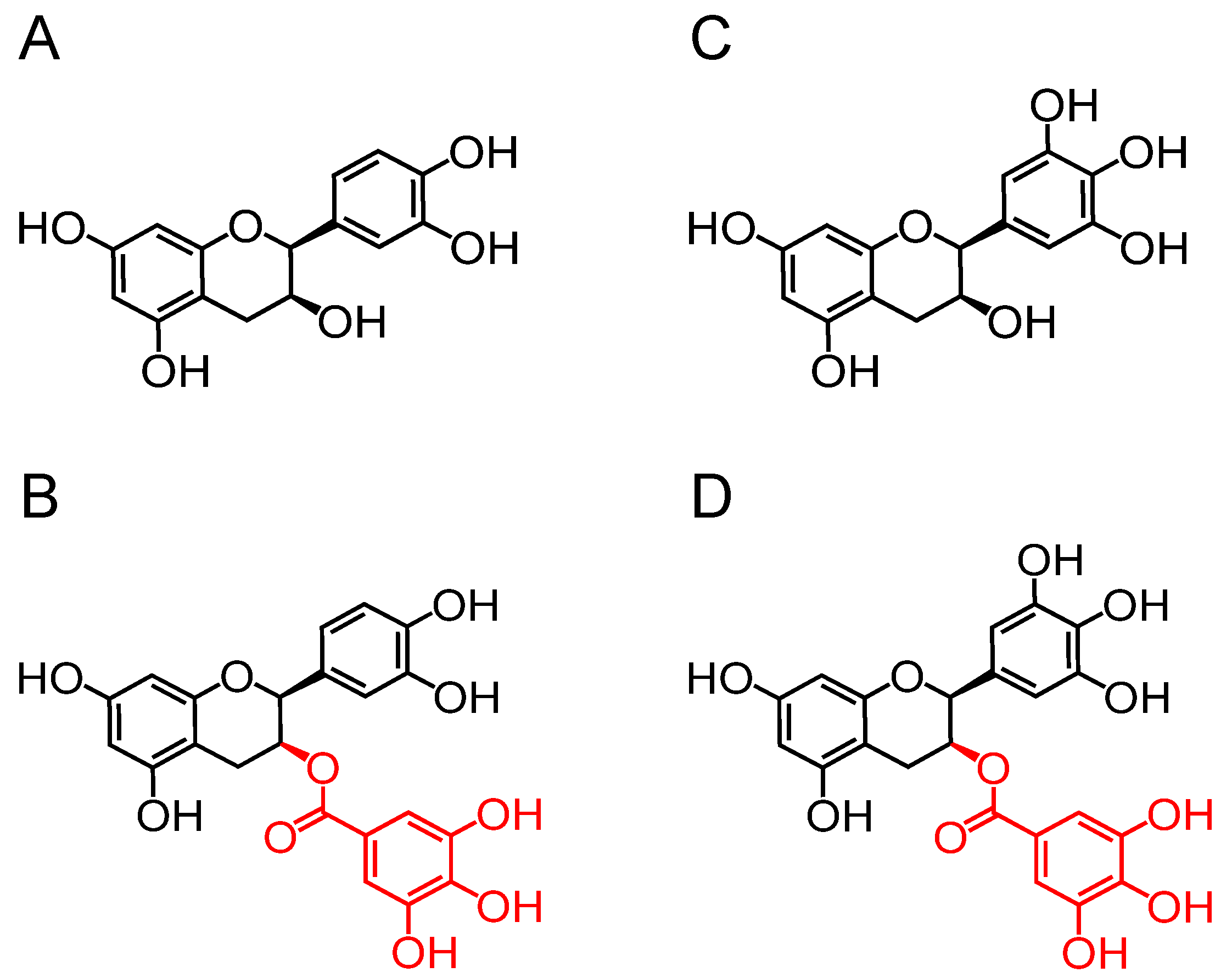
Figure 1. The chemical structures of four catechins: (A) EC, epicatechin; (B) ECG, epicatechin-3-gallate; (C) EGC, epigallocatechin; and (D) EGCG, epigallocatechin-3-gallate. The group in red is the galloyl group.
Foods 13 02616 g001
Figure 2. Influences of catechins (EC, epicatechin; ECG, epicatechin-3-gallate; EGC, epigallocatechin; EGCG, epigallocatechin-3-gallate) on RAW 264.7 cell viability. Results were displayed as mean ± SD from no less than three experimental replications.
Foods 13 02616 g002
Figure 3. Effects of catechins (EC, epicatechin; ECG, epicatechin-3-gallate; EGC, epigallocatechin; EGCG, epigallocatechin-3-gallate) on the levels of NO (A), PGE2 (B), IL-1β (C), and TNF-α (D) in RAW264.7 cells. Results were displayed as mean ± SD from no less than three experimental replications. Diverse lowercases in the histogram denote significant differences (p < 0.05).
Foods 13 02616 g003
Figure 4. Impacts of catechins (EC, epicatechin; ECG, epicatechin-3-gallate; EGC, epigallocatechin; EGCG, epigallocatechin-3-gallate) on ROS release in LPS-induced RAW264.7 cells. (A) Fluorescence microscopy observation. (B) Quantitative analysis. The cells were exposed to LPS (1 μg/mL) with or without the addition of catechins (100 µM) for 24 h. Results were displayed as mean ± SD from no less than three experimental replications. Diverse lowercases in the histogram denote significant differences (p < 0.05).
Foods 13 02616 g004
Figure 5. Regulatory influences of catechins (CAs: EC, epicatechin; ECG, epicatechin-3-gallate; EGC, epigallocatechin; EGCG, epigallocatechin-3-gallate) on cell cycle progression. (A) RAW264.7 cells were harvested after 24 h of LPS induction, colored with PI, and subsequently detected using flow cytometry. (B) The modulation of cell cycle by CAs was represented as the relative proportion of cells in three different phases.
Foods 13 02616 g005
Figure 6. Impacts of catechins (CAs: EC, epicatechin; ECG, epicatechin-3-gallate; EGC, epigallocatechin; EGCG, epigallocatechin-3-gallate) on the iNOS and COX-2 expression in macrophages. Cells were exposed to LPS (1 μg/mL) with or without the addition of catechins (100 µM) for 24 h. Results were displayed as mean ± SD from no less than three experimental replications. Diverse lowercases in the histogram denote significant differences (p < 0.05).
Foods 13 02616 g006
Figure 7. Effects of catechins (CAs: EC, epicatechin; ECG, epicatechin-3-gallate; EGC, epigallocatechin; EGCG, epigallocatechin-3-gallate) on MAPK/NF-κB pathway in LPS-triggered macrophages. (A) The NF-κB phosphorylation level in cells. (B) The ERK, JNK, and p38 phosphorylation levels in cells. Results were displayed as mean ± SD from no less than three experimental replications. Diverse lowercases in the histogram denote significant differences (p < 0.05).
Foods 13 02616 g007
Figure 8. Influences of catechins (CAs: EC, epicatechin; ECG, epicatechin-3-gallate; EGC, epigallocatechin; EGCG, epigallocatechin-3-gallate) on TLR4/CD14 pathway in LPS-triggered RAW264.7 cells. The expression levels of CD14, TLR4, MD-2, MyD88, and TRAF6 in cells. Results were displayed as mean ± SD from no less than three experimental replications. Diverse lowercases in the histogram denote significant differences (p < 0.05).
Foods 13 02616 g008
Figure 9. Docking results of the interaction between PU.1 and galloylated catechins (ECG, epicatechin-3-gallate; EGCG, epigallocatechin-3-gallate). (A) The 3D (A) and 2D (B) diagram of ECG-PU.1-DNA interaction. The 3D (C) and 2D (D) diagrams of EGCG-PU.1-DNA interaction.
Foods 13 02616 g009
Disclaimer/Publisher’s Note: The statements, opinions and data contained in all publications are solely those of the individual author(s) and contributor(s) and not of MDPI and/or the editor(s). MDPI and/or the editor(s) disclaim responsibility for any injury to people or property resulting from any ideas, methods, instructions or products referred to in the content.