Article

Bifidobacterium Relieved Fluoride-Induced Hepatic and Ileal Toxicity via Inflammatory Response and Bile Acid Transporters in Mice

1
College of Veterinary Medicine, Shanxi Agricultural University, 1 Mingxian South Road, Taigu 030801, China
2
College of Veterinary Medicine, Inner Mongolia Agricultural University, Huhehot 010018, China
3
College of Animal Science, Shanxi Agricultural University, Taigu 030801, China
*
Author to whom correspondence should be addressed.
These authors are equal contributions to this work.
Foods 2024, 13(7), 1011; https://doi.org/10.3390/foods13071011
Submission received: 26 February 2024 / Revised: 21 March 2024 / Accepted: 22 March 2024 / Published: 26 March 2024
(This article belongs to the Special Issue Food Safety Management: Contaminants and Toxicants)

Abstract

:
Fluoride is a pervasive environmental contaminant. Prolonged excessive fluoride intake can inflict severe damage on the liver and intestines. Previous 16S rDNA sequencing revealed a decrease in ileal Bifidobacterium abundance during fluoride-induced hepatointestinal injury. Hence, this work aimed to investigate the possible mitigating function of Bifidobacterium on hepatointestinal injury caused by fluoride. Thirty-six 6-week-old C57BL/6J mice (equally divided between males and females) were allotted randomly to three groups: Ctrl group (distilled water), NaF group, and NaF + Ba group (100 mg/L NaF distilled water). After 10 weeks, the mice were given 1 × 10⁹ CFU/mL Bifidobacterium solution (0.2 mL/day) intragastrically in the NaF + Ba group for 8 weeks, and the mice in other groups were given the same amount of distilled water. Dental damage, bone fluoride content, blood routine, liver and intestinal microstructure and function, inflammatory factors, and regulatory cholic acid transporters were examined. Our results showed that fluoride increased glutamic-oxalacetic transaminase (GOT), glutamic-pyruvic transaminase (GPT) activities, and the levels of lipopolysaccharide (LPS), IL-1β, IL-6, TNF-α, and IL-10 levels in serum, liver, and ileum. However, Bifidobacterium intervention alleviated fluoride-induced changes in the above indicators. In addition, Bifidobacterium reduced the mRNA expression levels of bile acid transporters ASBT, IBABP, OST-α, and OST-β in the ileum. In summary, Bifidobacterium supplementation relieved fluoride-induced hepatic and ileal toxicity via an inflammatory response and bile acid transporters in the liver and ileum of mice.

1. Introduction

Fluoride is a pervasive environmental contaminant, primarily found in groundwater [1]. The fluoride concentration in the drinking water of over 180 million people exceeds the thresholds recommended by the World Health Organization, leading to global public health concerns [2]. Research indicates that chronic fluoride exposure results in severe hepatocellular vacuolation, and damage to the rough endoplasmic reticulum and mitochondria of hepatocytes, affecting liver metabolism, diminishing hepatic detoxification capacity, and causing varying degrees of injury [3,4,5]. Elevated fluoride intake may lead to intestinal mucosal damage, triggering inflammatory responses in the intestine and impacting intestinal microbiota, resulting in gut dysbiosis [6,7,8]. Prolonged excessive fluoride intake can inflict severe damage on the liver and intestines. However, its underlying mechanisms remain unclear.
The intestinal microbiota plays a pivotal role in maintaining the integrity of the intestinal barrier, and its diversity is intricately linked to the overall health of the host [9]. Dysbiosis of the intestinal microbiota can precipitate various health ailments [10]. In recent years, research has revealed a close association between environmental fluoride exposure and intestinal inflammation and microbiota dysbiosis [11,12]. Fluoride substantially alters the components of the mouse intestinal microbiota [13,14]. Similarly, another study reported that sodium fluoride (NaF) disrupts the balance of the intestinal microbiota, potentially causing intestinal dysfunction in mice [15]. It is noteworthy that dysfunction and ecological imbalance of the intestinal barrier can also lead to liver damage [16]. In our previous study, based on 16S rDNA sequencing, fluoride exposure markedly altered the components of the mouse ileal microbiota, particularly reducing the abundance of the Bifidobacterium genus.
Bifidobacterium, as the predominant microbiota in the mammals and poultry host’s gastrointestinal tract, facilitates digestion, enhances nutrient absorption, boosts immune function, and preserves intestinal flora balance, thereby playing an indispensable role in overall health maintenance [17]. A recent study discovered Bifidobacterium can regulate bile acid metabolism through its bile acid hydrolase activity and alleviate the liver and intestinal damage caused by many factors (high-fat diet, non-steroidal anti-inflammatory drugs, harmful bacteria, etc.) [18]. Adhesin secreted by Bifidobacterium can significantly alleviate the inflammatory reaction of intestinal epithelial cells induced by LPS and H2O2, and protect the intestinal tract [19]. Current studies have observed that Bifidobacterium mitigated liver inflammation by altering the intestinal flora, thereby ameliorating non-alcoholic fatty liver disease, notably by diminishing the levels of LPS and inflammatory mediators in both serum and liver [20,21]. Therefore, the role of Bifidobacterium in fluoride-induced hepato-intestinal toxicity urgently needs further investigation.
This study established a Bifidobacterium-supplemented mouse model subjected to fluoride treatment. Subsequently, fluoride ion concentration in the femur, serum indicators of liver function (GOT and GPT), inflammatory factor (IL-1β, IL-6, IL-10, and TNF-α) content, and mRNA expression of genes associated with bile acid transport receptors in the liver (NTCP, MRP2, and BSEP) and ileum (ASBT, IBABP, and OST-α/β) were further detected. The research aimed to investigate Bifidobacterium’s protective effect against fluoride-induced hepatointestinal injury, focusing on inflammation. Additionally, it sought to elucidate the correlation between hepatointestinal injury resulting from environmental fluoride exposure and Bifidobacterium in the ileum and provide a reference for the influence of environmental factors on the imbalance of intestinal flora.

2. Material and Methods

2.1. Animals and Treatments

Male and female C57BL/6J mice, aged 6 weeks, were obtained from China Eaton Leverage Company Limited and bred in the laboratory. Eighteen mice of each gender were randomly assigned to three groups: control, sodium fluoride (NaF), and sodium fluoride with Bifidobacterium intervention (NaF + Ba). Each group comprised 12 mice, with an equal gender distribution. After the 10th week, the Bifidobacterium intervention began. Each NaF + Ba group of mice was orally administered 0.2 mL of Bifidobacterium solution per day, while the remaining groups were given the same amount of distilled water for 8 weeks. Subsequently, the mice were slaughtered, and the necessary samples were collected.
The mice were housed under controlled conditions with a temperature range of 20–25 °C and humidity between 50–60%, ensuring adequate ventilation and hygiene standards. They were provided ad libitum access to water and food (obtained from Jiangsu Collaborative Pharmaceutical and Biological Engineering Company Limited (Nanjing, China)). The feeding regimen spanned 18 weeks, during which weekly weight measurements were conducted. Sodium fluoride (NaF) was procured from Tianjin Chemical Reagent Factory No.3 (Tianjin, China), while Bifidobacterium JYBR-190 lyophilized powder was sourced from Shandong Zhongke Jiayi Biological Engineering Company Limited (Weifang, China). All animal procedures strictly adhered to the regulations and guidelines outlined by the Laboratory Animal Ethics Committee of Shanxi Agricultural University.

2.2. Organ Coefficient Analysis

The body weight of all mice was recorded before sacrifice, as was the weight of each organ weight after dissection. The organ coefficient was calculated using the formula: organ coefficient ratio = organ wet weight (mg)/body weight (g).

2.3. Bone Fluoride Determination

The fluoride content in the femur was assessed using the fluoride ion-selective electrode method. Following slaughter, both right and left femurs were removed, and the residual soft tissue on the surface was cleaned with medical gauze. Subsequently, the samples underwent drying at 105 °C for 4 h, followed by ashing in a muffle furnace at 550 °C for 4 h. Bone ash samples weighing 20–25 mg were dissolved in 0.25 mol/L hydrochloric acid; the pH was adjusted to 4 using sodium hydroxide, and the volume was made constant with buffer solution. Finally, the fluoride content was determined using a fluoride ion-selective electrode [22].

2.4. Grading of Dental Damage
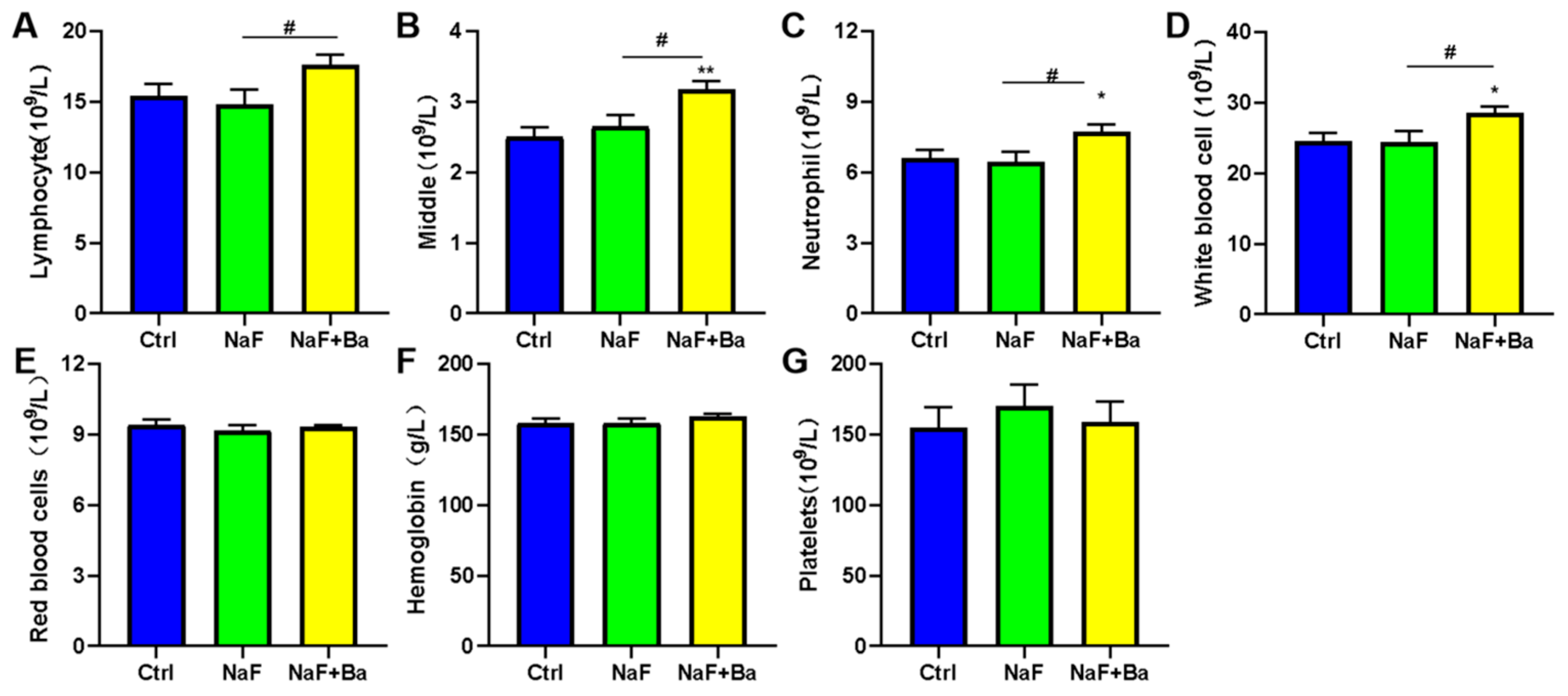
The evaluation of fluoride-induced dental damage was conducted using an 11-point grading scale [23]. The criteria for judging included transparency of the enamel mandibular incisors, color and size of chalky plaques, and degree of tooth defects. On the whole, grade 0 was pigeonholed as normal, grades 1–3 were classified as very mild dental damage, grades 4–5 were considered as mild dental damage, grades 6–8 were rated as moderate dental damage, and grades 9–10 were categorized as severe dental damage.

2.5. Blood Routine

Blood samples were analyzed by routine blood analysis instruments, including the number of lymphocytes, middle cells (sum of monocytes, eosinophils, and basophils), neutrophils, red blood cells, white blood cells, hemoglobin, and platelets.

2.6. Histopathological Examination

Liver and intestinal tissues (including the duodenum, ileum, colon, and cecum) were fixed in a 10% formalin solution. Following a 24-h fixation period, the tissues were rinsed, embedded in paraffin, and sectioned at a thickness of 5 µm. The sections were then stained with haematoxylin and eosin (H&E) solution and examined using light microscopy.

2.7. Biochemical Assays

Liver enzymes GOT, GPT, and TP levels were determined using commercial detection kits (Nanjing Jiancheng Bioengineering Institute, Nanjing, China) utilizing a microplate reader (Thermo Fisher, Waltham, MA, USA) following the manufacturer’s protocols.

2.8. ELISA Analysis

We exploited ELISA to detect LPS and inflammatory factor levels in the serum, liver, and ileum. Ileum and liver tissues weighing about 20 mg were homogenized in 180 μL PBS on ice. The liver tissue homogenate, ileum tissue homogenate, and serum were then centrifuged at 4 °C, 3000 rpm for 30 min. Afterward, 10 μL of supernatant was diluted five times, and the levels of LPS, IL-1β, IL-6, IL-10, and TNF-α were measured using ELISA kits following the manufacturer’s instructions. All enzyme-linked immunosorbent kits were purchased from Shanghai Jianglai Biological Company (Shanghai, China).

2.9. Real-Time PCR

Following the manufacturer’s protocol, total RNA was separated from liver and ileum tissues using Trizol Reagent (Beijing Quanshijin Biotechnology Company Limited, Beijing, China). A NanoDrop 2000 spectrophotometer (Thermo Fisher, USA) was used to detect the concentration and quality of RNA. Subsequently, the GoScript Reverse Transcriptase Reverse Transcription kit (Plommeg Biotechnology Company Limited, Shanghai, China) was used to reverse transcript total RNA into cDNA. Quantitative real-time PCR (qRT-PCR) was conducted with the GoTaq ® qPCR System (Beijing Quanshijin Biotechnology Company Limited, Beijing, China) using the MX3000P system (Stratagene, La Jolla, CA, USA). Primers (Table 1) designed online by Primer 3.0 plus were synthesized by China Shanghai Sangon Biotech Company Limited. The genes of interest included bile acid salt export protein (BSEP), apical sodium-dependent bile acid transporter (ASBT), organic solute transporters alpha and beta-α (OST-α), and organic solute transporters alpha and beta-β (OST-β). The thermocycling conditions were as follows: initial denaturation at 94 °C for 5 min, followed by 40 amplification cycles at 94 °C for 15s, 60 °C for 30s, and 72 °C for 15s. Finally, a melting curve analysis was performed at 95 °C for 15s, 94 °C for 60s, and 94 °C for 15s.

2.10. Statistical Analysis

All statistical analysis was carried out using GraphPad Prism 8 with a one-way analysis of variance with Tukey’s test. Data are displayed as means ± SEM. * means significant differences between the control group and the NaF group/the NaF + Ba group. # represents significant differences between the NaF group and the NaF + Ba group. Statistical significance is defined as a p-value below 0.001, 0.01, or 0.05.

3. Results

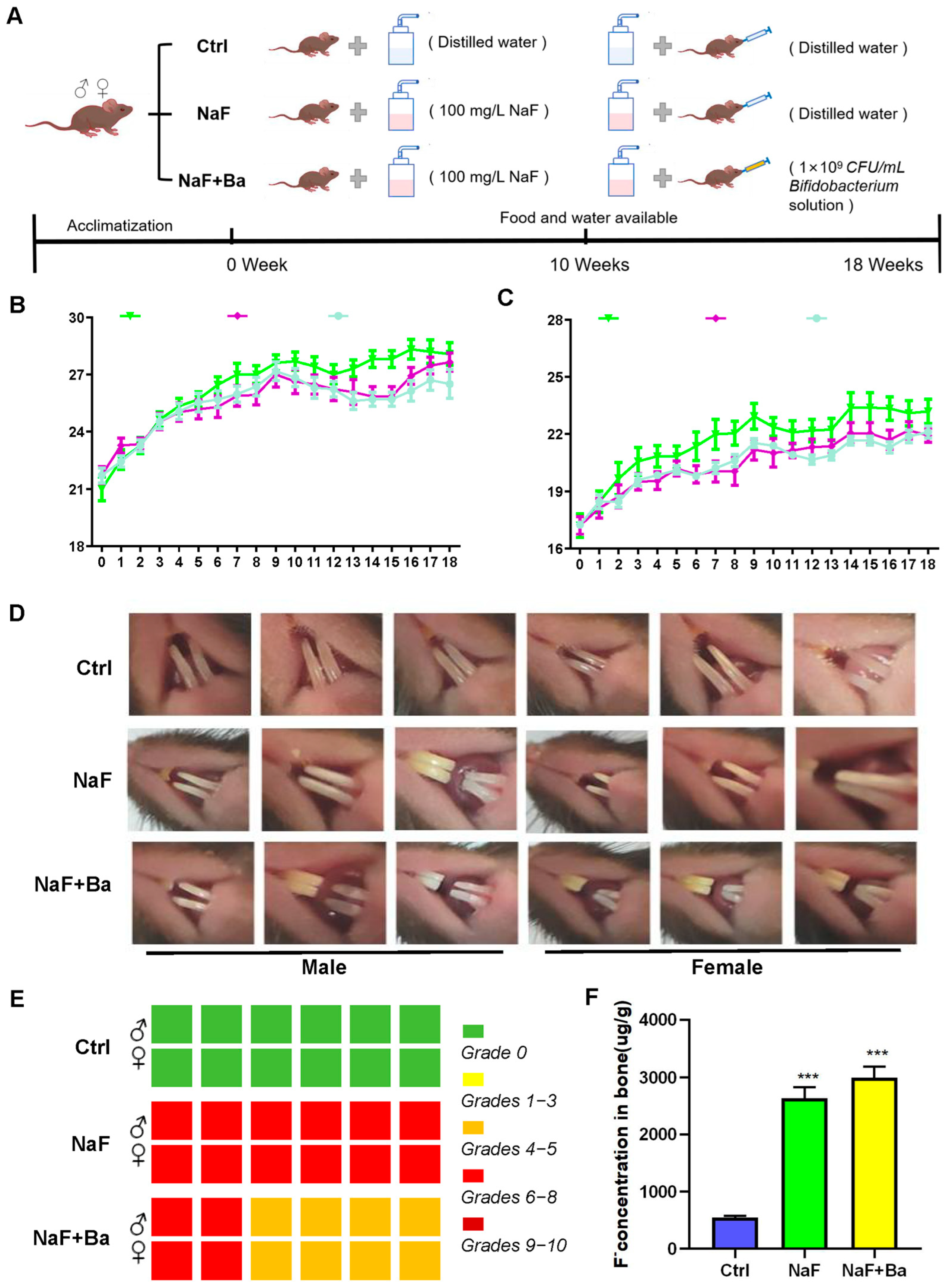
3.1. Mouse Model Design and Primary Evaluation of Fluoride Exposure and Bifidobacterium Intervention

The experimental design included recording and presenting the body weights of mice throughout the study, as depicted in Figure 1A–C. There was no statistically significant change in body weight, as well as the organ coefficients of the liver, kidney, spleen, left epididymis, testis, ovary, uterus, and brain among mice (Table 2 and Table 3). This suggests neither NaF nor/or Bifidobacterium treatment affected growth in mice.
Incisors of mice were well developed, with enamel luster and normal transparency in the Ctrl group. In the NaF group, the incisors became shorter, with chalky in a large area and severe cutting ends, showing 100% moderate dental damage symptoms. In the NaF + Ba group, the proportions of dental damage were 1/3 moderate dental damage and 2/3 middle dental damage, respectively. Otherwise, there was abrasion at the end of the incisors, and white spots and little pigmentation scattered in the middle of them (Figure 1D,E). In the same period, compared to the Ctrl group, bone F concentrations were increased significantly in both the NaF group and NaF + Ba group (p < 0.001), but no significant change was observed among the experimental groups (Figure 1F). These results suggest the fluoride-exposed mouse model was successfully established, and Bifidobacterium intervention could mitigate the grade of dental damage.

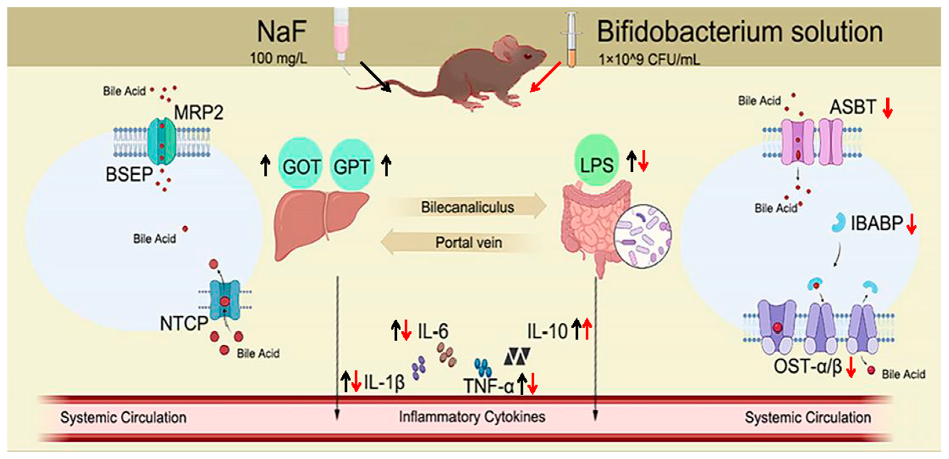
3.2. Bifidobacterium Alleviates Fluoride-Induced Liver and Ileal Inflammation

As seen from the routine blood indicators level in Figure 2, compared to the Ctrl group, the number of middle cells, neutrophils, and white blood cells were obviously increased in the NaF + Ba group (p < 0.05, p < 0.01), except there were no alterations in lymphocytes, red blood cells, hemoglobin concentration, and platelets, and no pronounced difference were noticed in the NaF group. Only lymphocytes, middle cells, neutrophils, and white blood cell numbers in the NaF + Ba group were higher than in the NaF group (p < 0.05), and other indicators had no notable changes. The results suggest that Bifidobacterium affects the immune response in mice.
The expression of inflammatory cytokines including pro-inflammatory factors IL-1β, IL-6, TNF-α, and anti-inflammatory factor IL-10 are shown in Figure 3. Compared to the Ctrl group, the level of serum, liver, and ileum inflammatory cytokines in the NaF group were significantly elevated after 10-weeks fluoride exposure (p < 0.05, p < 0.01, p < 0.001); In the NaF + Ba group, an obvious increment was only found in IL-10 and serum TNF-α after 8 weeks of Bifidobacterium intervention (p < 0.05, p < 0.001). Compared to the NaF group, the pro-inflammatory factor consistency was obviously reduced except for no notable alteration in the level of serum TNF-α (p < 0.05, p < 0.01, p < 0.001), and a dramatically enhanced anti-inflammatory factor density was observed in the NaF + Ba group (p < 0.05). The above results indicate that Bifidobacterium alleviated the inflammation occurring in the liver and ileum of fluoride-treated mice.

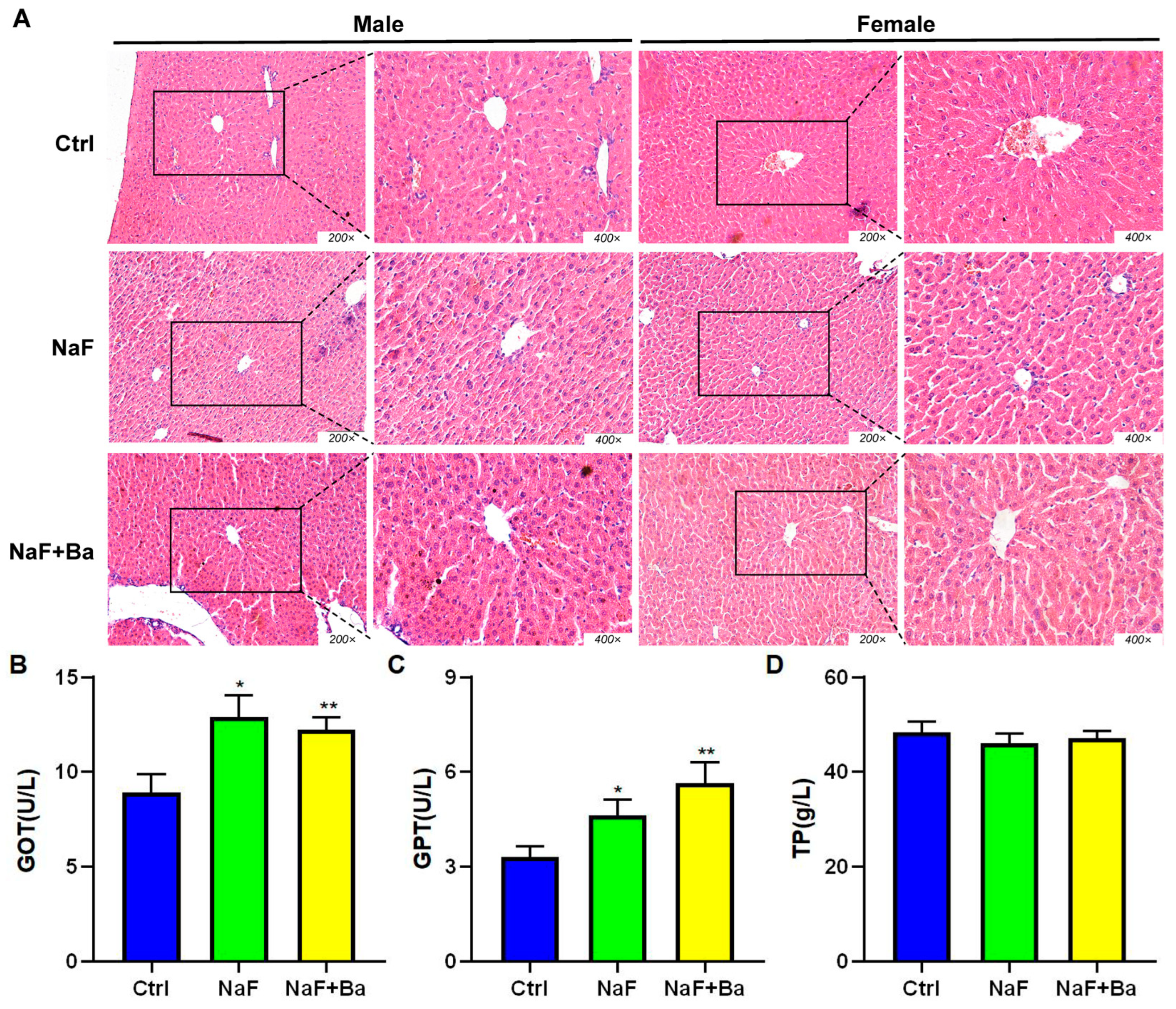
3.3. Bifidobacterium Restored the Alteration of Morphology of Liver Induced by Fluoride

The morphological changes of the liver tissue were observed by H.E staining and are shown in Figure 4A. The hepatocytes were regularly arranged around the central vein with a round shape, clear cell boundary, and round or oval nuclei in the Ctrl group and NaF + Ba group, while the histopathological lesions, manifested as a disordered arrangement of hepatocytes, blurred boundary, and Kupffer cell number upregulation occurred in the liver tissue of the NaF group.
The biochemical indicators related to liver function were detected. In comparison with the Ctrl group, GOT and GPT activity significantly increased in the NaF group (p < 0.05) and NaF + Ba group (p < 0.01), without a significant difference in TP concentration (Figure 4B–D). All indexes demonstrated no dramatic changes in the NaF + Ba group in comparison to the NaF group. The results showed that Bifidobacterium alleviated fluoride-induced liver inflammation but not impaired liver function.

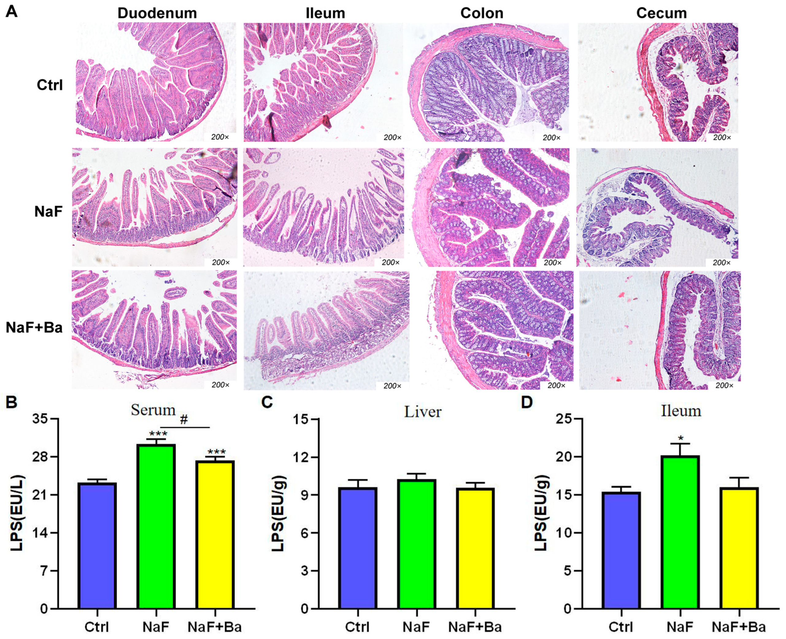
3.4. Bifidobacterium Intervention Alleviates Fluoride-Induced Intestinal Injury in Mice

The morphological characteristics of duodenum, ileum, colon, and cecum tissues in mice are shown in Figure 5A. In the Ctrl group, the intestinal epithelial cells had a neat arrangement and clear edges, intact intestinal mucosa, without significant injury. In the NaF group, the intestinal epithelial cells were disorderly arranged with a blurred edge of the nucleus, reduced height of the intestinal villi, damaged structure, velvet hollowing out, and rupture to varying degrees. The NaF + Ba group displayed mild intestinal injury with a slight reduction in villus length and disordered arrangement of intestinal epithelial cells.
The changes of LPS concentration in serum, ileum, and liver are shown in Figure 5B–D. In comparison to the Ctrl group, a significant increase was found in serum and ileum LPS concentrations (p < 0.05, p < 0.001) of the NaF group with no pronounced differences in the liver. The concentration of liver and ileum LPS in the NaF + Ba group had no notable differences except that serum LPS increased markedly (p < 0.001). Compared with the NaF group, the concentration of LPS was decreased significantly in serum (p < 0.05), with no obvious alteration in the liver and ileum of the NaF + Ba group. The results implied Bifidobacterium was able to restore the imbalance of homeostasis of intestinal barrier in mice caused by fluoride.

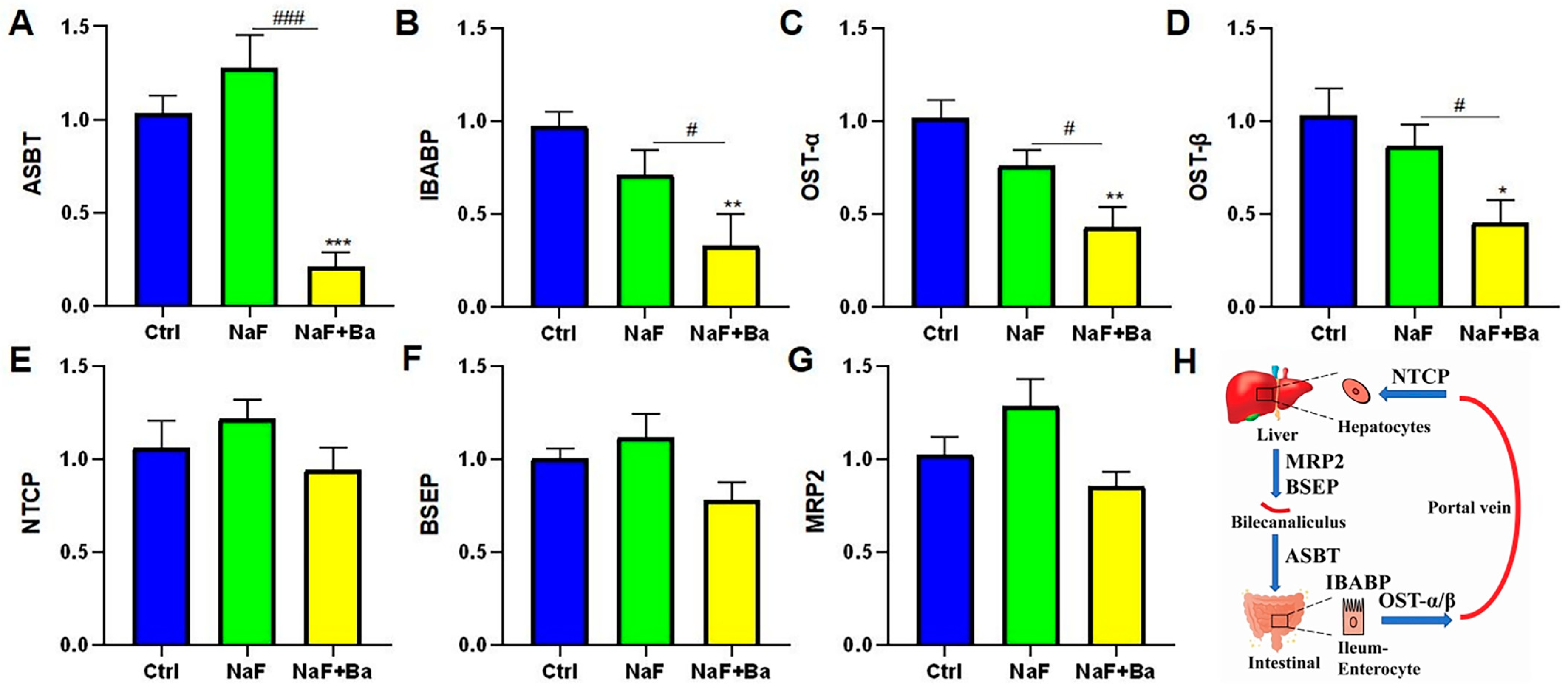
3.5. Bifidobacterium Regulated Bile Acid Transporters in Enterohepatic Circulation

In order to verify the changes in bile acid transporter after fluoride or/and Bifidobacterium treatment, the mRNA expression level of ASBT, IBABP, OST-α, and OST-β in the ileum, NTCP, BSEP, and MRP2 in the liver were tested (Figure 6A–G). The distribution of bile acid transporters in the hepatic–intestinal circulation is shown in Figure 6H. In the ileum, compared to the Ctrl group, the mRNA expression of ASBT, IBABP, OST-α, and OST-β were notably decreased in the NaF + Ba group (p < 0.05, p < 0.01, p < 0.001), and there were not any notable changes in the NaF group. In parallel, all these genes decreased markedly between the NaF + Ba group and the NaF group (p < 0.05, p < 0.001). In the liver, no pronounced changes were present in the mRNA expressions of NTCP, BSEP, and MRP2 among groups. The above results indicate that fluoride did not induce changes in bile acid transport in mice in this study. It is worth noting that Bifidobacterium may influence bile acid transporters in mice by reducing ASBT, IBABP, OST-α, and OST-β mRNA expression levels.

4. Discussion

Fluorosis can be categorized into different types, including drinking water-induced, tea-induced, coal-burning-induced, and industrial-induced fluorosis, with drinking water-induced fluorosis being the most significant due to contaminated water sources [24]. So, our research group chose to establish a fluoride-exposed mouse model by drinking water freely. The fluoride concentration in groundwater in China, India, and Pakistan has reached 48 mg/L [25], and the fluoride concentration in wastewater from copper smelting can exceed 10,000 mg/L [26]. In addition, the oral LD50/24 h of NaF for mice is 97.7 mg/kg body weight [27]. Based on the above background and previous fluoride studies in the laboratory [28], we constructed fluoride-treated mouse models by freely drinking distilled water containing 100 mg/L sodium fluoride for 18 weeks. Of note, Bifidobacterium was regarded as an effective therapeutic approach for alleviating liver and intestinal damage [29]. Here, the administration of 1 × 10⁹ CFU/mL Bifidobacterium is based on the research by Xin et al. [30].
The liver occupies an important role in toxicology studies. Most of the poisons are absorbed and metabolized through the liver [31]. Fluoride can accumulate in the liver and cause damage to the microstructure and function of the liver [3]. In this study, the normal morphological structure was destroyed post-fluoride exposure and mitigated after Bifidobacterium interposition. Both serum GPT and GOT activities were significantly increased in the NaF group. Here, Bifidobacterium did not alleviate the fluoride-induced impaired liver function.
Bones serve as the primary site for fluoride accumulation [32]. The study findings revealed that Bifidobacterium supplementation did not alleviate the accumulation of fluoride in bone. This indicates that Bifidobacterium has no effect on the absorption and excretion of fluoride ions, and Bifidobacterium may alleviate liver and intestine injury caused by fluoride in other ways. The results of the blood routine imply that we consider the role of the inflammatory response after Bifidobacterium supplementation. For that, a further study demonstrated that Bifidobacterium relieved the fluoride-induced increase in pro-inflammatory factors IL-1β, IL-6, and TNF-α in sera, liver, and ileum. Bifidobacterium can enhance the activity of natural killer cells and phagocytes, stimulate the activation, proliferation, and secretion of B lymphocytes, and boost the body’s immune response [33]. Our results indicated that Bifidobacterium can significantly increase the number of leukocytes, granulocytes, as well as intermediate cells (the sum of monocytes, eosinophils, and basophils) in blood. These results indicate that Bifidobacterium may alleviate liver and intestinal injury resulting from fluoride exposure by eliciting an anti-inflammatory response.
Fluoride is mainly absorbed and metabolized by gastrointestinal tissue, so the gastrointestinal tract is one of the tissues and organs most seriously affected by fluorosis [34]. Studies have found that patients with skeletal fluorosis have gastrointestinal symptoms after sampling and following tissue biopsy of the intestine, rupture of duodenal microvilli and abrasion on the mucosal surface were found [35]. The result stated clearly that Bifidobacterium intervention alleviated the fluoride-induced morphological structures of duodenum, ileum, colon, and cecum in mice. In our unpublished study, the ileum was the most sensitive to fluoride, and fluoride treatment significantly altered the microbial composition of ileal contents. The alteration in microbial composition prompted us to investigate the potential role of these microbes in inflammatory processes, particularly in the context of whether lipopolysaccharides (LPS) from Gram-negative cell walls exacerbate inflammation. LPS, a vital component of Gram-negative bacterial cell walls, releases inflammatory mediators through the activation of Kupffer cells in the liver, which in turn promote increased inflammation. In general, abnormal changes in LPS are also considered a marker of an imbalance of intestinal flora [36]. Nonetheless, the therapeutic role of Bifidobacterium in gastrointestinal diseases caused by fluoride is less studied. Former studies reported that when the intestine is stimulated for a long time, it can decrease the number of Bifidobacterium, destroy the integrity of the intestinal barrier, and release LPS entering the blood [37]. The present study discovered that Bifidobacterium can mitigate fluoride-induced damage to the intestinal microstructure and reduce the elevation of inflammatory factors in the ileum as well as LPS levels in the serum. These results suggest that Bifidobacterium restores the decline of intestinal barrier function resulting from fluoride exposure.
Our unpublished study has shown Bifidobacterium intervention significantly reduces the total bile acids concentration in serum and liver and alleviates the abnormalities of bile acid synthase and ileal FXR signaling pathway in fluoride-treated mice. Accordingly, we speculate Bifidobacterium could alleviate the hepatointestinal toxicity of fluoride through the hepatointestinal circulation metabolism of bile acid. For this, in this study, we further examined the bile acid transport receptors; however, fluoride did not cause the change of bile acid transporter, which did not confirm our previous conjecture. Interestingly, Bifidobacterium reduced bile acid receptor mRNA expression levels in the liver and ileum, and the specific mechanism remains to be further explored.

5. Conclusions

In conclusion, fluoride interfered with the enterohepatic circulation by altering the expression of serum liver function indicators GOT and GPT; pro-inflammatory factors IL-1β, IL-6, and TNF-α; anti-inflammatory factor IL-10; LPS; and intestinal barrier integrity, which then led to hepatointestinal injury. Bifidobacterium intervention ameliorated fluoride-induced damage to the liver and ileum in vivo, manifested as relief of inflammatory response and morphological structure (Figure 7). Additionally, Bifidobacterium intervention affected bile acid transport receptors. This study offers a theoretical basis and direction for the Bifidobacterium remission mechanism and reveals the connection between environmental fluoride exposure and Bifidobacterium in the gut.

Author Contributions

Conceptualization, X.R., Y.L. and E.S.; methodology, Y.L.; validation, Q.Z.; formal analysis, Q.Z.; investigation, Q.Z.; data curation, Y.W. (Yue Wu); writing—original draft preparation, Y.W. (Yue Wu) and A.C.; writing—review and editing, A.C., Y.W. (Yu Wang), X.R., C.Z., C.L. and J.Z.; visualization, A.C.; supervision, J.Z.; project administration, J.W. and J.Z.; funding acquisition, C.L. and J.Z. All authors have read and agreed to the published version of the manuscript.

Funding

This work was supported by National Natural Science Foundation of China [grant number, 32072934]; National Key R&D Program of China [grant number, 2023YFD1801100]; Earmarked Fund for Modern Agro-industry Technology Research System [grant number, 2023]; Research Project Supported by Shanxi Scholarship Council of China [grant number, 2022-101]; Top Youth Talent Plan of Shanxi Province [grant number, 2019]; Shanxi Province Outstanding Doctor Award Fund [grant number, SXBYKY2022046]; and Applied Basic Research Project of Shanxi Province [grant number, 202203021212434].

Institutional Review Board Statement

The research protocol of experimental animals involved in this research project was reviewed and approved by the Institutional Animal Care and Use Committee (IACUC) of Shanxi Agricultural University, which conforms to the welfare and ethical principles of experimental animals. Approval No.: (IACUC 20200305).

Informed Consent Statement

Not applicable.

Data Availability Statement

The original contributions presented in the study are included in the article, further inquiries can be directed to the corresponding author.

Conflicts of Interest

The authors declare no conflicts of interest.

Abbreviations

Sodium fluoride, NaF; Bifidobacterium, Ba; Glutamic-Oxalacetic Transaminase, GOT; Glutamic-pyruvic transaminase, GPT; Total protein, TP; Lipopolysaccharide, LPS; Interleukin-1β, IL-1β; Interleukin-6, IL-6; Tumor necrosis factor-α, TNF-α; Interleukin-10, IL-10; Bile acid salt export protein, BSEP; Apical sodium dependent bile acid transporter, ASBT; Organic solute transporters alpha and beta-α, OST-α; Organic solute transporters alpha and beta-β, OST-β.

References

  1. Wei, W.; Pang, S.; Sun, D. The pathogenesis of endemic fluorosis: Research progress in the last 5 years. J. Cell. Mol. Med. 2019, 23, 2333–2342. [Google Scholar] [CrossRef]
  2. Podgorski, J.; Berg, M. Global analysis and prediction of fluoride in groundwater. Nat. Commun. 2022, 13, 4232. [Google Scholar] [CrossRef] [PubMed]
  3. Malin, A.J.; Lesseur, C.; Busgang, S.A.; Curtin, P.; Wright, R.O.; Sanders, A.P. Fluoride exposure and kidney and liver function among adolescents in the United States: NHANES, 2013–2016. Environ. Int. 2019, 132, 105012. [Google Scholar] [CrossRef] [PubMed]
  4. Dec, K.; Łukomska, A.; Baranowska-Bosiacka, I.; Pilutin, A.; Maciejewska, D.; Skonieczna-Żydecka, K.; Derkacz, R.; Goschorska, M.; Wąsik, A.; Rębacz-Maron, E.; et al. Pre-and postnatal exposition to fluorides induce changes in rats liver morphology by impairment of antioxidant defense mechanisms and COX induction. Chemosphere 2018, 211, 112–119. [Google Scholar] [CrossRef] [PubMed]
  5. Wang, X.; Zheng, R.; Yao, Q.; Liang, Z.; Wu, M.; Wang, H. Effects of fluoride on the histology, lipid metabolism, and bile acid secretion in liver of Bufo gargarizans larvae. Environ. Pollut. 2019, 254 Pt B, 113052. [Google Scholar] [CrossRef]
  6. Aydin, Y.; Orta-Yilmaz, B. Synergistic effects of arsenic and fluoride on oxidative stress and apoptotic pathway in Leydig and Sertoli cells. Toxicology 2022, 475, 153241. [Google Scholar] [CrossRef] [PubMed]
  7. Wu, S.; Wang, Y.; Iqbal, M.; Mehmood, K.; Li, Y.; Tang, Z.; Zhang, H. Challenges of fluoride pollution in environment: Mechanisms and pathological significance of toxicity—A review. Environ. Pollut. 2022, 304, 119241. [Google Scholar] [CrossRef] [PubMed]
  8. Zuo, H.; Chen, L.; Kong, M.; Qiu, L.; Lü, P.; Wu, P.; Yang, Y.; Chen, K. Toxic effects of fluoride on organisms. Life Sci. 2018, 198, 18–24. [Google Scholar] [CrossRef] [PubMed]
  9. Paone, P.; Cani, P.D. Mucus barrier, mucins and gut microbiota: The expected slimy partners? Gut 2020, 69, 2232–2243. [Google Scholar] [CrossRef]
  10. Fan, Y.; Pedersen, O. Gut microbiota in human metabolic health and disease. Nat. Rev. Microbiol. 2021, 19, 55–71. [Google Scholar] [CrossRef]
  11. Cao, Q.; Li, R.; Fu, R.; Zhang, X.; Yue, B.; Wang, J.; Sun, Z.; Niu, R. Intestinal fungal dysbiosis in mice induced by fluoride. Chemosphere 2020, 245, 125617. [Google Scholar] [CrossRef]
  12. Nabwera, H.M.; Espinoza, J.L.; Worwui, A.; Betts, M.; Okoi, C.; Sesay, A.K.; Bancroft, R.; Agbla, S.C.; Jarju, S.; Bradbury, R.S.; et al. Interactions between fecal gut microbiome, enteric pathogens, and energy regulating hormones among acutely malnourished rural Gambian children. EBioMedicine 2021, 73, 103644. [Google Scholar] [CrossRef] [PubMed]
  13. Liu, J.; Wang, H.W.; Lin, L.; Miao, C.Y.; Zhang, Y.; Zhou, B.H. Intestinal barrier damage involved in intestinal microflora changes in fluoride-induced mice. Chemosphere 2019, 234, 409–418. [Google Scholar] [CrossRef] [PubMed]
  14. Liu, L.; Wang, M.; Li, Y.; Liu, H.; Hou, C.; Zeng, Q.; Li, P.; Zhao, Q.; Dong, L.; Yu, X.; et al. Low-to-moderate fluoride exposure in relation to overweight and obesity among school-age children in China. Ecotoxicol. Environ. Saf. 2019, 183, 109558. [Google Scholar] [CrossRef] [PubMed]
  15. Fu, R.; Niu, R.; Li, R.; Yue, B.; Zhang, X.; Cao, Q.; Wang, J.; Sun, Z. Fluoride-Induced alteration in the diversity and composition of bacterial microbiota in mice colon. Biol. Trace Elem. Res. 2020, 196, 537–544. [Google Scholar] [CrossRef] [PubMed]
  16. Chopyk, D.M.; Grakoui, A. Contribution of the Intestinal Microbiome and Gut Barrier to Hepatic Disorders. Gastroenterology 2020, 159, 849–863. [Google Scholar] [CrossRef] [PubMed]
  17. Fukuda, S.; Toh, H.; Hase, K.; Oshima, K.; Nakanishi, Y.; Yoshimura, K.; Tobe, T.; Clarke, J.M.; Topping, D.L.; Suzuki, T.; et al. Bifidobacteria can protect from enteropathogenic infection through production of acetate. Nature 2011, 469, 543–547. [Google Scholar] [CrossRef]
  18. Ponziani, F.R.; Bhoori, S.; Castelli, C.; Putignani, L.; Rivoltini, L.; Del Chierico, F.; Sanguinetti, M.; Morelli, D.; Paroni Sterbini, F.; Petito, V.; et al. Hepatocellular carcinoma is associated with gut microbiota profile and inflammation in nonalcoholic fatty liver disease. Hepatology 2019, 69, 107–120. [Google Scholar] [CrossRef] [PubMed]
  19. Ehrlich, A.M.; Pacheco, A.R.; Henrick, B.M.; Taft, D.; Xu, G.; Huda, M.N.; Mishchuk, D.; Goodson, M.L.; Slupsky, C.; Barile, D.; et al. Indole-3-lactic acid associated with Bifidobacterium-dominated microbiota significantly decreases inflammation in intestinal epithelial cells. BMC Microbiol. 2020, 20, 357. [Google Scholar] [CrossRef]
  20. Wang, L.; Jiao, T.; Yu, Q.; Wang, J.; Wang, L.; Wang, G.; Zhang, H.; Zhao, J.; Chen, W. Bifidobacterium bifidum Shows More Diversified Ways of Relieving Non-Alcoholic Fatty Liver Compared with Bifidobacterium adolescentis. Biomedicines 2021, 10, 84. [Google Scholar] [CrossRef]
  21. Fontana, L.; Plaza-Díaz, J.; Robles-Bolívar, P.; Valente-Godínez, H.; Sáez-Lara, M.J.; Abadía-Molina, F.; Gómez-Llorented, C.; Gil, Á.; Álvarez-Mercado, A.I. Bifidobacterium breve CNCM I-4035, Lactobacillus paracasei CNCM I-4034 and Lactobacillus rhamnosus CNCM I-4036 Modulate Macrophage Gene Expression and Ameliorate Damage Markers in the Liver of Zucker-Leprfa/fa Rats. Nutrients 2021, 13, 202. [Google Scholar] [CrossRef]
  22. Linhares, D.; Camarinho, R.; Garcia, P.V.; Rodrigues, A.D.S. Mus musculus bone fluoride concentration as a useful biomarker for risk assessment of skeletal fluorosis in volcanic areas. Chemosphere 2018, 205, 540–544. [Google Scholar] [CrossRef]
  23. Li, X.; Yang, J.; Liang, C.; Yang, W.; Zhu, Q.; Luo, H.; Liu, X.; Wang, J.; Zhang, J. Potential protective effect of riboflavin against pathological changes in the main organs of male mice induced by fluoride exposure. Biol. Trace Elem. Res. 2022, 200, 1262–1273. [Google Scholar] [CrossRef]
  24. Ando, M.; Tadano, M.; Asanuma, S.; Tamura, K.; Matsushima, S.; Watanabe, T.; Kondo, T.; Sakurai, S.; Ji, R.; Liang, C.; et al. Health effects of indoor fluoride pollution from coal burning in China. Environ. Health Perspect. 1998, 106, 239–244. [Google Scholar] [CrossRef]
  25. Yadav, K.K.; Kumar, S.; Pham, Q.B.; Gupta, N.; Rezania, S.; Kamyab, H.; Yadav, S.; Vymazal, J.; Kumar, V.; Tri, D.Q.; et al. Fluoride contamination, health problems and remediation methods in Asian groundwater: A comprehensive review. Ecotoxicol. Environ. Saf. 2019, 182, 109362. [Google Scholar] [CrossRef]
  26. Rasool, A.; Farooqi, A.; Xiao, T.; Ali, W.; Noor, S.; Abiola, O.; Ali, S.; Nasim, W. A review of global outlook on fluoride contamination in groundwater with prominence on the Pakistan current situation. Environ. Geochem. Health 2018, 40, 1265–1281. [Google Scholar] [CrossRef]
  27. Lim, J.K.; Renaldo, G.J.; Chapman, P. LD50 of SnF2, NaF, and Na2PO3F in the mouse compared to the rat. Caries Res. 1978, 12, 177–179. [Google Scholar] [CrossRef]
  28. Luo, H.; Liu, R.; Lang, Y.; Zhao, J.; Zhuang, C.; Wang, J.; Liang, C.; Zhang, J. Melatonin alleviated fluoride-induced impairment of spermatogenesis and sperm maturation process via Interleukin-17A. Food Chem. Toxicol. 2023, 178, 113867. [Google Scholar] [CrossRef]
  29. He, X.; Hao, P.; Wang, Y.; Wu, C.; Yin, W.; Shahid, M.A.; Wu, S.; Nawaz, S.; Du, W.; Xu, Y.; et al. Swertia bimaculata moderated liver damage in mice by regulating intestine microbiota. Ecotoxicol. Environ. Saf. 2023, 263, 115223. [Google Scholar] [CrossRef]
  30. Xin, J.; Wang, H.; Sun, N.; Bughio, S.; Zeng, D.; Li, L.; Wang, Y.; Khalique, A.; Zeng, Y.; Pan, K.; et al. Probiotic alleviate fluoride-induced memory impairment by reconstructing gut microbiota in mice. Ecotoxicol. Environ. Saf. 2021, 215, 112108. [Google Scholar] [CrossRef]
  31. Trefts, E.; Gannon, M.; Wasserman, D.H. The liver. Curr. Biol. 2017, 27, R1147–R1151. [Google Scholar] [CrossRef] [PubMed]
  32. Ciosek, Ż.; Kot, K.; Kosik-Bogacka, D.; Łanocha-Arendarczyk, N.; Rotter, I. The Effects of Calcium, Magnesium, Phosphorus, Fluoride, and Lead on Bone Tissue. Biomolecules 2021, 11, 506. [Google Scholar] [CrossRef] [PubMed]
  33. Cao, F.; Jin, L.; Gao, Y.; Ding, Y.; Wen, H.; Qian, Z.; Zhang, C.; Hong, L.; Yang, H.; Zhang, J.; et al. Artificial-enzymes-armed Bifidobacterium longum probiotics for alleviating intestinal inflammation and microbiota dysbiosis. Nat. Nanotechnol. 2023, 18, 617–627. [Google Scholar] [CrossRef] [PubMed]
  34. Yin, N.; Li, Y.; Yang, Y.; Fan, C.; Li, Y.; Du, X.; Sun, G.; Cui, Y. Human health risk assessment in aluminium smelting site: Soil fluoride bioaccessibility and relevant mechanism in simulated gastrointestinal tract. J. Hazard. Mater. 2021, 416, 125899. [Google Scholar] [CrossRef]
  35. Dasarathy, S.; Das, T.K.; Gupta, I.P.; Susheela, A.K.; Tandon, R.K. Gastroduodenal manifestations in patients with skeletal fluorosis. J. Gastroenterol. 1996, 31, 333–337. [Google Scholar] [CrossRef] [PubMed]
  36. Stephens, M.; von der Weid, P.Y. Lipopolysaccharides modulate intestinal epithelial permeability and inflammation in a species-specific manner. Gut Microbes 2020, 11, 421–432. [Google Scholar] [CrossRef]
  37. Wang, J.; Gu, X.; Yang, J.; Wei, Y.; Zhao, Y. Gut microbiota dysbiosis and increased plasma LPS and TMAO levels in patients with preeclampsia. Front. Cell. Infect. Microbiol. 2019, 9, 409. [Google Scholar] [CrossRef]
Figure 1. Fluoride exposure and Bifidobacterium intervention mouse model evaluation. (A) Schematic diagram of mouse model design and treatment. (B,C) Changes in body weight of mice from the Ctrl, NaF, and NaF + Ba groups. (D) Representative images of surface features of mouse incisors in the three groups. (E) Results of grading the degree of incisor damage in mice. (F) F concentrations in bone (femurs) of mice. Ctrl: Ctrl group; NaF: sodium fluoride group; NaF + Ba: co-administration of Bifidobacterium and sodium fluoride group. *** p < 0.001 indicates significant differences compared to the Ctrl group.
Foods 13 01011 g001
Figure 2. Bifidobacterium supplementation improves the alterations induced by fluoride in blood routine of mice. (AG) Changes in blood routine of mice from Ctrl, NaF, and NaF + Ba groups, including the number of lymphocytes, middle cells (sum of monocytes, eosinophils, and basophils), neutrophils, white blood cell, red blood cells, hemoglobin concentration, and platelets. Ctrl: Ctrl group; NaF: sodium fluoride group; NaF + Ba: co-administration of Bifidobacterium and sodium fluoride group. * p < 0.05 and ** p < 0.01 indicate significant differences compared to the Ctrl group. # p < 0.05 points to significant differences compared to the NaF group.
Foods 13 01011 g002
Figure 3. Bifidobacterium supplementation alleviates fluoride-induced alterations in inflammatory factors levels in serum, liver, and ileum of mice. (AC) The histogram of changes in pro-inflammatory factor IL-1β, IL-6, and TNF-α contents in serum, liver, and ileum tissue of mice. (D) The levels of anti-inflammatory factor IL-10 in serum, liver, and ileum tissue of mice. Ctrl: Ctrl group; NaF: sodium fluoride group; NaF + Ba: co-administration of Bifidobacterium and sodium fluoride group. * p < 0.05, ** p < 0.01 and *** p < 0.001 indicate significant differences compared to the Ctrl group. # p < 0.05, ## p < 0.01 and ### p < 0.001 point to significant differences compared to the NaF group.
Foods 13 01011 g003
Figure 4. Fluoride induces liver morphology and function injury and Bifidobacterium intervention in mice. (A) The representative morphological images of liver tissue in mice (H.E staining). (BD) Serum levels of the liver function marker enzymes aspartate aminotransferase (GOT), alanine aminotransferase (GPT), and total protein (TP) in mice. Ctrl: Ctrl group. NaF: sodium fluoride group; NaF + Ba: co-administration of Bifidobacterium and sodium fluoride group. * p < 0.05 and ** p < 0.01 indicate significant differences compared to the Ctrl group.
Foods 13 01011 g004
Figure 5. Fluoride induces the change of intestinal morphology and LPS content and Bifidobacterium intervention in mice. (A) The representative morphological images of the duodenum, ileum, colon, and cecum in mice (H.E staining, 200×). (BD) The histogram of changes in LPS content in serum, liver, and ileum tissue of mice. Ctrl: Ctrl group; NaF: sodium fluoride group; NaF + Ba: co-administration of Bifidobacterium and sodium fluoride group. * p < 0.05 and *** p < 0.001 indicate significant differences compared to the Ctrl group. # p < 0.05 points to significant differences compared to the NaF group.
Foods 13 01011 g005
Figure 6. Bifidobacterium regulated fluoride-induced changes in bile acid transporters in liver and ileum. (AD) The mRNA expression levels of bile acid transporter genes ASBT, IBABP, OST-α, and OST-β, in the ileum of mice. (EG) The mRNA expression levels of bile acid transporter genes NTCP, BSEP, and MRP2 in the liver of mice. (H) Distribution of bile acid transporters in the hepatic–intestinal circulation. Ctrl: Ctrl group; NaF: sodium fluoride group; NaF + Ba: co-administration of Bifidobacterium and sodium fluoride group. * p < 0.05, ** p < 0.01 and *** p < 0.001 indicate significant differences compared to the Ctrl group. # p < 0.05 and ### p < 0.001 point to significant differences compared to the NaF group.
Foods 13 01011 g006
Figure 7. Schematic diagram of how Bifidobacterium mitigated fluoride-induced enterohepatic toxicity via inflammatory response and bile acid transporters (Material from Biorender).
Foods 13 01011 g007
Table 1. qRT-PCR primer sequences.
Gene NamePrimer SequencesAccession No.Product Sizes (bp)
β-actinF:TACCCAGGCATTGCTGACAG
R:ACTCCTGCTTGCTGATCCAC
NM_007393.598
NTCPF:ATGCCTTTCACTGGCTTCC
R:CTGTTTCCATGCTGATGGTG
NM_011387.291
BSEPF:GGACAATGATGTGCTTGTGG
R:AGGGCCATTCTGAGATGTTG
NM_021022.380
MRP2F:AAGCAGGTGTTCGTTGTGTG
R:ACAGGAGGAATTGTGGCTTG
NM_013806.295
ASBTF:CATGACCACTTGCTCCACAC
R:AATCGTTCCCGAGTCAACC
NM_011388.391
IBABPF:GTCTTCCAGGAGACGTGATTG
R:CCGAAGTCTGGTGATAGTTGG
NM_008375.2114
OST-αF:TGCATCTGGGTGAACAGAAC
R:AGCGATCTGCCCACTGTTAG
NM_145932.3116
OST-βF:GACCTGCATCTTGATGACTCC
R:GGCCAAGTCTGGTTTCTCTG
NM_178933.290
Table 2. Effect of Bifidobacterium on organ coefficient of fluoride-treated male mice (n = 6, mean ± SEM).
MaleLiverKidneySpleenTestisLeft Epididymis
(10−2)
Brain
Crtl3.53 ± 0.201.33 ± 0.050.21 ± 0.070.82 ± 0.0317.52 ± 1.281.75 ± 0.10
NaF3.56 ± 0.161.33 ± 0.040.24 ± 0.010.72 ± 0.1015.37 ± 1.431.77 ± 0.12
NaF + Ba3.59 ± 0.061.24 ± 0.040.25 ± 0.010.82 ± 0.0317.44 ± 1.961.84 ± 0.05
Table 3. Effect of Bifidobacterium on organ coefficient of fluoride-treated female mice (n = 6, mean ± SEM).
FemaleLiverKidneySpleenOvary (10−2)UterusBrain
Crtl3.55 ± 0.161.26 ± 0.050.27 ± 0.035.49 ± 0.820.23 ± 0.072.14 ± 0.12
NaF3.60 ± 0.121.23 ± 0.040.30 ± 0.044.68 ± 1.620.26 ± 0.112.14 ± 0.11
NaF + Ba3.41 ± 0.191.21 ± 0.040.25 ± 0.035.30 ± 1.120.23 ± 0.072.19 ± 0.07
Disclaimer/Publisher’s Note: The statements, opinions and data contained in all publications are solely those of the individual author(s) and contributor(s) and not of MDPI and/or the editor(s). MDPI and/or the editor(s) disclaim responsibility for any injury to people or property resulting from any ideas, methods, instructions or products referred to in the content.

