Article

Technological and Enzymatic Characterization of Autochthonous Lactic Acid Bacteria Isolated from Viili Natural Starters

Dipartimento di Scienze Agrarie, Alimentari ed Ambientali, Università Politecnica delle Marche, via Brecce Bianche, 60131 Ancona, Italy
*
Author to whom correspondence should be addressed.
Foods 2024, 13(7), 1115; https://doi.org/10.3390/foods13071115
Submission received: 8 March 2024 / Revised: 2 April 2024 / Accepted: 3 April 2024 / Published: 5 April 2024
(This article belongs to the Special Issue Recent Advances in Cheese and Fermented Milk Production)

Abstract

:
Viili, a Finnish ropy fermented milk, is traditionally manufactured through spontaneous fermentation, by mesophilic lactic acid bacteria and yeast-like fungi, or back-slopping. This study evaluated four natural viili starters as sources of lactic acid bacteria for dairy production. Back-slopping activation of the studied viili samples was monitored through pH and titratable acidity measurements and enumeration of mesophilic lactic acid bacteria. Sixty lactic acid bacteria isolates were collected, molecularly identified, and assayed for acidification performance, enzymatic activities, production of exopolysaccharides (EPSs), presence of the histidine decarboxylase (hdcA) gene of Gram-positive bacteria, and production of bacteriocins. A neat predominance of Lactococcus lactis emerged among the isolates, followed by Enterococcus faecalis, Enterococcus faecium, Enterococcus durans, Enterococcus lactis, and Lactococcus cremoris. Most isolates exhibited proteolytic activity, whereas only a few enterococci showed lipase activity. Five isolates identified as L. cremoris, L. lactis, and E. faecalis showed a good acidification performance. Most of the isolates tested positive for leucine arylamidase, whereas only one E. durans and two L. lactis isolates were positive for valine arylamidase. A few isolates also showed a positive reaction for beta-galactosidase and alpha- and beta-glucosidase. None of the isolates produced EPSs or bacteriocins. The hdcA gene was detected in five isolates identified as L. lactis and E. faecium. A few L. cremoris and L. lactis isolates for potential use as starter or adjunct cultures for dairy processing were finally identified.

1. Introduction

The origin of fermented milks is not exactly defined; however, the production of these dairy products is undeniably intertwined with the domestication of mammals, which facilitated increased access to milk [1]. Over the years, numerous fermented dairy products have emerged worldwide as a mean to preserve milk from spoilage, and nowadays, they represent traditional foods with significant cultural value. However, the development of fermented milk products varied across geographical regions, likely driven by a combination of environmental factors and cultural practices of early farmers [2]. Indeed, the type of milk, the manufacturing practices, and the applied process conditions are the primary factors determining the physico-chemical, microbiological, and sensory properties of these products [3].
The Codex Alimentarius defines fermented milk as a milk product obtained by fermentation of milk by the action of suitable microorganisms resulting in a reduction in pH with or without coagulation (iso-electric precipitation of caseins) [4]. Moreover, the Codex Alimentarius also specifies that the starter microorganisms shall be viable, active, and abundant in the product to the date of minimum durability [4]. Three categories of fermented milks are generally recognized: (i) thermophilic sour milks, which undergo fermentation at 42–45 °C, resulting in the production of lactic acid; (ii) mesophilic sour milks, where the fermentation is conducted at 20–30 °C, resulting in the production of lactic acid; (iii) acid and alcoholic milks, where the fermentation occurs at 15–25 °C, resulting in the production of alcohol alongside lactic acid and carbon dioxide [5].
Before the introduction of modern refrigeration systems, the evolution of food fermentations strictly depended on climatic conditions; consequently, thermophilic lactic acid fermentations were favored in hot and subtropical regions, whereas mesophilic fermentations prevailed in colder climates [3,6]. Traditionally, fermented dairy products arose from the spontaneous fermentation of milk driven by the autochthonous microbiota. At the present time, the production of fermented milks usually involves the use of starter cultures, which are obtained either from a previous successful manufacture (back-slopping) or through the direct inoculation of selected microbial cultures [7]. The advantages of utilizing commercial starter cultures include enhanced microbial safety and the standardization of the final product. However, a commonly reported concern is the potential loss of biodiversity and the unique characteristics of naturally fermented products [8,9]. Indeed, artisanal fermented dairy products constitute ecosystems with a high microbial diversity that should be preserved [10,11,12]. This has driven research into the characterization of complex microbial communities to gain deeper insights into their species composition and functions [13]. Nevertheless, the use of artisanal starters in the development of fermented dairy products necessitates evaluating the safety and technological traits of the selected cultures [14,15,16,17].
In Northern European countries (e.g., Denmark, Finland, Iceland, Norway, and Sweden), milk and dairy products have historically been considered as essential components of the human diet [9]. This was due to the constraints on food crop cultivation imposed by climatic conditions in these regions [9]. As a result of the necessity for dietary diversity, a wide range of fermented dairy products has been developed. These include cultured creams, buttermilks, cultured milks, cultured buttermilks, ropy fermented milks, and concentrated fermented milks [9,18]. Among them, ropy fermented milk products, such as the Finnish viili, the Norwegian tettemelk, and the Swedish långfil, are noted for their distinctive slimy texture, which is attributed to the presence of exopolysaccharides (EPSs) produced by specific microbial strains occurring in the starter culture, primarily Lactocococcus lactis subsp. cremoris [9]. Interestingly, in Scandinavian folklore, the production of ropy fermented milks was obtained through the addition of leaves from the carnivorous plant Pinguicula vulgaris to milk [9]. Although this practice is no longer used, research by Porcellato et al. [19] has revealed the presence of Lactococcus spp. (carrying the EPS operon) in the microbiome of P. vulgaris.
Alongside L. lactis subsp. cremoris, the presence of other lactic acid bacteria species has also been documented in Nordic ropy fermented milks [20,21]. In more detail, Chen et al. [20] isolated the species Lactobacillus plantarum, Streptococcus thermophilus, Lactobacillus paracasei, and Lactobacillus delbrueckii from artisanal viili, whereas Kahala et al. [21] reported the isolation of L. lactis subsp. lactis, L. lactis subsp. lactis biovar diacetylactis, and Leuconostoc mesenteroides from industrial viili. Moreover, the yeast-like fungus Geotrichum candidum is known to commonly grow on the surface layer of this product [9]. However, most of the research on viili focuses on its health benefits associated with the presence of EPSs [22,23].
Ropy fermented milk products are commercially available in Nordic countries [9]. However, the use of commercial starter cultures in large-scale manufacturing may result in products that deviate from traditional ones [9]. Based on these premises, the objective of the present study was to isolate, identify, and characterize lactic acid bacteria from viili dry natural starters with the aim of evaluating the potential use of these microorganisms as starter or adjunct cultures for viili production resembling artisanal methods, as well as exploring their broader applications within the dairy industry.
Therefore, 60 lactic acid bacteria isolates were molecularly identified and characterized for (i) acidification performance; (ii) key enzymatic activities; (iii) production of sucrose-dependent and -independent EPSs; (iv) presence of the hdcA gene of Gram-positive bacteria encoding for histidine decarboxylase; and (v) production of bacteriocins against Listeria innocua, used as surrogate for Listeria monocytogenes [24].

2. Materials and Methods

2.1. Activation of Viili Starters and Monitoring of Fermentation

Four commercially available viili dry natural starters, denoted as V1, V2, V3, and V4, were activated according to the protocol described by Chen et al. [20]. Briefly, the starters were incubated at a concentration of 5% (w v−1) in sterilized bovine milk at 25 °C for 20 h. Subsequently, the inoculated milk was transferred into fresh sterilized milk under the same conditions. After three additional sub-culturing steps, the starters were considered active. No information on the production process of the starters was provided by the suppliers.
The fermentation process was monitored at each sub-culturing stage by measuring pH and titratable acidity, as detailed by Rampanti et al. [25]. Furthermore, plate counting of viable mesophilic lactic acid bacteria on M17 agar (Liofilchem, Roseto degli Abruzzi, Italy) incubated at 30 °C for 48 h was performed. At the end of the activation process, the content of lactic and acetic acid was determined using the D-/L-Lactic Acid (D-/L-Lactate) (Rapid) test kit and the Acetic Acid (Acetate Kinase Manual Format) test kit (Megazyme, Bray, Ireland), respectively. The results were expressed as mean ± standard deviation of three independent measurements.

2.2. Isolation and Characterization of Lactic Acid Bacteria

For each viili production performed using the dry natural starters, colonies grown on M17 agar were randomly selected and subsequently sub-cultured to purity under the same conditions. The obtained bacterial isolates were stored at −80 °C in a 1:1 sterile mixture of M17 broth and glycerol (50% in water) for long-term storage.
Cryopreserved bacteria were sub-cultured twice and subjected to DNA extraction according to Osimani et al. [26]. DNA was checked for quantity and purity with a NanoDrop ND 1000 (Thermo Fisher Scientific, Wilmington, DE, USA), standardized to a final concentration of 100 ng µL−1, and subjected to PCR in a Mastercycler X50a Thermocycler (Eppendorf, Hamburg, Germany) using the universal prokaryotic primers 27f and 1495r, as described by Osimani et al. [26]. The amplicons were then shipped to Genewiz (Leipzig, Germany) for purification and sequencing. The obtained sequences were compared to 16S rRNA sequences of type strains from the GenBank DNA database (http://www.ncbi.nlm.nih.gov/) (accessed on 14 November 2023) through the basic local alignment search tool (BLAST). Finally, lactic acid bacteria cultures were submitted to the GenBank DNA database to obtain accession numbers.

2.3. Enzymatic Activities

The enzymatic activities of lactic acid bacteria were evaluated by agar assays according to Linares-Morales et al. [27] with some modifications. Prior to the test, the isolates were retrieved from the frozen-stored suspensions and sub-cultured twice on the same medium used for the isolation. For each isolate, 5 µL of the overnight culture was spotted in triplicate on the following media: (i) skim milk agar (peptone 0.5%, malt extract 0.3%, yeast extract 0.3%, glucose 1%, sodium chloride 0.5%, agar 0.2%, skim milk 2%) for the evaluation of protease activity; (ii) tributyrin agar (peptone 0.5%, yeast extract 0.3%, agar 2%, tributyrin 1%) for lipase activity; (iii) tween 80 agar (peptone 1%, sodium chloride 0.5%, calcium chloride 0.01%, agar 0.2%, tween 80 1%) for esterase activity. The presence of an opaque precipitate (esterase activity) or a clear halo (protease and lipase activity) around the inoculum after incubation at 30 °C for 48 h indicated a positive result, with intensity levels denoted as + (1 mm), ++ (1–2 mm), and +++ (>2 mm).
To acquire further insight into enzymatic activities, the isolates were also tested using the semi-quantitative, color-based, micromethod API® ZYM (bioMérieux, Marcy l’Etoile, France) according to the manufacturer’s instruction. API® ZYM galleries are designed to determine the reactions of the enzymes listed in Figure 1. After color development (~5 min), a value ranging from 0 to 5 was assigned: 0, corresponding to a negative reaction; 1, 2, 3, 4, intermediate reactions depending on the level of intensity (3, 4, or 5 being considered as a positive reaction); 5, maximum intensity.

2.4. Acidification Performance

The acidification performance of the isolates was assessed as previously detailed by Nicosia et al. [16] with few modifications. First, the isolates were retrieved from the frozen-stored suspensions and sub-cultured twice on the same medium used for the isolation. After growth, bacterial suspensions were centrifuged (1610× g for 5 min), and the cell pellets were resuspended in physiological solution (NaCl 0.9% w v−1) to reach a turbidity level equivalent to 1 McFarland unit. The resulting suspension was inoculated in triplicate for each isolate at a concentration of 2% (v v−1) into sterile reconstituted skim milk (10% w v−1). The acidification was determined by measuring the pH prior to inoculation (t0) and after 6, 8, and 24 h of incubation at 30 °C using a pH meter equipped with a HI2031 solid electrode (Hanna Instruments, Padova, Italy).

2.5. In Vitro EPS Production

For detecting in vitro EPS-producing isolates, the method previously described by Hilbig et al. [28] was used with few adjustments. First, the isolates were retrieved from the frozen-stored suspensions and sub-cultured twice at 30 °C for 48 h. Then, 5 µL of each bacterial culture was spotted in triplicate on the following media: MRS agar added with sucrose at a concentration of 80 g L−1 to promote the synthesis of homopolysaccharides (HoPSs); MRS agar added with yeast extract (VWR Chemicals) (10 g L−1), meat extract (VWR Chemicals) (10 g L−1), lactose (Carlo Erba, Cornaredo, Italy) (20 g L−1), and galactose (VWR Chemicals) (20 g L−1) to promote the synthesis of heteropolysaccharides (HePSs). After 48 h incubation at 30 °C, colonies were classified as positive if they showed either a ropy consistency (able to form visible filaments with a sterile toothpick) or a mucoid appearance (displaying a visible glossy and slimy texture).

2.6. Detection of the hdcA Gene of Gram-Positive Bacteria

The isolates were tested for the presence of the histidine decarboxylase (hdcA) gene through qPCR performed using a CFX Connect Real-Time System machine (BioRad, Hercules, CA, USA), following the cycling conditions and primers previously described by Belleggia et al. [29] for the amplification of a 174 bp fragment of the hdcA gene [30]. The positive strain Lactobacillus parabuchneri DSM 5987 was used to create the standard curve. The analysis was performed in triplicate for each isolate, together with a blank. The results were expressed as the presence (+) or absence (−) of the target gene in the bacterial DNA samples.

2.7. Assessment of Antimicrobial Activity

The antimicrobial activity was assessed through the agar well diffusion assay as previously described by Cardinali et al. [31]. Prior to the test, the isolates were retrieved from frozen-stored suspensions and sub-cultured twice at 30 °C for 48 h; a third sub-culturing step was performed for 24 h. After the collection of an aliquot (500 µL) of the bacterial culture, the remaining broth cultures were centrifuged at 1610× g for 10 min. Then, the supernatant was neutralized (pH 7) with 0.1 N NaOH (AppliChem, Darmstadt, Germany). A filtration step on a sterile PES membrane filter of 0.22 μm pore size (Laboindustria S.p.A., Padova, Italy) was also performed. The Brain Heart Infusion (BHI) (VWR) soft agar (0.75%) growth medium was inoculated at a concentration of 2% (v v−1) with the target microorganism Listeria innocua and subsequently poured into a 90 mm diameter Petri dish. After solidification, a 200 µL sterile tip cone was used to create wells of ~50 µL. For each isolate, 4 wells were formed containing the following: (i) 50 μL of the sub-cultured suspension; (ii) 50 μL of the neutralized suspension; (iii) 50 μL of the filtered neutralized suspension; (iv) 50 μL of sterilized water as negative control. Finally, the Petri dishes were incubated at 37 °C for 24 h and checked for the presence of inhibition halos.

2.8. Statistical Analysis

Significant differences among fermented milks inoculated with different viili natural starters were determined by one-way analysis of variance (ANOVA) using the software JMP® Version 11.0.0 (SAS Institute Inc., Cary, NC, USA) and the Tukey–Kramer Honest Significant Difference (HSD) test (α = 0.05).

3. Results and Discussion

3.1. Physico-Chemical and Microbiological Parameters

Over time, food fermentation techniques have evolved from the back-slopping method, where portions of fermented substrates containing high microbial populations were added to raw materials to initiate subsequent fermentations. This method facilitated the selection of autochthonous microorganisms naturally adapted to specific fermentation conditions [32]. However, the microbial populations within natural starter cultures may exhibit variability in both load and species composition. Hence, in the present study, the fermentation of the four viili samples obtained using natural dry starters was monitored through physico-chemical measurements and viable counting of mesophilic lactic acid bacteria.
Table 1 provides the parameters monitored during the activation of the studied viili dry starters. The pH values of milk soon after inoculation ranged from 6.35 ± 0.07 (V2) to 6.55 ± 0.07 (V4), with no statistically significant difference among samples. Concurrently, the samples showed low levels of titratable acidity (approximately 0.14% of lactic acid equivalents). At t0, V3 showed the highest load of presumptive lactic acid bacteria (3.95 ± 0.11 Log cfu g−1), followed by V4 (3.26 ± 0.02 Log cfu g−1), V2 (2.68 ± 0.23 Log cfu g−1), and V1 (1.85 ± 0.00 Log cfu g−1). After 20 h of incubation at 30 °C (t1), a significant increase in the load of presumptive lactic acid bacteria was observed in all samples, ranging from 6.29 ± 0.06 (V2) to 9.25 ± 0.04 Log cfu g−1 (V3). At the same sampling time (t1), V3 showed statistically lower pH and higher titratable acidity values compared to the other samples. Conversely, higher pH and lower titratable acidity were observed in V1 and V2, whereas V4 showed intermediate values. All samples reached counts of presumptive lactic acid bacteria of approximately 9 Log cfu g−1 after the first sub-culturing step (t2). However, statistically significant differences for pH and titratable acidity were observed among samples. Specifically, V3 and V4 showed lower pH and higher titratable acidity (approx. 4.32 and 0.75%, respectively) compared to V1 and V2 (approx. 5.79 and 0.25%, respectively). In V2, V3, and V4, the load of presumptive lactic acid bacteria remained stable until the end of the activation process. However, V1 showed statistically lower counts compared to the other samples at t2, t3, and t4, ranging from 8.49 to 8.99 Log cfu g−1. Although the pH and titratable acidity in V1 were also statistically different at t3 compared to the other samples, no differences were observed after the last sub-culturing step (t4). Indeed, at this sampling time, pH and titratable acidity ranging from 4.27 to 4.40 and 0.73 to 0.76, respectively, were found in all samples. As for organic acids at the end of the activation process, V4 showed lower values of both lactic and acetic acid compared to the other samples (1.54 ± 0.27 and 0.06 ± 0.06 g 100 g−1, respectively). No statistically significant differences were found for lactic acid among V1, V2, and V3, with values ranging from 2.79 to 2.97 g 100 g−1. Moreover, V3 showed the highest acetic acid content, 0.63 ± 0.02 g 100 g−1, followed by V1 (0.48 ± 0.04 g 100 g−1) and V2 (0.24 ± 0.03 g 100 g−1).
The result obtained showed that the proper activation of the studied viili samples was then reached.

3.2. Identification of the Lactic Acid Bacteria Isolates

The alignment results of the 16S rRNA sequences obtained from 60 lactic acid bacteria isolated from the viili samples allowed their unambiguous identification to be achieved. The closest relatives, the percent identities, and the accession numbers of the obtained sequences are reported in Table 2.
In more detail, lactococci represented the most frequently isolated lactic acid bacteria (38 isolates), with Lactococcus lactis being the most isolated species (33 isolates), followed by Lactococcus cremoris (5 isolates).
Moreover, Enterococcus faecium (seven isolates), Enterococcus durans (seven isolates), Enterococcus faecalis (six isolates), and Enterococcus lactis (two isolates) were also isolated and identified.
Lactococcus spp., including L. lactis and L. cremoris, have previously been found among the predominant isolates in viili samples [21]. Of note, this lactic acid bacteria genus is widely associated with the milk environment and constitutes the primary component in the starter culture used to produce both artisanal and industrial dairy products worldwide [33,34].
On the contrary, to the best of the authors’ knowledge, this is the first research revealing the presence of Enterococcus spp. in viili and, more generally, in Nordic ropy fermented milks. However, enterococci have previously been isolated in other traditional fermented milks, such as Gariss and Shubat (obtained from camel milk) [35], Gioddu (obtained from ovine or goat milk) [36], and kumis (obtained from bovine milk) [37]. Among enterococci, the species E. faecium, E. faecalis, and E. durans are the most frequently found in cheese [38]. The occurrence of enterococci in fermented foods, particularly those of animal origin such as milk and dairy products, can be attributed to their association with the mammalian gastrointestinal tract and their ability to resist and grow in adverse extra-enteric environments [39]. Although enterococci have been demonstrated to have positive effects in fermented foods (e.g., enhancement of sensory properties, production of bacteriocins, and potential probiotic functions), they do not possess the Qualified Presumption of Safety (QPS) status in the EU and are not Generally Recognized as Safe (GRAS) in the USA [38]. In fact, their ability to disseminate virulence and antibiotic-resistance genes poses potential risks to consumers’ health that deserve further investigation [38].

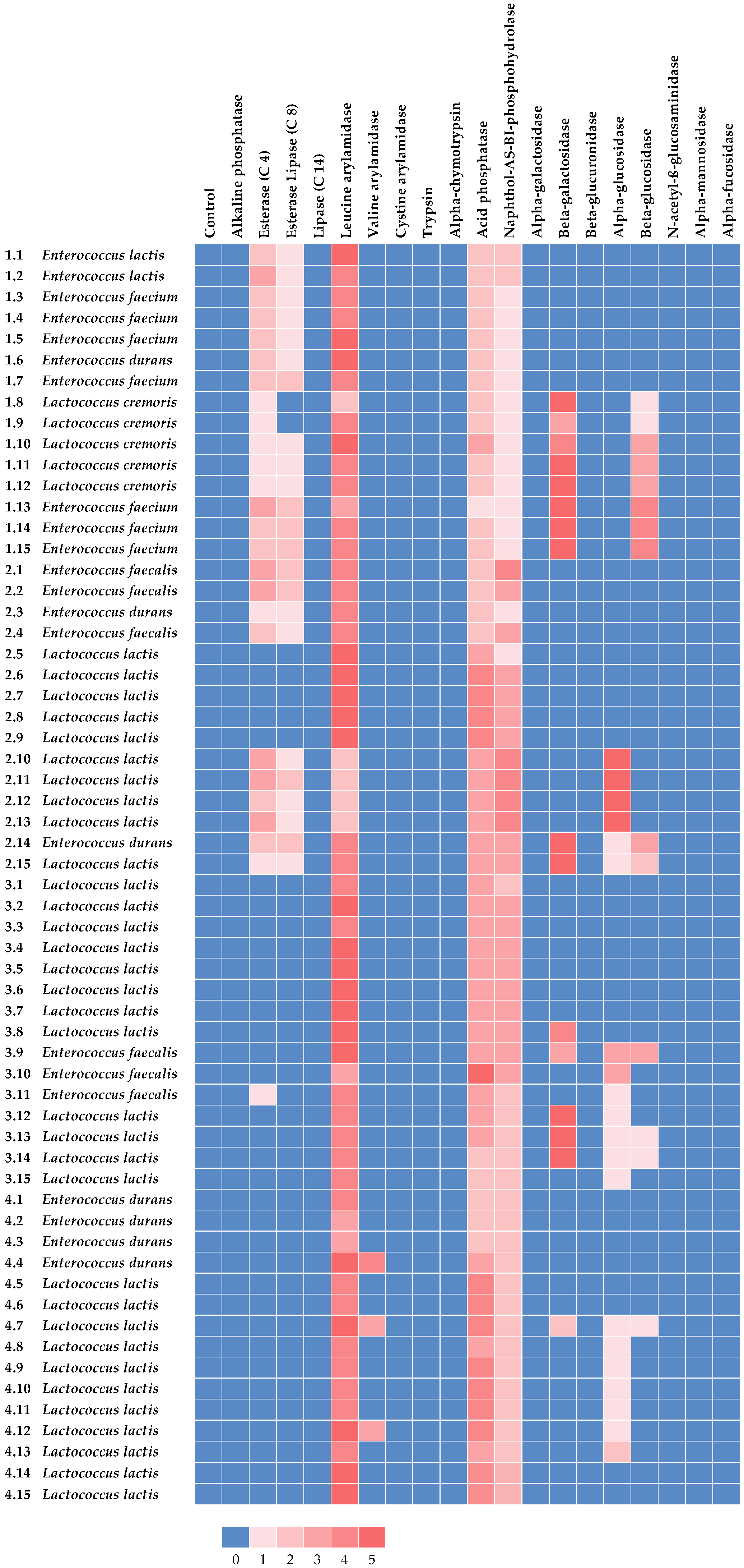
3.3. Enzymatic Activities of the Lactic Acid Bacteria Isolates

The results of the assessment of protease, lipase, and esterase activities of the lactic acid bacteria isolated from the viili samples are reported in Table 3. In more detail, most of the isolates (43 out of 60) showed a positive reaction for protease activity. Remarkably, isolates 1.3, 1.7, 1.12, 2.11, 2.14, 2.15, and 4.15 exhibited higher protease activity. Of note, almost all isolates belonging to the enterococci species showed protease activity, whereas higher variability was observed for lactococci. The proteolytic system of lactic acid bacteria consists of three main components: (i) cell-wall proteinase, which hydrolyzes extracellular caseins; (ii) peptide transporters, responsible for carrying oligopeptides into the cell; and (iii) intracellular peptidases, which break down peptides into shorter peptides and amino acids [40].
These mechanisms are essential for cells’ survival and multiplication during fermentation as they enable the supply of amino acids necessary for bacterial growth. Furthermore, the proteolysis of lactic acid bacteria is regarded as a crucial technological property in the dairy sector because it is associated with the development of volatile compounds that can affect the flavor of the end product [41,42]. Moreover, the proteolytic activity of lactic acid bacteria can provide potential health benefits for consumers, including improved digestibility and the release of bioactive peptides [41,42]. In general, lactic acid bacteria are weakly proteolytic compared to other microbial groups found in the microbiota of dairy products [43]. However, this property may vary depending on the species, the subspecies, and even the specific strain [44]. Lacticaseibacillus casei, Lactobacillus delbrueckii subsp. bulgaricus, Lactobacillus helveticus, and Lactobacillus acidophilus are the lactic acid bacteria that, due to their proteolytic activity, are the most used in the dairy industry. Among lactococci, L. cremoris typically exhibits higher proteolytic activity compared to L. lactis [44]. Moreover, the proteolytic activity of Enterococcus spp. is well documented in the literature [27,45,46,47].
In the present study, only 11 isolates (1.1, 1.5, 1.13, 1.14, 1.15, 2.1, 2.2, 2.14, 4.1, 4.2, 4.4) showed lipase activity, whereas no isolate exhibited esterase activity. Notably, all the isolates testing positive for lipolytic activity belonged to the enterococci group. Lipases are enzymes that catalyze the hydrolysis of triglycerides to glycerol and free fatty acids over an oil–water interface, whereas esterase catalyzes the hydrolysis of esters in solution and tri-, di-, and monoglycerides containing short-chain fatty acids [48,49]. Lactic acid bacteria are known to be weakly lipolytic compared to other microbial groups [49]. Thus, the results of this research are in accordance with those usually reported in the scientific literature. Indeed, the lipolytic activity of Lactococcus spp. isolated from dairy products has rarely been reported [16,50,51,52]. Instead, there is more evidence on the lipolytic activity of Enterococcus spp. [47,53,54].
The results of the semi-quantitative assessment of the enzymatic activities by API® ZYM are reported in Figure 1. In more detail, none of the isolates exhibited a positive reaction for alkaline phosphatase, lipase (C 14), cystine arylamidase, trypsin, alpha-chymotrypsin, alpha-galactosidase, beta-glucuronidase, N-acetyl-ß-glucosaminidase, alpha-mannosidase, and alpha-fucosidase. Conversely, all isolates showed leucine arylamidase, acid phosphatase, and naphthol-AS-BI-phosphohydrolase activities.
As for leucine arylamidase, all isolates except L. cremoris 1.8 and L. lactis 2.10, 2.11, 2.12, and 2.13 exhibited strong activity for this enzyme. The activity of leucine arylamidase in both Lactococcus spp. and Enterococcus spp. is widely documented in the literature [55,56,57] and considered as a desirable trait for dairy starters [55]. Conversely, the activity of other aminopeptidases is less frequently detected [57,58]. Of note, isolates E. durans 4.4 and L. lactis 4.7 and 4.12 exhibited a positive reaction for valine arylamidase. Weak esterase (C 4) and esterase lipase (C 8) activities were observed for a few isolates; however, only isolates E. lactis 1.2; E. faecium 1.13; E. faecalis 2.1, 2.2; and L. lactis 2.10, 2.11, 2.13 exhibited a positive reaction for esterase (C 4).
In the scientific literature, high esterase (C 4) and esterase lipase (C 8) activities are usually reported for enterococci [59,60,61]. Moreover, the esterase (C 4) activity of L. lactis has been already observed by Abarquero et al. [56] and Kamarinou et al. [58] in strains isolated from artisanal cheeses.
As for acid phosphatase, the activity of this enzyme may influence the hydrolysis of caseins in dairy products [62]. Except for isolate L. cremoris 1.10, all isolates of the same species sourcing from V1 exhibited a negative reaction to acid phosphatase. The same result was also observed for isolates 2.1, 2.2, 2.3, 2.4, 3.14, 3.15, 4.1, 4.2, and 4.3. Conversely, isolates 2.6, 2.7, 2.8, 2.9, 4.5, 4.6, 4.7, 4.9, 4.10, 4.11, and 4.12, all belonging to the L. lactis species, showed a strong reaction to acid phosphatase. Moreover, E. faecalis 3.10 was the sole isolate showing the strongest reaction to acid phosphatase. Of note, the acid phosphatase activity of Enterococcus spp. (E. durans, E. faecium, and E. faecalis) and L. lactis has previously been found in strains isolated from artisanal cheeses [56,60].
None of the isolates originating from V1 and V4 showed a positive naphthol-AS-BI-phosphohydrolase reaction. Conversely, except for 2.3, 2.5, 3.1, 3.11, 3.12, 3.13, 3.14, and 3.15, a positive reaction was observed for the other isolates. Similarly to acid phosphatase, the action of naphthol-AS-BI-phosphohydrolase is associated with potential technological and health benefits [63].
Few isolates exhibited positive reactions for beta-galactosidase, alpha-glucosidase, and beta-glucosidase, which are enzymes involved in sugar metabolism. In more detail, isolates L. cremoris 1.8, 1.9, 1.10, 1.11, 1.12, E. faecium 1.13, 1.14, 1.15, E. durans 2.14, L. lactis 2.15, 3.8, 3.12, 3.13, 3.14, and E. faecalis 3.9 exhibited a positive reaction for beta-galactosidase. This enzyme hydrolyzes the lactose beta-glycosidic bond between glucose and galactose [64], thereby holding significant importance in both technological and nutritional aspects. In fact, the activity of beta-galactosidase may contribute to milk acidification and the alleviation of lactose intolerance for consumers [65]. The beta-galactosidase activity exhibited by Enterococcus spp. is extensively documented in the literature, thus confirming their adaption to the dairy environment [57,59,60]. Of note, a low beta-galactosidase activity exhibited by Lactococcus spp. isolates was observed by Abarquero et al. [56]. In the present study, only the isolates L. lactis 2.10, 2.11, 2.12, and 2.13 and E. faecalis 3.9 and 3.10 showed alpha-glucosidase activity. Moreover, beta-glucosidase activity has also rarely been detected. In fact, only isolates L. cremoris 1.10, 1.11, 1.12, E. faecium 1.13, 1.14, 1.15, E. durans 2.14, and E. faecalis 3.9 exhibited a positive reaction for this enzymatic activity. Beta-glucosidase catalyzes the hydrolysis of terminal residues of beta-D-glucose [66] and has scarcely been found among the enzymatic activities of lactic acid bacteria isolated from dairy products [61,67,68].
It is noteworthy that all the isolates showed the absence of beta-glucuronidase activity, which is associated with the development of carcinogenic compounds in the human colon [31].
The assessment of enzymatic activities of lactic acid bacteria represents a valuable tool for the initial screening of potential starter or adjunct cultures in the dairy industry. Indeed, the metabolic reactions of microorganisms significantly impact the nutritional and sensory (volatile) properties of fermented foods [16,47,69]. However, criteria for selecting starter or adjunct cultures with specific technological properties may vary depending on the type of product. The collected results can provide a preliminary basis for future research aimed at further investigating the isolates’ capacity to produce aromatic or beneficial compounds in dairy products.

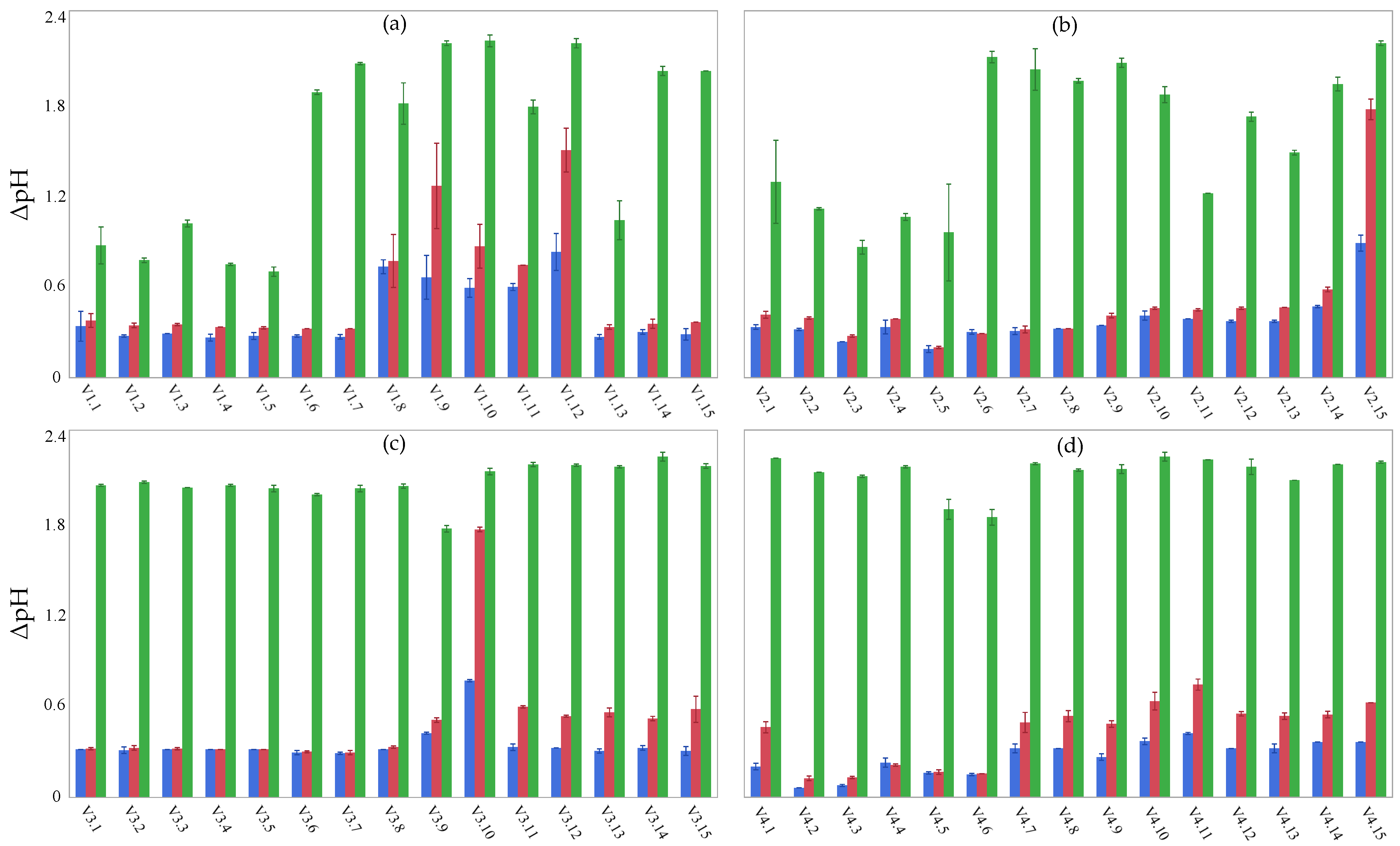
3.4. Acidification Performance

Based on their acidification performance, lactic acid bacteria are commonly classified into three categories: (i) fast acidifiers (pH < 5.3 after 6 h), medium acidifiers (pH < 5.3 after 8 h), and slow acidifiers (pH > 5.3 after 8 h) [16]. The results of the acidification performance of lactic acid bacteria isolated from the viili samples studied herein are reported in Figure 2 (V1, panel a; V2, panel b; V3, panel c; V4, panel d).
In more detail, none of the tested isolates lowered the pH of milk below 5.3 after 6 h of fermentation. The ΔpH after 6 h of fermentation ranged from 0.06 to 0.83. Notably, isolates L. cremoris 1.8, 1.9, 1.12, L. lactis 2.15, and E. faecalis 3.10 showed the best acidification performance after 6 h of fermentation. The ΔpH after 8 h of fermentation ranged from 0.12 to 1.73. A pH < 5.3 was measured in milk inoculated with the isolates 1.12, 2.15, and 3.10 after 8 h of fermentation. Moreover, isolate 1.9 also showed a good performance after 8 h of fermentation, resulting in a pH value of 5.33. The other isolates showed a slower acidification rate. However, except for isolates 1.1, 1.2, 1.3, 1.4, 1.5, 1.13, 2.2, 2.3, 2.4, 2.5, and 2.11, all isolates lowered the pH below 5.3 after 24 h of fermentation. The ΔpH after 24 h ranged from 0.65 to 2.20; at this time point, most of the isolates lowered the pH to values around 4.6, thus leading to milk coagulation.
The acidification performance is considered a primary criterion for selecting starter cultures with potential dairy applications [70]. Indeed, rapid fermentation and subsequent acidification are essential for ensuring product safety and texture [71,72]. In more detail, the well-known gel structure of fermented milk products is the result of acidic coagulation of caseins (namely αS1, αS2, β, and κ-casein) that is obtained at a pH of around 4.8, thus strongly affecting the rheological properties of the end product [73]. As reported by Beresford et al. [74], optimal starter bacteria for dairy purposes are expected to produce sufficient acid to lower the pH of milk to below 5.3 in 6 h. Cogan et al. [75] observed that mesophilic lactic acid bacteria exhibited lower acidification activity compared to thermophilic bacteria. Lactococcus spp. isolated from dairy products generally show weak acidification activity [16,76,77]; however, strain-specific variations may be observed [78]. Concerning the isolates belonging to enterococci species, the results obtained in the present study are consistent with those previously reported by Serio et al. [60].

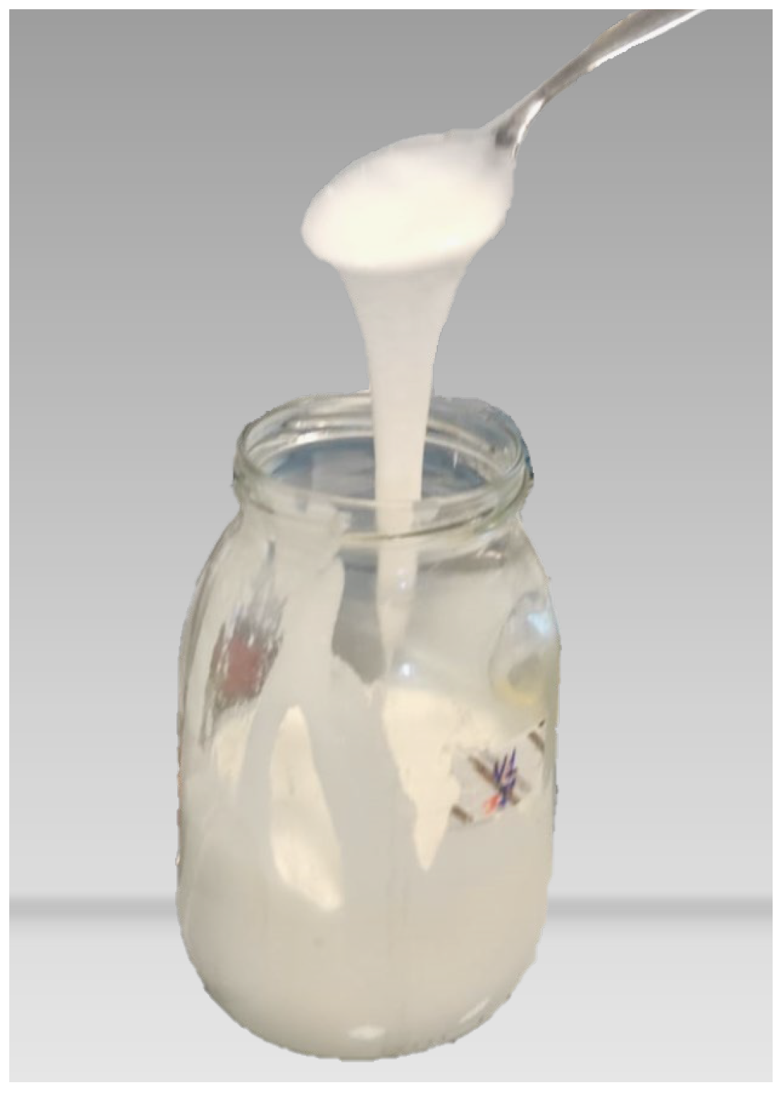
3.5. In Vitro EPS Production

None of the tested isolates exhibited in vitro EPS production, whether homopolysaccharides (HoPSs) or heteropolysaccharides (HePSs) (Table 3).
EPSs are biopolymers produced by specific microorganisms by extracellular synthesis or secretion during their growth [79]. The presence of EPSs positively influences the rheological and sensory characteristics of fermented dairy products, such as viscosity, syneresis, and sensory properties. Moreover, EPSs may exhibit various functional features, including antioxidant, antibacterial, cholesterol-lowering, immunoregulatory, antitumor, and anticoagulant activities [80]. Consequently, there has been a growing interest in recent years in the utilization of EPS-producing lactic acid bacteria in fermented foods to enhance product quality.
The distinctive feature of viili, along with other Nordic fermented milks, is their slimy texture, which arises from the production of EPSs by certain species of Lactococcus, mainly L. cremoris and L. lactis [9]. The in vitro assay for testing EPS-producing cultures applied in the present study represents a rapid and useful method for the preliminary selection of lactic acid bacteria as potential adjunct cultures [81]. However, this assay should be complemented by the molecular detection of EPS-related genes for further validation [28,82]. In the present study, the viili fermented milk samples obtained at the end of fermentation exhibited the typical ropy texture (Figure 3), thus suggesting the presence of EPSs in the end product.
It is noteworthy that EPS production by lactic acid bacteria depends on several factors, including genetic variation, environmental conditions, and strain-specific differences [83]. In the present study, the in vitro tests did not reveal EPS formation on agar plates, thus indicating the need for further in vivo tests of the isolates (e.g., use of pure cultures inoculated in milk).

3.6. Presence of hdcA Gene and Assessment of Antimicrobial Activity

The presence of the hdcA gene was revealed by qPCR in isolates E. faecium 1.5 and L. lactis 2.5, 2.8, 3.13, and 4.14 (Table 3). The gene cluster including the hdcA gene encodes for the activity of histidine decarboxylase, responsible for histamine production [84]. Of note, histamine represents a food hazard that is the causative agent of a wide range of foodborne diseases, including headache, hypotension, and vomiting [85]. Cheese and fish are the food categories mostly associated with histamine intoxication [86]. However, histamine content in dairy products is not subjected to any regulation by the European Union [87].
The detection of histamine-producing lactic acid bacteria represents a preventive strategy to avoid their use as starter or adjunct cultures, thus protecting the health of consumers [88,89]. Of note, histamine-producing L. lactis strains have already been detected by Roig-Sagués et al. [90] in traditional Spanish cheese. Moreover, Park et al. [91] and Tham et al. [92] previously reported the presence of histamine-producing Enterococcus spp. in goat cheese and in Cheonggukjang (a traditional Korean food made by fermenting soybeans), respectively.
Another safety-related aspect of interest to consider when selecting lactic acid bacteria to be used as starter or adjunct cultures is their antimicrobial activity against spoilage or pathogenic bacteria [88]. Indeed, some lactic acid bacteria have the capability to produce antimicrobial peptides, known as bacteriocins, which exhibit inhibitory activities against foodborne pathogens or spoilage microorganisms [93]. In more detail, strains of Lactococcus spp. and Enterococcus spp. isolated from dairy products have been found to produce bacteriocins (e.g., lactocins and enterocins) [94,95]. In the present study, none of the isolates showed antimicrobial activity against L. innocua (Table 3). However, the ability of lactic acid bacteria to synthesize bacteriocins is strictly strain-dependent and is affected by the expression of numerous genes [93].

4. Conclusions

The aim of this study was to isolate and characterize lactic acid bacteria from viili fermented milk samples obtained from natural starter cultures. This study aimed to assess the technological and safety traits of the isolates to identify potential candidates suitable for use as starter or adjunct cultures in dairy products.
Based on the results obtained in the present study, the isolates L. cremoris 1.12 and L. lactis 2.15 exhibited promising pro-technological traits (such as acidification performance and protease activity) for their use as starter cultures. Isolate 1.12 also showed leucine arylamidase, beta-galactosidase, and beta-glucosidase activities. Additionally, isolate 2.15 exhibited a positive reaction for acid phosphatase and naphthol-AS-BI-phosphohydrolase. Several E. faecium and E. faecalis isolates, namely 1.13, 1.14, 1.15, 2.1, and 2.2, showed significant lipase activity, a key feature for their potential use as adjunct cultures for flavor enhancement of the product. However, the presence of Enterococcus spp. in dairy products raises safety concerns due to virulence traits and antibiotic resistance occurring in certain strains.
Despite the observed slimy texture in the viili fermented milk samples studied herein, which suggested the presence of EPSs in the end product, no isolate exhibited the in vitro production of these natural biopolymers. Consequently, further investigation is needed to test the selected microbial cultures in in vivo viili models. Furthermore, future research should focus on investigating the presence and function of fungal populations in viili.
Of note, the detection of the hdcA gene in isolates 1.5, 2.7, 2.11, 3.13, and 4.14 highlights their unsuitability for dairy processing and sheds a first light on the potential histamine risk associated with the consumption of this fermented food.
Overall, the findings of this study have contributed to the identification and selection of lactic acid bacteria from artisanal viili, thereby enhancing the preservation of the quality and authenticity of this product. Moreover, several cultures of L. cremoris and L. lactis exhibited promising features for their application in dairy processing.

Author Contributions

Conceptualization, L.A.; formal analysis, G.R., V.M. and C.G.; investigation, G.R., A.C., F.C. and V.M.; resources, L.A. and A.O.; supervision, A.O.; writing—original draft, G.R.; writing—review and editing, F.C., L.A. and A.O. All authors have read and agreed to the published version of the manuscript.

Funding

The research was funded by the project Vitality—Project Code ECS00000041, CUP I33C22001330007—funded under the National Recovery and Resilience Plan (NRRP), Mission 4 Component 2 Investment 1.5—‘Creation and strengthening of innovation ecosystems,’ construction of ‘territorial leaders in R&D’—Innovation Ecosystems—Project ‘Innovation, digitalization and sustainability for the diffused economy in Central Italy—VITALITY’ Call for tender No. 3277 of 30/12/2021, and Concession Decree No. 0001057.23-06-2022 of Italian Ministry of University funded by the European Union—NextGenerationEU.

Data Availability Statement

The original contributions presented in the study are included in the article; further inquiries can be directed to the corresponding author.

Conflicts of Interest

The authors declare no conflicts of interest.

References

  1. Tamime, A.Y. Fermented Milks: A Historical Food with Modern Applications—A Review. Eur. J. Clin. Nutr. 2002, 56, S2–S15. [Google Scholar] [CrossRef] [PubMed]
  2. Evershed, R.P.; Payne, S.; Sherratt, A.G.; Copley, M.S.; Coolidge, J.; Urem-Kotsu, D.; Kotsakis, K.; Özdoǧan, M.; Özdoǧan, A.E.; Nieuwenhuyse, O.; et al. Earliest Date for Milk Use in the Near East and Southeastern Europe Linked to Cattle Herding. Nature 2008, 455, 528–531. [Google Scholar] [CrossRef] [PubMed]
  3. Bintsis, T.; Papademas, P. The Evolution of Fermented Milks, from Artisanal to Industrial Products: A Critical Review. Fermentation 2022, 8, 679. [Google Scholar] [CrossRef]
  4. Codex Stan 243-2003; Codex Standard for Fermented Milks. Codex Alimentarius: Rome, Italy, 2003.
  5. Macori, G.; Cotter, P.D. Novel Insights into the Microbiology of Fermented Dairy Foods. Curr. Opin. Biotechnol. 2018, 49, 172–178. [Google Scholar] [CrossRef] [PubMed]
  6. Robinson, R.K.; Tamime, A.Y.; Wszolek, M. Microbiology of Fermented Milks. In Dairy Microbiology Handbook; Robinson, R.K., Ed.; John Wiley and Sons, Inc.: New York, NY, USA, 2002; pp. 367–421. [Google Scholar]
  7. Maoloni, A.; Blaiotta, G.; Ferrocino, I.; Mangia, N.P.; Osimani, A.; Milanović, V.; Cardinali, F.; Cesaro, C.; Garofalo, C.; Clementi, F.; et al. Microbiological Characterization of Gioddu, an Italian Fermented Milk. Int. J. Food Microbiol. 2020, 323, 108610. [Google Scholar] [CrossRef] [PubMed]
  8. Leroy, F.; De Vuyst, L. Lactic Acid Bacteria as Functional Starter Cultures for the Food Fermentation Industry. Trends Food Sci. Technol. 2004, 15, 67–78. [Google Scholar] [CrossRef]
  9. Narvhus, J.A.; Abrahamsen, R.K. Traditional and Modern Nordic Fermented Milk Products: A Review. Int. Dairy J. 2023, 142, 105641. [Google Scholar] [CrossRef]
  10. Abi Khalil, R.; Yvon, S.; Couderc, C.; Jard, G.; El Rammouz, R.; Abi Nakhoul, P.; Eutamène, H.; Ayoub, M.J.; Tormo, H. Traditional Fermented Milk Products of Eastern Mediterranean Countries: A Cultural Heritage to Preserve. Int. Dairy J. 2023, 147, 105768. [Google Scholar] [CrossRef]
  11. Mallappa, R.H.; Balasubramaniam, C.; Nataraj, B.H.; Ramesh, C.; Kadyan, S.; Pradhan, D.; Muniyappa, S.K.; Grover, S. Microbial Diversity and Functionality of Traditional Fermented Milk Products of India: Current Scenario and Future Perspectives. Int. Dairy J. 2021, 114, 104941. [Google Scholar] [CrossRef]
  12. Moonga, H.B.; Schoustra, S.E.; Linnemann, A.R.; Shindano, J.; Smid, E.J. Towards Valorisation of Indigenous Traditional Fermented Milk: Mabisi as a Model. Curr. Opin. Food Sci. 2022, 46, 100835. [Google Scholar] [CrossRef]
  13. Ferrocino, I.; Rantsiou, K.; Cocolin, L. Investigating Dairy Microbiome: An Opportunity to Ensure Quality, Safety and Typicity. Curr. Opin. Biotechnol. 2022, 73, 164–170. [Google Scholar] [CrossRef] [PubMed]
  14. Goa, T.; Beyene, G.; Mekonnen, M.; Gorems, K. Isolation and Characterization of Lactic Acid Bacteria from Fermented Milk Produced in Jimma Town, Southwest Ethiopia, and Evaluation of Their Antimicrobial Activity against Selected Pathogenic Bacteria. Int. J. Food Sci. 2022, 2022, 2076021. [Google Scholar] [CrossRef] [PubMed]
  15. Chammas, G.I.; Saliba, R.; Corrieu, G.; Béal, C. Characterisation of Lactic Acid Bacteria Isolated from Fermented Milk “Laban”. Int. J. Food Microbiol. 2006, 110, 52–61. [Google Scholar] [CrossRef] [PubMed]
  16. Nicosia, F.D.; Pino, A.; Maciel, G.L.R.; Sanfilippo, R.R.; Caggia, C.; de Carvalho, A.F.; Randazzo, C.L. Technological Characterization of Lactic Acid Bacteria Strains for Potential Use in Cheese Manufacture. Foods 2023, 12, 1154. [Google Scholar] [CrossRef] [PubMed]
  17. Li, Y.; Zhang, Y.; Dong, L.; Li, Y.; Liu, Y.; Liu, Y.; Liu, L.; Liu, L. Fermentation of Lactobacillus Fermentum NB02 with Feruloyl Esterase Production Increases the Phenolic Compounds Content and Antioxidant Properties of Oat Bran. Food Chem. 2024, 437, 137834. [Google Scholar] [CrossRef] [PubMed]
  18. Narvhus, J.A.; Abrahamsen, R.K. Nordic Fermented Milks. Encycl. Dairy Sci. 2021, 464–471. [Google Scholar] [CrossRef]
  19. Porcellato, D.; Tranvåg, M.; Narvhus, J. Short Communication: Presence of Lactococcus and Lactococcal Exopolysaccharide Operons on the Leaves of Pinguicula Vulgaris Supports the Traditional Source of Bacteria Present in Scandinavian Ropy Fermented Milk. J. Dairy Sci. 2016, 99, 7049–7052. [Google Scholar] [CrossRef]
  20. Chen, T.; Tan, Q.; Wang, M.; Xiong, S.; Jiang, S.; Wu, Q.; Li, S.; Luo, C.; Wei, H. Identification of Bacterial Strains in Viili by Molecular Taxonomy and Their Synergistic Effects on Milk Curd and Exopolysaccharides Production. Afr. J. Biotechnol. 2011, 10, 16969–16975. [Google Scholar] [CrossRef]
  21. Kahala, M.; Mäki, M.; Lehtovaara, A.; Tapanainen, J.M.; Katiska, R.; Juuruskorpi, M.; Juhola, J.; Joutsjoki, V. Characterization of Starter Lactic Acid Bacteria from the Finnish Fermented Milk Product Viili. J. Appl. Microbiol. 2008, 105, 1929–1938. [Google Scholar] [CrossRef] [PubMed]
  22. Ruas-Madiedo, P.; Gueimonde, M.; De Los Reyes-Gavilán, C.G.; Salminen, S. Short Communication: Effect of Exopolysaccharide Isolated from “Viili” on the Adhesion of Probiotics and Pathogens to Intestinal Mucus. J. Dairy Sci. 2006, 89, 2355–2358. [Google Scholar] [CrossRef] [PubMed]
  23. Yamane, T.; Handa, S.; Imai, M.; Harada, N.; Sakamoto, T.; Ishida, T.; Nakagaki, T.; Nakano, Y. Exopolysaccharides from a Scandinavian Fermented Milk Viili Increase Butyric Acid and Muribaculum Members in the Mouse Gut. Food Chem. Mol. Sci. 2021, 3, 100042. [Google Scholar] [CrossRef] [PubMed]
  24. ANSES (Agence Nationale de Sécurité Sanitaire de L’alimentation, de L’environnement et du Travail). EURL Lm TECHNICAL GUIDANCE DOCUMENT for Conducting Shelflife Studies on Listeria Monocytogenes in Ready-to-Eat Foods. Version 3 of 6 June 2014—Amendment 1 of 21 February 2019. Available online: https://eurl-listeria.anses.fr/en/system/files/LIS-Cr-201909D2.pdf (accessed on 14 November 2023).
  25. Rampanti, G.; Raffo, A.; Melini, V.; Moneta, E.; Nardo, N.; Saggia Civitelli, E.; Bande-De León, C.; Tejada Portero, L.; Ferrocino, I.; Franciosa, I.; et al. Chemical, microbiological, textural, and sensory characteristics of pilot-scale Caciofiore cheese curdled with commercial Cynara Cardunculus Rennet and crude extracts from spontaneous and cultivated Onopordum Tauricum. Food Res. Int. 2023, 173, 113459. [Google Scholar] [CrossRef] [PubMed]
  26. Osimani, A.; Garofalo, C.; Aquilanti, L.; Milanović, V.; Clementi, F. Unpasteurised Commercial Boza as a Source of Microbial Diversity. Int. J. Food Microbiol. 2015, 194, 62–70. [Google Scholar] [CrossRef] [PubMed]
  27. Linares-Morales, J.R.; Cuellar-Nevárez, G.E.; Rivera-Chavira, B.E.; Gutiérrez-Méndez, N.; Pérez-Vega, S.B.; Nevárez-Moorillón, G.V. Selection of Lactic Acid Bacteria Isolated from Fresh Fruits and Vegetables Based on Their Antimicrobial and Enzymatic Activities. Foods 2020, 9, 1399. [Google Scholar] [CrossRef] [PubMed]
  28. Hilbig, J.; Gisder, J.; Prechtl, R.M.; Herrmann, K.; Weiss, J.; Loeffler, M. Influence of Exopolysaccharide-Producing Lactic Acid Bacteria on the Spreadability of Fat-Reduced Raw Fermented Sausages (Teewurst). Food Hydrocoll. 2019, 93, 422–431. [Google Scholar] [CrossRef]
  29. Belleggia, L.; Milanović, V.; Cardinali, F.; Garofalo, C.; Clementi, F.; Aquilanti, L.; Osimani, A. Prevalence of Histidine Decarboxylase Genes of Gram-Positive Bacteria in Surströmming as Revealed by QPCR. Indian J. Microbiol. 2021, 61, 96–99. [Google Scholar] [CrossRef] [PubMed]
  30. Fernández, M.; Del Río, B.; Linares, D.M.; Martín, M.C.; Alvarez, M.A. Real-Time Polymerase Chain Reaction for Quantitative Detection of Histamine-Producing Bacteria: Use in Cheese Production. J. Dairy Sci. 2006, 89, 3763–3769. [Google Scholar] [CrossRef] [PubMed]
  31. Cardinali, F.; Botta, C.; Harasym, J.; Reale, A.; Ferrocino, I.; Boscaino, F.; Orkusz, A.; Milanović, V.; Garofalo, C.; Rampanti, G.; et al. Tasting of Traditional Polish Fermented Cucumbers: Microbiology, Morpho-Textural Features, and Volatilome. Food Res. Int. 2024, 177, 113851. [Google Scholar] [CrossRef]
  32. Bassi, D.; Puglisi, E.; Cocconcelli, P.S. Comparing Natural and Selected Starter Cultures in Meat and Cheese Fermentations. Curr. Opin. Food Sci. 2015, 2, 118–122. [Google Scholar] [CrossRef]
  33. Cavanagh, D.; Fitzgerald, G.F.; McAuliffe, O. From Field to Fermentation: The Origins of Lactococcus Lactis and Its Domestication to the Dairy Environment. Food Microbiol. 2015, 47, 45–61. [Google Scholar] [CrossRef] [PubMed]
  34. Rampanti, G.; Belleggia, L.; Cardinali, F.; Milanović, V.; Osimani, A.; Garofalo, C.; Ferrocino, I.; Aquilanti, L. Microbial Dynamics of a Specialty Italian Raw Ewe’s Milk Cheese Curdled with Extracts from Spontaneous and Cultivated Onopordum Tauricum Willd. Microorganisms 2023, 11, 219. [Google Scholar] [CrossRef] [PubMed]
  35. Shori, A.B. Comparative Study of Chemical Composition, Isolation and Identification of Micro-Flora in Traditional Fermented Camel Milk Products: Gariss, Suusac, and Shubat. J. Saudi Soc. Agric. Sci. 2012, 11, 79–88. [Google Scholar] [CrossRef]
  36. Arrizza, S.; Ledda, A.; Sarra, P.G.; Dellaglio, F. Identification of Lactic Acid Bacteria in Gioddu. Sci. Tech. Latt.-Casearia 1983, 34, 87–102. [Google Scholar]
  37. Chaves-López, C.; Serio, A.; Martuscelli, M.; Paparella, A.; Osorio-Cadavid, E.; Suzzi, G. Microbiological Characteristics of Kumis, a Traditional Fermented Colombian Milk, with Particular Emphasis on Enterococci Population. Food Microbiol. 2011, 28, 1041–1047. [Google Scholar] [CrossRef] [PubMed]
  38. Dapkevicius, M.d.L.E.; Sgardioli, B.; Câmara, S.P.A.; Poeta, P.; Malcata, F.X. Current Trends of Enterococci in Dairy Products: A Comprehensive Review of Their Multiple Roles. Foods 2021, 10, 821. [Google Scholar] [CrossRef] [PubMed]
  39. Giraffa, G. Functionality of Enterococci in Dairy Products. Int. J. Food Microbiol. 2003, 88, 215–222. [Google Scholar] [CrossRef] [PubMed]
  40. Liu, M.; Bayjanov, J.R.; Renckens, B.; Nauta, A.; Siezen, R.J. The Proteolytic System of Lactic Acid Bacteria Revisited: A Genomic Comparison. BMC Genom. 2010, 11, 36. [Google Scholar] [CrossRef] [PubMed]
  41. Savijoki, K.; Ingmer, H.; Varmanen, P. Proteolytic Systems of Lactic Acid Bacteria. Appl. Microbiol. Biotechnol. 2006, 71, 394–406. [Google Scholar] [CrossRef]
  42. García-Cano, I.; Rocha-Mendoza, D.; Ortega-Anaya, J.; Wang, K.; Kosmerl, E.; Jiménez-Flores, R. Lactic Acid Bacteria Isolated from Dairy Products as Potential Producers of Lipolytic, Proteolytic and Antibacterial Proteins. Appl. Microbiol. Biotechnol. 2019, 103, 5243–5257. [Google Scholar] [CrossRef] [PubMed]
  43. Mcsweeney, P.L.H.; Sousa, M.J. Biochemical Pathways for the Production of Flavour Compounds in Cheeses during Ripening: A Review. Le Lait 2000, 80, 293–324. [Google Scholar] [CrossRef]
  44. Kieliszek, M.; Pobiega, K.; Piwowarek, K.; Kot, A.M. Characteristics of the Proteolytic Enzymes Produced by Lactic Acid Bacteria. Molecules 2021, 26, 1858. [Google Scholar] [CrossRef] [PubMed]
  45. El-Ghaish, S.; Dalgalarrondo, M.; Choiset, Y.; Sitohy, M.; Ivanova, I.; Haertlé, T.; Chobert, J.M. Screening of Strains of Lactococci Isolated from Egyptian Dairy Products for Their Proteolytic Activity. Food Chem. 2010, 120, 758–764. [Google Scholar] [CrossRef]
  46. Saidi, Y.; del Rio, B.; Senouci, D.E.; Redruello, B.; Martinez, B.; Ladero, V.; Kihal, M.; Alvarez, M.A. Polyphasic Characterisation of Non-Starter Lactic Acid Bacteria from Algerian Raw Camel’s Milk and Their Technological Aptitudes. Food Technol. Biotechnol. 2020, 58, 260–272. [Google Scholar] [CrossRef] [PubMed]
  47. Psomas, E.; Sakaridis, I.; Boukouvala, E.; Karatzia, M.A.; Ekateriniadou, L.V.; Samouris, G. Indigenous Lactic Acid Bacteria Isolated from Raw Graviera Cheese and Evaluation of Their Most Important Technological Properties. Foods 2023, 12, 370. [Google Scholar] [CrossRef] [PubMed]
  48. Treichel, H.; de Oliveira, D.; Mazutti, M.A.; Di Luccio, M.; Oliveira, J.V. A Review on Microbial Lipases Production. Food Bioproc. Tech. 2010, 3, 182–196. [Google Scholar] [CrossRef]
  49. Medina, R.B.; Katz, M.B.; González, S.; Oliver, G. Determination of Esterolytic and Lipolytic Activities of Lactic Acid Bacteria. In Public Health Microbiology: Methods and Protocols; Spencer, J.F., Ragout de Spencer, A.L., Eds.; Spencer Humana Press Inc.: Totowa, NJ, USA, 2004; Volume 268, pp. 465–470. [Google Scholar]
  50. Katz, M.; Medina, R.; Gonzalez, S.; Oliver, G. Esterolytic and Lipolytic Activities of Lactic Acid Bacteria Isolated from Ewe’s Milk and Cheese. J. Food Prot. 2002, 65, 1997–2001. [Google Scholar] [CrossRef] [PubMed]
  51. Yerlikaya, O. Probiotic Potential and Biochemical and Technological Properties of Lactococcus Lactis Ssp. Lactis Strains Isolated from Raw Milk and Kefir Grains. J. Dairy Sci. 2019, 102, 124–134. [Google Scholar] [CrossRef] [PubMed]
  52. El-Din, B.B.; El-Soda, M.; Ezzat, N. Proteolytic, Lipolytic and Autolytic Activities of Enterococci Strains Isolated from Egyptian Dairy Products. Lait 2002, 82, 289–304. [Google Scholar] [CrossRef]
  53. Morandi, S.; Brasca, M.; Andrighetto, C.; Lombardi, A.; Lodi, R. Technological and Molecular Characterisation of Enterococci Isolated from North-West Italian Dairy Products. Int. Dairy J. 2006, 16, 867–875. [Google Scholar] [CrossRef]
  54. Graham, K.; Stack, H.; Rea, R. Safety, Beneficial and Technological Properties of Enterococci for Use in Functional Food Applications–a Review. Crit. Rev. Food Sci. Nutr. 2020, 60, 3836–3861. [Google Scholar] [CrossRef]
  55. Domingos-Lopes, M.F.P.; Stanton, C.; Ross, P.R.; Dapkevicius, M.L.E.; Silva, C.C.G. Genetic Diversity, Safety and Technological Characterization of Lactic Acid Bacteria Isolated from Artisanal Pico Cheese. Food Microbiol. 2017, 63, 178–190. [Google Scholar] [CrossRef] [PubMed]
  56. Abarquero, D.; Bodelón, R.; Flórez, A.B.; Fresno, J.M.; Renes, E.; Mayo, B.; Tornadijo, M.E. Technological and Safety Assessment of Selected Lactic Acid Bacteria for Cheese Starter Cultures Design: Enzymatic and Antimicrobial Activity, Antibiotic Resistance and Biogenic Amine Production. LWT 2023, 180, 114709. [Google Scholar] [CrossRef]
  57. Rampanti, G.; Ferrocino, I.; Harasym, J.; Foligni, R.; Cardinali, F.; Orkusz, A.; Milanović, V.; Franciosa, I.; Garofalo, C.; Mannozzi, C.; et al. Queijo Serra Da Estrela PDO Cheese: Investigation into Its Morpho-Textural Traits, Microbiota, and Volatilome. Foods 2023, 12, 169. [Google Scholar] [CrossRef] [PubMed]
  58. Kamarinou, C.S.; Papadopoulou, O.S.; Doulgeraki, A.I.; Tassou, C.C.; Galanis, A.; Chorianopoulos, N.G.; Argyri, A.A. Mapping the Key Technological and Functional Characteristics of Indigenous Lactic Acid Bacteria Isolated from Greek Traditional Dairy Products. Microorganisms 2022, 10, 246. [Google Scholar] [CrossRef] [PubMed]
  59. Kirilov, N.; Dimov, S.; Dalgalarrondo, M.; Ignatova, T.; Kambarev, S.; Stoyanovski, S.; Danova, S.; Iliev, I.; Haertlé, T.; Chobert, J.M.; et al. Characterization of Enterococci Isolated from Homemade Bulgarian Cheeses and Katuk. Eur. Food Res. Technol. 2011, 233, 1029–1040. [Google Scholar] [CrossRef]
  60. Serio, A.; Chaves-López, C.; Paparella, A.; Suzzi, G. Evaluation of Metabolic Activities of Enterococci Isolated from Pecorino Abruzzese Cheese. Int. Dairy J. 2010, 20, 459–464. [Google Scholar] [CrossRef]
  61. Tsanasidou, C.; Asimakoula, S.; Sameli, N.; Fanitsios, C.; Vandera, E.; Bosnea, L.; Koukkou, A.I.; Samelis, J. Safety Evaluation, Biogenic Amine Formation, and Enzymatic Activity Profiles of Autochthonous Enterocin-Producing Greek Cheese Isolates of the Enterococcus Faecium/Durans Group. Microorganisms 2021, 9, 777. [Google Scholar] [CrossRef] [PubMed]
  62. Akuzawa, R.; Fox, P.F. Acid Phosphatase in Cheese. Anim. Sci. J. 2004, 75, 385–391. [Google Scholar] [CrossRef]
  63. Coimbra-Gomes, J.; Reis, P.J.M.; Tavares, T.G.; Faria, M.A.; Malcata, F.X.; Macedo, A.C. Evaluating the Probiotic Potential of Lactic Acid Bacteria Implicated in Natural Fermentation of Table Olives, Cv. Cobrançosa. Molecules 2023, 28, 3285. [Google Scholar] [CrossRef] [PubMed]
  64. Kolev, P.; Rocha-Mendoza, D.; Ruiz-Ramírez, S.; Ortega-Anaya, J.; Jiménez-Flores, R.; García-Cano, I. Screening and Characterization of β-Galactosidase Activity in Lactic Acid Bacteria for the Valorization of Acid Whey. JDS Commun. 2022, 3, 1–6. [Google Scholar] [CrossRef]
  65. Câmara, S.P.; Dapkevicius, A.; Riquelme, C.; Elias, R.B.; Silva, C.C.G.; Malcata, F.X.; Dapkevicius, M.L.N.E. Potential of Lactic Acid Bacteria from Pico Cheese for Starter Culture Development. Food Sci. Technol. Int. 2019, 25, 303–317. [Google Scholar] [CrossRef] [PubMed]
  66. Michlmayr, H.; Kneifel, W. β-Glucosidase Activities of Lactic Acid Bacteria: Mechanisms, Impact on Fermented Food and Human Health. FEMS Microbiol. Lett. 2014, 352, 1–10. [Google Scholar] [CrossRef] [PubMed]
  67. Dewan, S.; Tamang, J.P. Dominant Lactic Acid Bacteria and Their Technological Properties Isolated from the Himalayan Ethnic Fermented Milk Products. Antonie Van Leeuwenhoek Int. J. Gen. Mol. Microbiol. 2007, 92, 343–352. [Google Scholar] [CrossRef] [PubMed]
  68. Jawan, R.; Abbasiliasi, S.; Mustafa, S.; Kapri, M.R.; Halim, M.; Ariff, A.B. In Vitro Evaluation of Potential Probiotic Strain Lactococcus Lactis Gh1 and Its Bacteriocin-Like Inhibitory Substances for Potential Use in the Food Industry. Probiotics Antimicrob. Proteins 2021, 13, 422–440. [Google Scholar] [CrossRef] [PubMed]
  69. Liu, Y.; Dong, L.; Li, Y.; Chen, Q.; Wang, L.; Farag, M.A.; Liu, L.; Zhan, S.; Wu, Z.; Liu, L. Soy Protein Isolate-Citrus Pectin Composite Hydrogels Induced by TGase and Ultrasonic Treatment: Potential Targeted Delivery System for Probiotics. Food Hydrocoll. 2023, 143, 108901. [Google Scholar] [CrossRef]
  70. Ribeiro, S.C.; Coelho, M.C.; Silva, C.C.G. A Rapid Screening Method to Evaluate Acidifying Activity by Lactic Acid Bacteria. J. Microbiol. Methods 2021, 185, 106227. [Google Scholar] [CrossRef] [PubMed]
  71. Sionek, B.; Szydłowska, A.; Küçükgöz, K.; Kołożyn-Krajewska, D. Traditional and New Microorganisms in Lactic Acid Fermentation of Food. Fermentation 2023, 9, 1019. [Google Scholar] [CrossRef]
  72. Li, W.; Ren, M.; Duo, L.; Li, J.; Wang, S.; Sun, Y.; Li, M.; Ren, W.; Hou, Q.; Yu, J.; et al. Fermentation Characteristics of Lactococcus Lactis Subsp. Lactis Isolated from Naturally Fermented Dairy Products and Screening of Potential Starter Isolates. Front. Microbiol. 2020, 11, 1794. [Google Scholar] [CrossRef]
  73. Li, Q.; Zhao, Z. Acid and Rennet-Induced Coagulation Behavior of Casein Micelles with Modified Structure. Food Chem. 2019, 291, 231–238. [Google Scholar] [CrossRef] [PubMed]
  74. Beresford, T.P.; Fitzsimons, N.A.; Brennan, N.L.; Cogan, T.M. Recent Advances in Cheese Microbiology. Int. Dairy J. 2001, 11, 259–274. [Google Scholar] [CrossRef]
  75. Cogan, T.M.; Barbosa, M.; Beuvier, E.; Bianchi-salvadori, B.; Cocconcelli, P.S.; Fernandes, I.; Gomezr, J.; Gomez, R.; Kalantzopoulos, G.; Ledda, A.; et al. Characterization of the Lactic Acid Bacteria in Artisanal Dairy Products. J. Dairy Res. 1997, 64, 409–421. [Google Scholar] [CrossRef]
  76. Hadef, S.; Idoui, T.; Sifour, M.; Genay, M.; Dary-Mourot, A. Screening of Wild Lactic Acid Bacteria from Algerian Traditional Cheeses and Goat Butter to Develop a New Probiotic Starter Culture. Probiotics Antimicrob. Proteins 2022, 15, 387–399. [Google Scholar] [CrossRef] [PubMed]
  77. Ayad, E.H.E.; Nashat, S.; El-Sadek, N.; Metwaly, H.; El-Soda, M. Selection of Wild Lactic Acid Bacteria Isolated from Traditional Egyptian Dairy Products According to Production and Technological Criteria. Food Microbiol. 2004, 21, 715–725. [Google Scholar] [CrossRef]
  78. Delgado, S.; Delgado, T.; Mayo, B. Technological Performance of Several Lactococcus and Enterococcus Strains of Dairy Origin in Milk. J. Food Prot. 2002, 65, 1590–1596. [Google Scholar] [CrossRef] [PubMed]
  79. Welman, A.D.; Maddox, I.S. Exopolysaccharides from Lactic Acid Bacteria: Perspectives and Challenges. Trends Biotechnol. 2003, 21, 269–274. [Google Scholar] [CrossRef] [PubMed]
  80. Zhou, Y.; Cui, Y.; Qu, X. Exopolysaccharides of Lactic Acid Bacteria: Structure, Bioactivity and Associations: A Review. Carbohydr. Polym. 2019, 207, 317–332. [Google Scholar] [CrossRef] [PubMed]
  81. Ruas-Madiedo, P.; De Los Reyes-Gavilán, C.G. Invited Review: Methods for the Screening, Isolation, and Characterization of Exopolysaccharides Produced by Lactic Acid Bacteria. J. Dairy Sci. 2005, 88, 843–856. [Google Scholar] [CrossRef] [PubMed]
  82. Milanovic, V.; Osimani, A.; Garofalo, C.; Belleggia, L.; Maoloni, A.; Cardinali, F.; Mozzon, M.; Foligni, R.; Aquilanti, L.; Clementi, F. Selection of Cereal-Sourced Lactic Acid Bacteria as Candidate Starters for the Baking Industry. PLoS ONE 2020, 15, e0236190. [Google Scholar] [CrossRef] [PubMed]
  83. Nguyen, P.T.; Nguyen, T.T.; Bui, D.C.; Hong, P.T.; Hoang, Q.K.; Nguyen, H.T. Exopolysaccharide Production by Lactic Acid Bacteria: The Manipulation of Environmental Stresses for Industrial Applications. AIMS Microbiol. 2020, 6, 451–469. [Google Scholar] [CrossRef] [PubMed]
  84. Diaz, M.; Ladero, V.; Redruello, B.; Sanchez-Llana, E.; del Rio, B.; Fernandez, M.; Martin, M.C.; Alvarez, M.A. Nucleotide Sequence Alignment of HdcA from Gram-Positive Bacteria. Data Brief 2016, 6, 674–679. [Google Scholar] [CrossRef] [PubMed]
  85. Ordóñez, J.L.; Troncoso, A.M.; García-Parrilla, M.D.C.; Callejón, R.M. Recent Trends in the Determination of Biogenic Amines in Fermented Beverages—A Review. Anal. Chim. Acta 2016, 939, 10–25. [Google Scholar] [CrossRef] [PubMed]
  86. EFSA Scientific Opinion on Risk Based Control of Biogenic Amine Formation in Fermented Foods. EFSA J. 2011, 9, 2393. [CrossRef]
  87. Moniente, M.; García-Gonzalo, D.; Ontañón, I.; Pagán, R.; Botello-Morte, L. Histamine Accumulation in Dairy Products: Microbial Causes, Techniques for the Detection of Histamine-Producing Microbiota, and Potential Solutions. Compr. Rev. Food Sci. Food Saf. 2021, 20, 1481–1523. [Google Scholar] [CrossRef] [PubMed]
  88. Di Renzo, T.; Osimani, A.; Marulo, S.; Cardinali, F.; Mamone, G.; Puppo, C.; Garzón, A.G.; Drago, S.R.; Laurino, C.; Reale, A. Insight into the Role of Lactic Acid Bacteria in the Development of a Novel Fermented Pistachio (Pistacia vera L.) Beverage. Food Biosci. 2023, 53, 102802. [Google Scholar] [CrossRef]
  89. Osimani, A.; Belleggia, L.; Botta, C.; Ferrocino, I.; Milanović, V.; Cardinali, F.; Haouet, M.N.; Garofalo, C.; Mozzon, M.; Foligni, R.; et al. Journey to the Morpho-Textural Traits, Microbiota, and Volatilome of Ciauscolo PGI Salami. Food Biosci. 2023, 53, 102582. [Google Scholar] [CrossRef]
  90. Roig-Sagués, A.X.; Molina, A.P.; Hernández-Herrero, M.M. Histamine and Tyramine-Forming Microorganisms in Spanish Traditional Cheeses. Eur. Food Res. Technol. 2002, 215, 96–100. [Google Scholar] [CrossRef]
  91. Park, Y.K.; Jin, Y.H.; Lee, J.H.; Byun, B.Y.; Lee, J.; Jeong, K.C.C.; Mah, J.H. The Role of Enterococcus Faecium as a Key Producer and Fermentation Condition as an Influencing Factor in Tyramine Accumulation in Cheonggukjang. Foods 2020, 9, 915. [Google Scholar] [CrossRef] [PubMed]
  92. Tham, W.; Karp, G.; Danielsson-Tham, M.L. Histamine Formation by Enterococci in Goat Cheese. Int. J. Food Microbiol. 1990, 11, 225–229. [Google Scholar] [CrossRef] [PubMed]
  93. Perez, R.H.; Zendo, T.; Sonomoto, K. Multiple Bacteriocin Production in Lactic Acid Bacteria. J. Biosci. Bioeng. 2022, 134, 277–287. [Google Scholar] [CrossRef]
  94. de Niederhäusern, S.; Camellini, S.; Sabia, C.; Iseppi, R.; Bondi, M.; Messi, P. Antilisterial Activity of Bacteriocins Produced by Lactic Bacteria Isolated from Dairy Products. Foods 2020, 9, 1757. [Google Scholar] [CrossRef] [PubMed]
  95. Alegría, Á.; Delgado, S.; Roces, C.; López, B.; Mayo, B. Bacteriocins Produced by Wild Lactococcus Lactis Strains Isolated from Traditional, Starter-Free Cheeses Made of Raw Milk. Int. J. Food Microbiol. 2010, 143, 61–66. [Google Scholar] [CrossRef]
Figure 1. Semi-quantitative assessment of enzymatic activity of lactic acid bacteria isolated from viili samples.
Foods 13 01115 g001
Figure 2. Acidification performance of the lactic acid bacteria isolated from viili samples after 6 h (blue bar), 8 h (red bar), and 24 h of fermentation (green bar). Panel (a), isolates from V1; panel (b), isolates from V2; panel (c), isolates from V3; panel (d), isolates from V4.
Foods 13 01115 g002
Figure 3. Ropy consistency of viili after the activation of natural starters.
Foods 13 01115 g003
Table 1. pH, titratable acidity, and counts of presumptive lactic acid bacteria monitored during the activation of viili natural starters.
pHTitratable Acidity 1Presumptive Lactic Acid Bacteria 2
V1V2V3V4V1V2V3V4V1V2V3V4
t06.49 ± 0.07 A6.35 ± 0.07 A6.48 ± 0.07 A6.55 ± 0.07 A0.14 ± 0.01 D0.16 ± 0.01 C0.12 ± 0.01 C0.14 ± 0.01 C1.85 ± 0.00 d,D2.68 ± 0.23 c,C3.95 ± 0.11 a,B3.26 ± 0.02 b,C
t16.18 ± 0.01 a,B6.11 ± 0.01 a,B4.53 ± 0.01 c,B5.26 ± 0.01 b,B0.15 ± 0.01 c,D0.18 ± 0.01 c,B,C0.62 ± 0.00 a,B0.42 ± 0.01 b,B7.51 ± 0.05 c,C6.29 ± 0.06 d,B9.25 ± 0.04 a,A8.78 ± 0.07 b,B
t25.81 ± 0.01 a,C5.77 ± 0.00 a,C4.30 ± 0.01 b,C4.34 ± 0.01 b,C0.25 ± 0.03 b,C0.24 ± 0.01 b,B0.74 ± 0.03 a,A0.76 ± 0.02 a,A8.99 ± 0.00 b,A9.55 ± 0.06 a,A9.58 ± 0.03 a,A9.60 ± 0.01 a,A
t34.70 ± 0.01 a,D4.28 ± 0.01 b,D4.32 ± 0.00 b,C4.25 ± 0.01 b,C0.55 ± 0.02 b,B0.72 ± 0.03 a,A0.74 ± 0.02 a,A0.76 ± 0.02 a,A8.93 ± 0.01 b,A9.59 ± 0.14 a,A9.48 ± 0.01 a,A9.56 ± 0.09 a,A
t44.40 ± 0.01 E4.30 ± 0.00 D4.37 ± 0.01 C4.27 ± 0.01 C0.73 ± 0.02 A0.74 ± 0.02 A0.73 ± 0.01 A0.76 ± 0.05 A8.49 ± 0.02 b,B9.43 ± 0.02 a,A9.43 ± 0.14 a,A9.53 ± 0.02 a,A
t0: milk immediately after inoculation; t1: inoculated milk after incubation; t2, t3, t4: inoculated milk after the first, second, and third sub-culturing steps. Values are expressed as mean ± standard deviation of duplicate independent measurements. Within each row, means with different lowercase superscript letters are significantly different (p < 0.05). Within each column, means with different uppercase superscript letters are significantly different (p < 0.05). 1 Expressed as % of lactic acid equivalents. 2 Expressed as Log cfu g−1.
Table 2. Identification of lactic acid bacteria isolated from activated viili natural starters.
Isolation
Source
Isolate CodeClosest Relative% Identity *Accession
Number **
V11.1Enterococcus lactis99.25%NR_117562
1.2Enterococcus lactis99.30%NR_117562
1.3Enterococcus faecium99.33%NR_113904
1.4Enterococcus faecium99.37%NR_113904
1.5Enterococcus faecium99.61%NR_113904
1.6Enterococcus durans99.58%NR_036922
1.7Enterococcus faecium99.35%NR_113904
1.8Lactococcus cremoris99.71%NR_040954
1.9Lactococcus cremoris98.97%NR_040954
1.10Lactococcus cremoris99.01%NR_040954
1.11Lactococcus cremoris99.81%NR_040954
1.12Lactococcus cremoris98.99%NR_040954
1.13Enterococcus faecium99.25%NR_113904
1.14Enterococcus faecium99.79%NR_113904
1.15Enterococcus faecium99.48%NR_113904
V22.1Enterococcus faecalis99.64%NR_115765
2.2Enterococcus faecalis99.53%NR_115765
2.3Enterococcus durans99.58%NR_036922
2.4Enterococcus faecalis99.22%NR_115765
2.5Lactococcus lactis99.16%NR_040955
2.6Lactococcus lactis99.16%NR_040955
2.7Lactococcus lactis99.66%NR_040955
2.8Lactococcus lactis99.87%NR_040955
2.9Lactococcus lactis99.57%NR_040955
2.10Lactococcus lactis98.85%NR_040955
2.11Lactococcus lactis99.68%NR_040955
2.12Lactococcus lactis99.48%NR_040955
2.13Lactococcus lactis99.36%NR_040955
2.14Enterococcus durans99.34%NR_036922
2.15Lactococcus lactis99.62%NR_040955
V33.1Lactococcus lactis99.30%NR_040955
3.2Lactococcus lactis99.84%NR_040955
3.3Lactococcus lactis99.71%NR_040955
3.4Lactococcus lactis99.26%NR_040955
3.5Lactococcus lactis99.75%NR_040955
3.6Lactococcus lactis99.72%NR_040955
3.7Lactococcus lactis99.41%NR_040955
3.8Lactococcus lactis99.57%NR_040955
3.9Enterococcus faecalis99.33%NR_115765
3.10Enterococcus faecalis99.85%NR_115765
3.11Enterococcus faecalis99.12%NR_115765
3.12Lactococcus lactis99.71%NR_040955
3.13Lactococcus lactis99.87%NR_040955
3.14Lactococcus lactis99.61%NR_040955
3.15Lactococcus lactis99.49%NR_040955
V44.1Enterococcus durans99.23%NR_036922
4.2Enterococcus durans99.29%NR_036922
4.3Enterococcus durans99.42%NR_036922
4.4Enterococcus durans99.00%NR_036922
4.5Lactococcus lactis99.54%NR_040955
4.6Lactococcus lactis99.86%NR_040955
4.7Lactococcus lactis99.19%NR_040955
4.8Lactococcus lactis99.26%NR_040955
4.9Lactococcus lactis99.70%NR_040955
4.10Lactococcus lactis100.00%NR_040955
4.11Lactococcus lactis99.61%NR_040955
4.12Lactococcus lactis99.88%NR_040955
4.13Lactococcus lactis99.15%NR_040955
4.14Lactococcus lactis99.73%NR_040955
4.15Lactococcus lactis99.00%NR_040955
* Percentage of identical nucleotides in the sequence obtained from the bacterial isolates and the sequence of the closest relative found in the GenBank database. ** Accession number of the sequence of the closest relative found by BLAST search.
Table 3. Characterization of lactic acid bacteria isolated from viili samples.
Isolate
Code
Closest RelativeProtease
Activity *
Lipase
Activity *
Esterase
Activity *
EPS
Production
hdcA
Gene
Antimicrobial
Activity
1.1Enterococcus lactis++----
1.2Enterococcus lactis+-----
1.3Enterococcus faecium++-----
1.4Enterococcus faecium+-----
1.5Enterococcus faecium++--+-
1.6Enterococcus durans------
1.7Enterococcus faecium++-----
1.8Lactococcus cremoris+-----
1.9Lactococcus cremoris+-----
1.10Lactococcus cremoris+-----
1.11Lactococcus cremoris+-----
1.12Lactococcus cremoris++-----
1.13Enterococcus faecium+++----
1.14Enterococcus faecium+++----
1.15Enterococcus faecium+++----
2.1Enterococcus faecalis+++----
2.2Enterococcus faecalis+++----
2.3Enterococcus durans+-----
2.4Enterococcus faecalis+-----
2.5Lactococcus lactis+-----
2.6Lactococcus lactis+-----
2.7Lactococcus lactis----+-
2.8Lactococcus lactis------
2.9Lactococcus lactis------
2.10Lactococcus lactis+-----
2.11Lactococcus lactis++---+-
2.12Lactococcus lactis+-----
2.13Lactococcus lactis+-----
2.14Enterococcus durans+++----
2.15Lactococcus lactis++-----
3.1Lactococcus lactis+-----
3.2Lactococcus lactis+-----
3.3Lactococcus lactis+-----
3.4Lactococcus lactis------
3.5Lactococcus lactis------
3.6Lactococcus lactis------
3.7Lactococcus lactis------
3.8Lactococcus lactis------
3.9Enterococcus faecalis------
3.10Enterococcus faecalis+-----
3.11Enterococcus faecalis------
3.12Lactococcus lactis+-----
3.13Lactococcus lactis+---+-
3.14Lactococcus lactis+-----
3.15Lactococcus lactis+-----
4.1Enterococcus durans++----
4.2Enterococcus durans++----
4.3Enterococcus durans+-----
4.4Enterococcus durans++----
4.5Lactococcus lactis------
4.6Lactococcus lactis------
4.7Lactococcus lactis------
4.8Lactococcus lactis------
4.9Lactococcus lactis------
4.10Lactococcus lactis+-----
4.11Lactococcus lactis------
4.12Lactococcus lactis+-----
4.13Lactococcus lactis+-----
4.14Lactococcus lactis+---+-
4.15Lactococcus lactis++-----
* -, negative; +, positive (1 mm halo); ++, positive (1–2 mm halo).
Disclaimer/Publisher’s Note: The statements, opinions and data contained in all publications are solely those of the individual author(s) and contributor(s) and not of MDPI and/or the editor(s). MDPI and/or the editor(s) disclaim responsibility for any injury to people or property resulting from any ideas, methods, instructions or products referred to in the content.

