
Recent Advances in Wearable Healthcare Devices: From Material to Application


Abstract
1. Introduction
2. Materials and Methods
2.1. Elastomers
2.2. Thermosetting Polymers
2.3. Thermoplastic Polymers
2.4. Liquid Crystalline Polymers
2.5. Polymer Gels
2.6. Intrinsically Conducting Polymers and Piezoelectric Polymers
2.7. Biodegradable Materials

2.8. Self-Healing Materials

3. Applications
3.1. First Generation: Physical-Based
3.1.1. Bioelectrical Sensors
3.1.2. IMU
3.2. Second Generation: Biochemical-Based

| Sample | Target Biomarkers | Wearable Format | Diagnostic Examples | Advantages | Disadvantages |
|---|---|---|---|---|---|
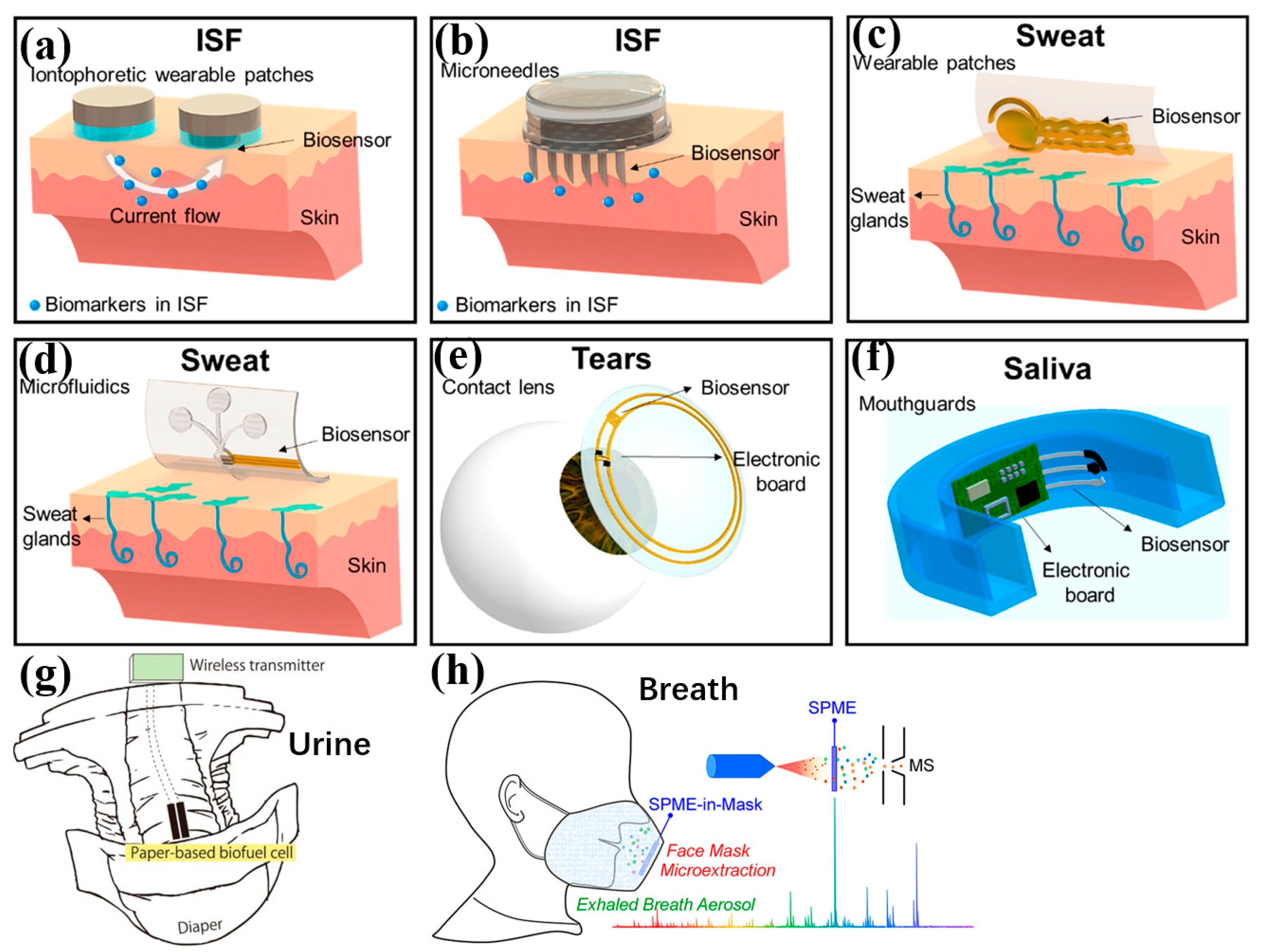
| ISF | Metabolites, ions, circulating RNAs, proteins, amino acids, fatty acids, peptides, coenzymes, neurotransmitters, hormones [111,116,200,211] | On-skin patch | Metabolite detection: glucose, lactate, ketone bodies, alcohol and uric acid PH sensing [212] Neurotransmitter detection Drug monitoring [213] | Rich source of Biomarkers [214,215,216] Minimally invasive Location (near the skin surface) Similar composition with blood plasma, serums Stead and continuous secretion rate Skin offers a large interface | Invasive Discomfort from sampling approaches. The time lag between interstitial and blood analyte levels. Low sample volume for analysis Skin thickness variation between individuals and sites |
| Sweat | Metabolites, electrolytes, irons, proteins, peptides, neurotransmitters, fatty acids, hormones [111,214,215,216] | On-skin patch, tattoos, clothes | Metabolite detection: glucose, lactate, alcohol and uric acid Protein biomarker detection: TNF Chronic disease monitoring: inflammatory bowel disease, cystic fibrosis [217] pH sensing [218,219,220] Hormone detection: cortisol | Convenient Non-invasive Location (on the skin surface) Skin offers a large interface Sample continuously secreted Sweat glands are widely distributed | Low volumes at normal sweat rates Contamination Evaporative loss Dilute analyte concentrations Compositional variation depending on the area of sampling Variation in sweating rates |
| Breath | Metabolites (volatilized or in aerosols), VOCs, viruses [111,221,222,223] | Face mask, Electronic nose | Metabolite detection: H2O2, SARS-CoV-2 testing | Non-invasive Convenient Sample continuously generated | Limited biomarkers, except for VOCs Face masks might be uncomfortable for users. VOC detection would require notable sensor engineering Unique sampling requirements for aerosol capture |
| Tears | Metabolites, electrolytes, proteins, hormones, lipids [111,224,225,226] | Contact lens | Metabolite detection: glucose and lactate | Convenient Non-invasive Sample continuously secreted | Lag between tear and blood analyte levels. Eye position requires considerable device engineering. The correlation between blood and tear analyte might be weak |
| Saliva | Metabolites, electrolytes, proteins, hormones, bacteria, and viruses [111,227,228,229] | Mouth-guard, on-tooth patch, pacifier [230,231] | Metabolite detection: glucose, lactate, alcohol and uric acid pH sensing [220] Specific bacterial monitoring Drug and hormone testing | Convenient Non-invasive Sample continuously secreted | High viscosity might pose sampling problems. Variation in analyte correlation between blood and saliva Saliva production changes due to talking, eating, or drinking. Eating or drinking brings contamination. Difficult for comfortable long-term use |
| Urine | Metabolites, electrolytes, metals, peptides, amino acids, fatty acids, toxins, hormones, proteins, coenzymes, neurotransmitters, circulating RNA and DNA [111,232,233] | Diaper [234] | Metabolite detection: glucose, nitrate pH sensing [220] | Rich source of biomarkers Convenient Non-invasive | Rely on urination events |

3.2.1. Interstitial Fluid
3.2.2. Sweat
3.2.3. Saliva
3.2.4. Breath
3.2.5. Tears
3.2.6. Urine
4. Future Perspectives and Conclusions
4.1. Data Reliability
4.2. Data Privacy
4.3. User-Friendly
4.4. Battery Harvesting and Storage
4.5. Healthcare System Integration between Devices and Medical Professionals
Author Contributions
Funding
Acknowledgments
Conflicts of Interest
References
- Shen, G. Recent advances of flexible sensors for biomedical applications. Prog. Nat. Sci. Mater. Int. 2021, 31, 872–882. [Google Scholar] [CrossRef]
- Pang, C.; Lee, C.; Suh, K.-Y. Recent advances in flexible sensors for wearable and implantable devices. J. Appl. Polym. Sci. 2013, 130, 1429–1441. [Google Scholar] [CrossRef]
- Iqbal, S.M.A.; Mahgoub, I.; Du, E.; Leavitt, M.A.; Asghar, W. Advances in healthcare wearable devices. NPJ Flex. Electron. 2021, 5, 9. [Google Scholar] [CrossRef]
- Updike, S.J.; Hicks, G.P. The Enzyme Electrode. Nature 1967, 214, 986–988. [Google Scholar] [CrossRef] [PubMed]
- Pillai, S.; Upadhyay, A.; Sayson, D.; Nguyen, B.H.; Tran, S.D. Advances in Medical Wearable Biosensors: Design, Fabrication and Materials Strategies in Healthcare Monitoring. Molecules 2022, 27, 165. [Google Scholar] [CrossRef] [PubMed]
- Nasiri, S.; Khosravani, M.R. Progress and challenges in fabrication of wearable sensors for health monitoring. Sens. Actuators A Phys. 2020, 312, 112105. [Google Scholar] [CrossRef]
- Windmiller, J.R.; Wang, J. Wearable Electrochemical Sensors and Biosensors: A Review. Electroanalysis 2013, 25, 29–46. [Google Scholar] [CrossRef]
- Liu, Y.; Wang, H.; Zhao, W.; Zhang, M.; Qin, H.; Xie, Y. Flexible, Stretchable Sensors for Wearable Health Monitoring: Sensing Mechanisms, Materials, Fabrication Strategies and Features. Sensors 2018, 18, 645. [Google Scholar] [CrossRef] [PubMed]
- Liu, J.; Liu, M.; Bai, Y.; Zhang, J.; Liu, H.; Zhu, W. Recent Progress in Flexible Wearable Sensors for Vital Sign Monitoring. Sensors 2020, 20, 4009. [Google Scholar] [CrossRef] [PubMed]
- Windmiller, J.R.; Bandodkar, A.J.; Parkhomovsky, S.; Wang, J. Stamp transfer electrodes for electrochemical sensing on non-planar and oversized surfaces. Analyst 2012, 137, 1570–1575. [Google Scholar] [CrossRef] [PubMed]
- Chan, B.-D.; Hsieh, K.-H.; Yang, S.-Y. Fabrication of organic flexible electrodes using transfer stamping process. Microelectron. Eng. 2009, 86, 586–589. [Google Scholar] [CrossRef]
- Li, Q.; Luo, S.; Wang, Y.; Wang, Q.-M. Carbon based polyimide nanocomposites thin film strain sensors fabricated by ink-jet printing method. Sens. Actuators A Phys. 2019, 300, 111664. [Google Scholar] [CrossRef]
- Bariya, M.; Shahpar, Z.; Park, H.; Sun, J.; Jung, Y.; Gao, W.; Nyein, H.Y.Y.; Liaw, T.S.; Tai, L.-C.; Ngo, Q.P.; et al. Roll-to-Roll Gravure Printed Electrochemical Sensors for Wearable and Medical Devices. ACS Nano 2018, 12, 6978–6987. [Google Scholar] [CrossRef] [PubMed]
- Hubble, L.J.; Wang, J. Sensing at Your Fingertips: Glove-based Wearable Chemical Sensors. Electroanalysis 2019, 31, 428–436. [Google Scholar] [CrossRef]
- Zhuo, Y.; Hu, H.; Wang, Y.; Marin, T.; Lu, M. Photonic crystal slab biosensors fabricated with helium ion lithography (HIL). Sens. Actuators A Phys. 2019, 297, 111493. [Google Scholar] [CrossRef]
- Han, T.; Kundu, S.; Nag, A.; Xu, Y. 3D Printed Sensors for Biomedical Applications: A Review. Sensors 2019, 19, 1706. [Google Scholar] [CrossRef] [PubMed]
- Gao, M.; Li, L.; Song, Y. Inkjet printing wearable electronic devices. J. Mater. Chem. C 2017, 5, 2971–2993. [Google Scholar] [CrossRef]
- Lou, Z.; Wang, L.; Jiang, K.; Wei, Z.; Shen, G. Reviews of wearable healthcare systems: Materials, devices and system integration. Mater. Sci. Eng. R Rep. 2020, 140, 100523. [Google Scholar] [CrossRef]
- Kim, S.; Kang, J.; Lee, I.; Jang, J.; Park, C.B.; Lee, W.; Bae, B.-S. An intrinsically stretchable multi-biochemical sensor for sweat analysis using photo-patternable ecoflex. npj Flex. Electron. 2023, 7, 33. [Google Scholar] [CrossRef]
- del Bosque, A.; Sánchez-Romate, X.X.F.; Calvo, D.L.L.; Fernández, P.R.; Borromeo, S.; Sánchez, M.; Ureña, A. Highly Flexible Strain Sensors Based on CNT-Reinforced Ecoflex Silicone Rubber for Wireless Facemask Breathing Monitoring via Bluetooth. ACS Appl. Polym. Mater. 2023, 5, 8589–8599. [Google Scholar] [CrossRef]
- Zheng, Q.; Jia, C.; Sun, F.; Zhang, M.; Wen, Y.; Xie, Z.; Wang, J.; Liu, B.; Mao, Y.; Zhao, C. Ecoflex Flexible Array of Triboelectric Nanogenerators for Gait Monitoring Alarm Warning Applications. Electronics 2023, 12, 3226. [Google Scholar] [CrossRef]
- Zhang, S.; Rana, S.S.; Bhatta, T.; Pradhan, G.B.; Sharma, S.; Song, H.; Jeong, S.; Park, J.Y. 3D printed smart glove with pyramidal MXene/Ecoflex composite-based toroidal triboelectric nanogenerators for wearable human-machine interaction applications. Nano Energy 2023, 106, 108110. [Google Scholar] [CrossRef]
- Liu, G.; Lv, Z.; Batool, S.; Li, M.; Zhao, P.; Guo, L.; Wang, Y.; Zhou, Y.; Han, S. Biocompatible Material-Based Flexible Biosensors: From Materials Design to Wearable/Implantable Devices and Integrated Sensing Systems. Small 2023, 19, e2207879. [Google Scholar] [CrossRef] [PubMed]
- Lee, P.; Lee, J.; Lee, H.; Yeo, J.; Hong, S.; Nam, K.H.; Lee, D.; Lee, S.S.; Ko, S.H. Highly Stretchable and Highly Conductive Metal Electrode by Very Long Metal Nanowire Percolation Network. Adv. Mater. 2012, 24, 3326–3332. [Google Scholar] [CrossRef] [PubMed]
- Chen, J.; Zheng, J.; Gao, Q.; Zhang, J.; Zhang, J.; Omisore, O.M.; Wang, L.; Li, H. Polydimethylsiloxane (PDMS)-Based Flexible Resistive Strain Sensors for Wearable Applications. Appl. Sci. 2018, 8, 345. [Google Scholar] [CrossRef]
- Hammock, M.L.; Chortos, A.; Tee, B.C.-K.; Tok, J.B.-H.; Bao, Z. 25th Anniversary Article: The Evolution of Electronic Skin (E-Skin): A Brief History, Design Considerations, and Recent Progress. Adv. Mater. 2013, 25, 5997–6037. [Google Scholar] [CrossRef] [PubMed]
- Sun, Y.; Rogers, J.A. Structural forms of single crystal semiconductor nanoribbons for high-performance stretchable electronics. J. Mater. Chem. 2007, 17, 832–840. [Google Scholar] [CrossRef]
- Zhang, A.; Shyam, A.B.; Cunningham, A.M.; Williams, C.; Brissenden, A.; Bartley, A.; Amsden, B.; Docoslis, A.; Kontopoulou, M.; Ameri, S.K. Adhesive Wearable Sensors for Electroencephalography from Hairy Scalp. Adv. Healthc. Mater. 2023, 12, e2300142. [Google Scholar] [CrossRef] [PubMed]
- Yang, J.; Liu, L.; Zhang, D.; Zhang, H.; Ma, J.; Zheng, J.; Wang, C. Dual-Stage Surficial Microstructure to Enhance the Sensitivity of MXene Pressure Sensors for Human Physiological Signal Acquisition. ACS Appl. Mater. Interfaces 2023, 16, 1096–1106. [Google Scholar] [CrossRef] [PubMed]
- Zhao, X.; Mei, D.; Tang, G.; Zhao, C.; Wang, J.; Luo, M.; Li, L.; Wang, Y. Strain and Pressure Sensors Based on MWCNT/PDMS for Human Motion/Perception Detection. Polymers 2023, 15, 1386. [Google Scholar] [CrossRef] [PubMed]
- Hua, Y.; Guan, M.; Xia, L.; Chen, Y.; Mai, J.; Zhao, C.; Liao, C. Highly Stretchable and Robust Electrochemical Sensor Based on 3D Graphene Oxide–CNT Composite for Detecting Ammonium in Sweat. Biosensors 2023, 13, 409. [Google Scholar] [CrossRef] [PubMed]
- Han, S.-T.; Peng, H.; Sun, Q.; Venkatesh, S.; Chung, K.-S.; Lau, S.C.; Zhou, Y.; Roy, V.A.L. An Overview of the Development of Flexible Sensors. Adv. Mater. 2017, 29, 1700375. [Google Scholar] [CrossRef] [PubMed]
- Zazoum, B.; Batoo, K.M.; Khan, M.A.A. Recent Advances in Flexible Sensors and Their Applications. Sensors 2022, 22, 4653. [Google Scholar] [CrossRef] [PubMed]
- Rim, Y.S.; Bae, S.H.; Chen, H.; De Marco, N.; Yang, Y. Recent Progress in Materials and Devices toward Printable and Flexible Sensors. Adv. Mater. 2016, 28, 4415–4440. [Google Scholar] [CrossRef] [PubMed]
- Zhang, T.; Chai, Y.; Wang, S.; Yu, J.; Jiang, S.; Zhu, W.; Fang, Z.; Li, B. Recent Study Advances in Flexible Sensors Based on Polyimides. Sensors 2023, 23, 9743. [Google Scholar] [CrossRef] [PubMed]
- Yi, C.; Li, W.; Shi, S.; He, K.; Ma, P.; Chen, M.; Yang, C. High-temperature-resistant and colorless polyimide: Preparations, properties, and applications. Sol. Energy 2020, 195, 340–354. [Google Scholar] [CrossRef]
- Wu, W.-Y.; Hsu, Y.-H.; Chen, Y.-F.; Wu, Y.-R.; Liu, H.-W.; Tu, T.-Y.; Chao, P.P.-C.; Tan, C.-S.; Horng, R.-H. Wearable Devices Made of a Wireless Vertical-Type Light-Emitting Diode Package on a Flexible Polyimide Substrate with a Conductive Layer. ACS Appl. Electron. Mater. 2021, 3, 979–987. [Google Scholar] [CrossRef]
- Lee, D.U.; Kim, S.-C.; Choi, D.Y.; Jung, W.-K.; Moon, M.J. Basic amino acid-mediated cationic amphiphilic surfaces for antimicrobial pH monitoring sensor with wound healing effects. Biomater. Res. 2023, 27, s40824-023-00355-0. [Google Scholar] [CrossRef] [PubMed]
- Kou, L.; Ye, N.; Waheed, A.; Auliya, R.Z.; Wu, C.; Ooi, P.C.; Li, F. High sensitivity and wide response range artificial synapse based on polyimide with embedded graphene quantum dots. Sci. Rep. 2023, 13, 8194. [Google Scholar] [CrossRef]
- Liu, W.; Huang, Y.; Peng, Y.; Walczak, M.S.; Wang, D.; Chen, Q.; Liu, Z.; Li, L. Stable Wearable Strain Sensors on Textiles by Direct Laser Writing of Graphene. ACS Appl. Nano Mater. 2020, 3, 283–293. [Google Scholar] [CrossRef]
- Lin, J.; Peng, Z.; Liu, Y.; Ruiz-Zepeda, F.; Ye, R.; Samuel, E.L.G.; Yacaman, M.J.; Yakobson, B.I.; Tour, J.M. Laser-induced porous graphene films from commercial polymers. Nat. Commun. 2014, 5, 5714. [Google Scholar] [CrossRef] [PubMed]
- Alemán, J.V.; Chadwick, A.V.; He, J.; Hess, M.; Horie, K.; Jones, R.G.; Kratochvíl, P.; Meisel, I.; Mita, I.; Moad, G.; et al. Definitions of terms relating to the structure and processing of sols, gels, networks, and inorganic-organic hybrid materials (IUPAC Recommendations 2007). Pure Appl. Chem. 2007, 79, 1801–1829. [Google Scholar] [CrossRef]
- Abrisham, M.; Panahi-Sarmad, M.; Sadeghi, G.M.M.; Arjmand, M.; Dehghan, P.; Amirkiai, A. Microstructural design for enhanced mechanical property and shape memory behavior of polyurethane nanocomposites: Role of carbon nanotube, montmorillonite, and their hybrid fillers. Polym. Test. 2020, 89, 106642. [Google Scholar] [CrossRef]
- Amirkiai, A.; Panahi-Sarmad, M.; Sadeghi, G.M.M.; Arjmand, M.; Abrisham, M.; Dehghan, P.; Nazockdast, H. Microstructural design for enhanced mechanical and shape memory performance of polyurethane nanocomposites: Role of hybrid nanofillers of montmorillonite and halloysite nanotube. Appl. Clay Sci. 2020, 198, 105816. [Google Scholar] [CrossRef]
- Zhang, Y.; Zhao, Y.; Zhai, W.; Zheng, G.; Ji, Y.; Dai, K.; Mi, L.; Zhang, D.; Liu, C.; Shen, C. Multifunctional interlocked e-skin based on elastic micropattern array facilely prepared by hot-air-gun. Chem. Eng. J. 2021, 407, 127960. [Google Scholar] [CrossRef]
- Tran, M.T.; Tung, T.T.; Sachan, A.; Losic, D.; Castro, M.; Feller, J.F. 3D Sprayed Polyurethane Functionalized Graphene / Carbon Nanotubes Hybrid Architectures to Enhance the Piezo-Resistive Response of Quantum Resistive Pressure Sensors. Carbon 2020, 168, 564–579. [Google Scholar] [CrossRef]
- Zhang, S.; Sun, K.; Liu, H.; Chen, X.; Zheng, Y.; Shi, X.; Zhang, D.; Mi, L.; Liu, C.; Shen, C. Enhanced piezoresistive performance of conductive WPU/CNT composite foam through incorporating brittle cellulose nanocrystal. Chem. Eng. J. 2020, 387, 124045. [Google Scholar] [CrossRef]
- Zhao, Y.; Ren, M.; Shang, Y.; Li, J.; Wang, S.; Zhai, W.; Zheng, G.; Dai, K.; Liu, C.; Shen, C. Ultra-sensitive and durable strain sensor with sandwich structure and excellent anti-interference ability for wearable electronic skins. Compos. Sci. Technol. 2020, 200, 108448. [Google Scholar] [CrossRef]
- Chen, T.; Xie, Y.; Wang, Z.; Lou, J.; Liu, D.; Xu, R.; Cui, Z.; Li, S.; Panahi-Sarmad, M.; Xiao, X. Recent Advances of Flexible Strain Sensors Based on Conductive Fillers and Thermoplastic Polyurethane Matrixes. ACS Appl. Polym. Mater. 2021, 3, 5317–5338. [Google Scholar] [CrossRef]
- Wu, S.-D.; Hsu, S.; Ketelsen, B.; Bittinger, S.C.; Schlicke, H.; Weller, H.; Vossmeyer, T. Fabrication of Eco-Friendly Wearable Strain Sensor Arrays via Facile Contact Printing for Healthcare Applications (Small Methods 9/2023). Small Methods 2023, 7, 2300170. [Google Scholar] [CrossRef] [PubMed]
- Bai, L.; Jin, Y.; Shang, X.; Jin, H.; Zhou, Y.; Shi, L. Highly synergistic, electromechanical and mechanochromic dual-sensing ionic skin with multiple monitoring, antibacterial, self-healing, and anti-freezing functions. J. Mater. Chem. A 2021, 9, 23916–23928. [Google Scholar] [CrossRef]
- Hu, X.; Wang, J.; Song, S.; Gan, W.; Li, W.; Qi, H.; Zhang, Y. Ionic conductive konjac glucomannan/liquid crystal cellulose composite hydrogels with dual sensing of photo- and electro-signals capacities as wearable strain sensors. Int. J. Biol. Macromol. 2024, 258, 129038. [Google Scholar] [CrossRef] [PubMed]
- Zhang, P.; Tong, X.; Gao, Y.; Qian, Z.; Ren, R.; Bian, C.; Wang, J.; Cai, G. A Sensing and Stretchable Polymer-Dispersed Liquid Crystal Device Based on Spiderweb-Inspired Silver Nanowires-Micromesh Transparent Electrode. Adv. Funct. Mater. 2023, 33, 2303270. [Google Scholar] [CrossRef]
- Tanaka, T. Collapse of Gels and the Critical Endpoint. Phys. Rev. Lett. 1978, 40, 820–823. [Google Scholar] [CrossRef]
- Detamornrat, U.; Parrilla, M.; Domínguez-Robles, J.; Anjani, Q.K.; Larrañeta, E.; De Wael, K.; Donnelly, R.F. Transdermal on-demand drug delivery based on an iontophoretic hollow microneedle array system. Lab Chip 2023, 23, 2304–2315. [Google Scholar] [CrossRef] [PubMed]
- Yin, R.; Zhang, C.; Shao, J.; Chen, Y.; Yin, A.; Feng, Q.; Chen, S.; Peng, F.; Ma, X.; Xu, C.-Y.; et al. Integration of flexible, recyclable, and transient gelatin hydrogels toward multifunctional electronics. J. Mater. Sci. Technol. 2023, 145, 83–92. [Google Scholar] [CrossRef]
- Yang, Z.; Bao, G.; Huo, R.; Jiang, S.; Yang, X.; Ni, X.; Mongeau, L.; Long, R.; Li, J. Programming hydrogel adhesion with engineered polymer network topology. Proc. Natl. Acad. Sci. USA 2023, 120, e2307816120. [Google Scholar] [CrossRef] [PubMed]
- Xu, L.; Liu, S.; Zhu, L.; Liu, Y.; Li, N.; Shi, X.; Jiao, T.; Qin, Z. Hydroxypropyl methyl cellulose reinforced conducting polymer hydrogels with ultra-stretchability and low hysteresis as highly sensitive strain sensors for wearable health monitoring. Int. J. Biol. Macromol. 2023, 236, 123956. [Google Scholar] [CrossRef] [PubMed]
- Peng, Y.; Peng, H.; Chen, Z.; Zhang, J. Ultrasensitive Soft Sensor from Anisotropic Conductive Biphasic Liquid Metal-Polymer Gels. Adv. Mater. 2024, 36, e2305707. [Google Scholar] [CrossRef]
- Li, Y.; Lu, D.; Wong, C.P. Intrinsically Conducting Polymers (ICPs). In Electrical Conductive Adhesives with Nanotechnologies; Springer: Boston, MA, USA, 2010; pp. 361–424. [Google Scholar] [CrossRef]
- Farrell, T.P.; Kaner, R.B. Conducting Polymers. In Encyclopedia of Polymeric Nanomaterials; Springer: Berlin/Heidelberg, Germany, 2021; pp. 1–8. [Google Scholar] [CrossRef]
- Ouyang, J. Application of intrinsically conducting polymers in flexible electronics. SmartMat 2021, 2, 263–285. [Google Scholar] [CrossRef]
- Li, T.; Liang, B.; Ye, Z.; Zhang, L.; Xu, S.; Tu, T.; Zhang, Y.; Cai, Y.; Zhang, B.; Fang, L.; et al. An integrated and conductive hydrogel-paper patch for simultaneous sensing of Chemical–Electrophysiological signals. Biosens. Bioelectron. 2022, 198, 113855. [Google Scholar] [CrossRef] [PubMed]
- Picchio, M.L.; Gallastegui, A.; Casado, N.; Lopez-Larrea, N.; Marchiori, B.; del Agua, I.; Criado-Gonzalez, M.; Mantione, D.; Minari, R.J.; Mecerreyes, D. Mixed Ionic and Electronic Conducting Eutectogels for 3D-Printable Wearable Sensors and Bioelectrodes. Adv. Mater. Technol. 2022, 7, 2101680. [Google Scholar] [CrossRef]
- Sha, L.; Chen, Z.; Chen, Z.; Zhang, A.; Yang, Z. Polylactic Acid Based Nanocomposites: Promising Safe and Biodegradable Materials in Biomedical Field. Int. J. Polym. Sci. 2016, 2016, 6869154. [Google Scholar] [CrossRef]
- Zarei, M.; Lee, G.; Lee, S.G.; Cho, K. Advances in Biodegradable Electronic Skin: Material Progress and Recent Applications in Sensing, Robotics, and Human–Machine Interfaces. Adv. Mater. 2023, 35, e2203193. [Google Scholar] [CrossRef] [PubMed]
- Sreejith, S.; Joseph, L.L.; Kollem, S.; Vijumon, V.T.; Ajayan, J. Biodegradable sensors: A comprehensive review. Measurement 2023, 219, 113261. [Google Scholar] [CrossRef]
- Zhu, J.; Wen, H.; Zhang, H.; Huang, P.; Liu, L.; Hu, H. Recent advances in biodegradable electronics- from fundament to the next-generation multi-functional, medical and environmental device. Sustain. Mater. Technol. 2023, 35, e00530. [Google Scholar] [CrossRef]
- Yin, L.; Farimani, A.B.; Min, K.; Vishal, N.; Lam, J.; Lee, Y.K.; Aluru, N.R.; Rogers, J.A. Mechanisms for Hydrolysis of Silicon Nanomembranes as Used in Bioresorbable Electronics. Adv. Mater. 2015, 27, 1857–1864. [Google Scholar] [CrossRef] [PubMed]
- Hosseini, E.S.; Dervin, S.; Ganguly, P.; Dahiya, R. Biodegradable Materials for Sustainable Health Monitoring Devices. ACS Appl. Bio Mater. 2020, 4, 163–194. [Google Scholar] [CrossRef] [PubMed]
- Guo, Y.; Zhong, M.; Fang, Z.; Wan, P.; Yu, G. A Wearable Transient Pressure Sensor Made with MXene Nanosheets for Sensitive Broad-Range Human–Machine Interfacing. Nano Lett. 2019, 19, 1143–1150. [Google Scholar] [CrossRef] [PubMed]
- Selamneni, V.; Sahatiya, P. Bolometric Effect Enhanced Ultrafast Graphene Based Do-It-Yourself Wearable Respiration Sensor for Personal Healthcare Monitoring. IEEE Sens. J. 2020, 20, 3452–3459. [Google Scholar] [CrossRef]
- Chao, M.; He, L.; Gong, M.; Li, N.; Li, X.; Peng, L.; Shi, F.; Zhang, L.; Wan, P. Breathable Ti3C2Tx MXene/Protein Nanocomposites for Ultrasensitive Medical Pressure Sensor with Degradability in Solvents. ACS Nano 2021, 15, 9746–9758. [Google Scholar] [CrossRef] [PubMed]
- Xu, M.; Yadavalli, V.K. Flexible Biosensors for the Impedimetric Detection of Protein Targets Using Silk-Conductive Polymer Biocomposites. ACS Sens. 2019, 4, 1040–1047. [Google Scholar] [CrossRef] [PubMed]
- Jadoun, S. Synthesis, Mechanism, and Applications of Self-healing Materials. Biomed. Mater. Devices 2024, 2, 225–240. [Google Scholar] [CrossRef]
- Wool, R.P. Self-healing materials: A review. Soft Matter 2008, 4, 400–418. [Google Scholar] [CrossRef] [PubMed]
- Arani, A.G.; Miralaei, N.; Farazin, A.; Mohammadimehr, M. An extensive review of the repair behavior of smart self-healing polymer matrix composites. J. Mater. Res. 2023, 38, 617–632. [Google Scholar] [CrossRef]
- Thangavel, G.; Tan, M.W.M.; Lee, P.S. Advances in self-healing supramolecular soft materials and nanocomposites. Nano Converg. 2019, 6, 29. [Google Scholar] [CrossRef]
- Wan, Y.; Li, X.-C.; Yuan, H.; Liu, D.; Lai, W.-Y. Self-Healing Elastic Electronics: Materials Design, Mechanisms, and Applications. Adv. Funct. Mater. 2024; 2316550, early view. [Google Scholar] [CrossRef]
- Yimyai, T.; Crespy, D.; Pena-Francesch, A. Self-Healing Photochromic Elastomer Composites for Wearable UV-Sensors. Adv. Funct. Mater. 2023, 33, 2213717. [Google Scholar] [CrossRef]
- Omidian, H.; Chowdhury, S.D. High-Performing Conductive Hydrogels for Wearable Applications. Gels 2023, 9, 549. [Google Scholar] [CrossRef] [PubMed]
- Luo, Y.; Li, J.; Ding, Q.; Wang, H.; Liu, C.; Wu, J. Functionalized Hydrogel-Based Wearable Gas and Humidity Sensors. Nano-Micro Lett. 2023, 15, 136. [Google Scholar] [CrossRef] [PubMed]
- Chen, S.; Fan, S.; Chan, H.; Qiao, Z.; Qi, J.; Wu, Z.; Yeo, J.C.; Lim, C.T. Liquid Metal Functionalization Innovations in Wearables and Soft Robotics for Smart Healthcare Applications. Adv. Funct. Mater. 2023; 2309989, early view. [Google Scholar] [CrossRef]
- Yang, J.; Cheng, J.; Qi, G.; Wang, B. Ultrastretchable, Multihealable, and Highly Sensitive Strain Sensor Based on a Double Cross-Linked MXene Hydrogel. ACS Appl. Mater. Interfaces 2023, 15, 17163–17174. [Google Scholar] [CrossRef]
- Seong, M.; Kondaveeti, S.; Choi, G.; Kim, S.; Kim, J.; Kang, M.; Jeong, H.E. 3D Printable Self-Adhesive and Self-Healing Ionotronic Hydrogels for Wearable Healthcare Devices. ACS Appl. Mater. Interfaces 2023, 15, 11042–11052. [Google Scholar] [CrossRef]
- Zhou, X.; Kateb, P.; Fan, J.; Kim, J.; Lodygensky, G.A.; Amilhon, B.; Pasini, D.; Cicoira, F. Conducting Polymer Films and Bioelectrodes Combining High Adhesion and Electro-Mechanical Self-Healing. J. Mater. Chem. C, 2023; advance article. [Google Scholar] [CrossRef]
- Sun, S.; Yuan, R.; Ling, S.; Zhou, T.; Wu, Z.; Fu, M.; He, H.; Li, X.; Zhang, C. Self-Healable, Self-Adhesive and Degradable MXene-Based Multifunctional Hydrogel for Flexible Epidermal Sensors. ACS Appl. Mater. Interfaces 2024, 16, 7826–7837. [Google Scholar] [CrossRef] [PubMed]
- Wang, Q.; Yu, Y.; Liu, J. Preparations, Characteristics and Applications of the Functional Liquid Metal Materials. Adv. Eng. Mater. 2018, 20, 1700781. [Google Scholar] [CrossRef]
- Won, P.; Jeong, S.; Majidi, C.; Ko, S.H. Recent advances in liquid-metal-based wearable electronics and materials. iScience 2021, 24, 102698. [Google Scholar] [CrossRef]
- Ma, J.; Krisnadi, F.; Vong, M.H.; Kong, M.; Awartani, O.M.; Dickey, M.D. Shaping a Soft Future: Patterning Liquid Metals. Adv. Mater. 2023, 35, e2205196. [Google Scholar] [CrossRef]
- Kim, S.; Saito, M.; Wei, Y.; Bhuyan, P.; Choe, M.; Fujie, T.; Mondal, K.; Park, S. Stretchable and wearable polymeric heaters and strain sensors fabricated using liquid metals. Sens. Actuators A Phys. 2023, 355, 114317. [Google Scholar] [CrossRef]
- Kim, H.; Zan, G.; Seo, Y.; Lee, S.; Park, C. Stimuli-Responsive Liquid Metal Hybrids for Human-Interactive Electronics. Adv. Funct. Mater. 2023; 2308703, early view. [Google Scholar] [CrossRef]
- Wang, L.; Lai, R.; Zhang, L.; Zeng, M.; Fu, L. Emerging Liquid Metal Biomaterials: From Design to Application. Adv. Mater. 2022, 34, e2201956. [Google Scholar] [CrossRef] [PubMed]
- Yu, L.; Yeo, J.C.; Soon, R.H.; Yeo, T.; Lee, H.H.; Lim, C.T. Highly Stretchable, Weavable, and Washable Piezoresistive Microfiber Sensors. ACS Appl. Mater. Interfaces 2018, 10, 12773–12780. [Google Scholar] [CrossRef] [PubMed]
- Liang, S.; Li, Y.; Chen, Y.; Yang, J.; Zhu, T.; Zhu, D.; He, C.; Liu, Y.; Handschuh-Wang, S.; Zhou, X. Liquid metal sponges for mechanically durable, all-soft, electrical conductors. J. Mater. Chem. C 2017, 5, 1586–1590. [Google Scholar] [CrossRef]
- Liu, H.; Xin, Y.; Lou, Y.; Peng, Y.; Wei, L.; Zhang, J. Liquid metal gradient fibers with reversible thermal programmability. Mater. Horiz. 2020, 7, 2141–2149. [Google Scholar] [CrossRef]
- Deng, X.; Chen, G.; Liao, Y.; Lu, X.; Hu, S.; Gan, T.; Handschuh-Wang, S.; Zhang, X. Self-Healable and Recyclable Dual-Shape Memory Liquid Metal–Elastomer Composites. Polymers 2022, 14, 2259. [Google Scholar] [CrossRef]
- Liu, R.; Gong, L.; Zhu, X.; Zhu, S.; Wu, X.; Xue, T.; Yan, L.; Du, J.; Gu, Z. Transformable Gallium-Based Liquid Metal Nanoparticles for Tumor Radiotherapy Sensitization. Adv. Healthc. Mater. 2022, 11, 2102584. [Google Scholar] [CrossRef] [PubMed]
- Yi, P.; Zou, H.; Yu, Y.; Li, X.; Li, Z.; Deng, G.; Chen, C.; Fang, M.; He, J.; Sun, X.; et al. MXene-Reinforced Liquid Metal/Polymer Fibers via Interface Engineering for Wearable Multifunctional Textiles. ACS Nano 2022, 16, 14490–14502. [Google Scholar] [CrossRef] [PubMed]
- Cao, L.; Yu, D.; Xia, Z.; Wan, H.; Liu, C.; Yin, T.; He, Z. Ferromagnetic Liquid Metal Putty-Like Material with Transformed Shape and Reconfigurable Polarity. Adv. Mater. 2020, 32, e2000827. [Google Scholar] [CrossRef]
- Markvicka, E.J.; Bartlett, M.D.; Huang, X.; Majidi, C. An autonomously electrically self-healing liquid metal–elastomer composite for robust soft-matter robotics and electronics. Nat. Mater. 2018, 17, 618–624. [Google Scholar] [CrossRef] [PubMed]
- Zhu, P.; Luo, X.; Lin, X.; Qiu, Z.; Chen, R.; Wang, X.; Wang, Y.; Deng, Y.; Mao, Y. A self-healable, recyclable, and flexible thermoelectric device for wearable energy harvesting and personal thermal management. Energy Convers. Manag. 2023, 285, 2308703. [Google Scholar] [CrossRef]
- Feng, X.; Wang, C.; Shang, S.; Liu, H.; Huang, X.; Jiang, J.; Song, Z.; Zhang, H. Self-healing, EMI shielding, and antibacterial properties of recyclable cellulose liquid metal hydrogel sensor. Carbohydr. Polym. 2023, 311, 120786. [Google Scholar] [CrossRef] [PubMed]
- Min, S.; Kim, D.H.; Joe, D.J.; Kim, B.W.; Jung, Y.H.; Lee, J.H.; Lee, B.; Doh, I.; An, J.; Youn, Y.; et al. Clinical Validation of a Wearable Piezoelectric Blood-Pressure Sensor for Continuous Health Monitoring. Adv. Mater. 2023, 35, e2301627. [Google Scholar] [CrossRef] [PubMed]
- Sun, J.; Xiu, K.; Wang, Z.; Hu, N.; Zhao, L.; Zhu, H.; Kong, F.; Xiao, J.; Cheng, L.; Bi, X. Multifunctional wearable humidity and pressure sensors based on biocompatible graphene/bacterial cellulose bioaerogel for wireless monitoring and early warning of sleep apnea syndrome. Nano Energy 2023, 108, 108215. [Google Scholar] [CrossRef]
- Zhao, H.; Zhang, L.; Deng, T.; Li, C. High-performance sensing, breathable, and biodegradable integrated wearable sweat biosensors for a wireless glucose early warning system. J. Mater. Chem. A 2023, 11, 12395–12404. [Google Scholar] [CrossRef]
- Engel, E.; Hill, A.C.; Granchelli, A.; Lockhart, G.C.; Lyng, G.; Kaye, L.; Lyson, H.; Bowler, R.P. Wearable Sensors for Early Detection of Copd Exacerbations. Chest 2023, 164, A5034. [Google Scholar] [CrossRef]
- Quinn, H.; Davis, J. Detecting the early onset of hyponatremia: An opportunity for wearable sensors? Curr. Opin. Electrochem. 2023, 39, 101302. [Google Scholar] [CrossRef]
- Khijmatgar, S.; Yong, J.; Rübsamen, N.; Lorusso, F.; Rai, P.; Cenzato, N.; Gaffuri, F.; Del Fabbro, M.; Tartaglia, G.M. Salivary biomarkers for early detection of oral squamous cell carcinoma (OSCC) and head/neck squamous cell carcinoma (HNSCC): A systematic review and network meta-analysis. Jpn. Dent. Sci. Rev. 2024, 60, 32–39. [Google Scholar] [CrossRef] [PubMed]
- Linh, V.T.N.; Kim, H.; Lee, M.-Y.; Mun, J.; Kim, Y.; Jeong, B.-H.; Park, S.-G.; Kim, D.-H.; Rho, J.; Jung, H.S. 3D plasmonic hexaplex paper sensor for label-free human saliva sensing and machine learning-assisted early-stage lung cancer screening. Biosens. Bioelectron. 2024, 244, 115779. [Google Scholar] [CrossRef] [PubMed]
- Ates, H.C.; Nguyen, P.Q.; Gonzalez-Macia, L.; Morales-Narváez, E.; Güder, F.; Collins, J.J.; Dincer, C. End-to-end design of wearable sensors. Nat. Rev. Mater. 2022, 7, 887–907. [Google Scholar] [CrossRef] [PubMed]
- Abramson, A.; Chan, C.T.; Khan, Y.; Mermin-Bunnell, A.; Matsuhisa, N.; Fong, R.; Shad, R.; Hiesinger, W.; Mallick, P.; Gambhir, S.S.; et al. A flexible electronic strain sensor for the real-time monitoring of tumor regression. Sci. Adv. 2022, 8, eabn6550. [Google Scholar] [CrossRef] [PubMed]
- Chun, K.-Y.; Seo, S.; Han, C.-S. A Wearable All-Gel Multimodal Cutaneous Sensor Enabling Simultaneous Single-Site Monitoring of Cardiac-Related Biophysical Signals. Adv. Mater. 2022, 34, 2110082. [Google Scholar] [CrossRef] [PubMed]
- Bi, Y.; Sun, M.; Wang, J.; Zhu, Z.; Bai, J.; Emran, M.Y.; Kotb, A.; Bo, X.; Zhou, M. Universal Fully Integrated Wearable Sensor Arrays for the Multiple Electrolyte and Metabolite Monitoring in Raw Sweat, Saliva, or Urine. Anal. Chem. 2023, 95, 6690–6699. [Google Scholar] [CrossRef] [PubMed]
- Quer, G.; Radin, J.M.; Gadaleta, M.; Baca-Motes, K.; Ariniello, L.; Ramos, E.; Kheterpal, V.; Topol, E.J.; Steinhubl, S.R. Wearable sensor data and self-reported symptoms for COVID-19 detection. Nat. Med. 2021, 27, 73–77. [Google Scholar] [CrossRef] [PubMed]
- Erdem, A.; Eksin, E.; Senturk, H.; Yildiz, E.; Maral, M. Recent developments in wearable biosensors for healthcare and biomedical applications. TrAC Trends Anal. Chem. 2024, 171, 117510. [Google Scholar] [CrossRef]
- Sharma, A.; Badea, M.; Tiwari, S.; Marty, J.L. Wearable Biosensors: An Alternative and Practical Approach in Healthcare and Disease Monitoring. Molecules 2021, 26, 748. [Google Scholar] [CrossRef] [PubMed]
- Zahradka, N.; Geoghan, S.; Watson, H.; Goldberg, E.; Wolfberg, A.; Wilkes, M. Assessment of Remote Vital Sign Monitoring and Alarms in a Real-World Healthcare at Home Dataset. Bioengineering 2023, 10, 37. [Google Scholar] [CrossRef] [PubMed]
- Hussain, T.; Ullah, S.; Fernández-García, R.; Gil, I. Wearable Sensors for Respiration Monitoring: A Review. Sensors 2023, 23, 7518. [Google Scholar] [CrossRef] [PubMed]
- Huang, X.; Zheng, Z.; Wang, H.; Xu, W.; Wu, M.; Wang, M.; Chen, C.; Wan, L.; Du, R.; Zhu, T.; et al. A Freeze-Resistant, Highly Stretchable and Biocompatible Organohydrogel for Non-Delayed Wearable Sensing at Ultralow-Temperatures. Adv. Funct. Mater. 2024; 2312149, early view. [Google Scholar] [CrossRef]
- Dang, X.; Fu, Y.; Wang, X. A temperature and pressure dual-responsive, stretchable, healable, adhesive, and biocompatible carboxymethyl cellulose-based conductive hydrogels for flexible wearable strain sensor. Biosens. Bioelectron. 2024, 246, 115893. [Google Scholar] [CrossRef] [PubMed]
- Hong, Q.; Liu, T.; Guo, X.; Yan, Z.; Li, W.; Liu, L.; Wang, D.; Hong, W.; Qian, Z.; Zhang, A.; et al. 3D dual-mode tactile sensor with decoupled temperature and pressure sensing: Toward biological skins for wearable devices and smart robotics. Sens. Actuators B Chem. 2024, 404, 135255. [Google Scholar] [CrossRef]
- Wang, J.; Sun, Y.; Jia, P.; Su, J.; Zhang, X.; Wu, N.; Yu, H.; Song, Y.; Zhou, J. Wearable nanocomposite hydrogel temperature sensor based on thermally-switchable and mechanical-deformation-insensitive structural colors. Chem. Eng. J. 2023, 476, 146602. [Google Scholar] [CrossRef]
- Faridi, S.; Allen, R.W.; Brook, R.D.; Yousefian, F.; Hassanvand, M.S.; Carlsten, C. An updated systematic review and meta-analysis on portable air cleaners and blood pressure: Recommendations for users and manufacturers. Ecotoxicol. Environ. Saf. 2023, 263, 115227. [Google Scholar] [CrossRef] [PubMed]
- Ismail, S.N.A.; Nayan, N.A.; Haniff, M.A.S.M.; Jaafar, R.; May, Z. Wearable Two-Dimensional Nanomaterial-Based Flexible Sensors for Blood Pressure Monitoring: A Review. Nanomaterials 2023, 13, 852. [Google Scholar] [CrossRef] [PubMed]
- Zhang, Q.; Zhang, H.; Liang, J.; Zhao, X.; Li, B.; Zang, J.; Gao, L.; Zhang, Z.D.; Xue, C. Ti3C2Tx-MXene/PET textile-based flexible pressure sensor for wearable pulse monitoring. J. Mater. Chem. C 2023, 11, 15638–15648. [Google Scholar] [CrossRef]
- Shi, C.; Zhang, H.; Ni, X.; Wang, K. An FBG-Based Sensor with Both Wearable and Handheld Forms for Carotid Arterial Pulse Waveform Measurement. IEEE Trans. Instrum. Meas. 2023, 72, 7506610. [Google Scholar] [CrossRef]
- Heimark, S.; Hove, C.; Stepanov, A.; Boysen, E.S.; Gløersen, Ø.; Bøtke-Rasmussen, K.G.; Gravdal, H.J.; Narayanapillai, K.; Larstorp, A.C.K.; Seeberg, T.M.; et al. 24-Hour Ambulatory Blood Pressure Measurement Using a Pulse Arrival Time Based Blood Pressure Model by a Prototype Wearable Sensor. J. Hypertens. 2023, 41, e248. [Google Scholar] [CrossRef]
- Kumar, R.; Fu, J.; Ortiz, B.L.; Cao, X.; Shedden, K.; Choi, S.W. Dyadic and Individual Variation in 24-Hour Heart Rates of Cancer Patients and Their Caregivers. Bioengineering 2024, 11, 95. [Google Scholar] [CrossRef] [PubMed]
- Marasco, I.; Niro, G.; Demir, S.M.; Marzano, L.; Fachechi, L.; Rizzi, F.; Demarchi, D.; Ros, P.M.; D’orazio, A.; Grande, M.; et al. Wearable Heart Rate Monitoring Device Communicating in 5G ISM Band for IoHT. Bioengineering 2023, 10, 113. [Google Scholar] [CrossRef] [PubMed]
- Yang, Y.; Xu, S.; Gai, Y.; Zhang, B.; Chen, L. Recent Progresses in Lanthanide Metal-Organic Frameworks (Ln-MOFs) as Chemical Sensors for Ions, Antibiotics and Amino Acids. Chin. J. Struct. Chem. 2022, 41, 2211045–2211070. [Google Scholar] [CrossRef]
- Kaiqiang, W.; Xingyang, L. Wearable pressure sensor for athletes’ full-range motion signal monitoring. Mater. Res. Express 2020, 7, 105003. [Google Scholar] [CrossRef]
- Warmerdam, E.; Orth, M.; Pohlemann, T.; Ganse, B. Gait Analysis to Monitor Fracture Healing of the Lower Leg. Bioengineering 2023, 10, 255. [Google Scholar] [CrossRef] [PubMed]
- Lee, C.; Ahn, J.; Lee, B.-C. A Systematic Review of the Long-Term Effects of Using Smartphone- and Tablet-Based Rehabilitation Technology for Balance and Gait Training and Exercise Programs. Bioengineering 2023, 10, 1142. [Google Scholar] [CrossRef] [PubMed]
- May, D.S.; Tueth, L.E.; Earhart, G.M.; Mazzoni, P. Using Wearable Sensors to Assess Freezing of Gait in the Real World. Bioengineering 2023, 10, 289. [Google Scholar] [CrossRef] [PubMed]
- Pieruccini-Faria, F.; Black, S.E.; Masellis, M.; Smith, E.E.; Almeida, Q.J.; Li, K.Z.H.; Bherer, L.; Camicioli, R.; Montero-Odasso, M. Gait variability across neurodegenerative and cognitive disorders: Results from the Canadian Consortium of Neurodegeneration in Aging (CCNA) and the Gait and Brain Study. Alzheimer’s Dement. 2021, 17, 1317–1328. [Google Scholar] [CrossRef] [PubMed]
- Cicirelli, G.; Impedovo, D.; Dentamaro, V.; Marani, R.; Pirlo, G.; D’Orazio, T.R. Human Gait Analysis in Neurodegenerative Diseases: A Review. IEEE J. Biomed. Health Inform. 2021, 26, 229–242. [Google Scholar] [CrossRef] [PubMed]
- Marković, V.; Stanković, I.; Radovanović, S.; Petrović, I.; Lukić, M.J.; Mišković, N.D.; Svetel, M.; Kostić, V. Gait alterations in Parkinson’s disease at the stage of hemiparkinsonism—A longitudinal study. PLoS ONE 2022, 17, e0269886. [Google Scholar] [CrossRef] [PubMed]
- Ahn, J.W.; Ku, Y.; Kim, H.C. A Novel Wearable EEG and ECG Recording System for Stress Assessment. Sensors 2019, 19, 1991. [Google Scholar] [CrossRef] [PubMed]
- Pourmohammadi, S.; Maleki, A. Stress detection using ECG and EMG signals: A comprehensive study. Comput. Methods Programs Biomed. 2020, 193, 105482. [Google Scholar] [CrossRef] [PubMed]
- Umer, W. Simultaneous monitoring of physical and mental stress for construction tasks using physiological measures. J. Build. Eng. 2022, 46, 103777. [Google Scholar] [CrossRef]
- Ritsert, F.; Elgendi, M.; Galli, V.; Menon, C. Heart and Breathing Rate Variations as Biomarkers for Anxiety Detection. Bioengineering 2022, 9, 711. [Google Scholar] [CrossRef] [PubMed]
- Zhu, G.; Huang, Y.; Wang, X. Basic Theory of EEG. In Multi-Modal EEG Monitoring of Severely Neurologically Ill Patients; Wang, X., Li, F., Pan, S., Eds.; Springer: Singapore, 2022; pp. 3–25. ISBN 9789811644931. [Google Scholar]
- Li, F. Application of Multimodal EEG in the Determination of Brain Death. In Multi-Modal EEG Monitoring of Severely Neurologically Ill Patients; Wang, X., Li, F., Pan, S., Eds.; Springer: Singapore, 2022; pp. 323–333. ISBN 9789811644931. [Google Scholar]
- Wang, Y.; Lin, Y. Patterns and Clinical Significance of Abnormal Sleep EEG. In Multi-Modal EEG Monitoring of Severely Neurologically Ill Patients; Wang, X., Li, F., Pan, S., Eds.; Springer: Singapore, 2022; pp. 141–158. ISBN 9789811644931. [Google Scholar]
- Zhou, Y.; Li, F. Application of aEEG in Severely Ill Patients. In Multi-Modal EEG Monitoring of Severely Neurologically Ill Patients; Wang, X., Li, F., Pan, S., Eds.; Springer: Singapore, 2022; pp. 347–360. ISBN 9789811644931. [Google Scholar]
- Wang, X.; Yan, Y. Abnormal EEG Background Activity. In Multi-Modal EEG Monitoring of Severely Neurologically Ill Patients; Wang, X., Li, F., Pan, S., Eds.; Springer: Singapore, 2022; pp. 123–139. ISBN 9789811644931. [Google Scholar]
- Li, F.; Huang, Z. qEEG Monitoring System in Severely Ill Patients. In Multi-Modal EEG Monitoring of Severely Neurologically Ill Patients; Wang, X., Li, F., Pan, S., Eds.; Springer: Singapore, 2022; pp. 27–57. ISBN 9789811644931. [Google Scholar]
- Wang, X.; Li, J.; Jing, W. Application of Multimodal EEG in SE. In Multi-Modal EEG Monitoring of Severely Neurologically Ill Patients; Wang, X., Li, F., Pan, S., Eds.; Springer: Singapore, 2022; pp. 197–227. ISBN 9789811644931. [Google Scholar]
- Zhuang, Y.; Ge, Q.; Li, Q.; Xu, L.; Geng, X.; Wang, R.; He, J. Combined behavioral and EEG evidence for the 70 Hz frequency selection of short-term spinal cord stimulation in disorders of consciousness. CNS Neurosci. Ther. 2024, 30, e14388. [Google Scholar] [CrossRef] [PubMed]
- Mehmood, I.; Li, H.; Qarout, Y.; Umer, W.; Anwer, S.; Wu, H.; Hussain, M.; Antwi-Afari, M.F. Deep learning-based construction equipment operators’ mental fatigue classification using wearable EEG sensor data. Adv. Eng. Inform. 2023, 56, 101978. [Google Scholar] [CrossRef]
- Zhao, S.; Dai, G.; Li, J.; Zhu, X.; Huang, X.; Li, Y.; Tan, M.; Wang, L.; Fang, P.; Chen, X.; et al. An interpretable model based on graph learning for diagnosis of Parkinson’s disease with voice-related EEG. npj Digit. Med. 2024, 7, 3. [Google Scholar] [CrossRef] [PubMed]
- Zhang, Z.; Meng, Q.; Jin, L.; Wang, H.; Hou, H. A Novel EEG-Based Graph Convolution Network for Depression Detection: Incorporating Secondary Subject Partitioning and Attention Mechanism. Expert Syst. Appl. 2023, 239, 122356. [Google Scholar] [CrossRef]
- Srinivasan, S.; Johnson, S.D. A novel approach to schizophrenia Detection: Optimized preprocessing and deep learning analysis of multichannel EEG data. Expert Syst. Appl. 2024, 246, 122937. [Google Scholar] [CrossRef]
- Chen, J.; Cui, Y.; Wang, H.; He, E.; Alhudhaif, A. Deep learning approach for detection of unfavorable driving state based on multiple phase synchronization between multi-channel EEG signals. Inf. Sci. 2024, 658, 120070. [Google Scholar] [CrossRef]
- Song, Y.; Fan, C.; Mao, X. Optimization of Epilepsy Detection Method Based on Dynamic EEG Channel Screening. Neural Netw. 2024, 172, 106119. [Google Scholar] [CrossRef] [PubMed]
- Li, A.; Deng, Z.; Zhang, W.; Xiao, Z.; Choi, K.-S.; Liu, Y.; Hu, S.; Wang, S. Multiview Transfer Representation Learning with TSK Fuzzy System for EEG Epilepsy Detection. IEEE Trans. Fuzzy Syst. 2024, 32, 38–52. [Google Scholar] [CrossRef]
- Anita, M.; MeenaKowshalya, A.M. Automatic epileptic seizure detection using MSA-DCNN and LSTM techniques with EEG signals. Expert Syst. Appl. 2024, 238, 121727. [Google Scholar] [CrossRef]
- Han, Y.; Zeng, X.; Hua, L.; Quan, X.; Chen, Y.; Zhou, M.; Chuang, Y.; Li, Y.; Wang, S.; Shen, X.; et al. The fusion of multi-omics profile and multimodal EEG data contributes to the personalized diagnostic strategy for neurocognitive disorders. Microbiome 2024, 12, 12. [Google Scholar] [CrossRef] [PubMed]
- Deng, J.; Sun, B.; Kavcic, V.; Liu, M.; Giordani, B.; Li, T. Novel methodology for detection and prediction of mild cognitive impairment using resting-state EEG. Alzheimer’s Dement. 2024, 20, 145–158. [Google Scholar] [CrossRef] [PubMed]
- Ntolkeras, G.; Makaram, N.; Bernabei, M.; De La Vega, A.C.; Bolton, J.; Madsen, J.R.; Stone, S.S.D.; Pearl, P.L.; Papadelis, C.; Grant, E.P.; et al. Interictal EEG source connectivity to localize the epileptogenic zone in patients with drug-resistant epilepsy: A machine learning approach. Epilepsia, 2024; early view. [Google Scholar] [CrossRef]
- Bernabei, J.M.; Li, A.; Revell, A.Y.; Smith, R.J.; Gunnarsdottir, K.M.; Ong, I.Z.; Davis, K.A.; Sinha, N.; Sarma, S.; Litt, B. Quantitative approaches to guide epilepsy surgery from intracranial EEG. Brain 2023, 146, 2248–2258. [Google Scholar] [CrossRef] [PubMed]
- Kučikienė, D.; Rajkumar, R.; Timpte, K.; Heckelmann, J.; Neuner, I.; Weber, Y.; Wolking, S. EEG microstates show different features in focal epilepsy and psychogenic nonepileptic seizures. Epilepsia, 2024; early view. [Google Scholar] [CrossRef]
- Chai, X.; Cao, T.; He, Q.; Wang, N.; Zhang, X.; Shan, X.; Lv, Z.; Tu, W.; Yang, Y.; Zhao, J. Brain–computer interface digital prescription for neurological disorders. CNS Neurosci. Ther. 2024, 30, e14615. [Google Scholar] [CrossRef] [PubMed]
- Friedman, P.A. The Electrocardiogram at 100 Years: History and Future. Circulation 2024, 149, 411–413. [Google Scholar] [CrossRef] [PubMed]
- Pham, H.N.; Holmstrom, L.; Chugh, H.; Uy-Evanado, A.; Nakamura, K.; Zhang, Z.; Salvucci, A.; Jui, J.; Reinier, K.; Chugh, S.S. Dynamic electrocardiogram changes are a novel risk marker for sudden cardiac death. Eur. Heart J. 2024, 45, 809–819. [Google Scholar] [CrossRef] [PubMed]
- Soh, C.H.; de Sá, A.G.C.; Potter, E.; Halabi, A.; Ascher, D.B.; Marwick, T.H. Use of the energy waveform electrocardiogram to detect subclinical left ventricular dysfunction in patients with type 2 diabetes mellitus. Cardiovasc. Diabetol. 2024, 23, 91. [Google Scholar] [CrossRef] [PubMed]
- Zhang, S.; Lian, C.; Xu, B.; Su, Y.; Alhudhaif, A. 12-Lead ECG Signal Classification for Detecting ECG Arrhythmia via An Information Bottleneck-Based Multi-Scale Network. Inf. Sci. 2024, 662, 120239. [Google Scholar] [CrossRef]
- Lee, E.; Ito, S.; Miranda, W.R.; Lopez-Jimenez, F.; Kane, G.C.; Asirvatham, S.J.; Noseworthy, P.A.; Friedman, P.A.; Carter, R.E.; Borlaug, B.A.; et al. Artificial intelligence-enabled ECG for left ventricular diastolic function and filling pressure. npj Digit. Med. 2024, 7, 4. [Google Scholar] [CrossRef] [PubMed]
- Chen, J.; Huang, S.; Zhang, Y.; Chang, Q.; Zhang, Y.; Li, D.; Qiu, J.; Hu, L.; Peng, X.; Du, Y.; et al. Congenital heart disease detection by pediatric electrocardiogram based deep learning integrated with human concepts. Nat. Commun. 2024, 15, 976. [Google Scholar] [CrossRef] [PubMed]
- Xiong, P.; Wu, C.; Zhou, H.; Song, A.; Hu, L.; Liu, X.P. Design of an accurate end-of-arm force display system based on wearable arm gesture sensors and EMG sensors. Inf. Fusion 2018, 39, 178–185. [Google Scholar] [CrossRef]
- Chand, S.; McDaid, A.; Lu, Y. Dynamic muscle fatigue assessment using s-EMG technology towards human-centric human-robot collaboration. J. Manuf. Syst. 2023, 68, 508–522. [Google Scholar] [CrossRef]
- Jonkman, A.H.; Warnaar, R.S.P.; Baccinelli, W.; Carbon, N.M.; D’cruz, R.F.; Doorduin, J.; van Doorn, J.L.M.; Elshof, J.; Estrada-Petrocelli, L.; Graßhoff, J.; et al. Analysis and applications of respiratory surface EMG: Report of a round table meeting. Crit. Care 2024, 28, 2. [Google Scholar] [CrossRef] [PubMed]
- Fan, J.; Jiang, X.; Liu, X.; Meng, L.; Jia, F.; Dai, C. Surface EMG feature disentanglement for robust pattern recognition. Expert Syst. Appl. 2024, 237, 121224. [Google Scholar] [CrossRef]
- Park, J.; Jeong, J.; Kang, M.; Pritish, N.; Cho, Y.; Ha, J.; Yea, J.; Jang, K.-I.; Kim, H.; Hwang, J.; et al. Imperceptive and reusable dermal surface EMG for lower extremity neuro-prosthetic control and clinical assessment. npj Flex. Electron. 2023, 7, 49. [Google Scholar] [CrossRef]
- Li, J.-W.; Chen, H.-F.; Liu, Y.-Z.; Wang, J.-H.; Lu, M.-C.; Chiu, C.-W. Photocurable 3D-printed AgNPs/Graphene/Polymer nanocomposites with high flexibility and stretchability for ECG and EMG smart clothing. Chem. Eng. J. 2024, 484, 149452. [Google Scholar] [CrossRef]
- Inertial Sensors and Inertial Measurement Units. In Pedestrian Inertial Navigation with Self-Contained Aiding; John Wiley & Sons, Ltd.: Hoboken, NJ, USA, 2021; pp. 17–36. ISBN 978-1-119-69991-0.
- Chatterjee, G.; Latorre, L.; Mailly, F.; Nouet, P.; Hachelef, N.; Oudea, C. Smart-MEMS based inertial measurement units: Gyro-free approach to improve the grade. Microsyst. Technol. 2017, 23, 3969–3978. [Google Scholar] [CrossRef]
- Washabaugh, E.P.; Kalyanaraman, T.; Adamczyk, P.G.; Claflin, E.S.; Krishnan, C. Validity and repeatability of inertial measurement units for measuring gait parameters. Gait Posture 2017, 55, 87–93. [Google Scholar] [CrossRef] [PubMed]
- Young, F.; Mason, R.; Morris, R.E.; Stuart, S.; Godfrey, A. IoT-Enabled Gait Assessment: The Next Step for Habitual Monitoring. Sensors 2023, 23, 4100. [Google Scholar] [CrossRef] [PubMed]
- Rahmani, M.H.; Berkvens, R.; Weyn, M. Chest-Worn Inertial Sensors: A Survey of Applications and Methods. Sensors 2021, 21, 2875. [Google Scholar] [CrossRef] [PubMed]
- Figueira, V.; Silva, S.; Costa, I.; Campos, B.; Salgado, J.; Pinho, L.; Freitas, M.; Carvalho, P.; Marques, J.; Pinho, F. Wearables for Monitoring and Postural Feedback in the Work Context: A Scoping Review. Sensors 2024, 24, 1341. [Google Scholar] [CrossRef] [PubMed]
- Blair, S.; Duthie, G.; Robertson, S.; Hopkins, W.; Ball, K. Concurrent validation of an inertial measurement system to quantify kicking biomechanics in four football codes. J. Biomech. 2018, 73, 24–32. [Google Scholar] [CrossRef] [PubMed]
- Xia, K.; Chen, X.; Chang, X.; Liu, C.; Guo, L.; Xu, X.; Lv, F.; Wang, Y.; Sun, H.; Zhou, J. Hand Exoskeleton Design and Human–Machine Interaction Strategies for Rehabilitation. Bioengineering 2022, 9, 682. [Google Scholar] [CrossRef] [PubMed]
- Dey, S.; Schilling, A.F. A Function Approximator Model for Robust Online Foot Angle Trajectory Prediction Using a Single IMU Sensor: Implication for Controlling Active Prosthetic Feet. IEEE Trans. Ind. Inform. 2023, 19, 1467–1475. [Google Scholar] [CrossRef]
- Peng, R.; Wang, Z.; Lu, P. AeCoM: An Aerial Continuum Manipulator With IMU-Based Kinematic Modeling and Tendon-Slacking Prevention. IEEE Trans. Syst. Man Cybern. Syst. 2023, 53, 4740–4752. [Google Scholar] [CrossRef]
- Zhang, X.; Tricomi, E.; Missiroli, F.; Lotti, N.; Masia, L. Real-Time Assistive Control via IMU Locomotion Mode Detection in a Soft Exosuit: An Effective Approach to Enhance Walking Metabolic Efficiency. IEEE/ASME Trans. Mechatron. 2023; 1–12, early view. [Google Scholar] [CrossRef]
- Hong, S.; Yoon, J.; Ham, Y.; Lee, B.; Kim, H. Monitoring safety behaviors of scaffolding workers using Gramian angular field convolution neural network based on IMU sensing data. Autom. Constr. 2023, 148, 104748. [Google Scholar] [CrossRef]
- Xu, Z.; Huang, B.; Jia, B.; Mao, G. Enhancing WiFi Fingerprinting Localization Through a Co-Teaching Approach Using Crowdsourced Sequential RSS and IMU Data. IEEE Internet Things J. 2024, 11, 3550–3562. [Google Scholar] [CrossRef]
- Huang, H.; Yang, J.; Fang, X.; Jiang, H.; Xie, L. VariFi: Variational Inference for Indoor Pedestrian Localization and Tracking Using IMU and WiFi RSS. IEEE Internet Things J. 2023, 10, 9049–9061. [Google Scholar] [CrossRef]
- Lee, S.; Lim, Y.; Lim, K. Multimodal sensor fusion models for real-time exercise repetition counting with IMU sensors and respiration data. Inf. Fusion 2024, 104, 102153. [Google Scholar] [CrossRef]
- Tan, T.; Wang, D.; Shull, P.B.; Halilaj, E. IMU and Smartphone Camera Fusion for Knee Adduction and Knee Flexion Moment Estimation During Walking. IEEE Trans. Ind. Inform. 2023, 19, 1445–1455. [Google Scholar] [CrossRef]
- Gomaa, W.; Khamis, M.A. A perspective on human activity recognition from inertial motion data. Neural Comput. Appl. 2023, 35, 20463–20568. [Google Scholar] [CrossRef]
- Edwards, N.A.; Talarico, M.K.; Chaudhari, A.; Mansfield, C.J.; Oñate, J. Use of accelerometers and inertial measurement units to quantify movement of tactical athletes: A systematic review. Appl. Ergon. 2023, 109, 103991. [Google Scholar] [CrossRef] [PubMed]
- Demeco, A.; Frizziero, A.; Nuresi, C.; Buccino, G.; Pisani, F.; Martini, C.; Foresti, R.; Costantino, C. Gait Alteration in Individual with Limb Loss: The Role of Inertial Sensors. Sensors 2023, 23, 1880. [Google Scholar] [CrossRef] [PubMed]
- Kang, G.E.; Stout, A.; Waldon, K.; Kang, S.; Killeen, A.L.; Crisologo, P.A.; Siah, M.; Jupiter, D.; Najafi, B.; Vaziri, A.; et al. Digital Biomarkers of Gait and Balance in Diabetic Foot, Measurable by Wearable Inertial Measurement Units: A Mini Review. Sensors 2022, 22, 9278. [Google Scholar] [CrossRef] [PubMed]
- Liang, W.; Wang, F.; Fan, A.; Zhao, W.; Yao, W.; Yang, P. Extended Application of Inertial Measurement Units in Biomechanics: From Activity Recognition to Force Estimation. Sensors 2023, 23, 4229. [Google Scholar] [CrossRef]
- Weizman, Y.; Tirosh, O.; Fuss, F.K.; Tan, A.M.; Rutz, E. Recent State of Wearable IMU Sensors Use in People Living with Spasticity: A Systematic Review. Sensors 2022, 22, 1791. [Google Scholar] [CrossRef] [PubMed]
- Yogesh, V.; Buurke, J.H.; Veltink, P.H.; Baten, C.T.M. Integrated UWB/MIMU Sensor System for Position Estimation towards an Accurate Analysis of Human Movement: A Technical Review. Sensors 2023, 23, 7277. [Google Scholar] [CrossRef] [PubMed]
- Hu, Y.; Chatzilakou, E.; Pan, Z.; Traverso, G.; Yetisen, A.K. Microneedle Sensors for Point-of-Care Diagnostics. Adv. Sci. 2024, 11, e2306560. [Google Scholar] [CrossRef] [PubMed]
- Zhang, X.; Yao, B.; Hu, Q.; Hong, Y.; Wallace, A.; Reynolds, K.; Ramsey, C.; Maeder, A.; Reed, R.; Tang, Y. Detection of biomarkers in body fluids using bioprobes based on aggregation-induced emission fluorogens. Mater. Chem. Front. 2020, 4, 2548–2570. [Google Scholar] [CrossRef]
- Kho, K.W.; Qing, K.Z.M.; Shen, Z.X.; Ahmad, I.B.; Lim, S.S.C.; Mhaisalkar, S.; White, T.J.; Watt, F.; Soo, K.C.; Olivo, M. Polymer-based microfluidics with surface-enhanced Raman-spectroscopy-active periodic metal nanostructures for biofluid analysis. J. Biomed. Opt. 2008, 13, 054026. [Google Scholar] [CrossRef] [PubMed]
- Guo, J.-W.; Yang, Z.-W.; Liu, X.-L.; Zhang, L.-W.; Guo, W.-B.; Zhang, J.; Ding, L.-H. 2D Co metal-organic framework nanosheet as an oxidase-like nanozyme for sensitive biomolecule monitoring. Rare Met. 2023, 42, 797–805. [Google Scholar] [CrossRef]
- Mustafa, Y.L.; Keirouz, A.; Leese, H.S. Molecularly imprinted polymers in diagnostics: Accessing analytes in biofluids. J. Mater. Chem. B 2022, 10, 7418–7449. [Google Scholar] [CrossRef]
- Shende, P.; Trivedi, R. Biofluidic material-based carriers: Potential systems for crossing cellular barriers. J. Control. Release 2021, 329, 858–870. [Google Scholar] [CrossRef] [PubMed]
- Li, W.; Wei, H.; Li, N.; Li, S.; Liu, Y.; Liu, R.; Zou, W.; Hu, P.; Zhang, Z.; Wang, C. Rapid identification and quantification of diquat in biological fluids within 30 s using a portable Raman spectrometer. Biosens. Bioelectron. 2023, 225, 115083. [Google Scholar] [CrossRef] [PubMed]
- Li, H.; Gu, S.; Zhang, Q.; Song, E.; Kuang, T.; Chen, F.; Yu, X.; Chang, L. Recent advances in biofluid detection with micro/nanostructured bioelectronic devices. Nanoscale 2021, 13, 3436–3453. [Google Scholar] [CrossRef] [PubMed]
- Chen, Z.-B.; Jin, H.-H.; Yang, Z.-G.; He, D.-P. Recent advances on bioreceptors and metal nanomaterials-based electrochemical impedance spectroscopy biosensors. Rare Met. 2023, 42, 1098–1117. [Google Scholar] [CrossRef]
- Jin, C.; Bai, Z. MXene-Based Textile Sensors for Wearable Applications. ACS Sens. 2022, 7, 929–950. [Google Scholar] [CrossRef] [PubMed]
- Saha, T.; Del Caño, R.; Mahato, K.; De la Paz, E.; Chen, C.; Ding, S.; Yin, L.; Wang, J. Wearable Electrochemical Glucose Sensors in Diabetes Management: A Comprehensive Review. Chem. Rev. 2023, 123, 7854–7889. [Google Scholar] [CrossRef]
- Saha, T.; Mukherjee, S.; Dickey, M.D.; Velev, O.D. Harvesting and manipulating sweat and interstitial fluid in microfluidic devices. Lab Chip 2024, 24, 1244–1265. [Google Scholar] [CrossRef] [PubMed]
- Dervisevic, M.; Dervisevic, E.; Esser, L.; Easton, C.D.; Cadarso, V.J.; Voelcker, N.H. Wearable microneedle array-based sensor for transdermal monitoring of pH levels in interstitial fluid. Biosens. Bioelectron. 2023, 222, 114955. [Google Scholar] [CrossRef] [PubMed]
- Samant, P.P.; Niedzwiecki, M.M.; Raviele, N.; Tran, V.; Mena-Lapaix, J.; Walker, D.I.; Felner, E.I.; Jones, D.P.; Miller, G.W.; Prausnitz, M.R. Sampling interstitial fluid from human skin using a microneedle patch. Sci. Transl. Med. 2020, 12, eaaw0285. [Google Scholar] [CrossRef] [PubMed]
- Min, J.; Tu, J.; Xu, C.; Lukas, H.; Shin, S.; Yang, Y.; Solomon, S.A.; Mukasa, D.; Gao, W. Skin-Interfaced Wearable Sweat Sensors for Precision Medicine. Chem. Rev. 2023, 123, 5049–5138. [Google Scholar] [CrossRef] [PubMed]
- Yang, D.S.; Ghaffari, R.; Rogers, J.A. Sweat as a diagnostic biofluid. Science 2023, 379, 760–761. [Google Scholar] [CrossRef]
- Davis, N.; Heikenfeld, J.; Milla, C.; Javey, A. The challenges and promise of sweat sensing. Nat. Biotechnol. 2024; 1–12, early view. [Google Scholar] [CrossRef]
- Ha, J.-H.; Jeong, Y.; Ahn, J.; Hwang, S.H.; Jeon, S.; Kim, D.; Ko, J.; Kang, B.; Jung, Y.; Choi, J.; et al. A wearable colorimetric sweat pH sensor-based smart textile for health state diagnosis. Mater. Horiz. 2023, 10, 4163–4171. [Google Scholar] [CrossRef] [PubMed]
- Wang, W.; Chen, Y.; Xiao, C.; Xiao, S.; Wang, C.; Nie, Q.; Xu, P.; Chen, J.; You, R.; Zhang, G.; et al. Flexible SERS wearable sensor based on nanocomposite hydrogel for detection of metabolites and pH in sweat. Chem. Eng. J. 2023, 474, 145953. [Google Scholar] [CrossRef]
- Xu, Z.; Qiao, X.; Tao, R.; Li, Y.; Zhao, S.; Cai, Y.; Luo, X. A wearable sensor based on multifunctional conductive hydrogel for simultaneous accurate pH and tyrosine monitoring in sweat. Biosens. Bioelectron. 2023, 234, 115360. [Google Scholar] [CrossRef] [PubMed]
- Yoon, J.H.; Kim, S.-M.; Park, H.J.; Kim, Y.K.; Oh, D.X.; Cho, H.-W.; Lee, K.G.; Hwang, S.Y.; Park, J.; Choi, B.G. Highly self-healable and flexible cable-type pH sensors for real-time monitoring of human fluids. Biosens. Bioelectron. 2020, 150, 111946. [Google Scholar] [CrossRef] [PubMed]
- Hajivand, P.; Jansen, J.C.; Pardo, E.; Armentano, D.; Mastropietro, T.F.; Azadmehr, A. Application of metal-organic frameworks for sensing of VOCs and other volatile biomarkers. Coord. Chem. Rev. 2024, 501, 215558. [Google Scholar] [CrossRef]
- Pan, D.; Williams, C.M.; Decker, J.; Fletcher, E.; Sze, S.; Assadi, S.; Haigh, R.; Saleem, B.; Nazareth, J.; Garton, N.J.; et al. Exhaled SARS-CoV-2 RNA viral load kinetics measured by facemask sampling associates with household transmission. Clin. Microbiol. Infect. 2023, 29, 254.e1–254.e6. [Google Scholar] [CrossRef] [PubMed]
- Alsved, M.; Nyström, K.; Thuresson, S.; Nygren, D.; Patzi-Churqui, M.; Hussein, T.; Fraenkel, C.-J.; Medstrand, P.; Löndahl, J. Infectivity of exhaled SARS-CoV-2 aerosols is sufficient to transmit COVID-19 within minutes. Sci. Rep. 2023, 13, 21245. [Google Scholar] [CrossRef] [PubMed]
- Hagan, S.; Tomlinson, A. Tear Fluid Biomarker Profiling: A Review of Multiplex Bead Analysis. Ocul. Surf. 2013, 11, 219–235. [Google Scholar] [CrossRef] [PubMed]
- Hamm-Alvarez, S.F.; Janga, S.R.; Edman, M.C.; Madrigal, S.; Shah, M.; Frousiakis, S.E.; Renduchintala, K.; Zhu, J.; Bricel, S.; Silka, K.; et al. Tear Cathepsin S as a Candidate Biomarker for Sjögren’s Syndrome. Arthritis Rheumatol. 2014, 66, 1872–1881. [Google Scholar] [CrossRef] [PubMed]
- Wijesinghe, P.; Xi, J.; Cui, J.; Campbell, M.; Pham, W.; Matsubara, J.A. MicroRNAs in tear fluids predict underlying molecular changes associated with Alzheimer’s disease. Life Sci. Alliance 2023, 6, e202201757. [Google Scholar] [CrossRef] [PubMed]
- Cardoso, A.G.; Viltres, H.; Ortega, G.A.; Phung, V.; Grewal, R.; Mozaffari, H.; Ahmed, S.R.; Rajabzadeh, A.R.; Srinivasan, S. Electrochemical sensing of analytes in saliva: Challenges, progress, and perspectives. TrAC Trends Anal. Chem. 2023, 160, 116965. [Google Scholar] [CrossRef]
- Nijakowski, K.; Owecki, W.; Jankowski, J.; Surdacka, A. Salivary Biomarkers for Alzheimer’s Disease: A Systematic Review with Meta-Analysis. Int. J. Mol. Sci. 2024, 25, 1168. [Google Scholar] [CrossRef]
- Deng, Q.; Wong, H.M.; Peng, S. Salivary and gingival crevicular fluid biomarkers of periodontal health and/or obesity among children and adolescents: A systematic review and meta-analysis. Heliyon 2024, 10, e23782. [Google Scholar] [CrossRef] [PubMed]
- García-Carmona, L.; Martín, A.; Sempionatto, J.R.; Moreto, J.R.; González, M.C.; Wang, J.; Escarpa, A. Pacifier Biosensor: Toward Noninvasive Saliva Biomarker Monitoring. Anal. Chem. 2019, 91, 13883–13891. [Google Scholar] [CrossRef] [PubMed]
- Arakawa, T.; Kuroki, Y.; Nitta, H.; Chouhan, P.; Toma, K.; Sawada, S.-I.; Takeuchi, S.; Sekita, T.; Akiyoshi, K.; Minakuchi, S.; et al. Mouthguard biosensor with telemetry system for monitoring of saliva glucose: A novel cavitas sensor. Biosens. Bioelectron. 2016, 84, 106–111. [Google Scholar] [CrossRef] [PubMed]
- Jordaens, S.; Zwaenepoel, K.; Tjalma, W.; Deben, C.; Beyers, K.; Vankerckhoven, V.; Pauwels, P.; Vorsters, A. Urine biomarkers in cancer detection: A systematic review of preanalytical parameters and applied methods. Int. J. Cancer 2023, 152, 2186–2205. [Google Scholar] [CrossRef] [PubMed]
- Koukourikis, P.; Papaioannou, M.; Papanikolaou, D.; Apostolidis, A. Urine Biomarkers in the Management of Adult Neurogenic Lower Urinary Tract Dysfunction: A Systematic Review. Diagnostics 2023, 13, 468. [Google Scholar] [CrossRef] [PubMed]
- Shitanda, I.; Fujimura, Y.; Takarada, T.; Suzuki, R.; Aikawa, T.; Itagaki, M.; Tsujimura, S. Self-Powered Diaper Sensor with Wireless Transmitter Powered by Paper-Based Biofuel Cell with Urine Glucose as Fuel. ACS Sens. 2021, 6, 3409–3415. [Google Scholar] [CrossRef] [PubMed]
- Yuan, Z.-C.; Li, W.; Wu, L.; Huang, D.; Wu, M.; Hu, B. Solid-Phase Microextraction Fiber in Face Mask for in Vivo Sampling and Direct Mass Spectrometry Analysis of Exhaled Breath Aerosol. Anal. Chem. 2020, 92, 11543–11547. [Google Scholar] [CrossRef] [PubMed]
- Saifullah, K.M.; Rad, Z.F. Sampling Dermal Interstitial Fluid Using Microneedles: A Review of Recent Developments in Sampling Methods and Microneedle-Based Biosensors. Adv. Mater. Interfaces 2023, 10, 2201763. [Google Scholar] [CrossRef]
- Abbasiasl, T.; Mirlou, F.; Mirzajani, H.; Bathaei, M.J.; Istif, E.; Shomalizadeh, N.; Cebecioğlu, R.E.; Özkahraman, E.E.; Yener, U.C.; Beker, L. A Wearable Touch-Activated Device Integrated with Hollow Microneedles for Continuous Sampling and Sensing of Dermal Interstitial Fluid. Adv. Mater. 2023, 36, e2304704. [Google Scholar] [CrossRef]
- Baker, L.B. Sweating Rate and Sweat Sodium Concentration in Athletes: A Review of Methodology and Intra/Interindividual Variability. Sports Med. 2017, 47, 111–128. [Google Scholar] [CrossRef] [PubMed]
- I Medbø, J.; Sejersted, O.M. Plasma potassium changes with high intensity exercise. J. Physiol. 1990, 421, 105–122. [Google Scholar] [CrossRef] [PubMed]
- Hekmat, F.; Kachouei, M.A.; Foshtomi, S.T.; Shahrokhian, S.; Zhu, Z. Direct decoration of commercial cotton fabrics by binary nickel-cobalt metal-organic frameworks for flexible glucose sensing in next-generation wearable sensors. Talanta 2023, 257, 124375. [Google Scholar] [CrossRef] [PubMed]
- Mo, L.; Ma, X.; Fan, L.; Xin, J.H.; Yu, H. Weavable, large-scaled, rapid response, long-term stable electrochemical fabric sensor integrated into clothing for monitoring potassium ions in sweat. Chem. Eng. J. 2023, 454, 140473. [Google Scholar] [CrossRef]
- Zhang, Y.; Liao, J.; Li, Z.; Hu, M.; Bian, C.; Lin, S. All fabric and flexible wearable sensors for simultaneous sweat metabolite detection and high-efficiency collection. Talanta 2023, 260, 124610. [Google Scholar] [CrossRef] [PubMed]
- Kim, J.; Campbell, A.S.; de Ávila, B.E.-F.; Wang, J. Wearable biosensors for healthcare monitoring. Nat. Biotechnol. 2019, 37, 389–406. [Google Scholar] [CrossRef] [PubMed]
- Cui, Y.; Zhang, H.; Zhu, J.; Liao, Z.; Wang, S.; Liu, W. Correlations of Salivary and Blood Glucose Levels among Six Saliva Collection Methods. Int. J. Environ. Res. Public Health 2022, 19, 4122. [Google Scholar] [CrossRef] [PubMed]
- Ilea, A.; Andrei, V.; Feurdean, C.N.; Băbțan, A.-M.; Petrescu, N.B.; Câmpian, R.S.; Boșca, A.B.; Ciui, B.; Tertiș, M.; Săndulescu, R.; et al. Saliva, a Magic Biofluid Available for Multilevel Assessment and a Mirror of General Health—A Systematic Review. Biosensors 2019, 9, 27. [Google Scholar] [CrossRef] [PubMed]
- Nguyen, P.Q.; Soenksen, L.R.; Donghia, N.M.; Angenent-Mari, N.M.; de Puig, H.; Huang, A.; Lee, R.; Slomovic, S.; Galbersanini, T.; Lansberry, G.; et al. Wearable materials with embedded synthetic biology sensors for biomolecule detection. Nat. Biotechnol. 2021, 39, 1366–1374. [Google Scholar] [CrossRef] [PubMed]
- Wang, B.; Yang, D.; Chang, Z.; Zhang, R.; Dai, J.; Fang, Y. Wearable bioelectronic masks for wireless detection of respiratory infectious diseases by gaseous media. Matter 2022, 5, 4347–4362. [Google Scholar] [CrossRef] [PubMed]
- Srinivasan, P.; Dhingra, K.; Kailasam, K. A critical insight into porous organic polymers (POPs) and its perspectives for next-generation chemiresistive exhaled breath sensing: A state-of-the-art review. J. Mater. Chem. A 2023, 11, 17418–17451. [Google Scholar] [CrossRef]
- Chung, J.; Akter, S.; Han, S.; Shin, Y.; Choi, T.G.; Kang, I.; Kim, S.S. Diagnosis by Volatile Organic Compounds in Exhaled Breath from Patients with Gastric and Colorectal Cancers. Int. J. Mol. Sci. 2022, 24, 129. [Google Scholar] [CrossRef] [PubMed]
- Li, J.; Hannon, A.; Yu, G.; Idziak, L.A.; Sahasrabhojanee, A.; Govindarajan, P.; Maldonado, Y.A.; Ngo, K.; Abdou, J.P.; Mai, N.; et al. Electronic Nose Development and Preliminary Human Breath Testing for Rapid, Non-Invasive COVID-19 Detection. ACS Sens. 2023, 8, 2309–2318. [Google Scholar] [CrossRef] [PubMed]
- de Vries, R.; Farzan, N.; Fabius, T.; De Jongh, F.H.C.; Jak, P.M.C.; Haarman, E.G.; Snoey, E.; Veen, J.C.C.M.I.; Dagelet, Y.W.F.; Der Zee, A.-H.M.-V.; et al. Prospective Detection of Early Lung Cancer in Patients With COPD in Regular Care by Electronic Nose Analysis of Exhaled Breath. Chest 2023, 164, 1315–1324. [Google Scholar] [CrossRef] [PubMed]
- Mohan, A.; Pattnaik, B.R.; Vadala, R.; Bhatraju, N.; Rai, D.; Mittal, S.; Tiwari, P.; Madan, K.; Hadda, V.; Tak, J.; et al. Discriminating Chronic Respiratory Diseases from Lung Cancer Using Exhaled Breath Signatures by E-Nose: A Pilot Study. Chest 2023, 164, A4146–A4147. [Google Scholar] [CrossRef]
- Kort, S.; Brusse-Keizer, M.; Schouwink, H.; Citgez, E.; de Jongh, F.H.; van Putten, J.W.G.; Borne, B.v.D.; Kastelijn, E.A.; Stolz, D.; Schuurbiers, M.; et al. Diagnosing Non-Small Cell Lung Cancer by Exhaled Breath Profiling Using an Electronic Nose. Chest 2023, 163, 697–706. [Google Scholar] [CrossRef] [PubMed]
- Chen, L.; Hu, K.; Lu, M.; Chen, Z.; Chen, X.; Zhou, T.; Liu, X.; Yin, W.; Casiraghi, C.; Song, X. Wearable Sensors for Breath Monitoring Based on Water-Based Hexagonal Boron Nitride Inks Made with Supramolecular Functionalization. Adv. Mater. 2024; e2312621, early view. [Google Scholar] [CrossRef]
- Luib, E.; Demleitner, A.F.; Cordts, I.; Westenberg, E.; Rau, P.; Pürner, D.; Haller, B.; Lingor, P. Reduced tear fluid production in neurological diseases: A cohort study in 708 patients. J. Neurol. 2023, 271, 1824–1836. [Google Scholar] [CrossRef] [PubMed]
- Shean, R.; Yu, N.; Guntipally, S.; Nguyen, V.; He, X.; Duan, S.; Gokoffski, K.; Zhu, Y.; Xu, B. Advances and Challenges in Wearable Glaucoma Diagnostics and Therapeutics. Bioengineering 2024, 11, 138. [Google Scholar] [CrossRef] [PubMed]
- Shi, Y.; Wang, L.; Hu, Y.; Zhang, Y.; Le, W.; Liu, G.; Tomaschek, M.; Jiang, N.; Yetisen, A.K. Contact lens sensor for ocular inflammation monitoring. Biosens. Bioelectron. 2024, 249, 116003. [Google Scholar] [CrossRef] [PubMed]
- Yao, H.; Shum, A.J.; Cowan, M.; Lähdesmäki, I.; Parviz, B.A. A contact lens with embedded sensor for monitoring tear glucose level. Biosens. Bioelectron. 2011, 26, 3290–3296. [Google Scholar] [CrossRef] [PubMed]
- Li, Z.; Yun, J.; Li, X.; Kim, M.; Li, J.; Lee, D.; Wu, A.; Lee, S.W. Power-Free Contact Lens for Glucose Sensing. Adv. Funct. Mater. 2023, 33, 2304647. [Google Scholar] [CrossRef]
- Yong, C.; Tracy, C.R.; Antes, L.M. Urine Microscopy—Urine Made Crystal Clear. In Urine Tests: A Case-Based Guide to Clinical Evaluation and Application; Sharp, V.J.A., Antes, L.M., Sanders, M.L., Lockwood, G.M., Eds.; Springer International Publishing: Cham, Switzerland, 2020; pp. 233–258. ISBN 978-3-030-29138-9. [Google Scholar]
- Pape, P.T.; Sharp, V.J.A.; Rockafellow, J. Urine Dipstick: An Approach to Glucosuria, Ketonuria, pH, Specific Gravity, Bilirubin and Urobilinogen—Undeniable Chemistry. In Urine Tests: A Case-Based Guide to Clinical Evaluation and Application; Sharp, V.J.A., Antes, L.M., Sanders, M.L., Lockwood, G.M., Eds.; Springer International Publishing: Cham, Switzerland, 2020; pp. 117–141. ISBN 978-3-030-29138-9. [Google Scholar]
- Appenheimer, A.B.; Ford, B. Urine Dipstick: Urinary Nitrites and Leukocyte Esterase—Dipping into Murky Waters. In Urine Tests: A Case-Based Guide to Clinical Evaluation and Application; Sharp, V.J.A., Antes, L.M., Sanders, M.L., Lockwood, G.M., Eds.; Springer International Publishing: Cham, Switzerland, 2020; pp. 97–115. ISBN 978-3-030-29138-9. [Google Scholar]
- Mann, L.; Antes, L.M.; Sanders, M.L. Urine Dipstick: Proteinuria—Causes, Consequences and Diagnostic Approach. In Urine Tests: A Case-Based Guide to Clinical Evaluation and Application; Sharp, V.J.A., Antes, L.M., Sanders, M.L., Lockwood, G.M., Eds.; Springer International Publishing: Cham, Switzerland, 2020; pp. 73–96. ISBN 978-3-030-29138-9. [Google Scholar]
- Lockwood, G.M.; Sharp, V.J.A. Going with the Flow: Proper Urine Testing Methods for Clinical Practice. In Urine Tests: A Case-Based Guide to Clinical Evaluation and Application; Sharp, V.J.A., Antes, L.M., Sanders, M.L., Lockwood, G.M., Eds.; Springer International Publishing: Cham, Switzerland, 2020; pp. 25–47. ISBN 978-3-030-29138-9. [Google Scholar]
- Schubbe, M.; Dahmoush, L.; Nepple, K.G. Urine Based Tests in the Diagnosis of Genitourinary Cancers. In Urine Tests: A Case-Based Guide to Clinical Evaluation and Application; Sharp, V.J.A., Antes, L.M., Sanders, M.L., Lockwood, G.M., Eds.; Springer International Publishing: Cham, Switzerland, 2020; pp. 281–297. ISBN 978-3-030-29138-9. [Google Scholar]
- Steinman, J.; Kuehn, C.; Antes, L.M. Kidney Excretions: The Lyter Side of Urine. In Urine Tests: A Case-Based Guide to Clinical Evaluation and Application; Sharp, V.J.A., Antes, L.M., Sanders, M.L., Lockwood, G.M., Eds.; Springer International Publishing: Cham, Switzerland, 2020; pp. 299–328. ISBN 978-3-030-29138-9. [Google Scholar]
- Sharp, A.J.; Sharp, V.J.A. Urine Dipstick: Blood—The Spectrum of Red. In Urine Tests: A Case-Based Guide to Clinical Evaluation and Application; Sharp, V.J.A., Antes, L.M., Sanders, M.L., Lockwood, G.M., Eds.; Springer International Publishing: Cham, Switzerland, 2020; pp. 49–71. ISBN 978-3-030-29138-9. [Google Scholar]
- van Abswoude, D.H.; Pellikaan, K.; Nguyen, N.; Rosenberg, A.G.W.; Davidse, K.; Hoekstra, F.M.E.; Rood, I.M.; Poitou, C.; Grugni, G.; Høybye, C.; et al. Kidney disease in adults with Prader-Willi syndrome: International cohort study and systematic literature review. Front. Endocrinol. 2023, 14, 1168648. [Google Scholar] [CrossRef] [PubMed]
- Yambem, L.; Yapici, M.K.; Zou, J. A New Wireless Sensor System for Smart Diapers. IEEE Sens. J. 2008, 8, 238–239. [Google Scholar] [CrossRef]
- Tanaka, A.; Utsunomiya, F.; Douseki, T. Wearable Self-Powered Diaper-Shaped Urinary-Incontinence Sensor Suppressing Response-Time Variation With 0.3 V Start-Up Converter. IEEE Sens. J. 2015, 16, 3472–3479. [Google Scholar] [CrossRef]
- Tajin, M.A.S.; Mongan, W.M.; Dandekar, K.R. Passive RFID-Based Diaper Moisture Sensor. IEEE Sens. J. 2021, 21, 1665–1674. [Google Scholar] [CrossRef]
- Seo, W.; Yu, W.; Tan, T.; Ziaie, B.; Jung, B. Diaper-Embedded Urinary Tract Infection Monitoring Sensor Module Powered by Urine-Activated Batteries. IEEE Trans. Biomed. Circuits Syst. 2017, 11, 681–691. [Google Scholar] [CrossRef] [PubMed]
- Li, X.; Zhan, C.; Huang, Q.; He, M.; Yang, C.; Yang, C.; Huang, X.; Chen, M.; Xie, X.; Chen, H.-J. Smart Diaper Based on Integrated Multiplex Carbon Nanotube-Coated Electrode Array Sensors for In Situ Urine Monitoring. ACS Appl. Nano Mater. 2022, 5, 4767–4778. [Google Scholar] [CrossRef]
- Wang, Q.; Han, N.; Shen, Z.; Li, X.; Chen, Z.; Cao, Y.; Si, W.; Wang, F.; Ni, B.-J.; Thakur, V.K. MXene-based electrochemical (bio) sensors for sustainable applications: Roadmap for future advanced materials. Nano Mater. Sci. 2023, 5, 39–52. [Google Scholar] [CrossRef]
- Liu, X.-L.; Guo, J.-W.; Wang, Y.-W.; Wang, A.-Z.; Yu, X.; Ding, L.-H. A flexible electrochemical sensor for paracetamol based on porous honeycomb-like NiCo-MOF nanosheets. Rare Met. 2023, 42, 3311–3317. [Google Scholar] [CrossRef]
- Chen, K.-Y.; Xu, Y.-T.; Zhao, Y.; Li, J.-K.; Wang, X.-P.; Qu, L.-T. Recent progress in graphene-based wearable piezoresistive sensors: From 1D to 3D device geometries. Nano Mater. Sci. 2023, 5, 247–264. [Google Scholar] [CrossRef]
- Li, B.; Wang, C.; Qin, Z.; Luan, C.; Zhan, C.; Li, L.; Lv, R.; Shen, W.; Huang, Z.-H. ZnS/CuS nanoparticles encapsulated in multichannel carbon fibers as high-performance anode materials for flexible Li-ion capacitors. Energy Mater. Devices 2023, 1, 9370012. [Google Scholar] [CrossRef]
- Zhang, H.; Zhang, D.-Z.; Wang, D.-Y.; Xu, Z.-Y.; Yang, Y.; Zhang, B. Flexible single-electrode triboelectric nanogenerator with MWCNT/PDMS composite film for environmental energy harvesting and human motion monitoring. Rare Met. 2022, 41, 3117–3128. [Google Scholar] [CrossRef]


Disclaimer/Publisher’s Note: The statements, opinions and data contained in all publications are solely those of the individual author(s) and contributor(s) and not of MDPI and/or the editor(s). MDPI and/or the editor(s) disclaim responsibility for any injury to people or property resulting from any ideas, methods, instructions or products referred to in the content. |
© 2024 by the authors. Licensee MDPI, Basel, Switzerland. This article is an open access article distributed under the terms and conditions of the Creative Commons Attribution (CC BY) license (https://creativecommons.org/licenses/by/4.0/).
Share and Cite
Luo, X.; Tan, H.; Wen, W. Recent Advances in Wearable Healthcare Devices: From Material to Application. Bioengineering 2024, 11, 358. https://doi.org/10.3390/bioengineering11040358
Luo X, Tan H, Wen W. Recent Advances in Wearable Healthcare Devices: From Material to Application. Bioengineering. 2024; 11(4):358. https://doi.org/10.3390/bioengineering11040358
Chicago/Turabian StyleLuo, Xiao, Handong Tan, and Weijia Wen. 2024. "Recent Advances in Wearable Healthcare Devices: From Material to Application" Bioengineering 11, no. 4: 358. https://doi.org/10.3390/bioengineering11040358
APA StyleLuo, X., Tan, H., & Wen, W. (2024). Recent Advances in Wearable Healthcare Devices: From Material to Application. Bioengineering, 11(4), 358. https://doi.org/10.3390/bioengineering11040358