Article

Characterization Studies on the sugC Gene of Streptococcus suis Serotype 2 in Adhesion, Invasion, and Virulence in Mice

1
Tianjin Key Laboratory of Animal Molecular Breeding and Biotechnology, Tianjin Engineering Research Center of Animal Healthy Farming, Institute of Animal Science and Veterinary, Tianjin Academy of Agricultural Sciences, Tianjin 300381, China
2
National Data Center of Animal Health, Tianjin 300381, China
3
Institute of Agro-Product Safety and Nutrition, Tianjin Academy of Agricultural Sciences, Tianjin 300381, China
4
State Key Laboratory for Animal Disease Control and Prevention, Harbin Veterinary Research Institute, Chinese Academy of Agricultural Sciences, Harbin 150069, China
*
Author to whom correspondence should be addressed.
Vet. Sci. 2024, 11(9), 447; https://doi.org/10.3390/vetsci11090447
Submission received: 3 August 2024 / Revised: 8 September 2024 / Accepted: 20 September 2024 / Published: 21 September 2024

Abstract

:

Simple Summary

As a zoonotic pathogen, the exploration of new virulence-related genes and the functional identification of Streptococcus suis are of positive significance in revealing the pathogenic mechanism of the strain and developing new vaccines. This study provides evidence that the sugC gene is a virulence-related gene of the Streptococcus suis serotype 2 strain and plays a crucial role in regulating its adhesion and invasion. The sugC gene is a coding gene for the ATP-binding transporter-associated protein, which has been reported to exist in the highly virulent Streptococcus suis. In order to reveal the effect of the sugC gene on the virulence of Streptococcus suis serotype 2, TJS75, a wild-type strain, was used as a parent strain, and a knockout sugC strain and complementary strain were successfully constructed via homologous recombination technology. We found that the adhesion and invasion abilities of the knockout sugC strain in PK-15 cells were decreased. Most importantly, the knockout of the sugC gene could reduce the incidence rate, mortality and LD50 value of TJS75 in BALB/c mice. These results provide a reference for us to further study the pathogenic mechanism of Streptococcus suis.

Abstract

The sugC gene of Streptococcus suis (S. suis) is a coding gene for the ATP-binding transporter-associated protein with strong pathogenicity. In order to reveal the effect of the sugC gene on the virulence of S. suis serotype 2, a wild-type strain of TJS75, isolated from fattening pigs’ brain tissue samples, was used as a parent strain, and a knockout sugC gene (ΔsugC) and complementary strain (CΔsugC) were successfully constructed via homologous recombination technology. The biological characteristics of TJS75, ΔsugC and CΔsugC were compared and analyzed through growth curves, biochemical characteristics, hemolysis characteristics, cell infection tests and pathogenicity tests on BALB/c mice. The results of the growth characteristic experiments in vitro showed that the plateau stage growth period of ΔsugC was delayed compared to the TJS75 strain, but there was no difference in the total number of bacteria. The biochemical characteristics and hemolysis ability of ΔsugC in sheep blood had no difference compared with TJS75, but its adhesion and invasion abilities in PK-15 cells were decreased. Knockout of the sugC gene had no impact on the expression levels of adhesion-related genes in TJS75 in real-time PCR analysis. In addition, the LD50 of ΔsugC in BALB/c mice was 1.47 × 10⁸ CFU, seven times higher than that of TJS75 (LD50 = 2.15 × 10⁷ CFU). These results illustrate that the deletion of sugC reduced the virulence of TJS75 to BALB/c mice, but its role in the adhesion and invasion of PK-15 cells in this strain needs to be further explored. In summary, this study provides evidence that the sugC gene is a virulence-related gene in the S. suis serotype 2 strain and plays a crucial role in the adhesion and invasion of S. suis. This study lays a foundation for the further exploration of the potential virulence factors and pathogenesis of S. suis.

1. Introduction

Streptococcus suis (S. suis) can infect pig herds and trigger serious diseases such as meningitis, sepsis, arthritis, pneumonia and endocarditis [1]. This bacterium is usually a co-infection agent with other pathogens that cause respiratory diseases in pigs, and it can cause high mortality of up to 80% and result in severe economic losses in the swine industry [2]. Between 1998 and 2005, incidents of S. suis serotype 2 infections in human cases and death were reported in China [3]. To date, a total of 29 serotypes of S. suis have been reported worldwide, and serotype 2 is the most prevalent and highly pathogenic to humans and pigs. At present, many research studies have focused on analyzing the pathogenic mechanism of S. suis and exploring the role of virulence-related factors in the infection processes of highly pathogenic strains [4,5,6]. Research findings have revealed that more than 100 virulence-related factors are associated with the virulence of S. suis, including muramidase-released protein (MRP), suilysin (SLY), extracellular protein factor (EPF), fibrinogen-binding protein (FBPS), glutamate dehydrogenase (GDH), phosphate-3-glyceraldehyde dehydrogenase (GAPDH) and capsular biosynthesis (CPS) [7,8,9,10]. However, due to the wide variety of virulence factors and complex modes of action, we still lack a comprehensive understanding of the specific virulence factors and their pathogenesis in S. suis.
ATP-binding cassette (ABC) transporters are a type of membrane-integrated protein commonly existing in prokaryotic and eukaryotic organisms and are one of the largest protein families in organisms [11,12]. Various cellular processes, such as the acquisition of essential nutrients, adhesion, conjugation, biofilm formation, toxin secretion and multidrug resistance (MDR), are mediated by ABC transporters [13]. In addition, ABC transporters have been confirmed to be important for the virulence and pathogenesis of Escherichia coli (E. coli), Staphylococcus aureus (S. aureus) and Streptococcus pneumoniae (S. pneumoniae) through mutagenesis studies [14,15]. A sugC gene coding for an ABC transporter-related protein was reported to exist in highly pathogenic strains of S. suis [15], but its effect on the virulence of S. suis remains unknown.
In this study, a wild-type S. suis TJS75 strain was used as the parent strain, and a sugC gene knockout strain (ΔsugC) was successfully constructed by gene homologous recombination technology. The differences in the pathogenicity-related characteristics between ΔsugC and the TJS75 strain were compared and analyzed, laying the foundation for further research on the effect of sugC in S. suis.

2. Materials and Methods

2.1. Bacterial Strains, Plasmids and Culture Conditions

The S. suis TJS75 strain was identified and preserved in the Institute of Animal Husbandry and Veterinary Medicine, Tianjin Academy of Agricultural Sciences. The TJS75 strain and ΔsugC strain were cultured in tryptone soy broth (TSB; Solarbio Science & Technology Co., Ltd., Beijing, China) containing 10% fetal bovine serum, and the CΔsugC strain was cultured in TSB (containing 10% fetal bovine serum and an appropriate concentration of spectinomycin). The E. coli DH5α was cultured in Luria–Bertani broth (LB; Solarbio Science & Technology Co., Ltd., Beijing, China). Spectinomycin (Spc; Solarbio Science & Technology Co., Ltd., Beijing, China) was added for S. suis (100 μg/mL) and E. coli (50 μg/mL). Table 1 shows the strains and plasmids used in this study.

2.2. Construction of sugC Gene Knockout Strain and Complementary Strain

A sugC gene-deficient strain was constructed using homologous recombination technology [17]. The primers used in the experiment are listed in Table 1. Briefly, by using the genomic DNA of the TJS75 strain as a template, the upstream and downstream homologous arms of sugC were amplified using primers L1/L2 and R1/R2, respectively. These fragments were connected to the pSET4s plasmid using a DNA Ligation Kit (D6022 DNA Ligation Kit Ver. 2.0.; TaKaRa Biomedical Technology (Beijing) Co., Ltd., Beijing, China) to construct the vector pSET4s-ΔsugC. Thereafter, pSET4s-ΔsugC was electro-transformed into the TJS75 strain [18]. The screening of strains lacking the sugC gene was performed using tryptic soy agar (TSA; Solarbio Science & Technology Co., Ltd., Beijing, China) plates and tryptic soy broth, which were contaminated with spectinomycin (Solarbio Science & Technology Co., Ltd., Beijing, China) resistance. Suspected positive strains were identified with primers L1/R2 and S1/S2, and the PCR products were entrusted to BGI TECH SOLUTIONS (BEIJING LIUHE) CO., Beijing, China, LIMITED for nucleotide sequencing. The obtained sequences were analyzed and identified using DNAStar5.0.
The sequence fragments of sugC and the promoter were amplified with primers CΔsugC-F and CΔsugC-R (Table 1) and cloned into the shuttle plasmid pSET2 (Hangzhou BIO SCI Biotechnology Co., Ltd., Zhejiang, China) of E. coli and S. suis. Procedures reported in previous studies [7] were used to obtain complement strain ΔsugC through cultivation methods and for the identification of suspected positive strains using primers L1/R2 and S1/S2.
In addition, the method of relative quantitative PCR [19] was used to detect the relative expression levels of the sugC gene and internal reference gene (16S rRNA gene) in the TJS75, ΔsugC and CΔsugC strains by qsugC-F/qsugC-R and q16S rRNA-F/q16S rRNA-R.

2.3. Cultivation Characteristics

Referring to reported studies [20], the morphology and staining characteristics of the TJS75, ΔsugC and CΔsugC strains cultured in vitro were observed through Gram staining and microscopy. Briefly, (1) the TJS75, ΔsugC and CΔsugC strains were inoculated into a 4.5 mL TSB tube containing 10% fetal bovine serum, and the tube was incubated at 37 °C for 24 h; (2) the 24 h culture medium of TJS75, ΔsugC and CΔsugC was used to inoculate TSA plates containing 10% fetal bovine serum; the plates were incubated at 37 °C for 24 h; and the colony characteristics and morphology were observed; (3) Gram staining was conducted to observe the characteristics and microscopic morphologies of the TJS75, ΔsugC and CΔsugC strains.

2.4. Growth Curve

A sample of the overnight cultures of the TJS75, ΔsugC and CΔsugC strains was diluted at a ratio of 1:1000, inoculated into TSB containing 10% fetal bovine serum, and cultured on a shaking platform at 37 °C for 24 h. During the 24 h culture period, 1 mL culture medium was collected every 2 h to inoculate TSA plates containing 10% fetal bovine serum to count the number of growth colonies.

2.5. Biochemical Characteristics

A sample of the overnight cultures of the TJS75, ΔsugC and CΔsugC strains was inoculated into fermentation broth tubes (Qingdao Hi-Tech Industrial Park Hope Bio-Technology Co., Ltd., Qingdao, China), including xylose, lactose, glucosidase, mannitol, galactose, maltose, sucrose, glycerol, salicin, sorbitol and trehalose. Under sufficient lighting conditions, the reaction tubes and the blank control group were compared and the color changes of the reaction tubes were observed [21].

2.6. Hemolytic Characteristics

Samples of the overnight cultures of the TJS75, ΔsugC and CΔsugC strains were inoculated onto Columbia blood agar plates (Qingdao Hi-Tech Industrial Park Hope Bio-Technology Co., Ltd., Qingdao, China), and incubated in a 37 °C incubator for 24 h to observe and compare the hemolytic characteristics of the gene-deficient and wild strains. Hemolytic characteristics were tested as per a published procedure [22]. Briefly, TJS75, ΔsugC and CΔsugC were mixed with a concentration of 50 mL/L defibrated sheep whole blood (Beijing Solarbio Science & Technology Co., Ltd., Beijing, China)–RPMI 1640 medium (Beijing BOAOtoda Technology Co., Ltd., Beijing, China), centrifugated at 2000 rpm/min for 2 min, and then incubated at 37 °C. A total of 4 groups in an hourly interval (i.e., 0 h, 1 h, 2 h, and 3 h) were set, and each group was set with a negative control. When each group of tubes was taken out, they were centrifugated at 12,000 rpm/min for 1 min to collect the supernatant and measure the OD630 values.

2.7. Characteristics of Adhesion and Invasion

2.7.1. Exploring the Minimum Bactericidal Concentration of GEN for TJS75, ΔsugC and CΔsugC

To prepare for the adhesion and invasion testing, the broth microdilution method was conducted with a 96-well cell culture plate, using E. coli ATCC25922 as the quality control strain, to verify the effectiveness of gentamicin (GEN; Beijing Solarbio Science & Technology Co., Ltd., Beijing, China) (NEST; Wuxi NEST Biotechnology Co., Ltd., Wuxi, China) [23]. Subsequently, this method was applied to determine the minimum inhibitory concentrations (MICs) of TJS75, ΔsugC and CΔsugC. According to the MIC measurement results, the appropriate concentration of GEN was interacted with the test strains (1.0 × 10⁸ CFU/mL) for 1 h, and the test strain solution was recorded as 100 at this time. A volume of 100 μL of the bacterial solution was added to 900 μL TSB, vortexed for 30 s, and denoted as the 101 bacterial solution. According to this method, 102, 103, 104, 105, 106 and 107 bacterial solutions were obtained sequentially. The 100 μL bacterial solution with different dilution ratios was coated on TSA plates and incubated at 37 °C to observe whether there was bacterial growth on the plates. The minimum bactericidal concentration (MBC) of GEN at 1 h against TJS75, ΔsugC and CΔsugC was explored.

2.7.2. The Effect of GEN on the Morphology of PK-15 Cells

A concentration of 1×MBC of GEN at 1 h was mixed with PK-15 cells (1.0 × 10⁹/L), and they were incubated at 37 °C for 1 h to observe the cell state. At the same time, the supernatant was removed and cell growth medium (MEM containing 10% fetal bovine serum) was added to PK-15 cells for 48 h to explore the effect of the appropriate concentration of GEN by MTT [24].

2.7.3. Bacterial Adherence and Invasion Assays

The adhesion and invasion abilities of TJS75, ΔsugC and CΔsugC on PK-15 cells were analyzed in accordance with the published methods [20]. In brief, the bacterial suspensions of TJS75, ΔsugC and CΔsugC were added into a 24-well plate pre-cultured with a monolayer of PK-15 cells (approximately 1.0 × 10⁹/L), and they were inoculated at an MOI of 100:1. Each strain was divided into 6 groups and incubated at 37 °C for 3 h. Randomly, 3 groups were selected and washed with sterile PBS (Beijing Solarbio Science & Technology Co., Ltd., Beijing, China) 6 times, and then 1 mL of sterile distilled water was added for digestion for 10 min. The above liquid was applied to the TSA plates, and the cells were grown for 24 h to count the number of colonies. The result was recorded as the number of cell adhesion bacteria in 3 h. Then, the remaining 3 parallel groups were treated with GEN (final concentration 100 μg/mL) for 1 h. The supernatant was removed, the pallet was washed with PBS 6 times, and we then added 1 mL of sterile distilled water for digestion for 10 min. Then, the above liquid was applied to the TSA plates, and the cells were grown for 24 h to count the number of colonies. The counting result was the number of viable intracellular bacteria grown in 3 h.

2.7.4. The Relative Expression of fbps, cps2J, gdh and gapdh

A real-time PCR [19] was used to detect the relative expression of fbps, cps2J, gdh and gapdh. The raw data were analyzed using 16S rRNA as an internal reference gene. The primers used for this analysis are listed in Table 1.

2.8. Animal Pathogenicity Tests

The tested strains were grown overnight or for 24 h at 37 °C and then diluted with 0.9% NaCl to 1 × 10⁹ CFU/mL, 1 × 10⁸ CFU/mL, 1 × 10⁷ CFU/mL and 1 × 10⁶ CFU/mL. BALB/c mice (4-week-old females) (SPF (Beijing) Biotechnology Co., Ltd., Beijing, China) were intraperitoneally injected with the test strains of the bacterial solution [25]. According to the different doses of the tested strains, the mice were randomly divided into 4 groups (three study groups with 40 mice in each group and one control group with 10 mice) (Table 2). All 4 groups of experimental mice were fed in IVC mouse cages and observed every 12 h for clinical manifestations and mortality within the trial period of 7 days. The median lethal dose (LD50) was calculated according to the deaths of the BALB/c mice. In addition, surviving mice were deeply anesthetized and euthanized through cervical dislocation. The death of a mouse was considered when it was approaching death and its main symptoms included coarse and messy fur, loss of appetite, weight loss, increased secretion from the corners of the eyes, a hunched back, and lying still.

2.9. Statistical Analysis

All experimental data obtained in this study were analyzed using GraphPad 5.0 and represented as the average and standard deviation of three experimental results. Two-way analysis of variance (ANOVA) was used for the data; p < 0.05 was considered significant difference, p < 0.01 was considered very significant difference and p < 0.001 was considered extremely significant difference.

3. Results

3.1. Identification of sugC Gene Knockout Strain and Complementary Strain

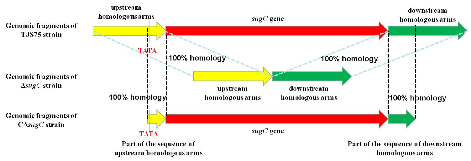
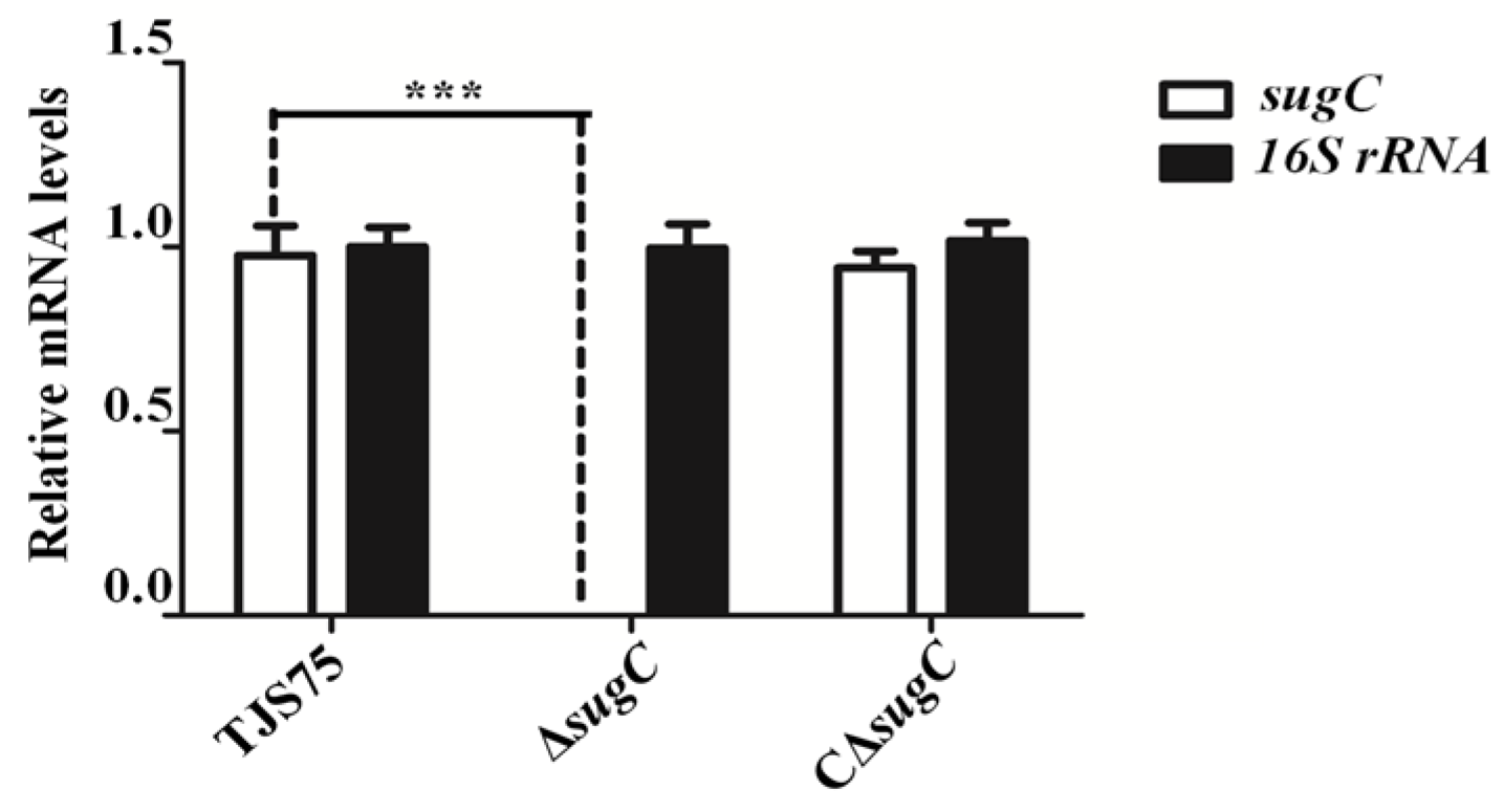
The primers listed in Table 1 were used to verify the deletion through PCR. Some fragments were amplified using primers L1/R2 in ΔsugC. The fragments in ΔsugC were smaller than those from the parent strain TJS75 (Figure 1 and Figure 2; Supplementary Materials Sequences 1 to 3). The sugC expression was detected by qRT-PCR with primers qsugC-F/qsugC-R. The results showed that the expression of the sugC gene could be detected in TJS75 and CΔsugC, and there was no significant difference in the expression level (Figure 3), but it was not found in ΔsugC. These results indicate that we constructed a sugC gene knockout strain (ΔsugC) and its complementary strain (CΔsugC). The original gel images of Figure 1 can be found in the Supplementary Materials Figure S1.

3.2. Observation of the Culture Characteristics of S. suis Strains

When ΔsugC, CΔsugC and TJS75 were inoculated in TSB containing 10% fetal bovine serum, ΔsugC reached a logarithmic growth phase after 8 h cultivation and reached its peak at 16 h; afterwards, its growth tended to flatten out. The CΔsugC and TJS75 strains reached a logarithmic growth phase at 8 h cultivation, reached a peak about 14 h, and then tended to flatten out. Although the growth rate of ΔsugC decreased compared to TJS75 and CΔsugC, there were no significant differences from 14 h to 24 h (Figure 4) in the total bacterial counts, which indicated that the time for ΔsugC to reach the plateau stage was relatively delayed, but the effect of the bacterial content after reaching the peak did not have a significant difference. Gram staining and microscopic examination showed a similar morphology to TJS75 and CΔsugC, and all strains were Gram-positive, with single or multiple short-chain arranged cocci.

3.3. Effect of the sugC Gene on the Biochemical Characteristics of the TJS75 Strain

ΔsugC, CΔsugC and TJS75 could ferment α-glucosidase, galactose, maltose, sucrose, salicin and trehalose, but could not ferment sorbitol, mannitol, xylose, lactose and glycerol. These results indicate that the deletion of the sugC gene had no significant influence on the biochemical characteristics in TJS75.

3.4. Lacking the sugC Gene Does Not Change the Hemolytic Characteristics of the TJS75 Strain

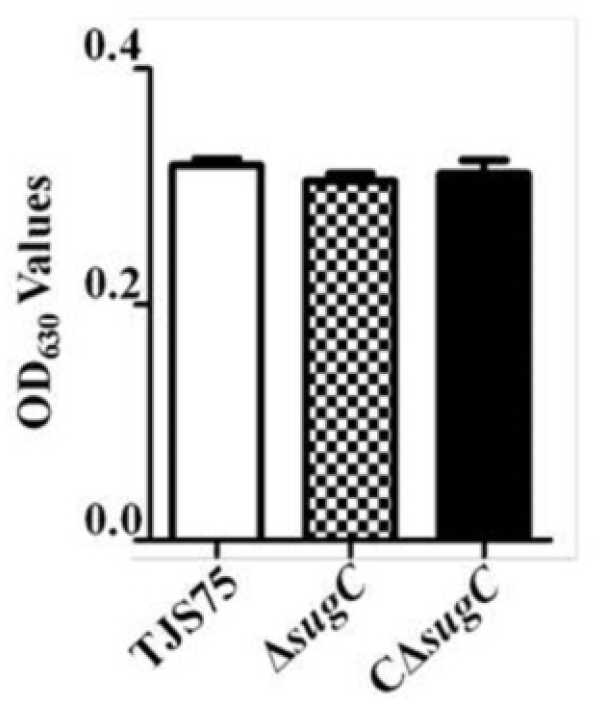
The semi-transparent and round colonies of ΔsugC on the Columbia blood agar plate were accompanied by α-hemolysis, and this result was consistent with CΔsugC and TJS75. At the same time, ΔsugC, CΔsugC and TJS75 were interacted with sheep red blood cells, and the average OD630 values of the treated solution were 0.3050, 0.3110 and 0.3180, respectively. The difference for ΔsugC was not significant compared to CΔsugC and TJS75 (Figure 5), indicating that the deletion of sugC did not significantly affect the hemolytic ability of TJS75 in sheep red blood cells.

3.5. Knocking Out the sugC Gene Reduces the Adhesion and Invasiveness of TJS75 in PK-15 Cells

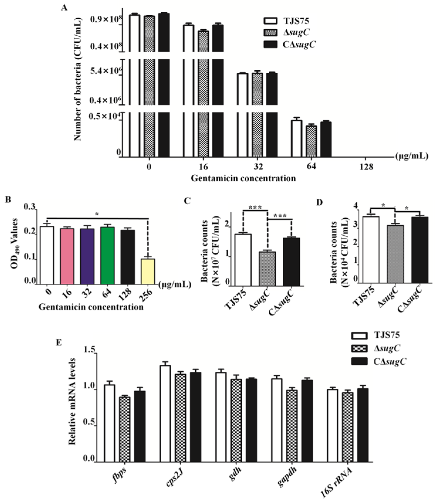
The MICs of GEN (Supplementary Materials Figure S2A) against ΔsugC, CΔsugC and TJS75 were determined to be 16 μg/mL (Supplementary Materials Figure S2B to Figure S2D), and the MBCs for 1 h were 128 μg/mL (Figure 6A). Next, 128 μg/mL GEN was used to interact with PK-15 cells for 1 h, and the cell viability of PK-15 cells (Figure 6B) did not significantly change compared with the control group.
The results of the cell adhesion tests showed that the number of viable bacteria on the surface and in the interior of PK-15 cells of ΔsugC was significantly reduced compared to TJS75 and CΔsugC showed no significant change compared to TJS75 (Figure 6C). After using GEN to inactivate the living bacteria adhering to the surfaces of PK-15 cells, the number of ΔsugC living bacteria inside PK-15 cells decreased compared to TJS75 and CΔsugC (Figure 6D). After analyzing the expression levels of genes mainly involved in adhesion-related virulence in TJS75, ΔsugC and CΔsugC, it was found that the deletion of the sugC gene had no significant effect on the mRNA expression levels of genes such as fbps, cps2J, gdh and gapdh (Figure 6E). These results indicate that the deletion of sugC reduced the adhesion and invasion abilities of TJS75 in PK-15 cells.

3.6. Deletion of the sugC Gene Reduced the Virulence of TJS75 in BALB/c Mice

After inoculation, the three study groups of BALB/c mice showed varying degrees of rough and matte fur, decreased appetite, weight loss, mental depression, and increased secretion from the corners of the eyes. The control group of mice treated with 0.9% NaCl showed no abnormalities. At the dose of 1.0 × 10⁹ CFU/mL, all BALB/c mice attacked with TJS75 began to develop the disease within 12 h and all died within 24 h, as seen for mice challenged with ΔsugC and CΔsugC. At the dose of 1.0 × 10⁸ CFU/mL, BALB/c mice infected with TJS75 began to develop the disease at 12 h (the incidence rate was 100% at 24 h) and died within 24 h (the death rate was 60% at the end of the experiment). BALB/c mice infected with CΔsugC were similar to those with TJS75, but the death rate was 70% at the end of the experiment. Meanwhile, BALB/c mice infected with ΔsugC were also similar to those with TJS75 and CΔsugC in the time of onset, but the incidence rate, time of death and mortality rate were 36 h, 80% and 40%, respectively, which were different from those of TJS75 and CΔsugC. At the doses of 1.0×10⁷ CFU/mL and 1.0 × 10⁶ CFU/mL, the times of onset (24 h) in all BALB/c mice infected with the three tested strains were consistent, but the morbidity rates and mortality rates of CΔsugC and TJS75 were higher than those of ΔsugC (Table 3 and Table 4). According to the Reed–Munch method, the LD50 of each group was calculated, and the results showed that the LD50 values of ΔsugC, CΔsugC and TJS75 were 1.47 × 10⁸ CFU, 1.75 × 10⁷ CFU and 2.15 × 10⁷ CFU (Table 4). Interestingly, the LD50 of ΔsugC in BALB/c mice was seven times higher than that of TJS75, and these results indicate that the deletion of sugC reduced the virulence of TJS75 in BALB/c mice. In addition, the distribution of bacteria in the organs showed that the three tested strains could be isolated from the brains, hearts, livers, spleens, lungs and kidneys of dead mice in each experimental group. After 48–96 h of infection, varying amounts of the three tested strains could be isolated from the brains, heart blood and lungs of dying mice. It should be noted that a small amount of bacteria could be isolated from surviving mice with infection at 7 days (Table 5). At the same time, the number of dead mice (including dead and near dead) in ΔsugC was lower than that in TJS75 and CΔsugC, and the number of surviving mice was higher than that in the TJS75 and CΔsugC groups.

4. Discussion

This study used homologous recombination technology to exchange the upstream and downstream homologous fragments of the sugC gene with the genome of the S. suis wild strain TJS75, thereby replacing the sugC gene fragments and obtaining the target gene deletion strain. Then, the TJS75 strain was used as the research object to explore the main functions of the knockout sugC gene. The above method was adopted as the main principle of target gene deletion and mutation [16]. In order to reveal the virulence relationship between the sugC gene and wild-type S. suis TJS75, the construction of sugC gene mutant strains was performed to explore the function of virulence-related factors, and this method is widely used [7,17,18,20,25,26].
ABC transporters are a family of membrane proteins that have multiple functions and are widely distributed in bacteria [27]. Their main function is to utilize the energy generated by ATP hydrolysis to achieve the transmembrane transport of substrates [28]. Most ABC transporters were initially discovered through the study of drug resistance in eukaryotic organisms [29]. There is currently extensive research on the role of ABC transporters in bacterial pathogenicity. For example, during the process of human infection with Streptococcus agalactiae CS101, the Opp (oligopeptide) ABC transporters, which are involved in amino acid intake, heme synthesis, spore formation, and the expression of the major virulence factor SpeB cysteine protease, were observed to reduce the expression of fibrinogen-binding protein (FbsA), indicating a decrease in the adhesion of the CS101 strain to epithelial cells [30]. Research has shown that Fusarium graminearum, which is a destructive fungal pathogen of small grain crops worldwide, contains an ABC transporter protein (called FgAtm1) involved in regulating iron homeostasis (which is important for the growth, reproduction, and other metabolic processes of all eukaryotes). Here, the deficiency of FgAtm1 reduced the activity of Fe-S protein nitrite reductase and xanthine dehydrogenase in the cytoplasm, thereby activating the transcription factor FgAreA and leading to the high expression of FgHopX, which directly inhibits the transcription of iron-consuming protein genes [31]. The ABC transporter encoded by the sugC gene in Mycobacterium could provide trehalose for Mycobacterium and enhance its adaptability to the environment [32,33]. The survival time of mice infected with the sugC gene deletion strain (ΔsugC) was 33 days, which was longer than the survival time of mice infected with the wild strain; this indicates that the sugC gene enhanced the virulence of Mycobacteria and is an important virulence factor [32]. Recently, the genes of different serotypes of S. suis were examined through comparative genomics, and the results showed that the sugC gene existed in highly virulent S. suis [14], but these studies did not explore the correlation between the sugC gene and the virulence of S. suis. Fortunately, we obtained a TJS75 strain carrying the sugC gene and possessing pathogenic abilities from veterinary clinical practice. On the basis of the TJS75 strain, we designed specific primers to knock out and supplement the sugC gene, obtaining ΔsugC and CΔsugC strains, respectively.
The infection mechanisms of S. suis can be divided into adhesion and colonization, blood survival and diffusion, inflammatory activation and septic shock, invasion of the central nervous system and meningitis, among which adhesion to the host tissue or cells is the most important step in the invasion and infection of host cells [34]. At present, CPS, MRP, GDH, FBPS and GAPDH are known to be the adhesins of S. suis serotype 2, which are involved in the adhesion of S. suis to host tissue or cells and enhance the pathogenicity to the host [35,36,37,38]. The absence of one or more adhesins could reduce the adhesion ability of S. suis serotype 2 to host tissue or cells, but it would not be completely lost, indicating the presence of other adhesins. The growth and biochemical characteristics, hemolysis, adhesion, invasiveness, and pathogenicity to mice were evaluated between ΔsugC and its parent strain. Interestingly, we found that there was no significant difference in the growth characteristics, biochemical characteristics, and hemolysis ability of ΔsugC compared to the wild strain TJS75, but the ability to adhere and invade PK-15 cells was decreased. At the same time, we found that there were no obvious impact on the relative expression levels of adhesion-related genes such as fbps, gdh, gapdh and cps2J between ΔsugC and its parent strain TJS75. This means that the deletion of the sugC gene would not affect the expression levels of the fbps, gdh, gapdh and cps2J genes in the TJS75 strain. It could be seen that the deletion of the sugC gene could reduce the adhesion and invasion abilities of TJS75 in PK-15 cells, but it was not completely blocked. Meanwhile, the results of the animal pathogenicity test also showed that the LD50 of ΔsugC in BALB/c mice was 1.47 × 10⁸ CFU, and there was a seven-fold difference compared to the TJS75 strain (LD50 = 2.15 × 10⁷ CFU), indicating that the virulence of TJS75 to BALB/c mice was reduced by the deletion of the sugC gene. Thus, the above results indicate that the sugC gene is a virulence-related gene in S. suis serotype 2.

5. Conclusions

In this study, a wild-type strain of TJS75 isolated from fattening pigs’ brain tissue samples was used as a parent strain, and a knockout sugC strain (ΔsugC) and complementary strain (CΔsugC) were successfully constructed using homologous recombination technology. Through experimental analysis, it has been confirmed that the sugC gene is a new virulence-related gene in the TJS75 strain both in vivo and in vitro. However, the specific mechanism of action of this gene remains to be explored. This study lays a foundation for the further exploration of the potential virulence factors and pathogenesis of S. suis.

Supplementary Materials

The following supporting information can be downloaded at https://www.mdpi.com/article/10.3390/vetsci11090447/s1. Sequence 1. Part of the sequence of the TJS75 strain (sequence position 5′-3′: 838904–840584). Sequence 2. Part of the sequence of the ΔsugC strain (sequence position 5′-3′: 838904–839215 + 840230–840584). Sequence 3. Part of the sequence of the CΔsugC strain (sequence position 5′-3′: 839140–840370). Figure S1. The original gel images of Figure 1. Figure S2. The results of the MICs for ΔsugC, CΔsugC, and TJS75.

Author Contributions

Conceptualization, Z.D. and M.Y.; methodology, C.L., X.T., X.G., W.R. and F.L.; software, C.L.; validation, X.L. and L.Z.; formal analysis, J.C.; investigation, X.T.; resources, L.Z.; data curation, X.G. and Y.Z.; writing—original draft preparation, C.L. and W.R.; writing—review and editing, Z.D.; visualization, X.L.; supervision, M.Y. and W.Z.; project administration, J.C.; funding acquisition, Z.D. and M.Y. All authors have read and agreed to the published version of the manuscript.

Funding

This research was funded by the Innovative Research and Experimental Projects for Young Researchers of Tianjin Academy of Agricultural Sciences (grant number: 2021019), the Key Laboratory Open Fund Project of the Ministry of Education of System Bioengineering in Tianjin University (grant number: 2022003), the Central Public-Interest Scientific Institution Basal Research Fund and Tianjin Modern Agricultural Industry Technology System Project (grant number: ITTPRS2021003), the Tianjin Enterprise Science and Technology Commissioner Project (grant number: 23YDTPJC00830) and Research on Innovation and Efficient Breeding Technology of Livestock and Poultry Germplasm Resources (grant number: 2024ZYCX012).

Institutional Review Board Statement

The current study was approved by the Animal Care and Use Committee of the Institute of Animal Science and Veterinary, Tianjin Academy of Agricultural Sciences (protocol number 2023002).

Informed Consent Statement

Informed consent was obtained from all subjects involved in the study.

Data Availability Statement

The complete genome sequence of the TJS75 strain (accession: CP095162.1) has been submitted to GenBank.

Conflicts of Interest

The authors declare no conflicts of interest.

References

  1. López-Martínez, M.J.; Ornelas, M.A.S.; Amarie, R.E.; Manzanilla, E.G.; Martínez-Subiela, S.; Tecles, F.; Tvarijonaviciute, A.; Escribano, D.; González-Bulnes, A.; Cerón, J.J.; et al. Changes in salivary biomarkers of stress, inflammation, redox status, and muscle damage due to Streptococcus suis infection in pigs. BMC Vet. Res. 2023, 1, 100. [Google Scholar] [CrossRef] [PubMed]
  2. Zhou, Y.; Dong, X.; Li, Z.; Zou, G.; Lin, L.; Wang, X.; Chen, H.; Gasser, R.B.; Li, J. Predominance of Streptococcus suis ST1 and ST7 in human cases in China, and detection of a novel sequence type, ST658. Virulence 2017, 8, 1031–1035. [Google Scholar] [CrossRef] [PubMed]
  3. Tang, J.; Wang, C.; Feng, Y.; Yang, W.; Song, H.; Chen, Z.; Yu, H.; Pan, X.; Zhou, X.; Wang, H.; et al. Streptococcal toxic shock syndrome caused by Streptococcus suis serotype 2. PLoS Med. 2006, 3, e151. [Google Scholar] [CrossRef]
  4. Peng, W.; Yang, X.; Wang, N.; Gao, T.; Liu, Z.; Liu, W.; Zhou, D.; Yang, K.; Guo, R.; Liang, W.; et al. PerR-regulated manganese import contributes to Oxidative stress defense in Streptococcus suis. Appl. Environ. Microbiol. 2022, 88, e0008622. [Google Scholar] [CrossRef]
  5. Nicholson, T.L.; Bayles, D.O. Comparative virulence and antimicrobial resistance distribution of Streptococcus suis isolates obtained from the United States. Front. Microbiol. 2022, 13, 1043529. [Google Scholar] [CrossRef]
  6. Aradanas, M.; Poljak, Z.; Fittipaldi, N.; Ricker, N.; Farzan, A. Serotypes, virulence-associated factors, and antimicrobial resistance of Streptococcus suis isolates recovered from sick and healthy pigs determined by Whole-Genome Sequencing. Front. Vet. Sci. 2021, 8, 742345. [Google Scholar] [CrossRef]
  7. Wang, Y.; Wang, Y.; Liu, B.; Wang, S.; Li, J.; Gong, S.; Sun, L.; Yi, L. pdh modulate virulence through reducing stress tolerance and biofilm formation of Streptococcus suis serotype 2. Virulence 2019, 10, 588–599. [Google Scholar] [CrossRef]
  8. Liu, L.; Zhang, Q.; Xu, Z.; Chen, B.; Zhang, A.; Sun, X.; Jin, M. Screening of virulence-related transcriptional regulators in Streptococcus suis. Genes 2020, 11, 972. [Google Scholar] [CrossRef]
  9. Roodsant, T.J.; Van Der Putten, B.C.L.; Tamminga, S.M.; Schultsz, C.; Van Der Ark, K.C.H. Identification of Streptococcus suis putative zoonotic virulence factors: A systematic review and genomic meta-analysis. Virulence 2021, 12, 2787–2797. [Google Scholar] [CrossRef]
  10. Roy, S.; Zhu, Y.; Ma, J.; Roy, A.C.; Zhang, Y.; Zhong, X.; Pan, Z.; Yao, H. Role of ClpX and ClpP in Streptococcus suis serotype 2 stress tolerance and virulence. Microbiol. Res. 2019, 223–225, 99–109. [Google Scholar] [CrossRef]
  11. Akhtar, A.A.; Turner, D.P. The role of bacterial ATP-binding cassette (ABC) transporters in pathogenesis and virulence: Therapeutic and vaccine potential. Microb. Pathog. 2022, 171, 105734. [Google Scholar] [CrossRef] [PubMed]
  12. Martin, A.; Daniel, J. The ABC transporter Rv1272c of Mycobacterium tuberculosis enhances the import of long-chain fatty acids in Escherichia coli. Biochem. Bioph. Res. Commun. 2018, 496, 667–672. [Google Scholar] [CrossRef] [PubMed]
  13. Basavanna, S.; Khandavilli, S.; Yuste, J.; Cohen, J.M.; Hosie, A.H.; Webb, A.J.; Thomas, G.H.; Brown, J.S. Screening of Streptococcus pneumoniae ABC transporter mutants demonstrates that LivJHMGF, a branched-chain amino acid ABC transporter, is necessary for disease pathogenesis. Infect. Immun. 2009, 77, 3412–3423. [Google Scholar] [CrossRef] [PubMed]
  14. Rosinha, G.M.; Freitas, D.A.; Miyoshi, A.; Azevedo, V.; Campos, E.; Cravero, S.L.; Rossetti, O.; Splitter, G.; Oliveira, S.C. Identification and characterization of a Brucella abortus ATP-binding cassette transporter homolog to Rhizobium meliloti ExsA and its role in virulence and protection in mice. Infect. Immun. 2002, 70, 5036–5044. [Google Scholar] [CrossRef] [PubMed]
  15. Zhang, A.; Yang, M.; Hu, P.; Wu, J.; Chen, B.; Hua, Y.; Yu, J.; Chen, H.; Xiao, J.; Jin, M. Comparative genomic analysis of Streptococcus suis reveals significant genomic diversity among different serotypes. BMC Genom. 2011, 12, 523. [Google Scholar] [CrossRef]
  16. Takamatsu, D.; Osaki, M.; Sekizaki, T. Construction and characterization of Streptococcus suis-Escherichia coli shuttle cloning vectors. Plasmid 2001, 45, 101–113. [Google Scholar] [CrossRef]
  17. Yu, Y.; Qian, Y.; Du, D.; Xu, C.; Dai, C.; Li, Q.; Liu, H.; Shao, J.; Wu, Z.; Zhang, W. SBP2 plays an important role in the virulence changes of different artificial mutants of Streptococcus suis. Mol. Biosyst. 2016, 12, 1948–1962. [Google Scholar] [CrossRef]
  18. Ji, X.; Sun, Y.; Liu, J.; Zhu, L.; Guo, X.; Lang, X.; Feng, S. A novel virulence-associated protein, vapE, in Streptococcus suis serotype 2. Mol. Med. Rep. 2016, 13, 2871–2877. [Google Scholar] [CrossRef]
  19. Zuo, J.; Fan, Q.; Li, J.; Liu, B.; Xue, B.; Zhang, X.; Yi, L.; Wang, Y. Sub-Inhibitory concentrations of amoxicillin and tylosin affect the biofilm formation and virulence of Streptococcus suis. Int. J. Environ. Res. Public Health 2022, 19, 8359. [Google Scholar] [CrossRef]
  20. Liu, H.; Fu, H.; Jiang, X.; Liao, X.; Yue, M.; Li, X.; Fang, W. PrsA contributes to Streptococcus suis serotype 2 pathogenicity by modulating secretion of selected virulence factors. Vet. Microbiol. 2019, 236, 108375. [Google Scholar] [CrossRef]
  21. Gottschalk, M.; Higgins, R.; Jacques, M.; Beaudoin, M.; Henrichsen, J. Characterization of six new capsular types (23 through 28) of Streptococcus suis. J. Clin. Microbiol. 1991, 29, 2590–2594. [Google Scholar] [CrossRef] [PubMed]
  22. Zheng, F.; Ji, H.; Cao, M.; Wang, C.; Feng, Y.; Li, M.; Pan, X.; Wang, J.; Qin, Y.; Hu, F.; et al. Contribution of the Rgg transcription regulator to metabolism and virulence of Streptococcus suis serotype 2. Infect. Immun. 2011, 79, 1319–1328. [Google Scholar] [CrossRef] [PubMed]
  23. CLSI. Performance Standards for Antimicrobial Disk and Dilution Susceptibility Tests for Bacteria Isolated from Animals, Approved Standard, 4th ed.; CLSI document VET01-A4; Clinical and Laboratory Standards Institute: Wayne, PA, USA, 2013. [Google Scholar]
  24. Cetin, Y.; Bullerman, L.B. Cytotoxicity of Fusarium mycotoxins to mammalian cell cultures as determined by the MTT bioassay. Food. Chem. Toxicol. 2005, 43, 755–764. [Google Scholar] [CrossRef] [PubMed]
  25. Hu, Y.; Hu, Q.; Wei, R.; Li, R.; Zhao, D.; Ge, M.; Yao, Q.; Yu, X. The XRE family transcriptional regulator SrtR in Streptococcus suis is involved in Oxidant tolerance and virulence. Front. Cell. Infect. Microbiol. 2019, 8, 452. [Google Scholar] [CrossRef] [PubMed]
  26. Ni, C.; Gao, S.; Li, X.; Zheng, Y.; Jiang, H.; Liu, P.; Lv, Q.; Huang, W.; Li, Q.; Ren, Y.; et al. Fpr2 exacerbates Streptococcus suis-induced streptococcal toxic shock-like syndrome via attenuation of neutrophil recruitment. Front. Immunol. 2023, 14, 1094331. [Google Scholar] [CrossRef] [PubMed]
  27. Theodoulou, F.L.; Kerr, I.D. ABC transporter research: Going strong 40 years on. Biochem. Soc. Trans. 2015, 43, 1033–1040. [Google Scholar] [CrossRef]
  28. Thomas, C.; Tampé, R. Structural and mechanistic principles of ABC transporters. Annu. Rev. Biochem. 2020, 89, 605–636. [Google Scholar] [CrossRef]
  29. Orelle, C.; Mathieu, K.; Jault, J.M. Multidrug ABC transporters in bacteria. Res. Microbiol. 2019, 170, 381–391. [Google Scholar] [CrossRef]
  30. Lewis, V.G.; Ween, M.P.; McDevitt, C.A. The role of ATP-binding cassette transporters in bacterial pathogenicity. Protoplasma 2012, 249, 919–942. [Google Scholar] [CrossRef]
  31. Wang, Z.; Ma, T.; Huang, Y.; Wang, J.; Chen, Y.; Kistler, H.C.; Ma, Z.; Yin, Y. A fungal ABC transporter FgAtm1 regulates iron homeostasis via the transcription factor cascade FgAreA-HapX. PLoS Pathog. 2019, 15, e1007791. [Google Scholar] [CrossRef]
  32. Kalscheuer, R.; Weinrick, B.; Veeraraghavan, U.; Besra, G.S.; Jr, W.R.J. Trehalose-recycling abc transporter lpqy-suga-sugb-sugc is essential for virulence of Mycobacterium tuberculosis. Proc. Natl. Acad. Sci. USA 2010, 107, 21761–21766. [Google Scholar] [CrossRef] [PubMed]
  33. Babu-Sait, M.R.; Koliwer-Brandl, H.; Stewart, J.A.; Swarts, B.M.; Jacobsen, M.; Ioerger, T.R.; Kalscheuer, R. PPE51 mediates uptake of trehalose across the mycomembrane of Mycobacterium tuberculosis. Sci. Rep. 2022, 12, 2097. [Google Scholar] [CrossRef] [PubMed]
  34. Tan, C.; Zhang, A.; Chen, H.; Zhou, R. Recent proceedings on prevalence and pathogenesis of Streptococcus suis. Curr. Issues Mol. Biol. 2019, 32, 473–520. [Google Scholar] [CrossRef]
  35. Wang, J.; Kong, D.; Zhang, S.; Jiang, H.; Zheng, Y.; Zang, Y.; Hao, H.; Jiang, Y. Interaction of fibrinogen and muramidase-released protein promotes the development of Streptococcus suis meningitis. Front. Microbiol. 2015, 6, 1001. [Google Scholar] [CrossRef] [PubMed]
  36. De Greeff, A.; Buys, H.; Verhaar, R.; Dijkstra, J.; Van Alphen, L.; Smith, H.E. Contribution of fibronectin-binding protein to pathogenesis of Streptococcus suis serotype 2. Infect. Immun. 2002, 70, 1319–1325. [Google Scholar] [CrossRef] [PubMed]
  37. Brassard, J.; Gottschalk, M.; Quessy, S. Cloning and purification of the Streptococcus suis serotype 2 glyceraldehyde-3-phosphate dehydrogenase and its involvement as an adhesin. Vet. Microbiol. 2004, 102, 87–94. [Google Scholar] [CrossRef]
  38. Tan, C.; Fu, S.; Liu, M.; Jin, M.; Liu, J.; Bei, W.; Chen, H. Cloning, expression and characterization of a cell wall surface protein, 6-phosphogluconate-dehydrogenase, of Streptococcus suis serotype 2. Vet. Microbiol. 2008, 130, 363–370. [Google Scholar] [CrossRef]
Figure 1. Construction and confirmation of ΔsugC and CΔsugC. (A) Construction and confirmation of ΔsugC. M = DL2000 marker; 1 = ddH2O; 2 and 4 = randomly chosen clone strains; 3 = pSET4s-ΔsugC; 5 = TJS75. (B) Construction and confirmation of CΔsugC. M = DL2000 marker; 1 = ddH2O; 2 = randomly chosen clone strain; 3 = pSET2-sugC; 4 = TJS75.
Vetsci 11 00447 g001
Figure 2. Amino acid sequence analysis of PCR products from multiple target fragments with TJS75, ΔsugC and CΔsugC.
Vetsci 11 00447 g002
Figure 3. Transcription of target molecules in ΔsugC, CΔsugC and TJS75. ΔsugC vs. TJS75: psugC = 0, p16S rRNA = 0.8839; CΔsugC vs. TJS75: psugC = 0.7962, p16S rRNA = 0.6983. ***: p < 0.001.
Vetsci 11 00447 g003
Figure 4. In vitro growth curves of different generations of TJS75, ΔsugC and CΔsugC. ΔsugC vs. TJS75: p6h = 0.0064, p8h = 0.0035, p10h = 0.0051, p12h = 0.0063, p14h = 0.0912, p16h = 0.2954, p20h = 0.2149, p24h = 0.0506; CΔsugC vs. TJS75: p6h = 0.1460; p8h = 0.0116; p10h = 0.0092, p12h = 0.1077, p14h = 0.2254, p16h = 0.0965, p20h = 0.5040, p24h = 0.1165.
Vetsci 11 00447 g004
Figure 5. Hemolytic characteristic results of detection of sugC in S. suis. OD630 values of solutions in which ΔsugC, CΔsugC and TJS75 were interacted with sheep red blood cells. MeanTJS75 = 0.3180, MeanΔsugC = 0.3050, MeansugC = 0.3110, pΔsugC vs. TJS75 = 0.1421, pCΔsugC vs. TJS75 = 0.3714.
Vetsci 11 00447 g005
Figure 6. Analysis of adhesion and invasiveness. (A) The MBCs for 1 h of ΔsugC, CΔsugC, and TJS75. (B) The effect of GEN on the viability of PK-15 cells by MTT. Mean0 μg/mL = 0.2320, Mean16 μg/mL = 0.2224, Mean32 μg/mL = 0.2229, Mean64 μg/mL = 0.2296, Mean128 μg/mL = 0.2173, Mean256 μg/mL = 0.1021; p0 μg/mL vs. 16 μg/mL= 0.5014, p0 μg/mL vs. 32 μg/mL= 0.5805, p0 μg/mL vs. 64 μg/mL = 0.8622, p0 μg/mL vs. 128 μg/mL = 0.1952, p0 μg/mL vs. 256 μg/mL= 0.0102. (C) Results of cell adhesion test of ΔsugC, CΔsugC and TJS75. MeanTJS75 = 1.76 × 10⁷ CFU/mL, MeanΔsugC = 1.16 × 10⁷ CFU/mL, MeansugC = 1.62 × 10⁷ CFU/mL, pΔsugC vs. TJS75 = 0.0001, pCΔsugC vs. TJS75 = 0.0918, pΔsugC vs. sugC = 0.0001. (D) Results of cell invasion test of ΔsugC, CΔsugC, and TJS75. MeanTJS75 = 3.63 × 10⁴ CFU/mL, MeanΔsugC= 3.14 × 10⁴ CFU/mL, MeansugC = 3.60 × 10⁴ CFU/mL, pΔsugC vs. TJS75 = 0.0307, pCΔsugC vs. TJS75 = 0.8327, pΔsugC vs. sugC = 0.0165. (E) The relative expression of fbps, cps2J, gdh and gapdh. ΔsugC vs. TJS75: pfbps = 0.0624, p cps2J = 0.1124, p gdh = 0.2038, p gapdh = 0.0528, p16S rRNA = 0.6622; CΔsugC vs. TJS75: pfbps = 0.8320, p cps2J = 0.2907, p gdh = 0.2851, p gapdh = 0.6814, p16S rRNA = 0.8105. *: p < 0.05; ***: p < 0.001.
Vetsci 11 00447 g006
Table 1. Information about strains, plasmids and primers used in this study.
Type and NameSequence (5′-3′)Purpose
TJS75/S. suis serotype 2 virulence strain isolated from diseased pigs in Tianjin in 2015 (Accession: CP095162.1)
ΔsugC/TJS75 strain with sugC gene knocked out
sugC/Complementary strain of ΔsugC; SpcR
E. coli DH5α/For cloning of recombinant plasmids
pSET4s [16]/S. suis temperature-sensitive suicide vector
pSET2/E. coliS. suis shuttle vector; SpcR
pSET4s-ΔsugC/Recombinant vector with background of pSET4s, designed to knock out sugC gene; SpcR
pSET2-sugC/pSET2 containing complete sugC gene and its promoter; SpcR
S1TACTACTTACCTCCGTATTGCADetecting full length of sugC gene
S2TGATTACCTTTAACGATAT
L1GAAGCTGCAGTCAAAGAAGACATATACCCAAG 1Detecting upstream homologous arms of sugC gene
L2GAGGTGTGATTGCTCAAAGATAT
R1TAGCCACGTTACACACCTCDetecting downstream homologous arms of sugC gene
R2CCCCCCGGGCGAAGCTGAACGTGGCTAT
sugC-FCCCCGGGTATATGATGAAGGCTACCAGCAACCACAsugC gene and its upstream promoter carrying relevant restriction enzyme sites at both ends
sugC-RGCTGCAGGAAATTAAAGACTTTGCAAGCAGCGT
qsugC-FCTACTTACCTCCGTATTGCATAATGRelative quantitative detection of sugC gene
qsugC-RCCATGTTATTGATGATGTCGTGACT
q16S rRNA-FGGCGTGCCTAATACATGRelative quantitative detection of internal reference genes
q16S rRNA-RGCTATGAGGCAGGTT
qgdh-FCGGCGGTGGTAAAGGTGGTTRelative quantitative detection of gdh gene
qgdh-RCGTCAAGTGAAGGTCCGATGTG
qfbps-FTGCCATTTGCCAATAGCCCTGAARelative quantitative detection of fbps gene
qfbps-RTCCCGCTCCGCCTTATCCTG
qcps2J-FGTTACTTGCTACTTTTGATGGRelative quantitative detection of cps2J gene
qcps2J-RTTTTCATTTCCTAAGTCTCG
qgapdh-FGTTTGATGACTACAATCCTCGGTTACRelative quantitative detection of gapdh gene
qgapdh-RGCTTTAGCAGCACCAGTTGAG
1. Underlined and bold type indicates the restriction sites in the primer sequences.
Table 2. Test group.
Dose (CFU/mL)Strain and Number of Mice
TJS75ΔsugCsugCControl (0.9% NaCl)
1 × 109101010/ 1
1 × 108101010/
1 × 107101010/
1 × 106101010/
Total40404010
1 “/” means that there was no such content.
Table 3. The initial times of onset and death and the number of infected mice in each group.
Dose (CFU/mL)Initial Time of Onset (h)Initial Time of Death (h)Duration of Onset (h)Morbidity Rate %
TJS75ΔsugCsugCTJS75ΔsugCsugCTJS75ΔsugCsugCTJS75ΔsugCsugC
1.0 × 109121212121212121212100100100
1.0 × 10812121224242413213213210080100
1.0 × 10712241248024132132132804080
1.0 × 10624242460060241236602060
Table 4. The LD50 of ΔsugC, CΔsugC and TJS75 in BALB/c mice.
Dose (CFU/mL)Number of Dead Mice/Total Mice
TJS75ΔsugCsugC
1.0 × 10910/10 10/10 10/10 10/1010/1010/1010/1010/1010/10
1.0 × 1086/107/105/104/104/104/107/107/107/10
1.0 × 1074/104/104/100/100/100/104/104/104/10
1.0 × 1062/102/102/100/100/100/102/102/102/10
Mean LD50 value2.15 × 107 CFU1.47 × 108 CFU1.75 × 107 CFU
Table 5. Distribution and bacterial count determination of three tested strains in BALB/c mouse organs.
GroupDose (CFU/mL)Dying MiceAverage Bacterial CountSurvivorsAverage Bacterial Count
Heart BloodBrainLungsHeart BloodBrainLungs
TJS751.0 × 10843.40 × 1061.10 × 1055.10 × 10541.10 × 1031.40×1013.0 × 101
1.0 × 10724.90 × 1055.40 × 1046.40 × 10464.80 × 10141
1.0 × 10615.80 × 1041.30 × 1028.40 × 1018200
ΔsugC1.0 × 10817.80 × 1046.30 × 1023.80 × 10165.20 × 10291.40 × 101
1.0 × 1070/ *//101.50 × 10100
1.0 × 1060///10000
sugC1.0 × 10838.60 × 1067.10 × 1044.90 × 10533.20 × 10246.70 × 101
1.0 × 10713.80 × 1051.70 × 1034.50 × 10462.60 × 10140
1.0 × 10615.70 × 1044.10 × 1025.90 × 1018100
* “/” means no such result.
Disclaimer/Publisher’s Note: The statements, opinions and data contained in all publications are solely those of the individual author(s) and contributor(s) and not of MDPI and/or the editor(s). MDPI and/or the editor(s) disclaim responsibility for any injury to people or property resulting from any ideas, methods, instructions or products referred to in the content.