Article

Five Amino Acid Substitutions in the S1 Unit of Infectious Bronchitis Virus Are Critical Determinants Enhancing Its Adaptation to Vero Cells

College of Agriculture, Yangtze University, Jingzhou 434025, China
*
Author to whom correspondence should be addressed.
Vet. Sci. 2025, 12(5), 394; https://doi.org/10.3390/vetsci12050394
Submission received: 18 March 2025 / Revised: 17 April 2025 / Accepted: 21 April 2025 / Published: 22 April 2025
(This article belongs to the Section Veterinary Microbiology, Parasitology and Immunology)

Simple Summary

The S1 subunit of the spike protein in avian infectious bronchitis virus (IBV) plays a crucial role in determining its host range, cellular tropism, and tissue specificity. Following the continuous passage of IBV-EP3 through Vero cells, the adaptability of IBV to Vero cells has progressively increased, leading to the accumulation of 19 amino acid substitutions in the S1 region of IBV-P65. In this study, we identify five specific amino acid changes as critical determinants that enhance the adaptation of IBV to Vero cells. Our findings provide valuable insights into the mechanisms underlying the adaptation of IBV to Vero cells and may facilitate the development of Vero cell-derived IBV vaccines by improving replication efficiency via targeted genetic modification.

Abstract

The S1 subunit of the spike protein of avian infectious bronchitis virus (IBV) plays a crucial role in determining its host range and cell and tissue tropism. Following the continuous passage of IBV-EP3 through Vero cells over up to 65 generations, a total of 19 amino acid mutations accumulated in the S1 region of IBV-P65. To investigate the impact of these mutations on the adaptability of IBV to Vero cells, six recombinant viruses carrying either a subset or all of the identified mutations were constructed and obtained via a reverse genetics system. Analyses on the growth characteristics of these recombinant viruses and Western blot detection of the expression levels of their spike proteins indicated that the IBV mutant obtained by replacing the amino acid sequence from positions 179 to 323 in the S1 region of IBV-P65 with the corresponding segment from IBV-EP3 S1 significantly impaired viral growth and exhibited a lower replication efficiency in Vero cells, suggesting that five amino acid substitutions (T181I, I246T, F267C, T273I, Q296K) within this region could enhance the adaptation of IBV to Vero cells.

1. Introduction

Coronaviruses are enveloped, single-stranded RNA viruses that infect a broad spectrum of mammalian and avian species [1]. According to phylogenetic and genomic analyses, coronaviruses are classified into four genera: Alphacoronavirus, Betacoronavirus, Gammacoronavirus, and Deltacoronavirus [2]. The viral genome encodes four primary structural proteins: the spike (S) protein, envelope (E) protein, membrane (M) protein, and nucleocapsid (N) protein. Among them, the S protein plays a crucial role in determining the viral host range, facilitating viral entry, dictating cell and tissue tropism, enabling cross-species transmission, and adapting to new hosts [3].
Infectious bronchitis virus (IBV), a member of the Gammacoronavirus, is prevalent globally in both industrial and backyard poultry populations [4,5,6]. Similar to other coronaviruses, the IBV S protein comprises two subunits: S1 and S2 [7]. The S1 subunit is responsible for receptor binding, whereas the S2 subunit mediates the fusion of the viral envelope with the host cell membrane [8,9]. The S1 subunit contains two domains: the N-terminal domain (NTD, amino acids 21–237) and the C-terminal domain (CTD, amino acids 269–414) [10]. One or both of these domains function as the receptor-binding domain (RBD) [11,12,13].
A multitude of diverse IBV strains circulate globally. The S1 subunit exhibits significant amino acid sequence diversity, whereas the S2 subunit remains relatively conserved across different strains [14]. Phylogenetic analysis of the S1 subunit has classified IBVs into six genotypes, encompassing 32 distinct viral lineages and a number of inter-lineage recombinants [15]. Naturally, IBV primarily infects the respiratory tract [16,17], while some strains can also infect additional tissues such as the kidney, oviduct, and gastrointestinal tract [4,18,19]. Different strains exhibit variations in tissue tropism, morbidity, mortality, pathogenicity, and production losses in poultry, which are believed to be attributed to amino acid variations in the S1 subunit [13,20,21].
Most field strains of IBV are incapable of infecting mammalian cell lines; however, the Beaudette strain represents an exception. The Beaudette strain was developed through serial passaging of the virulent Massachusetts M41 strain in embryonated chicken eggs and cultured cells. As a result of this adaptation process, the Beaudette strain has acquired the ability to infect baby hamster kidney cells (BHK-21), monkey kidney cells (Vero) [11,22], and even the human cell lines H1299 and Huh7 [23]. Research has demonstrated that the S-protein of IBV plays a crucial role in determining the extended host cell tropism of the Beaudette strain [7].
IBV-EP3 was generated by passaging the Beaudette strain through chicken embryonated eggs for three passages, while IBV-P65 was obtained by passing IBV-EP3 through Vero cells for 65 generations. During the serial passage through Vero cells, IBV gradually adapted to the Vero cells [24]. Compared with IBV-EP3, a total of 49 amino acid substitutions were identified in IBV-P65. Specifically, 26 of these substitutions occurred in the S protein, with 19 amino acid changes clustered in the S1 region. These mutations in the S protein likely expanded the host range of IBV [24].
In this study, using the established IBV reverse genetics system, six recombinant viruses that encompass the partial or total amino acid changes of the aforementioned 19 amino acid substitutions in the S1 subunit were generated by replacing the corresponding regions of IBV-P65 with those from IBV-EP3. Our findings indicate that the mutant virus, which replaces the 20,884–21,335 nucleotide sequence of the IBV-P65 S gene with the corresponding sequence from the IBV-EP3 S1 gene, exhibits significantly reduced and delayed growth kinetics as well as a lower replication efficiency in Vero cells compared to IBV-P65. Considering that only five amino acid substitutions (I181T, T246I, C267F, I273T, K296Q) differentiate this region between IBV-P65 and IBV-EP3, it is plausible to infer that these specific mutations in the S1 domain may enhance the adaptation of IBV to Vero cells.

2. Materials and Methods

2.1. Cell Culture and Virus

Vero cells (mycoplasma test negatively; passaged three generations after recovery) were cultured in Dulbecco’s Modified Eagle’s Medium (DMEM) (Invitrogen, Waltham, MA, USA) supplemented with 10% fetal bovine serum (FBS) (Hyclone, Logan, UT, USA), penicillin (100 units/mL), and streptomycin (100 μg/mL) (Invitrogen, USA) at 37 °C. IBV-EP3 (GenBank accession number DQ001338) is a Beaudette strain (GenBank accession number M95169.1) propagated in chicken embryonated eggs for three passages. IBV-P65 is a Vero cell-adapted IBV Beaudette strain (GenBank accession number DQ001339.1) obtained by passaging IBV-EP3 for 65 generations in Vero cells.

2.2. Generation of Mutant Viruses

Several fragments containing the sequences of IBV-P65 or IBV-EP3 were amplified by PCR and cloned into pGEM-T easy (Promega, Madison, WI, USA). The primers (synthesized by BGI, Shenzhen, China) and the obtained plasmids are detailed in Table 1 and Table 2, respectively. After confirmation via sequencing analysis, the plasmids were subjected to digestion with BsmB I or Bsa I, and the corresponding enzyme-digested fragments were subsequently purified. The full-length cDNA was assembled by substituting the corresponding fragment with the mutant fragment, as previously described [25]. The construction process of the recombinant viruses is illustrated in Figure 1. Full-length transcripts were synthesized in vitro by utilizing the mMessage mMachine T7 kit (Ambion, Austin, TX, USA) according to the manufacturer’s protocol. These transcripts were subsequently electroporated into Vero cells using a single pulse at 450 V and 50 µF with a Bio-Rad Gene Pulser II electroporator. Post-electroporation, the transfected Vero cells were incubated overnight in DMEM supplemented with 1% fetal bovine serum (FBS), followed by further cultivation in FBS-free DMEM. The transfected cells were monitored daily for the development of cytopathic effects (CPE). Recovered viruses underwent plaque purification and were passaged through Vero cells.

2.3. Growth Curves of Recombinant Viruses on Vero Cells

Vero cells were infected with IBV-P65 and recombinant IBV and harvested at different times post-infection. Viral stocks were prepared by freezing/thawing the cells three times. The 50% tissue culture infection dose (TCID50) of each virus was determined by infecting five wells of Vero cells on 96-well plates in duplicate with 10-fold serial dilution of each viral stock.

2.4. Analysis of RNA Synthesis of EP3 by Real-Time PCR

Confluent monolayers of Vero cells in six-well plates were inoculated with IBV-P65 and IBV-EP3 at a multiplicity of infection (MOI) of 1. Following a 1 h period of incubation at 4 °C, the cells were washed twice with phosphate-buffered saline (PBS) and subsequently cultured in 2 mL of serum-free DMEM at 37 °C. Virus-infected cells were harvested at 0, 4, 8, 12, 16, and 24 h post-infection then lysed using TRizol reagent (Invitrogen, USA), and total RNA was extracted according to the manufacturer’s protocol. The RNA samples were resuspended in nuclease-free water, and their concentrations were quantified using a Nanodrop spectrophotometer. Reverse transcription was conducted using Expand Reverse Transcriptase (Roche, Basel, Switzerland) with equal amounts of RNA and specific primers (P-IBV4527F: 5′-4527TTTAGCAGAACATTTTGACGCAGA4551-3′ and P-IBV4805R: 5′-4805TTAGTAGAACCAACAAACACGACAG4781-3′). Real-time PCR was performed using the LightCycler FastStart DNA Master SYBR Green I kit, following the manufacturer’s instructions (Roche, Switzerland). Normalized target gene expression levels were recorded for each time point and virus. The GAPDH primers (forward 5′-GTCAAGGCTGAGAACGGGAA-3′ and reverse 5′-AGTGATGGCATGGACTGTGG-3′) were employed to quantify the relative expression levels. The relative expression levels were calculated using the 2−ΔΔCt method, with GAPDH serving as the reference gene. All experiments were performed in triplicate.

2.5. Growth Curves of Viruses

To examine the growth kinetics of viruses, confluent monolayers of Vero cells in 96-well plates were inoculated with IBV-P65 and mutant viruses at a MOI of 0.5. Virus-infected cells were collected at 0, 4, 8, 12, 16, 24, and 36 h post-infection. Virus stocks were prepared by subjecting the infected cells to three cycles of freeze–thaw. TCID50 was determined for each virus stock. All experiments were conducted in triplicate, and the mean values were calculated.

2.6. Western Blotting

Confluent monolayers of Vero cells in six-well plates were inoculated with IBV-P65 and mutant viruses at a MOI of 0.5. Cells were harvested at 0, 4, 8, 12, 16, 24, and 36 h post-infection. At each time point, infected cells were washed with phosphate-buffered saline (PBS), lysed in 2× SDS loading buffer containing 100 mM dithiothreitol (DTT), and heated to 100 °C for 5 min before clarification. Proteins were separated by SDS-PAGE and transferred onto a polyvinylidene difluoride (PVDF) membrane (Stratagene, San Diego, CA, USA). The membrane was blocked overnight at 4 °C or for 2 h at room temperature using blocking buffer (5% fat-free milk powder in PBS containing 0.1% Tween 20). It was then incubated with primary antibodies (anti-S2 at 1:5000; actin at 1:500) diluted in blocking buffer for 2 h at room temperature. Following three washes with PBST, the membrane was incubated with horseradish peroxidase-conjugated secondary antibodies (anti-mouse or anti-rabbit IgG, Dako, Glostrup, Denmark) diluted at 1:2000 in blocking buffer for 1 h at room temperature. After additional washes with PBST, polypeptides were visualized using a chemiluminescence detection kit (ECL kit, Amersham Biosciences, Piscataway, NJ, USA) according to the manufacturer’s protocol. The intensities of the protein bands were quantified using the Image J (1.53e) program.

3. Results

3.1. Assessment of Infectivity of IBV-EP3 and IBV-P65 in Vero Cells

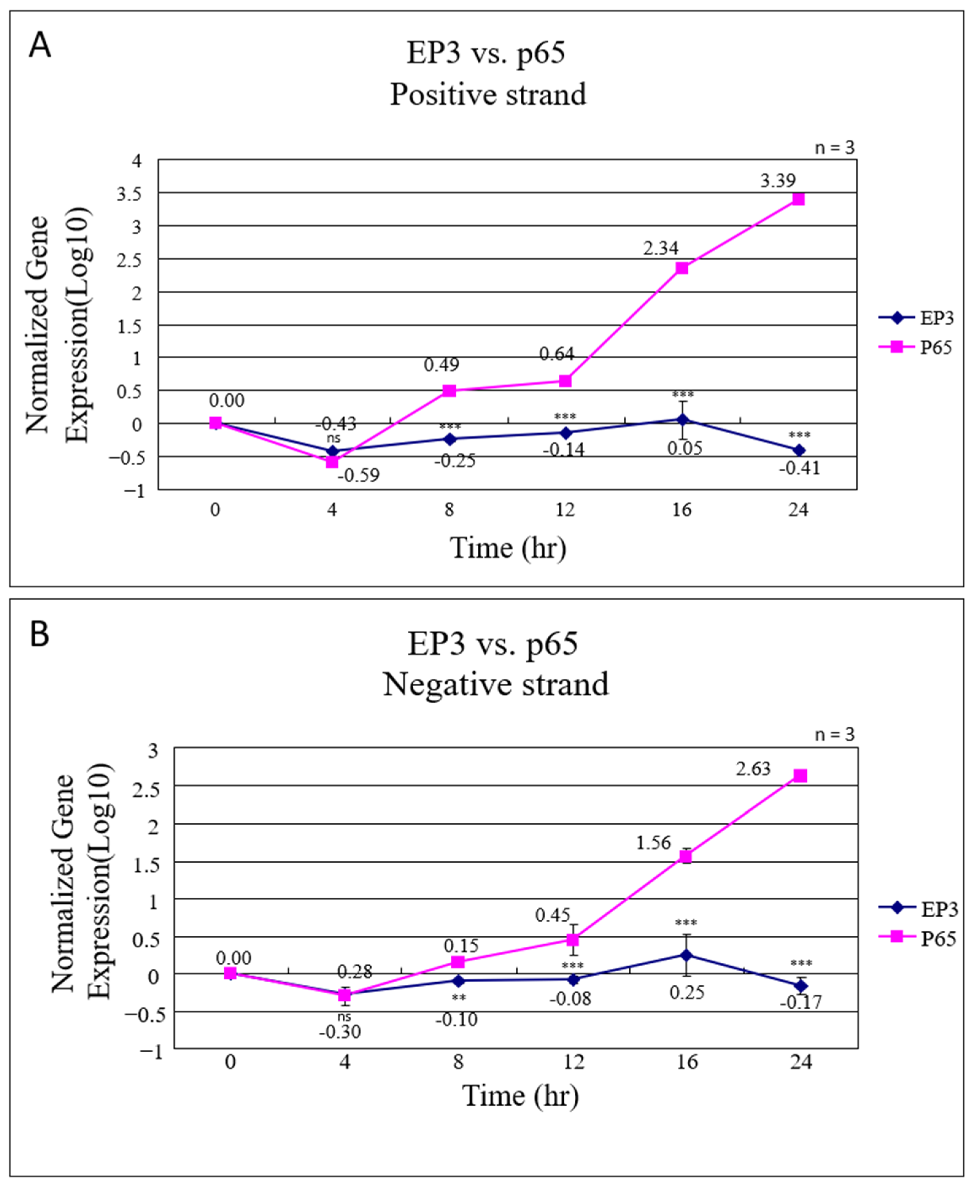
To compare the infectivity of IBV-EP3 and IBV-P65 in Vero cells, real-time PCR was employed to quantify the synthesis of positive-sense and negative-sense viral RNAs in infected Vero cells. The gene expression levels at each time point were normalized relative to the expression level at 0 h. The normalized gene expression levels of both positive and negative RNA strands are presented in Figure 2. For IBV-EP3, there was no significant increase in RNA levels, with only a 1.12-fold and 1.78-fold increase observed for the positive and negative strands, respectively, at 16 h post-infection. In contrast, the levels of both positive and negative RNA strands in IBV-P65-infected cells progressively increased over time, reaching 169.82-fold and 36.31-fold increases at 16 h (Figure 2A,B). These findings confirm that IBV-EP3 exhibits significantly lower infectivity in Vero cells compared to IBV-P65.

3.2. Recovery of Recombinant Viruses

Sequence alignment revealed 19 amino acid variations in the S1 region (1–537 aa) between IBV-P65 and IBV-EP3 [24]. To investigate the functional significance of these mutations in the adaptation of IBV to Vero cells and to identify critical amino acids, six recombinant viruses were generated by substituting the corresponding sequence regions in IBV-P65 with those from IBV-EP3, following the previously established protocol [26]. The characteristics of the resulting recombinant viruses and the associated amino acid changes are summarized in Table 3 (amino acid alignment of the S1 region for both IBV-P65 and IBV-EP3 is shown in Figure S1). Following plaque purification and five consecutive passages through Vero cells, RT-PCR products from the S1 regions were sequenced. Sequence analysis confirmed that no additional mutations occurred beyond the intended amino acid substitutions, indicating the genetic stability of these recombinants in Vero cells.

3.3. Growth Characteristics of Recombinant Viruses

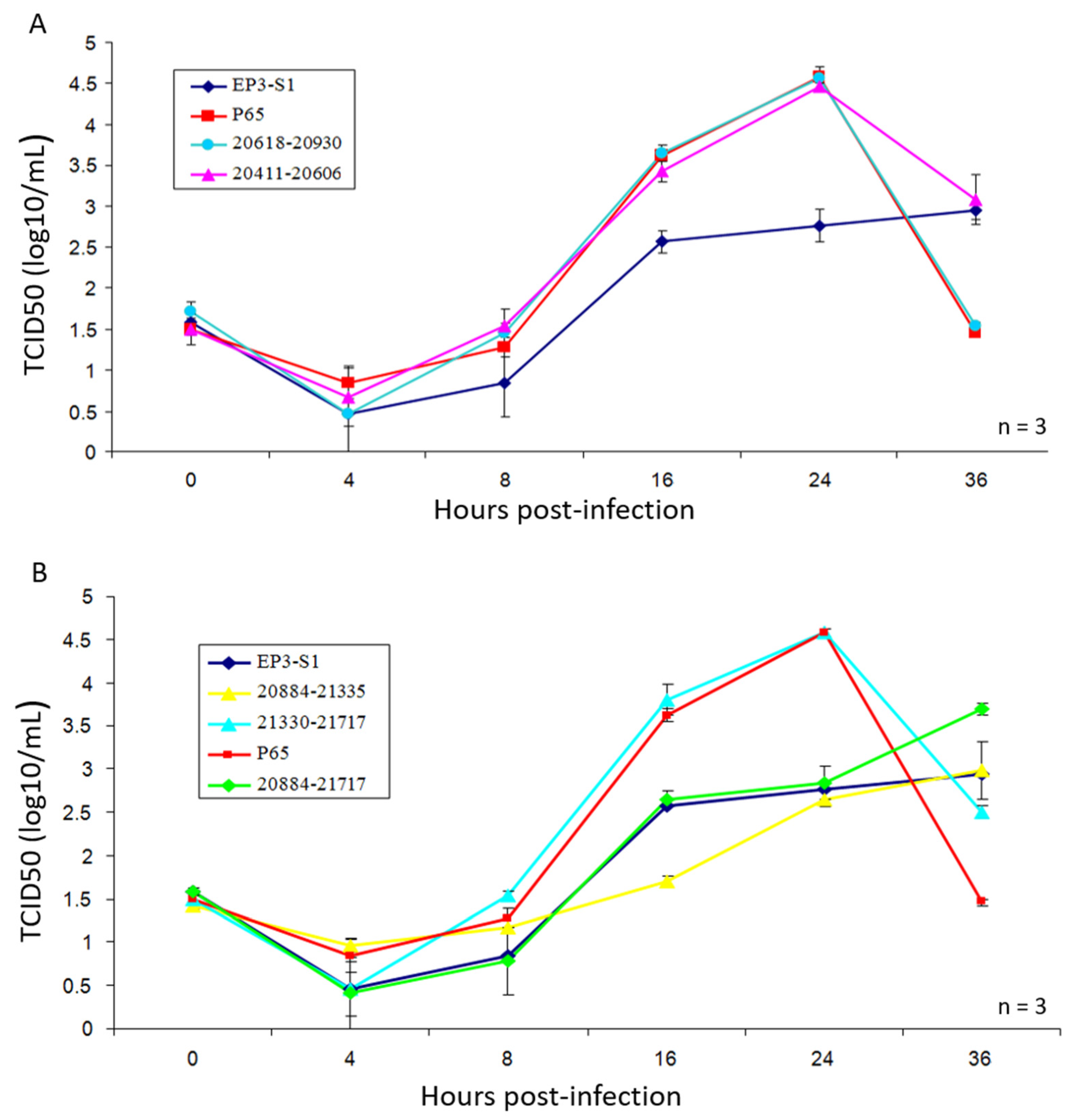
Titers of IBV-P65 and six recombinant viruses were determined using the TCID50 method. The growth characteristics of each mutant virus were evaluated by TCID50 analysis (Figure 3A,B). As illustrated in these figures, IBV-P65, IBV-21330-21717 (321–451 aa), IBV-20618-20930 (86–180 aa), and IBV-20411-20606 (17–82 aa) exhibited highly similar growth profiles, achieving comparable viral titers at each time point and reaching peak TCID50 values at 24 h post-infection. These results suggest that the 14 amino acid substitutions (S38N, Q43H, F56S, S63P, T66I, I69T, H117Y, G118D, Q128K, R156K, K330N, K364S, H391L, D405G) had no significant impact on IBV replication in Vero cells. In contrast, IBV-EP3-S1 (17–451 aa), IBV-20884-21335 (179–323 aa), and IBV-20884-21717 (179–451 aa) displayed markedly delayed and reduced growth phenotypes compared to IBV-P65, with titers approximately 100-fold lower than those of IBV-P65 at 24 h post-infection (Figure 3B). Collectively, these findings indicate that the replacement of the amino acid sequence from positions 179 to 323 in the S1 region of IBV-P65 with the corresponding segment from IBV-EP3 S1 significantly impaired viral growth in Vero cells.

3.4. Effects of Amino Acid Changes on Expression of IBV-S

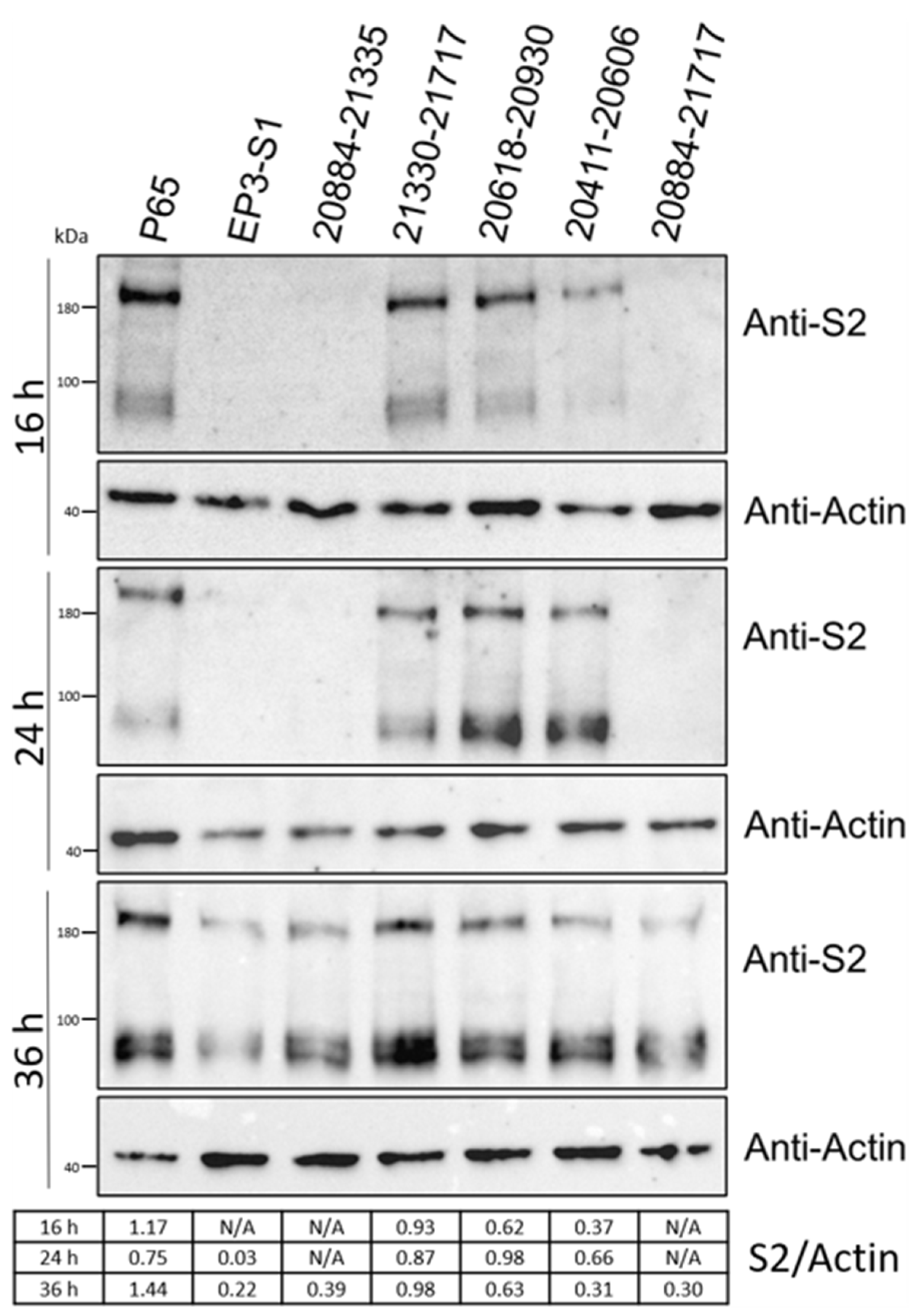
We used Western blot analysis to evaluate the expression of the S proteins of the recombinant viruses. As illustrated in Figure 3, at 16 h post-infection (hpi), the S proteins were clearly detected in Vero cells infected with IBV-P65, IBV-21330-21717, 20,411–20,606, and 20,618–20,930. In contrast, for Vero cells infected with IBV-EP3-S1, 20,884–21,335, and 20,884–21,717, the S proteins were only detectable at 36 hpi, with significantly lower expression levels (Figure 4). These results indicate that the replacement of amino acids 179–323, 179–451, and 17–451 in IBV-P65 with the corresponding amino acids from IBV-EP3 decreased the replication efficiency of IBV in Vero cells.

3.5. Effects of Amino Acid Changes on CPE Formation

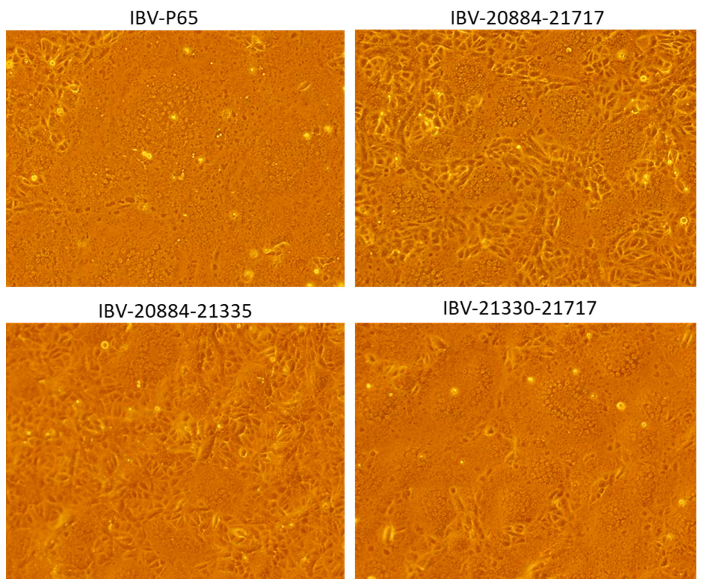
To visually assess cytopathic effects (CPE) in Vero cells, images were captured at 24 hpi for cells infected with IBV-P65, 20,884–21,335, 20,884–21,717, and 21,330–21,717. The CPEs induced by IBV-P65 and 21,330–21,717 nearly extended across the entire monolayer, whereas those caused by IBV-20884-21717 and 20,884–21,335 were more localized (Figure 5), indicating reduced infectivity.

3.6. Amino Acid Sequence Alignment of Different IBV Strains

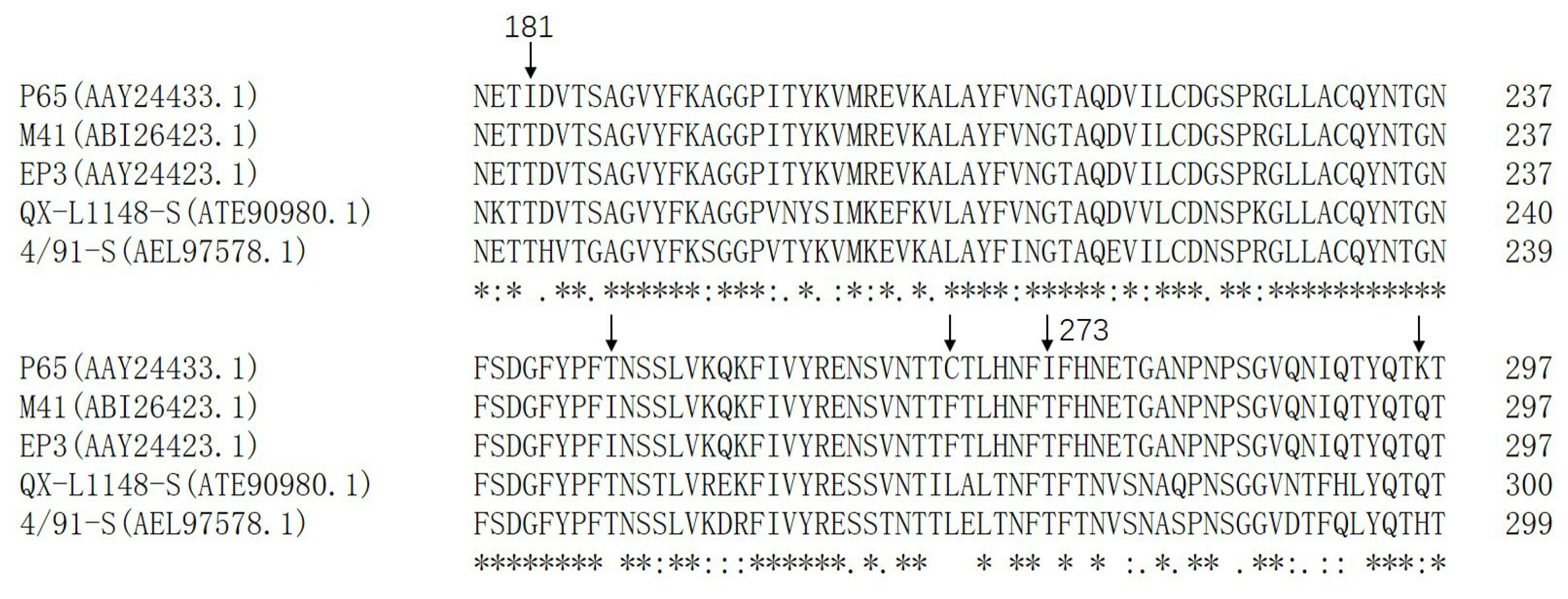
As shown in Figure 6, the amino acid sequence comparison results demonstrate that threonine (T) was conserved in IBV-M41, EP3, QX-L1148, and 4/91 at the corresponding sites I181 and I273 in S1 of IBV-P65, respectively. Furthermore, T246 in IBV-P65 was also observed in QX-L1148 and 4/91, whereas isoleucine (I) was present at this position in IBV-M41 and EP3. Additionally, cysteine (C) at position 267 in IBV-P65 corresponded to phenylalanine (F) in IBV-M41 and EP3, and leucine (L) in QX-L1148 and 4/91. Moreover, lysine (K) at position 296 in IBV-P65 corresponded to glutamine (Q) in IBV-M41, EP3, and QX-L1148, and histidine (H) in 4/91. Given that IBV-M41 lacks the ability to infect Vero cells and IBV-EP3 exhibits limited replication efficiency in Vero cells, it is likely that the five amino acid changes (T181I, I246T, F267C, T273I, Q296K) play crucial roles in the adaptation of IBV to Vero cells.
In summary, the aforementioned findings indicate that the mutant virus obtained by replacing the 20,884–21,335 nucleotide sequence of the IBV-P65 S1 gene with the corresponding sequence from the IBV-EP3 S1 gene exhibits significantly reduced and delayed growth kinetics, as well as a lower replication efficiency in Vero cells compared to IBV-P65. Considering that only five amino acid substitutions (I181T, T246I, C267F, I273T, K296Q) differentiate this region between IBV-P65 and IBV-EP3, it is plausible to infer that these specific mutations in the S1 domain may enhance the adaptation of IBV to Vero cells.

4. Discussion

Field IBV strains generally lack replication capabilities in Vero cells, although some strains are capable of replicating in primary chicken cells, such as chick kidney (CK) cells and chick embryo fibroblasts (DF1). In contrast, the Beaudette strain demonstrates a broader host range, enabling it to replicate efficiently in Vero cells as well as various human and animal cell lines [23,24]. Previous studies have shown that continuous passaging of IBV-EP3 through Vero cells gradually enhances its adaptability to these cells. This process culminates in the generation of IBV-P65 by the 65th passage, a relatively stable strain adapted to Vero and other human and animal cell lines [23,24]. However, the underlying mechanism of this adaptation remains unclear. Sequence comparison revealed that the S1 subunit of the IBV-P65 spike protein contains 19 amino acid mutations relative to IBV-EP3. These mutations are hypothesized to contribute to the improved adaptation. To investigate the effects of these mutations on viral adaptation, in this study, six recombinant mutant viruses were constructed, each harboring partial or complete amino acid substitution in the S1 subunit of IBV-EP3, based on the genetic background of IBV-P65 (Table 3). Our results indicate that five specific amino acid changes (T181I, I246T, F267C, T273I, Q296K) from IBV-EP3 to IBV-P65 S1 enhanced the adaptation of IBV to Vero cells (Figure 3, Figure 4 and Figure 5), while the remaining 14 amino acid changes had no significant effect on IBV’s adaptation to Vero cells. Structurally, 10 conserved amino acids (N38, H43, S56, P63, I66, T69, Y117, D118, K128, and K156) in the S1 units of IBV-EP3 and IBV-M41 are clustered in the S1-NTD region [10]. Although S1-NTD has been reported as the receptor-binding domain (RBD) of the spike protein in IBV M41 and QX-like strains, it contributes to sialic acid binding and plays a critical role in IBV infection, tissue tropism, and pathogenicity [26,27]. Moreover, N38, H43, P63, and T69 in IBV-M41 appear to be critical for binding the spike to the chicken respiratory tract [26]. However, our results indicate that these amino acid changes did not significantly impact the adaptation of IBV-P65 to Vero cells in this study (Figure 3 and Figure 4). Similar results have been reported for recombinant IBV-P65 expressing the S1 region of IBV-H120, as well as the hypervariable regions of QX-like IBV strains [28,29]. These viruses are capable of replicating in Vero cells, suggesting that the acquired ability of IBV to infect Vero cells may not be associated with these specific amino acid changes.
Amino acid sequence alignment demonstrated that the conserved residues T181, I246, F267, T273, and Q296 in IBV-M41 and IBV-EP3 are mutated to I, T, C, I, and K, respectively (Figure 6). IBV-20884-21335, which harbors these specific amino acids, exhibited markedly reduced and delayed growth kinetics as well as diminished replication efficiency in Vero cells compared to IBV-P65 (Figure 3 and Figure 4), suggesting that the specified amino acid substitutions are likely to play pivotal roles in the adaptation of IBV to Vero cells. However, whether a single point mutation or a combination of multiple mutations underlies the adaptability of IBV-P65 to Vero cells remains unclear. Further mapping and detailed analysis of key amino acids will be necessary to elucidate this mechanism. Among the five amino acid residues, I181 in IBV-P65 is located within the NTD, whereas I273 and K296 are situated within the CTD [10]. It remains to be determined whether these amino acid substitutions alter the receptor-binding affinity of the S protein by either directly enhancing receptor interaction or modifying its three-dimensional structure. Further investigation is required to elucidate this mechanism. Determining the three-dimensional structure of this chimeric S protein would provide valuable insights into this issue. Additionally, the IBV S1-CTD functions as an RBD that binds to an unidentified receptor on the surface of chicken cells [10]. Further study is required to determine whether the substitutions T273I and Q296K, located within the CTD, enhance receptor-binding affinity.
N-glycosylation of the IBV-S protein plays a critical role in determining receptor specificity, cellular and tissue tropism, and pathogenicity [30,31,32]. Notably, when employing NetNGlyc-1.0 [33] to predict N-glycosylation sites within the S1 region, it was found that the substitution of threonine at position 273 (T273) in M41 and EP3 to isoleucine at position 273 (I273) in IBV-P65 resulted in the loss of glycosylation at site N271. Further investigation is required to determine whether this loss contributes to enhancing the adaptation of IBV to Vero cells.
Due to their ability to be cultivated in both suspension and adherent cultures, Vero cells facilitate consistent and scalable production of viral yields. As such, they have undergone extensive validation for their efficiency in viral propagation and are officially approved for use in human vaccine production, including the manufacture of polio [34], rabies [35], influenza virus [36], and SARS-CoV-2 vaccines [37]. IBV vaccines, including both live-attenuated and inactivated forms, are currently produced using embryonated hens’ eggs. This process is labor-intensive and costly, primarily because field IBV strains fail to replicate efficiently in cultured cells. Recombinant infectious bronchitis viruses (IBVs) expressing heterologous spike (S) proteins, including either the full S protein or the S1 region derived from the H120, M41, or QX strains, are capable of replicating in Vero cells and confer varying degrees of protection against homologous challenge [7,29,38,39]. These results demonstrate that recombinant rIBVs can effectively express S1 subunits derived from genetically diverse strains of IBV. This functionality will support the rational development of a new generation of IBV vaccines, which have the potential to be propagated in Vero cells. Given that field IBV strains cannot replicate in Vero cells, our findings are expected to facilitate the production of IBV vaccines in Vero cells by enhancing replication efficiency through genetic modification.

5. Conclusions

Five amino acid substitutions in the S1 region of the IBV-65 spike protein were identified as critical determinants that enhance the adaptation of infectious bronchitis virus (IBV) to Vero cells. However, the contributions of these substitutions require further investigation.

Supplementary Materials

The following supporting information can be downloaded at: https://www.mdpi.com/article/10.3390/vetsci12050394/s1, Figure S1: The amino acid alignment of the S1 region for both IBV-P65 and IBV-EP3. The amino acid changes are highlighted in boldface.

Author Contributions

Z.C. assisted in the study’s design, performed experiments, analyzed and interpreted the data, and drafted the manuscript; M.Z. performed partial experiments; S.F. designed experiments, reviewed and edited the manuscript, and carried out project administration and funding acquisition. All authors have read and agreed to the published version of the manuscript.

Funding

This work was financially supported by grants from the National Natural Science Foundation of China (no. 31572490).

Institutional Review Board Statement

Not applicable.

Informed Consent Statement

Not applicable.

Data Availability Statement

The datasets generated and/or analyzed during the current study are available from the corresponding author on reasonable request.

Conflicts of Interest

The authors declare no conflicts of interest. The author Zhichao Cai is an employee of MDPI; however, they do not work for the Veterinary Sciences journal at the time of submission and publication.

References

  1. Graham, R.L.; Donaldson, E.F.; Baric, R.S. A decade after SARS: Strategies for controlling emerging coronaviruses. Nat. Rev. Microbiol. 2013, 11, 836–848. [Google Scholar] [CrossRef] [PubMed]
  2. de Wilde, A.H.; Snijder, E.J.; Kikkert, M.; van Hemert, M.J. Host Factors in Coronavirus Replication. Curr. Top. Microbiol. Immunol. 2018, 419, 1–42. [Google Scholar] [CrossRef] [PubMed]
  3. Li, F. Structure, Function, and Evolution of Coronavirus Spike Proteins. Annu. Rev. Virol. 2016, 3, 237–261. [Google Scholar] [CrossRef] [PubMed]
  4. Cook, J.K.; Jackwood, M.; Jones, R.C. The long view: 40 years of infectious bronchitis research. Avian Pathol. 2012, 41, 239–250. [Google Scholar] [CrossRef]
  5. Jackwood, M.W. Review of infectious bronchitis virus around the world. Avian Dis. 2012, 56, 634–641. [Google Scholar] [CrossRef]
  6. Sjaak de Wit, J.J.; Cook, J.K.; van der Heijden, H.M. Infectious bronchitis virus variants: A review of the history, current situation and control measures. Avian Pathol. 2011, 40, 223–235. [Google Scholar] [CrossRef]
  7. Casais, R.; Dove, B.; Cavanagh, D.; Britton, P. Recombinant avian infectious bronchitis virus expressing a heterologous spike gene demonstrates that the spike protein is a determinant of cell tropism. J. Virol. 2003, 77, 9084–9089. [Google Scholar] [CrossRef]
  8. Luo, Z.; Weiss, S.R. Roles in cell-to-cell fusion of two conserved hydrophobic regions in the murine coronavirus spike protein. Virology 1998, 244, 483–494. [Google Scholar] [CrossRef]
  9. Belouzard, S.; Millet, J.K.; Licitra, B.N.; Whittaker, G.R. Mechanisms of coronavirus cell entry mediated by the viral spike protein. Viruses 2012, 4, 1011–1033. [Google Scholar] [CrossRef]
  10. Shang, J.; Zheng, Y.; Yang, Y.; Liu, C.; Geng, Q.; Luo, C.; Zhang, W.; Li, F. Cryo-EM structure of infectious bronchitis coronavirus spike protein reveals structural and functional evolution of coronavirus spike proteins. PLoS Pathog. 2018, 14, e1007009. [Google Scholar] [CrossRef]
  11. Madu, I.G.; Chu, V.C.; Lee, H.; Regan, A.D.; Bauman, B.E.; Whittaker, G.R. Heparan sulfate is a selective attachment factor for the avian coronavirus infectious bronchitis virus Beaudette. Avian Dis. 2007, 51, 45–51. [Google Scholar] [CrossRef] [PubMed]
  12. Promkuntod, N.; Wickramasinghe, I.N.; de Vrieze, G.; Gröne, A.; Verheije, M.H. Contributions of the S2 spike ectodomain to attachment and host range of infectious bronchitis virus. Virus Res. 2013, 177, 127–137. [Google Scholar] [CrossRef] [PubMed]
  13. Wickramasinghe, I.N.; van Beurden, S.J.; Weerts, E.A.; Verheije, M.H. The avian coronavirus spike protein. Virus Res. 2014, 194, 37–48. [Google Scholar] [CrossRef] [PubMed]
  14. Cavanagh, D.; Casais, R.; Armesto, M.; Hodgson, T.; Izadkhasti, S.; Davies, M.; Lin, F.; Tarpey, I.; Britton, P. Manipulation of the infectious bronchitis coronavirus genome for vaccine development and analysis of the accessory proteins. Vaccine 2007, 25, 5558–5562. [Google Scholar] [CrossRef]
  15. Valastro, V.; Holmes, E.C.; Britton, P.; Fusaro, A.; Jackwood, M.W.; Cattoli, G.; Monne, I. S1 gene-based phylogeny of infectious bronchitis virus: An attempt to harmonize virus classification. Infect. Genet. Evol. 2016, 39, 349–364. [Google Scholar] [CrossRef]
  16. Dwars, R.M.; Matthijs, M.G.; Daemen, A.J.; van Eck, J.H.; Vervelde, L.; Landman, W.J. Progression of lesions in the respiratory tract of broilers after single infection with Escherichia coli compared to superinfection with E. coli after infection with infectious bronchitis virus. Vet. Immunol. Immunopathol. 2009, 127, 65–76. [Google Scholar] [CrossRef]
  17. Matthijs, M.G.; van Eck, J.H.; Landman, W.J.; Stegeman, J.A. Ability of Massachusetts-type infectious bronchitis virus to increase colibacillosis susceptibility in commercial broilers: A comparison between vaccine and virulent field virus. Avian Pathol. 2003, 32, 473–481. [Google Scholar] [CrossRef]
  18. Ignjatović, J.; Sapats, S. Avian infectious bronchitis virus. Rev. Sci. Tech. 2000, 19, 493–508. [Google Scholar] [CrossRef]
  19. Raj, G.D.; Jones, R.C. Infectious bronchitis virus: Immunopathogenesis of infection in the chicken. Avian Pathol. 1997, 26, 677–706. [Google Scholar] [CrossRef]
  20. Hodgson, T.; Casais, R.; Dove, B.; Britton, P.; Cavanagh, D. Recombinant infectious bronchitis coronavirus Beaudette with the spike protein gene of the pathogenic M41 strain remains attenuated but induces protective immunity. J. Virol. 2004, 78, 13804–13811. [Google Scholar] [CrossRef]
  21. Bouwman, K.M.; Parsons, L.M.; Berends, A.J.; de Vries, R.P.; Cipollo, J.F.; Verheije, M.H. Three Amino Acid Changes in Avian Coronavirus Spike Protein Allow Binding to Kidney Tissue. J. Virol. 2020, 94, e01363-19. [Google Scholar] [CrossRef] [PubMed]
  22. Otsuki, K.; Noro, K.; Yamamoto, H.; Tsubokura, M. Studies on avian infectious bronchitis virus (IBV). II. Propagation of IBV in several cultured cells. Arch. Virol. 1979, 60, 115–122. [Google Scholar] [CrossRef] [PubMed]
  23. Tay, F.P.; Huang, M.; Wang, L.; Yamada, Y.; Liu, D.X. Characterization of cellular furin content as a potential factor determining the susceptibility of cultured human and animal cells to coronavirus infectious bronchitis virus infection. Virology 2012, 433, 421–430. [Google Scholar] [CrossRef]
  24. Fang, S.G.; Shen, S.; Tay, F.P.; Liu, D.X. Selection of and recombination between minor variants lead to the adaptation of an avian coronavirus to primate cells. Biochem. Biophys. Res. Commun. 2005, 336, 417–423. [Google Scholar] [CrossRef]
  25. Fang, S.; Chen, B.; Tay, F.P.; Ng, B.S.; Liu, D.X. An arginine-to-proline mutation in a domain with undefined functions within the helicase protein (Nsp13) is lethal to the coronavirus infectious bronchitis virus in cultured cells. Virology 2007, 358, 136–147. [Google Scholar] [CrossRef] [PubMed]
  26. Promkuntod, N.; van Eijndhoven, R.E.; de Vrieze, G.; Gröne, A.; Verheije, M.H. Mapping of the receptor-binding domain and amino acids critical for attachment in the spike protein of avian coronavirus infectious bronchitis virus. Virology 2014, 448, 26–32. [Google Scholar] [CrossRef]
  27. You, R.; Liu, K.; Huang, M.; Tang, L.; Zhang, X.; Huang, Y.; Zhao, J.; Zhao, Y.; Ye, L.; Zhang, G. Identification and Comparison of the Sialic Acid-Binding Domain Characteristics of Avian Coronavirus Infectious Bronchitis Virus Spike Protein. J. Virol. 2023, 97, e0048923. [Google Scholar] [CrossRef]
  28. Wei, Y.Q.; Guo, H.C.; Dong, H.; Wang, H.M.; Xu, J.; Sun, D.H.; Fang, S.G.; Cai, X.P.; Liu, D.X.; Sun, S.Q. Development and characterization of a recombinant infectious bronchitis virus expressing the ectodomain region of S1 gene of H120 strain. Appl. Microbiol. Biotechnol. 2014, 98, 1727–1735. [Google Scholar] [CrossRef]
  29. Shan, D.; Fang, S.; Han, Z.; Ai, H.; Zhao, W.; Chen, Y.; Jiang, L.; Liu, S. Effects of hypervariable regions in spike protein on pathogenicity, tropism, and serotypes of infectious bronchitis virus. Virus Res. 2018, 250, 104–113. [Google Scholar] [CrossRef]
  30. Bouwman, K.M.; Habraeken, N.; Laconi, A.; Berends, A.J.; Groenewoud, L.; Alders, M.; Kemp, V.; Verheije, M.H. N-glycosylation of infectious bronchitis virus M41 spike determines receptor specificity. J. Gen. Virol. 2020, 101, 599–608. [Google Scholar] [CrossRef]
  31. Zheng, J.; Yamada, Y.; Fung, T.S.; Huang, M.; Chia, R.; Liu, D.X. Identification of N-linked glycosylation sites in the spike protein and their functional impact on the replication and infectivity of coronavirus infectious bronchitis virus in cell culture. Virology 2018, 513, 65–74. [Google Scholar] [CrossRef] [PubMed]
  32. Parsons, L.M.; Bouwman, K.M.; Azurmendi, H.; de Vries, R.P.; Cipollo, J.F.; Verheije, M.H. Glycosylation of the viral attachment protein of avian coronavirus is essential for host cell and receptor binding. J. Biol. Chem. 2019, 294, 7797–7809. [Google Scholar] [CrossRef] [PubMed]
  33. Gupta, R.; Brunak, S. Prediction of glycosylation across the human proteome and the correlation to protein function. In Biocomputing; 2002; pp. 310–322. Available online: https://www.worldscientific.com (accessed on 20 April 2025). [PubMed]
  34. Bandyopadhyay, A.S.; Garon, J.; Seib, K.; Orenstein, W.A. Polio vaccination: Past, present and future. Future Microbiol. 2015, 10, 791–808. [Google Scholar] [CrossRef]
  35. Fooks, A.R.; Banyard, A.C.; Ertl, H.C.J. New human rabies vaccines in the pipeline. Vaccine 2019, 37 (Suppl. S1), A140–A145. [Google Scholar] [CrossRef] [PubMed]
  36. Ehrlich, H.J.; Berezuk, G.; Fritsch, S.; Aichinger, G.; Singer, J.; Portsmouth, D.; Hart, M.K.; El-Amin, W.; Kistner, O.; Barrett, P.N. Clinical development of a Vero cell culture-derived seasonal influenza vaccine. Vaccine 2012, 30, 4377–4386. [Google Scholar] [CrossRef]
  37. Xia, S.; Duan, K.; Zhang, Y.; Zhao, D.; Zhang, H.; Xie, Z.; Li, X.; Peng, C.; Zhang, Y.; Zhang, W.; et al. Effect of an Inactivated Vaccine Against SARS-CoV-2 on Safety and Immunogenicity Outcomes: Interim Analysis of 2 Randomized Clinical Trials. JAMA 2020, 324, 951–960. [Google Scholar] [CrossRef]
  38. Bickerton, E.; Dowgier, G.; Britton, P. Recombinant infectious bronchitis viruses expressing heterologous S1 subunits: Potential for a new generation of vaccines that replicate in Vero cells. J. Gen. Virol. 2018, 99, 1681–1685. [Google Scholar] [CrossRef] [PubMed]
  39. Ting, X.; Xiang, C.; Liu, D.X.; Chen, R. Establishment and Cross-Protection Efficacy of a Recombinant Avian Gammacoronavirus Infectious Bronchitis Virus Harboring a Chimeric S1 Subunit. Front. Microbiol. 2022, 13, 897560. [Google Scholar] [CrossRef] [PubMed] [PubMed Central]
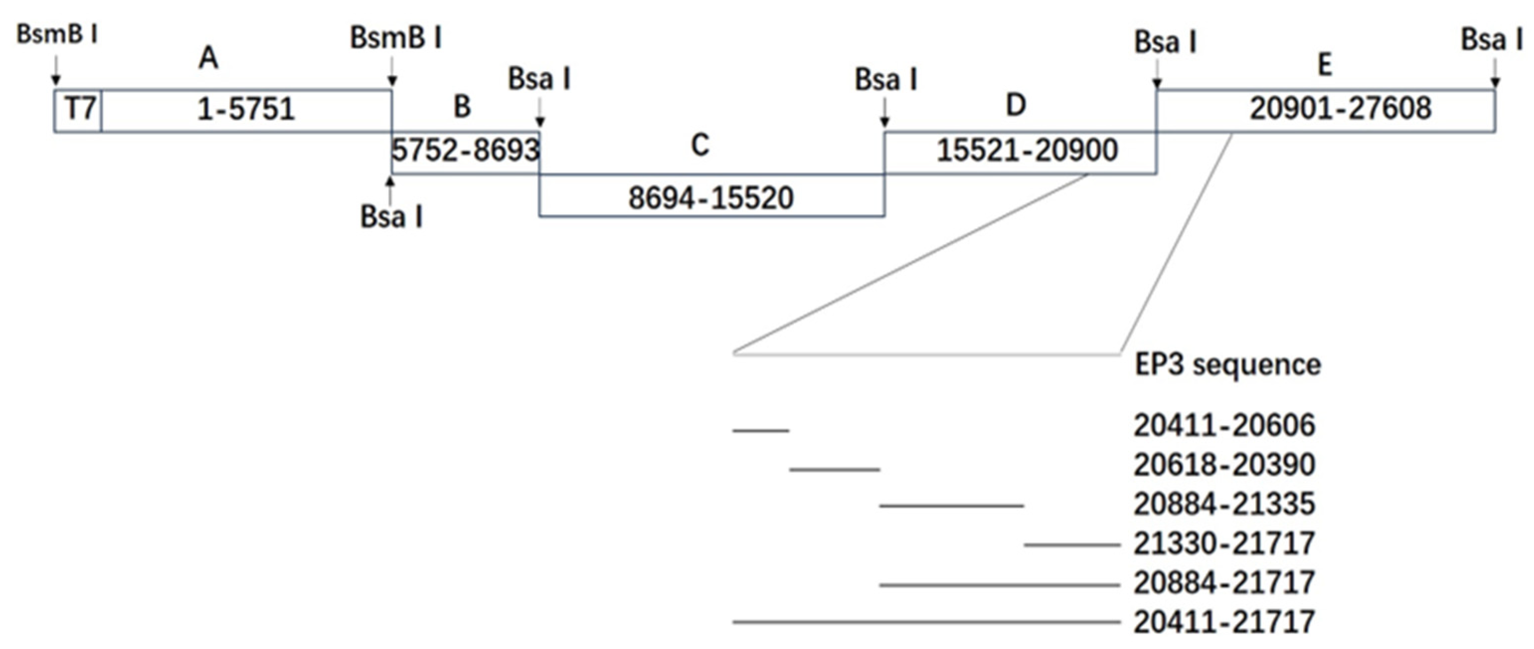
Figure 1. The construction of the recombinant viruses. The figure shows the five RT-PCR fragments of IBV-P65, including the T7 promoter at the 5′-end of fragment A, 30 As at the 3′-end of fragment E, and the replacement regions of EP3-S1 (nucleotides 20,411–21,717).
Vetsci 12 00394 g001
Figure 2. Real-time PCR analysis of positive (A) and negative (B) viral RNAs from IBV-EP3- and P65-infected Vero cells. Virus-infected cells were harvested at the indicated time points post-infection, and total RNA was extracted for the RT-qPCR analysis of positive and negative RNA synthesis. Normalized target gene expression levels were recorded for each time point and virus. The data represent the mean ± SD from three independent experiments. ns, p > 0.05, non-significant; **, p < 0.01; ***, p < 0.001.
Vetsci 12 00394 g002
Figure 3. The growth characteristics of IBV-P65 and the recovered recombinant viruses IBV-EP3-S1, 20,618–20,930, and 20,411–20,606 (A), as well as 20,884–21,335, 21,330–21,717, and 20,884–21,717 (B). The error bars represent the standard error of the mean.
Vetsci 12 00394 g003
Figure 4. A Western blot analysis of the expression levels of the S protein in Vero cells infected with IBV-P65 and six recombinant viruses. N/A, not applicable.
Vetsci 12 00394 g004
Figure 5. The CPEs induced by IBV-P65 and the recombinant IBV strains (20,884–21,717, 20,884–21,335, and 20,330–21,717).
Vetsci 12 00394 g005
Figure 6. Multiple sequence alignment of the amino acid sequences (179–323 aa) within the S1 region of IBV-P65, IBV-EP3, and IBV-M41. The arrow denotes the position of the mutated amino acid residue. “*”, fully conserved residue; “:”, conserved mutation; “.”, semi-conserved mutation.
Vetsci 12 00394 g006
Table 1. The primers utilized for the construction of mutant plasmids. The boldface indicates the restriction site of the introduced BsaI enzyme.
PrimerNucleotide Sequence
BsaI-20411-F5′-CGGGTCTCTATGTAGTGCTGTTTTGTATGA
BsaI-20416-R5′-CGGGTCTCTACATAGTGCACACAAAAGAGT
BsaI-21330-F5′-CGGTCTCGTATGGATCTTATCACCCAA
BsaI-21335-R5′-CGGTCTCCCATACATAAAATTAGACTC
BsaI-20618-F5′-CGGGTCTCCGGCACCGTCATCAGGTAT
BsaI-20623-R5′-CGGGTCTCGTGCCGTCATAGCTATAGA
BsaI-21712-F 5′-CGGGTCTCACCGACTCAGCTGTTAGTTAT
BsaI-21717-R5′-CGGGTCTCGTCGGTTACATTAGTAATAAA
IBV-20884-F5′-GTTTACACCTCTAATGAGACCATAG
IBV-20930-R5′-ACACCTGCAGATGTAACATC’
IBV-15511-F5′-GTGATTCATTGAGACCTTTTGC
IBV-15532-R5′-GCAAAAGGTCTCAATGAATCAC
IBV-3′ end5′-CggTCTCgTTTTTTTTTTTTTTTTTTTTTTTTTTTTTTgCTC
Table 2. The plasmids used in the construction of the mutants.
PlasmidRegion (nt)SourcePrimerNote
pGEM-IBV-F15,511–20,416IBV-P65IBV-15511-F BsaI-20416-RConstruction of mutant 20,411–20,606
pGEM-IBV-G20,618–20,930IBV-P65BsaI-20618-F IBV-20930-R
pGEM-EP3-120,411–20,623IBV-EP3BsaI-20411-F BsaI-20623-R
pGEM-IBV-H15,511–20,623IBV-P65IBV-15511-F BsaI-20623-RConstruction of mutant 20,618–20,930
pGEM-EP3-220,618–20,930IBV-EP3BsaI-20618-F IBV-20930-R
pGEM-IBV-I21,712–27,608IBV-P65BsaI-21712-F IBV-3′ endConstruction of mutant 20,884–21,717
pGEM-EP3-320,884–21,717IBV-EP3IBV-20884-F BsaI-21717-R
pGEM-IBV-J21,330–27,608IBV-P65BsaI-21330-F IBV-3′ endConstruction of mutant 20,884–21,335
GEM-IBV-K20,884–21,335IBV-P65IBV-20884-F BsaI-21335-R
pGEM-EP3-421,330–21,717IBV-EP3BsaI-21330-F BsaI-21717-RConstruction of mutant 21,330–21,717
pGEM-EP3-520,411–21,717IBV-EP3BsaI-20411-F BsaI-21717-RConstruction for mutant EP3-S1
Table 3. Detailed information on the recombinant viruses.
Recombinant VirusesReplaced Nucleotide Sequences (nt)Replaced Amino Acid Sequences (aa)Amino Acid Changes from IBV-P65 to IBV-EP3
20,411–20,60620,411–20,60617–82S38N, Q43H, F56S, S63P, T66I, I69T
20,618–20,93020,618–20,93086–180H117Y, G118D, Q128K, R156K
20,884–21,33520,884–21,335179–323I181T, T246I, C267F, I273T, K296Q
21,330–21,71721,330–21,717321–451K330N, K364S, H391L, D405G
20,884–21,71720,884–21,717179–451I181T, T246I, C267F, I273T, K296Q, K330N, K364S, H391L, D405G
EP3-S120,411–21,71717–451S38N, Q43H, F56S, S63P, T66I, I69T, H117Y, G118D, Q128K, R156K, I181T, T246I, C267F, I273T, K296Q, K330N, K364S, H391L, D405G
Disclaimer/Publisher’s Note: The statements, opinions and data contained in all publications are solely those of the individual author(s) and contributor(s) and not of MDPI and/or the editor(s). MDPI and/or the editor(s) disclaim responsibility for any injury to people or property resulting from any ideas, methods, instructions or products referred to in the content.