Article

Regulatory Role of Vacuolar Calcium Transport Proteins in Growth, Calcium Signaling, and Cellulase Production in Trichoderma reesei

by
Letícia Harumi Oshiquiri
1,
Lucas Matheus Soares Pereira
1,
David Batista Maués
1,
Elizabete Rosa Milani
2,
Alinne Costa Silva
1,
Luiz Felipe de Morais Costa de Jesus
1,
Julio Alves Silva-Neto
3,
Flávio Protásio Veras
3,
Renato Graciano de Paula
4,5 and
Roberto Nascimento Silva
1,5,*
1
Department of Biochemistry and Immunology, Ribeirão Preto Medical School, University of São Paulo, Ribeirão Preto 14049-900, SP, Brazil
2
Department of Cellular and Molecular Biology and Pathogenic Bioagents, Ribeirão Preto Medical School, University of São Paulo, Ribeirão Preto 14049-900, SP, Brazil
3
Department of Pharmacology, Ribeirão Preto Medical School, University of São Paulo, Ribeirão Preto 14049-900, SP, Brazil
4
Department of Physiological Sciences, Health Sciences Centre, Federal University of Espirito Santo, Vitoria 29047-105, ES, Brazil
5
National Institute of Science and Technology in Human Pathogenic Fungi, Brazil
*
Author to whom correspondence should be addressed.
J. Fungi 2024, 10(12), 853; https://doi.org/10.3390/jof10120853
Submission received: 4 November 2024 / Revised: 23 November 2024 / Accepted: 4 December 2024 / Published: 11 December 2024
(This article belongs to the Special Issue Omics Approaches in Trichoderma Research)

Abstract

:
Recent research has revealed the calcium signaling significance in the production of cellulases in Trichoderma reesei. While vacuoles serve as the primary calcium storage within cells, the function of vacuolar calcium transporter proteins in this process remains unclear. In this study, we conducted a functional characterization of four vacuolar calcium transport proteins in T. reesei. This was accomplished by the construction of the four mutant strains ∆trpmc1, ∆tryvc1, ∆tryvc3, and ∆tryvc4. These mutants displayed enhanced growth when subjected to arabinose, xylitol, and xylose. Furthermore, the mutants ∆trpmc1, ∆tryvc1, and ∆tryvc4 showed a reduction in growth under conditions of 100 mM MnCl2, implying their role in manganese resistance. Our enzymatic activity assays revealed a lack of the expected augmentation in cellulolytic activity that is typically seen in the parental strain following the introduction of calcium. This was mirrored in the expression patterns of the cellulase genes. The vacuolar calcium transport genes were also found to play a role in the expression of genes involved with the biosynthesis of secondary metabolites. In summary, our research highlights the crucial role of the vacuolar calcium transporters and, therefore, of the calcium signaling in orchestrating cellulase and hemicellulase expression, sugar utilization, and stress resistance in T. reesei.

1. Introduction

Each year, the global food industry discards over 500 million tons of plant-based organic waste, largely composed of cellulose, hemicellulose, and lignin, which form lignocellulosic biomass [1,2]. This has led to a growing interest in using these molecules to produce second-generation biofuels, which utilize non-edible substrates, unlike first-generation biofuel substrates [3,4]. However, the industrial-scale production of biofuels faces significant challenges. A key obstacle is the requirement for enzymes to catalyze the conversion of biomass to biofuel, contributing to about 48% of the minimum sales value in bioethanol production [5]. As such, future research should focus on devising cost-effective production methods to make the process economically feasible and contribute to the wider goal of sustainable energy production.
The filamentous fungus, Trichoderma reesei, is recognized for its capacity to produce enzymes that degrade lignocellulosic biomass, primarily cellulases and hemicellulases. The production of these enzymes is influenced by several factors. For instance, the substrate can function as an inducer (e.g., cellulose or lactose) or a repressor (e.g., glucose) [6]. Other influencing factors include medium conditions, such as pH, cultivation duration, and temperature. Additionally, the presence of metals, like zinc, manganese, strontium, and calcium ions [7,8,9,10,11], also plays a role, with the last being the central point of this study. Previous research has demonstrated that the supplementation of the culture medium with CaCl2 triggers the production of cellulases via a calcium-dependent signaling pathway [10]. This observation emphasizes the importance of understanding the role of calcium ions in enzyme production for biofuel applications.
In fungi, it has been established that external stress stimuli trigger the opening of calcium channels in the plasma membrane or intracellular compartments, facilitating the influx of calcium ions [12]. Three such ions interact with calmodulin (CAM), activating it and enabling it to bind to the A subunit of calcineurin (CNA1). Simultaneously, other calcium ions bind to the B subunit of calcineurin (CNB1), thereby activating it. Calcineurin, a serine-threonine phosphatase, is composed of the catalytic subunit CNA1 and the regulatory subunit CNB1. CNA1 is known to interact with a variety of proteins, including transcription factors and other enzymes. Among these, the transcription factor CRZ1 (calcineurin-responsive zinc-finger transcription factor 1) is a notable example. Upon interaction, CNA1 dephosphorylates CRZ1, which consequently is translocated to the cell nucleus via Nmd5, which permits it to bind to CDREs (calcineurin-dependent response elements) in target promoters. This binding event induces the production of enzymes, such as cellulases and calcium transport proteins, including PMC1 and PMR1. This process ultimately leads to a reduction in the cytoplasmic calcium concentration until it reaches its basal level [13,14]. In fungi, vacuoles serve as the primary storage sites for calcium. As such, the transport system plays a pivotal role in maintaining calcium homeostasis and ensuring proper signal transduction. Fungi possess three types of vacuolar calcium transport proteins. In the case of Saccharomyces cerevisiae, calcium is released from the vacuoles via the Ca2+ channels YVC1 (or TRPY1). Following a peak in calcium levels, the Ca2+ ATPase PMC1 sequesters the calcium, while the Ca2+/H+ antiporter VCX1 maintains the basal level of calcium [12,15].
In T. reesei, numerous genes have been identified that encode proteins involved in calcium signaling. These include 3 ion channels, 10 ATPases, 10 transporters, 5 phospholipases C (PLC), 1 CAM, 1 CNA1, 1 CNB1, 9 genes that interact with calcium or CAM, and 8 additional genes involved in calcium-dependent signaling [16]. Our research group has observed the modulation of the expression of these calcium-related genes during the cultivation of T. reesei in the presence of various substrates, such as cellulose, sophorose, glucose, sugarcane bagasse, and glycerol. Notably, this modulation includes calcium transport proteins [6,17,18,19,20]. Furthermore, it has been demonstrated that the transcription factor CRZ1 plays a crucial role in the induction of the transcription factor XYR1, which subsequently leads to the production of cellulases, hemicellulases, and calcium and sugar transporters. The presence of calcium is a decisive factor for this process to occur [11].
Other studies have explored the influence of calcium on T. reesei. In 2016, Chen et al. [10] established that the transcription factor CRZ1 competes with the ACE1 repressor for the xyr1 promoter, leading to the induction of cellulases. Subsequently, in 2018, Chen et al. [7] demonstrated that an elevated Mn2+ concentration results in an increase in intracellular Ca2+ levels via the P-type ATPase, TPMR1. This increase triggers the induction of genes encoding cellulases through the calcium-dependent signaling pathway. In 2019, Chen et al. [21] found that the addition of N, N-dimethylformamide (DMF) also stimulates the production of cellulases, an effect linked to the activation of PLC-E. In 2021, the mechanism underlying the production of cellulases from Mn2+ and DMF was explored in greater detail. Chen et al. [22] showed that both substances augment the amount of cyclic adenosine 3′,5′-monophosphate (cAMP) in the cell cytosol, leading to an increase in intracellular calcium concentration, a process dependent on PLC-E. In 2022, Li et al. [9] revealed that the addition of Sr2+ to the culture medium also causes an increase in intracellular calcium concentration, activating CRZ1 and resulting in enhanced cellulase activity. In 2023, Li et al. [8] demonstrated that Zn2+ boosts the activity of cellulases and xylanases, with PLC-E playing a role in this process by promoting the release of calcium from intracellular stores to the cytoplasm and by regulating the transcription factor ZAFA, which is involved in zinc metabolism and may influence cellulase production. Most recently, a study by Liu et al. [23] explored the effect of polyethylene glycol 8000 (PEG 8000) stress on cellulase biosynthesis in T. reesei via calcium signaling, providing further evidence of the crucial role of calcium in cellulase production. From these studies, it is evident that multiple factors can stimulate the production of cellulases via the calcium-dependent signaling pathway.
In this study, we undertook a comprehensive characterization of four vacuolar calcium transport proteins in T. reesei by deleting the genes encoding these proteins, leading to the generation of the mutant strains ∆trpmc1, ∆tryvc1, ∆tryvc3, and ∆tryvc4. These mutants exhibited enhanced growth when arabinose, xylitol, and xylose were used as a sole carbon source. Moreover, the mutants ∆trpmc1, ∆tryvc1, and ∆tryvc4 displayed diminished growth in the presence of 100 mM MnCl2, suggesting a role in manganese resistance. Our enzymatic activity assays indicated the absence of the usual increase in cellulolytic activity observed in the parental strain upon the addition of calcium. The genes also influenced secondary metabolites’ expression, and confocal microscopy suggested calcium retention within the vacuoles in the absence of these transport proteins. These findings contribute to our understanding of calcium’s role in T. reesei’s growth and enzyme production.

2. Materials and Methods

2.1. Strains and Culture Conditions

The T. reesei strain QM6aΔtmus53Δpyr4, characterized by a deficiency in the nonhomologous end joining (NHEJ) repair pathway and auxotrophy for uridine, is herein designated as the parental strain. This strain was obtained from the Technical University of Vienna, Austria [24], and for its cultivation, 5 mM of uridine was added due to the deletion of the pyr4 gene. The strains QM6aΔtmus53Δ58952, QM6aΔtmus53Δ74057, QM6aΔtmus53Δ55731, and QM6aΔtmus53Δ56440 (hereafter referred to as Δtrpmc1, Δtryvc1, Δtryvc3 and Δtryvc4 respectively) were generated during this study. These strains are distinguished by the deletion of calcium transport genes. For experimental procedures, 2 µL of a 107 spore suspension of these strains in 0.08% NaCl and 0.05% Tween 80 were inoculated on MEX medium plates (3% malt extract (w/v) and 2% bacteriological agar (w/v)). The plates were subsequently incubated at 30 °C for 7–12 days.
To conduct the growth experiments, 107 conidia in spore suspension were inoculated in minimal medium [25] agar plates containing 1% carboxymethylcellulose (CMC) or 2% glucose + 100 mM Congo red (CR). These plates were incubated for 3–5 days, and their growth was recorded daily. The microplate assays were conducted in minimal medium with the addition of 25 mM of cellobiose, galactose, glycerol, glucose, lactose, maltose, mannose, or xylose to obtain growth profiles in carbon sources, or with the addition of 0; 2.7; 5; 10; 15; 50; 100 or 150 mM of CaCl2, accompanied by 2% glucose to verify calcium tolerance. In addition, 0.25% phytagel and 0.03 Tween 20 was added to the medium. In these experiments, the microplates were incubated at 30 °C for 96 h. Absorbance readings at 750 nm were performed every 24 h to determine the degree of turbidity, as described in [26], indicating fungal growth. The experiments were carried out in three technical replicates and at least two biological replicates.
To perform the enzymatic assays and the gene expression quantification, we cultured the strains in Mandels Andreotti (MA) medium [27] with 24 h pre-growth in 1% glycerol followed by a transference of the mycelia to fresh MA media containing 1% sugarcane bagasse or 1% cellulose and incubated it for up to 72 h at 30 °C. Alternatively, we cultured the strains directly in 1% cellulose, 25 mM xylose, or 2% glucose for up to 72 h at 30 °C. Right after, the mycelia were filtrated, frozen using liquid nitrogen, and stored at −80 °C until use. The supernatant was collected, centrifuged for 10 min at 12,000× g, and frozen at −20 °C until use.

2.2. Vector Construction for Gene Deletion

The knockout strains were designed using the orotidine-5′-phosphate decarboxylase gene of T. reesei (pyr4, Tr_74020) as a selection marker, which enables the identification of transformants through auxotrophic complementation of the T. reesei parental strain. The full sequences of the genes cited in this study can be searched in the Trichoderma reesei v2.0 genome available in JGI database (https://mycocosm.jgi.doe.gov/Trire2/Trire2.home.html, accessed on 30 August 2024), except for 74057 (tryvc1), which the full coding region annotation can be found in Trichoderma reesei QM6a genome (https://mycocosm.jgi.doe.gov/Trire_Chr/Trire_Chr.home.html, accessed on 30 August 2024).
To facilitate genomic integration via homologous recombination, we constructed the cassettes, including regions with homology to approximately 1000 base pairs (bp) upstream and downstream of the target genes in T. reesei parental strain. For this purpose, two strategies were used. First, to construct the plasmids containing the cassettes of the genes tryvc1 and tryvc2, primers for the amplification of its upstream and downstream regions were designed with the addition of restriction sites, allowing for ligation into the pJET-pyr4 plasmid [28] (Figure S1), which already contains the sequence of the pyr4 gene. The enzyme T4 DNA Ligase (Thermo Fisher Scientific, Waltham, MA, USA) was used to join the fragments, according to the manufacturer’s instructions. For the transformation of the ligation reactions, the heat-shock method was used in thermocompetent cells of Escherichia coli strain DH5α, according to [29]. After bacterial growth, plasmid DNA was extracted, as in [30], and digested with the respective restriction enzymes to confirm the ligation. In this strategy, the upstream region was first ligated, followed by the downstream region.
To construct the deletion cassettes of the genes tryvc3 and trpmc1, primers were designed for the amplification of its upstream and downstream regions (Table S1), as well as for the coding region of the pyr4 gene. These primers included sequences of approximately 20 bp overlap between the fragments to be joined and the pRS426 plasmid (Figure S1), allowing the use of a yeast-mediated homologous recombination technique. In this case, the yeast shuttle vector pRS426 (ampR lacZ URA3) [31] was digested with EcoRI and XhoI (Thermo Scientific, Waltham, MA, USA) and was purified with the QIAquick PCR Purification Kit (Qiagen, San Diego, CA, USA). Yeast transformation was essentially conducted, as previously described [32,33,34]. Here, an overnight culture (200 rpm, 30 °C) of the yeast S. cerevisiae strain SC9721 (MATα his3-Δ200 URA3-52 leu2Δ1 lys2Δ202 trp1Δ63) (Fungal Genetic Stock Center) was prepared. Then, 1 mL of the overnight culture was added to 50 mL of fresh YPD (1% yeast extract, 2% peptone, 1% glucose) (all from Sigma Aldrich, St. Louis, MO, USA) medium and incubated at 30 °C until OD600 = 1. Thereafter, the cells were centrifuged and resuspended in 100 mM lithium acetate for transformation. To accomplish this, we mixed equal amounts of the 5′ and 3′ flanking regions, pyr4, and the digested pRS426, and then used this mixture for yeast transformation using the lithium acetate method [32]. S. cerevisiae SC9721 transformants were selected for their ability to grow on YPD medium supplemented with lysine, histidine, leucine, and tryptophan, without uracil. After DNA extraction [35], the cassettes amplified by PCR were used for transformation in T. reesei parental strain. The deletion cassettes of tryvc3 and trpmc1 were PCR-amplified using 5F and 3R primers (Table S1) [36]. All amplifications were performed from T. reesei parental strain genomic DNA, isolated according to [37]. After amplification, the fragments were purified using the QIAquick PCR Purification Kit (Qiagen). The sequences of the primers are listed in Table S1.

2.3. Transformation of T. reesei

T. reesei QM6aΔtmus53Δpyr4 was used for transformation to achieve highly efficient homologous integration of the deletion cassette. Protoplast transformation was carried out as previously described [24]. For the transformation of the T. reesei parental strain, approximately 10–40 µg of the linearized plasmid or cassette (amplified by PCR) was used for protoplast transformation [24].
Transformants were grown on selective minimal medium [1 g/L MgSO4·7H2O, 10 g/L 1% KH2PO4, 6 g/L (NH4)2SO4, 3 g/L trisodium citrate·2H2O, 10 g/L glucose, 20 mL/L 50× trace elements solution (0.25 g/L FeSO4·7H2O, 0.07 g/L ZnSO4·2H2O, 0.1 g/L CoCl2·6H2O, 0.085 g/L MnSO4·H2O), 2% (w/v) agar lacking uridine] (all from Sigma Aldrich). Additionally, the transformants underwent 3 rounds in Mandels-Andreotti medium [27] + 2% glucose without uridine with the addition of 0.1% (v/v) Triton X-100 for homokaryons selection Finally, to confirm the obtaining of the mutant strains, we used conventional PCR and RT-qPCR, as illustrated in Figure S2. The sequences of the primers are listed in Table S1.

2.4. Enzyme Activity

Filter paper activity (FPase) was determined by an enzymatic reaction employing Whatman filter paper no. 1, 30 µL of culture supernatant, and 30 µL of 100 mM citrate-phosphate buffer (pH 5.0). The reactions were incubated at 50 °C for 20 h. Subsequently, 60 µL of 3,5-dinitrosalicylic acid (DNS) was added to the reaction, which was then heated at 95 °C for 5 min. The FPase activity was assayed in a 96-well microplate, and absorbance was read at 540 nm. Additionally, CMCase (Endoglucanase activity), β-glucosidase, xylanase, and β-xylosidase were assessed following the method described by [38] with minor modifications [17,39,40]. The CMCase activity was determined using 30 μL of 1% CMC (Sigma Aldrich, St. Louis, MO, USA) in sodium acetate buffer (50 mM, pH 4.8) and 30 µL of the enzyme (culture supernatant) at 50 °C for 1 h. Right after, 60 μL of DNS was added, followed by heating at 95 °C for 5 min to allow color development. Then, the absorbance of the samples was read at 540 nm.
To determine the β-glucosidase and β-xylosidase activities, p-nitrophenyl-derived substrates: p-Nitrophenol-β-D-glucopyranoside (pNP-Gluc) (5 mM) and p-Nitrophenyl-β-D-xylopyranoside (pNP-Xyl) (5 mM) were used, respectively. The β-glucosidase and β-xylosidase reactions were carried out in a microplate assay format and the assay mixture contained 10 μL of enzyme solution (culture supernatant), 50 μL of 50 mM sodium acetate buffer, and 40 μL of p-nitrophenyl-derived solution. The mixtures were buffered at pH 5.5 (β-glucosidase) and pH 4.8 (β-xylosidase) and after 15 min at 50 °C the reactions were stopped by adding 100 μL of 1 M sodium carbonate. The amount of p-nitrophenol was determined using a spectrophotometer at 405 nm.
Finally, xylanase activity was determined by mixing 25 µL of 1% xylan beechwood (Sigma Aldrich, St. Louis, MO, USA) in sodium acetate buffer (100 mM, pH 5.0) and 10 µL of enzyme and incubating at 50 °C for 30 min. The reaction was stopped by adding 75 µL of DNS, and the samples were heated for 5 min at 95 °C, followed by measuring the absorbance at 540 nm. In all enzymatic assays, one enzyme unit was defined as the amount of enzyme capable of liberating 1 µmol of reducing sugar or p-nitrophenol per minute per mL of sample solution (U/mL) [41]. All enzyme assays were completed in triplicate for each sample.

2.5. RNA Extraction and Transcript Analysis Using Quantitative PCR (RT-qPCR)

Total RNA was isolated from the mycelia of T. reesei strains using TRIZOL® reagent (Thermo Fisher Scientific, Waltham, MA, USA) according to the manufacturer’s instructions. The quantification of RNAs was carried out by OD 260/280 spectrophotometry and their integrity was verified using the Agilent 2100 Bioanalyzer and electrophoresis in 1% agarose gel. Right after, 1 µg of total RNA was treated with DNase I (Thermo Fisher Scientific, Waltham, MA, USA) to eliminate genomic DNA contamination and used for cDNA synthesis using Maxima™ First Strand cDNA Synthesis kit (Thermo Fisher Scientific, Waltham, MA, USA) according to the manufacturer’s instructions. Then, cDNA was diluted 1:50 and analyzed using the CFX96™ Real-Time PCR Detection System (Bio-Rad Laboratories, Hercules, CA, USA) and SsoFast™EvaGreen® Supermix (Bio-Rad Laboratories, Hercules, CA, USA), in accordance with the manufacturer’s instructions. Each reaction (10 µL) contained 5 µL of SsoFast™ EvaGreen® Supermix (Bio-Rad Laboratories, Hercules, CA, USA), forward and reverse primers (300 nm each; Table S1), cDNA template, and nuclease-free water. PCR cycling conditions were as follows: 10 min at 95 °C, followed by 40 cycles of 10 s at 95 °C and 30 s at 60 °C. Melt analysis used a ramp of 60–95 °C at a rate of 0.5 °C/10 s to evaluate primer dimers and nonspecific amplification. The β-actin gene was used as an endogenous control to normalize the total amount of cDNA present in each reaction [17,42].

2.6. Confocal Microscopy

To detect intracellular calcium, the Fluo4-AM reagent (Thermo Fisher Scientific, Waltham, MA, USA) was used. Firstly, 107 conidia from each strain were incubated in MA medium for 48 h at 200 rpm and 30 °C in the presence or absence of calcium. After incubation, the cells were washed three times with PBS and loaded with 5 µM Fluo4-AM for 30 min at 30 °C. Then, the fluorescence intensity of Fluo4-AM was measured by excitation at a wavelength of 340 nm with an emission of 550 nm.

2.7. Bioinformatics and Statistical Analysis

To identify the calcium transporters and channels in T. reesei vacuoles, a set of homologous proteins previously characterized in other fungi were selected as references (Table S2). Using the amino acid sequences of these proteins, three alignments with MAFFT were performed [43] and three profiles with hmmbuild were created [44]: (a) YVC1, (b) VCX1, and (c) PMC1. Then, these profiles were used to search the T. reesei proteome [45] using hmmsearch [44], and annotations were made using InterProScan [36]. For the cladogram design, the identified sequences and sequences of characterized proteins presented in [12] were employed. Finally, the sequences were aligned using MAFFT [43], cladogram was inferred using FastTree 2 [46] and displayed by iTOL v5 [47].
To analyze the gene expression of the identified proteins in the RNA-Seq assays conducted by our research group [6,17,18,19,20,48], were used a cut-off of 1.0 × 10−45 for the full e-value. We then used the gene expression levels under various conditions to create heatmaps using the python library seaborn [49].
All the results in this study were analyzed and statistically compared using the GraphPad Prism version 8.0 for Windows (www.graphpad.com, accessed on 30 August 2024). Student’s t-test was used for comparisons between the two groups. For comparisons between multiple groups, two-way ANOVA followed by Tukey’s multiple comparisons test was employed. All statistical tests assumed a 95% confidence interval.

3. Results

3.1. Identification of Putative Homologues in T. reesei: Three for PMC1, Five for YVC1, and Seven for VCX1 of S. cerevisiae

To identify calcium transporters and channels in T. reesei vacuoles, we used a set of homologs, previously characterized in other fungi, as references (Table S2). We performed an alignment for each protein type—YVC1, VCX1, and PMC1 and used these alignments to create sequence profiles. These profiles were then applied to search the T. reesei proteome. This search led to the identification of a significant number of proteins, totaling 33 (Table 1). However, it is important to note that, while these proteins were identified, not all may be involved in calcium transport within the vacuoles.
To further investigate this hypothesis, we constructed a cladogram using the sequences of characterized calcium-transporting proteins located in various cellular compartments, as discussed in [12], and the proteins identified in this study (Figure 1). In terms of vacuolar proteins, we identified three proteins of T. reesei (75347, 62362 and 58952) that grouped with PMC1 homologs, five proteins (56440, 55731, 71037, 63125 and 740257) that grouped with YVC1 homologs and seven proteins (68169, 79599, 56744, 79398, 55595, 82544 and 62835) that grouped with VCX1 homologs. Other proteins initially identified as potential PMC1 homologs were found to be closest in the cladogram to ECA1/NCA-1, PMR1, SPF1, or isolated (unknown). In fact, the annotation in Table 1 indicated only three calcium-translocating P-type ATPases of the PMCA-type, but it is likely that other proteins were identified in our search due to their similarities with proteins localized in the endoplasmic reticulum and the Golgi apparatus. The number of calcium transport proteins in T. reesei underscores the complexity of the calcium flow control system in this organism.
Next, we analyzed the expression profiles of the predicted T. reesei vacuolar transport proteins using RNA-Seq data from our research group across a range of carbon sources and strains [6,17,18,19,20,48]. Our analysis specifically targeted the homologs of YVC1 and PMC1.
For the genes encoding YVC1 homologs (Figure 2a), we noted that in the commonly used parental strains QM9414, QM6a, and TU6, the genes 55731, 74057, and 56440 were notably upregulated under the conditions analyzed, particularly those that induce the production of cellulolytic enzymes, such as cellulose compared to glucose. In the mutant strains, we observed the downregulation of genes 71037, 55731, and 63125 in the absence of XYR1 in cellulose and sophorose, and of TMK2 in sugarcane bagasse. We also noted the upregulation of genes 74057 and 71037 in the absence of AZF1 in sugarcane bagasse and TMK2 in both sugarcane bagasse and glucose. Interestingly, all genes homologous to YVC1 were upregulated in the absence of CRE1 in sophorose, except for 63125.
In relation to the genes homologous to PMC1 (Figure 2c), we primarily observed that gene 58952 is upregulated in QM6a under both conditions analyzed. Interestingly, we found that gene 58952 was differentially expressed only in the absence of AZF1. The genes 75347 and 62362 were slightly upregulated in the commonly used parental strains in all conditions, except in QM9414 in the presence of sophorose in relation to cellulose. Finally, gene 75347 was also downregulated in the mutant strains lacking CRE1, TMK1, and TMK2, while gene 62362 was downregulated only in the absence of TMK2.
Based on the RNA-Seq results from our research group and existing annotations in the JGI database, the genes 74057, 55371, 56440, and 58952 were selected for further investigation in this study. The proteins encoded by these genes were designated as TrYVC1, TrYVC3, TrYVC4, and TrPMC1 respectively, based on the data obtained in the cladogram (Figure 1).

3.2. The Vacuolar Calcium Transport Proteins Are Involved in Sugars Assimilation, Manganese and Osmotic Stress Responses, Cellulose Deconstruction, and Cell Wall Stress Resistance

To elucidate the roles of TrYVC1, TrYVC2, TrYVC3, and TrPMC1 proteins of T. reesei, we engineered mutant strains lacking these genes (Figures S3–S5). Then, we conducted a comprehensive evaluation of the effects of these gene deletions on growth under various conditions, such as different carbon sources with or without calcium supplementation, varying metal concentrations, and other stress factors.
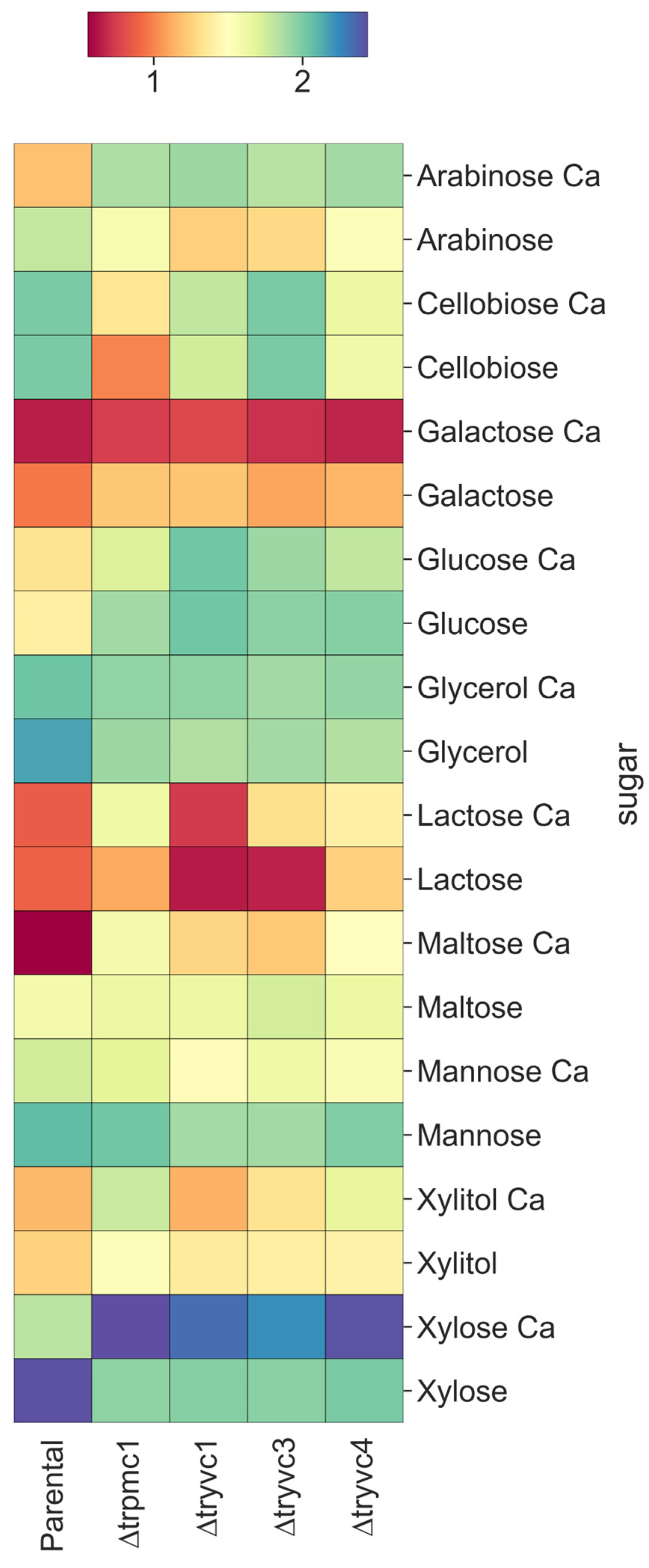
In our experiments, we monitored growth in minimal medium over a period of 72 h, using the carbon sources arabinose, cellobiose, galactose, glucose, glycerol, lactose, maltose, mannose, xylitol, and xylose (Figure 3 and Figure S6). On the first day, all mutant strains, except ∆tryvc4 with CaCl2, exhibited enhanced growth compared to the parental strain when grown in the presence of arabinose, xylitol, and xylose (Figure S6). However, by the third day, calcium supplementation, which promoted growth in the parental strain, either inhibited growth or had no effect on the mutant strains (Figure 3). In the case of cellobiose, we observed a decline in the growth of mutant strains compared to the parental strain, particularly at 72 h (Figure 3). For maltose, all strains showed improved growth with calcium supplementation (Figure 3). These results suggest that these proteins are important to sugar assimilation and calcium supplementation might increase sugar uptake or metabolism in the absence of functional vacuolar calcium transport. However, these effects are carbon source dependent.
We further analyzed the growth of the strains in the presence of different metals and concentrations over a period of 48 or 72 h, using xylose as the carbon source (Figure 4). This approach was based on our observation that the mutant strains were more impacted by calcium supplementation in arabinose, xylitol, and xylose (Figure 3), and considering the cost-effectiveness of xylose. The parental strain demonstrated increased growth in the presence of 10 mM of all metals, except for cobalt. In higher concentrations, growth persisted only in the presence of calcium, manganese, and magnesium (Figure 4a,b,f,m). Interestingly, in contrast to the parental strain, the mutant strains displayed heightened sensitivity to 100 mM manganese (Figure 4e,f) and 10 mM zinc (Figure 4i,j), suggesting their importance in conferring resistance to these metals. Moreover, the growth enhancement typically induced by calcium in the parental strain was not observed in the mutant strains cultured in the absence of calcium (Figure 4a,b).
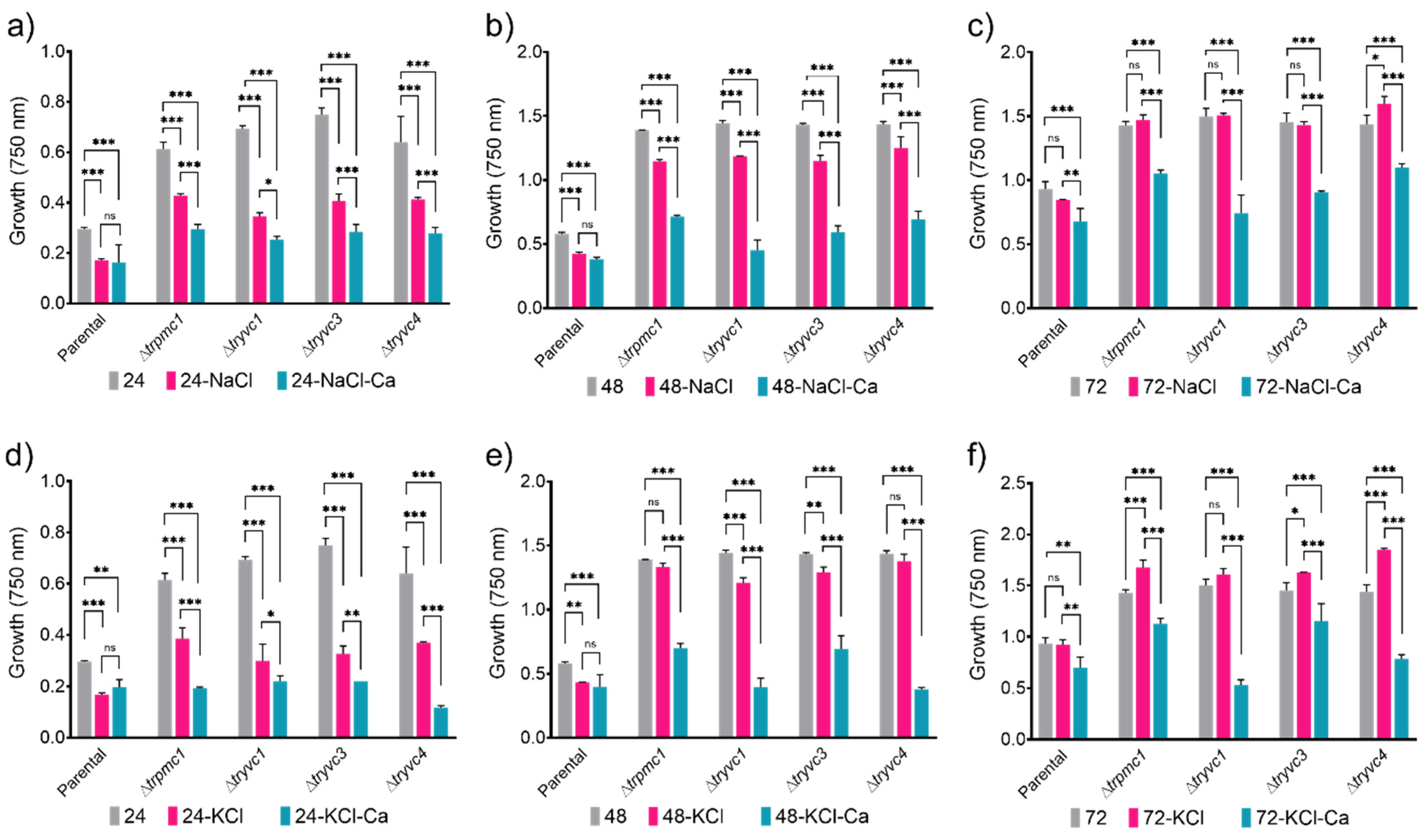
We examined the tolerance of T. reesei parental and mutant strains to elevated osmotic pressure by growing them on plates under osmotic stress conditions, specifically in the presence of 0.5 M NaCl or 0.5 M KCl, with and without calcium (Figure 5). Both NaCl and KCl inhibited growth of all strains at 24 h (Figure 5a,d), but by 72 h, the strains recovered and exhibited growth levels comparable to or higher than the control condition (Figure 5c,f). Notably, calcium supplementation led to a reduction in growth in the mutant strains, but not in the parental strain, at both 24 and 48 h (Figure 5a,b,d,e). This observation suggests that effective calcium signaling plays a pivotal role in T. reesei’s initial response to osmotic stresses.
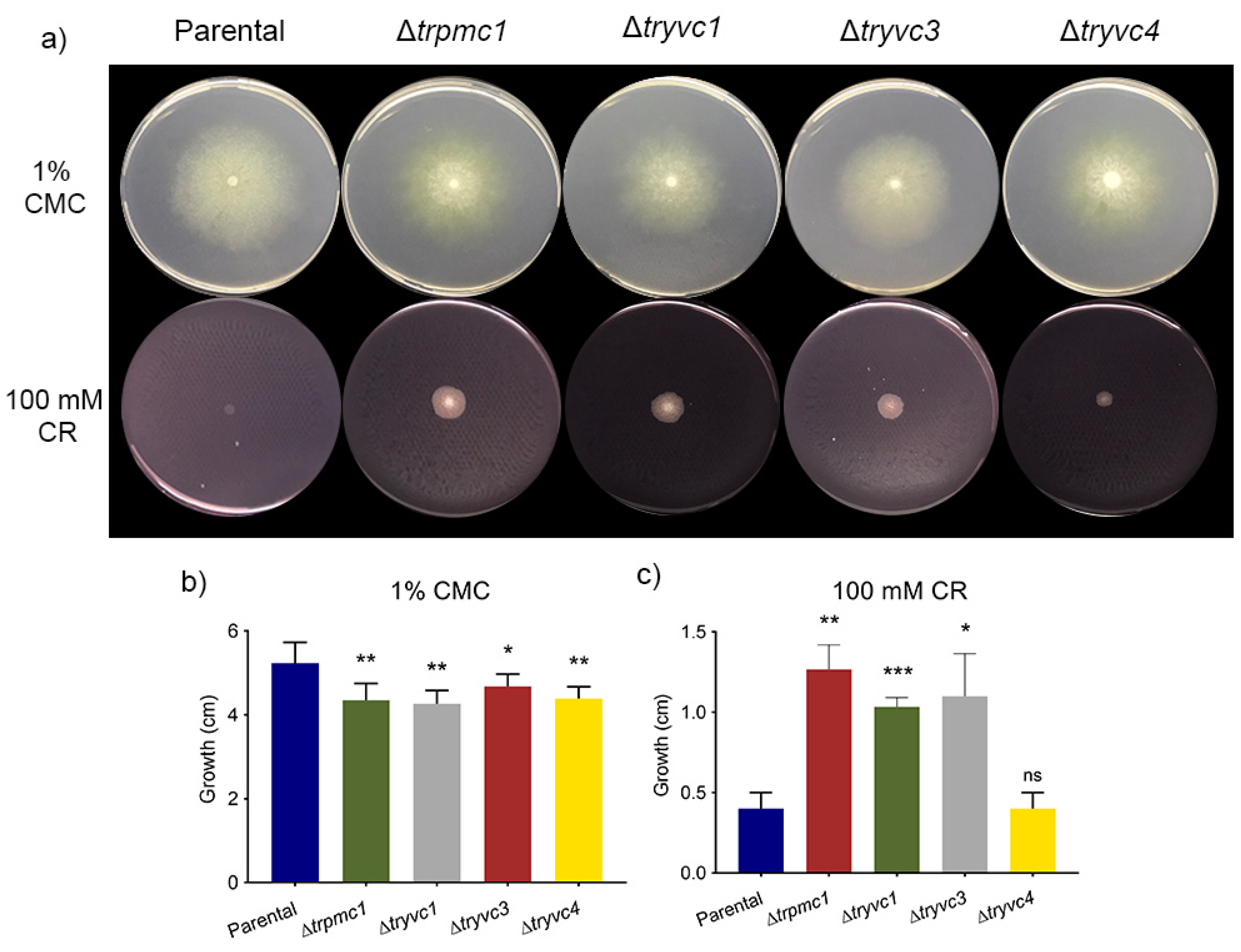
Furthermore, the integrity of T. reesei parental e mutant strain’s cell wall was investigated by testing of sensibility to cell wall interfering substance Congo Red (CR). For this, we monitored growth in MA media with 1% CMC or 2% glucose + 100 mM Congo Red (CR) (Figure 6a). Interestingly, all mutant strains showed reduced growth at 1% CMC (Figure 6b), suggesting a role for vacuolar transport proteins in cellulose metabolism. Moreover, the strains ∆trpmc1, ∆tryvc1, and ∆tryvc3 demonstrated enhanced growth on glucose in the presence of the cell wall stressor CR (Figure 6c). Given that the mutant strains exhibit reduced growth in glucose at the 72-h mark (Figure 3), it is plausible that the observed growth enhancement in the presence of CR may be attributed to the impact of the absence of vacuolar calcium transport proteins on the cell wall, suggesting the knockout strains have a high tolerance to stress agents such as CR.

3.3. TrPMC1, TrYVC1, TrYVC3, and TrYVC4 Are Key Factors in Cellulases Production Under Calcium Supplementation

We have shown that the strains lacking the proteins TrPMC1, TrYVC1, TrYVC3, and TrYVC4 exhibit impaired growth when CMC is used as the carbon source in the media (Figure 6b). To assess the impact of deleting genes encoding vacuolar calcium transport proteins on cellulase production, we cultivated both the parental and the mutant strains in Mandels-Andreotti (MA) medium with 1% cellulose, both in the presence or absence of 10 mM CaCl2.
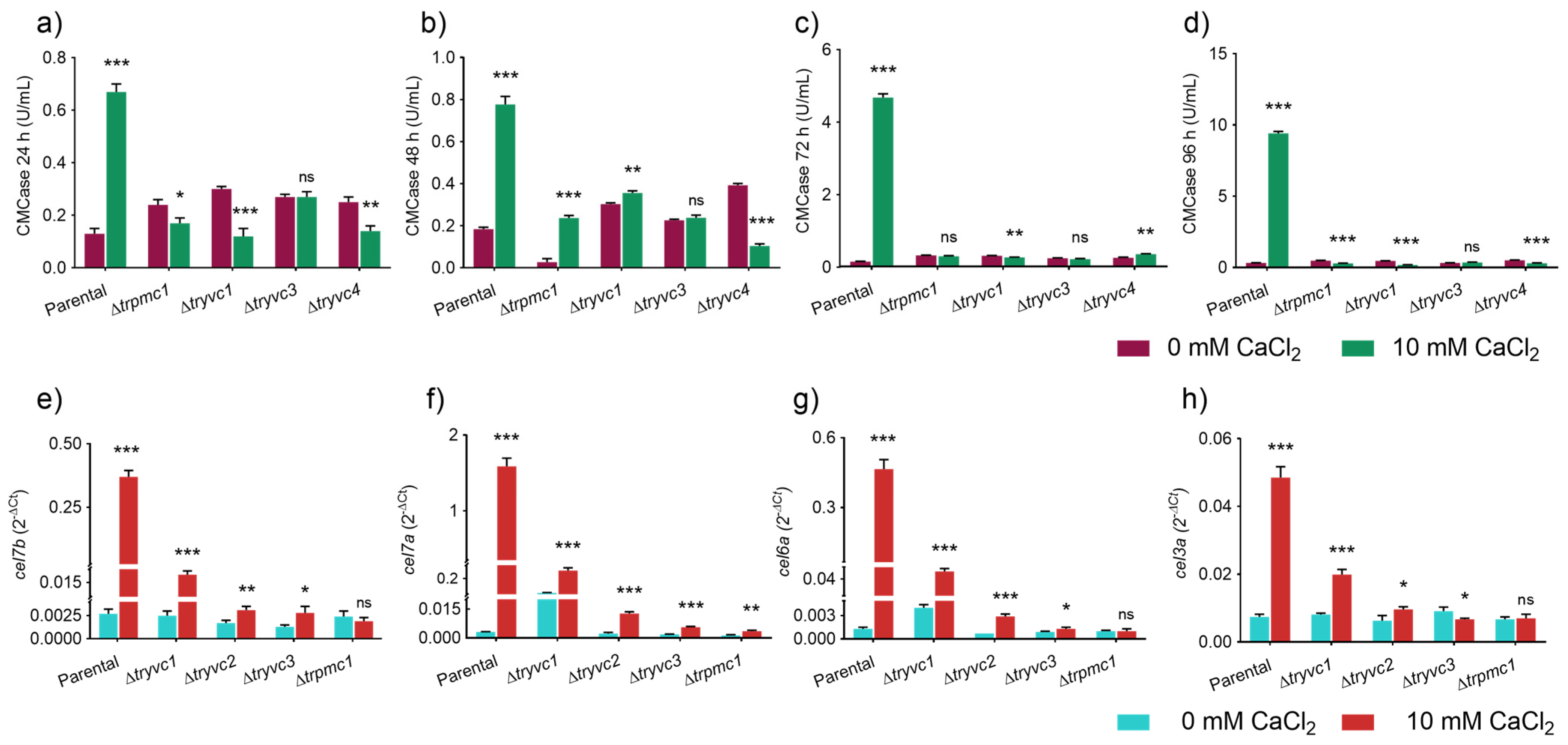
Regarding CMCase activity we noted an increase in the parental strain supplemented with calcium at all analyzed time points (Figure 7a) being this increase also observed in ∆trpmc1 and ∆tryvc1 at 48 h of cultivation in the presence of calcium, albeit at a significantly lower level compared to the parental strain (Figure 7b). At 96 h, all mutant strains except ∆tryvc3 exhibited a reduction in CMCase activity when comparing the condition with 10 mM CaCl2 supplementation to the condition without the metal (Figure 7d). These findings suggest that the vacuolar transport proteins TrPMC1, TrYVC1, TrYVC3, and TrYVC4 play a crucial role in triggering endoglucanase production in response to calcium.
To understand how cellulase expressions are affected in the mutant strains, we analyzed the expression of the genes cel7b (endoglucanase I) (Figure 7e), cel7a (cellobiohydrolase I) (Figure 7f), cel6a (cellobiohydrolase II) (Figure 7g), and cel3a (beta-glucosidase I) (Figure 7h) in MA + 1% cellulose for 24 h, both with and without CaCl2 supplementation. Our results show that all these genes are induced in the parental strain when calcium is supplemented to the culture medium (Figure 7b). In contrast, in the mutant strains, although induction occurs, the levels are significantly lower or non-existent. For instance, the parental strain’s level of cel7a is approximately 512 times higher when 10 mM CaCl2 is added, while in ∆trpmc1, ∆tryvc1, ∆tryvc3, and ∆tryvc4, it is about 8, 6, 3, and 3 times higher, respectively (Figure 7f). These findings underscore that the calcium transport proteins characterized in this study are a key factor in cellulase production when calcium is supplemented to the medium. It is important to note that the MA medium already contains calcium at a concentration of 5.4 mM, so the supplementation resulted in a final concentration of 15.4 mM.
To gain a better understanding of the mechanism behind the observed cellulase inhibition in the mutant strains, we analyzed the expression of the genes encoding the transcription factors XYR1, ACE3, HAC1a, CRE1, and CRZ1, as well as the calcium signaling components CAM and CNA1 (Figure 8).
With respect to the transcription factors, we observed statistically significant differences only for xyr1 and hac1a in the parental strain supplemented with CaCl2, compared to the control without supplementation (Figure 8a,c). This observation is consistent with the detected increase in cellulase expression. In the mutant strains, however, we did not observe an increase in xyr1, and for hac1a, an increase was only noted in the ∆tryvc3 and ∆tryvc4 strains, but to a lesser extent than in the parental strain (Figure 8a,c). Furthermore, we observed an increase in crz1 expression in ∆tryvc3 when supplemented with CaCl2 (Figure 8e), and a decrease in ace3 in ∆tryvc1 when supplemented with CaCl2 (Figure 8b), both when compared to the control condition without the addition of the metal.
Upon analyzing the expression of the components of the calcium-dependent signaling pathway, we observed an increase in cam expression in the parental strain and in ∆tryvc4 (Figure 8f), as well as the upregulation of cna1 in ∆trpmc1, and ∆tryvc3 under calcium supplementation (Figure 8g). These results suggest that the production of cellulases is influenced by a combination of factors, including different transcription factors and the calcium signaling components.

3.4. The Production of Xylanases and Secondary Metabolites Are Impacted by the Vacuolar Calcium Transport Proteins in T. reesei in the Presence of Xylose

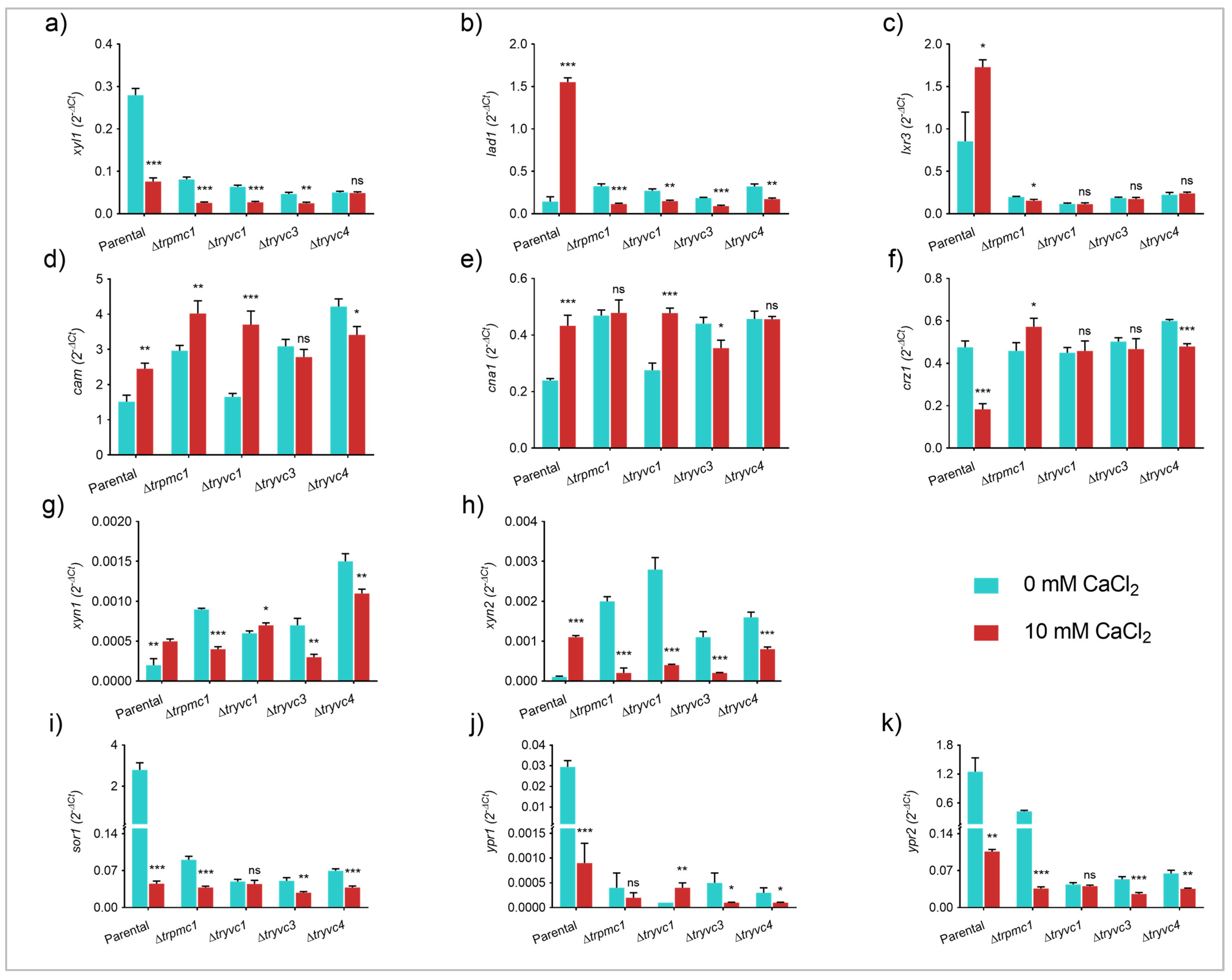
To comprehend the correlation between calcium supplementation and the observed growth enhancement in the parental strain in xylose, as well as the increased growth of the mutant strains relative to the parental strain without calcium supplementation, we cultivated these strains in liquid MA media with 25 mM xylose, both with and without calcium. We then analyzed the expression of genes encoding enzymes involved in xylose metabolism (XYL1, LAD1, and LXR3), xylanases (XYN1 and XYN2), and calcium signaling components (CAM, CNA1, and CRZ1) using the fungal mycelia.
Our analysis of the expression of genes encoding xylose metabolism enzymes revealed that xyl1 and lxr3 are more expressed in the parental strain than in the mutant strains without calcium supplementation (Figure 9a,c). Upon the addition of 10 mM CaCl2, we observed negative regulation of xyl1 (Figure 9a) and positive regulation of lad1 (Figure 9b) and lxr3 (Figure 9c) in the parental strain, compared to the same strain without the metal. These genes may be induced in response to calcium supplementation, contributing to the enhanced growth observed in Figure 3. Despite the mutant strains showing increased growth without calcium supplementation, the only observed differences were the inhibition of xyl1 in the ∆trpmc1, ∆tryvc1, and ∆tryvc3 (Figure 9a), and lxr3 in ∆trpmc1 (Figure 9c), and a slight induction of lad1 compared to the parental strain (Figure 9b).
As depicted in Figure 9d, we observed that the expression of cam was induced by calcium in both the parental and the mutant strains ∆trpmc1 and ∆tryvc1; however, an increase in growth (Figure 3) was only evident in the parental strain. In the case of cna1, we noted an upregulation in the parental strain and in ∆tryvc1 when calcium was supplemented to the culture medium (Figure 9e). Regarding the transcription factor CRZ1, its transcript levels decreased in the parental and ∆tryvc4 strains upon calcium supplementation (Figure 9f). These findings suggest that the elevated levels of cam and cna1, observed in the absence of calcium supplementation, may contribute to the enhanced growth seen in the mutant strains. Furthermore, the lack of additional growth promotion upon calcium supplementation could be due to a compromised calcium signaling pathway.
Considering that xylanases are induced by media containing xylose [50], we investigated the effect of deleting vacuolar calcium transport proteins on their gene expression (Figure 9g,h). First, we observed that xylanase expression levels are elevated in the mutant strains compared to the parental strain. However, while calcium supplementation led to an increase in the expression of xyn1 and xyn2 in the parental strain, it resulted in inhibition in the mutant strains, except for xyn1 in the ∆tryvc1 strain. Notably, the repression of xyn2 was significantly pronounced in the mutant strains compared to the same strains without calcium supplementation, suggesting that calcium-mediated signaling could play a crucial role in the expression of xylanases.
Interestingly, during the cultivation of the fungi for gene expression analysis, we observed that the addition of calcium resulted in the parental strain losing its characteristic yellow pigmentation (Figure S7). Consequently, we analyzed the expression of genes related to the production of the yellow pigment, a sorbicillinoid, produced by T. reesei [28]. Both the transcription factors ypr1 (Figure 9j) and ypr2 (Figure 9k), as well as the sor1 gene (Figure 9i) involved in pigment production, are repressed when calcium is supplemented to the culture medium. This effect also occurs in the mutant strains ∆trpmc1, ∆tryvc3, and ∆tryvc4, albeit to a lesser extent, indicating that the production of this secondary metabolite is impaired when the calcium transport proteins are absent and when calcium is supplemented.

3.5. Microscopy Analysis Reveals the Role of TrPMC1, TrYVC1, TrYVC3, and TrYVC4 in Cell Wall Thickness and Calcium Dynamics in T. reesei

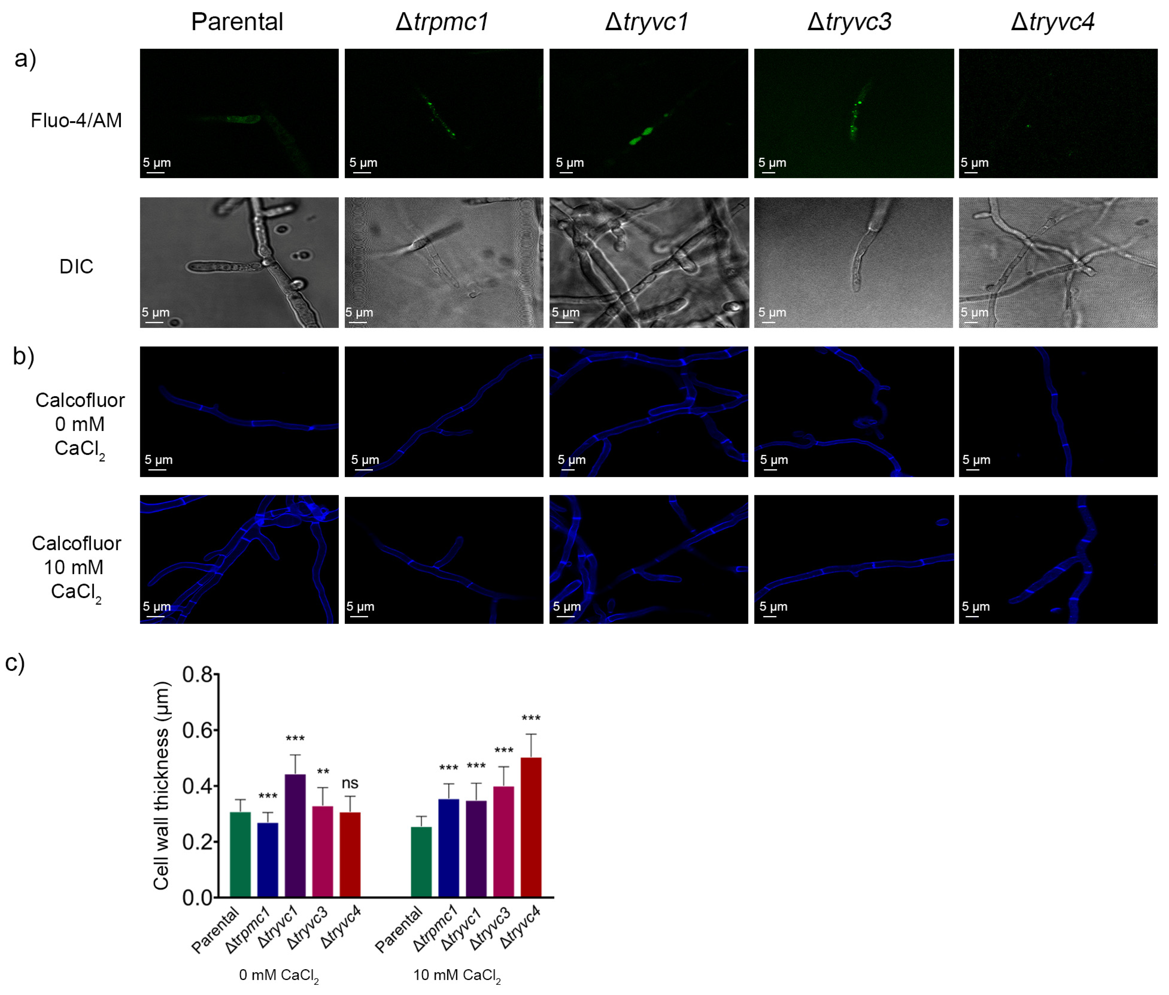
To gain deeper insights into the observed effects in mutant strains lacking vacuolar calcium transport proteins, we marked intracellular calcium using Fluo-4/AM in cultures with MA medium supplemented with 25 mM xylose and 10 mM CaCl2. As depicted in Figure 10, calcium appears more evenly distributed in the parental strain, without any distinct points indicating higher concentration, unlike in the mutant strains (Figure 10a). This suggests that in these strains, calcium may be accumulating in the vacuoles due to the deletion of the proteins involved in its transport.
Given our observation that the ∆trpmc1, ∆tryvc1, and ∆tryvc3 mutant strains exhibit greater resistance to wall stress induced by CR than the parental strain (Figure 6a,c), we measured and calculated the wall thickness of the strains under study using calcofluor white labeling (CFW) (Figure 10b,c). Our analysis revealed that all mutant strains exhibited cell wall thickening when calcium was added (Figure 10c). However, in the absence of calcium, only the ∆tryvc1 and ∆tryvc3 strains were found to have thicker walls than the parental strain (Figure 10b). So, this result suggests the knockout strains have a high tolerance to stress agents, which could be explained by cell wall thickening.

4. Discussion

In this study, we functionally characterized four vacuolar calcium transport proteins in T. reesei: TrPMC1, TrYVC1, TrYVC3, and TrYVC4. First, we identified genes encoding proteins that are homologous to PMC1, YVC1, and VCX1 of S. cerevisiae, and then, we analyzed the expression profiles of these genes using various RNA-Seq datasets obtained by our group and constructed mutant lines with deletions of genes 58952, 74057, 55731, and 56440, which were subsequently named ∆trpmc1, ∆tryvc1, ∆tryvc3, and ∆tryvc4, respectively. We examined the growth phenotype of these strains, their cellulolytic enzyme activity, and the expression of genes associated with the degradation of lignocellulosic biomass, secondary metabolite biosynthesis, and the calcium signaling pathway. Finally, we conducted microscopy assays to confirm the intracellular localization of calcium in the parental and mutant strains of T. reesei.
Through bioinformatics analyses, we identified 5 potential YVC1, 7 potential VCX1, and 21 potential PMC1 proteins (Table 1). However, only 15 of these were identified as proteins that transport calcium in the vacuoles (5 YVC1, 7 VCX1, and 3 PMC1), with the others likely involved in the transport of phospholipids, calcium, and other metals in other organelles. We also identified proteins of the SERCA (sarcoplasmic/endoplasmic reticulum Ca2+ ATPase) and PMCA (plasma membrane Ca2+ ATPase) types, as proteins present in other cellular compartments may share similarities with vacuolar transport proteins [15]. It is important to note that proteins classified as PMC1 can be located both in the plasma membrane and in the vacuoles, as is the case with the Ca2+ ATPases NCA-2 and NCA-3 from Neurospora crassa [51]. A similar number of transport proteins involved in calcium signaling was reported by [16], who identified three ion channels, 10 ATPases, and 10 calcium transporters in T. reesei. Additionally, other organisms share a similar number of YVC1, VCX1, and PMC1 homologs. For instance, four homologs of YVC1 were identified in Colletotrichum graminicola (CgTRPF1-4) [52], three homologs of PMC1 were identified in Aspergillus fumigatus (PMCAA-C) [53] and in Beauveria bassiana (PMCA-C) [54], and five VCX1 homologs were also identified in B. bassiana (VCX1A-E) [55].
When analyzing the expression profiles of proteins identified in the RNA-Seq data obtained by our group [17,18,41,48,56], we observed that expression levels vary even between the strains that are generally used as parental in functional studies (Figure 2): QM6a, QM9414, and TU6 [57]. This indicates that the mutations present in QM9414 and TU6 strains, which result in greater cellulase production, may be involved in the calcium-mediated signaling pathway. Furthermore, we observed that the expression of the genes under study can be regulated by the transcription factors XYR1, CRE1, and AZF1, and are also involved with the MAPK (mitogen-activated protein kinase) signaling pathway, as these proteins have also been differentially expressed in these analyses. Consistent with these data, it has been demonstrated that calmodulin, an important component of the calcium-mediated signaling pathway, binds to an integral membrane protein, activating the MAPK signaling pathway in Candida albicans [58]. Furthermore, in B. bassiana, it was demonstrated that the inactivation of Slt2/Mpk1 repressed the phosphatase activity of calcineurin [38]. The carbon source can also influence the expression of the proteins under study. For example, in S. cerevisiae [59] it was demonstrated that the addition of glucose causes the entry of calcium into the cell and that this process may be related to the YVC1 vacuolar channel, as its deletion resulted in a strain in which this response was attenuated.
Based on the collected data, the proteins identified by JGI IDs 58952, 74057, 55731, and 56440 were selected for further investigation. The objective was to generate mutant strains with the deletion of the respective genes, thereby facilitating a more comprehensive understanding of the calcium-dependent signaling pathway. These proteins were subsequently designated as TrPMC1, TrYVC1, TrYVC3, and TrYVC4, respectively.
Our phenotypic growth analyses revealed an enhanced growth rate in the presence of arabinose, xylitol, and xylose in the mutant strains compared to the parental strain (Figure 3). While previous studies have demonstrated that calcium can enhance fungal growth in media containing glucose [10], there is limited evidence regarding its effect in media containing xylose. In Mucor circinelloides, it has been shown that zinc and calcium can augment xylose consumption. The underlying mechanism remains elusive, but it is hypothesized that the concentration of calcium may regulate zinc transport into the cell [60]. In this study, we examined the expression of genes involved in xylose metabolism and calcium signaling in both parental and mutant strains cultured in xylose (Figure 9). It is plausible that the inhibition of xyl1 and lxr3, coupled with the induction of lad1, may play a role in this process. This could be accompanied by an upregulation of cam and cna1, although the precise mechanism requires further investigation.
We also demonstrated that calcium influences the production of secondary metabolites and that vacuolar calcium transport proteins may be implicated in this process (Figure 9). This observation aligns with findings in B. bassiana, where cam plays a significant role in secondary metabolism by interacting with a ketoisovalerate reductase, thereby inhibiting its activity [61]. These findings are consistent with a study conducted in Penicillium oxalicum, which showed that PoCrz1 regulates secondary metabolism. In the absence of this transcription factor, five clusters were negatively regulated in the mutant strain [62]. Furthermore, it has been shown that intracellular calcium is involved in the expression of genes related to the biosynthesis and secretion of metabolites in filamentous fungi, with YVC1 playing a role. The exact mechanism remains unknown, but it is suggested that calcium signaling may induce the expression of the regulator crc (a zinc-finger type regulator), which is responsible for the induction of secondary metabolite biosynthesis genes [63]. Additionally, it has been demonstrated that PenV, a putative calcium channel in Penicillium chrysogenum, plays a crucial role in secondary metabolite production. By transporting amino acids from the vacuoles to the cytosol, PenV supports the biosynthesis of penicillin. Furthermore, PenV regulates the expression of two penicillin biosynthesis genes, pcbC, and penDE, highlighting its significance in secondary metabolite production [63,64].
In xylose, we observed that the mutant strains exhibit sensitivity to high concentrations of manganese, and interestingly, the growth enhancement typically induced by calcium in the parental strain was absent in these mutants (Figure 4). This aligns with previous findings indicating that manganese plays a role in calcium signaling in T. reesei, as it triggers an increase in cytosolic calcium concentration [7]. Furthermore, it has been shown that YVC1 can be activated by calcium or other metals, such as manganese, magnesium, and zinc [15], thereby facilitating the release of calcium from the vacuoles. In addition, when the strains were cultivated under osmotic stress conditions, we found that calcium supplementation hindered growth in the mutant strains, but not in the parental strain, within the first 48 h (Figure 5). This is consistent with several studies indicating that calcium signaling contributes to osmotic stress tolerance. For instance, in B. bassiana and N. crassa, disruption of calcineurin function resulted in increased sensitivity to osmotic stress [65,66]. Additionally, it has been shown that phospholipase C may also play a role in osmotic stress resistance in S. cerevisiae [66].
Here, we also observed that the mutant strains exhibited increased resistance to cell wall stress induced by CR (Figure 6). Using confocal microscopy, we found that the cell wall of T. reesei is thicker in mutant strains (Figure 10), which could explain their heightened resistance to the cell wall stress caused by CR. In Aspergillus nidulans, it was shown that deletion of CchA, a plasma membrane calcium channel of the high-affinity calcium influx system (HACS) and MidA, its regulatory unit, rendered the mutant strains more resistant to stress induced by CR and CFW, likely due to alterations in the composition of the fungal cell wall [67]. In fact, the transcription factor CRZ1/CRZA is involved in cell wall biosynthesis by the induction of the expression of the genes chs1, crh1, rho1, scw10, and kre6 in Saccharomyces cerevisiae [68], chitin synthase (chs6) in Cryptococcus neoformans [69] and chsA-G in A. fumigatus [70]. In this study, we found that in the presence of xylose and with the supplementation with 10 mM CaCl2, the mutant strains show induced Crz1 in relation to the parental strain, which could explain the resistance that was observed in the presence of the metal, this suggesting that the vacuolar transport proteins TrPMC1, TrYVC1, TrYVC3 and TrYVC4 might be involved in the cell wall resistance though CRZ1.
Our results also showed that the growth of mutant strains is compromised when cultivated in CMC (Figure 6), and that the expression and activity of cellulases are significantly reduced in these strains (Figure 7). While calcium supplementation is known to enhance cellulase production in T. reesei [10], it had a negative impact on the mutant strains. This suggests that the uptake and release of calcium from the vacuoles are crucial for the induction of cellulase expression in T. reesei. Other studies have also linked calcium signaling to cellulase production in T. reesei [7,9,11,22,23], but this is the first to demonstrate that vacuolar calcium transport proteins are also involved in this process. We noted the downregulation of the inducers ace3 and crz1 in the TrYVC1-deficient mutant strain and the upregulation of the repressor cre1 in the TrYVC3-deficient strain both in the presence of calcium (Figure 8).
Given the lack of a clear pattern, it is possible that the reduced production of cellulases in the mutant strains could be associated with other proteins. In addition, despite the involvement of YVC1 deletion mutants in the release of calcium and the PMC1 mutant in the sequestration of calcium, we did not observe distinct phenotypic differences. This could be due to the presence of other homologs in T. reesei. However, we found that the deletion of a single gene is sufficient to impair cellulase production (Figure 7). It is possible that the observed effects are due to the retention of calcium within the vacuoles, as shown using confocal microscopy (Figure 10). It is conceivable that this retention hampers calcium-dependent signaling, which has been shown to be crucial for the induction of cellulases [7]. This result is in accordance with the study of Martins-Santana et al. (2020) [11], in which they showed that the promoter regions of cellulase genes, as well as calcium transport-related genes, have binding sites for the regulator CRZ1. In addition, other calcium signaling components were shown to be involved in cellulases production. For example, cam and cna1 are upregulated when manganese is added to the culture medium, a condition in which endoglucanases and cellobiohydrolases production is induced [7]. Altogether, the data presented here suggests that the calcium signaling pathway is crucial to cellulases production in T. reesei.

5. Conclusions

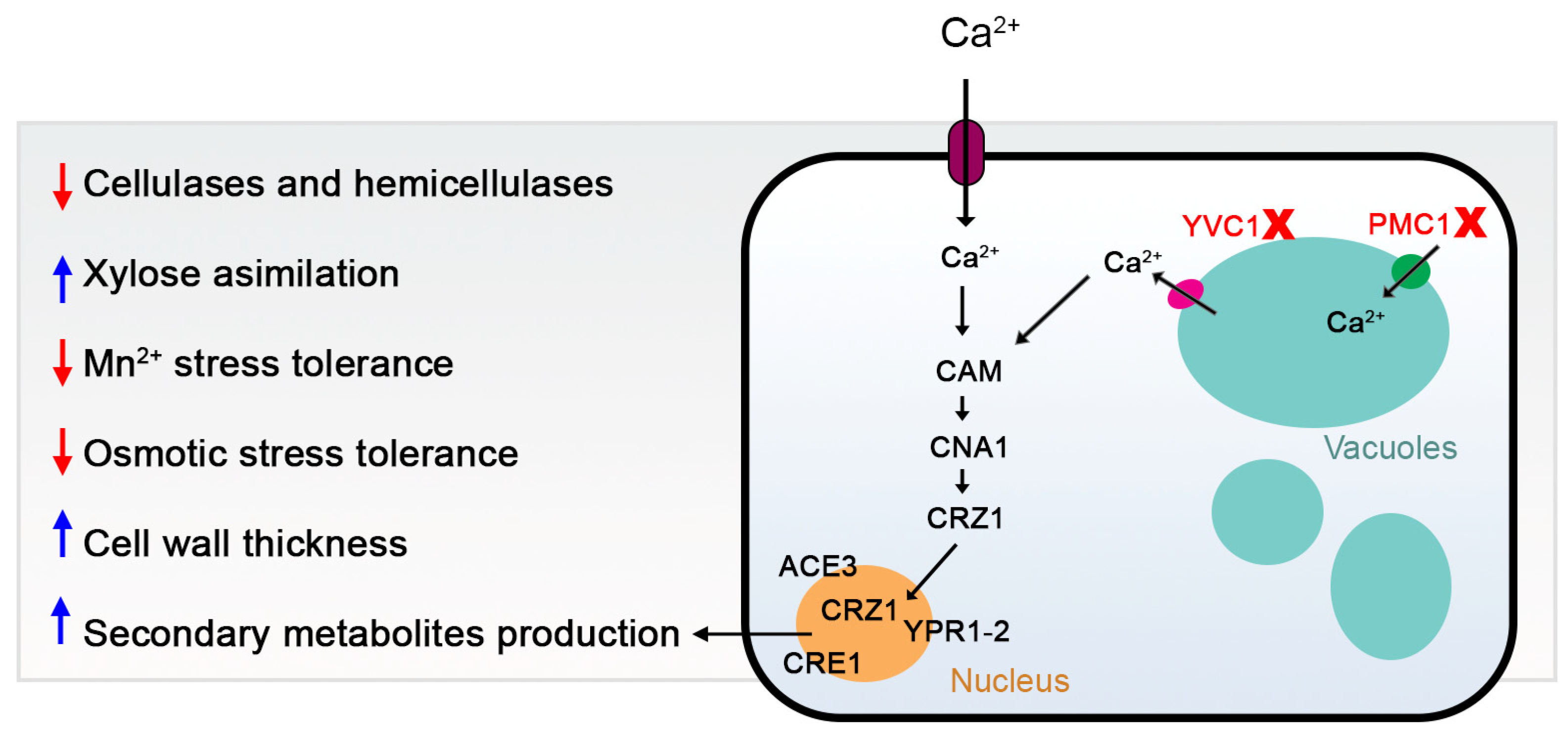
In conclusion, our study provides novel insights into the role of calcium signaling and vacuolar calcium transport proteins in the regulation of growth, stress response, secondary metabolism biosynthesis, and cellulase production in T. reesei (Figure 11). We demonstrated that the deletion of specific genes involved in calcium transport can significantly impact these biological processes, suggesting their potential as targets for genetic manipulation to enhance the industrial utility of this organism. However, the exact mechanisms underlying these effects remain to be elucidated, highlighting the need for further research in this area. Our findings pave the way for future studies exploring the intricate network of calcium signaling in fungi and its implications for industrial biotechnology.

Supplementary Materials

The following supporting information can be downloaded at: https://www.mdpi.com/article/10.3390/jof10120853/s1, Figure S1. Utilized plasmids to construct the deletion cassettes. (a) pJET-pyr4 and (b) pRS426; Figure S2. Strategy used to confirm the mutant strain. Primers to detect the target gene are in green. Primers for detecting cassette insertion in the correct orientation are in black and blue; Figure S3. Confirmation of the mutant strains ∆tryvc3 and ∆tryvc4 using RT-qPCR. Amplification and melt peak chart of ∆tryvc3 (a) and ∆tryvc4 (b). The reaction used the indicated sample-primer, e.g., P strain was the sample and Act was the primer. P = parental strain. Act = actin, 55 = ∆tryvc3, and 56 = ∆tryvc4; Figure S4. Confirmation of the mutant strains ∆trpmc1 and ∆tryvc1 using RT-qPCR. Amplification and melt peak chart of ∆trpmc1 (a) and ∆tryvc1 (b). The reaction used the indicated sample-primer, e.g., P strain was the sample and Act was the primer. P = parental strain. Act = actin, 58 = ∆trpmc1, and 74 = ∆tryvc1; Figure S5. Confirmation of the deletion strains using conventional PCR. Deletion of trpmc1 (58952), tryvc1 (74057), tryvc3 (55731) and tryvc4 (56440), according to the primers of Figure S2. The positive mutants in each confirmation phase are highlighted in red. (a) ORF detection for trpmc1, (b) 5′ orientation detection for trpmc1, (c) 3′ orientation detection for trpmc1, (d) ORF detection for tryvc1, (e) 5′ orientation detection for tryvc1, (f) 3' orientation detection for tryvc1, (g) ORF detection for tryvc3, (h) 5′ orientation detection for tryvc3, (i) 3′ orientation detection for tryvc3, (j) ORF detection for tryvc4, (k) 5′ orientation detection for tryvc4, (l) 3′ orientation detection for tryvc4. M—DNA molecular size marker (GeneRuler 1 kb Plus DNA Ladder (Thermo Fisher Scientific, Waltham, MA, USA); Q—positive control (DNA of QM6a∆tmus53pyr4—reaction with parental strain DNA; H—negative control (ultrapure water reaction); Figure S6. Growth in different carbon sources. Growth of strains QM6a∆tmus53pyr4 (parental), ∆trpmc1, ∆tryvc1, ∆tryvc3, and ∆tryvc4 in minimal media in the presence of 26 mM of different carbon sources for (a) 24 h and (b) 48 h, with and without 10 mM CaCl2 supplementation. The values represent the absorbance readings at 750 nm; Figure S7. Culture of the parental strain in MA medium + xylose with and without calcium supplementation. Table S1. Primers used in this study. The bold regions are the restriction sites, the underlined regions are the homology regions. Table S2. Vacuolar calcium transport proteins characterized in fungi. References [51,52,53,54,71,72,73,74,75,76,77,78,79,80,81,82,83,84] are cited in supplementary materials.

Author Contributions

Conceptualization, R.N.S. and R.G.d.P.; methodology, R.N.S., R.G.d.P. and L.H.O.; validation, L.H.O.; formal analysis, L.H.O.; investigation, L.H.O., R.G.d.P., L.M.S.P., D.B.M., A.C.S., E.R.M., L.F.d.M.C.d.J., J.A.S.-N. and F.P.V.; writing—original draft preparation, L.H.O.; writing—review and editing, R.G.d.P., R.N.S. and L.H.O.; supervision, R.N.S. and R.G.d.P. All authors have read and agreed to the published version of the manuscript.

Funding

This work was supported by the Fundação de Amparo à Pesquisa do Estado de São Paulo (FAPESP) (grant numbers 2016/23233-9, 2019/19569-0 and, 2019/11655-4), Conselho Nacional de Desenvolvimento Científico e Tecnológico (CNPq) grant number 405934/2022-0 (The National Institute of Science and Technology INCT Funvir, Brazil) and 301873/2022-4 (RNS supporting), Fundação Coordenação de Aperfeiçoamento de Pessoal de Nível Superior, and the Fundação de Apoio à Pesquisa do Espírito Santo (FAPES) process number 2021-RZN24 (TO 438/2021).

Institutional Review Board Statement

Not applicable.

Informed Consent Statement

Not applicable.

Data Availability Statement

The original contributions presented in the study are included in the article/Supplementary Material, further inquiries can be directed to the corresponding author.

Conflicts of Interest

The authors declare no conflicts of interest.

References

  1. Saini, J.K.; Saini, R.; Tewari, L. Lignocellulosic agriculture wastes as biomass feedstocks for second-generation bioethanol production: Concepts and recent developments. 3 Biotech 2015, 5, 337–353. [Google Scholar] [CrossRef] [PubMed]
  2. Zhang, Z.; O’hara, I.M.; Mundree, S.; Gao, B.; Ball, A.S.; Zhu, N.; Bai, Z.; Jin, B. Biofuels from food processing wastes. Curr. Opin. Biotechnol. 2016, 38, 97–105. [Google Scholar] [CrossRef] [PubMed]
  3. de Paula, R.G.; Antoniêto, A.C.C.; Ribeiro, L.F.C.; Carraro, C.B.; Nogueira, K.M.V.; Lopes, D.C.B.; Silva, A.C.; Zerbini, M.T.; Pedersoli, W.R.; do Nascimento Costa, M.; et al. New Genomic Approaches to Enhance Biomass Degradation by the Industrial Fungus Trichoderma reesei. Int. J. Genom. 2018, 2018, 1974151. [Google Scholar] [CrossRef]
  4. Jansen, M.L.A.; Bracher, J.M.; Papapetridis, I.; Verhoeven, M.D.; de Bruijn, H.; de Waal, P.P.; van Maris, A.J.A.; Klaassen, P.; Pronk, J.T. Saccharomyces cerevisiae strains for second-generation ethanol production: From academic exploration to industrial implementation. FEMS Yeast Res. 2017, 17, fox044. [Google Scholar] [CrossRef]
  5. Liu, G.; Zhang, J.; Bao, J. Cost evaluation of cellulase enzyme for industrial-scale cellulosic ethanol production based on rigorous Aspen Plus modeling. Bioprocess Biosyst. Eng. 2016, 39, 133–140. [Google Scholar] [CrossRef]
  6. dos Santos Castro, L.; Pedersoli, W.; Antoniêto, A.C.; Steindorff, A.; Silva-Rocha, R.; Martinez-Rossi, N.M.; Rossi, A.; Brown, N.A.; Goldman, G.H.; Faça, V.M.; et al. Comparative metabolism of cellulose, sophorose and glucose in Trichoderma reesei using high-throughput genomic and proteomic analyses. Biotechnol. Biofuels 2014, 7, 41. [Google Scholar] [CrossRef]
  7. Chen, Y.; Shen, Y.; Wang, W.; Wei, D. Mn2+ modulates the expression of cellulase genes in Trichoderma reesei Rut-C30 via calcium signaling. Biotechnol. Biofuels 2018, 11, 54. [Google Scholar] [CrossRef]
  8. Li, N.; Li, J.; Chen, Y.; Shen, Y.; Wei, D.; Wang, W. Mechanism of Zn2+ regulation of cellulase production in Trichoderma reesei Rut-C30. Biotechnol. Biofuels Bioprod. 2023, 16, 73. [Google Scholar] [CrossRef]
  9. Li, N.; Zeng, Y.; Chen, Y.; Shen, Y.; Wang, W. Induction of cellulase production by Sr2+ in Trichoderma reesei via calcium signaling transduction. Bioresour. Bioprocess. 2022, 9, 96. [Google Scholar] [CrossRef]
  10. Chen, L.; Zou, G.; Wang, J.; Wang, J.; Liu, R.; Jiang, Y.; Zhao, G.; Zhou, Z. Characterization of the Ca2+-responsive signaling pathway in regulating the expression and secretion of cellulases in Trichoderma reesei Rut-C30. Mol. Microbiol. 2016, 100, 560–575. [Google Scholar] [CrossRef]
  11. Martins-Santana, L.; de Paula, R.G.; Silva, A.G.; Lopes, D.C.B.; Silva, R.D.N.; Silva-Rocha, R. CRZ1 regulator and calcium cooperatively modulate holocellulases gene expression in Trichoderma reesei QM6a. Genet. Mol. Biol. 2020, 43, e20190244. [Google Scholar] [CrossRef] [PubMed]
  12. Lange, M.; Peiter, E. Calcium Transport Proteins in Fungi: The Phylogenetic Diversity of Their Relevance for Growth, Virulence, and Stress Resistance. Front. Microbiol. 2020, 10, 3100. [Google Scholar] [CrossRef] [PubMed]
  13. Liu, S.; Hou, Y.; Liu, W.; Lu, C.; Wang, W.; Sun, S. Components of the calcium-calcineurin signaling pathway in fungal cells and their potential as antifungal targets. Eukaryot. Cell 2015, 14, 324–334. [Google Scholar] [CrossRef] [PubMed]
  14. Yang, Y.; Xie, P.; Li, Y.; Bi, Y.; Prusky, D.B. Updating Insights into the Regulatory Mechanisms of Calcineurin-Activated Transcription Factor Crz1 in Pathogenic Fungi. J. Fungi 2022, 8, 1082. [Google Scholar] [CrossRef]
  15. Tisi, R.; Rigamonti, M.; Groppi, S.; Belotti, F. Calcium homeostasis and signaling in fungi and their relevance forpathogenicity of yeasts and filamentous fungi. AIMS Mol. Sci. 2016, 3, 505–549. [Google Scholar] [CrossRef]
  16. Schmoll, M.; Dattenböck, C.; Carreras-Villaseñor, N.; Mendoza-Mendoza, A.; Tisch, D.; Alemán, M.I.; Baker, S.E.; Brown, C.; Cervantes-Badillo, M.G.; Cetz-Chel, J.; et al. The Genomes of Three Uneven Siblings: Footprints of the Lifestyles of Three Trichoderma Species. Microbiol. Mol. Biol. Rev. 2016, 80, 205–327. [Google Scholar] [CrossRef]
  17. De Paula, R.G.; Antoniêto, A.C.C.; Carraro, C.B.; Lopes, D.C.B.; Persinoti, G.F.; Peres, N.T.A.; Martinez-Rossi, N.M.; Silva-Rocha, R.; Silva, R.N. The Duality of the MAPK Signaling Pathway in the Control of Metabolic Processes and Cellulase Production in Trichoderma reesei. Sci. Rep. 2018, 8, 14931. [Google Scholar] [CrossRef]
  18. Silva, A.C.; Oshiquiri, L.H.; de Morais Costa de Jesus, L.F.; Maués, D.B.; do Nascimento Silva, R. The Cerato-Platanin EPL2 from Trichoderma reesei Is Not Directly Involved in Cellulase Formation but in Cell Wall Remodeling. Microorganisms 2023, 11, 1965. [Google Scholar] [CrossRef]
  19. Campos Antoniêto, A.C.; Graciano de Paula, R.; Santos Castro L dos Silva-Rocha, R.; Felix Persinoti, G.; Nascimento Silva, R. Trichoderma reesei CRE1-mediated Carbon Catabolite Repression in Response to Sophorose Through RNA Sequencing Analysis. Curr. Genom. 2015, 17, 119–131. [Google Scholar] [CrossRef]
  20. Dos Santos Castro, L.; de Paula, R.G.; Antoniêto, A.C.C.; Persinoti, G.F.; Silva-Rocha, R.; Silva, R.N. Understanding the Role of the Master Regulator XYR1 in Trichoderma reesei by Global Transcriptional Analysis. Front. Microbiol. 2016, 7, 175. [Google Scholar] [CrossRef]
  21. Chen, Y.; Wu, C.; Shen, Y.; Ma, Y.; Wei, D.; Wang, W. N,N-dimethylformamide induces cellulase production in the filamentous fungus Trichoderma reesei. Biotechnol. Biofuels 2019, 12, 36. [Google Scholar] [CrossRef] [PubMed]
  22. Chen, Y.; Fan, X.; Zhao, X.; Shen, Y.; Xu, X.; Wei, L.; Wang, W.; Wei, D. cAMP activates calcium signalling via phospholipase C to regulate cellulase production in the filamentous fungus Trichoderma reesei. Biotechnol. Biofuels 2021, 14, 62. [Google Scholar] [CrossRef] [PubMed]
  23. Liu, S.; Quan, L.; Yang, M.; Wang, D.; Wang, Y.Z. Regulation of cellulase production via calcium signaling in Trichoderma reesei under PEG8000 stress. Appl. Microbiol. Biotechnol. 2024, 108, 178. [Google Scholar] [CrossRef] [PubMed]
  24. Derntl, C.; Kiesenhofer, D.P.; Mach, R.L.; Mach-Aigner, A.R. Novel Strategies for Genomic Manipulation of Trichoderma reesei with the Purpose of Strain Engineering. Appl. Environ. Microbiol. 2015, 81, 6314–6323. [Google Scholar] [CrossRef]
  25. Nogueira, K.M.V.; de Paula, R.G.; Antoniêto, A.C.C.; dos Reis, T.F.; Carraro, C.B.; Silva, A.C.; Almeida, F.; Rechia, C.G.V.; Goldman, G.H.; Silva, R.N. Characterization of a novel sugar transporter involved in sugarcane bagasse degradation in Trichoderma reesei. Biotechnol. Biofuels 2018, 11, 84. [Google Scholar] [CrossRef]
  26. Przylucka, A.; Akcapinar, G.B.; Chenthamara, K.; Cai, F.; Grujic, M.; Karpenko, J.; Livoi, M.; Shen, Q.; Kubicek, C.P.; Druzhinina, I.S. HFB7—A novel orphan hydrophobin of the Harzianum and Virens clades of Trichoderma, is involved in response to biotic and abiotic stresses. Fungal Genet. Biol. 2017, 102, 63–76. [Google Scholar] [CrossRef]
  27. Schmoll, M.; Schuster, A.; Silva, R.D.N.; Kubicek, C.P. The G-alpha protein GNA3 of Hypocrea jecorina (anamorph Trichoderma reesei) regulates cellulase gene expression in the presence of light. Eukaryot. Cell 2009, 8, 410–420. [Google Scholar] [CrossRef]
  28. Derntl, C.; Rassinger, A.; Srebotnik, E.; Mach, R.L.; Mach-Aigner, A.R. Identification of the main regulator responsible for synthesis of the typical yellow pigment produced by Trichoderma reesei. Appl. Environ. Microbiol. 2016, 82, 6247–6257. [Google Scholar] [CrossRef]
  29. Sambrook, J.; Russell, D.W. Preparation and Transformation of Competent E. coli Using Calcium Chloride. Cold Spring Harb. Protoc. 2006, 2006, pdb.prot3932. [Google Scholar] [CrossRef]
  30. Bimboim, H.C.; Doly, J. A rapid alkaline extraction procedure for screening recombinant plasmid DNA. Nucleic Acids Res. 1979, 7, 1513–1523. [Google Scholar] [CrossRef]
  31. Teepe, A.G.; Loprete, D.M.; He, Z.; Hoggard, T.A.; Hill, T.W. The protein kinase C orthologue PkcA plays a role in cell wall integrity and polarized growth in Aspergillus nidulans. Fungal Genet. Biol. 2007, 44, 554–562. [Google Scholar] [CrossRef] [PubMed]
  32. Gietz, R.D.; Schiestl, R.H. High-efficiency yeast transformation using the LiAc/SS carrier DNA/PEG method. Nat. Protoc. 2007, 2, 31–34. [Google Scholar] [CrossRef] [PubMed]
  33. Collopy, P.D.; Colot, H.V.; Park, G.; Ringelberg, C.; Crew, C.M.; Borkovich, K.A.; Dunlap, J.C. High-throughput construction of gene deletion cassettes for generation of Neurospora crassa knockout strains. Methods Mol. Biol. 2010, 638, 33–40. [Google Scholar] [PubMed]
  34. Colot, H.V.; Park, G.; Turner, G.E.; Ringelberg, C.; Crew, C.M.; Litvinkova, L.; Weiss, R.L.; Borkovich, K.A.; Dunlap, J.C. A high-throughput gene knockout procedure for Neurospora reveals functions for multiple transcription factors. Proc. Natl. Acad. Sci. USA 2006, 103, 10352–10357. [Google Scholar] [CrossRef]
  35. Goldman, G.H.; Dos Reis Marques, E.; Duarte Ribeiro, D.C.; De Souza Bernardes, L.Â.; Quiapin, A.C.; Vitorelli, P.M.; Savoldi, M.; Semighini, C.P.; de Oliveira, R.C.; Nunes, L.R.; et al. Expressed sequence tag analysis of the human pathogen Paracoccidioides brasiliensis yeast phase: Identification of putative homologues of Candida albicans virulence and pathogenicity genes. Eukaryot. Cell 2003, 2, 34–48. [Google Scholar] [CrossRef]
  36. Jones, P.; Binns, D.; Chang, H.Y.; Fraser, M.; Li, W.; McAnulla, C.; McWilliam, H.; Maslen, J.; Mitchell, A.; Nuka, G.; et al. InterProScan 5: Genome-scale protein function classification. Bioinformatics 2014, 30, 1236–1240. [Google Scholar] [CrossRef]
  37. Vitikainen, M.; Arvas, M.; Pakula, T.; Oja, M.; Penttilä, M.; Saloheimo, M. Array comparative genomic hybridization analysis of Trichoderma reesei strains with enhanced cellulase production properties. BMC Genom. 2010, 11, 441. [Google Scholar] [CrossRef]
  38. Huang, S.; He, Z.; Zhang, S.; Keyhani, N.O.; Song, Y.; Yang, Z.; Jiang, Y.; Zhang, W.; Pei, Y.; Zhang, Y. Interplay between calcineurin and the Slt2 MAP-kinase in mediating cell wall integrity, conidiation and virulence in the insect fungal pathogen Beauveria bassiana. Fungal Genet. Biol. 2015, 83, 78–91. [Google Scholar] [CrossRef]
  39. Miller, G.L. Use of Dinitrosalicylic Acid Reagent for Determination of Reducing Sugar. Anal. Chem. 1959, 31, 426–428. [Google Scholar] [CrossRef]
  40. Xiao, Z.; Storms, R.; Tsang, A. Microplate-based carboxymethylcellulose assay for endoglucanase activity. Anal. Biochem. 2005, 342, 176–178. [Google Scholar] [CrossRef]
  41. Castro, L.D.S.; Antoniêto, A.C.C.; Pedersoli, W.R.; Silva-Rocha, R.; Persinoti, G.F.; Silva, R.N. Expression pattern of cellulolytic and xylanolytic genes regulated by transcriptional factors XYR1 and CRE1 are affected by carbon source in Trichoderma reesei. Gene Expr. Patterns 2014, 14, 88–95. [Google Scholar] [CrossRef]
  42. Verbeke, J.; Coutinho, P.; Mathis, H.; Quenot, A.; Record, E.; Asther, M.; Heiss-Blanquet, S. Transcriptional profiling of cellulase and expansin-related genes in a hypercellulolytic Trichoderma reesei. Biotechnol. Lett. 2009, 31, 1399–1405. [Google Scholar] [CrossRef] [PubMed]
  43. Katoh, K.; Rozewicki, J.; Yamada, K.D. MAFFT online service: Multiple sequence alignment, interactive sequence choice and visualization. Brief. Bioinform. 2018, 20, 1160–1166. [Google Scholar] [CrossRef] [PubMed]
  44. Potter, S.C.; Luciani, A.; Eddy, S.R.; Park, Y.; Lopez, R.; Finn, R.D. HMMER web server: 2018 update. Nucleic Acids Res. 2018, 46, W200–W204. [Google Scholar] [CrossRef] [PubMed]
  45. Martinez, D.; Berka, R.M.; Henrissat, B.; Saloheimo, M.; Arvas, M.; Baker, S.E.; Chapman, J.; Chertkov, O.; Coutinho, P.M.; Cullen, D.; et al. Genome sequencing and analysis of the biomass-degrading fungus Trichoderma reesei (syn. Hypocrea jecorina). Nat. Biotechnol. 2008, 26, 553–560. [Google Scholar] [CrossRef]
  46. Price, M.N.; Dehal, P.S.; Arkin, A.P. FastTree 2—Approximately maximum-likelihood trees for large alignments. Poon AFY, editor. PLoS ONE 2010, 5, e9490. [Google Scholar] [CrossRef]
  47. Letunic, I.; Bork, P. Interactive tree of life (iTOL) v5: An online tool for phylogenetic tree display and annotation. Nucleic Acids Res. 2021, 49, W293–W296. [Google Scholar] [CrossRef]
  48. Antonieto, A.C.C.; Nogueira, K.M.V.; de Paula, R.G.; Nora, L.C.; Cassiano, M.H.A.; Guazzaroni, M.-E.; Almeida, F.; da Silva, T.A.; Ries, L.N.A.; de Assis, L.J.; et al. A Novel Cys2His2 Zinc Finger Homolog of AZF1 Modulates Holocellulase Expression in Trichoderma reesei. mSystems 2019, 4, 10.1128. [Google Scholar] [CrossRef]
  49. Waskom, M. Seaborn: Statistical data visualization. J. Open Source Softw. 2021, 6, 3021. [Google Scholar] [CrossRef]
  50. Herold, S.; Bischof, R.; Metz, B.; Seiboth, B.; Kubicek, C.P. Xylanase gene transcription in Trichoderma reesei is triggered by different inducers representing different hemicellulosic pentose polymers. Eukaryot. Cell 2013, 12, 390–398. [Google Scholar] [CrossRef]
  51. Bowman, B.J.; Abreu, S.; Margolles-Clark, E.; Draskovic, M.; Bowman, E.J. Role of four calcium transport proteins, encoded by nca-1, nca-2, nca-3, and cax, in maintaining intracellular calcium levels in Neurospora crassa. Eukaryot. Cell 2011, 10, 654–661. [Google Scholar] [CrossRef] [PubMed]
  52. Lange, M.; Weihmann, F.; Schliebner, I.; Horbach, R.; Deising, H.B.; Wirsel, S.G.R.; Peiter, E. The transient receptor potential (TRP) channel family in colletotrichum graminicola: A molecular and physiological analysis. PLoS ONE 2016, 11, e0158561. [Google Scholar] [CrossRef] [PubMed]
  53. Dinamarco, T.M.; Freitas, F.Z.; Almeida, R.S.; Brown, N.A.; dos Reis, T.F.; Ramalho, L.N.Z.; Savoldi, M.; Goldman, M.H.S.; Bertolini, M.C.; Goldman, G.H. Functional Characterization of an Aspergillus fumigatus Calcium Transporter (PmcA) that Is Essential for Fungal Infection. PLoS ONE 2012, 7, e37591. [Google Scholar] [CrossRef]
  54. Wang, J.; Zhu, X.G.; Ying, S.H.; Feng, M.G. Differential Roles for Six P-Type Calcium ATPases in Sustaining Intracellular Ca2+ Homeostasis, Asexual Cycle and Environmental Fitness of Beauveria bassiana. Sci. Rep. 2017, 7, 1040. [Google Scholar] [CrossRef]
  55. Ortiz-Urquiza, A.; Keyhani, N.O. Molecular Genetics of Beauveria bassiana Infection of Insects. Adv. Genet. 2016, 94, 165–249. [Google Scholar]
  56. Antoniêto, A.C.C.; Castro, L.S.; Silva-Rocha, R.; Persinoti, G.F.; Silva, R.N. Defining the genome-wide role of CRE1 during carbon catabolite repression in Trichoderma reesei using RNA-Seq analysis. Fungal Genet. Biol. 2014, 73, 93–103. [Google Scholar] [CrossRef]
  57. Peterson, R.; Nevalainen, H. Trichoderma reesei RUT-C30—Thirty years of strain improvement. Microbiology 2012, 158, 58–68. [Google Scholar] [CrossRef]
  58. Davis, T.R.; Zucchi, P.C.; Kumamoto, C.A. Calmodulin Binding to Dfi1p Promotes Invasiveness of Candida albicans. PLoS ONE 2013, 8, e76239. [Google Scholar] [CrossRef]
  59. Groppi, S.; Belotti, F.; Brandão, R.L.; Martegani, E.; Tisi, R. Glucose-induced calcium influx in budding yeast involves a novel calcium transport system and can activate calcineurin. Cell Calcium 2011, 49, 376–386. [Google Scholar] [CrossRef]
  60. Fonseca-Peralta, H.M.; Pineda-Hidalgo, K.V.; Castro-Martínez, C.; Contreras-Andrade, I. Effect of Zinc-Calcium on Xylose Consumption by Mucor circinelloides (MN128960): Xylitol and Ethanol Yield Optimization. Energies 2022, 15, 906. [Google Scholar] [CrossRef]
  61. Kim, M.H.; Choi, Y.J.; Kwon, B.; Choo, Y.M.; Yu, K.Y.; Kim, J. Regulation of secondary metabolism by calmodulin signaling in filamentous fungi. Rev. Iberoam. Micol. 2019, 36, 167–168. [Google Scholar] [CrossRef] [PubMed]
  62. Zhao, K.; Liu, Z.; Li, M.; Hu, Y.; Yang, L.; Song, X.; Qin, Y. Drafting Penicillium oxalicum calcineurin-CrzA pathway by combining the analysis of phenotype, transcriptome, and endogenous protein–protein interactions. Fungal Genet. Biol. 2022, 158, 103652. [Google Scholar] [CrossRef] [PubMed]
  63. Martín, J.F. Vacuolal and Peroxisomal Calcium Ion Transporters in Yeasts and Fungi: Key Role in the Translocation of Intermediates in the Biosynthesis of Fungal Metabolites. Genes 2022, 13, 1450. [Google Scholar] [CrossRef] [PubMed]
  64. Martín, J.F.; Liras, P. The PenV vacuolar membrane protein that controls penicillin biosynthesis is a putative member of a subfamily of stress-gated transient receptor calcium channels. Curr. Res. Biotechnol. 2021, 3, 317–322. [Google Scholar] [CrossRef]
  65. Li, F.; Wang, Z.L.; Zhang LBin Ying, S.H.; Feng, M.G. The role of three calcineurin subunits and a related transcription factor (Crz1) in conidiation, multistress tolerance and virulence in Beauveria bassiana. Appl. Microbiol. Biotechnol. 2015, 99, 827–840. [Google Scholar] [CrossRef]
  66. Kumar, A.; Roy, A.; Deshmukh, M.V.; Tamuli, R. Dominant mutants of the calcineurin catalytic subunit (CNA-1) showed developmental defects, increased sensitivity to stress conditions, and CNA-1 interacts with CaM and CRZ-1 in Neurospora crassa. Arch. Microbiol. 2020, 202, 921–934. [Google Scholar] [CrossRef]
  67. Wang, S.; Cao, J.; Liu, X.; Hu, H.; Shi, J.; Zhang, S.; Keller, N.P.; Lu, L. Putative Calcium Channels CchA and MidA Play the Important Roles in Conidiation, Hyphal Polarity and Cell Wall Components in Aspergillus nidulans. PLoS ONE 2012, 7, e46564. [Google Scholar] [CrossRef]
  68. Yoshimoto, H.; Saltsman, K.; Gasch, A.P.; Li, H.X.; Ogawa, N.; Botstein, D.; Brown, P.O.; Cyert, M.S. Genome-wide analysis of gene expression regulated by the calcineurin/Crz1p signaling pathway in Saccharomyces cerevisiae. J. Biol. Chem. 2002, 277, 31079–31088. [Google Scholar] [CrossRef]
  69. Annick Ries, L.N.; Rocha, M.C.; De Castro, P.A.; Silva-Rocha, R.; Silva, R.N.; Freitas, F.Z.; de Assis, L.J.; Bertolini, M.C.; Malavazi, I.; Goldman, G.H. The Aspergillus fumigatus CrzA transcription factor activates chitin synthase gene expression during the caspofungin paradoxical effect. mBio 2017, 8, e00705-17. [Google Scholar]
  70. Lev, S.; Desmarini, D.; Chayakulkeeree, M.; Sorrell, T.C.; Djordjevic, J.T. The Crz1/Sp1 Transcription Factor of Cryptococcus neoformans Is Activated by Calcineurin and Regulates Cell Wall Integrity. PLoS ONE 2012, 7, e51403. [Google Scholar] [CrossRef]
  71. Palmer, C.P.; Zhou, X.L.; Lin, J.; Loukin, S.H.; Kung, C.; Saimi, Y. A TRP homolog in Saccharomyces cerevisiae forms an intracellular Ca2+-permeable channel in the yeast vacuolar membrane. Proc. Natl. Acad. Sci. USA 2001, 98, 7801–7805. [Google Scholar] [CrossRef] [PubMed]
  72. Pozos, T.C.; Sekler, I.; Cyert, M.S. The product of HUM1, a novel yeast gene, is required for vacuolar Ca2+/H+ exchange and is related to mammalian Na+/Ca2+ exchangers. Mol. Cell Biol. 1996, 16, 3730–3741. [Google Scholar] [CrossRef] [PubMed]
  73. Cunningham, K.W.; Fink, G.R. Calcineurin-dependent growth control in Saccharomyces cerevisiae mutants lacking PMC1, a homolog of plasma membrane Ca2+ ATPases. J. Cell Biol. 1994, 124, 351–363. [Google Scholar] [CrossRef] [PubMed]
  74. Yu, Q.; Wang, F.; Zhao, Q.; Chen, J.; Zhang, B.; Ding, X.; Wang, H.; Yang, B.; Lu, G.; Zhang, B.; et al. A novel role of the vacuolar calcium channel Yvc1 in stress response, morphogenesis and pathogenicity of Candida albicans. Int. J. Med. Microbiol. 2014, 304, 339–350. [Google Scholar] [CrossRef] [PubMed]
  75. Jia, C.; Zhang, K.; Zhang, D.; Yu, Q.; Xiao, C.; Dong, Y.; Chu, M.; Zou, S.; Li, M. Effects of Disruption of PMC1 in the tfp1∆/∆ Mutant on Calcium Homeostasis, Oxidative and Osmotic Stress Resistance in Candida albicans. Mycopathologia 2018, 183, 315–327. [Google Scholar] [CrossRef] [PubMed]
  76. De Castro, P.A.; Chiaratto, J.; Winkelströter, L.K.; Bom, V.L.P.; Ramalho, L.N.Z.; Goldman, M.H.S.; Brown, N.A.; Goldman, G.H. The involvement of the Mid1/Cch1/Yvc1 calcium channels in Aspergillus fumigatus virulence. PLoS ONE 2014, 9, e103957. [Google Scholar] [CrossRef]
  77. Kim, H.-S.; Kim, J.-E.; Frailey, D.; Nohe, A.; Duncan, R.; Czymmek, K.J.; Kang, S. Roles of three Fusarium oxysporum calcium ion (Ca2+) channels in generating Ca2+ signatures and controlling growth. Fungal Genet. Biol. 2015, 82, 145–157. [Google Scholar] [CrossRef]
  78. Nguyen, Q.B.; Kadotani, N.; Kasahara, S.; Tosa, Y.; Mayama, S.; Nakayashiki, H. Systematic functional analysis of calcium-signalling proteins in the genome of the rice-blast fungus, Magnaporthe oryzae, using a high-throughput RNA-silencing system. Mol. Microbiol. 2008, 68, 1348–1365. [Google Scholar] [CrossRef]
  79. Kmetzsch, L.; Staats, C.C.; Simon, E.; Fonseca, F.L.; de Oliveira, D.L.; Sobrino, L.; Rodrigues, J.; Leal, A.L.; Nimrichter, L.; Rodrigues, M.L.; et al. The vacuolar Ca2+ exchanger vcx1 is involved in calcineurin-dependent Ca2+ tolerance and virulence in Cryptococcus neoformans. Eukaryot. Cell 2010, 9, 1798–1805. [Google Scholar] [CrossRef]
  80. Kmetzsch, L.; Staats, C.C.; Cupertino, J.B.; Fonseca, F.L.; Rodrigues, M.L.; Schrank, A.; Vainstein, M.H. The calcium transporter Pmc1 provides Ca2+ tolerance and influences the progression of murine cryptococcal infection. FEBS J. 2013, 280, 4853–4864. [Google Scholar] [CrossRef]
  81. Hu, Y.; Wang, J.; Ying, S.H.; Feng, M.G. Five vacuolar Ca2+ exchangers play different roles in calcineurin-dependent Ca2+/Mn2+ tolerance, multistress responses and virulence of a filamentous entomopathogen. Fungal Genet. Biol. 2014, 73, 12–19. [Google Scholar] [CrossRef] [PubMed]
  82. Fokina, A.V.; Sokolov, S.S.; Kang, H.A.; Kalebina, T.S.; Ter-Avanesyan, M.D.; Agaphonov, M.O. Inactivation of Pmc1 vacuolar Ca2+ ATPase causes G2 cell cycle delay in Hansenula polymorpha. Cell Cycle 2012, 11, 778–784. [Google Scholar] [CrossRef] [PubMed]
  83. Cortés, J.C.G.; Katoh-Fukui, R.; Moto, K.; Ribas, J.C.; Ishiguro, J. Schizosaccharomyces pombe Pmr1p is essential for cell wall integrity and is required for polarized cell growth and cytokinesis. Eukaryot. Cell 2004, 3, 1124–1135. [Google Scholar] [CrossRef] [PubMed]
  84. Jiang, H.; Liu, F.; Zhang, S.; Lu, L. Putative PmrA and PmcA are important for normal growth, Morphogenesis and cell wall integrity, But not for viability in Aspergillus nidulans. Microbiology 2014, 160, 2387–2395. [Google Scholar] [CrossRef]
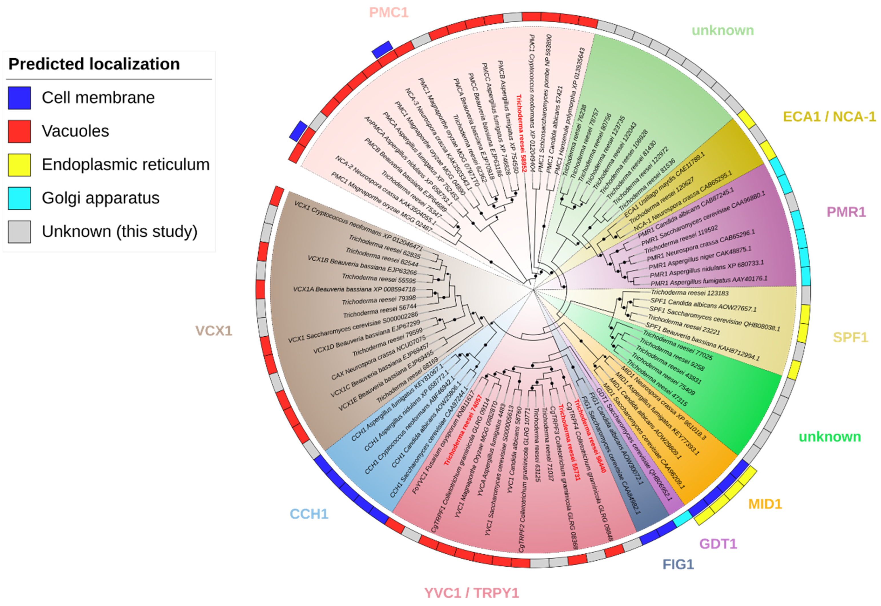
Figure 1. Cladogram of calcium transport proteins. We used sequences of characterized proteins present in the cell membrane, vacuoles, endoplasmic reticulum, and Golgi apparatus together with T. reesei proteins of unknown localization identified in this study. The support values from 1000 resamples are indicated by the black circles, varying from 0 to 1. The figure only shows values from 0.8 to 1, from smaller to larger size.
Jof 10 00853 g001
Figure 2. Comparative expression analysis of T. reesei putative vacuolar calcium transport proteins, homologous to S. cerevisiae’s YVC1 and PMC1. The RNA-Seq data were obtained from studies [6,17,18,19,20,48]. (a) yvc1 expression in commonly used parental strains and (b) the mutant strains lacking Xyr1, Azf1, Cre1, Tmk1, Tmk2 and Epl2, respectively. (c,d) pmc1 expression under the same conditions. Cel = cellulose, Glu = glucose, Soph = sophorose, SCB = sugarcane bagasse, Gly = glycerol.
Jof 10 00853 g002
Figure 3. Growth of strains QM6a∆tmus53pyr4 (parental), ∆trpmc1, ∆tryvc1, ∆tryvc3, and ∆tryvc4 in minimal media in the presence of 25 mM of different carbon sources for 72 h, with and without 10 mM CaCl2 supplementation. The values represent the absorbance readings at 750 nm.
Jof 10 00853 g003
Figure 4. Growth of strains QM6a∆tmus53pyr4 (parental), ∆trpmc1, ∆tryvc1, ∆tryvc3, and ∆tryvc4 in 25 mM xylose minimal media for 48 or 72 h in the presence of 10, 100, or 300 mM of the metals calcium (a,b), copper (c,d), manganese (e,f), cobalt (g,h), zinc (i,j), iron (k,l), magnesium (m,n) or aluminum (o,p). Significance levels are indicated as follows: * p < 0.05, ** p < 0.01, and *** p < 0.001 in relation to the parental strain (Student’s t-test). The control condition (gray bars) was calculated for each microplate.
Jof 10 00853 g004
Figure 5. Growth of strains QM6a∆tmus53pyr4 (parental), ∆trpmc1, ∆tryvc1, ∆tryvc3, and ∆tryvc4 in 25 mM xylose Minimal media in the presence of 0.5 M NaCl (ac) or 0.5 M KCl (df) and in presence or absence of 10 mM CaCl2. * p < 0.05, ** p < 0.01, and *** p < 0.001 in relation to the parental strain (two-way ANOVA, followed by Tukey’s multiple comparisons). Non-significant results are indicated as ‘ns’. The control condition (gray bars) was calculated for each microplate.
Jof 10 00853 g005
Figure 6. Growth analysis of strains QM6a∆tmus53pyr4 (parental), ∆trpmc1, ∆tryvc1, ∆tryvc3, and ∆tryvc4 in MA with 1% carboxymethylcellulose (CMC) for 5 days or 2% glucose + 100 mM Congo red (CR) for 3 days. (a) growth in plates, (b) growth measurements in 1% CMC and (c) 100 mM CR. Significance levels are indicated as follows: * p < 0.05, ** p < 0.01, and *** p < 0.001 (Student’s t-test). Non-significant results are indicated as ‘ns’.
Jof 10 00853 g006
Figure 7. CMCase activity of parental and mutant strains grown for 24 h in MA media with 1% cellulose with or without 10 mM CaCl2 supplementation for 24 h (a), 48 h (b), 72 h (c), and 96 h (d), and gene expression of endoglucanase (cel7b) (e), cellobiohydrolases (cel7a and cel6a) (f,g) and beta-glucosidase (cel3a) (h). Significance levels are indicated as follows: * p < 0.05, ** p < 0.01, and *** p < 0.001 (Student’s t-test). Non-significant results are indicated as ‘ns’.
Jof 10 00853 g007
Figure 8. Gene expression of transcription factors and calcium signaling components of parental and mutant strains grown on MA media with 1% cellulose for 24 h. Gene expression of xyr1 (a), ace3 (b), hac1a (c), cre1 (d), crz1 (e), cam (f), and cna1 (g). * p < 0.05, ** p < 0.01, and *** p < 0.001 (Student’s t-test). Non-significant results are indicated as ‘ns’.
Jof 10 00853 g008
Figure 9. Gene expression of xylose metabolism enzymes (ac), calcium signaling components (df), xylanases (g,h), and secondary metabolites components (ik) of parental and mutant strains grown on MA media with 25 mM xylose for 48 h. * p < 0.05, ** p < 0.01, and *** p < 0.001 (Student’s t-test). Non-significant results are indicated as ‘ns’.
Jof 10 00853 g009
Figure 10. Confocal microscopy and analysis of cell wall thickness. (a) the strains were grown in MA media with 25 mM xylose supplemented with 10 mM CaCl2 and stained with 5 µM Fluo-4/AM. (b) the strains were grown in MEX media supplemented with 0 or 10 mM CaCl2 for 1 day in a microscope slide and stained with 0.001% calcofluor white. (c) measurements of cell wall thickness. ** p < 0.01 and *** p < 0.001 (Student’s t-test). Non-significant results are indicated as ‘ns’.
Jof 10 00853 g010
Figure 11. Putative model of the function of the vacuolar calcium transport proteins YVC1 and PMC1 of T. reesei. The deletion of these proteins impairs the dynamics of calcium transport in the vacuoles, therefore, when extracellular calcium enters the cells, the calcium-mediated signaling does not occur properly, as the expression of the transcription factors ACE3, CRZ1, CRE1, YPR1, and YPR2 are increased/decreased, causing a reduction in cellulases/hemicellulases expression, manganese and osmotic stress tolerance and an increase in xylose assimilation and in cell wall thickness. red arrows down—down-regulation effect and blue arrows up—up-regulation effect.
Jof 10 00853 g011
Table 1. Potential vacuolar calcium transporters and channels in T. reesei. The proteins under study are highlighted in bold and underlined.
JGI IDPossible Homologue in S. cerevisiaeSize (aa)AnnotationFull e-Value (Hmmsearch)
74057YVC1625Nonselective cation channel protein1.8 × 10−256
55731YVC11167Nonselective cation channel protein7 × 10−164
56440YVC1701Nonselective cation channel protein5 × 10−145
63125YVC1631Nonselective cation channel protein3.7 × 10−121
71037YVC11104Nonselective cation channel protein3.4 × 10−117
79599VCX1462Vacuolar calcium ion transporter1.4 × 10−178
55595VCX1421Vacuolar calcium ion transporter2 × 10−166
79398VCX1339Vacuolar calcium ion transporter2.1 × 10−151
56744VCX1381Vacuolar calcium ion transporter8.5 × 10−100
82544VCX1433Vacuolar calcium ion transporter4.3 × 10−87
68169VCX1394Vacuolar calcium ion transporter1 × 10−81
62835VCX11115Vacuolar calcium ion transporter7.4 × 10−48
75347PMC11379Calcium-translocating P-type ATPase, PMCA-type0
58952PMC11204Calcium-translocating P-type ATPase, PMCA-type0
62362PMC11281Calcium-translocating P-type ATPase, PMCA-type0
120627PMC1998Calcium-translocating P-type ATPase, SERCA-type2.4 × 10−142
119592PMC11062Calcium-transporting ATPase1.5 × 10−137
81536PMC11101Calcium-transporting ATPase4.3 × 10−111
122972PMC11071Calcium-transporting ATPase8.4 × 10−109
81430PMC11023Calcium-transporting ATPase1.3 × 10−106
106928PMC11049Sodium/potassium-transporting ATPase subunit alpha6.3 × 10−94
78757PMC1923Calcium-transporting ATPase1.9 × 10−47
76238PMC1982Calcium-transporting ATPase1.4 × 10−45
123183PMC11309Cation-transporting ATPase-related1.1 × 10−38
122043PMC11171Heavy metal translocating P-type ATPase2.2 × 10−36
43831PMC11354Probable phospholipid-transporting ATPase1.2 × 10−32
23221PMC11318Cation-transporting ATPase-related3.6 × 10−31
123735PMC11105Heavy metal translocating P-type ATPase5 × 10−30
77025PMC11300Probable phospholipid-transporting ATPase7.4 × 10−30
80756PMC11133Heavy metal translocating P-type ATPase1 × 10−27
79258PMC11534Probable phospholipid-transporting ATPase3.2 × 10−27
47315PMC11392Probable phospholipid-transporting ATPase2.7 × 10−23
75409PMC11368Probable phospholipid-transporting ATPase2.4 × 10−20
Disclaimer/Publisher’s Note: The statements, opinions and data contained in all publications are solely those of the individual author(s) and contributor(s) and not of MDPI and/or the editor(s). MDPI and/or the editor(s) disclaim responsibility for any injury to people or property resulting from any ideas, methods, instructions or products referred to in the content.