Case Report

Clinical and Radiological Features of Pneumocystis jirovecii Pneumonia in Children: A Case Series

1
Division of Infectious Diseases, IRCCS Istituto Giannina Gaslini, Via Gerolamo Gaslini 5, 16147 Genoa, Italy
2
Division of Infectious Diseases, Department of Health Sciences (DISSAL), University of Genoa, 16132 Genoa, Italy
3
IRCCS Ospedale Policlinico San Martino, 16132 Genoa, Italy
4
Department of Neuroscience, Rehabilitation, Ophthalmology, Genetics, Maternal and Child Health (DINOGMI), University of Genova, 16132 Genoa, Italy
5
Division of Ematology, IRCCS Istituto Giannina Gaslini, 16147 Genoa, Italy
6
Unit of Bone Marrow Transplantation, IRCCS Istituto Giannina Gaslini, 16147 Genoa, Italy
7
Division of Neonatal and Pediatric Critical Care and Semi-Intensive Care, IRCCS Istituto Giannina Gaslini, 16147 Genoa, Italy
8
Center for Autoinflammatory Diseases and Immunodeficiencies, IRCCS Istituto Giannina Gaslini, 16147 Genoa, Italy
9
Hygiene Unit, Department of Health Sciences, Ospedale Policlinico San Martino, University of Genoa, 16132 Genoa, Italy
10
Divison of Radiology, IRCCS Istituto Giannina Gaslini, 16147 Genoa, Italy
11
Microbiology Unit, Department of Surgical Sciences and Integrated Diagnostics (DISC), University of Genoa, 16132 Genoa, Italy
*
Author to whom correspondence should be addressed.
These authors contributed equally to this work.
J. Fungi 2024, 10(4), 276; https://doi.org/10.3390/jof10040276
Submission received: 9 February 2024 / Revised: 27 March 2024 / Accepted: 4 April 2024 / Published: 9 April 2024
(This article belongs to the Special Issue Diagnosis of Invasive Fungal Diseases)

Abstract

:
Background: Pneumocytis jirovecii pneumonia (PJP) has high mortality rates in immunocompromised children, even though routine prophylaxis has decreased in incidence. The aim of this case series is to present the radiological and clinical pathway of PJP in a pediatric population. Description of Cases: All PJP cases in non-HIV/AIDS patients diagnosed at Istituto Giannina Gaslini Pediatric Hospital in Genoa (Italy) from January 2012 until October 2022 were retrospectively evaluated. Nine cases were identified (median age: 8.3 years), and of these, 6/9 underwent prophylaxis with trimethoprim/sulfamethoxazole (TMP/SMX; five once-a-week schedules and one three times-a-week schedule), while 3/9 did not receive this. PJP was diagnosed by real-time PCR for P. jirovecii-DNA in respiratory specimens in 7/9 cases and two consecutive positive detections of β-d-glucan (BDG) in the serum in 2/9 cases. Most patients (6/8) had a CT scan with features suggestive of PJP, while one patient did not undergo a scan. All patients were treated with TMP/SMX after a median time from symptoms onset of 3 days. In 7/9 cases, empirical TMP/SMX treatment was initiated after clinical suspicion and radiological evidence and later confirmed by microbiological data. Clinical improvement with the resolution of respiratory failure and 30-day survival included 100% of the study population. Discussion: Due to the difficulty in obtaining biopsy specimens, PJP diagnosis is usually considered probable in most cases. Moreover, the severity of the clinical presentation often leads physicians to start TMP/SMX treatment empirically. BDG proved to be a useful tool for diagnosis, and CT showed good accuracy in identifying typical patterns. In our center, single-day/week prophylaxis was ineffective in high-risk patients; the three-day/week schedule would, therefore, seem preferable and, in any case, should be started promptly in all patients who have an indication of pneumonia.

1. Introduction

The incidence of pneumocystis jirovecii pneumonia (PJP) has gradually decreased in the Acquired Immune Deficiency Syndrome (AIDS) population [1], whereas, over the last few decades, it has increased in patients affected by immunocompromising conditions [2], with infection rate ranging from 15 to 45% in the absence of antimicrobial prophylaxis [3]. Conditions associated with a higher risk of PJP are chemotherapy, hematopoietic stem cell transplantation (HCT), severe defects in T-cell immunity, lymphopenia, receiving high doses of corticosteroids for any reasons, and autoimmune diseases in particular [4,5,6,7]. In immunocompromised patients, the introduction of trimethoprim-sulfamethoxazole (TMP/SMX) prophylaxis led to a decline in the rate of PJP infections [4,8,9,10,11]. However, despite the introduction of this highly effective prophylaxis [12,13,14], the PJP-associated mortality rate among children with the non-human immunodeficiency virus (HIV) is still high in developing countries, ranging from 40 to 50% [15,16].
In non-HIV patients, PJP presents as acute and rapidly progressive respiratory deterioration [17,18,19]. A wide variety of radiological features are found both in AIDS- and non-AIDS-associated diseases [20,21], with typical bilateral ground-glass opacities on the upper lobes and peripheral sparing in non-HIV immunocompromised patients [22,23,24,25]. Less common radiological features are nodules, cavities, cystic or honeycomb lesions, pneumothorax, pleural effusion, hilar enlargement, and pneumothorax [23,26,27,28,29]. The gold standard for identifying pulmonary findings is high-resolution computerized tomography (CT) [27,30,31].
The proven diagnosis of PJP relies on the histological identification of trophic and cystic forms of trophozoites. However, since lung biopsy is not always feasible, often, clinicians rely on probable diagnosis, which needs clinical, radiological, and microbiological criteria [12,32], e.g., a polymerase chain reaction (PCR) showing high sensitivity [33,34,35], and serum/plasmatic (1–3)-β-D-glucan (BDG) [36,37,38,39].
This case series aims to describe the typical and suggestive clinical presentation and the most common CT scan features of PJP in an immunocompromised non-AIDS pediatric population.

2. Materials and Methods

2.1. Study Population

We retrospectively evaluated data from patients under 20 years old who were diagnosed with proven or probable PJP at the IRCCS Istituto Giannina Gaslini Pediatric Hospital in Genoa (Italy) during the period 1 January 2012–31 October 2022. The identification of episodes was based on data extraction from the laboratories that performed a PCR for Pneumocystis jirovecii and BDG, as well as from the DRG (diagnosis-related group) register of our center. These data were then compared with the medical records. Of the extracted results, we selected children with proven or probable PJP diagnosis by analyzing the patients’ clinical records.

2.2. Inclusion Criteria

Five inclusion criteria had to be met by each child included in this study as follows: (i) the presence of relevant pulmonary symptoms (i.e., cough or dyspnea), (ii) pulmonary infiltration observed by chest radiography or CT, (iii) the detection of P. jirovecii by real-time PCR in respiratory specimens and/or two consecutive positive detections of BDG in the serum, (iv) the ruling out of another possible invasive fungal diseases (IFDs), and (v) the availability of 30-day follow-up.

2.3. Diagnosis of PJP

Proven and probable PJP was defined according to the “Revised EORTC/MSGERC Invasive Fungal Disease Definitions” [12], in which “proven PJP” was defined by the presence of the clinical and microbiological criteria of PJP diagnosis plus the demonstration of P. jirovecii by microscopy using conventional or immunofluorescence straining on tissue or respiratory specimens; “probable PJP” was defined by the presence of the clinical and microbiological criteria of a PJP diagnosis plus the detection of P. jirovecii by real-time PCR in the respiratory specimens or detection of BDG in the serum. Other possible IFDs were ruled out thanks to BAL cultures for molds, serum galactomannan if the patient was neutropenic, and blood cultures.

2.4. Data Collection

Clinical data collected included general demographic information, underlying diseases, immunosuppressive therapies, clinical symptomatology, laboratory values, and 30-day hospital mortality. Any further episodes of PJP after the first episode in the subsequent two-year period were also retrospectively analyzed.
Demographical and clinical details were retrieved from the hospital’s computerized record system. For each patient, symptoms from clinical onset to diagnosis were recorded.

2.5. Imaging Review

All CT scans were reviewed by a radiologist experienced in pediatric radiology (the author M.B.D), who provided a detailed description of the images for each patient. After a literature review on the typical radiological features of PJP in both the adult and pediatric population, the radiologist classified the CT scans as “typical features associated with PJP” and “non-typical features associated with PJP”.

2.6. Statistical Analysis

Continuous variables were described by the median and range or interquartile range (IQR). Categorical variables were described by numbers and percentages.

3. Results

3.1. Clinical and Epidemiological Characteristics of Study Population

A total of 9 children were included in this study, with a median age of 8.3 years (IQR 3.3–13.9), of which 6/9 (66.6%) were female. They all received a diagnosis of probable PJP, though none of them had the criteria for a proven one. The clinical and epidemiological characteristics of the study population are summarized in Table 1. Steroids (with an equivalent dose of prednisone ≥ 0.3 mg/kg per day for at least 2 weeks before diagnosis) and/or other immunosuppressive/anti-inflammatory drugs were administered to 7/9 patients. In the 3 patients who developed PJP after allogeneic HCT, the infection was diagnosed at a median of 27 days (IQR 26.5–73.5) after the procedure, and, at that time, 2/3 (66.6%) were affected by active graft-versus-host-disease (GvHD) with gastrointestinal involvement.
Five out of nine (55.5%) patients had profound lymphocytopenia with CD4 cell count < 200/mm3 at the time of PJP diagnosis.

3.2. Study Population

Six patients (66.6%) were receiving TMP/SMX prophylaxis with a once-a-week (n = 5) or three-weekly (n = 1) regimen and one adolescent patient (patient 5) had poor treatment adherence due to his non-acceptance of the underlying disease and social difficulties.
The remaining three patients did not receive prophylaxis, although they should have since they were all receiving a dose equivalent of prednisone ≥ 0.3 mg/kg per day for at least 2 weeks before PJP diagnosis.

3.3. Clinical Presentation

All patients presented with acute respiratory failure, characterized by a rapid respiratory deterioration requiring supplemental oxygen therapy, which in 4/9 cases (44.4%) was represented by high-flow nasal cannula (HFNC) and/or continuous positive airway pressure (CPAP) and in the remaining cases by oxygen at low flows.

3.4. PJP Diagnostic Criteria

Microbiological criteria for diagnosis were represented by the positive detection of P. jirovecii deoxyribonucleic acid (DNA) by quantitative real-time PCR in respiratory specimens in 4/9 cases (44.4%), qualitative real-time PCR in 3/9 cases (33.3%) and two consecutive positive detections of BDG in serum in 2/9 cases (22.2%). In the study population, 6/9 (66.6%) patients had at least one positive detection of BDG in the serum, with a median value of 486 pg/mL (IQR 405–523); no BDG test was performed on the other three patients. The median time from clinical presentation to microbiological positivity was 2 days (IQR 1–7).

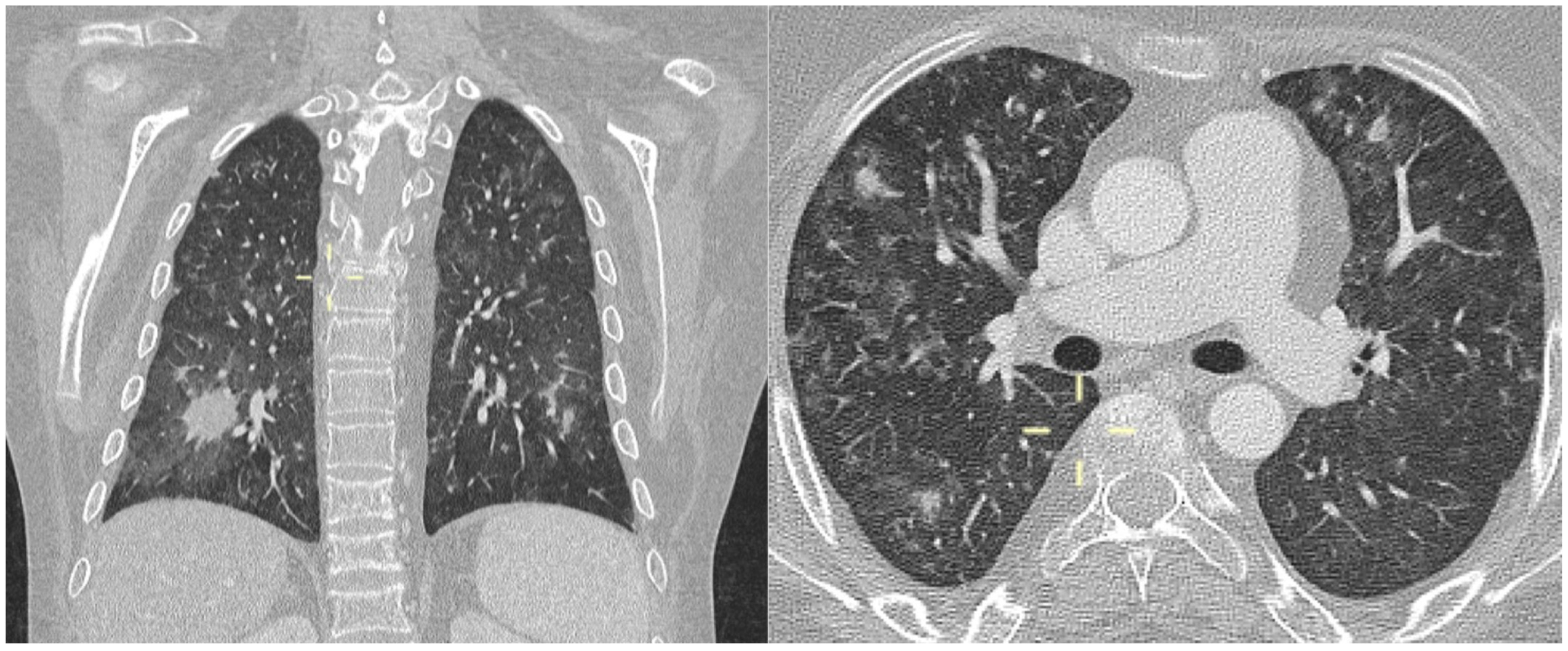
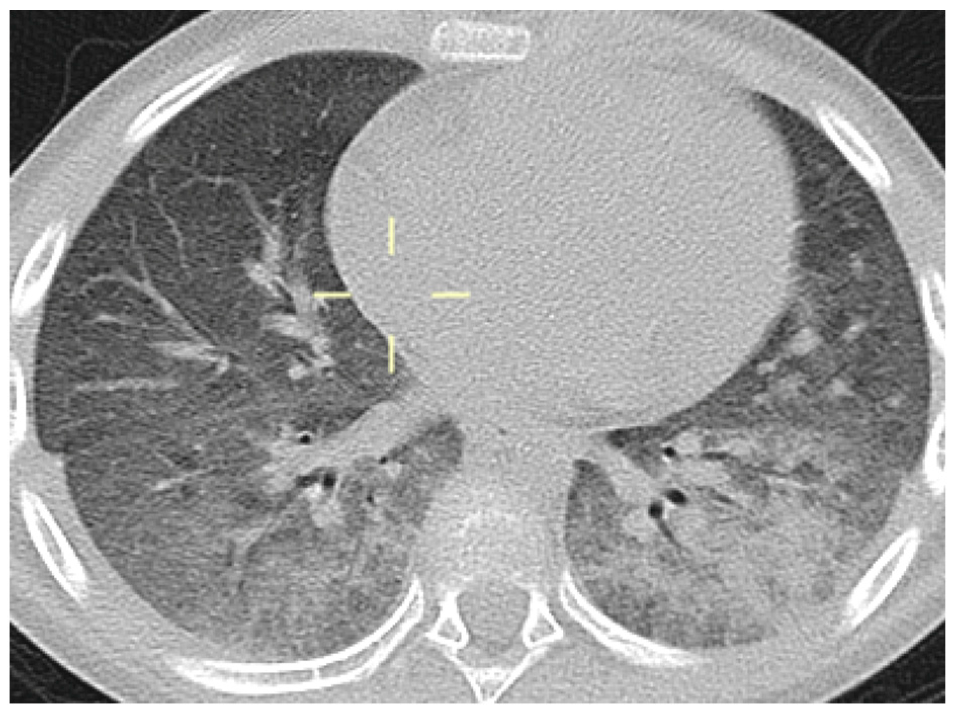
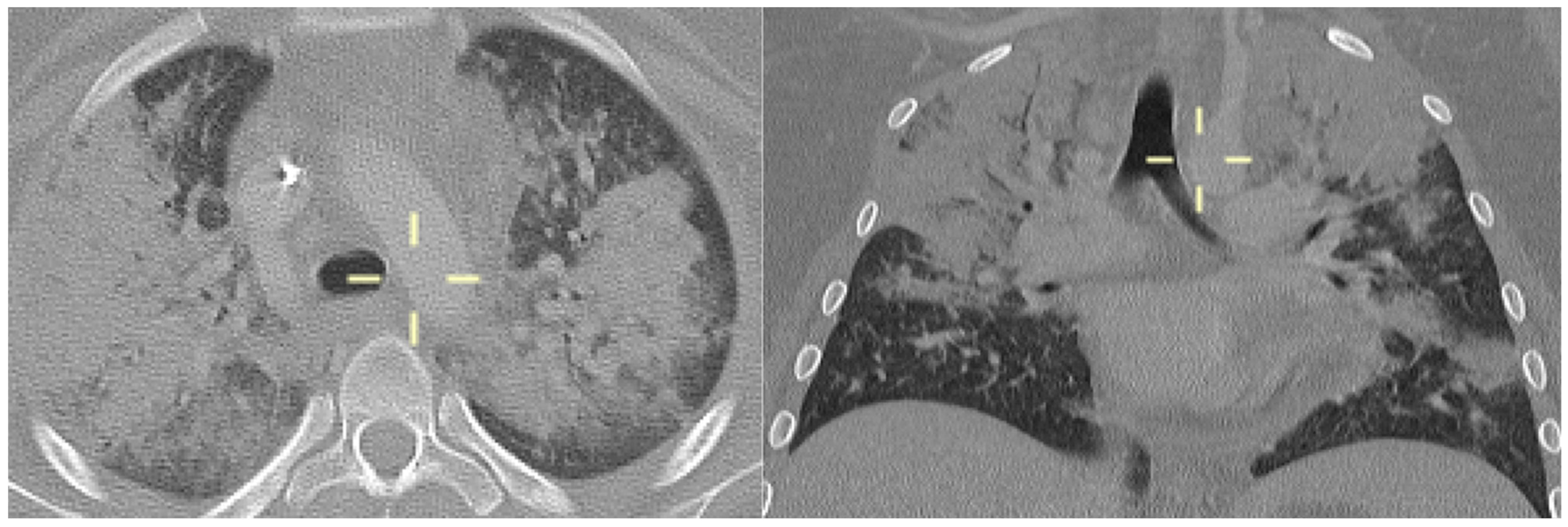
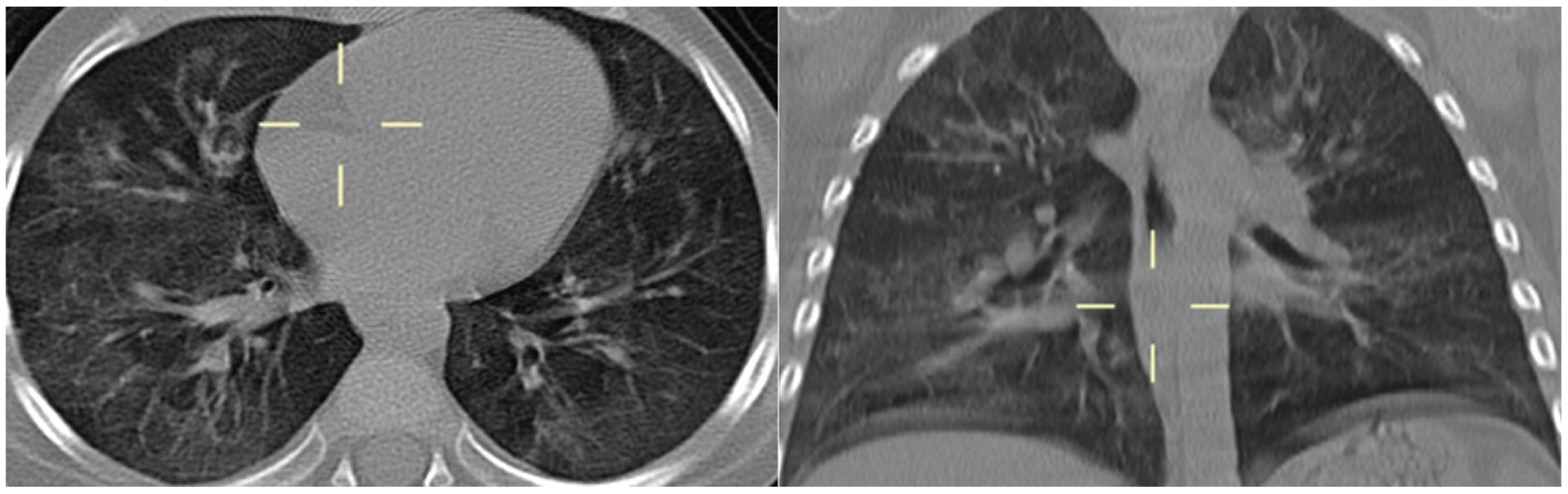
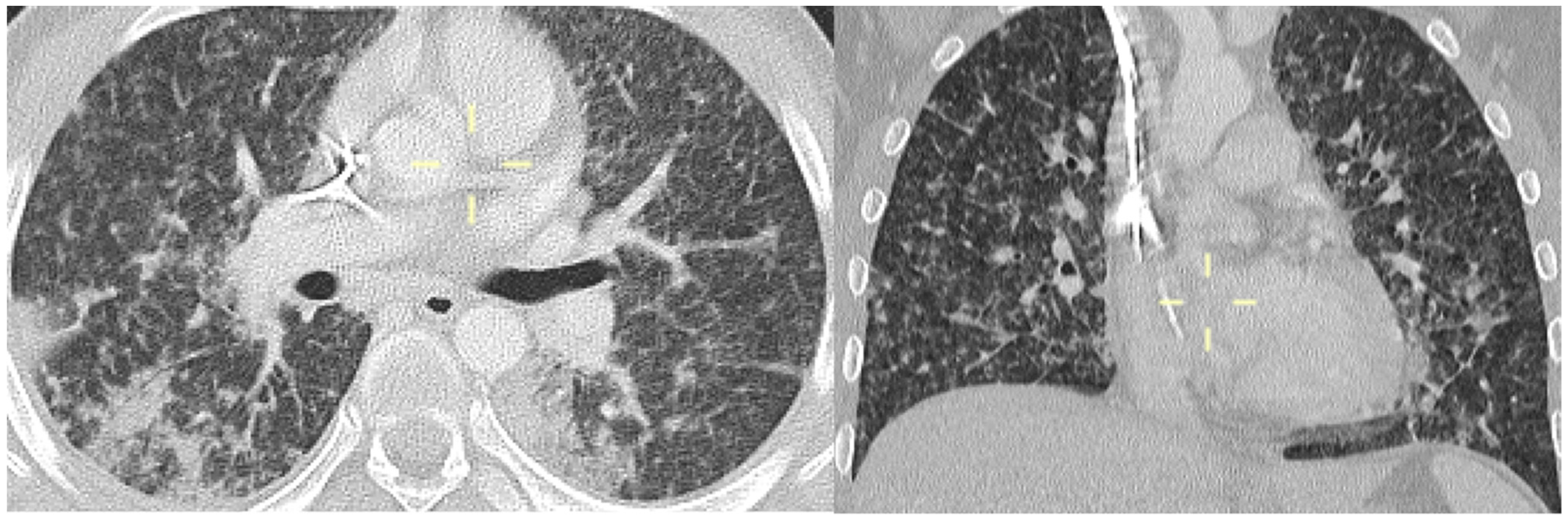
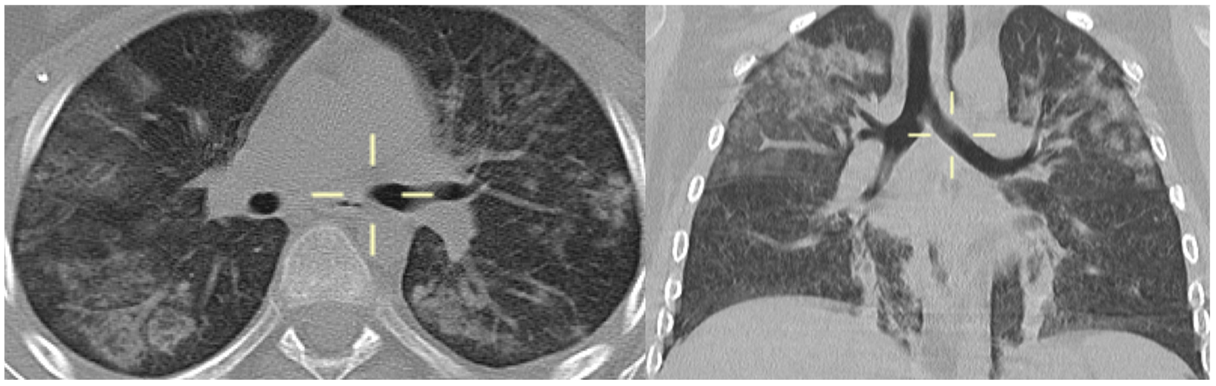
3.5. Radiological Features of PJP in the Study Population

Given the acute clinical presentation, all patients received an early radiological examination in a median of 2 days (IQR 0–5) after the onset of respiratory symptoms.
Overall, 8/9 patients (88.8%) underwent a CT scan for a median of 4 days (IQR 0.75–5.75) after symptoms onset. Two patients were unable to perform a CT scan due to clinical severity, which prevented transport to the radiology department for a CT scan. For those two patients, treatment was started based on chest X-ray (CRX) findings, and in one of them, a CT scan was nevertheless performed on day 23 after clinical stabilization to better characterize the pulmonary radiological pattern.
All CT scans were reviewed by an expert pediatric radiologist, who, after a literature review of typical radiological features of PJP in both the adult and pediatric population, classified the CT scans as “typical features associated with PJP” in 6/8 (75%) patients and “non-typical images associated with PJP” in 2/8 (25%) patients.
A detailed description of the TC scans classified as “typical” is presented in Figure 1, Figure 2, Figure 3, Figure 4, Figure 5 and Figure 6.
The two “non-typical images” episodes were both characterized by multiple thickenings, some of which demonstrated air bronchogram spread throughout the entire lung parenchyma without pleural effusion. Multiple thickenings were also observed, some of which had air bronchogram spread throughout the entire lung parenchyma.

3.6. Treatment and Outcomes

All patients were treated with a 21-day course of treatment with TMP/SMX, with a median time from symptoms onset to treatment initiation of 3 days (IQR 2–8). In 7/9 (77.7%) cases, TMP-SMX was started empirically based on clinical and radiological findings while awaiting microbiological confirmation; in two cases, TMP-SMX was started based on microbiological positivity, which preceded radiological examination.
Clinical improvement with the resolution of respiratory failure was achieved in all patients, and 30-day survival was achieved for 100% of the study population.

3.7. Secondary Prophylaxis

After completing the 21-day treatment cycle, all patients included in the study received secondary prophylaxis with TMP/SMX. Among these, 5/9 patients received three-weekly administration, while 4/9 patients received a single-day course of prophylaxis with once-a-week administration, attributing the failure of prophylaxis to poor compliance rather than to a lack of effectiveness.
In the case of one patient (patient 7) receiving once-weekly prophylaxis, a second episode of PJP occurred 1 year after the previous one. He presented with acute respiratory failure and radiological evidence of interstitial pneumonia, which led to a second course of treatment with TMP/SMX. Unfortunately, the patient died before the completion of therapy due to complications related to the underlying disease.

4. Discussion

The study included nine immunocompromised, non-HIV/AIDS children who met specific criteria for probable PJP diagnosis. The study population was at high risk for PJP due to various factors, including immunosuppressive therapies and lymphopenia. The majority of patients were receiving high-dose corticosteroids, which are known to increase the risk of PJP [4,5,6,7]. Indeed, almost half of them were patients with hematological malignancies, confirming the finding that PJP is 1 of the 14 acute toxic effects of antileukemic therapy recognized by the Delphi consensus by 15 international childhood acute lymphoblastic leukemia study groups [40]. It is noteworthy that six patients developed PJP while on prophylaxis with TMP/SMX, with a once-a-week administration for five patients and a three-weekly administration for one patient. Although the effectiveness of once-a-week prophylaxis in the pediatric population has been well described by means of a large epidemiological study [41], failures have been reported in categories of children particularly at risk, such as allogeneic HCT [42]. In these cases, the concomitant presence of GvHD, present in 2/3 of the patients undergoing HCT and under prophylaxis at the time of diagnosis, may have altered the intestinal absorption of TMP/SMX, playing an important role in the failure of prophylaxis. The association between the presence of GvHD and the incidence of PJP is described [43], and this finding made us rethink the type of anti-PJP prophylaxis that should be undertaken in this particular patient setting [42].
Regarding treatment adherence to chronic care in the pediatric population, our data on poor adherence in an adolescent patient highlight how delicate it is to treat patients with daily therapies in this specific age group [44,45] and also for PJP [46].
Another remarkable aspect to consider is incomplete adherence to the criteria for initiating ani-PJP prophylaxis in the study population, as three patients (patients 1, 8, and 9) in our case series developed an infection in the absence of appropriate prophylaxis, even though this was indicated due to the presence of risk factors (all three patients were receiving high-dose corticosteroid therapy).
The clinical presentation of PJP in our series was characterized by acute and severe respiratory failure, necessitating oxygen therapy in all cases. The prompt radiological examinations, including CT scans, aided in the diagnosis and early treatment of PJP. The majority of CT scans (75%) were classified as showing “typical features associated with PJP”, including diffuse bilateral interstitial pulmonary infiltrates [20,21]. Additionally, CT scans revealed ground-glass opacities on the upper lobes with peripheral sparing, which is a common finding in non-HIV immunocompromised patients with PJP [23,26,27,28]. It is important to note that no radiological findings were pathognomonic of PJP. In fact, in two patients, the radiological presentation was not typical, so non-suggestive imaging should not exclude a diagnosis in case of high-level clinical suspicion. However, in the majority of cases, high-resolution CT proved to be the best radiological method for diagnosing PJP. It has been documented that CRX can be initially normal or specific [27,30,31], and therefore, CT scans play a crucial role in the diagnostic assessment of suspected PJP cases, especially in immunosuppressed pediatric patients.
PJP diagnoses were all probable diagnoses according to the EORTC/MSGERC criteria [12], with a Pneumocystis PCR and/or BDG appearing positive in all patients. It has been demonstrated that the sensitivity and specificity of PCR on non-invasive respiratory samples (e.g., sputum) are high at 99 and 96%, while on serum, its sensitivity is 77% and the specificity is 90%, though potentially without distinguishing between colonization and infection [38]. In order to reduce possible confounding factors suggestive of colonization rather than infection, in our clinical practice, a Pneumocystis PCR was exclusively performed in patients with strong clinical suspicion, predisposing underlying conditions, and/or compatible radiological findings. It is usually performed only on BAL in patients who have undergone bronchoscopy, which, despite being a safe procedure, is an invasive test, particularly for pediatric patients. The role of PCR and serum BDG showed potential as a supportive diagnostic tool, especially when combined with other P. jirovecii-specific assays in adults [36,37,38,39], and the combination of these two tests increased both positive and negative predictive values [47]; therefore, the use of BDG for diagnosing PJP in the pediatric population could be useful, while it is generally not recommended for the diagnosis of IFD [48].
Treatment with TMP/SMX was initiated promptly in all patients upon clinical and radiological suspicion, leading to clinical improvement and 100% 30-day survival in the study population.
A remarkable aspect that emerged from the analysis of this case series was the failure of secondary prophylaxis, which became apparent after the first episode during a once-weekly administration schedule of TMP/SMX as secondary prophylaxis. This finding, in addition to the data on the failure of such a scheme of applying for primary prophylaxis [42], should make one rethink the indications of a daily or three-weekly administration in the pediatric population.
In conclusion, PJP is a significant cause of morbidity and mortality in immunocompromised children, and early recognition of this disease is important to enable the rapid initiation of target therapy. In this setting, CT plays a crucial role in recognizing compatible radiological patterns, and combined with molecular detection by PCR and the detection of serum BDG in suspected cases, allows the diagnosis to be confirmed in pediatric patients.

Author Contributions

Conceptualization, E.R. and A.M. (Andrea Moscatelli); investigation, E.M., F.P., G.B., A.M. (Andrea Moscatelli) and R.C.; formal analysis, M.M., M.B.D. and A.M. (Anna Marchese) and B.B.; resources, data curation, C.R. and C.S.; writing—original draft preparation, C.B.; writing—review and editing, E.R.; supervision, E.C. All authors have read and agreed to the published version of the manuscript.

Funding

This research was partially supported by grants from Ministero della Salute—Ricerca Corrente 2023.

Institutional Review Board Statement

The study was performed in accordance with the ethical standards laid down in the 1964 Declaration of Helsinki and its later amendments or comparable ethical standards. Written informed consent was obtained from parents/legal guardians, or the patients themselves if they were adults. Clinical and laboratory data were collected from the patient’s medical record in the context of clinical routine. All information and metadata were anonymized. Samples were coded and downstream retrospective analyses were performed with anonymized data.

Informed Consent Statement

Informed consent was obtained from all subjects involved in the study.

Data Availability Statement

The data presented in this study are not available for privacy reasons.

Conflicts of Interest

The authors declare no conflicts of interest.

References

  1. Gona, P.; Van Dyke, R.B.; Williams, P.L.; Dankner, W.M.; Chernoff, M.C.; Nachman, S.A.; Seage, G.R. Incidence of Opportunistic and Other Infections in HIV-Infected Children in the HAART Era. J. Am. Med. Assoc. 2006, 296, 292. [Google Scholar] [CrossRef] [PubMed]
  2. Morris, A.; Lundgren, J.D.; Masur, H.; Walzer, P.D.; Hanson, D.L.; Frederick, T.; Huang, L.; Beard, C.B.; Kaplan, J.E. Current Epidemiology of Pneumocystis Pneumonia. Emerg. Infect. Dis. 2004, 10, 1713–1720. [Google Scholar] [CrossRef] [PubMed]
  3. Sepkowitz, K.A. Opportunistic Infections in Patients with and Patients without Acquired Immunodeficiency Syndrome. Clin. Infect. Dis. 2002, 34, 1098–1107. [Google Scholar] [CrossRef] [PubMed]
  4. Yale, S.H.; Limper, A.H. Pneumocystis carinii Pneumonia in Patients without Acquired Immunodeficiency Syndrome: Associated Illnesses and Prior Corticosteroid Therapy. Mayo Clin. Proc. 1996, 71, 5–13. [Google Scholar] [CrossRef] [PubMed]
  5. Roblot, F.; Godet, C.; Le Moal, G.; Garo, B.; Faouzi Souala, M.; Dary, M.; De Gentile, L.; Gandji, J.A.; Guimard, Y.; Lacroix, C.; et al. Analysis of underlying diseases and prognosis factors associated with Pneumocystis carinii pneumonia in immunocompromised HIV-negative patients. Eur. J. Clin. Microbiol. Infect. Dis. 2002, 21, 523–531. [Google Scholar] [PubMed]
  6. Ling, C.; Qian, S.; Wang, Q.; Zeng, J.; Jia, X.; Liu, J.; Li, Z. Pneumocystis pneumonia in non-HIV children: A 10-year retrospective study. Clin. Respir. J. 2018, 12, 16–22. [Google Scholar] [CrossRef]
  7. Basiaga, M.L.; Ross, M.E.; Gerber, J.S.; Ogdie, A. Incidence of Pneumocystis jirovecii and Adverse Events Associated with Pneumocystis Prophylaxis in Children Receiving Glucocorticoids. J. Pediatr. Infect. Dis. Soc. 2017, 7, 283–289. [Google Scholar] [CrossRef] [PubMed]
  8. Thomas, C.F.; Limper, A.H. Pneumocystis Pneumonia. N. Engl. J. Med. 2004, 350, 2487–2498. [Google Scholar] [CrossRef]
  9. Pyrgos, V.; Shoham, S.; Roilides, E.; Walsh, T.J. Pneumocystis pneumonia in children. Paediatr. Respir. Rev. 2009, 10, 192–198. [Google Scholar] [CrossRef]
  10. Saltzman, R.W.; Albin, S.; Russo, P.; Sullivan, K.E. Clinical conditions associated with PCP in children. Pediatr. Pulmonol. 2012, 47, 510–516. [Google Scholar] [CrossRef]
  11. Russian, D.A.; Levine, S.J. Pneumocystis carinii Pneumonia in Patients without HIV Infection. Am. J. Med. Sci. 2001, 321, 56–65. [Google Scholar] [CrossRef] [PubMed]
  12. Lagrou, K.; Chen, S.; Masur, H.; Viscoli, C.; Decker, C.F.; Pagano, L.; Groll, A.H. Pneumocystis jirovecii Disease: Basis for the Revised EORTC/MSGERC Invasive Fungal Disease Definitions in Individuals without Human Immunodeficiency Virus. Clin. Infect. Dis. 2021, 72 (Suppl. S2), S114–S120. [Google Scholar] [CrossRef] [PubMed]
  13. Stern, A.; Green, H.; Paul, M.; Vidal, L.; Leibovici, L. Prophylaxis for Pneumocystis pneumonia (PCP) in non-HIV immunocompromised patients. Cochrane Database Syst. Rev. 2014, 2016. [Google Scholar] [CrossRef] [PubMed]
  14. Maertens, J.; Cesaro, S.; Maschmeyer, G.; Einsele, H.; Donnelly, J.P.; Alanio, A.; Hauser, P.M.; Lagrou, K.; Melchers, W.J.G.; Helweg-Larsen, J.; et al. ECIL guidelines for preventing Pneumocystis jirovecii pneumonia in patients with haematological malignancies and stem cell transplant recipients. J. Antimicrob. Chemother. 2016, 71, 2397–2404. [Google Scholar] [CrossRef] [PubMed]
  15. Sepkowitz, K.A. Pneumocystis carinii Pneumonia in Patients without AIDS. Clin. Infect. Dis. 1993, 17 (Suppl. S2), S416–S422. [Google Scholar] [CrossRef] [PubMed]
  16. Bakeera-Kitaka, S.; Musoke, P.; Downing, R.; Tumwine, J.K. Pneumocystis carinii in children with severe pneumonia at Mulago Hospital, Uganda. Ann. Trop. Paediatr. 2004, 24, 227–235. [Google Scholar] [CrossRef] [PubMed]
  17. Kovacs, J.A. Pneumocystis carinii Pneumonia: A Comparison between Patients with the Acquired Immunodeficiency Syndrome and Patients with Other Immunodeficiencies. Ann. Intern. Med. 1984, 100, 663. [Google Scholar] [CrossRef] [PubMed]
  18. Monnet, X.; Vidal-Petiot, E.; Osman, D.; Hamzaoui, O.; Durrbach, A.; Goujard, C.; Miceli, C.; Bourée, P.; Richard, C. Critical care management and outcome of severe Pneumocystis pneumonia in patients with and without HIV infection. Crit. Care 2008, 12, R28. [Google Scholar] [CrossRef] [PubMed]
  19. Yun, K.S.; Anh, B.; Choi, S.H.; Hong, K.T.; Choi, J.Y.; Yun, K.W.; Kang, H.J.; Choi, E.H. Clinical Characteristics and Prognosis of the Modified Probable Pneumocystis jirovecii Pneumonia in Korean Children, 2001–2021. Children 2022, 9, 1596. [Google Scholar] [CrossRef]
  20. Katragkou, A.; Fisher, B.T.; Groll, A.H.; Roilides, E.; Walsh, T.J. Diagnostic Imaging and Invasive Fungal Diseases in Children. J. Pediatr. Infect. Dis. Soc. 2017, 6 (Suppl. S1), S22–S31. [Google Scholar] [CrossRef]
  21. Orlowski, H.L.P.; McWilliams, S.; Mellnick, V.M.; Bhalla, S.; Lubner, M.G.; Pickhardt, P.J.; Menias, C.O. Imaging Spectrum of Invasive Fungal and Fungal-Like Infections. RadioGraphics 2017, 37, 1119–1134. [Google Scholar] [CrossRef]
  22. Vogel, M.N.; Vatlach, M.; Weissgerber, P.; Goeppert, B.; Claussen, C.D.; Hetzel, J.; Horger, M. HRCT-features of Pneumocystis jiroveci pneumonia and their evolution before and after treatment in non-HIV immunocompromised patients. Eur. J. Radiol. 2012, 81, 1315–1320. [Google Scholar] [CrossRef]
  23. Toma, P.; Bertaina, A.; Castagnola, E.; Colafati, G.S.; D’Andrea, M.L.; Finocchi, A.; Lucidi, V.; Mastronuzzi, A.; Granata, C. Fungal infections of the lung in children. Pediatr. Radiol. 2016, 46, 1856–1865. [Google Scholar] [CrossRef]
  24. Cereser, L.; Dallorto, A.; Candoni, A.; Volpetti, S.; Righi, E.; Zuiani, C.; Girometti, R. Pneumocystis jirovecii pneumonia at chest High-resolution Computed Tomography (HRCT) in non-HIV immunocompromised patients: Spectrum of findings and mimickers. Eur. J. Radiol. 2019, 116, 116–127. [Google Scholar] [CrossRef]
  25. Hardak, E.; Brook, O.; Yigla, M. Radiological Features of Pneumocystis jirovecii Pneumonia in Immunocompromised Patients with and without AIDS. Lung 2010, 188, 159–163. [Google Scholar] [CrossRef]
  26. Selwyn, P.A.; Pumerantz, A.S.; Durante, A.; Alcabes, P.G.; Gourevitch, M.N.; Boiselle, P.G.; Elmore, J.G. Clinical predictors of Pneumocystis carinii pneumonia, bacterial pneumonia and tuberculosis in HIV-infected patients. AIDS 1998, 12, 885–893. [Google Scholar] [CrossRef]
  27. Kanne, J.P.; Yandow, D.R.; Meyer, C.A. Pneumocystis jiroveci Pneumonia: High-Resolution CT Findings in Patients with and without HIV Infection. Am. J. Roentgenol. 2012, 198, W555–W561. [Google Scholar] [CrossRef]
  28. Mantadakis, E. Pneumocystis jirovecii Pneumonia in Children with Hematological Malignancies: Diagnosis and Approaches to Management. J. Fungi 2020, 6, 331. [Google Scholar] [CrossRef]
  29. Castagnola, E.; Fioredda, F.; Moroni, C.; Loy, A.; Viscoli, C. Pneumothorax and Pneumocystis Pneumonia in an Infant with Acquired Immunodeficiency Syndrome. Pediatr. Infect. Dis. J. 1992, 11, 504. [Google Scholar] [CrossRef]
  30. Gruden, J.F.; Huang, L.; Turner, J.; Webb, W.R.; Merrifield, C.; Stansell, J.D.; Gamsu, G.; Hopewell, P.C. High-resolution CT in the evaluation of clinically suspected Pneumocystis carinii pneumonia in AIDS patients with normal, equivocal, or nonspecific radiographic findings. Am. J. Roentgenol. 1997, 169, 967–975. [Google Scholar] [CrossRef]
  31. Hsu, J.M.; Hass, A.; Gingras, M.-A.; Chong, J.; Costiniuk, C.; Ezer, N.; Fraser, R.S.; McDonald, E.G.; Lee, T.C. Radiographic features in investigated for Pneumocystis jirovecii pneumonia: A nested case-control study. BMC Infect. Dis. 2020, 20, 492. [Google Scholar] [CrossRef]
  32. Alanio, A.; Hauser, P.M.; Lagrou, K.; Melchers, W.J.G.; Helweg-Larsen, J.; Matos, O.; Cesaro, S.; Maschmeyer, G.; Einsele, H.; Donnelly, J.P.; et al. ECIL guidelines for the diagnosis of Pneumocystis jirovecii pneumonia in patients with haematological malignancies and stem cell transplant recipients. J. Antimicrob. Chemother. 2016, 71, 2386–2396. [Google Scholar] [CrossRef]
  33. Guegan, H.; Robert-Gangneux, F. Molecular diagnosis of Pneumocystis pneumonia in immunocompromised patients. Curr. Opin. Infect. Dis. 2019, 32, 314–321. [Google Scholar] [CrossRef]
  34. Summah, H.; Zhu, Y.G.; Falagas, M.E.; Vouloumanou, E.K.; Qu, J.M. Use of real-time polymerase chain reaction for the diagnosis of Pneumocystis pneumonia in immunocompromised patients: A meta-analysis. Chin. Med. J. 2013, 126, 1965–1973. [Google Scholar] [CrossRef]
  35. Fan, L.-C.; Lu, H.-W.; Cheng, K.-B.; Li, H.-P.; Xu, J.-F. Evaluation of PCR in Bronchoalveolar Lavage Fluid for Diagnosis of Pneumocystis jirovecii Pneumonia: A Bivariate Meta-Analysis and Systematic Review. PLoS ONE 2013, 8, e73099. [Google Scholar] [CrossRef]
  36. Desoubeaux, G.; Chesnay, A.; Mercier, V.; Bras-Cachinho, J.; Moshiri, P.; Eymieux, S.; De Kyvon, M.A.; Lemaignen, A.; Goudeau, A.; Bailly, É. Combination of β-(1,3)-D-glucan testing in serum and qPCR in nasopharyngeal aspirate for facilitated diagnosis of Pneumocystis jirovecii pneumonia. Mycoses 2019, 62, 1015–1022. [Google Scholar] [CrossRef]
  37. Morjaria, S.; Frame, J.; Franco-Garcia, A.; Geyer, A.; Kamboj, M.; Babady, N.E. Clinical Performance of (1,3) Beta-D Glucan for the Diagnosis of Pneumocystis Pneumonia (PCP) in Cancer Patients Tested with PCP Polymerase Chain Reaction. Clin. Infect. Dis. 2019, 69, 1303–1309. [Google Scholar] [CrossRef]
  38. Senécal, J.; Smyth, E.; Del Corpo, O.; Hsu, J.M.; Amar-Zifkin, A.; Bergeron, A.; Cheng, M.P.; Butler-Laporte, G.; McDonald, E.G.; Lee, T.C. Non-invasive diagnosis of Pneumocystis jirovecii pneumonia: A systematic review and meta-analysis. Clin. Microbiol. Infect. 2022, 28, 23–30. [Google Scholar] [CrossRef]
  39. White, P.L.; Backx, M.; Barnes, R.A. Diagnosis and management of Pneumocystis jirovecii infection. Expert Rev. Anti-Infect. Ther. 2017, 15, 435–447. [Google Scholar] [CrossRef]
  40. Schmiegelow, K.; Attarbaschi, A.; Barzilai, S.; Escherich, G.; Frandsen, T.L.; Halsey, C.; Hough, R.; Jeha, S.; Kato, M.; Liang, D.-C.; et al. Consensus definitions of 14 severe acute toxic effects for childhood lymphoblastic leukaemia treatment: A Delphi consensus. Lancet Oncol. 2016, 17, e231–e239. [Google Scholar] [CrossRef]
  41. Caselli, D.; Petris, M.G.; Rondelli, R.; Carraro, F.; Colombini, A.; Muggeo, P.; Ziino, O.; Melchionda, F.; Russo, G.; Pierani, P.; et al. Single-Day Trimethoprim/Sulfamethoxazole Prophylaxis for Pneumocystis Pneumonia in Children with Cancer. J. Pediatr. 2014, 164, 389–392.e1. [Google Scholar] [CrossRef]
  42. Castagnola, E.; Mesini, A.; Saffioti, C.; Moscatelli, A.; Pierri, F.; Giardino, S.; Faraci, M. Failures of once-a-week trimethoprim-sulfamethoxazole prophylaxis in children undergoing allogeneic hematopoietic stem cell transplant. Transpl. Infect. Dis. 2020, 22, e13231. [Google Scholar] [CrossRef]
  43. Williams, K.M.; Ahn, K.W.; Chen, M.; Aljurf, M.D.; Agwu, A.L.; Chen, A.R.; Walsh, T.J.; Szabolcs, P.; Boeckh, M.J.; Auletta, J.J.; et al. The incidence, mortality and timing of Pneumocystis jiroveci pneumonia after hematopoietic cell transplantation: A CIBMTR analysis. Bone Marrow Transplant. 2016, 51, 573–580. [Google Scholar] [CrossRef]
  44. Taddeo, D.; Egedy, M.; Frappier, J.-Y. Adherence to treatment in adolescents. Paediatr. Child Health 2008, 13, 19–24. [Google Scholar] [CrossRef]
  45. Metzner, G.; von der Warth, R.; Glattacker, M. The concept of treatment beliefs in children and adolescents with chronic health conditions: A scoping review. Health Psychol. Rev. 2023, 1–35. [Google Scholar] [CrossRef]
  46. Castagnola, E.; Zarri, D.; Caprino, D.; Losurdo, G.; Micalizzi, C. Cotrimoxazole prophylaxis of Pneumocystis carinii infection during the treatment of childhood acute lymphoblastic leukemia—Beware non compliance in older children and adolescents. Support. Care Cancer 2001, 9, 552–553. [Google Scholar] [CrossRef]
  47. Giacobbe, D.R.; Dettori, S.; Di Pilato, V.; Asperges, E.; Ball, L.; Berti, E.; Blennow, O.; Bruzzone, B.; Calvet, L.; Capra Marzani, F.; et al. Pneumocystis jirovecii pneumonia in intensive care units: A multicenter study by ESGCIP and EFISG. Crit. Care 2023, 27, 323. [Google Scholar] [CrossRef]
  48. Lehrnbecher, T.; Robinson, P.D.; Fisher, B.T.; Castagnola, E.; Groll, A.H.; Steinbach, W.J.; Zaoutis, T.E.; Negeri, Z.F.; Beyene, J.; Phillips, B.; et al. Galactomannan, β-D-Glucan, and Polymerase Chain Reaction–Based Assays for the Diagnosis of Invasive Fungal Disease in Pediatric Cancer and Hematopoietic Stem Cell Transplantation: A Systematic Review and Meta-Analysis. Clin. Infect. Dis. 2016, 63, 1340–1348. [Google Scholar] [CrossRef]
Figure 1. Coronal and axial MPR CT reconstitution (patient 1)—diffuse ground-glass opacities with nodules due to alveolitis with intra-alveolar fibrin and debris. In the coronal view, a definite nodule is evident in the lower right lobe, with ground-glass opacities around the nodule.
Jof 10 00276 g001
Figure 2. Axial CT reconstitution (patient 2)—bilateral ground-glass opacities, consolidation with air bronchogram, and interstitial thickening with the prevalent involvement of the lower lobes.
Jof 10 00276 g002
Figure 3. Axial and coronal MPR CT reconstitution (patient 3)—bilateral areas of pulmonary consolidation with air bronchogram with a typical distribution preferentially involving the upper lobes and diffuse interstitial thickening.
Jof 10 00276 g003
Figure 4. Axial and coronal CT reconstitution (patient 4)—ground-glass opacities with a typical mosaic pattern.
Jof 10 00276 g004
Figure 5. Axial and coronal CT reconstitution (patient 5)—peripheral interstitial thickening with intralobular nodules; subpleural regions are not involved.
Jof 10 00276 g005
Figure 6. Axial and coronal CT reconstitution (patient 8)—diffuse bilateral alveolar consolidation partly confluent with typical perihilar distribution in the upper lobes.
Jof 10 00276 g006
Table 1. Clinical and demographic characteristics of study population.
Patient IDAge (Years)/SexUnderlining ConditionHCT TypeTime from HCT to PJP (Days)Ongoing GvHDComorbidities at DiagnosisCS at DiagnosisImmunosuppressive Drugs at DiagnosisOngoing Lympho-peniaOngoing PJP Prophylaxis RegimenOutcome
#119/MJIANoNANASARS-CoV-2 pneumoniaYesCsA, canakinumab, MAS 825, anakinra, eculizumabYesNoFull recovery
#28/FALLNoNANANoNoMTX, mercaptopurineNoTMP/SMX 1/wFull recovery
#313/FALLhaplo26Yes (grade 2)NoYesCT, rituximabYesTMP/SMX 3/wFull recovery
#42/FMDSMUD120Yes (grade 4)NoYesCsA, ruxolitinibNoTMP/SMX 1/wFull recovery
#517/MMPALMUD27NoBSI E. faeciumYesCT, dasatinibYesTMP/SMX 1/wFull recovery
#61/FPINoNANANoYesanakinra, sirolimusYesTMP/SMX 1/wFull recovery
#73/MPINoNANABSI C. koseri, esophageal candidiasisNoNoYesTMP/SMX 1/wFull recovery
#814/FependymomaNoNANANoYesCT, RTNoNoFull recovery
#94/FCDNoNANANoYesNoNoNoFull recovery
CS: corticosteroids; HCT: hematopoietic stem cell transplantation; GvHD: graft versus host disease; M: male; F: female; JIA: juvenile idiopathic arthritis; ALL: acute lymphoblastic leukemia; MDS: myelodysplastic syndrome; MPAL: mixed-phenotype acute leukemia; PI: primary immunodeficiency; CD: Crohn’s disease; BSI: bloodstream infection; CsA: cyclosporine A; CT: chemotherapy; RT: radiotherapy; MTX: methotrexate; NA: not applicable.
Disclaimer/Publisher’s Note: The statements, opinions and data contained in all publications are solely those of the individual author(s) and contributor(s) and not of MDPI and/or the editor(s). MDPI and/or the editor(s) disclaim responsibility for any injury to people or property resulting from any ideas, methods, instructions or products referred to in the content.

