Article

Ophiostomatalean Fungi (Ascomycota, Ophiostomatales) Associated with Three Beetles from Pinus sylvestris var. mongolica in Heilongjiang, China

1
College of Plant Protection, Shandong Agricultural University, Tai’an 271018, China
2
Key Laboratory of Forest Protection of National Forestry and Grassland Administration, Ecology and Nature Conservation Institute, Chinese Academy of Forestry, Beijing 100091, China
*
Author to whom correspondence should be addressed.
J. Fungi 2025, 11(1), 27; https://doi.org/10.3390/jof11010027
Submission received: 22 November 2024 / Revised: 26 December 2024 / Accepted: 1 January 2025 / Published: 2 January 2025
(This article belongs to the Special Issue Taxonomy, Systematics and Evolution of Forestry Fungi, 2nd Edition)

Abstract

:
Globally, forest decline and tree mortality are rising due to climate change. As one of the important afforestation trees in northeast China, Pinus sylvestris var. mongolica is suffering from forest decline and the accompanying pests. Certain fungi from the ophiostomatalean contribute to forest pest outbreaks and can be pathogenic to pine trees. However, only a limited number of ophiostomatalean fungi associated with beetles infesting Pinus sylvestris var. mongolica have been identified. In this study, 293 ophiostomatalean fungi were isolated from Acanthocinus griseus, Ips chinensis, and Pissodes nitidus infesting Pinus sylvestris var. mongolica in Heilongjiang Province, including Graphilbum griseum sp. nov., Gra. nitidum sp. nov., Graphilbum sp., and Ophiostoma ips. Ophiostoma ips was the dominant species, followed by Graphilbum sp., Graphilbum griseum, and Gra. nitidum, which accounted for 73.38, 17.41, 7.17, and 2.05% of the isolated ophiostomatalean fungi, respectively. Fungi associated with different beetles are diverse, even within the same host. This study deepens our understanding of the pest-associated fungi of P. sylvestris var. mongolica and provides a basis for exploring the causes of forest decline.

1. Introduction

Climate change has led to increased forest decline and tree mortality worldwide [1]. Mongolian Scots pine (Pinus sylvestris var. mongolica) is a variety of Scots pine (Pinus sylvestris) mainly distributed in northeast China [2]. As one of the main afforestation species in the Three North Protective Forest Program (Green Great Wall), planting areas of P. sylvestris var. mongolica have exceeded 3.0×10⁵ ha [3]. Recently, a decline in the population of P. sylvestris var. mongolica plantations has been observed, with outbreaks of insect pests and diseases accompanied by climate change being considered an important factor [4].
Boring insects, such as bark beetles, weevils, and longhorned beetles, are major pests affecting pine trees and are prevalent in China [5]. Microorganisms associated with these insects, especially ophiostomatalean fungi, are beneficial for the development of beetle vectors and play a key role in tree mortality. Ophiostomatales belong to Sordariomycetidae of Ascomycota, containing more than 400 species in 20 genera, and are common beetle associates [6,7,8,9,10,11]. For example, Leptographium clavigerum, associated with Dendroctonus ponderosae, assists beetles by detoxifying the host tree’s chemical defenses and causing canker staining in Pinus contorta [12]. Leptographium procerum is considered an important factor in the successful invasion of its vector Dendroctonus valens in China, acting as a lethal pathogen responsible for pine root diseases [13]. Thus, understanding beetle–fungal associations is crucial for studying forest pest outbreaks.
In northeastern China, a total of 62 ophiostomatalean fungi-associated bark beetles have been reported [10]. While 39 species were associated with beetles infesting pine trees, only three were isolated from P. sylvestris var. mongolica. Among these, Ophiostoma minus is widely distributed in the northern hemisphere and is associated with a variety of bark beetles, such as Dendroctonus spp., Tomicus spp., and Ips spp., whereas Ceratocystiopsis subelongati and Masuyamyces lotiformis are endemic and only associated with Ips subelongatus from P. sylvestris var. mongolica in Inner Mongolia [14,15]. The limited research on ophiostomatalean fungi associated with boring beetles infesting P. sylvestris var. mongolica has constrained the understanding of forest decline in this afforestation species.
In the present study, P. sylvestris var. mongolica trees infested by beetles were surveyed in Heilongjiang Province through 2023. Fungal isolates from three beetle species were identified based on their morphological features and multigene phylogenetic analyses. This study provides new insights into the diversity of ophiostomatalean fungi associated with P. sylvestris var. mongolica and advances understanding of the decline in plantations of this tree.

2. Materials and Methods

2.1. Sample Collection and Fungal Isolation

In June 2023, Pinus sylvestris var. mongolica with a withered crown and scattered sawdust and beetle feces on the trunk were collected from a pine plantation in Huanan County, Heilongjiang Province. The trunk close to the ground was cut into one-meter-long logs and transported to the laboratory, where they were stored at room temperature. The discoloration around the beetle tunnels was observed after peeling off some bark. Over the next two months, adults of three beetle species, Acanthocinus griseus, Ips chinensis, and Pissodes nitidus, emerged from the logs. Ten adults of each species were used for fungal isolation. We did not identify the sex of the beetles nor find their mycangia. Depending on the size of the adult beetles, they were separated into 15 to 40 tissue pieces, which were placed on 2% water agar (20 g agar and 1 L distilled water) and incubated in the dark at 25 °C until mycelium grew from the sample. A single hyphal tip was transferred to 2% malt extract agar (MEA, 20 g malt extract, 20 g agar, and 1 L distilled water) medium in the dark at 25 °C to obtain a pure culture.

2.2. Morphological Analyses

Conidia and conidiogenous structures were observed and recorded using an Olympus BX43 microscope (Olympus Corporation, Tokyo, Japan) equipped with a BioHD-A20c color digital camera (FluoCa Scientific, Shanghai, China). Thirty conidia and conidiogenous structures were randomly selected, and their lengths and widths were measured and recorded. Five millimeter agar plugs were transferred onto the center of a 90-mm-diameter Petri plate containing 2% MEA in the dark at 25 °C from the actively growing colonies to observe and record cultural characteristics. To assess the optimal growth temperature, the cultures were incubated at temperatures ranging from 5 °C to 40 °C at 5 °C intervals in darkness.

2.3. DNA Extraction, PCR Amplification, and Sequencing

Actively growing mycelia were collected for DNA extraction using a Fungal Genomic DNA Extraction Kit (Solarbio Co., Ltd., Beijing, China), according to the manufacturer’s instructions. Three gene fragments—the internal transcribed spacer regions 1 and 2 of the nuclear ribosomal DNA operon, including the 5.8S region (ITS), β-tubulin gene region (tub2), and transcription elongation factor 1-α gene region (tef1-α)—were amplified using the primer pairs with different conditions, as listed in Table 1. The 2 × Taq PCR Master Mix (Tiangen Biotech Co., Ltd., Beijing, China) was used for PCR amplification following the manufacturer’s instructions. Sequencing of the PCR products was conducted by Rui Biotech Co., Ltd. (Beijing, China).

2.4. Phylogenetic Analysis

Newly obtained sequences were subjected to a standard nucleotide BLAST search through the National Center for Biotechnology Information (NCBI) to determine species affinities. Reference sequences were selected based on previous publications and were downloaded from GenBank. Alignments were performed using the MAFFT v.7 online web server [20] with default settings and then edited and improved manually using MEGA 7.0 [21]. Maximum likelihood (ML) phylogenetic analyses were performed using RAxML-HPC v.8.2.3 [22] in the GTRGAMMA model with 1000 replicates. MrBayes v. 3.1.2 [23] was used for Bayesian inference (BI) under the best substitution models, which were determined using jModelTest v.2.1.7 [24]. Four Markov chain Monte Carlo chains were run simultaneously from a random starting tree for 5,000,000 generations.

3. Results

3.1. Sampling Collection and Fungal Isolation

In the present study, ten adult beetles of Acanthocinus griseus, Ips chinensis, and Pissodes nitidus from Pinus sylvestris var. mongolica were used for fungal isolation. In total, 157, 59, and 77 ophiostomatalean strains were isolated from these beetles, respectively. Of these, 13 representative strains were used for phylogenetic analyses (Table 2).

3.2. Phylogenetic Analysis

We performed phylogenetic inferences for Graphilbum using the ITS, tub2, and tef1-α datasets. The alignments of the three datasets contained 570, 574, and 875 characters, respectively (including gaps). The best evolutionary models were GTR+G (ITS dataset) and HKY+I+G (tub2 and tef1-α datasets). In the ITS phylogenetic tree (Figure S1), our seven isolates represented three taxa (taxa 1–3). Among these, taxon 1, taxon 3, Gra. acuminatum, and Gra. hongsongense formed a monophyletic clade with Gra. xianjuensis, Graphilbum sp. 1, Gra. anningense, Gra. translucens, and Gra. puerense. Taxon 2 formed a well-supported terminal clade. In the tub2 tree (Figure S2), taxon 2 formed a branch between the Gra. niveum and Gra. laoshanense clades. The tef1-α sequence provided the highest-resolution DNA barcode for dividing species boundaries in Graphilbum. In the tef1-α phylogenetic tree (Figure 1), our seven isolates formed three well-supported terminal clades, representing three undescribed taxa. Taxon 1 was a phylogenetic sister to Gra. hongsongense and Graphilbum sp.; it formed a subclade with Gra. translucens and Graphilbum sp. 1. Taxon 2 was a phylogenetic sister to Gra. crescericum and formed a subclade with Gra. niveum and Gra. laoshanense.
The ITS and tub2 datasets were used to perform a phylogenetic analysis of the Ophiostoma ips complex. The alignments of these datasets contained 624 and 271 characters (including gaps). The best evolutionary models of the ITS and tub2 datasets were GTR+G and HKY+I, respectively. Our six isolates clustered in a clade with known isolates of O. ips based on the ITS and tub2 trees (Figures S3 and S4).

3.3. Taxonomy

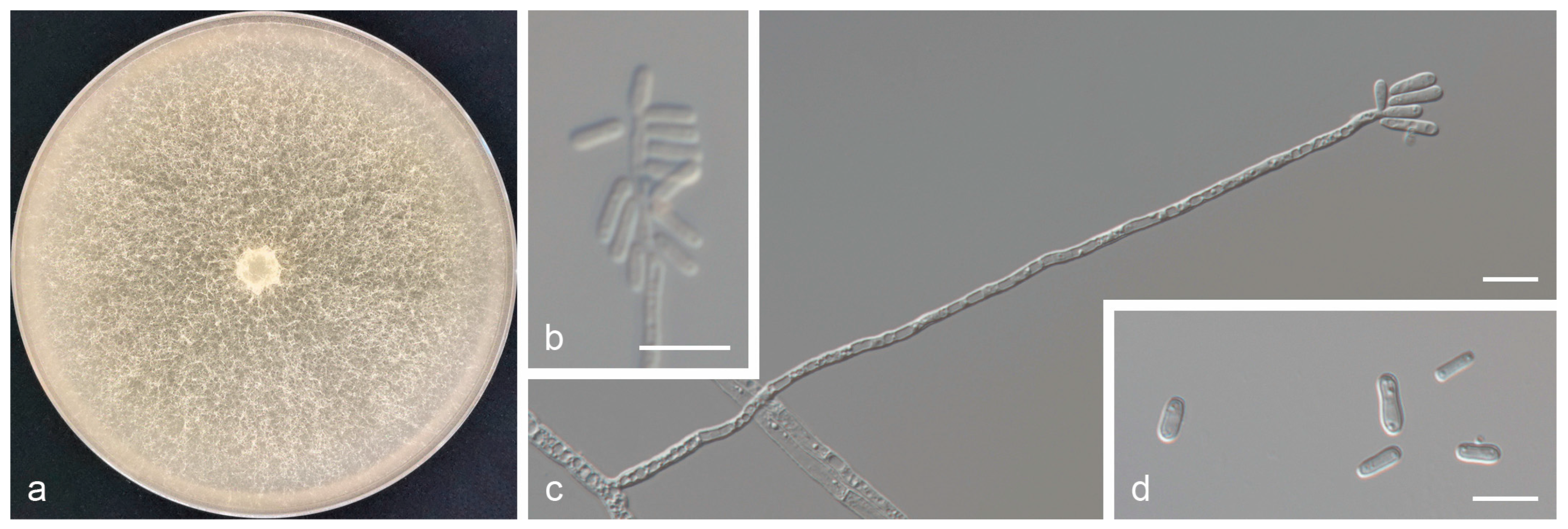
Graphilbum griseum Z. Wang & Q. Lu, sp. nov. (Taxon 1 Figure 2).
Mycobank: 856720
Etymology: The epithet griseum (Latin) refers to its vector, Acanthocinus griseus.
Holotype: CXY3358
Description: Sexual morph: not observed. Asexual morph: hyalorhinocladiella-like. Conidiophores are simple, arising directly from the mycelium; conidiogenous cells are subulate, hyaline, smooth or rough, (13.9–)23.4–53.8(–69.8) × (2.0–)2.2–3.1(–3.6) μm. Conidia are hyaline, smooth, aseptate, cylindrical to obovate, (5.8–)5.9–8.7(–11.5) × (2.4–)2.5–3.2(–3.7) μm.
Culture characteristics: Colony diameters reached 78.7 mm in seven days on 2% MEA at 25 °C. Colonies were initially hyaline or light white. Mycelia later became grayish-white and superficial, with abundant aerial mycelia. Radial thinning of the colony margins. Colonies grow fastest at 30 °C and do not grow at 5 °C or 40 °C.
Associated insects: Acanthocinus griseus.
Hosts: Pinus sylvestris var. mongolica.
Material examined: CHINA, Heilongjiang Province, Jiamusi City, Huanan County, from Acanthocinus griseus infesting Pinus sylvestris var. mongolica, June 2023, Z. Wang and Q. Lu, holotype: CXY3358, ex-type culture CFCC71126, ibid. CFCC71130, CFCC71124.
Notes: Graphilbum griseum was closely related to Gra. acuminatum and Gra. hongsongense in phylogenetic inferences (Figure 1 and Figures S1 and S2) [10,25]. Although these three species had an identical ITS sequence (Figure S1), they were distinct species based on phylogenetic analysis of tef1-α sequences (Figure 1). Graphilbum griseum can be distinguished from Gra. acuminatum and Gra. hongsongense by a hyalorhinocladiella-like asexual morph, which is pesotum-like in Gra. acuminatum and Gra. hongsongense. The optimal growth temperatures for the three species were 30 °C (Gra. griseum) and 25 °C (Gra. acuminatum and Gra. hongsongense). At 25 °C on 2% MEA, Gra. griseum grew slower than Gra. hongsongense (78.7 mm in 7 days vs. 76.3 mm in 4 days) and faster than Gra. acuminatum (11.2 mm/d vs. 5.8 mm/d). Furthermore, Gra. griseum was associated with Acanthocinus griseus from Pinus sylvestris var. mongolica in Jiamusi, China, whereas Gra. hongsongense was associated with Ips chinensis from Pinus koraiensis in Hegang, China. Graphilbum acuminatum used Pinus sylvestris and Picea abies as hosts and was associated with Ips acuminantus, Orthotomicus laricis, Pityogenes bidentatus, and Pityogenes quadridens in Europe.
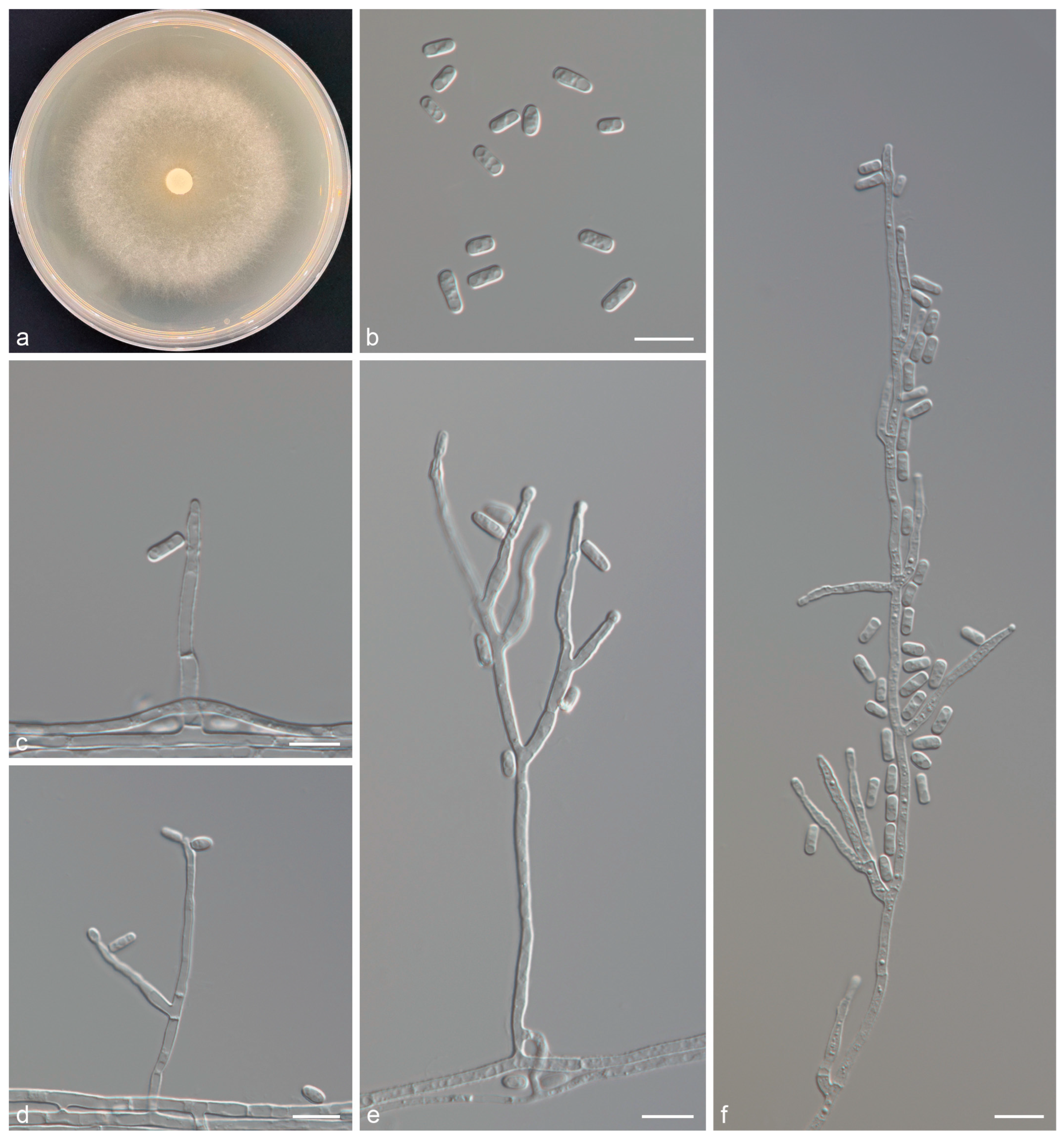
Graphilbum nitidum Z. Wang & Q. Lu, sp. nov. (Taxon 2 Figure 3).
Mycobank: 856721
Etymology: The epithet nitidum (Latin) refers to its vector, Pissodes nitidus.
Holotype: CXY3362
Description: Sexual morph: not observed. Asexual morph: hyalorhinocladiella-like. Conidiophores are simple or sparingly branched, arising directly from the mycelium; conidiogenous cells are sympodial, subulate, hyaline, 1–3 per branch, smooth or rough, (11.7–)19.1–35.5(–47.5) × (1.9–)2.1–2.8(–3.4) μm. Conidia are hyaline, smooth, aseptate, cylindrical to obovate, (4.7–)5.3–6.6(–7.6) × (2.5–)2.7–3.2(–3.5) μm.
Culture characteristics: Colony diameters reached 40.2 mm in eight days on 2% MEA at 25 °C. Colonies were initially hyaline or light white. Mycelia later became grayish white and superficial, with aerial mycelia. Radial thinning of the colony margins. Colonies grow fastest at 30 °C and do not grow at 5 °C and 40 °C.
Associated insects: Pissodes nitidus.
Hosts: Pinus sylvestris var. mongolica.
Material examined: CHINA, Heilongjiang Province, Jiamusi City, Huanan County, from Pissodes nitidus infesting Pinus sylvestris var. mongolica, June 2023, Z. Wang and Q. Lu, holotype: CXY3362, ex-type culture CFCC71143, ibid. CFCC71144.
Notes: Graphilbum nitidum was phylogenetically close to Gra. crescericum (Figure 1 and Figures S1 and S2), both of which were observed in a hyalorhinocladiella-like asexual morph [26]. There were differences in conidial shape and size (Gra. nitidum: cylindrical to obovate, 5.3–6.6 μm long; Gra. crescericum: globose-subglobose, 4.5–5.7 μm long). The optimal growth temperatures for the two species were 30 °C (Gra. nitidum) and 25 °C (Gra. crescericum). At 25 °C on 2% MEA, Gra. nitidum grew slower than Gra. crescericum (8 days: 40.2 mm vs. 60.4 mm). In addition, Gra. nitidum was associated with Pissodes nitidus from Pinus sylvestris var. mongolica in China, whereas Gra. crescericum was isolated from Hylurgops palliatus, Hylastes ater, and Orthotomicus erosus, infesting Pinus radiata in Europe [25,26].

4. Discussion

The results of this study showed that four ophiostomatalean fungi were associated with three beetles infesting Pinus sylvestris var. mongolica, including Graphilbum griseum sp. nov., Gra. nitidum sp. nov., Graphilbum sp., and Ophiostoma ips (Table 2 and Table 3). The dominant species was O. ips, with an isolation rate of 73.38%, a fungus commonly found in pine forests worldwide [27]. In China, O. ips is widely distributed, associated with various pine-infesting bark beetles, and has also been isolated from pine trees infested by Bursaphelenchus xylophilus [7,28,29,30,31]. In this study, O. ips was obtained from all three beetles, viz. Acanthocinus griseus, Ips chinensis, and Pissodes nitidus (Table 3). However, there is no evidence of specificity between O. ips and particular beetle vectors [27].
The genus Graphilbum, belonging to the family Ophiostomataceae, is commonly associated with conifer-infesting bark beetles [6,25]. This study identified two new species of Graphilbum, increasing the total number of species described in this genus to 32 [6,7,9,10,32]. Graphilbum griseum and Gra. nitidum were specific associated with A. griseus and P. nitidus, respectively, whereas Graphilbum sp. was associated with I. chinensis and P. nitidus (Table 3). The three beetles occupy different ecological niches within the trunk, with A. griseus living in the xylem, and I. chinensis and P. nitidus in the bark. This distinction may explain the differences in the fungal species associated with these beetles. Furthermore, species-specific associations between Ips bark beetles and ophiostomatoid fungi have been observed [10], and similar specific associations may exist between beetles of different families and these fungi. However, a larger sample size is necessary to confirm this hypothesis.
There may also be community succession among fungi associated with beetles. Zheng et al. found that the fungal communities associated with the 2nd–3rd- and 4th–5th-instar larvae of Monochamus alternates differed [32]. Fungal associates of 4th-5th-instar larvae were fewer than those of 2nd–3rd-instar larvae, with 14 and 6 species, respectively, and only two shared species. Sixteen ophiostomatalean fungi have been reported to be associated with Ips chinensis, including eight from northeast China [10,29]. However, the two ophiostomatalean fungal associates of I. chinensis in the present study did not overlap with these eight. Compared with the findings of Wang et al. [10], this study only used emerging adults for fungal isolation, which may explain why only two associates of I. chinensis were identified. Thus, fungal isolation across different beetle lifestyles can better reflect the associated fungal communities.
Beetles are known to carry various tree disease pathogens. For example, the diplodia tip blight pathogen of P. sylvestris var. mongolica, Diplodia sapinea, is the dominant fungus associated with Pityophthorus morosovi. Additionally, the associate of Ips subelongatus infesting P. sylvestris var. mongolica, Ceratocystiopsis subelongati, has been found to be pathogenic to pine trees [14]. Interestingly, Ophiostoma minus, which causes canker staining in pines, has been detected in the endophytic fungal communities of P. sylvestris var. mongolica infected with diplodia tip blight [33]. Therefore, pathogenicity tests of the fungi identified in this study are essential to assess their role in the decline of P. sylvestris var. mongolica.

5. Conclusions

In summary, four ophiostomatalean fungal associates from three beetles (A. griseus, I. chinensis, and P. nitidus) infesting P. sylvestris var. mongolica were identified in this study, including Graphilbum griseum, Gra. nitidum, Graphilbum sp., and O. ips. The results of this study revealed that the fungi associated with different beetles are diverse, even when they share the same host. Moreover, the correlation between ophiostomatalean fungi and forest decline needs to be further studied by means of, for example, pathogenicity tests.

Supplementary Materials

The following supporting information can be downloaded at: https://www.mdpi.com/article/10.3390/jof11010027/s1, Figure S1: Maximum likelihood (ML) tree based on ITS sequence data of Graphilbum spp. ML bootstrap values above 70% and Bayesian posterior probability values above 0.85 are indicated at the nodes. Sequences generated from this study are shown in bold. T = ex-type; Figure S2: Maximum likelihood (ML) tree based on tub2 sequence data of Graphilbum spp. ML bootstrap values above 70% and Bayesian posterior probability values above 0.85 are indicated at the nodes. Sequences generated from this study are shown in bold. T = ex-type; Figure S3: Maximum likelihood (ML) tree based on ITS sequence data of Ophiostoma ips complex. ML bootstrap values above 70% and Bayesian posterior probability values above 0.85 are indicated at the nodes. Sequences generated from this study are shown in bold. T = ex-type; Figure S4: Maximum likelihood (ML) tree based on tub2 sequence data of Ophiostoma ips complex. ML bootstrap values above 70% and Bayesian posterior probability values above 0.85 are indicated at the nodes. Sequences generated from this study are shown in bold. T = ex-type.

Author Contributions

Conceptualization, Z.W., H.W. and Q.L.; methodology, Z.W. and Y.T.; software, Z.W. and X.S.; validation, Z.W., C.L., H.W. and Q.L.; formal analysis, Z.W. and Y.T.; investigation, Z.W. and C.L.; resources, Z.W. and Q.L.; data curation, Z.W. and C.L.; writing—original draft preparation, Z.W. and Q.L.; writing—review and editing, Z.W. and Q.L.; visualization, Z.W.; supervision, Z.W. and Q.L.; project administration, Q.L. and Z.W.; funding acquisition, Q.L. and Z.W. All authors have read and agreed to the published version of the manuscript.

Funding

This research was funded by the National Natural Science Foundation of China (Project No.: 32230071) and National Key R & D Program of China (Project No.: 2023YFC2604801-4).

Informed Consent Statement

Not applicable.

Data Availability Statement

The data presented in this study are openly available in [GenBank].

Conflicts of Interest

The authors declare no conflicts of interest.

References

  1. Allen, C.D.; Macalady, A.K.; Chenchouni, H.; Bachelet, D.; McDowell, N.; Vennetier, M.; Kitzberger, T.; Rigling, A.; Breshears, D.D.; Hogg, E.H.; et al. A global overview of drought and heat-induced tree mortality reveals emerging climate change risks for forests. For. Ecol. Manag. 2010, 259, 660–684. [Google Scholar] [CrossRef]
  2. Dang, H.; Han, H.; Zhang, X.; Chen, S.; Li, M.; Liu, C. Key strategies underlying the adaptation of Mongolian scots pine (Pinus sylvestris var. mongolica) in sandy land under climate change: A review. Forests 2022, 13, 846. [Google Scholar] [CrossRef]
  3. Li, Y.; Li, Z.; Sun, S.; Hu, X.; Guan, C.; Gao, W. Differences in growth and wood anatomy in relation to meteorological variables between healthy and declining Mongolian pine trees. Chin. J. Ecol. 2024, in press. [Google Scholar]
  4. Lu, W.W.; Wu, B.; Bai, J.H.; Song, X.D.; Shi, Z.J.; Dang, H.Z.; Li, H.; Yin, M. Causes and research prospects of the decline of Pinus sylvestris var. mongolica plantation. Chin. Sci. Bull. 2023, 68, 1286–1297. [Google Scholar] [CrossRef]
  5. Liu, B.; Yan, J.; Wang, D.; Wang, Y.; Zhou, Y.; Chen, Y. Occurrence of major forest pests in China in 2023 and prediction for trend in 2024. Pest Dis. 2024, 43, 41–45. [Google Scholar]
  6. de Beer, Z.W.; Procter, M.; Wingfield, M.J.; Marincowitz, S.; Duong, T.A. Generic boundaries in the Ophiostomatales reconsidered and revised. Stud. Mycol. 2022, 101, 57–120. [Google Scholar] [CrossRef] [PubMed]
  7. Song, X.; Tie, Y.; Wang, Z.; Kong, L.; Liu, H.; Lu, Q. Three Ophiostomatalean fungi associated with bark beetles from Pinus thunbergii infested by Bursaphelenchus xylophilus in Laoshan Mountain (Shandong, China). Forests 2024, 15, 1990. [Google Scholar] [CrossRef]
  8. Wang, H.; Liu, C.; Yue, F.; Yan, D.H.; Lu, Q. Identification of ophiostomatalean fungi associated with Tomicus pilifer infesting Pinus koraiensis in Northeastern China. Front. Microbiol. 2022, 13, 919302. [Google Scholar] [CrossRef] [PubMed]
  9. Wang, H.; Wang, T.; Liu, Y.; Zeng, F.; Zhang, H.; Decock, C.; Zhang, X.; Lu, Q. Diversity of ophiostomatoid fungi associated with Dendroctonus armandi infesting Pinus armandii in western China. J. Fungi 2022, 8, 214. [Google Scholar] [CrossRef]
  10. Wang, Z.; Liang, L.; Wang, H.; Decock, C.; Lu, Q. Ophiostomatoid fungi associated with Ips bark beetles in China. Fungal Divers. 2024, 129, 283–364. [Google Scholar] [CrossRef]
  11. Wang, Z.; Liu, C.; Song, X.; Tie, Y.; Wang, H.; Liu, H.; Lu, Q. Ophiostomatalean fungi associated with Polygraphus bark beetles in the Qinghai-Tibet Plateau, China. MycoKeys 2024, 110, 93–115. [Google Scholar] [CrossRef]
  12. DiGuistini, S.; Wang, Y.; Liao, N.Y.; Taylor, G.; Tanguay, P.; Feau, N.; Henrissat, B.; Chan, S.K.; Hesse-Orce, U.; Alamouti, S.M.; et al. Genome and transcriptome analyses of the mountain pine beetle fungal symbiont Grosmannia clavigera, a lodgepole pine pathogen. Proc. Natl. Acad. Sci. USA 2011, 108, 2504–2509. [Google Scholar] [CrossRef]
  13. Lu, Q.; Decock, C.; Zhang, X.Y.; Maraite, H. Ophiostomatoid fungi (Ascomycota) associated with Pinus tabuliformis infested by Dendroctonus valens (Coleoptera) in northern China and an assessment of their pathogenicity on mature trees. Antonie Van. Leeuwenhoek 2009, 96, 275–293. [Google Scholar] [CrossRef] [PubMed]
  14. Wang, Z.; Liu, Y.; Liu, C.; Liu, Z.; Liang, L.; Lu, Q. Morphological and phylogenetic analyses reveal a new species of Ceratocystiopsis (Ophiostomataceae, Ophiostomatales) associated with Ips subelongatus in Inner Mongolia (China) with weak host pathogenicity. Forests 2021, 12, 1795. [Google Scholar] [CrossRef]
  15. Wang, Z.; Liu, Y.; Wang, H.; Meng, X.; Liu, X.; Decock, C.; Zhang, X.; Lu, Q. Ophiostomatoid fungi associated with Ips subelongatus, including eight new species from northeastern China. IMA Fungus 2020, 11, 1–29. [Google Scholar]
  16. White, T.J.; Bruns, T.; Lee, S.J.; Taylor, J. Amplification and direct sequencing of fungal ribosomal RNA genes for phylogenetics. PCR Protoc. A Guide Methods Appl. 1990, 18, 315–322. [Google Scholar]
  17. Gardes, M.; Bruns, T.D. ITS primers with enhanced specificity for basidiomycetes-application to the identification of mycorrhizae and rusts. Mol. Ecol. 1993, 2, 113–118. [Google Scholar] [CrossRef] [PubMed]
  18. Glass, N.L.; Donaldson, G.C. Development of primer sets designed for use with the PCR to amplify conserved genes from filamentous Ascomycetes. Appl. Environ. Microbiol. 1995, 61, 1323–1330. [Google Scholar] [CrossRef] [PubMed]
  19. Jacobs, K.; Bergdahl, D.R.; Wingfield, M.J.; Halik, S.; Seifert, K.A.; Bright, D.E.; Wingfield, B.D. Leptographium wingfieldii introduced into North America and found associated with exotic Tomicus piniperda and native bark beetles. Mycol. Res. 2004, 108, 411–418. [Google Scholar] [CrossRef]
  20. Katoh, K.; Rozewicki, J.; Yamada, K.D. MAFFT online service: Multiple sequence alignment, interactive sequence choice and visualization. Brief. Bioinform. 2019, 20, 1160–1166. [Google Scholar] [CrossRef]
  21. Kumar, S.; Stecher, G.; Tamura, K. MEGA7: Molecular evolutionary genetics analysis version 7.0 for bigger datasets. Mol. Biol. Evol. 2016, 33, 1870–1874. [Google Scholar] [CrossRef] [PubMed]
  22. Stamatakis, A. RaxML Version 8: A tool phylogenetic analysis and post-analysis of large phylogenies. Bioinformatics 2014, 30, 1312–1313. [Google Scholar] [CrossRef]
  23. Ronquist, F.; Huelsenbeck, J.P. MrBayes 3: Bayesian phylogenetic inference under mixed models. Bioinformatics 2003, 19, 1572–1574. [Google Scholar] [CrossRef] [PubMed]
  24. Darriba, D.; Taboada, G.L.; Doallo, R.; Posada, D. jModelTest 2: More models, new heuristics and parallel computing. Nat. Methods 2012, 9, 772. [Google Scholar] [CrossRef]
  25. Jankowiak, R.; Solheim, H.; Bilański, P.; Marincowitz, S.; Wingfield, M.J. Seven new species of Graphilbum from conifers in Norway, Poland, and Russia. Mycologia 2020, 112, 1240–1262. [Google Scholar] [CrossRef]
  26. Romón, P.; De Beer, Z.W.; Zhou, X.D.; Duong, T.A.; Wingfield, B.D.; Wingfield, M.J. Multigene phylogenies of Ophiostomataceae associated with Monterey pine bark beetles in Spain reveal three new fungal species. Mycologia 2014, 106, 119–132. [Google Scholar] [CrossRef] [PubMed]
  27. Zhou, X.; Burgess, T.I.; De Beer, Z.W.; Lieutier, F.; Yart, A.; Klepzig, K.; Carnegie, A.; Portales, J.M.; Wingfield, B.D.; Wingfield, M.J. High intercontinental migration rates and population admixture in the sapstain fungus Ophiostoma ips. Mol. Ecol. 2007, 16, 89–99. [Google Scholar] [CrossRef]
  28. Lu, M.; Zhou, X.D.; De Beer, Z.W.; Wingfield, M.J.; Sun, J.H. Ophiostomatoid fungi associated with the invasive pine-infesting bark beetle, Dendroctonus valens, in China. Fungal Divers. 2009, 38, 133–145. [Google Scholar]
  29. Chang, R.; Duong, T.A.; Taerum, S.J.; Wingfield, M.J.; Zhou, X.; de Beer, Z.W. Ophiostomatoid fungi associated with conifer-infesting beetles and their phoretic mites in Yunnan, China. MycoKeys 2017, 28, 19–64. [Google Scholar] [CrossRef] [PubMed]
  30. Chang, R.; Zhang, X.; Si, H.; Zhao, G.; Yuan, X.; Liu, T.; Bose, T.; Dai, M. Ophiostomatoid species associated with pine trees (Pinus spp.) infested by Cryphalus piceae from eastern China, including five new species. MycoKeys 2021, 83, 181–208. [Google Scholar] [CrossRef]
  31. Pan, Y.; Lu, J.; Chen, P.; Yu, Z.; Zhang, H.; Ye, H.; Zhao, T. Ophiostomatales (Ascomycota) associated with Tomicus species in southwestern China with an emphasis on Ophiostoma canum. J. For. Res. 2020, 31, 2549–2562. [Google Scholar] [CrossRef]
  32. Zheng, G.; You, M.; Li, X.; Zhou, Q.; Wang, Z.; Wang, H.; Lu, Q. Diversity of fungi associated with Monochamus alternatus larval habitats in Bursaphelenchus xylophilus-infected Pinus massoniana and identification of two new ophiostomatalean species (Ascomycota, Ophiostomatales). MycoKeys 2022, 92, 1. [Google Scholar] [CrossRef] [PubMed]
  33. Liu, Y.; Wang, L.; Ren, N.; You, C. Community structure of endophytic fungi in asymptomatic and symptomatic Pinus sylvestris var. mongolica infected by diplodia tip blight. J. Beijing For. Univ. 2024, 46, 119–131. [Google Scholar]
Figure 1. Maximum likelihood (ML) tree based on tef1-α sequence data of Graphilbum spp. ML bootstrap values above 70% and Bayesian posterior probability values above 0.85 are indicated at the nodes. Sequences generated from this study are shown in bold. T = ex-type.
Jof 11 00027 g001
Figure 2. Graphilbum griseum sp. nov. (a) Eight-day-old culture on 2% MEA; (bd) Hyalorhinocladiella-like asexual morph: conidiogenous cells and conidia. Scale bars of (bd) = 10 μm.
Jof 11 00027 g002
Figure 3. Graphilbum nitidum sp. nov. (a) Eight-day-old culture on 2% MEA; (bf) Hyalorhinocladiella-like asexual morph: conidiogenous cells and conidia. Scale bars of (bf) = 10 μm.
Jof 11 00027 g003
Table 1. Primers and PCR conditions used in this study.
Gene FragmentPrimersPCR ConditionsReferences
ITSITS1-F/ITS494 °C for 3 min, 35 cycles of 94 °C for 1 min, 55 °C for 45 s, and 72 °C for 1 min, 72 °C for 8 min[16,17]
tub2Bt2a/Bt2b95 °C for 2 min, 35 cycles of 95 °C for 30 s, 56 °C for 30 s, and 72 °C for 1 min, 72 °C for 8 min[18]
tef1-αEF1F/EF2R95 °C for 2 min, 35 cycles of 95 °C for 30 s, 56 °C for 30 s, and 72 °C for 1 min, 72 °C for 8 min[19]
Table 2. Representative strains in this study.
SpeciesTaxonIsolate No. 1Other No. 2Insect Vector 3GenBank Accession No.
ITStub2tef1-α
Gra. griseum sp. nov.1CFCC71126CXY3358A. griseusPQ623400-PQ619704
CFCC71130CXY3359A. griseusPQ623401-PQ619705
CFCC71124CXY3360A. griseusPQ623402-PQ619706
Gra. nitidum sp. nov.2CFCC71144CXY3361P. nitidusPQ623403-PQ619707
CFCC71143CXY3362P. nitidusPQ623404PQ619697PQ619708
Graphilbum sp.3CFCC71127CXY3363P. nitidusPQ623405-PQ619709
CFCC71139CXY3364I. chinensisPQ623406-PQ619710
Ophiostoma ips4CFCC71129CXY3365A. griseusPQ623407PQ619698-
CFCC71135CXY3366A. griseusPQ623408PQ619699-
CFCC71128CXY3367I. chinensisPQ623409PQ619700-
CFCC71136CXY3368I. chinensisPQ623410PQ619701-
CFCC71137CXY3369P. nitidusPQ623411PQ619702-
CFCC71138CXY3370P. nitidusPQ623412PQ619703-
1 CFCC: the China Forestry Culture Collection Center. 2 CXY: the culture collection of the Forest Pathology Laboratory at the Chinese Academy of Forestry. 3 A.: Acanthocinus; I.: Ips; P.: Pissodes.
Table 3. Isolates from Pinus sylvestris var. mongolica infested by three beetles in this study.
TaxonSpeciesNumbers of Isolates 1TotalTotal Percentage
A. griseusI. chinensisP. nitidus
1Graphilbum griseum21 217.17%
2Gra. nitidum 662.05%
3Graphilbum sp. 25265117.41%
4Ophiostoma ips136344521573.38%
Total 1575977293100.00%
1 A.: Acanthocinus; I.: Ips; P.: Pissodes.
Disclaimer/Publisher’s Note: The statements, opinions and data contained in all publications are solely those of the individual author(s) and contributor(s) and not of MDPI and/or the editor(s). MDPI and/or the editor(s) disclaim responsibility for any injury to people or property resulting from any ideas, methods, instructions or products referred to in the content.