Article

Development of Marker Recycling Systems for Sequential Genetic Manipulation in Marine-Derived Fungi Spiromastix sp. SCSIO F190 and Aspergillus sp. SCSIO SX7S7

1
CAS Key Laboratory of Tropical Marine Bio-Resources and Ecology, Guangdong Key Laboratory of Marine Materia Medica, RNAM Center for Marine Microbiology, South China Sea Institute of Oceanology, Chinese Academy of Sciences, 164 West Xingang Road, Guangzhou 510301, China
2
Southern Marine Science and Engineering Guangdong Laboratory (Guangzhou), No. 1119, Haibin Road, Nansha District, Guangzhou 511458, China
3
College of Oceanology, University of Chinese Academy of Sciences, Qingdao 266400, China
*
Authors to whom correspondence should be addressed.
These authors contributed equally to this work.
J. Fungi 2023, 9(3), 302; https://doi.org/10.3390/jof9030302
Submission received: 16 January 2023 / Revised: 23 February 2023 / Accepted: 24 February 2023 / Published: 26 February 2023
(This article belongs to the Special Issue The Gift of Marine Fungi: Abundant Secondary Metabolites)

Abstract

:
Marine-derived fungi are emerging as prolific workhorses of structurally novel natural products (NPs) with diverse bioactivities. However, the limitation of available selection markers hampers the exploration of cryptic NPs. Recyclable markers are therefore valuable assets in genetic engineering programs for awaking silent SM clusters. Here, both pyrG and amdS-based recyclable marker cassettes were established and successfully applied in marine-derived fungi Aspergillus sp. SCSIO SX7S7 and Spiromastix sp. SCSIO F190, respectively. Using pyrG recyclable marker, a markerless 7S7-∆depH strain with a simplified HPLC background was built by inactivating a polyketide synthase (PKS) gene depH and looping out the pyrG recyclable marker after depH deletion. Meanwhile, an amdS recyclable marker system was also developed to help strains that are difficult to use pyrG marker. By employing the amdS marker, a backbone gene spm11 responsible for one major product of Spiromastix sp. SCSIO F190 was inactivated, and the amdS marker was excised after using, generating a relatively clean F190-∆spm11 strain for further activation of novel NPs. The collection of two different recycle markers will guarantee flexible application in marine-derived fungi with different genetic backgrounds, enabling the exploitation of novel structures in various fungi species with different genome mining strategies.

1. Introduction

Marine-derived fungi have yielded an increasing number of structurally unique and biologically active natural products (polyketides, alkaloids, steroids, terpenoids, and peptides) [1,2,3]. The chemical diversity of the compounds generated from marine-derived fungi, along with the strains’ novelty, shows that this group of microeukaryotes are tremendous sources for the isolation of unusual bioactive natural products [4]. Nonetheless, most of the natural product biosynthetic gene clusters (BGCs) are either dormant or weakly expressed under laboratory conditions. The plethora of advanced tools for genome sequencing and mining enabled the discovery of new secondary metabolites which are “silent” and are not expressed under laboratory conditions. For example, our previous studies have shown that metabolic blockade-based genome mining via generating a “multiple-deletion” mutant is an efficient method for exploring novel secondary metabolites (SMs) [5,6]. Likewise, genetic manipulations of pathway-specific regulatory genes and global transcription regulators are alternative methods for activating cryptic gene clusters [7,8]. Despite of the strategy used, a crucial element common to all of these genetic manipulation techniques is the use of a selection marker that enables discrimination between transformed and untransformed cells. However, for most marine-derived fungi, only a few markers are useful due to intrinsic resistance to drugs, which precludes the ability to sequentially disrupt/activate multiple genes, especially when chassis cells with simplified HPLC backgroud are needed before exploring novel natural products. Therefore, it is helpful to establish marker recycling systems to resolve the limitation of selection markers.
Currently, pyrG or amdS-based marker recycling systems are the two widely used methods in multistep gene editing. The pyrG gene encodes orotidine-5-phosphate (OMP) decarboxylase that catalyzes the decarboxylation of orotidine-5′-monophosphate to form uridine monophosphate [9]. In filamentous fungi, mutants of pyrG cannot grow without exogenous uridine/uracil (uridine/uracil auxotrophs). Additionally, OMP decarboxylase could also convert 5-fluoroorotic acid (5-FOA) to fluoroorotidine monophosphate, which can be further converted into fluorodeoxyuridine, a toxic compound that inhibits DNA and RNA synthesis. Resistance to 5-FOA can be achieved through the inactivation of OMP decarboxylase [10]. Therefore, the pyrG gene can be used as a maker allowing auxotrophic selection and 5-FOA counterselection permitting unlimited rounds of genetic manipulation. The generation of 5-FOA-resistant pyrG mutants has been reported in the different genera of filamentous fungi including Aspergillus sp. [10,11,12,13], Acremonium sp. [14], Talaromyces versatilis [10], and Trichoderma reesei [15]. Nevertheless, when applied to marine fungal strains with limited available sequencing and genetic information, the pyrG recycling system can be a laborious, time-consuming, and inconclusive process. An excellent alternative to the pyrG-based counterselection is the amdS selection marker as it can be directly used in the strain without the requirement of the null mutant as the recipient strain [16]. The amdS gene from A. nidulans, encoding acetamidase (amdS), was first characterized in 1970 by Hynes and Pateman [17]. Acetamidase catalyzes acetamide into acetate and ammonium allowing the organism to utilize acetamide as a sole carbon or nitrogen source. Acetamidase also hydrolyses the toxic fluoroacetamide to produce fluoroacetate that can be further transformed into toxic fluoroacetyl CoA and fluorocitrate, leading to inhibition of the citric acid cycle [17,18,19]. Hence, amdS can serve as a bidirectional selectable marker and has been employed in several members of Ascomycota that grow poorly on acetamide medium [16,20,21,22]. Despite the successful use of pyrG and/or amdS as recycling makers in various fungal taxa, these two counterselectable markers are seldom utilized in genetic manipulation of marine fungi.
Aspergillus sp. SCSIO SX7S7 and Spiromastix sp. SCSIO F190 are different fungal species isolated from marine environments [23,24,25]. Several antimicrobial natural products (e.g.,spiromarmycin, spiromastol L, and nornidulin) have been identified from these two strains with high yielding [24,25,26]. Genome sequencing and antiSMASH analyses revealed that both strains possess various cryptic secondary metabolites that need to be further activated by genetic approaches. Due to the high abundance of constitutively produced compounds in these two strains, inactivation of the BGCs corresponding to these major products before activation of the silent SMs would simplify the isolation steps of novel natural products. Moreover, we expected that, in some cases, inhibiting the production of major secondary metabolites would free up subunits for incorporation into other NPs. Therefore, multiple rounds of genetic manipulation are required for sequential inactivation/activation of the secondary metabolite BGCs. Our previous study has established a CRISPR-Cas9-based gene editing vector (pBSKII-toCas9-hph-sgRNA) with a hygromycin B resistant maker for these two strains [23]. However, it is difficult to recycle the hph maker gene once it is integrated into the genome of the host strain, which limits its application in multistep genome editing.
In this study, we developed a platform including both pyrG and amdS-based marker recycling systems by replacing hygromycin B phosphotransferase (hph) expression element in pBSKII-toCas9-hph-sgRNA with direction repeats (DRs) flanked pyrG or amdS expression cassette. The two new constructed vectors named pBSKII-toCas9-pyrG-sgRNA and pBSKII-toCas9-amdS-sgRNA were successfully applied for gene disruption in Aspergillus sp. SCSIO SX7S7 and Spiromastix sp. SCSIO F190, respectively. In Aspergillus sp. SCSIO SX7S7, the tricyclic depsidones and bicyclic depsides (DEPs) biosynthesis cluster was inactivated by disrupting of a PKS gene (depH), and the pyrG marker was removed after deletion of the depH gene, generating a 7S7-∆depH strain with a relatively simple HPLC background compared to the wild type strain for further activating of cryptic NPs by sequential genetic editing. In Spiromastix sp. SCSIO F190, the production of one major product spiromarmycin was abolished via inactivation of the PKS gene spm11 using the amdS-based marker recycling system. Subsequential removal of the amdS marker in the ∆spm11 strain allowed multiple rounds of genetic manipulation for inducing the expression of silent NPs.

2. Materials and Methods

2.1. Strains, Plasmids, and Culture Conditions

All strains and plasmids used in this study are listed in Tables S1 and S2. Both Spiromastix sp. SCSIO F190 and Aspergillus sp. SCSIO SX7S7 are marine-derived as indicated in our previous study [23]. All fungi strains (Table S1) were routinely maintained on potato dextrose agar (PDA) and in potato dextrose broth (PDB) at 28 °C for the sporulation and the protoplast preparation, respectively. A 5-FOA sensitivity assay was performed on a PDA medium with different concentrations of 5-FOA. The acetamide plate was prepared according to the Aspergillus Nitrogen-free Minimal (ANM) medium [27] recipe using acetamide instead of glucose and ammonium [28]. Removing of the amdS marker gene was conducted on an ANM plate supplementing with 10 mM FAA. Escherichia coli strain DH5α was cultured in the Luria–Bertani (LB) medium with 1 mg/mL ampicillin for plasmid DNA isolation. The pBluescript II SK plasmid (Table S2) for generating pyrG/amdS expression cassette [28] was kindly provided by Prof. Wong, K.H. from the University of Macau. The pBSKII-toCas9-hph plasmid used for constructing pBSKII-toCas9-pyrG and pBSKII-toCas9-amdS plasmids was kindly provided by Prof. Dan Hu from Jinan University [29]. The pFC330 plasmid (Table S2) containing the sgRNA expression sequence and AfpyrG coding sequence was generously offered by Prof. Yi Tang from the University of California, Los Angeles, CA, USA [30]. The pBSKII-toCas9-pyrG and pBSKII-toCas9-amdS vectors (Table S2) were used to construct the Cas9 and sgRNA-expressing vector pBSKII-toCas9-pyrG/amdS-sgRNA in this study.

2.2. DNA Manipulation

The primer synthesis and DNA sequencing were performed by Qingke Biotech Co., Ltd. (Guangzhou, China) and Sangong Biotech Co., Ltd. (Shanghai, China) (Table S3). The plasmid extraction and DNA purification were carried out with commercial kits (Omega Bio-Tech, Inc., Norcross, GA, USA), following the manufacturer’s protocol. The one step cloning of multiple DNA fragments was carried out using a pEASY®-Uni Seamless Cloning and Assembly Kit from TransGen Biotechnology Co., Ltd. (Beijing, China), following the manufacturer’s protocol. The restriction enzymes and other DNA modification reagents were purchased from New England Biolabs, Inc. (NEB, Hitchin, UK) and Takara Biotechnology Co., Ltd. (Dalian, China), following the manufacturer’s protocol. The DNA amplification was performed on a PCR thermal cycler from Applied Biosystems (Thermo Fisher, Shanghai, China) with either Taq DNA polymerase (TransGen, Beijing, China) or High-fidelity Fastpfu DNA polymerase (TransGen, Beijing, China), following the manufacturer’s protocol.

2.3. Plasmid Construction

The pBSKII-toCas9-pyrG-sgRNA and pBSKII-toCas9-amdS-sgRNA plasmids were constructed by three steps as follows. First, the pyrG and amdS genes were amplified from FC330 and A. niduans using primers pyrG-F1/R1 and amdS-F1/R1, respectively. Either of the pyrG and amdS sequences together with A. nidulans derived PtrpC and TtrpC fragments were added in SmaI digested pBluescript II SK plasmid by one step cloning kit, yielding pBSKII-pyrG or pBSKII-amdS. Second, the pyrG and amdS expression cassettes under the control of PtrpC and TtrpC were amplified from the pBSKII-pyrG and pBSKII-amdS plasmids using primers DR-Ptrpc-F and PsiI-Ttrpc-R. In addition, the 300 bp DR sequence was amplified from pBSKII-toCas9-hph plasmid. Then, the pyrG or amdS expression cassette along with the DR sequence were ligated into PsiI cut pBSKII-toCas9-hph plasmid to replace the hph expression cassette, generating pBSKII-toCas9-pyrG or pBSKII-toCas9-amdS. Third, the sgRNA for targeting gene together with ribozyme sequences controlled by the PgdpA and TtrpC sequences was separately amplified from pFC330 using two primer pairs Frag1-F/R and Frag2-F/R. Next, the two sgRNA expression fragments were inserted into the BsaAI digested pBSKII-toCas9-pyrG or pBSKII-toCas9-amdS plasmid by one step cloning.

2.4. 5-FOA Sensitivity Assay

The 5-FOA sensitivity was determined in PDA plate with varying concentrations of 5-FOA. The spores of Aspergillus sp. SCSIO SX7S7 were inoculated on a PDA medium supplemented with 5-FOA at gradient concentrations (0, 0.5, 1, 2 mg/mL). Control plates without 5-FOA were used. The strains were incubated at 28 °C for 7 days, and all experiments were conducted in triplicate.

2.5. Acetamide Utilization Assay

Spiromastix sp. SCSIO F190 was tested for the ability to grow on 10 mM acetamide agar using a plate assay. Spores were inoculated onto plates using acetamide as the sole carbon and nitrogen source supplemented with different concentrations of cesium chloride. Plates were incubated at 28 °C for 14 days and then compared with the control plate containing both glucose and ammonium tartrate.

2.6. Protoplast Transformation

The protoplasts of Aspergillus sp. SCSIO SX7S7 and Spiromastix sp. SCSIO F190 were prepared as previously described [23]. In brief, the conidia were cultured in 50 mL potato dextrose broth (PDB) at 100 rpm and 28 °C until germination. After completion of the culture, the mycelia were collected and digested with Lysing enzymes (Sigma-Aldrich, St. Louis, MO, USA) and Yatalase (TaKaRa, Dalian, China). The digested protoplasts and mycelial debris were separated by gradient centrifugation. Finally, the protoplasts were resuspended in STC solution to a concentration around 2 × 10⁷ mL−1 for the subsequent transformation.
The transformation of exogenous plasmid was also performed according to our previously established method [23]. Briefly, 2–5 μg plasmid was added into the protoplast suspension, and the plasmid was introduced into the protoplast via PEG-mediated protoplast transformation. Next, the PEG-treated protoplasts were collected and poured onto 3–4 regeneration plates. ANM with 1.2 M sorbitol was used for preparing regeneration plates in the pyrG marker recycling system, while ANM with 1.2 M sorbitol but only containing acetamide as the sole carbon and nitrogen source was used in the amdS marker recycling system. After 4–20 days of cultivation at 28 °C, the transformants were inoculated onto a new PDA plate (without sorbitol), and the genome DNA of the transformants was extracted for genotyping PCR as described in our previous research [23].

2.7. Secondary Metabolite Analysis by HPLC

To examine the production of secondary metabolites, wild-type and mutants were cultivated in a PDB liquid medium for 7 days at 28 °C, 180 rpm. Then, the cultures were extracted with an equal volume of butanone by sonication, and the organic phases were evaporated to dryness. Next, the remaining residues were redissolved in 1 mL MeOH, and 50 μL of which was injected into the analytical HPLC for analysis. The HPLC analysis was performed on an Agilent 1260 HPLC system (Agilent Technologies Inc., Santa Clara, CA, USA) equipped with a binary pump and a diode array detector using an analytical Phenomenex column (250 × 4.60 mm, 5 microns). The injected samples were eluted with a linear gradient from 5% to 80% solvent B over 20 min, followed by 80% to 100% solvent B in 1.5 min, and then eluted with 100% solvent B in 5.5 min, at a flow rate of 1.0 mL/min using UV detection at 220 nm, 254 nm, 275 nm, and 354 nm.

3. Results and Discussion

3.1. Construction of CRISPR-Cas9 Vectors Containing pyrG/amdS Marker Gene

CRISPR−Cas9 genome editing technology plays an extremely significant role in synthetic biology and metabolic engineering [31]. In our previous study, a rearranged CRISPR-Cas9 vector containing T. reesei codon-optimized cas9 (toCas9), hph coding sequences, and sgRNA expression cassette driven by RNA polymerase II promoters has shown to be an efficient gene editing tool in Aspergillus sp. SCSIO SX7S7 and Spiromastix sp. SCSIO F190 [23]. However, one marker gene is not sufficient to reveal the hidden BGCs in marine fungi using multi-round genetic engineering. Recyclable markers that can be removed from the targeted site after using can be valuable tools in ambitious metabolic engineering programs. The pyrG marker serves as a strong, recyclable, auxotrophic selection marker that can be used for uridine/uracil auxotrophic complementary transformation and then counter-selected using 5-FOA. The pyrG gene from A. fumigatus has been widely applied in filamentous fungi [29,32]. In addition to the pyrG marker, the amdS gene is another broadly used recyclable marker. In the fungi species lacking acetamidase activity, the expression of acetamidase gene from A. nidulans under a strong promoter provides selection on media containing acetamide as the sole nitrogen source that can be counter-selected using FAA [20,33,34]. The efficiency of looping out counterselectable markers can be drastically increased by employing the blaster strategy, flanking the marker with around 300 bp direct repeats (DRs) [16,35]. However, to our knowledge, this technique has not been employed in marine-derived fungi.
Thus, the pyrG sequence from A. fumigatus and the amdS gene from A. nidulans were selected to test their function in the marine-derived fungi. Since the promoter (PtrpC) and terminator (TtrpC) originated from A. nidulans are functioning well in Aspergillus sp. SCSIO SX7S7 and Spiromastix sp. SCSIO F190 [23], we kept the expression of pyrG and amdS under the control of PtrpC and TtrpC. Consequently, the sequence of pyrG or amdS along with PtrpC and TtrpC were ligated into the pBluescript II SK plasmid to generate marker expression vectors pBSKII-pyrG or pBSKII-amdS. Given the high efficiency of blaster counterselection methodology in removing counterselective markers, a 300 bp sequence located directly after the TtrpC terminator in pBSKII-toCas9-hph plasmid was designed as a direct repeat (DR) sequence. The same 300 bp DR sequence was amplified for inserting before the PtrpC promoter. Next, two new vectors called pBSKII-toCas9-pyrG and pBSKII-toCas9-amdS, respectively, were generated by inserting the marker expression cassette and the DR sequences into the PsiI digested pBSKII-toCas9-hph to replace the hph expression cassette (Figure 1). Finally, the amplified gene-specific sgRNA expression fragments were inserted into either pyrG or amdS vector via one step isothermal assembly to generate recyclable gene editing vectors, named pBSKII-toCas9-pyrG-sgRNA and pBSKII-toCas9-amdS-sgRNA (Figure 1).
Application of the pyrG selection marker is laborious in generating uracil/uridine auxotrophs but shows high gene editing efficiency due to the near-nominal level of false positive rate of transformed clones and the normal growth rate on a uracil/uridine depleted medium [10]. The amdS selection marker takes advantage of its ability to transform wild-type cells that lack a background auxotrophy [16]. However, the amdS selection marker can only be applied in the strains unable to grow (or with poor growth) on acetamide as the sole carbon- or nitrogen-source. Therefore, the simplicity and gene editing efficiency of these two systems would be species dependent. For example, our initial trial of the amdS-based recyclable system in Aspergillus sp. SCSIO SX7S7 failed (data not shown), which may be due to the high expression of endogenous acetamidase. Thus, it is important to establish a gene editing toolkit containing different recyclable markers, which will guarantee flexible application in marine-derived fungi with different and complicated genetic backgrounds.

3.2. Construction of Uracil Auxotroph Mutant by Disruption of the pyrG Gene in Aspergillus sp. SCSIO SX7S7

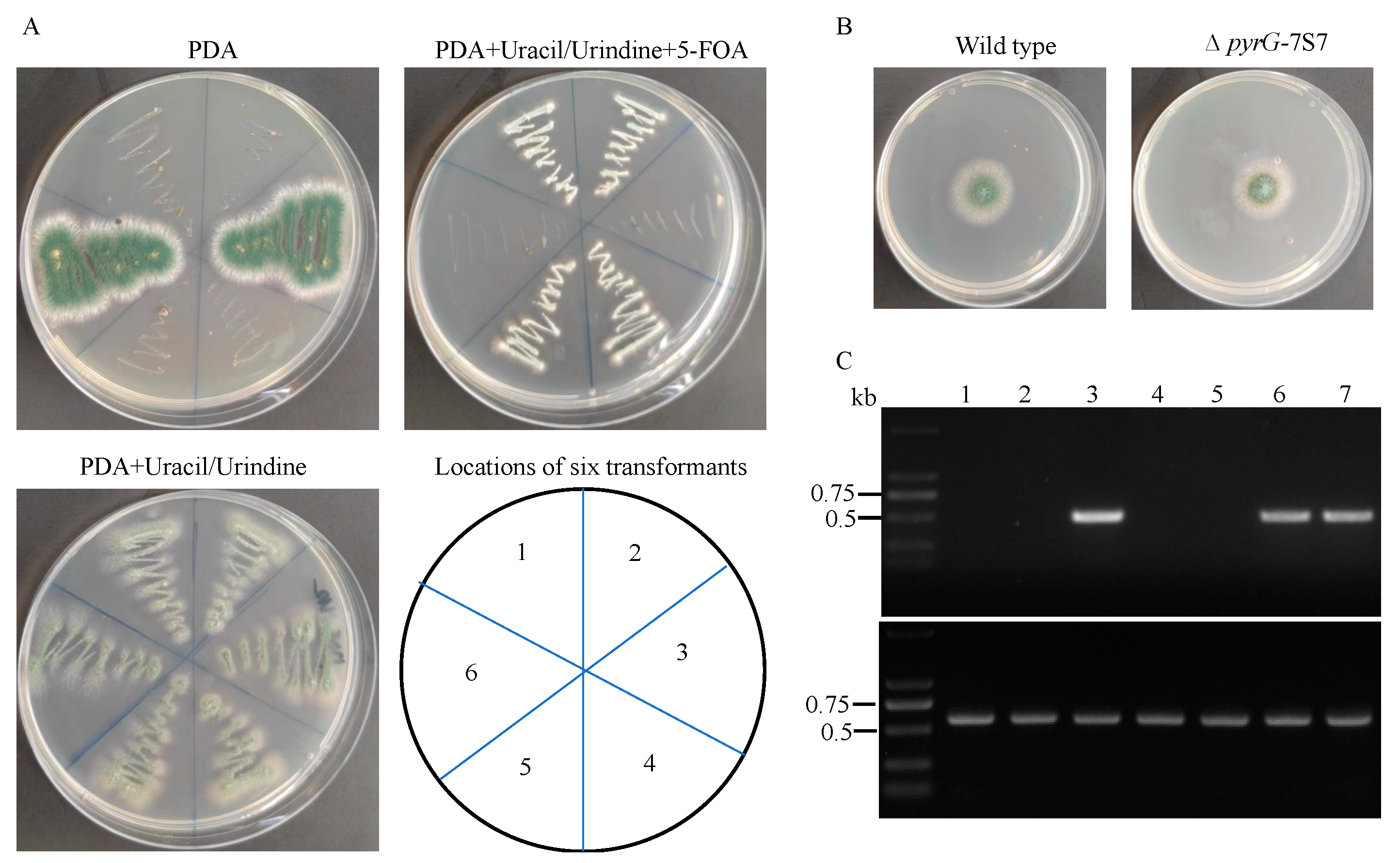
The first step for developing a pyrG-based marker recycling system is to generate a recipient strain with mutation of the pyrG gene for performing uracil auxotrophic complementary transformation and subsequent gene disruption. Most pyrG mutants were obtained following either ultraviolet (UV) or 5-FOA induced mutagenesis, which may include multiple undesirable mutations [12]. Moreover, the mutation rate of the pyrG gene under 5-FOA pressure is species dependent. In this study, the sensibility of Aspergillus sp. SCSIO SX7S7 to 5-FOA was tested, and we found that 1 mg/mL 5-FOA can completely inhibit the growth of SCSIO SX7S7 (Figure S1). We tried to induce spontaneous pyrG mutation in Aspergillus sp. SCSIO SX7S7 using the uridine/uracil-containing media supplemented with different concentrations of 5-FOA (1–10 mg/mL). We were unable to retrieve 5-FOA-resistant colonies. As our previously established CRISPR-Cas9 system has shown to be an efficient tool for gene disruption in Aspergillus sp. SCSIO SX7S7 [23], a CRISPR-Cas9 plasmid containing a sgRNA targeting the exon of pyrG-7S7 was generated and introduced into the protoplast of SCSIO SX7S7. After 4 days of growth, six colonies were picked, and four of them showed no growth on PDA but were able to grow on PDA supplemented with uridine/uracil. Moreover, the four colonies were 5-FOA resistant, suggesting that the four colonies may be pyrG-7S7 deficient (Figure 2A). PCR application using primers targeting pyrG-7S7 further validated the disruption of pyrG-7S7 gene (Figure 2C). The growth test revealed that disruption of pyrG-7S7 did not induce growth change compared with the wild type strain (Figure 2B), indicating the CRISPR-Cas9 system may not induce non-specific mutation. Therefore, one of the pyrG-7S7 mutants was selected for testing the feasibility of pyrG-based marker recycling system using the new plasmid built in this study.

3.3. Gene Disruption Using the CRISPR-Cas9-pyrG System in Aspergillus sp. SCSIO SX7S7

Our previous study showed that tricyclic depsidones and bicyclic depsides (DEPs) are constitutively produced by Aspergillus sp. SCSIO SX7S7 with high abundance [24]. To eliminate depsidones and depsides in Aspergillus sp. SCSIO SX7S7, the genome of SCSIO SX7S7 was analyzed by SM biosynthetic gene clusters annotation tool AntiSMASH [36], and a gene cluster containing two PKS genes (depD and depH) was proposed to be responsible for DEPs biosynthesis. Then, a sgRNA targeting the sequences of the PKS gene depH were designed and ligated into pBSKII-toCas9-pyrG. After transformation and selection on the plate without uridine/uracil, six transformants were randomly picked out for PCR validation using primers surrounding the sgRNA binding site. Our previous study has shown that gene disruption by the CRISPR-Cas9 system in the two tested marine-derived fungi can result in high frequency integration of the exogenous plasmid at the Cas9 cut site [23] and thus result in failed PCR when using primers flanking the PAM site. Similarly, five transformants harboring the depH disruption construct failed to be PCR amplified for obtaining targeted bands. (Figure S2). To assess whether the ∆depH mutant abolishes the ability to produce DEPs, the secondary metabolites from ∆depH mutant were extracted and analyzed with HPLC. HPLC analysis revealed that the ∆depH mutant was not able to produce DEPs (Figure 3), which further confirmed that the depH has been successfully mutated by the pyrG-based gene editing system and validated that the depH is a key gene for DEPs biosynthesis. In addition to DEPs, other differences were also observed in the metabolic profile between the wild type and ∆depH strains, suggesting that inactivation of DEPs biosynthesis cluster may also alter the biosynthesis of other NPs. Moreover, lack of visible growth differences between wild type and ΔdepH mutant suggested that the editing happened at the loci of interest without non-specific disruptions (Figure 3).

3.4. Remove of pyrG Marker by Counter Selection

In the ΔdepH mutant, all high-yielding DEPs in wild type strain were abolished, which provide us with a clean chassis cell for further natural product activation. To apply the chassis cell for the second round of genetic manipulation, we tried to remove the pyrG marker by counterselection. Due to the importance of DRs for looping out the pyrG marker [35] and the random break of transformed plasmid when integrated into the genome [23], PCR with the primers targeting the complete DRs franked pyrG marker expression cassette was carried out to check the integrity of DRs. All mutants retained the intact pyrG marker expression cassette flanked by DRs (Figure S3). One of the 7S7-ΔdepH-afpyrG mutants was selected to remove the pyrG marker for yielding a marker-free mutant. The spores of the ΔdepH mutant were spread on the 5-FOA (1 mM) plates, and the plates were incubated at 28 °C for 1 week until colonies became visible. Six colonies were picked out from the 5-FOA plate, and PCR amplifications with primers targeting the pyrG marker were used to confirm the deletion of the pyrG marker. As shown in Figure 4, selected colonies failed to be PCR amplified with the primers targeting pyrG marker, which depicted that the pyrG marker is absent from ΔdepH mutants, and thus the marker-free ΔdepH mutant (7S7-ΔdepH) can be used for the stepwise gene manipulation for identifying novel natural products.

3.5. Gene Disruptions Using CRISPR-Cas9-amdS System in Spiromastix sp. SCSIO F190

The successful application of the pyrG-based CRISPR-Cas9 system in Aspergillus sp. SCSIO SX7S7 promoted us to use the same gene editing tool into Spiromastix sp. SCSIO F190. Unlike Aspergillus sp. SCSIO SX7S7, generation of the pyrG mutant in Spiromastix sp. SCSIO F190 failed after repeated attempts. This may be due to the high GC content of the four available sgRNAs targeting the pyrG gene in Spiromastix sp. SCSIO F190, which can reduce cleavage efficiency [37]. To improve the efficiency of the genetic engineering on the strains with complicated genetic backgrounds makes the construction of auxotrophic strains a laborious task. Thus, it is important to develop another marker recycling system that does not need to generate auxotrophic mutants.
The amdS gene encoding acetamidase from A. nidulans has been successfully used as a recycle marker in several filamentous fungi that are not able to use acetamide as the sole nitrogen or carbon source. To apply the amdS marker recycle system in Spiromastix sp. SCSIO F190, the metabolic capability of Spiromastix sp. SCSIO F190 for using acetamide was first evaluated. The spores of Spiromastix sp. SCSIO F190 were inoculated on the ANM plate with acetamide as the sole carbon and nitrogen source with different concentrations of cesium chloride. After 14 days of growth, Spiromastix sp. SCSIO F190 can grow weakly on the acetamide plate without cesium chloride, but the growth can be further inhibited by cesium chloride with increasing concentrations (Figure 5). The rationale for the effects of cesium chloride is not completely clear, but it has been used widely for suppressing the growth of recipient strains when the amdS marker recycle system was applied [20,38]. Therefore, the acetamide plate with 100 mM cesium chloride that can significantly block the growth of Spiromastix sp. SCSIO F190 was used to differentiate the transformed and untransformed cells.
Next, to evaluate whether the amdS marker from A.nidulans was suitable for gene knock-out in Spiromastix sp. SCSIO F190, a polyketide synthase gene (spm11) was selected for this proof-of-principle experiment. In our previous study, a gene cluster responsible for the biosynthesis of spiromarmycin, a high-abundance major product, was proposed, in which spm11 was predicted as a backbone gene for spiromarmycin biosynthesis [25]. Thus, the absence of spm11 would stop producing the brown spiromarmycin, giving a fast preliminary evaluation of targeted mutation by comparing the color difference on a solid medium. More importantly, clearance of the constantly produced major product is a key step for generating chassis cells with simplified HPLC background for exploring novel compounds. The pBSKII-toCas9-amdS-sgRNA plasmid containing a sgRNA targeting the spm11 was transformed into Spiromastix sp. SCSIO F190 to inactivate the polyketide synthase gene. After transformation, cells were grown on the regeneration plates containing acetamide as the sole nitrogen source with 100 mM cesium chloride for 14 days, and the transformants were transferred into PDA plates. Among the ten picked colonies, eight of them showed different phenotypes with wild type strain (Figure S4).
The genome DNA of the eight colonies without significant accumulation of secreted compounds was extracted, and PCR was performed to check the mutation of the spm11 gene. Unexpectedly, only one colony fail to obtain the PCR band when using the primers flanking the PAM site (Figure 6A). However, when the primers with one forward primer targeted the sgRNA binding site were used, all of the other seven colonies failed to obtain the PCR bands, suggesting the amdS-based CRISPR-Cas9 system-induced mutations are predominantly short deletions around the PAM site. Sequencing of the PCR bands of the seven colonies further confirmed that the spm11 gene was disrupted by several DNA bases deletion/mutation (Figure 6B). In addition, HPLC analysis of a ∆spm11 mutant showed that the mutant is not able to produce spiromarmycin (Figure 6C), validating the previous prediction that the spm11 is a key gene for spiromarmycin biosynthesis.

3.6. Remove of amdS Marker by Counter Selection

To apply the resulting strain F190-∆spm11-amdS for another round of genetic manipulation, amdS maker excision aided by the DRs created upon integration under counter-selection pressure was performed. Similar to the pyrG marker looping out experiment, the integrity of DRs was first checked. Fortunately, six of the eight ∆spm11 mutants containing the complete DRs franked amdS marker expression cassette (Figure S5). One of the six F190-∆spm11-amdS mutants was cultured on PDA plate for 10 days, and the spores were collected and spread on the 5 mM FAA plates. PCR analysis of the growing colonies on FAA plates confirmed correct marker removal (Figure 7). The resulting strain F190-∆spm11 is ready for multiple rounds of genetic manipulation using an amdS maker, including inactivation of the other constitutively produced major products [26] and activation or heterogeneous expression of novel NPs in the chassis cells with simplified HPLC background.

4. Conclusions

In summary, we have successfully developed a genetic engineering toolbox containing two different marker recycling systems (pyrG and amdS) for gene disruption in marine-derived fungi. Applications of these marker recycling systems have proven to be efficient strategies for the clearance of the constitutively produced SMs in different marine fungi species. Especially, in Spiromastix species, it is the first application of the amdS-based recycle system for genome editing. Thus, the genetic editing systems established in this study showed promising potential in assisting sequential genetic manipulation of marine-derived fungi with distinct phylogenetics, providing powerful tools for accessing the structural diversity. Furthermore, although several model organisms such as Aspergillus nidulans and Aspergillus oryzae have been developed, there is no marine-derived model fungus. Here, with the assistance of recyclable markers, we are developing marine-derived fungi as model organisms/chassis cells for heterologous expressing BGCs originating from marine-derived fungi and investing marine fungi biology.

Supplementary Materials

The following supporting information can be downloaded at: https://www.mdpi.com/article/10.3390/jof9030302/s1, Figure S1: Growth of Aspergillus sp. SCSIO SX7S7 on PDA plate with different concentrations of 5-FOA; Figure S2: Verification of the depH mutants by genotyping PCR; Figure S3: Verification of the integrity of DR flanked pyrG expression cassette; Figure S4: Growth of Spiromastix sp. SCSIO F190 strains; Figure S5: Verification of the integrity of DR flanked amdS expression cassette. Table S1: Strains used in this study; Table S2: Plasmids used in this study; Table S3: Primers used in this study.

Author Contributions

J.J. and Y.C. designed the experiment. Y.C., J.Y., C.C. and J.S. performed the experiment and processed the experimental data. Y.C. drafted the manuscript. J.J., J.M. and Y.S. reviewed the draft. J.J. and J.M. supervised the project. All authors have read and agreed to the published version of the manuscript.

Funding

This research was funded by “Key science and Technology project of Hainan Province (ZDKJ202018)”, “GuangDong Basic and Applied Basic Research Foundation (2023A1515011840)”, “Key-Area Research and Development Program of Guangdong Province (2020B1111030005)”, “National Natural Science Foundation of China (22037006, 82022067)”, “Local Innovation and Entrepreneurship Team Project of Guangdong (2019BT02Y262)”, “Open program of ShenZhen Bay Laboratory (SZBL2021080601006)”, “Nansha District Science and technology award (NSJL202102)” and “Fundamental Research & Applied Fundamental Research Major Project of Guangdong Province (2019B030302004)”.

Institutional Review Board Statement

Not applicable.

Informed Consent Statement

Not applicable.

Data Availability Statement

The data presented in this study are available in this manuscript, and constructs can be requested from the corresponding author.

Acknowledgments

We are grateful to Dan Hu (Jinan University, Guangzhou, China) for providing the pBSKII-toCas9-hph plasmid. We thank Yi Tang (University of California, Los Angeles, CA, USA) for providing the pFC330 plasmid. We thank Wong, K.H. (University of Macau) for providing the pBluescript II SK plasmid.

Conflicts of Interest

The authors declare no conflict of interest.

References

  1. Hasan, S.; Ansari, M.I.; Ahmad, A.; Mishra, M. Major bioactive metabolites from marine fungi: A Review. Bioinformation 2015, 11, 176–181. [Google Scholar] [CrossRef] [PubMed] [Green Version]
  2. Liu, Z.; Frank, M.; Yu, X.; Yu, H.; Tran-Cong, N.M.; Gao, Y.; Proksch, P. Secondary metabolites from marine-derived fungi from China. Prog. Chem. Org. Nat. Prod. 2020, 111, 81–153. [Google Scholar] [PubMed]
  3. Shin, H.J. Natural Products from Marine Fungi. Mar. Drugs 2020, 18, 230. [Google Scholar] [CrossRef] [PubMed]
  4. Shabana, S.; Lakshmi, K.R.; Satya, A.K. An Updated Review of Secondary Metabolites from Marine Fungi. Mini. Rev. Med. Chem. 2021, 21, 602–642. [Google Scholar] [CrossRef] [PubMed]
  5. Zhang, H.; Zhang, C.; Li, Q.; Ma, J.; Ju, J. Metabolic blockade-based genome mining reveals lipochain-linked dihydro-beta-alanine synthetases involved in autucedine biosynthesis. Org. Lett. 2022, 24, 5535–5540. [Google Scholar] [CrossRef] [PubMed]
  6. Zhang, C.; Ding, W.; Qin, X.; Ju, J. Genome sequencing of Streptomyces olivaceus SCSIO T05 and activated production of lobophorin CR4 via metabolic engineering and genome mining. Mar. Drugs 2019, 17, 593. [Google Scholar] [CrossRef] [Green Version]
  7. Macheleidt, J.; Mattern, D.J.; Fischer, J.; Netzker, T.; Weber, J.; Schroeckh, V.; Valiante, V.; Brakhage, A.A. Regulation and role of fungal secondary metabolites. Annu. Rev. Genet. 2016, 50, 371–392. [Google Scholar] [CrossRef]
  8. Brakhage, A.A. Regulation of fungal secondary metabolism. Nat. Rev. Microbiol. 2013, 11, 21–32. [Google Scholar] [CrossRef]
  9. Boeke, J.D.; LaCroute, F.; Fink, G.R. A positive selection for mutants lacking orotidine-5’-phosphate decarboxylase activity in yeast: 5-fluoro-orotic acid resistance. Mol. Gen. Genet. 1984, 197, 345–346. [Google Scholar] [CrossRef]
  10. Delmas, S.; Llanos, A.; Parrou, J.L.; Kokolski, M.; Pullan, S.T.; Shunburne, L.; Archer, D.B. Development of an unmarked gene deletion system for the filamentous fungi Aspergillus niger and Talaromyces versatilis. Appl. Environ. Microbiol. 2014, 80, 3484–3487. [Google Scholar] [CrossRef] [Green Version]
  11. D’Enfert, C.; Diaquin, M.; Delit, A.; Wuscher, N.; Debeaupuis, J.P.; Huerre, M.; Latge, J.P. Attenuated virulence of uridine-uracil auxotrophs of Aspergillus fumigatus. Infect. Immun. 1996, 64, 4401–4405. [Google Scholar] [CrossRef] [PubMed] [Green Version]
  12. Ling, S.O.; Storms, R.; Zheng, Y.; Rodzi, M.R.; Mahadi, N.M.; Illias, R.M.; Abdul Murad, A.M.; Abu Bakar, F.D. Development of a pyrG mutant of Aspergillus oryzae strain S1 as a host for the production of heterologous proteins. Sci. World J. 2013, 2013, 634317. [Google Scholar] [CrossRef] [PubMed] [Green Version]
  13. Nielsen, M.L.; Albertsen, L.; Lettier, G.; Nielsen, J.B.; Mortensen, U.H. Efficient PCR-based gene targeting with a recyclable marker for Aspergillus nidulans. Fungal Genet. Biol. 2006, 43, 54–64. [Google Scholar] [CrossRef]
  14. Sun, R.; Xu, H.; Feng, Y.; Hou, X.; Zhu, T.; Che, Q.; Pfeifer, B.; Zhang, G.; Li, D. An efficient marker recycling system for sequential gene deletion in a deep sea-derived fungus Acremonium sp. HDN16-126. Synth. Syst. Biotechnol. 2021, 6, 127–133. [Google Scholar] [CrossRef]
  15. Jorgensen, M.S.; Skovlund, D.A.; Johannesen, P.F.; Mortensen, U.H. A novel platform for heterologous gene expression in Trichoderma reesei (Teleomorph Hypocrea jecorina). Microb. Cell Fact. 2014, 13, 33. [Google Scholar] [CrossRef] [Green Version]
  16. Erpf, P.E.; Stephenson, C.J.; Fraser, J.A. amdS as a dominant recyclable marker in Cryptococcus neoformans. Fungal Genet. Biol. 2019, 131, 103241. [Google Scholar] [CrossRef] [PubMed] [Green Version]
  17. Hynes, M.J.; Pateman, J.A. The genetic analysis of regulation of amidase synthesis in Aspergillus nidulans. II. Mutants resistant to fluoroacetamide. Mol. Gen. Genet. 1970, 108, 107–116. [Google Scholar] [CrossRef] [PubMed]
  18. Clarke, D.D. Fluoroacetate and fluorocitrate: Mechanism of action. Neurochem. Res. 1991, 16, 1055–1058. [Google Scholar] [CrossRef]
  19. Apirion, D. The two-way selection of mutants and revertants in respect of acetate utilization and resistance to fluoro-acetate in Aspergillus nidulans. Genet. Res. 1965, 6, 317–329. [Google Scholar] [CrossRef]
  20. Nan, Y.; Ouyang, L.; Chu, J. In vitro CRISPR/Cas9 system for genome editing of Aspergillus niger based on removable bidirectional selection marker AmdS. Biotechnol. Appl. Biochem. 2021, 68, 964–970. [Google Scholar] [CrossRef]
  21. Fu, J.; Blaylock, M.; Wickes, C.F.; Welte, W.; Mehrtash, A.; Wiederhold, N.; Wickes, B.L. Development of a Candida glabrata dominant nutritional transformation marker utilizing the Aspergillus nidulans acetamidase gene (amdS). FEMS. Yeast. Res. 2016, 16, fow023. [Google Scholar] [CrossRef] [Green Version]
  22. Beri, R.K.; Turner, G. Transformation of Penicillium chrysogenum using the Aspergillus nidulans amdS gene as a dominant selective marker. Curr. Genet. 1987, 11, 639–641. [Google Scholar] [CrossRef]
  23. Chen, Y.; Cai, C.; Yang, J.; Shi, J.; Song, Y.; Hu, D.; Ma, J.; Ju, J. Development of the CRISPR-Cas9 system for the marine-derived fungi Spiromastix sp. SCSIO F190 and Aspergillus sp. SCSIO SX7S7. J. Fungi 2022, 8, 715. [Google Scholar] [CrossRef]
  24. Yang, J.; Zhou, L.; Zhou, Z.; Song, Y.; Ju, J. Anti-pathogenic depsidones and its derivatives from a coral-derived fungus Aspergillus sp. SCSIO SX7S7. Biochem. Syst. Ecol. 2022, 102, 104415. [Google Scholar] [CrossRef]
  25. Shao, M.; Sun, C.; Liu, X.; Wang, X.; Li, W.; Wei, X.; Li, Q.; Ju, J. Upregulation of a marine fungal biosynthetic gene cluster by an endobacterial symbiont. Commun. Biol. 2020, 3, 527. [Google Scholar] [CrossRef]
  26. Cai, C.; Chen, Y.; Zhou, L.; Gong, N.; Zhang, H.; Sun, C.; Ma, J.; Ju, J. Antimicrobial polyketides from the marine-derived fungus Spiromastix sp. SCSIO F190. J. Nat. Prod. 2022. [Google Scholar] [CrossRef]
  27. Todd, R.B.; Davis, M.A.; Hynes, M.J. Genetic manipulation of Aspergillus nidulans: Meiotic progeny for genetic analysis and strain construction. Nat. Protoc. 2007, 2, 811–821. [Google Scholar] [CrossRef] [PubMed]
  28. Qin, L.; Li, A.; Tan, K.; Guo, S.; Chen, Y.; Wang, F.; Wong, K.H. Universal plasmids to facilitate gene deletion and gene tagging in filamentous fungi. Fungal Genet. Biol. 2019, 125, 28–35. [Google Scholar] [CrossRef] [PubMed]
  29. Zheng, Y.M.; Lin, F.L.; Gao, H.; Zou, G.; Zhang, J.W.; Wang, G.Q.; Chen, G.D.; Zhou, Z.H.; Yao, X.S.; Hu, D. Development of a versatile and conventional technique for gene disruption in filamentous fungi based on CRISPR-Cas9 technology. Sci. Rep. 2017, 7, 9250. [Google Scholar] [CrossRef]
  30. Nodvig, C.S.; Nielsen, J.B.; Kogle, M.E.; Mortensen, U.H. A CRISPR-Cas9 system for genetic engineering of filamentous Fungi. PLoS ONE 2015, 10, e0133085. [Google Scholar] [CrossRef] [PubMed] [Green Version]
  31. Song, R.; Zhai, Q.; Sun, L.; Huang, E.; Zhang, Y.; Zhu, Y.; Guo, Q.; Tian, Y.; Zhao, B.; Lu, H. CRISPR/Cas9 genome editing technology in filamentous fungi: Progress and perspective. Appl. Microbiol. Biotechnol. 2019, 103, 6919–6932. [Google Scholar] [CrossRef] [PubMed] [Green Version]
  32. Dong, H.; Zheng, J.; Yu, D.; Wang, B.; Pan, L. Efficient genome editing in Aspergillus niger with an improved recyclable CRISPR-HDR toolbox and its application in introducing multiple copies of heterologous genes. J. Microbiol. Methods 2019, 163, 105655. [Google Scholar] [CrossRef] [PubMed]
  33. Michielse, C.B.; Ram, A.F.; van den Hondel, C.A. The Aspergillus nidulans amdS gene as a marker for the identification of multicopy T-DNA integration events in Agrobacterium-mediated transformation of Aspergillus awamori. Curr. Genet. 2004, 45, 399–403. [Google Scholar] [CrossRef]
  34. Penttila, M.; Nevalainen, H.; Ratto, M.; Salminen, E.; Knowles, J. A versatile transformation system for the cellulolytic filamentous fungus Trichoderma reesei. Gene 1987, 61, 155–164. [Google Scholar] [CrossRef] [PubMed]
  35. Alani, E.; Cao, L.; Kleckner, N. A method for gene disruption that allows repeated use of URA3 selection in the construction of multiply disrupted yeast strains. Genetics 1987, 116, 541–545. [Google Scholar] [CrossRef]
  36. Blin, K.; Shaw, S.; Kloosterman, A.M.; Charlop-Powers, Z.; van Wezel, G.P.; Medema, M.H.; Weber, T. antiSMASH 6.0: Improving cluster detection and comparison capabilities. Nucleic Acids. Res. 2021, 49, W29–W35. [Google Scholar] [CrossRef]
  37. Liu, X.; Homma, A.; Sayadi, J.; Yang, S.; Ohashi, J.; Takumi, T. Sequence features associated with the cleavage efficiency of CRISPR/Cas9 system. Sci. Rep. 2016, 6, 19675. [Google Scholar] [CrossRef] [PubMed] [Green Version]
  38. Tilburn, J.; Scazzocchio, C.; Taylor, G.G.; Zabicky-Zissman, J.H.; Lockington, R.A.; Davies, R.W. Transformation by integration in Aspergillus nidulans. Gene 1983, 26, 205–221. [Google Scholar] [CrossRef]
Figure 1. The construction of new CRISPR-Cas9 vectors (pBSKII-toCas9-pyrG/amdS-sgRNA) for multistep gene deletion in marine-derived fungi. The sequence of the pyrG/amdS expression cassette including PtrpC promoter, TtrpC terminator, and direct repeat was amplified. Then the amplified fragments were ligated into PsiI digested pBSKII-toCas9-hph vector using isothermal assembly to generate two vectors named pBSKII-toCas9-pyrG and pBSKII-toCas9-amdS which were further digested by the BsaAI restriction enzyme for building the final gene disruption construct. The Fragment1 and Fragment2 were amplified by Frag1 F/R and Frag2 F/R primers using FC330 plasmid as a template, respectively. The resultant PCR products of flanking regions and the BsaAI-digested pBSKII-toCas9- pyrG/amdS plasmid can be joined together to form the final construct pBSKII-toCas9-pyrG/amdS-sgRNA. For simplicity, all complementary ends are visualized in the same color and no DNA elements in the above figure are drawn to scale.
Jof 09 00302 g001
Figure 2. The inactivation of the pyrG-7S7 gene in Aspergillus sp. SCSIO SX7S7 using the CRISPR-Cas9 system. (A) Growth of the transformants carrying the CRISPR-Cas9 plasmids containing a sgRNA targeting exon of pyrG-7S7 on a PDA medium with or without uridine-uracil (5 mM) and a PDA medium supplemented with both uridine/uracil (5 mM) and 5-FOA (1 mg/mL). (B) Growth of the Aspergillus sp. SCSIO SX7S7 wild type and ∆ pyrG-7S7 strains on a PDA medium with uridine/uracil (5 mM) at 28 °C for 4 days. (C) DNA was extracted from the six randomly selected transformants (shown on the left side) and amplified by PCR using the primers flanking the PAM site (upper panel), and the DNA template quality of the tested samples was validated using primers flanking the ITS region (lower panel). PCR amplification with the same primers of DNA from the wild type strain served as positive control. (Lane 1–6: tested transformants; lane 7: wild-type strain).
Jof 09 00302 g002
Figure 3. Characterization of the ΔdepH mutant of Aspergillus sp. SCSIO SX7S7. (A) Growth of the wild-type and ΔdepH mutation strains on PDA plates at 28 °C for 4 days. (B) HPLC-DAD contour plot of extracts from the wild-type and ΔdepH mutation strains. The boxed region shows DEPs.
Jof 09 00302 g003
Figure 4. Verification of AfpyrG gene looping out. Upper panel: PCR was carried out using the primers targeting the AfpyrG marker. Lower panel: DNA template quality of the tested samples was validated using primers flanking the ITS region. The ΔdepH strain carrying the AfpyrG marker was set as positive control. (Lane 1–6: ΔdepH mutants without AfpyrG marker; Lane 7: positive control).
Jof 09 00302 g004
Figure 5. Growth of Spiromastix sp. SCSIO F190 on ANM plate with acetamide as sole nitrogen and carbon source. The spores of Spiromastix sp. SCSIO F190 were inoculated on the acetamide plate with different concentrations of cesium chloride and incubated at 28 °C for 10 days. The growth on the ANM plate containing glucose and ammonium tartrate was set as control.
Jof 09 00302 g005
Figure 6. Inactivation of spm11 in Spiromastix sp. SCSIO F190 using the amdS-based CRISPR-Cas9 system. (A) DNA amplification of the DNA region surrounding the sgRNA binding site of the eight clones with different phenotypes from wild type plate using primers flanking the PAM site in the spm11 gene (Upper panel) and primers with one forward primer targeted the sgRNA binding site (Lower panel). PCR amplification with the same primers of DNA from the wild type strain served as positive control. (Lane 1–8: tested transformants; lane 9: wild-type strain.) (B) Sequence analysis of PCR products of clones 2–8. (C) HPLC-DAD contour plot of extracts from the wild-type and Δspm11 mutation strains. The boxed region indicates spiromarmycin.
Jof 09 00302 g006
Figure 7. Verification of AnamdS gene looping out. Upper: PCR was carried out using the primers targeting the AnamdS marker. Lower: DNA template quality of the tested samples was validated using primers flanking the ITS region The F190-∆spm11-AnamdS strain carrying AnamdS marker was set as positive control. (Lane 1–6: Δspm11 mutants without AnamdS marker; Lane 7: positive control).
Jof 09 00302 g007
Disclaimer/Publisher’s Note: The statements, opinions and data contained in all publications are solely those of the individual author(s) and contributor(s) and not of MDPI and/or the editor(s). MDPI and/or the editor(s) disclaim responsibility for any injury to people or property resulting from any ideas, methods, instructions or products referred to in the content.