Article

Phenylalanine Ammonia-Lyase: A Key Gene for Color Discrimination of Edible Mushroom Flammulina velutipes

1
Mushroom Research Division, National Institute of Horticultural and Herbal Science, Rural Development Administration, 92, Bisan-ro, Eumseong-gun 27709, Republic of Korea
2
Department of Medicinal Biosciences, Research Institute for Biomedical & Health Science, College of Biomedical and Health Science, Konkuk University, 268 Chungwon-daero, Chungju-si 27478, Republic of Korea
*
Author to whom correspondence should be addressed.
J. Fungi 2023, 9(3), 339; https://doi.org/10.3390/jof9030339
Submission received: 18 February 2023 / Revised: 7 March 2023 / Accepted: 8 March 2023 / Published: 9 March 2023
(This article belongs to the Special Issue Biotechnology of Edible Fungi 2.0)

Abstract

:
In nature; Flammulina velutipes, also known as winter mushrooms, vary in the color of their fruiting bodies, from black, yellow, pale yellow, or beige to white. The purpose of this study was to compare the genome sequences of different colored strains of F. velutipes and to identify variations in the genes associated with fruiting body color. Comparative genomics of six F. velutipes strains revealed 70 white-strain-specific variations, including single nucleotide polymorphisms (SNPs) and insertions/deletions (indels), in the genome sequences. Among them, 36 variations were located in the open reading frames, and only one variation was identified as a mutation with a disruptive in-frame deletion (ΔGCGCAC) within the annotated gene phenylalanine ammonia-lyase 1 (Fvpal1). This mutation was found to cause a deletion, without a frameshift, of two amino acids at positions 112 and 113 (arginine and threonine, respectively) in the Fvpal1 gene of the white strain. Specific primers to detect this mutation were designed, and amplification refractory mutation system (ARMS) polymerase chain reaction (PCR) was performed to evaluate whether the mutation is color specific for the F. velutipes fruiting body. PCR analysis of a total of 95 F. velutipes strains revealed that this mutation was present only in white strains. In addition, monospores of the heterozygous mutant were isolated, and whether this mutation was related to the color of the fruiting body was evaluated by a mating assay. In the mating analysis of monospores with mutations in Fvpal1, it was found that this mutation plays an important role in determining the color of the fruiting body. Furthermore, the deletion (Δ112RT113) in Fvpal1 is located between motifs that play a key role in the catalytic function of FvPAL1. These results suggest that this mutation can be used as an effective marker for the color-specific breeding of F. velutipes, a representative edible mushroom.

1. Introduction

The edible mushroom Flammulina velutipes belongs to the family Tricholomataceae within Agaricales and grows on old trees or the stumps of various broadleaf trees from late autumn to the following spring. This mushroom is a cold-resistant fungus that occurs even in winter; therefore, it is also called the winter mushroom [1,2]. Wild-type F. velutipes strains vary in color from yellowish-brown to dark brown, whereas artificial cultivars are predominantly white [3] (Figure 1). The first artificially cultivated variety was a brown F. velutipes strain, but the cultivation of F. velutipes began in earnest after white varieties were bred in Japan, and these are still the main cultivated varieties today [3,4]. The white F. velutipes mainly cultivated in Korea and Japan were discovered by chance, and the genetic cause of the discoloration of the fruiting body is not known. Since the artificial cultivation of F. velutipes began, various white cultivars have been actively developed, but the close genetic relationship between these varieties limits the development of new cultivars [3,4]. To overcome these limitations, new varieties are being actively developed through crossbreeding with various wild strains, including non-white ones [3,5,6,7,8,9,10,11]. Recent studies have analyzed nutritional components, such as saccharides, amino acids, and organic acids, as well as the growth characteristics of new varieties, and a comparative analysis of nutrients between white and non-white varieties was also conducted [3,5,6,7,8,9,10,11,12,13,14,15,16,17]. Information on the physiological characteristics and differences of these F. velutipes varieties, including the color of the F. velutipes fruiting body, can be used for breeding newly developed cultivars. While research on the development of non-white F. velutipes varieties is being conducted, studies have also been carried out to suppress the browning of mushrooms. Browning of Agaricus bisporus (button mushrooms) is a serious problem that shortens the shelf life after harvest. Mushroom browning is mainly caused by the activation of tyrosinase, belonging to the polyphenol oxidase (PPO) family, or spontaneous oxidation [18]. In mushrooms, it has been reported that the enzymatic oxidation of phenolic compounds leads to the biosynthesis of melanin, resulting in the formation of a brown color [19]. The key enzymes in the melanin biosynthesis pathway are PPO, laccase (EC 1.10.3.2), and tyrosinase (EC 1.14.18.1) and are involved in the conversion of phenolic compounds [20]. Tyrosinase converts monophenols to ortho-diphenols by ortho-hydroxylation, and ortho-diphenols to ortho-quinones by oxidation. Finally, quinone is converted to melanin via a non-enzymatic reaction. Laccases oxidize a variety of phenolic substrates by performing one-electron oxidation, leading to crosslinking and facilitating the biosynthesis of melanin pigments [21]. Another enzyme involved in browning is peroxidase, which catalyzes the oxidation of various compounds using hydrogen peroxide as a substrate [22,23]. Recently, the PPO gene relating to the color of the fruiting body of Agaricus bisporus was identified, and the development of a variety that suppresses browning through gene editing has been attempted [24]. The color of mushroom fruiting bodies is an important factor that affects not only the development of new varieties but also the mushroom industry.
As the genome sequences of F. velutipes species have been determined and in-depth genetic information has been revealed, comparative studies on the biological characteristics and diversity of these mushrooms have been actively conducted [1,25,26,27,28,29]. The genetic repertoire of F. velutipes mushrooms revealed through genomic studies has proven that this mushroom can be used for a wide variety of industrial applications and has ample potential for the development of new varieties. In this study, comparative genome analysis of F. velutipes was performed to identify key genes or mutations related to the color of the F. velutipes fruiting body. The color-related genes or mutations of F. velutipes identified in this study can be used to selectively develop the color of this mushroom variety in the future.

2. Materials and Methods

2.1. Fungal Strain Culture and Genomic DNA Isolation

F. velutipes strains (Table 1) were obtained from the Mushroom Research Division, National Institute of Horticultural and Herbal Science (Rural Development Administration, Jeonju, Republic of Korea) and were grown on potato dextrose agar (PDA; 4 g potato starch, 20 g dextrose, 15 g agar per liter) at 25 °C for 15 days. Genomic DNA was then extracted using extraction buffer (0.25 M Tris-HCl, 100 mM NaCl, 50 mM ethylenediaminetetraacetic acid, 5% SDS), 2 × CTAB buffer (100 mM Tris-HCl pH 8, 20 mM EDTA pH 8, 2% CTAB, 1.4 M NaCl, and 1% polyvinyl pyrrolidone), and phenol-chloroform-isoamyl alcohol (25:24:1) as previously described [29]. The extracted DNA samples were treated with RNase A (Qiagen, Hilden, Germany).

2.2. Genome Sequencing and Identification of Variations

Genome sequencing of F. velutipes strains was performed using the HiSeq 2000 platform (Illumina, Inc., San Diego, CA, USA). Sequenced reads were processed using FastQC (http://www.bioinformatics.babraham.ac.uk/projects/fastqc/, accessed on 11 August 2022) and Trimmomatic (version 0.39) [30] for quality control. The final reads were used for mapping to the reference genome (F. velutipes KACC42780; accession number) [1] and to identify sequence variations using the genome analysis toolkit (GATK) pipeline [31] with the Burrows–Wheeler Aligner (BWA) [32], SAMtools [33], and PICARD [34]. SnpEff [35] and BEDTools [36] were used to identify the locations of variations within the genes. Gene modeling of the reference genome was performed by the Funannotate pipelines [37], using F. velutipes KACC43778 transcriptome data as “evidence”. Gene models of F. velutipes KACC42780 were annotated using KEGG, InterPro, and UniProt databases. Genome sequencing reads were deposited in NABIC (National Agricultural Biotechnology Information Center, RDA, Korea) (see Data Availability Statement).

2.3. Primer Design and Amplification Refractory Mutation System PCRs

Primers that amplified specific variation sites within the phenylalanine ammonia-lyase 1 (Fvpal1) gene of white and non-white F. velutipes strains were designed based on the amplification refractory mutation system (ARMS) [38]. Three primer sets were designed with expected amplicons of 464 bp, 293 bp, and 200 bp from all F. velutipes strains, F. velutipes white strains, and F. velutipes non-white strains, respectively (Table 2). Genomic DNA was used as a template (100 ng/μL) for ARMS PCR reactions using the Taq PreMix kit (TNT Research, Anyang, Korea) and 0.25 pmol of each primer in a 20 μL reaction mixture. PCR conditions were 10 min of initial denaturation at 94 °C, followed by 30 cycles of denaturation at 94 °C for 30 s, annealing at 56 °C for 30 s, extension at 72 °C for 1 min, and a final extension at 72 °C for 10 min using Bio-Rad thermal cycler (Bio-Rad Laboratories Inc., Hercules, CA, USA). The amplified products were separated on a 2% agarose gel in 0.5 × TAE buffer (Tris–acetic acid–EDTA, TNT Research, Anyang, Korea) buffer and visualized with ethidium bromide on a UV transilluminator.

2.4. Mating of F. velutipes Strains

For spore collection of F. velutipes ASI4175 (non-white, heterozygote for ΔGCGCAC Fvpal1) and F. velutipes w1-8 (white, homozygote for ΔGCGCAC Fvpal1) strains, the stipe of the fruiting body was removed, and only part of the pileus was separated and placed in a Petri dish with the fold of the pileus facing down, and spores were collected for 24 h. The collected spores were diluted to a concentration of 1.0 × 10⁻³ cfu/mL, and 100 μL of this dilution was spread on PDA medium and cultured for 7 d in a 25 °C incubator, protected from light. The germinated spores were inoculated into PDA medium and further cultured for 7 d in an incubator at 25 °C. Monokaryons without clamp connection were selected among the cultured mycelia. The Fvpal1 genotype of the isolated monokaryons was analyzed by ARMS PCR to select those for mating.
The monokaryons isolated from F. velutipes ASI4175 and F. velutipes w1-8 were inoculated on PDA medium at intervals of 1–2 cm and cultured for 7 d in a 25 °C incubator; hybrid dikaryons with clamp connection were then selected for fruiting. The hybrid dikaryons were inoculated into fruiting medium (80% sawdust and 20% rice bran) and incubated at 20 °C and 65% humidity for 30 days; the temperature was then sequentially changed from 14 °C (95% humidity) to 7 °C (80% humidity) for fruiting.

3. Results and Discussion

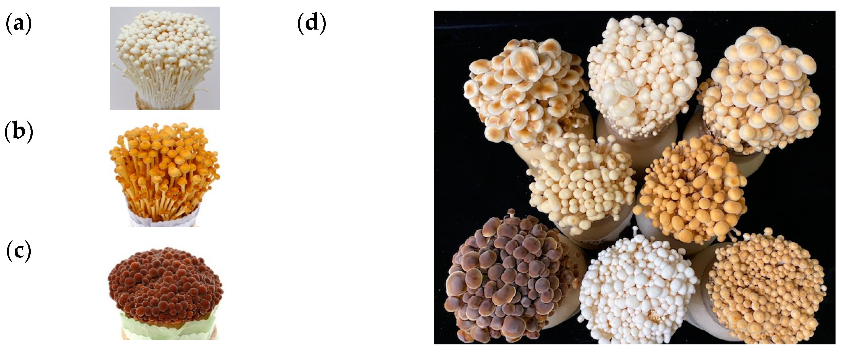
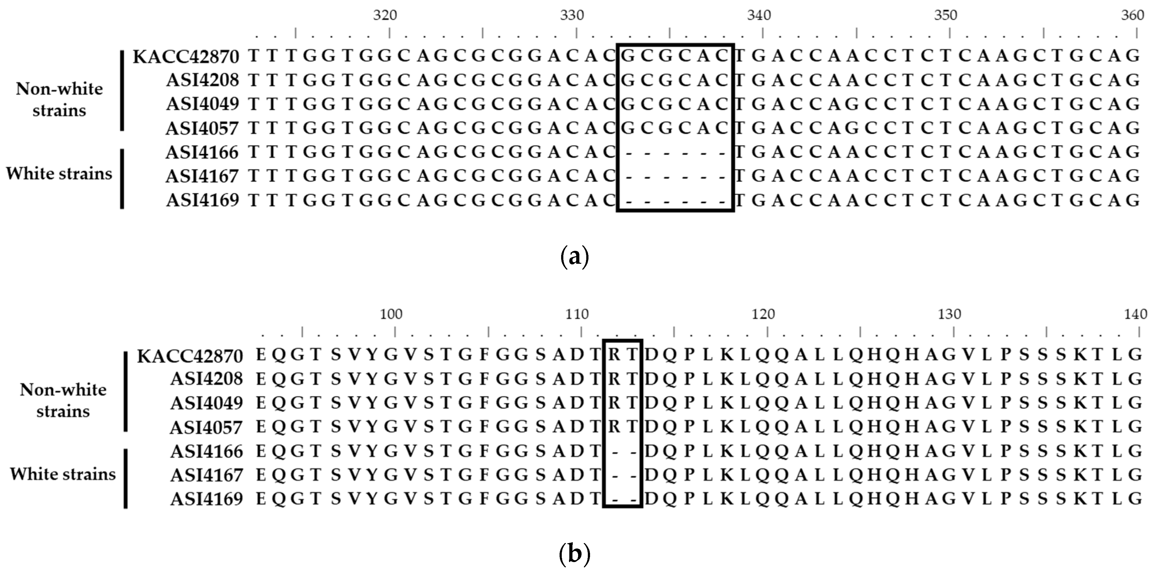
3.1. Identification of Fruiting Body Color-Specific Mutation of F. velutipes

The quality-trimmed reads of the six F. velutipes genome sequences were mapped to the reference genome sequence (F. velutipes KACC42870) at a rate of 73.15–80.13% (Table 3).
A total of 70 white-color-specific variations, including 57 single nucleotide polymorphisms (SNPs) and 13 indels, were identified from the genome comparison of non-white and white F. velutipes strains (Table S1). Among the 11 chromosomes of F. velutipes, chromosome 7 had the highest number of variations (60%, 33 SNPs, and 9 indels), suggesting that genetic variations among F. velutipes strains mainly occurred on chromosome 7. Gene modeling of the reference strain (KACC42870) was conducted to identify variations in the genes. Using transcriptome data from Funannotate pipelines, a total of 15,874 gene models were identified from the reference genome (KACC42870) (Table S2). Gene annotations revealed that 36 predicted genes were associated with the identified variations (Table S1). Among the variations in the genes, only one variation (indel) in Fvpal1 was identified; with a disruptive in-frame deletion in the exon of white F. velutipes strains. This variation was caused by a six-nucleotide (GCGCAC) deletion in the Fvpal1 gene of the white F. velutipes strains (Figure 2 and Figure S1). The six-nucleotide deletion was located in the third exon of the Fvpal1 gene and resulted in arginine and threonine deletions without frameshift or reading frame interruption.

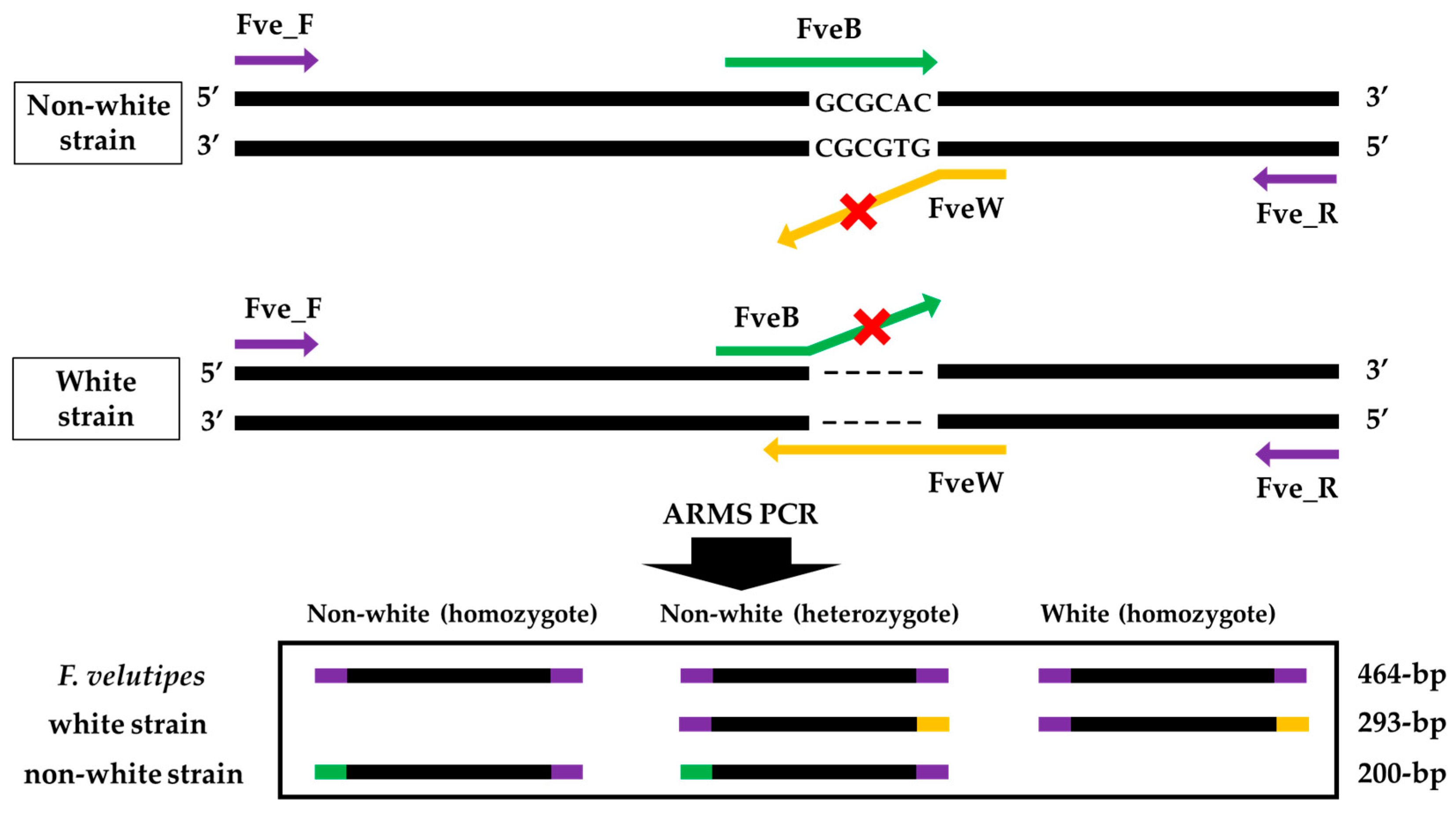
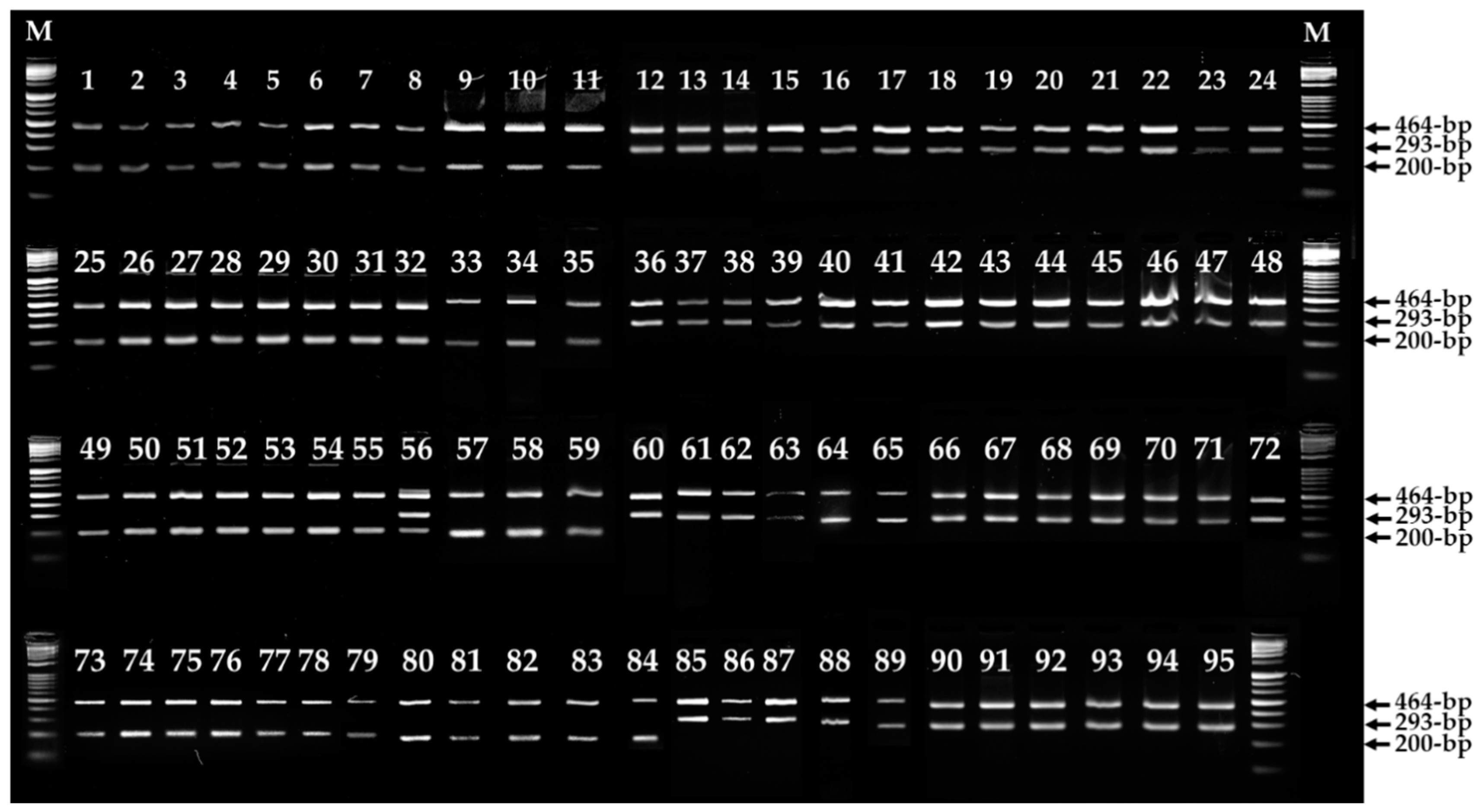
3.2. Primers Design and ARMS PCR for Fruiting Body Color Discrimination of F. velutipes

Figure 3 shows the scheme of ARMS PCR primer design to detect the specific variation ΔGCGCAC in the Fvpal1 gene and to discriminate the fruiting body color of F. velutipes. A four-primer set was designed to amplify all F. velutipes, non-white, or white strains (Table 2). The FveF and FveR primers were expected to amplify the 464 bp product from all F. velutipes strains. In addition, the FveF/FveW and FveR/FveB primer sets were expected to amplify 293 bp and 200 bp products for F. velutipes white and non-white strains, respectively.
ARMS is a simple and reliable method for identifying single nucleotide variations (SNVs) or deletions [38]. Since ARMS uses PCR primers that allow the amplification of DNA only in the presence of specific mutations, the amplification of ARMS PCR products determines the presence of mutations. As shown in Figure 4, ARMS PCR analysis revealed the specific detection of the ΔGCGCAC variation in Fvpal1, as well as the specific discrimination of non-white and white F. velutipes strains. Among the 95 F. velutipes strains tested, ARMS PCR amplified 464 bp and 293 bp products in all white strains. The non-white strains amplified either 464 bp and 200 bp or 464 bp, 200 bp, and 293 bp products. The F. velutipes ASI4175 non-white strain amplified all three products, indicating that it was heterozygous for the normal and ΔGCGCAC Fvpal1 gene. These results suggest that the normal Fvpal1 gene has a dominant effect on the fruiting body color of F. velutipes, as F. velutipes ASI4175 is a non-white strain. In this study, Fvpal1 gene mutations in 50 white strains were detected using specific primers and ARMS PCR analysis, and as a result, the fruiting body color of F. velutipes strains was accurately discriminated.
Although the color of the fruiting body was accurately discriminated by analysis of the 95 strains used in this study, future research should continuously analyze an additional F. velutipes strain to determine whether the variation in Fvpal1 affects the color of the fruiting body.

3.3. Mutation in the Fvpal1 Gene Affect Fruiting Body Color of F. velutipes

Phenylalanine ammonia-lyase (PAL; EC 4.3.1.24) catalyzes the deamination of l-phenylalanine to trans-cinnamic acid and is commonly found in plants and fungi [39,40]. PAL is involved in the first step of the phenylpropanoid pathway, leading to the synthesis of various phenylpropanoids such as flavonoids, isoflavonoids, anthocyanins, lignins, and other phenolic compounds [40]. Therefore, PAL is considered to be a key initiator of the phenylpropanoid pathway, a transition process from primary to secondary metabolism.
In plants, particularly in lettuce, 5-caffeoylquinic acid, 3,5-dicaffeoylquinic acid, caffeoyltartaric acid, and dicaffeoyltartaric acid have been reported to be associated with browning [41,42]. It has also been reported that the phenylpropanoid pathway is activated by wounds or hormones, such as ethylene, to increase the synthesis of phenolic compounds [41,43]. Therefore, PAL has been extensively studied to increase the understanding of metabolic processes as well as browning. It has also been reported that PAL enzyme and phenolic compounds are essential for the browning of mung bean sprouts [44]. In addition, phenolic compounds, including trans-caffeoyltartronic acid and trans-coumaroyltartronic acid, are substrates of polyphenol oxidase (PPO; EC 1.10.3.1) in mung bean sprouts and act as major factors for browning [44]. It has been suggested that the polyphenols in mung bean sprouts increase gradually during storage and are oxidized by PPO to form a brown pigment [45,46].
It has been reported that the fungal PAL enzyme degrades phenylalanine via a pathway similar to that in plants [47,48]. Further research revealed the phenylalanine metabolic pathway in some basidiomycetes, including Rhodotorula glutinis, Schizophyllum commune, and Sporobolomyces roseus [48,49,50]. In the phytopathogenic fungi Moniliophthora perniciosa, PAL has been found to accumulate during the infection stage, suggesting that it may be associated with pathogenicity [51]. Additionally, Tricholoma matsutake and F. velutipes PAL mRNAs were expressed specifically at the developmental stage, and in F. velutipes, the highest expression was found in the mycelium and when l-tyrosine was added [52,53]. Although there have been reports of various roles for PAL, it has not been reported that it is associated with the color of fungi, including mushrooms.
Sixty monospores were isolated from each of F. velutipes ASI4175 (non-white, heterozygote for ΔGCGCAC Fvpal1) and w1-8 (white, homozygote for ΔGCGCAC Fvpal1), based on the ARMS PCR results (Figure 4). Each monospore was evaluated for mutations (ΔGCGCAC) in the Fvpal1 gene through ARMS PCR analysis, and monospores containing 15 mutated Fvpal1 (ΔGCGCAC) genes and 15 non-mutated Fvpal1 genes were obtained from F. velutipes ASI4175 (Figure S2). As shown in Figure 5, progenies from the mating of monospores with the ΔGCGCAC deletion and monospores without the ΔGCGCAC deletion in the Fvpal1 gene showed non-white fruiting bodies. However, the mating of monospores containing the mutated Fvpal1 gene (ΔGCGCAC) isolated from F. velutipes ASI4175 (non-white, heterozygote for ΔGCGCAC Fvpal1) and w1-8 (white, homozygote for ΔGCGCAC Fvpal1) produced progenies with only white-colored fruiting bodies.
In this study, mating analysis revealed that the Fvpal1 gene plays an important role in the fruiting body color of F. velutipes, suggesting that this gene could be a useful marker for the selective breeding of new varieties of F. velutipes, especially for fruiting body color.

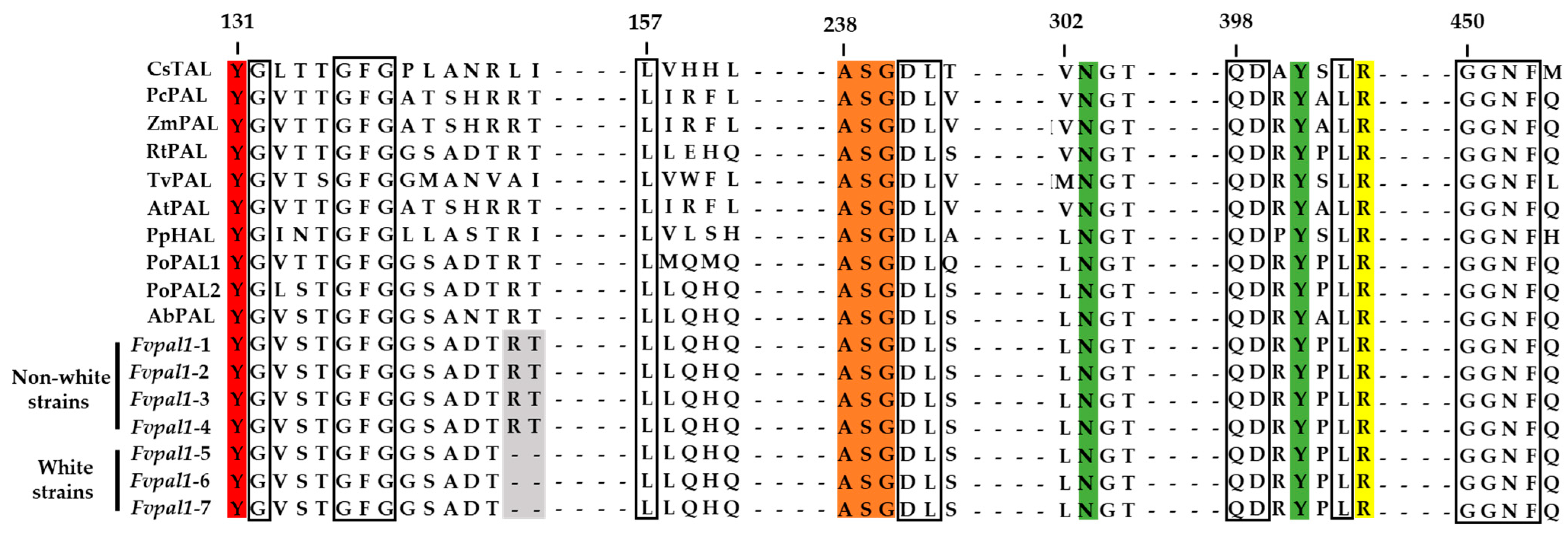
3.4. Structural Characteristics of the PAL of F. velutipes

The catalytic prosthetic 3,5-dihydro-5-methylidine-4H-imidazol-4-one group (MIO) is essential for the catalytic activity of PAL and is produced by the autocatalytic crystallization of amino acids, including alanine, serine, and glycine [40,54,55]. The MIO group is commonly found in ammonia lyases, including PAL, tyrosine ammonia lyase (TAL), and histidine ammonia lyase (HAL), and the ASG motif plays an important role in this enzymatic activity [56,57,58,59]. A highly conserved MIO group (ASG motif) was also found at positions 238–240 in Fvpal1 of F. velutipes in both the white and non-white strains (Figure 6). Another highly conserved motif was also found in Fvpal1 of F. velutipes, including stabilizing residues for the MIO group (N303 and Y401) and carboxylic-acid-binding residues for the substrate (R404).
Among the PAL and TAL motifs, specific residues for substrate specificity are phenylalanine-leucine (FL) for phenylalanine and histidine-leucine (HL) for tyrosine. A characteristic histidine-glycine (HQ) motif of the PAL enzyme, which exhibits substrate activity for both tyrosine and phenylalanine, has also been reported [60,61]. For PAL and TAL enzymes with an HQ motif, it has been reported to have dual substrate activity with a KmPhenylalanine/Tyrosine ratio greater than one [61,62,63,64]. A histidine-glycine (160HQ161) motif was also found in the FvPAL1 of F. velutipes as well as other species, including P. ostreatus and A. bisporus (Figure 6). Although specific activity assays for phenylalanine and tyrosine are required, the HQ motif in the FvPAL1 of F. velutipes suggests that this enzyme could possibly catalyze both substrates.
In this study, two PAL genes, Fvpal1 and Fvpal2, were identified in the genome of F. velutipes. No mutations were found in the sequence of the Fvpal2 gene (Figure S3). As shown in Figure 7, the fruiting body color-related mutation Δ112RT113 was not found in the Fvpal2 gene of either non-white or white F. velutipes strains. Motifs essential for the catalytic activity of PAL enzymes were found in Fvpal2 genes. However, substrate-specific residues of Fvpal2 (138MQ139) were found to be different from those of the Fvpal1 gene (160HQ161) but identical to those of the PoPAL1 gene (160MQ161) of P. ostreatus (Figure 6 and Figure 7).
In a previous study [53], Fvpal was identified in F. velutipes and was found to have the same sequence as the Fvpal1 gene of the F. velutipes non-white strain identified in this study, which consisted of 2746 bp and 12 exons and showed 100% identity to Fvpal, with 724 amino acids (2175 bp cDNA). However, Δ112RT113 deletions identified in the Fvpal1 gene were not found in the Fvpal gene sequence. These results indicate that the previously reported Fvpal gene [53] was identified in non-white F. velutipes, and this was confirmed with the information that the non-white strain F. velutipes 4164 was used for the identification of the Fvpal gene [53]. The Fvpal2 gene is 2617 bp in size with eight exons and consists of a 2208 bp cDNA that is translated into 735 amino acids (Figure S3). Furthermore, among the Fvpal2 genes identified from F. velutipes strains, no mutations specific to non-white or white strains were found, except for variations for each strain. Although further studies are required, these results suggest that the Fvpal2 gene is essential for the physiological and metabolic functions of F. velutipes but that the Fvpal1 gene has the potential to function selectively in determining the color of the F. velutipes fruiting body.

4. Conclusions

In nature, Flammulina velutipes forms non-white fruiting bodies, but white fruiting body varieties were accidentally developed by artificial breeding. However, until recently the physiological, biochemical, and genetic causes associated with the formation of white fruiting bodies in F. velutipes had not been elucidated. A recent comparative analysis reported that components of non-white F. velutipes strains, such as amino acids, saccharides, and β-glucan, were relatively higher or lower than those of white strains [6,12,13,14,15,16,17]. These results suggest that the fruiting body color of F. velutipes can be used as a criterion for breeding new varieties. Therefore, the selective breeding of fruiting body color is considered a great advantage for efficient breeding.
In this study, comparative genomics of six F. velutipes strains showed 70 variations unique to white strains, including SNPs and indels. Of these, 36 were found in open reading frames and only one caused a disruptive in-frame deletion (ΔGCGCAC) in the Fvpal1 gene, resulting in the deletion of two amino acids (arginine and threonine) at positions 112 and 113. Specific primers were designed to detect this mutation, and PCR analysis of 95 F. velutipes strains revealed that this mutation was present only in white strains. In addition, monospores of the heterozygous mutant were isolated, and a mating assay was performed to evaluate the mutation’s relationship to fruiting body color. As a result, progeny resulting from mating monospores with and without the ΔGCGCAC deletion in the Fvpal1 gene showed non-white fruiting bodies. However, mating monospores with the mutated Fvpal1 gene resulted in progeny with only white-colored fruiting bodies.
Although the effect of mutations in the Fvpal1 gene of F. velutipes on enzyme activity and metabolic function remains to be studied, it is considered that this gene can be effectively used for selective breeding of this mushroom.

Supplementary Materials

The following supporting information can be downloaded at https://www.mdpi.com/article/10.3390/jof9030339/s1: Figure S1: Alignments of phenylalanine ammonia-lyase 1 (Fvpal1) genes in Flammulina velutipes strains. (a) cDNA sequence and (b) amino acid sequence. F. velutipes KACC42870 (non-white), F. velutipes ASI4208 (non-white), F. velutipes ASI4049 (non-white), F. velutipes ASI4057 (non-white), F. velutipes ASI4166 (white), F. velutipes ASI4167 (white), F. velutipes ASI4169 (white). Figure S2: Amplification refractory mutation system (ARMS) PCR of Fvpal1 in monospores from Flammulina velutipes ASI4175 (heterozygote, non-white) (a) and w1-8 (homozygote, white) (b) strains. Asterisks indicate F. velutipes monospores without mutation in the Fvpal1 gene. Figure S3: Alignments of phenylalanine ammonia-lyase 2 (Fvpal2) genes of Flammulina velutipes strains. (a) cDNA sequence and (b) amino acid sequence. F. velutipes KACC42870 (non-white), F. velutipes ASI4208 (non-white), F. velutipes ASI4049 (non-white), F. velutipes ASI4057 (non-white), F. velutipes ASI4166 (white), F. velutipes ASI4167 (white), and F. velutipes ASI4169 (white). Table S1: White-color-specific variations identified in Flammulina velutipes strains. Table S2: Predicted gene models of Flammulina velutipes KACC42780.

Author Contributions

Conceptualization and methodology, Y.-J.P. and J.-H.I.; software, Y.-J.P. and H.-W.Y.; validation, J.-H.I., H.-W.Y., C.-H.P., J.-W.K., and J.-H.S.; investigation and data curation, J.-H.I., C.-H.P., J.-W.K. and J.-H.S.; software, Y.-J.P.; writing—original draft preparation, Y.-J.P. and J.-H.I.; writing—review and editing, Y.-J.P. and J.-H.I.; resources, K.-Y.J.; supervision, Y.-J.P. and K.-Y.J.; project administration, Y.-J.P.; funding acquisition, Y.-J.P. and J.-H.I. All authors have read and agreed to the published version of the manuscript.

Funding

This work was carried out with the support of the Cooperative Research Program for Agriculture Science and Technology Development (Project No. PJ01516403), Rural Development Administration, Republic of Korea.

Institutional Review Board Statement

Not applicable.

Informed Consent Statement

Not applicable.

Data Availability Statement

NGS reads were deposited in the NABIC (National Agricultural Biotechnology Information Center, RDA, Korea; https://nabic.rda.go.kr/, accessed on 11 August 2022) Sequence Read Archive (SRA) (Table 3).

Conflicts of Interest

The authors declare no conflict of interest.

References

  1. Park, Y.J.; Baek, J.H.; Lee, S.; Kim, C.; Rhee, H.; Kim, H.; Seo, J.S.; Park, H.R.; Yoon, D.E.; Nam, J.Y.; et al. Whole genome and global gene expression analyses of the model mushroom Flammulina velutipes reveal a high capacity for lignocellulose degradation. PLoS ONE 2014, 9, e93560. [Google Scholar] [CrossRef] [PubMed]
  2. Kim, E.S.; Woo, S.I.; Oh, M.; Oh, Y.L.; Shin, P.G.; Jnag, K.Y.; Kong, W.S.; Lee, C.S. Characteristics of ‘Baekjung’, a variety adaptable to high temperature in Flammulina velutipes. J. Mushrooms 2015, 13, 203–206. [Google Scholar] [CrossRef]
  3. Kong, W.S.; Jang, K.Y.; Lee, C.Y.; Koo, J.; Shin, P.G.; Jhune, C.S.; Oh, Y.; Yoo, Y.B.; Suh, J.S. Breeding progress and characterization of a Korean white variety ‘Baek-A’ in Flammulina velutipes. J. Mushrooms 2013, 11, 159–163. [Google Scholar] [CrossRef]
  4. Kitamoto, Y.; Nakamata, M.; Masuda, P. Production of novel white Flammulina velutipes by breeding. In Geneticsand Breeding of Edible Mushrooms, 1st ed.; Chang, S.T., Buswell, J.A., Miles, P.G., Eds.; Routledge: London, UK, 1993; pp. 65–86. [Google Scholar] [CrossRef]
  5. Woo, S.I.; Kong, W.S.; Jang, K.Y. Characteristics of ’Baekseung’, a new cultivar Flammulina velutipes. J. Mushrooms 2017, 15, 25–30. [Google Scholar] [CrossRef] [Green Version]
  6. Kim, M.J.; Lee, K.W.; Chang, W.B.; Jeon, J.O.; Kim, I.J. Characteristics and breeding of ‘Yeoreumhyang2ho’: A new blackish-brown variety of Flammulina velutipes that is adaptable to high temperature. J. Mushrooms 2018, 15, 192–197. [Google Scholar] [CrossRef]
  7. Kim, M.J.; Lee, K.W.; Chang, W.B.; Jeon, J.O.; Kim, I.J. Characteristics and breeding of ‘Yeoreumhyang1ho’: A new light brown variety of Flammulina velutipes adaptable to high temperature. J. Mushrooms 2018, 16, 287–292. [Google Scholar] [CrossRef]
  8. Kim, M.J.; Lee, K.W.; Chang, W.B.; Jeon, J.O.; Kim, I.J. Characteristics and breeding of ‘Geumhyang2ho’, a new brown and labor-saving variety of Flammulina velutipes. J. Mushrooms 2018, 16, 293–298. [Google Scholar] [CrossRef]
  9. Im, J.H.; Jang, K.Y.; Oh, Y.L.; Oh, M.; Raman, J.; Kong, W.S. Breeding on a new cultivar of golden Flammulina velutipes ‘Auram’. J. Mushrooms 2019, 17, 218–223. [Google Scholar] [CrossRef]
  10. Kim, M.J.; Chang, H.B.; Choi, J.S.; Lee, K.W.; Joo, G.N.; Kim, Y.G. Characteristics and breeding of a new brown variety ‘Geumhyang’ with short cultivation period in Flammulina velutipes. J. Mushrooms 2015, 13, 92–96. [Google Scholar] [CrossRef] [Green Version]
  11. Kim, M.J.; Chang, H.B.; Choi, J.S.; Lee, K.W.; Joo, G.N.; Kim, Y.G. Characteristics and breeding of a new brown variety ‘Heukhyang’ with good taste in Flammulina velutipes. J. Mushrooms 2015, 13, 103–107. [Google Scholar] [CrossRef] [Green Version]
  12. Jhune, C.S.; Yun, H.S.; Leem, H.T.; Kong, W.S.; Lee, K.H.; Sung, G.H.; Cho, J.H. Changes of saccharide content in fruitbody composition of Flammulina velutipes during storage. J. Mushrooms 2011, 9, 123–131. [Google Scholar] [CrossRef]
  13. Jhune, C.S.; Yun, H.S.; Leem, H.T.; Gong, W.S.; Sung, G.H.; Cho, J.H.; Park, K.M. The change of amino acid content in fruit-body of winter mushroom according to the storage period and strains. J. Mushrooms 2012, 10, 224–235. [Google Scholar] [CrossRef]
  14. Kim, J.T.; Kim, M.J.; Jhune, C.S.; Shin, P.G.; Oh, Y.L.; Yoo, Y.B.; Kong, W.S. Comparison of amino acid and free amino acid contents between cap and stipe in Flammulina velutipes and Pleurotus ostreatus. J. Mushrooms 2014, 12, 341–349. [Google Scholar] [CrossRef] [Green Version]
  15. Kim, K.J.; Jin, S.W.; Choi, B.S.; Kim, J.K.; Koh, Y.W.; Ban, S.E.; Seo, K.S. Evaluation of the nutrition properties of Flammulina velutipes. J. Mushrooms 2016, 14, 44–50. [Google Scholar] [CrossRef] [Green Version]
  16. An, G.H.; Han, J.G.; Cho, J.H. Comparisons of biological activities and amino acid contents of edible mushrooms extracted using different solvents. J. Mushrooms 2020, 18, 53–62. [Google Scholar] [CrossRef]
  17. An, G.H.; Han, J.G.; Kim, O.T.; Cho, J.H. Changes of biological activity and nutritional content by processing methods of Flammulina velutipes, Grifola frondosa, and Sparassis crispa. J. Mushrooms 2020, 18, 403–409. [Google Scholar] [CrossRef]
  18. Nerya, O.; Ben-Arie, R.; Luzzatto, T.; Musa, R.; Khativ, S.; Vaya, J. Prevention of Agaricus bisporus postharvest browning with tyrosinase inhibitors. Postharvest Biol. Technol. 2006, 39, 272–277. [Google Scholar] [CrossRef]
  19. Jolivet, S.; Arpin, N.; Wichers, H.J.; Pellon, G. Agaricus bisporus browning: A review. Mycol. Res. 1988, 102, 1459–1483. [Google Scholar] [CrossRef]
  20. Claus, H.; Decker, H. Bacterial tyrosinases. Sys. Appl. Microbiol. 2006, 29, 3–14. [Google Scholar] [CrossRef]
  21. Lee, D.; Jang, E.H.; Lee, M.; Kim, S.W.; Lee, Y.; Lee, K.T.; Bahn, Y.S. Unraveling melanin biosynthesis and signaling networks in Cryptococcus neoformans. mBio 2019, 10, e02267-19. [Google Scholar] [CrossRef] [Green Version]
  22. Conesa, A.; Punt, P.J.; van den Hondel, C.A.M.J.J. Fungal peroxidases: Molecular aspects and applications. J. Biotechnol. 2002, 93, 143–158. [Google Scholar] [CrossRef] [PubMed]
  23. Hofrichter, M.; Ullrich, R.; Pecyna, M.J.; Liers, C.; Lundell, T. New and classic families of secreted fungal heme peroxidases. Appl. Microbiol. Biotechnol. 2010, 87, 897–971. [Google Scholar] [CrossRef] [PubMed]
  24. Waltz, E. Gene-edited CRISPR mushroom escapes US regulation. Nature 2016, 532, 293. [Google Scholar] [CrossRef] [PubMed] [Green Version]
  25. Park, Y.J.; Jeong, Y.U.; Kong, W.S. Genome sequencing and carbohydrate-active enzyme (CAZyme) repertoire of the white rot fungus Flammulina elastica. Int. J. Mol. Sci. 2018, 19, 2379. [Google Scholar] [CrossRef] [PubMed] [Green Version]
  26. Lee, C.S.; Kong, W.S.; Park, Y.J. Genome sequencing and genome-wide identification of carbohydrate-active enzymes (CAZymes) in the white rot fungus Flammulina fennae. Microbiol. Biotechnol. Lett. 2018, 46, 300–312. [Google Scholar] [CrossRef]
  27. Park, Y.J.; Kong, W.S. Genome-wide comparison of carbohydrate-active enzymes (CAZymes) repertoire of Flammulina ononidis. Mycobiology 2018, 46, 349–360. [Google Scholar] [CrossRef] [Green Version]
  28. Park, Y.J.; Lee, C.S.; Kong, W.S. Genomic insights into the fungal lignocellulolytic machinery of Flammulina rossica. Microorganisms 2019, 7, 421. [Google Scholar] [CrossRef] [PubMed] [Green Version]
  29. Yu, H.W.; Im, J.H.; Kong, W.S.; Park, Y.J. Comparative analysis of carbohydrate active enzymes in the Flammulina velutipes var. lupinicola genome. Microorganisms 2021, 9, 20. [Google Scholar] [CrossRef]
  30. Bolger, A.M.; Lohse, M.; Usadel, B. Trimmomatic: A flexible trimmer for Illumina sequence data. Bioinformatics 2014, 30, 2114–2120. [Google Scholar] [CrossRef] [Green Version]
  31. Van der Auwera, G.A.; Carneiro, M.; Hartl, C.; Poplin, R.; del Angel, G.; Levy-Moonshine, A.; Jordan, T.; Shakir, K.; Roazen, D.; Thibault, J.; et al. From fastQ data to high-confidence variant calls: The genome analysis toolkit best practices pipeline. Curr. Protoc. Bioinform. 2013, 43, 11.10.1–11.10.33. [Google Scholar] [CrossRef] [Green Version]
  32. Li, H.; Durbin, R. Fast and accurate short read alignment with Burrows-Wheeler Transform. Bioinformatics 2009, 25, 1754–1760. [Google Scholar] [CrossRef] [Green Version]
  33. Danecek, P.; Bonfield, J.K.; Liddle, J.; Marshall, J.; Ohan, V.; Pollard, M.O.; Whitwham, A.; Keane, T.; McCarthy, S.A.; Davies, R.M.; et al. Twelve years of SAMtools and BCFtools. Gigascience 2021, 10, giab008. [Google Scholar] [CrossRef] [PubMed]
  34. Broad Institute. Available online: http://broadinstitute.github.io/picard/ (accessed on 11 August 2022).
  35. Cingolani, P.; Platts, A.; Wang, L.L.; Coon, M.; Nguyen, T.; Wang, L.; Land, S.J.; Lu, X.; Ruden, D.M. A program for annotating and predicting the effects of single nucleotide polymorphisms, SnpEff: SNPs in the genome of Drosophila melanogaster strain w1118; iso-2; iso-3. Fly 2012, 6, 80–92. [Google Scholar] [CrossRef] [PubMed] [Green Version]
  36. Quinlan, A.R.; Hall, I.M. BEDTools: A flexible suite of utilities for comparing genomic features. Bioinformatics 2010, 26, 841–842. [Google Scholar] [CrossRef] [Green Version]
  37. Palmer, J.M.; Stajich, J.E. Funannotate: Eukaryotic Genome Annotation Pipeline. Available online: http://funannotate.readthedocs.io (accessed on 11 August 2022).
  38. Little, S. Amplification-refractory mutation system (ARMS) analysis of point mutations. Curr. Protoc. Hum. Genet. 2001, 7, 9–18. [Google Scholar] [CrossRef]
  39. Hyun, M.W.; Yun, Y.H.; Kim, J.Y.; Kim, S.H. Fungal and plant phenylalanine ammonia-lyase. Mycobiology 2011, 39, 257–265. [Google Scholar] [CrossRef] [Green Version]
  40. Kong, J.Q. Phenylalanine ammonia-lyase, a key component used for phenylpropanoids production by metabolic engineering. RCS Adv. 2015, 5, 62587–62603. [Google Scholar] [CrossRef]
  41. Tomás-Barberán, F.A.; Loaiza-Velarde, J.; Bonfanti, A.; Saltveit, M.E. Early wound- and ethylene-induced changes in phenylpropanoid metabolism in harvested lettuce. J. Am. Soc. Hortic. Sci. 1997, 122, 399–404. [Google Scholar] [CrossRef]
  42. Tomás-Barberán, F.A.; Gil-Muñoz, M.I.; Castañer, M.; Artés, F.; Saltveit, M.E. Effect of selected browning inhibitors on phenolic metabolism in stem tissue of harvested lettuce. J. Agric. Food Chem. 1997, 45, 583–589. [Google Scholar] [CrossRef]
  43. Ke, D.; Saltveit, M.E. Wound-induced ethylene production, phenolic metabolism and susceptibility to russet spotting in iceberg lettuce. Physiol. Plant. 1989, 76, 412–418. [Google Scholar] [CrossRef]
  44. Sameshima, N.; Nishimura, M.; Murakami, K.; Kogo, Y.; Shimamura, Y.; Sakuta, M.; Murata, M. Cloning of phenylalanine ammonia-lyase and its role in enzymatic browning of mung bean sprout during cold storage. Food Sci. Technol. Res. 2016, 22, 255–260. [Google Scholar] [CrossRef] [Green Version]
  45. Barroga, C.F.; Laurena, A.C.; Mendoza, E.M.T. Polyphenols in mung bean (Vigna radiata (L.) Wilczek): Determination and removal. J. Agric. Food Chem. 1985, 33, 1006–1009. [Google Scholar] [CrossRef]
  46. Nishimura, M.; Sameshima, N.; Joshita, K.; Murata, M. Regulation of enzymatic browning of mung bean sprout by heat-shock treatment. Food Sci. Technol. Res. 2012, 18, 412–417. [Google Scholar] [CrossRef] [Green Version]
  47. Power, D.M.; Towers, G.H.; Neish, A.C. Biosynthesis of phenolic acids by certain wood-destroying Basidiomycetes. Can. J. Biochem. 1965, 43, 1397–1407. [Google Scholar] [CrossRef] [PubMed]
  48. Moore, K.; Subba Rao, P.V.; Towers, G.H. Degradation of phenylalanine and tyrosine by Basidiomycetes. Life Sci. 1967, 6, 2629–2633. [Google Scholar] [CrossRef] [PubMed]
  49. Yamada, S.; Nabe, K.; Izuo, N.; Nakamichi, K.; Chibata, I. Production of l-phenylalanine from trans-cinnamic acid with Rhodotorula glutinis containing l-phenylalanine ammonia lyase activity. Appl. Environ. Microbiol. 1981, 42, 773–778. [Google Scholar] [CrossRef] [Green Version]
  50. Moore, K.; Subba Rao, P.V.; Towers, G.H. Degradation of phenylalanine and tyrosine by Sporobolomyces roseus. Biochem. J. 1968, 106, 507–514. [Google Scholar] [CrossRef] [Green Version]
  51. Leal, G.A.; Gomes, L.H.; Albuquerque, P.S.; Tavares, F.C.; Figueira, A. Searching for Moniliophthora perniciosa pathogenicity genes. Fungal Biol. 2010, 114, 842–854. [Google Scholar] [CrossRef]
  52. Yoon, H.; You, Y.H.; Kim, Y.E.; Kim, Y.J.; Kong, W.S.; Kim, J.G. Cloning and mRNA expression analysis of the gene encoding phenylalanine ammonia-lyase of the ectomycorrhizal fungus Tricholoma matsutake. J. Microbiol. Biotechnol. 2013, 23, 1055–1059. [Google Scholar] [CrossRef] [Green Version]
  53. Yun, Y.H.; Koo, J.S.; Kim, S.H.; Kong, W.S. Cloning and expression analysis of phenylalanine ammonia-lyase gene in the mycelium and fruit body of the edible mushroom Flammulina velutipes. Mycobiology 2015, 43, 327–332. [Google Scholar] [CrossRef] [Green Version]
  54. Oufedjikh, H.; Mahrouz, M.; Amiot, M.J.; Lacroix, M. Effect of γ-irradiation on phenolic compounds and phenylalanine ammonia-lyase activity during storage in relation to peel injury from peel of Citrus clementina Hort. Ex. Tanaka. J. Agric. Food Chem. 2000, 48, 559–565. [Google Scholar] [CrossRef] [PubMed]
  55. Poppe, L. Methylidene-imidazolone: A novel electrophile for substrate activation. Curr. Opin. Chem. Biol. 2001, 5, 512–524. [Google Scholar] [CrossRef] [PubMed]
  56. Heberling, M.M.; Wu, B.; Bartsch, S.; Janssen, D.B. Priming ammonia lyases and aminomutases for industrial and therapeutic applications. Curr. Opin. Chem. Biol. 2013, 17, 250–260. [Google Scholar] [CrossRef] [PubMed]
  57. Cui, J.D.; Qiu, J.Q.; Fan, X.W.; Jia, S.R.; Tan, Z.L. Biotechnological production and applications of microbial phenylalanine ammonia lyase: A recent review. Crit. Rev. Biotechnol. 2014, 34, 258–268. [Google Scholar] [CrossRef]
  58. Rother, D.; Poppe, L.; Morlock, G.; Viergutz, S.; Retey, J. An active site homology model of phenylalanine ammonia-lyase from Petroselinum crispum. Eur. J. Biochem. 2002, 269, 3065–3075. [Google Scholar] [CrossRef]
  59. Schuster, B.; Retey, J. The mechanism of action of phenylalanine ammonia-lyase: The role of prosthetic dehydroalanine. Proc. Natl. Acad. Sci. USA. 1995, 92, 8433–8437. [Google Scholar] [CrossRef] [Green Version]
  60. Abell, C.W.; Shen, R.S. Phenylalanine ammonia-lyase from the yeast Rhodotorula glutinis. Methods Enzymol. 1987, 142, 242–253. [Google Scholar] [CrossRef]
  61. Goldson-Barnaby, A.; Scaman, C.H. Purification and characterization of phenylalanine ammonia lyase from Trichosporon cutaneum. Enzym. Res. 2013, 2013, 670702. [Google Scholar] [CrossRef] [Green Version]
  62. Vannelli, T.; Xue, Z.; Breinig, S.; Qi, W.W.; Sariaslani, F.S. Functional expression in Escherichia coli of the tyrosine inducible tyrosine ammonia-lyase enzyme from yeast Trichosporon cutaneum for production of p-hydroxycinnamic acid. Enzym. Microb. Technol. 2007, 41, 413–422. [Google Scholar] [CrossRef]
  63. Ameyama, M.; Shinagawa, E.; Matsushita, K.; Adachi, O. Growth stimulation of microorganisms by pyrroloquinoline quinone. Agri. Biol. Chem. 1984, 48, 2909–2911. [Google Scholar] [CrossRef]
  64. Xue, Z.; McCluskey, M.; Cantera, K.; Ben-Bassat, A.; Sariaslani, F.S.; Huang, L. Improved production of p-hydroxycinnamic acid from tyrosine using a novel thermostable phenylalanine/tyrosine ammonia lyase enzyme. Enzyme Microb. Technol. 2007, 42, 58–64. [Google Scholar] [CrossRef]
Figure 1. Color of the fruiting bodies of Flammulina velutipes strains. (a) F. velutipes white strain; (b) F. velutipes yellowish-brown strain; (c) F. velutipes dark-brown strain; (d) various colors of F. velutipes strains.
Jof 09 00339 g001
Figure 2. Alignments of phenylalanine ammonia-lyase 1 (Fvpal1) genes of Flammulina velutipes strains. (a) cDNA sequences; (b) amino acid sequences. Boxes indicate the variation sites of the Fvpal1 gene. The white strains of F. velutipes showed a deletion of GCGCAC (112RT113; arginine and threonine) in the Fvpal1 gene.
Jof 09 00339 g002
Figure 3. Primer design for non-white or white Flammulina velutipes strain discrimination by amplification refractory mutation system (ARMS) PCR. Primers were designed to amplify the Fvpal1 gene with 464 bp and 200 bp, 464 bp and 293 bp from non-white and white strain, respectively. A heterozygote of the Fvpal1 gene would be expected to show all three bands.
Jof 09 00339 g003
Figure 4. Amplification refractory mutation system (ARMS) PCR of Fvpal1 in 95 Flammulina velutipes strains (Table 1). Non-white and white strains of F. velutipes showed 464 bp and 200 bp, 464 bp and 293 bp PCR products, respectively. A heterozygous strain (No. 56, F. velutipes ASI4175) showed all three amplification products: 464 bp, 293 bp, and 200 bp. M, size marker (1 kb ladder, TNT Research).
Jof 09 00339 g004
Figure 5. Matings of monospores from Flammulina velutipes strains ASI4175 (non-white, heterozygote) and w1-8 (white, homozygote). The isolated monospores were analyzed for the deletion of arginine and threonine; Δ112RT113 in the Fvpal1 gene using ARMS PCR. The number in the box indicates the isolated monospore.
Jof 09 00339 g005
Figure 6. Alignment of the highly conserved domains of phenylalanine/tyrosine/histidine ammonia-lyase amino acids. Red: catalytically essential tyrosine residue, orange: the MIO forming amino acid triad, green: amino acid stabilizing MIO group, yellow: arginine responsible for binding the carboxylic group of the substrate, gray: variation site (arginine and threonine) of F. velutipes, boxes: other conserved catalytic and binding residues. Cereibacter sphaeroides TAL (CsTAL, UniProtKB ID; Q3IWB0), Petroselinum crispum PAL (PcPAL, UniProtKB ID; P24481), Zea mays PAL (ZmPAL, UniProtKB ID; C0HJ40), Rhodosporidium toruloides PAL (RtPAL, UniProtKB ID; P11544), Trichormus variabilis PAL (TvPAL, UniProtKB ID; Q3M5Z3), Arabidopsis thaliana PAL (AtPAL, UniProtKB ID; P45724), Pseudomonas putida HAL (PpHAL, UniProtKB ID; Q88CZ7), Pleurotus ostreatus Fvpal1 (PoPAL1, UniProtKB ID; A0A4Y6HUI7), Pleurotus ostreatus PAL2 (PoPAL2, UniProtKB ID; A0A4Y6HUD7), Agaricus bisporus PAL (AbPAL, KEGG ID; T03136), F. velutipes KACC42870 (non-white strain) Fvpal1 (Fvpal1-1), F. velutipes ASI4028 (non-white strain) Fvpal1 (Fvpal1-2), F. velutipes ASI4049 (non-white strain) Fvpal1 (Fvpal1-3), F. velutipes ASI4057 (non-white strain) Fvpal1 (Fvpal1-4), F. velutipes ASI4166 (white strain) Fvpal1 (Fvpal1-5), F. velutipes ASI4157 (white strain) Fvpal1 (Fvpal1-6), F. velutipes ASI4169 (white strain) Fvpal1 (Fvpal1-7).
Jof 09 00339 g006
Figure 7. Alignment of the highly conserved domains of phenylalanine ammonia-lyase 2 amino acids of Flammulina velutipes strains. Red: catalytically essential tyrosine residue, orange: the MIO forming amino acid triad, green: amino acid stabilizing MIO group, yellow: arginine responsible for binding the carboxylic group of the substrate, gray: variation site of Fvpal1 gene of F. velutipes, boxes: other conserved catalytic and binding residues. F. velutipes KACC42870 (non-white strain) Fvpal2 (Fvpal2-1), F. velutipes ASI4028 (non-white strain) Fvpal2 (Fvpal2-2), F. velutipes ASI4049 (non-white strain) Fvpal2 (Fvpal2-3), F. velutipes ASI4057 (non-white strain) Fvpal2 (Fvpal2-4), F. velutipes ASI4166 (white strain) Fvpal2 (Fvpal2-5), F. velutipes ASI4157 (white strain) Fvpal2 (Fvpal2-6), F. velutipes ASI4169 (white strain) Fvpal2 (Fvpal2-7).
Jof 09 00339 g007
Table 1. Flammulina velutipes strains used in this study.
No.StrainsColorNo.StrainsColorNo.StrainsColor
1ASI4019non-white41T011white81B21non-white
2ASI4019-20non-white42honamwhite82B8non-white
3ASI4019-1820non-white43daeheung white83B26non-white
4ASI4028non-white44W6-8white84B39non-white
5ASI4049non-white45W6-18white85baegi white
6ASI4019-2003non-white46W6-19white86baekseung white
7ASI4057non-white47W6-14white87baekjung white
8ASI4064non-white48W1-9white887937white
9ASI4067non-white49hwanggeum non-white898492white
10ASI4069non-white50B151non-white90naju white
11ASI4072non-white51B129non-white91ASI4100white
12ASI4074white52B112non-white92ASI4102white
13ASI4166white53B13non-white93hampyeong white
14ASI4167white54yeoreumhyang2ho non-white94W8-17white
15ASI4168white55B16non-white95ASI4168white
16ASI4169white564175non-white
17ASI4178white57B17non-white
18ASI4029white58B18non-white
19ASI4210white59B62non-white
20ASI4216white60W5-2white
21ASI4217white61W1-18white
22ASI4226white62W6-13white
23ASI4228white63W8-18white
24ASI4230white64W1-23white
25ASI4083non-white65W1-8white
26ASI4090non-white66W3-24white
27ASI4103non-white67W4-16white
28ASI4111non-white68W5-3white
29ASI4146non-white69W5-16white
30ASI4148non-white70W6-13white
31ASI4149non-white71W6-16white
32ASI4163non-white72W8-1white
33ASI4218non-white73B70non-white
34ASI4219non-white74B74non-white
35ASI4232non-white75B87non-white
36ASI4231white76B66non-white
37ASI0003white77B127non-white
38ASI0019white78B162non-white
39jeonnam white79B63non-white
40cheongdo white80B121non-white
Table 2. Primers used in this study.
PrimerSequences (5′–3′)TargetAmplicon Size (Base Pair)
Fve_FTCTCCACTTACCTTCTCCTAFlammulina velutipes strains464
Fve_RTATGGTAAGTACACGGTCAG
Fve_FTCTCCACTTACCTTCTCCTAFlammulina velutipes white strains293
FveWTTGAGAGGTTGGTCAGTGTC
Fve_RTATGGTAAGTACACGGTCAGFlammulina velutipes
non-white strains
200
FveBTTCCTAGCGGACACGCGCAC
Table 3. List of Flammulina velutipes strains and genome sequencing statics.
StrainsFruiting Body ColorSequencing ReadsMapping Rate (%)References or
Accession Number
Total ReadsTrimmed Reads (%)
F. velutipes KACC42870non-whiteReference strain[1]
F. velutipes KACC43778non-whiteReference strain[1]
F. velutipes ASI4028non-white7,510,2786,355,902 (84.63)77.93NN-1534-000001
F. velutipes ASI4049non-white11,535,9489,447,436 (81.9)80.13NN-1535-000001
F. velutipes ASI4057non-white8,134,5566,949,358 (85.43)77.58NN-1537-000001
F. velutipes ASI4166white29,145,72826,852,658 (92.13)73.15NN-0798-000001
F. velutipes ASI4167white13,665,79411,211,378 (82.04)75.89NN-1545-000001
F. velutipes ASI4169white17,619,23614,367,908 (81.55)78.61NN-1546-000001
Disclaimer/Publisher’s Note: The statements, opinions and data contained in all publications are solely those of the individual author(s) and contributor(s) and not of MDPI and/or the editor(s). MDPI and/or the editor(s) disclaim responsibility for any injury to people or property resulting from any ideas, methods, instructions or products referred to in the content.

