Article

Double Encapsulation of Resveratrol and Doxorubicin in Composite Nanogel—An Opportunity to Reduce Cardio- and Neurotoxicity of Doxorubicin

by
Lyubomira Radeva
1,*,
Yordan Yordanov
1,
Ivanka Spassova
2,
Daniela Kovacheva
2,
Virginia Tzankova
1 and
Krassimira Yoncheva
1,*
1
Faculty of Pharmacy, Medical University of Sofia, 1000 Sofia, Bulgaria
2
Institute of General and Inorganic Chemistry, Bulgarian Academy of Sciences, 1113 Sofia, Bulgaria
*
Authors to whom correspondence should be addressed.
Gels 2024, 10(11), 699; https://doi.org/10.3390/gels10110699
Submission received: 23 September 2024 / Revised: 24 October 2024 / Accepted: 25 October 2024 / Published: 28 October 2024
(This article belongs to the Special Issue Hydrogels, Oleogels and Bigels Used for Drug Delivery)

Abstract

:
The simultaneous encapsulation of drugs into nanosized delivery systems could be beneficial for cancer therapies since it could alleviate adverse reactions as well as provide synergistic effects. However, the encapsulation of hydrophobic drugs into hydrophilic nanoparticles, such as nanogels, could be challenging. Therefore, innovative technological approaches are needed. In this research, a composite nanogel system was prepared from chitosan, albumin, and hydroxypropyl-β-cyclodextrin for co-delivery of the hydrophilic anticancer drug doxorubicin and hydrophobic antioxidant resveratrol. The nanoparticles were characterized using dynamic light scattering and found to have a hydrodynamic diameter of approx. 31 nm, narrow size distribution (PDI = 0.188), positive ƺ-potential (+51.23 mV), and pH-dependent release of the loaded drugs. FTIR and X-ray analyses proved the successful development of the composite nanogel. Moreover, the double-loaded system showed that the loading of resveratrol exerted protection against doxorubicin-induced toxicity in cardioblast H9c2 and neuroblast SH-SY5Y cells. The simultaneous loading did not influence the cytostatic effect of the antitumor agent in lymphoma L5178Y and L5178MDR cell lines.

Graphical Abstract

1. Introduction

Developing new strategies to improve the application of drugs and to enhance the effectiveness of different therapies is a widely researched topic nowadays. Nanosized drug delivery systems offer a huge number of advantages that could be beneficial for modern medicine. Nanogels are a type of hydrophilic nanoparticles with deformable and elastic structures that provide the possibility of passive targeting and easier crossing through biological barriers [1,2,3]. They are also highly stable structures and have the ability to protect the incorporated active substances from internal and external factors [4]. Moreover, the biodegradability and biocompatibility of nanogel particles make them appropriate for parenteral administration [5]. Their hydrophilicity and small size lead to decreased levels of opsonization and formation of protein corona, resulting in longer blood circulation [6]. The possibility of developing nanogels with polymers containing ionizable functional groups, such as amines and carboxylic acids, allows for achieving pH-dependent release, making them suitable carriers for anticancer therapies [7]. For instance, Manzanares-Guevara et al. developed smart N,N′-diethylaminoethyl methacrylate-poly(ethyleneglycol) methacrylate nanogels, which had no toxicity in vitro and in vivo [8]. The system was stable in physiological conditions for 30 days and improved the anticancer activity of curcumin in vitro. Gelatin-hydroxyapatite nanogel provided a pH-dependent release of curcumin (more pronounced in an acidic medium) and improved cellular uptake and anticancer activity of the drug in human lung cancer cells A549 [9]. The loading of curcumin in chondroitin sulfate nanogel also resulted in improved cellular uptake in breast cancer MCF-7 cells [10]. As mentioned, the encapsulation of unstable drugs into nanogels could ensure their protection. For example, the photostability of doxorubicin has been significantly increased via encapsulation into citric acid-pentane-1,2,5-triol and chitosan-albumin nanogels [11,12].
The simultaneous delivery of drugs, especially for anticancer therapy, is gaining significant attention nowadays. This approach could result in a reduction of the toxic effects of the loaded drugs, overcoming multidrug resistance, and synergistic anticancer action [13]. Synergistic antitumor effect in resistant breast cancer MCF-7/ADR cells and in vivo in murine bearing MCF-7/ADR cells model was achieved via double-loading of doxorubicin and baicalein into nanostructured lipid carriers [14]. Similarly, Amjadi et al. encapsulated doxorubicin and betanin simultaneously into pH-responsive gelatin-based nanoparticles and enhanced the antitumor activity in MCF-7 cells [15]. The double incorporation of docetaxel and resveratrol into methoxyl poly(ethylene glycol)-poly(d,l-lactide) micelles also led to decreased IC50 in MCF-7 cells [16]. Camptothecin and curcumin were loaded into chitosan-coated poly(lactic-co-glycolic acid) nanoparticles, resulting in a synergistic anticancer effect in colon adenocarcinoma Colon-26 cells [17]. Furthermore, the co-delivery of anticancer drugs with natural antioxidants could alleviate the adverse effects. For instance, the simultaneous encapsulation of doxorubicin, docosahexaenoic acid, and α-tocopherol succinate into nanostructured lipid carriers resulted in ameliorated heart and liver toxicity of doxorubicin and reduced mortality in mice [18]. Moreover, the systems showed synergistic anticancer effects in vitro in breast cancer 4T1 cells and in vivo in 4T1 tumor-bearing mice. The co-encapsulation of doxorubicin and silybin into distearoylphosphatidylethanolamine–polyethylene glycol–cholic acid-modified liposomes decreased cardiotoxicity of doxorubicin in cardioblast H9c2 cells [19]. The proliferation of hepatocellular cancer HepG2 cells and migration of HCC97H cells was also inhibited by the system. Furthermore, the authors observed decreased accumulation in heart tissue and increased accumulation in liver tissue.
Despite the significant advantages of hydrophilic nanogels, in some cases, the incorporation of hydrophobic drugs into such structures is problematic [6]. The homogeneous distribution of hydrophobic substances into hydrogel matrices is limited, leading to low loading efficiency [20,21]. Therefore, new approaches for overcoming these drawbacks are needed. Developing composite nanoparticles consisting of two or more different nanoscale components could be considered an appropriate strategy for improving the loading of hydrophobic drugs into hydrophilic systems as well as for providing an opportunity for double-loading. For instance, zein-carboxymethyl cellulose composite nanoparticles were developed for the co-delivery of quercetin and resveratrol [22]. Since both drugs are hydrophobic, they were first encapsulated into a zein core, and then the system was coated with cellulose to increase stability. The hydrophilic doxorubicin and hydrophobic hydroxycamptothecin were simultaneously encapsulated into polydopamine-upconversion nanoparticle-mesoporous silica yolk-shell composite nanoparticles [23]. Thus, by developing composite nanogel systems, the advantages of nanogels, such as hydrophilicity and elasticity, as well as achieving high encapsulation efficiency of hydrophobic substances, could be combined.
The aim of this study was to develop a composite nanogel system that could enable the encapsulation of drugs with different water affinity, particularly doxorubicin and resveratrol. Since doxorubicin hydrochloride possesses serious adverse effects such as cardiotoxicity [24] and neurotoxicity [25], the simultaneous encapsulation with natural substances with antioxidant and anti-inflammatory effects (e.g., resveratrol) could be a suitable strategy to cope with these problematic issues. Thus, resveratrol was included in hydroxypropyl-β-cyclodextrin complex that was further simultaneously encapsulated with doxorubicin into chitosan-bovine serum albumin nanogel. The system was characterized via dynamic light scattering (DLS), transmission electron microscopy (TEM), infrared spectroscopy (IR), X-ray, and in vitro dissolution analyses. The ability of the system to reduce the cardiotoxicity and neurotoxicity of doxorubicin was evaluated in vitro in cardioblast H9c2 and neuroblast SH-SY5Y cells. Moreover, the cytostatic effects of the system were tested in lymphoma L5178Y and L5178MDR cells.

2. Results and Discussion

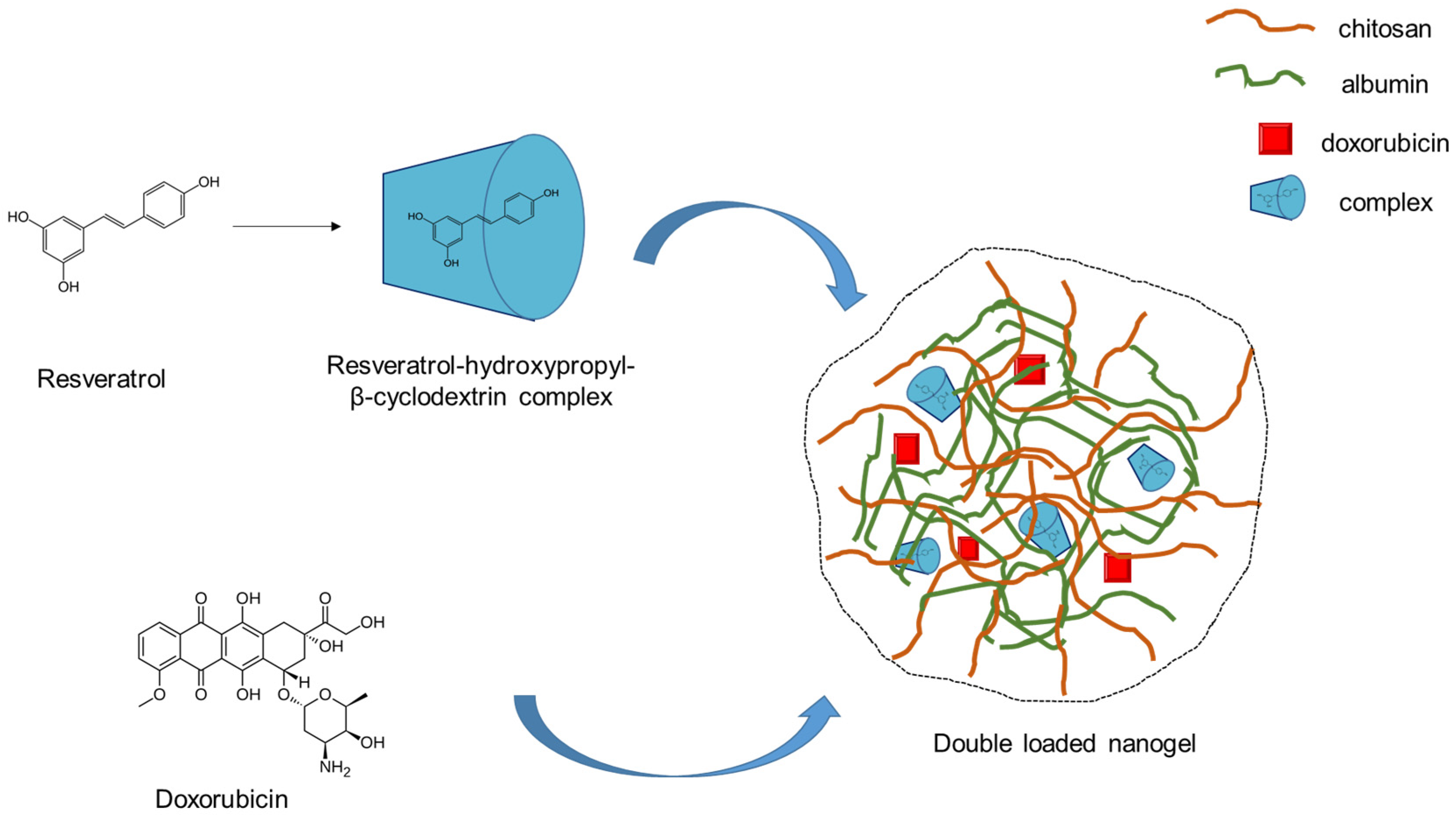
2.1. Preparation of Double-Loaded with Doxorubicin and Resveratrol Complex Composite Chitosan-Albumin Nanogels

The double incorporation of substances with different water affinity into one nanosized delivery system could provide many advantages, but it is still a challenge [26,27,28]. Moreover, the loading of hydrophobic substances into hydrophilic systems such as nanogels requires technological approaches [29]. Resveratrol is known to possess low aqueous solubility (50 µg/mL) and high lipophilicity (logP = 3.1) [30]. Therefore, we applied a strategy for obtaining a composite nanosystem that will provide the successful encapsulation of both doxorubicin and resveratrol. First, a complex between resveratrol and hydroxypropyl-β-cyclodextrin at a ratio of 1:10 was developed via the solvent evaporation method [31]. Therefore, the double-loaded nanogel particles were prepared via electrostatic gelation and heating by modifying a previously reported procedure [11] (Figure 1). The encapsulation efficiency and loading degree of doxorubicin and resveratrol were 73.3% and 516 µg/mL and 97.8% and 324 µg/mL, respectively. These high values for the hydrophobic resveratrol confirmed that the developed approach was successful. For instance, the encapsulation efficiency for resveratrol in hydrophilic systems was found to be approx. 71% in alginate nanoparticles [32], 71–75% in sericin nanoparticles [33], and approx. 67% in chitosan nanoparticles [34]. Thus, the complexation of the drug in the current study led to improved loading into the hydrophilic nanogel. Furthermore, this approach allowed high loading efficiency without using organic solvents, which have potential toxic effects.

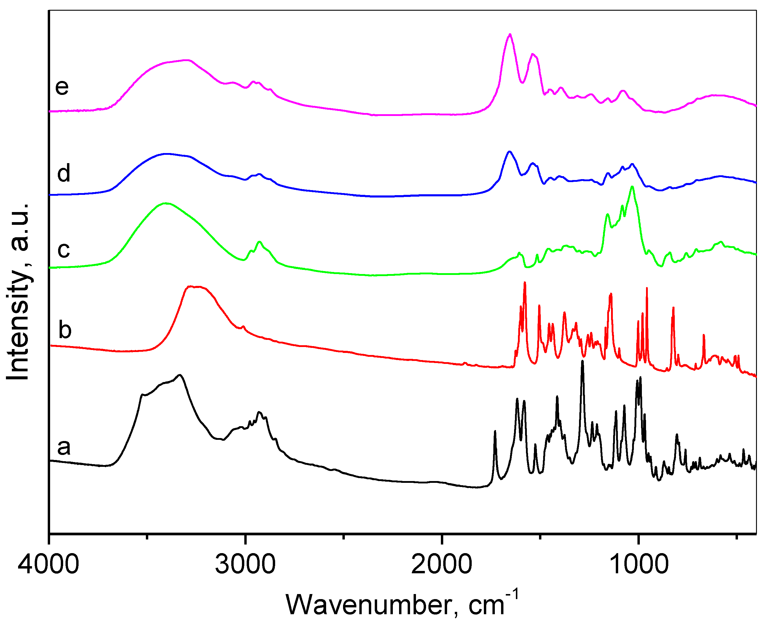
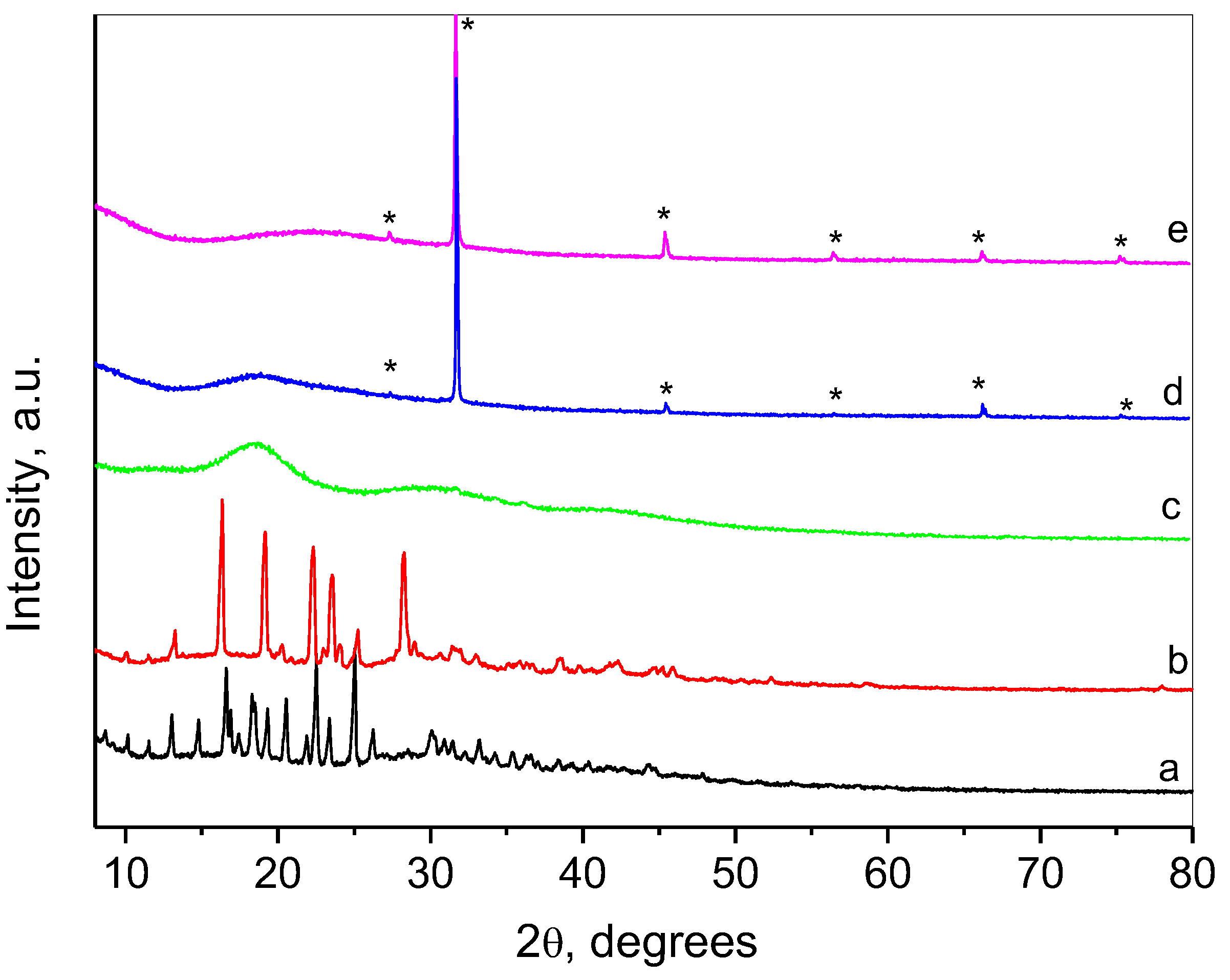
2.2. Characterization of the Composite Nanogel

The double-loaded nanogel particles were examined via dynamic light scattering (DLS) and transmission electron microscopy (TEM). The analyses revealed that the nanoparticles were characterized with a small average size of approx. 31 nm, narrow size distribution, spherical shape, and positive zeta potential (Figure 2a,b, Table 1). The zeta potential of the system increased after the loading process. Since the positive charge of the surface of the empty nanogel was a result of the free amino groups of chitosan, probably more of these groups did not react with the carboxyl groups of albumin when the complex was added. A possible explanation could be the interaction between the cyclodextrin and albumin, leading to complexation and occupying the protein’s functional groups [35]. Moreover, a zeta potential in the range of 30–60 mV absolute value could provide excellent stability due to the repulsion between the nanoparticles and avoiding agglomeration [36].
Figure 3 presents the FTIR spectra of initial drugs, complex with resveratrol and nanogel. The spectrum of doxorubicin and resveratrol coincide with those reported in the literature [37,38]. The characteristic bands of pure doxorubicin appearing at 1733 cm−1 indicated the presence of C=O bonds, and the vibrations of phenol rings at 1615 cm−1 and 1585 cm−1 were visible. The lines at 1418 cm−1, 1282 cm−1, and 989 cm−1 were due to the vibrations of N–H, C–C, and C–OH bonds, respectively. The following bands considered as characteristic of resveratrol were registered at 1603 cm−1, ascribed to aromatic C=C bond stretching, 1582 cm−1 for olefinic C=C stretching, and 967 cm−1 typical for trans-olefinic bond [39]. The disappearance of the line at 967 cm−1 in the FTIR spectrum of resveratrol complex with HP-β-CD suggests the successful complex formation [31]. The spectrum of the empty nanogel presented a set of bands typical for both chitosan at 1081 cm−1 and 1021 cm−1 for stretching vibrations of C–O–C and P=O bonds [40], and albumin at 1651 cm−1 for C=O stretching, amide I, -NH bending at 1538 cm−1, amide II, and 1395 cm−1 for C–N stretching [41]. The FTIR spectrum of DOX/RES-NG consisted of the main characteristic bands of resveratrol complex, doxorubicin, and the nanogel particles. The successful inclusion of doxorubicin was proved by the presence of 989 cm−1 shoulder band, and the encapsulation of the resveratrol complex into the nanogel was suggested by the increase of the intensities of the peaks in 1157 cm−1, 1084 cm−1, and 1032 cm−1 due to vibrations of C–O and C–O–C bonds.
Collected XRD patterns of the drugs, complex, and nanogels are shown in Figure 4. The pattern of doxorubicin consisted of a number of sharp, intense peaks revealing the high crystalline nature of the drug [37,42]. Resveratrol is also highly crystalline, with typical peaks corresponding to the crystal structure of the trans-form of the drug [43]. The pattern of resveratrol complex with HP-β-CD comprised three broad humps that showed successful interaction between the two components [31,44,45]. A typical amorphous compound diffraction pattern was observed for the empty nanogel (NG) with one very broad hump centered at 22.5° 2θ. The lines of crystalline NaCl (marked with asterisks in the diffraction patterns) were also registered using the preparation method. The pattern of the nanogel loaded with both drugs (DOX/RES-NG) consisted of an amorphous broad peak with a center coinciding with the first peak of the resveratrol complex. The disappearance of the sharp peaks of doxorubicin at this stage indicated its presence in a molecular form in the final DOX/RES-NG.

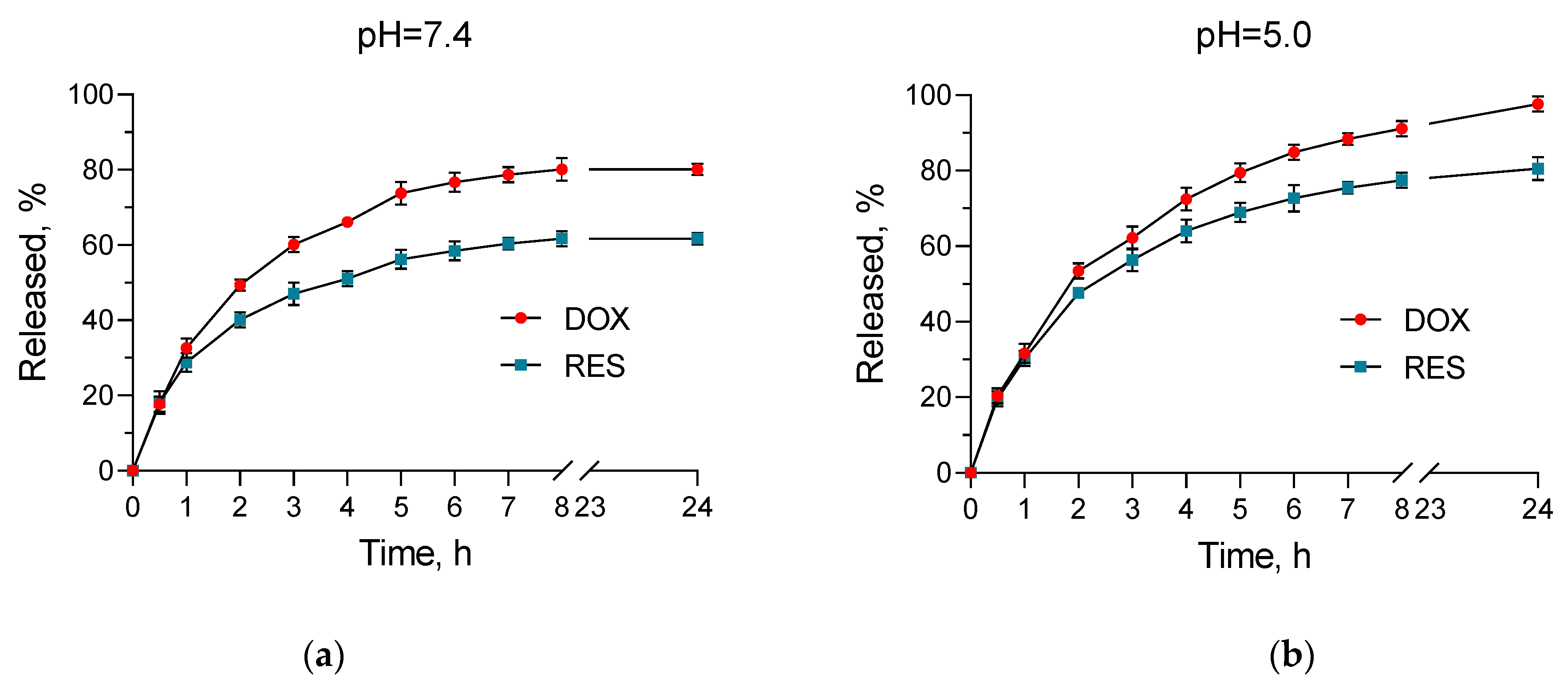
2.3. In Vitro Release Studies

The pH of the extracellular medium surrounding healthy cells is around 7.4, while the extracellular pH value for cancer cells could decrease to approx. 6.0 [46]. Furthermore, the pH value in the endosomes and lysosomes is around 4.5 [47]. Therefore, we conducted in vitro dissolution tests in media with neutral (7.4) and slightly acidic pH (5.0). The tests showed a pH-dependent release. There was a higher amount of both drugs released in the medium with pH = 5.0 compared to the medium with pH = 7.4 (Figure 5). In particular, approx. 98% of doxorubicin was released in the acidic medium vs. around 80% in the neutral one. For resveratrol, these values were approx. 81 vs. 62%, respectively. This pH-dependent release manner of both doxorubicin and resveratrol draws attention to the system’s ability to provide specific delivery of the encapsulated drugs. The main reason for this is probably the solubility of chitosan, which is more pronounced in an acidic medium [48,49]. Similarly, more pronounced release from chitosan nanoparticles in such a slightly acidic medium was observed for different active substances such as tamoxifen [50], Cinnamomum zeylanicum essential oil [51], curcumin [52], and oxaliplatin [47]. Thus, the ability of the developed nanogel system to achieve pH-dependent release of the encapsulated drugs, as well as the small size, could enable the targeting of cancer cells, the efficiency of therapies, and the protection of healthy tissues.
Moreover, the release mechanism for doxorubicin and resveratrol was studied by fitting the data until the 8th hour by applying zero-order, first-order, and Higuchi mathematical models [53]. The results from the analyses are presented in Table 2 and Table 3. For doxorubicin in the medium with pH = 7.4, the highest coefficient (r2) was calculated for the Higuchi model, meaning that the process is controlled via diffusion through the polymer matrix. The greatest coefficient (r2) for doxorubicin in the neutral medium and for resveratrol in both acidic and neutral medium was found for first-order kinetics. Therefore, in these cases, the release of the drugs depended on their remaining concentration in the nanogel matrix.

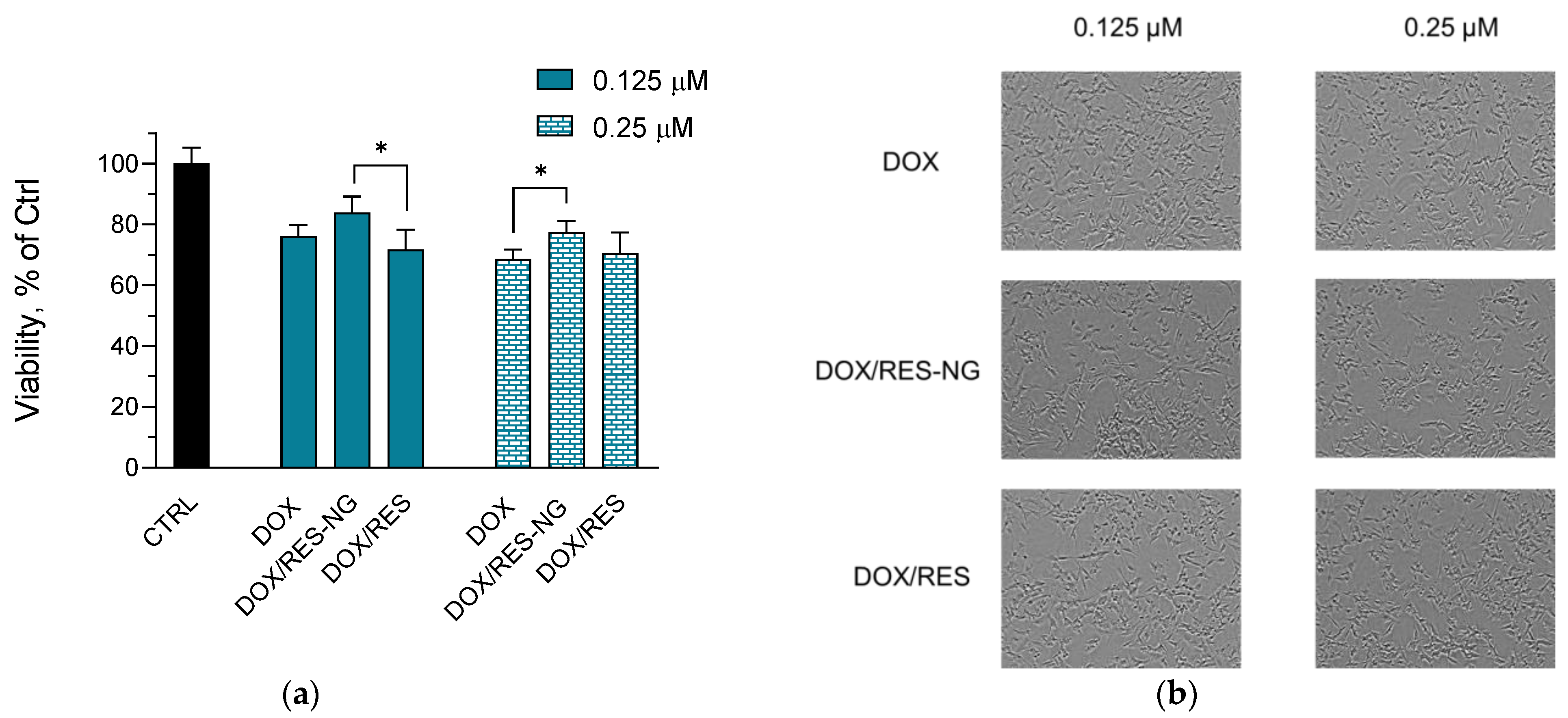
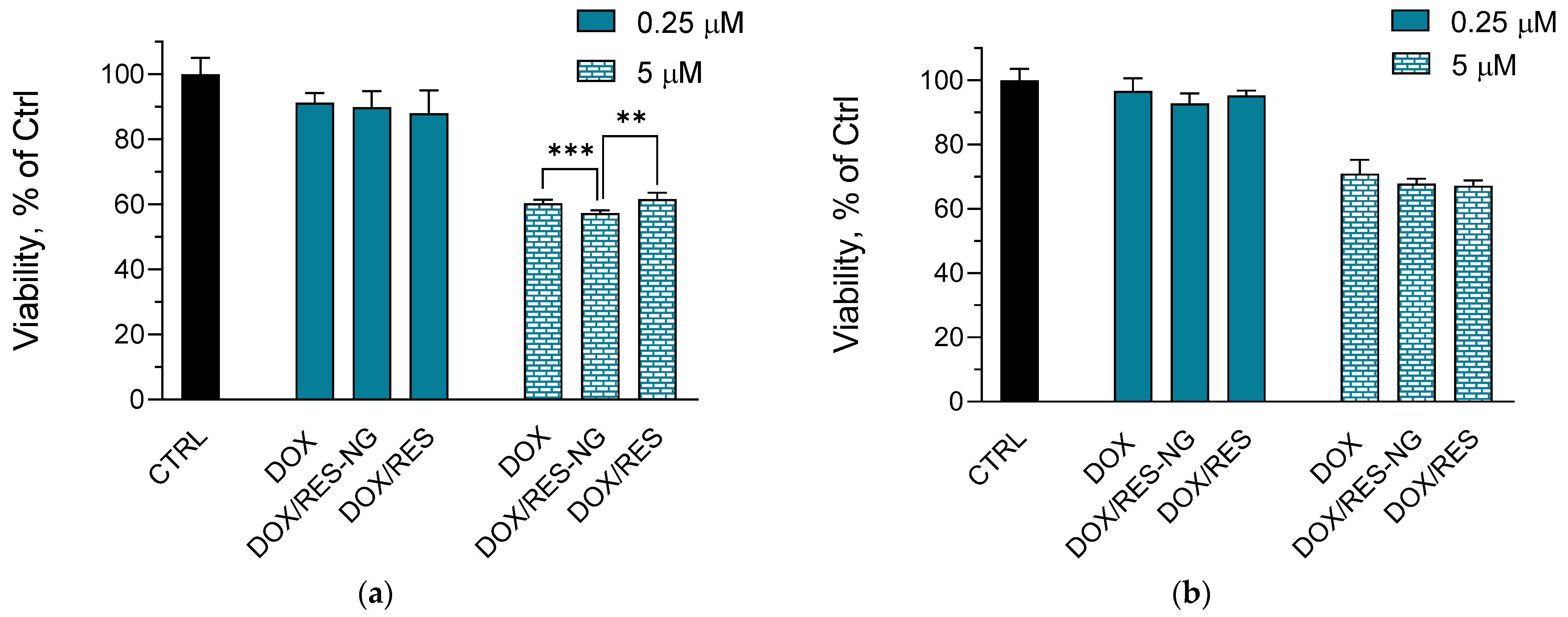
2.4. Cytotoxicity Studies

One of the main adverse effects of doxorubicin in anticancer therapies is its cardiotoxicity [54]. The induced oxidative stress by the antitumor drug is one of the reasons for this toxicity [55]. Therefore, the simultaneous delivery of doxorubicin and antioxidants, such as resveratrol, could be considered an appropriate strategy to ameliorate this toxicity. Furthermore, the advantages of the nanogel systems could additionally reduce the adverse effect via passive targeting. Taking this into consideration, we examined the potential of the double-loaded system to have a protective effect on H9c2 cardioblasts. First, the empty nanogel particles did not show any toxic effects on the cells. The in vitro test revealed that the simultaneous encapsulation of both substances is associated with decreased cardiotoxicity of doxorubicin (Figure 6a,b). In particular, the double-loaded nanogel showed a significant protective effect at 0.25 µM concentration of both drugs. At the higher concentration of 5 µM, a tendency for protection was also observed when the cells were treated with the encapsulated substances. On the contrary, the referent solutions of the non-encapsulated drugs showed cytotoxicity similar to that of pure doxorubicin in both concentrations. This is also confirmed by the microscopic images of the cells. Namely, the typical morphology of apoptotic cells (shrinkage and more prominent phase contrast halo) [56,57] is more common in the images of the cells treated with doxorubicin or with the referent solution in comparison with the cells treated with the double-loaded nanogels. Thus, the encapsulation of resveratrol seems to enable its cardioprotective effect, whereas its non-encapsulated form did not exert such an effect on the cardioblasts.
Another adverse effect of doxorubicin is its neurotoxicity [58]. Oxidative stress, particularly lipid peroxidation, is considered to be a part of the mechanisms responsible for this adverse effect [59]. This was a prerequisite for evaluating the potential of the simultaneous delivery of doxorubicin, along with resveratrol, to alleviate neurotoxicity in vitro. The in vitro assays on neuroblastoma SH-SY5Y cells revealed that the empty nanogel did not have any cytotoxic effects. Similar to the cardioblasts, only the double-loaded nanogel showed protective effects against doxorubicin-induced neurotoxicity (Figure 7a,b). At 0.125 µM concentration, we observed a tendency for protection, while at 0.25 µM, there was a significant protective effect vs. the doxorubicin-treated group. The referent solution of doxorubicin and resveratrol did not show any protective effects. This could be confirmed from the microscopic images, where the morphological changes such as shrinkage and fragmentation are observed more often for the cells treated with doxorubicin and the referent solution, compared to the cell groups treated with the loaded nanogels [56,57].
The simultaneous delivery of anticancer drugs with antioxidants could influence the cytostatic activity of the antitumor agent. However, resveratrol is known to possess antitumor effects via its prooxidant activity, induction of autophagy, and apoptosis [60,61]. Therefore, we evaluated the cytotoxic effects of the double-loaded nanogel in lymphoma L5178Y and multidrug-resistant L5178MDR cell lines. The empty nanogel did not reduce the viability of the cell lines. As can be seen in Figure 8, the double encapsulation of doxorubicin and resveratrol did not reduce the cytostatic effect of the antitumor drug in both L5178Y and L5178MDR cell lines. Moreover, at 5 µM concentration of both drugs, there was a decrease in the cell viability of L5178Y cells when they were only treated with the loaded nanogel (Figure 8a).
Thus, the double encapsulation of doxorubicin and resveratrol in the nanogel system resulted in reduced cardiotoxicity and neurotoxicity, especially at 0.25 µM concentration. The system did not decrease the cytostatic effect of doxorubicin in lymphoma cells; besides, at 5 µM concentration, there was an enhanced cytostatic effect in L5178Y cells. This is probably due to the concentration-dependent prooxidant effects of resveratrol [62].

3. Conclusions

Composite nanogel particles were successfully developed from chitosan, bovine serum albumin, and hydroxypropyl-β-cyclodextrin. The composite system enabled high encapsulation efficiency of hydrophilic doxorubicin (73.3%) and hydrophobic resveratrol (97.8%). The double-loaded nanogel particles were characterized by small size of approximately 30 nm, narrow size distribution, and pH-dependent release of both drugs, which was faster in medium with pH = 5.0. Therefore, we consider the double-loaded nanogel appropriate for application in cancer treatment. Preliminary in vitro studies revealed the potential of the double-loaded nanogel to alleviate cardiotoxicity and neurotoxicity related to doxorubicin, as well as to maintain its anticancer activity in lymphoma cells.

4. Materials and Methods

4.1. Materials

Chitosan (Mv 110,000–150,000), bovine serum albumin (fraction V), hydroxypropyl-β-cyclodextrin, doxorubicin hydrochloride, trans-resveratrol, Dulbecco’s Modified Eagle’s Medium, Roswell Park Memorial Institute 1640 Medium, McCoy’s 5A Medium, fetal bovine serum (FBS), L-glutamine and colchicine were obtained from Sigma Chemical Co. (Germany). Neutral red and Resazurin were supplied by Thermo Fisher Scientific (Waltham, MA, USA). The cardioblast cell line H9c2 and the neuroblastoma SH-SY5Y were purchased from the European Collection of Cell Cultures (ECACC, Salisbury, UK). The mouse lymphoma cell line L5178Y and resistant L5178MDR were donated by Dr. M. M. Gottesman (National Cancer Institute, Bethesda, MD, USA).

4.2. Preparation of Double-Loaded with Doxorubicin and Resveratrol Complex Composite Chitosan-Albumin Nanogel

The loaded composite nanogels were prepared at a molar ratio of 1:1 between the drugs via electrostatic gelation followed by a heating method [11] with a modification. Firstly, a complex between trans-resveratrol (1.25 mg) and hydroxypropyl-β-cyclodextrin at a ratio of 1:10 (wt./wt.) was obtained via solvent evaporation method according to previously described procedure [31]. Then, 3 mg of doxorubicin and 25 mg of bovine serum albumin were mixed with the complex dispersion. A 1 mL 0.5% solution of chitosan (w/v) in hydrochloric acid buffer (pH = 1.2) was dripped into the dispersion, and the mixture was stirred at 700 rpm for 90 min. Therefore, it was alkalized with a sufficient amount of 1M NaOH to a pH of approx. 4.6. The system was heated at 78 °C for 20 min and then stirred for 3 h at 700 rpm. After that, the dispersion was filtered (0.2 µm), the filter was rinsed with 50% ethanol, and the concentration of unloaded drugs in the filter fraction was determined spectrophotometrically at 480 nm for doxorubicin and 306 nm for resveratrol (Thermo Fisher Scientific, Waltham, MA, USA). Standard curves of doxorubicin were prepared in the range of 10–80 µg/mL in buffers with pH = 5 (r > 0.9997) and pH = 7.4 (r > 0.9991). A standard curve of resveratrol was prepared in 50% ethanol in the range of 2–10 µg/mL (r > 0.9996). The encapsulation efficiency (EE) and loading degree (LD) were determined according to the following equations:
EE = (Total amount of drug − Non-loaded drug)/Total amount of drug
LD = (Total amount of drug − Non-loaded drug)/Volume of drug loaded micellar dispersion

4.3. Physicochemical Characterization of the Loaded Nanogels

The mean diameter and polydispersity index of the systems were determined after 10 times dilution of the aqueous nanogel dispersion with purified water. The measurements were performed via photon correlation spectroscopy at a scattering angle of 90°. Phase analysis light scattering (PALS) method at a scattering angle of 15° was applied for evaluation of the zeta potential (Zetasizer NanoBrook 90Plus PALS, Brookhaven Instruments Corporation, Holtsville, NY, USA). The size and shape of the nanoparticles were confirmed via transmission electron microscopy (TEM, HR STEM JEOL JEM 2100, Tokyo, Japan). The diluted (1:10 in water) nanogel dispersion (approx. 5 µL) was pipetted onto a TEM grid and was allowed to dry at room temperature. The grid was directly observed in the TEM after the medium was evaporated.
Nicolet iS5 FTIR spectrometer (Thermo Fisher Scientific, Waltham, MA, USA), accumulating 64 scans at a spectral resolution of 2 cm−1, was used for collecting the IR spectra. The samples were prepared in KBr pellets.
Bruker D8 Advance diffractometer (Bruker Corporation, Billerica, MA, USA) with Cu Kα radiation and a LynxEye detector was used for recording the powder X-ray diffraction patterns of doxorubicin, trans-resveratrol, chitosan, albumin, and the loaded nanoparticles in the 5–80° 2θ range with a step of 0.02°. The samples were placed in a standard sample holder.

4.4. In Vitro Release Studies

In vitro release tests were conducted in two 10% ethanol buffer media with pH values of 7.4 (phosphate buffer) and 5.0 (citrate buffer) via dialysis method [63]. Nanogel dispersion (2 mL, containing 1.058 mg doxorubicin and 0.624 mg resveratrol) was introduced into a dialysis bag (10,000 MWCO, Spectrum Labs, San Francisco, CA, USA) and then placed in 40 mL of the media. The test was performed in a water bath at 37 °C under gentle shaking (IKA Labortechnik HS-B20, Staufen, Germany). Samples of 3 ml were taken at predetermined time intervals, and the same amount of fresh buffer was returned to maintain sink conditions. The concentrations of doxorubicin and trans-resveratrol were determined spectrophotometrically as described above.
The data from the release tests was fitted to zero-order (Equation (3)), first-order (Equation (4)) and Higuchi (Equation (5)) models in order to evaluate the release kinetics:
Ct = C0 + K0·t,
where Ct is the amount of active substance released during the time t; C0 is the initial concentration of the drug released; and K0 is the zero-order rate constant.
ln(Ci − Ct) = ln(Ci) − K1·t,
where Ct is the amount of active substance released during the time t; Ci is the initial concentration of the drug before release; and K1 is the first-order rate constant.
Ct = KH·t1/2,
where Ct is the amount of drug released during the time t, and KH is the release constant of Higuchi.

4.5. Cell Cytotoxicity Studies

The H9c2 and SH-SY5Y cells were maintained in Dulbecco’s Modified Eagle’s Medium and Roswell Park Memorial Institute 1640 Medium, respectively, which were supplemented with 10% fetal bovine serum and 4 mM L-glutamine. The L5178Y and L51L5178 MDR cells were maintained in McCoy’s 5A Medium, supplemented with 10% horse serum, 4 mM L-glutamine, and 60 ng/mL colchicine only for the MDR cell line. The cell lines were incubated under standard conditions (5% CO2, 37 °C, high humidity, Esco CelCulture® CO2 Incubator, CCL-170B-8-IVF, Esco Micro Pte. Ltd., Singapore) and subcultured according to the protocols for adherent [64] and suspension [65] cell lines.
Cell cytotoxicity assays were conducted on rat cardioblast (H9c2) and neuroblastoma (SH-SY5Y) cells via neutral red assay [66] and on lymphoma L5178Y and L5178MDR cells via Alamar blue assay [67]. The cells were seeded in 96-well plates at a cell density of 0.5 × 10⁴ (H9c2), 2.5 × 10⁴ (SH-SY5Y) and 1 × 10⁴ (L5178Y and L5178MDR). Then, they were incubated overnight at standard conditions, namely 37 °C, 5% CO2, and high humidity (Esco CelCulture® CO2 Incubator, CCL-170B-8-IVF, Esco Micro Pte. Ltd., Singapore). Thereafter, the cells were treated with an aqueous solution of doxorubicin, double-loaded nanogel, and a non-encapsulated hydroalcoholic mixture of doxorubicin and resveratrol. The cardioblast and lymphoma cell lines were treated with 0.25 and 5 µM concentrations of doxorubicin and resveratrol, whereas the neuroblastoma cell line was with 0.125 and 0.25 µM. For H9c2 and SH-SY5Y cells, neutral red solution in the appropriate mediums (40 µg/mL) was added to each well (100 µL/well), and the plates were incubated for 3 h at 37 °C. Phosphate-buffered saline (PBS) was used for washing the cells, and then 100 µL of a destaining solution per well was added. The plates were rapidly shaken for 10 min, and the optical density was measured in a Synergy 2 microplate reader (BioTek Instruments, Inc., Highland Park, Winooski, VT, USA) at 540 nm. For L5178Y and L5178MDR, a resazurin solution in PBS at 44 µM final concentration was added to each well. The fluorescence at 540 nm was measured immediately after resazurin addition for baseline fluorescence evaluation and after 3 h of incubation for viability evaluation in the Synergy 2 microplate reader.
The cells were also photographed with an inverted light microscope (Optika XDS-2, Ponteranica, Italy) and digital camera (Optikam Pro 8LT—4083.18LT, Montreal, QC, Canada). In order to facilitate visual interpretation, the background effects in images were attenuated by applying a Bandpass Filter in Fiji software (version 1.54g) [68].

4.6. Statistical Analysis

All the experiments were conducted in triplicate, and the results are expressed as mean values ± SD. Statistical analysis was performed using GraphPad Prism 8 Software (Dotmatics, San Diego, CA, USA). The groups of cells treated with pure doxorubicin, the double-loaded nanogel, or the non-encapsulated mixture of doxorubicin and resveratrol were compared via multiple t-tests with Holm–Sidak correction.

Author Contributions

Conceptualization, L.R. and K.Y.; methodology, L.R., Y.Y., I.S., D.K., V.T. and K.Y.; formal analysis, L.R., I.S. and D.K.; investigation, L.R., I.S. and D.K.; data curation, Y.Y., V.T. and K.Y.; writing—original draft preparation, L.R., I.S. and D.K.; writing—review and editing, Y.Y., V.T. and K.Y.; visualization, L.R., I.S. and D.K.; supervision, V.T. and K.Y.; project administration, K.Y.; funding acquisition, L.R. and K.Y. All authors have read and agreed to the published version of the manuscript.

Funding

This research was funded by the Medical University of Sofia (Council of Medical Science, Contract number D-103/29.05.2024).

Institutional Review Board Statement

Not applicable.

Informed Consent Statement

Not applicable.

Data Availability Statement

Data are contained within the article.

Conflicts of Interest

The authors declare no conflicts of interest.

References

  1. Maeda, H.; Wu, J.; Sawa, T.; Matsumura, Y.; Hori, K. Tumor Vascular Permeability and the EPR Effect in Macromolecular Therapeutics: A Review. J. Control. Release 2000, 65, 271–284. [Google Scholar] [CrossRef] [PubMed]
  2. Ali, A.A.; Al-Othman, A.; Al-Sayah, M.H. Multifunctional Stimuli-Responsive Hybrid Nanogels for Cancer Therapy: Current Status and Challenges. J. Control. Release 2022, 351, 476–503. [Google Scholar] [CrossRef] [PubMed]
  3. Pinelli, F.; Ferracin, F.; Perale, G.; Rossi, F. Chapter Two—Synthesis and Applications of Nanogels via Covalent Cross-Linking Strategies. In Advances in Chemical Engineering; Mauri, E., Zhang, Z.J., Eds.; Soft Particles; Academic Press: Cambridge, MA, USA, 2023; Volume 62, pp. 35–58. [Google Scholar]
  4. Yin, Y.; Hu, B.; Yuan, X.; Cai, L.; Gao, H.; Yang, Q. Nanogel: A Versatile Nano-Delivery System for Biomedical Applications. Pharmaceutics 2020, 12, 290. [Google Scholar] [CrossRef] [PubMed]
  5. Suhail, M.; Rosenholm, J.M.; Minhas, M.U.; Badshah, S.F.; Naeem, A.; Khan, K.U.; Fahad, M. Nanogels as Drug-Delivery Systems: A Comprehensive Overview. Ther. Deliv. 2019, 10, 697–717. [Google Scholar] [CrossRef]
  6. Keskin, D.; Zu, G.; Forson, A.M.; Tromp, L.; Sjollema, J.; van Rijn, P. Nanogels: A Novel Approach in Antimicrobial Delivery Systems and Antimicrobial Coatings. Bioact. Mater. 2021, 6, 3634–3657. [Google Scholar] [CrossRef]
  7. Li, Z.; Huang, J.; Wu, J. pH-Sensitive Nanogels for Drug Delivery in Cancer Therapy. Biomater. Sci. 2021, 9, 574–589. [Google Scholar] [CrossRef]
  8. Manzanares-Guevara, L.A.; Licea-Claverie, A.; Oroz-Parra, I.; Bernaldez-Sarabia, J.; Diaz-Castillo, F.; Licea-Navarro, A.F. Smart Nanoformulation Based on Stimuli-Responsive Nanogels and Curcumin: Promising Therapy against Colon Cancer. ACS Omega 2020, 5, 9171–9184. [Google Scholar] [CrossRef]
  9. Verma, G.; Gajipara, A.; Shelar, S.B.; Priyadarsini, K.I.; Hassan, P.A. Development of Water-Dispersible Gelatin Stabilized Hydroxyapatite Nanoformulation for Curcumin Delivery. J. Drug Deliv. Sci. Technol. 2021, 66, 102769. [Google Scholar] [CrossRef]
  10. Setayesh, A.; Bagheri, F.; Boddohi, S. Self-Assembled Formation of Chondroitin Sulfate-Based Micellar Nanogel for Curcumin Delivery to Breast Cancer Cells. Int. J. Biol. Macromol. 2020, 161, 771–778. [Google Scholar] [CrossRef]
  11. Radeva, L.; Zaharieva, M.M.; Spassova, I.; Kovacheva, D.; Pencheva-El Tibi, I.; Najdenski, H.; Yoncheva, K. Biopolymeric Nanogel as a Drug Delivery System for Doxorubicin—Improved Drug Stability and Enhanced Antineoplastic Activity in Skin Cancer Cells. Pharmaceuticals 2024, 17, 186. [Google Scholar] [CrossRef]
  12. Kamenova, K.; Radeva, L.; Yoncheva, K.; Ublekov, F.; Ravutsov, M.A.; Marinova, M.K.; Simeonov, S.P.; Forys, A.; Trzebicka, B.; Petrov, P.D. Functional Nanogel from Natural Substances for Delivery of Doxorubicin. Polymers 2022, 14, 3694. [Google Scholar] [CrossRef] [PubMed]
  13. Zeinali, M.; Abbaspour-Ravasjani, S.; Ghorbani, M.; Babazadeh, A.; Soltanfam, T.; Santos, A.C.; Hamishehkar, H.; Hamblin, M.R. Nanovehicles for Co-Delivery of Anticancer Agents. Drug Discov. Today 2020, 25, 1416–1430. [Google Scholar] [CrossRef] [PubMed]
  14. Liu, Q.; Li, J.; Pu, G.; Zhang, F.; Liu, H.; Zhang, Y. Co-Delivery of Baicalein and Doxorubicin by Hyaluronic Acid Decorated Nanostructured Lipid Carriers for Breast Cancer Therapy. Drug Deliv. 2016, 23, 1364–1368. [Google Scholar] [CrossRef] [PubMed]
  15. Amjadi, S.; Hamishehkar, H.; Ghorbani, M. A Novel Smart PEGylated Gelatin Nanoparticle for Co-Delivery of Doxorubicin and Betanin: A Strategy for Enhancing the Therapeutic Efficacy of Chemotherapy. Mater. Sci. Eng. C 2019, 97, 833–841. [Google Scholar] [CrossRef] [PubMed]
  16. Guo, X.; Zhao, Z.; Chen, D.; Qiao, M.; Wan, F.; Cun, D.; Sun, Y.; Yang, M. Co-Delivery of Resveratrol and Docetaxel via Polymeric Micelles to Improve the Treatment of Drug-Resistant Tumors. Asian J. Pharm. Sci. 2019, 14, 78–85. [Google Scholar] [CrossRef]
  17. Xiao, B.; Si, X.; Han, M.K.; Viennois, E.; Zhang, M.; Merlin, D. Co-Delivery of Camptothecin and Curcumin by Cationic Polymeric Nanoparticles for Synergistic Colon Cancer Combination Chemotherapy. J. Mater. Chem. B 2015, 3, 7724–7733. [Google Scholar] [CrossRef]
  18. Lages, E.B.; Fernandes, R.S.; Silva, J.d.O.; de Souza, Â.M.; Cassali, G.D.; de Barros, A.L.B.; Miranda Ferreira, L.A. Co-Delivery of Doxorubicin, Docosahexaenoic Acid, and α-Tocopherol Succinate by Nanostructured Lipid Carriers Has a Synergistic Effect to Enhance Antitumor Activity and Reduce Toxicity. Biomed. Pharmacother. 2020, 132, 110876. [Google Scholar] [CrossRef]
  19. Li, Y.; Yang, D.; Wang, Y.; Li, Z.; Zhu, C. Co-Delivery Doxorubicin and Silybin for Anti-Hepatoma via Enhanced Oral Hepatic-Targeted Efficiency. Int. J. Nanomed. 2019, 14, 301–315. [Google Scholar] [CrossRef]
  20. Larrañeta, E.; Stewart, S.; Ervine, M.; Al-Kasasbeh, R.; Donnelly, R.F. Hydrogels for Hydrophobic Drug Delivery. Classification, Synthesis and Applications. J. Funct. Biomater. 2018, 9, 13. [Google Scholar] [CrossRef]
  21. McKenzie, M.; Betts, D.; Suh, A.; Bui, K.; Kim, L.D.; Cho, H. Hydrogel-Based Drug Delivery Systems for Poorly Water-Soluble Drugs. Molecules 2015, 20, 20397–20408. [Google Scholar] [CrossRef]
  22. Yang, Z.; McClements, D.J.; Peng, X.; Xu, Z.; Meng, M.; Chen, L.; Jin, Z. Fabrication of Zein–Carboxymethyl Cellulose Nanoparticles for Co-Delivery of Quercetin and Resveratrol. J. Food Eng. 2023, 341, 111322. [Google Scholar] [CrossRef]
  23. Chen, X.; Song, L.; Li, X.; Zhang, L.; Li, L.; Zhang, X.; Wang, C. Co-Delivery of Hydrophilic/Hydrophobic Drugs by Multifunctional Yolk-Shell Nanoparticles for Hepatocellular Carcinoma Theranostics. Chem. Eng. J. 2020, 389, 124416. [Google Scholar] [CrossRef]
  24. Thorn, C.F.; Oshiro, C.; Marsh, S.; Hernandez-Boussard, T.; McLeod, H.; Klein, T.E.; Altman, R.B. Doxorubicin Pathways: Pharmacodynamics and Adverse Effects. Pharmacogenet. Genom. 2011, 21, 440–446. [Google Scholar] [CrossRef] [PubMed]
  25. Du, J.; Zhang, A.; Li, J.; Liu, X.; Wu, S.; Wang, B.; Wang, Y.; Jia, H. Doxorubicin-Induced Cognitive Impairment: The Mechanistic Insights. Front. Oncol. 2021, 11, 673340. [Google Scholar] [CrossRef] [PubMed]
  26. Español, L.; Larrea, A.; Andreu, V.; Mendoza, G.; Arruebo, M.; Sebastian, V.; Aurora-Prado, M.S.; Kedor-Hackmann, E.R.M.; Santoro, M.I.R.M.; Santamaria, J. Dual Encapsulation of Hydrophobic and Hydrophilic Drugs in PLGA Nanoparticles by a Single-Step Method: Drug Delivery and Cytotoxicity Assays. RSC Adv. 2016, 6, 111060–111069. [Google Scholar] [CrossRef]
  27. Sun, J.; Chen, Y.; Xu, J.; Song, X.; Wan, Z.; Du, Y.; Ma, W.; Li, X.; Zhang, L.; Li, S. High Loading of Hydrophobic and Hydrophilic Agents via Small Immunostimulatory Carrier for Enhanced Tumor Penetration and Combinational Therapy. Theranostics 2020, 10, 1136–1150. [Google Scholar] [CrossRef]
  28. Hueppe, N.; Wurm, F.R.; Landfester, K. Nanocarriers with Multiple Cargo Load—A Comprehensive Preparation Guideline Using Orthogonal Strategies. Macromol. Rapid Commun. 2023, 44, 2200611. [Google Scholar] [CrossRef]
  29. Sun, M.; Gao, M.; Bi, J.; Zhao, Y.; Gong, J. Highly Efficient Hydrogel Encapsulation of Hydrophobic Drugs via an Organic Solvent-Free Process Based on Oiling-Out Crystallization and a Mechanism Study. ACS Sustain. Chem. Eng. 2024, 12, 4813–4824. [Google Scholar] [CrossRef]
  30. Robinson, K.; Mock, C.; Liang, D. Pre-Formulation Studies of Resveratrol. Drug Dev. Ind. Pharm. 2015, 41, 1464–1469. [Google Scholar] [CrossRef]
  31. Radeva, L.; Yordanov, Y.; Spassova, I.; Kovacheva, D.; Tibi, I.P.-E.; Zaharieva, M.M.; Kaleva, M.; Najdenski, H.; Petrov, P.D.; Tzankova, V.; et al. Incorporation of Resveratrol-Hydroxypropyl-β-Cyclodextrin Complexes into Hydrogel Formulation for Wound Treatment. Gels 2024, 10, 346. [Google Scholar] [CrossRef]
  32. Saralkar, P.; Dash, A.K. Alginate Nanoparticles Containing Curcumin and Resveratrol: Preparation, Characterization, and In Vitro Evaluation Against DU145 Prostate Cancer Cell Line. AAPS PharmSciTech 2017, 18, 2814–2823. [Google Scholar] [CrossRef] [PubMed]
  33. Suktham, K.; Koobkokkruad, T.; Wutikhun, T.; Surassmo, S. Efficiency of Resveratrol-Loaded Sericin Nanoparticles: Promising Bionanocarriers for Drug Delivery. Int. J. Pharm. 2018, 537, 48–56. [Google Scholar] [CrossRef] [PubMed]
  34. Bu, L.; Gan, L.-C.; Guo, X.-Q.; Chen, F.-Z.; Song, Q.; Zhao, Q.; Gou, X.-J.; Hou, S.-X.; Yao, Q. Trans-Resveratrol Loaded Chitosan Nanoparticles Modified with Biotin and Avidin to Target Hepatic Carcinoma. Int. J. Pharm. 2013, 452, 355–362. [Google Scholar] [CrossRef] [PubMed]
  35. Ghosh, S.; Paul, B.K.; Chattopadhyay, N. Interaction of Cyclodextrins with Human and Bovine Serum Albumins: A Combined Spectroscopic and Computational Investigation. J. Chem. Sci. 2014, 126, 931–944. [Google Scholar] [CrossRef]
  36. Németh, Z.; Csóka, I.; Semnani Jazani, R.; Sipos, B.; Haspel, H.; Kozma, G.; Kónya, Z.; Dobó, D.G. Quality by Design-Driven Zeta Potential Optimisation Study of Liposomes with Charge Imparting Membrane Additives. Pharmaceutics 2022, 14, 1798. [Google Scholar] [CrossRef]
  37. Yoncheva, K.; Tzankov, B.; Yordanov, Y.; Spassova, I.; Kovacheva, D.; Frosini, M.; Valoti, M.; Tzankova, V. Encapsulation of Doxorubicin in Chitosan-Alginate Nanoparticles Improves Its Stability and Cytotoxicity in Resistant Lymphoma L5178 MDR Cells. J. Drug Deliv. Sci. Technol. 2020, 59, 101870. [Google Scholar] [CrossRef]
  38. Moyano-Mendez, J.R.; Fabbrocini, G.; De Stefano, D.; Mazzella, C.; Mayol, L.; Scognamiglio, I.; Carnuccio, R.; Ayala, F.; La Rotonda, M.I.; De Rosa, G. Enhanced Antioxidant Effect of Trans-Resveratrol: Potential of Binary Systems with Polyethylene Glycol and Cyclodextrin. Drug Dev. Ind. Pharm. 2014, 40, 1300–1307. [Google Scholar] [CrossRef]
  39. Shi, G.; Rao, L.; Yu, H.; Xiang, H.; Yang, H.; Ji, R. Stabilization and Encapsulation of Photosensitive Resveratrol within Yeast Cell. Int. J. Pharm. 2008, 349, 83–93. [Google Scholar] [CrossRef]
  40. Song, C.; Yu, H.; Zhang, M.; Yang, Y.; Zhang, G. Physicochemical Properties and Antioxidant Activity of Chitosan from the Blowfly Chrysomya megacephala Larvae. Int. J. Biol. Macromol. 2013, 60, 347–354. [Google Scholar] [CrossRef]
  41. Militello, V.; Casarino, C.; Emanuele, A.; Giostra, A.; Pullara, F.; Leone, M. Aggregation Kinetics of Bovine Serum Albumin Studied by FTIR Spectroscopy and Light Scattering. Biophys. Chem. 2004, 107, 175–187. [Google Scholar] [CrossRef]
  42. Bezzon, V.D.N.; Caturello, N.A.M.D.S.; Dalpian, G.M.; Ferreira, F.F. Crystal Structure Determination and DFT Analysis of Doxorubicin Hydrochloride for Controlled-Release Drug Formulations. J. Mol. Struct. 2023, 1294, 136412. [Google Scholar] [CrossRef]
  43. Caruso, F.; Tanski, J.; Villegas-Estrada, A.; Rossi, M. Structural Basis for Antioxidant Activity of Trans-Resveratrol: Ab Initio Calculations and Crystal and Molecular Structure. J. Agric. Food Chem. 2004, 52, 7279–7285. [Google Scholar] [CrossRef] [PubMed]
  44. Hao, X.; Sun, X.; Zhu, H.; Xie, L.; Wang, X.; Jiang, N.; Fu, P.; Sang, M. Hydroxypropyl-β-Cyclodextrin-Complexed Resveratrol Enhanced Antitumor Activity in a Cervical Cancer Model: In Vivo Analysis. Front. Pharmacol. 2021, 12, 573909. [Google Scholar] [CrossRef] [PubMed]
  45. Silva, A.F.R.; Monteiro, M.; Resende, D.; Braga, S.S.; Coimbra, M.A.; Silva, A.M.S.; Cardoso, S.M. Inclusion Complex of Resveratrol with γ-Cyclodextrin as a Functional Ingredient for Lemon Juices. Foods 2021, 10, 16. [Google Scholar] [CrossRef] [PubMed]
  46. Ward, C.; Meehan, J.; Gray, M.E.; Murray, A.F.; Argyle, D.J.; Kunkler, I.H.; Langdon, S.P. The Impact of Tumour pH on Cancer Progression: Strategies for Clinical Intervention. Explor. Target. Anti-Tumor Ther. 2020, 1, 71–100. [Google Scholar] [CrossRef]
  47. Vivek, R.; Thangam, R.; Nipunbabu, V.; Ponraj, T.; Kannan, S. Oxaliplatin-Chitosan Nanoparticles Induced Intrinsic Apoptotic Signaling Pathway: A “Smart” Drug Delivery System to Breast Cancer Cell Therapy. Int. J. Biol. Macromol. 2014, 65, 289–297. [Google Scholar] [CrossRef]
  48. Qin, C.; Li, H.; Xiao, Q.; Liu, Y.; Zhu, J.; Du, Y. Water-Solubility of Chitosan and Its Antimicrobial Activity. Carbohydr. Polym. 2006, 63, 367–374. [Google Scholar] [CrossRef]
  49. Aranaz, I.; Alcántara, A.R.; Civera, M.C.; Arias, C.; Elorza, B.; Heras Caballero, A.; Acosta, N. Chitosan: An Overview of Its Properties and Applications. Polymers 2021, 13, 3256. [Google Scholar] [CrossRef]
  50. Vivek, R.; Nipun Babu, V.; Thangam, R.; Subramanian, K.S.; Kannan, S. pH-Responsive Drug Delivery of Chitosan Nanoparticles as Tamoxifen Carriers for Effective Anti-Tumor Activity in Breast Cancer Cells. Colloids Surf. B Biointerfaces 2013, 111, 117–123. [Google Scholar] [CrossRef]
  51. Matshetshe, K.I.; Parani, S.; Manki, S.M.; Oluwafemi, O.S. Preparation, Characterization and in Vitro Release Study of β-Cyclodextrin/Chitosan Nanoparticles Loaded Cinnamomum zeylanicum Essential Oil. Int. J. Biol. Macromol. 2018, 118, 676–682. [Google Scholar] [CrossRef]
  52. Nair, R.S.; Morris, A.; Billa, N.; Leong, C.-O. An Evaluation of Curcumin-Encapsulated Chitosan Nanoparticles for Transdermal Delivery. AAPS PharmSciTech 2019, 20, 69. [Google Scholar] [CrossRef] [PubMed]
  53. Trucillo, P. Drug Carriers: A Review on the Most Used Mathematical Models for Drug Release. Processes 2022, 10, 1094. [Google Scholar] [CrossRef]
  54. Linders, A.N.; Dias, I.B.; López Fernández, T.; Tocchetti, C.G.; Bomer, N.; Van der Meer, P. A Review of the Pathophysiological Mechanisms of Doxorubicin-Induced Cardiotoxicity and Aging. npj Aging 2024, 10, 9. [Google Scholar] [CrossRef] [PubMed]
  55. Dulf, P.L.; Mocan, M.; Coadă, C.A.; Dulf, D.V.; Moldovan, R.; Baldea, I.; Farcas, A.-D.; Blendea, D.; Filip, A.G. Doxorubicin-Induced Acute Cardiotoxicity Is Associated with Increased Oxidative Stress, Autophagy, and Inflammation in a Murine Model. Naunyn. Schmiedebergs Arch. Pharmacol. 2023, 396, 1105–1115. [Google Scholar] [CrossRef] [PubMed]
  56. Huh, S.; Ker, D.F.E.; Su, H.; Kanade, T. Apoptosis Detection for Adherent Cell Populations in Time-Lapse Phase-Contrast Microscopy Images. In Medical Image Computing and Computer-Assisted Intervention—MICCAI 2012; Ayache, N., Delingette, H., Golland, P., Mori, K., Eds.; Lecture Notes in Computer Science; Springer: Berlin/Heidelberg, Germany, 2012; Volume 7510, pp. 331–339. ISBN 978-3-642-33414-6. [Google Scholar]
  57. Ölander, M.; Handin, N.; Artursson, P. Image-Based Quantification of Cell Debris as a Measure of Apoptosis. Anal. Chem. 2019, 91, 5548–5552. [Google Scholar] [CrossRef]
  58. Kamińska, K.; Cudnoch-Jędrzejewska, A. A Review on the Neurotoxic Effects of Doxorubicin. Neurotox. Res. 2023, 41, 383–397. [Google Scholar] [CrossRef]
  59. Alhowail, A.H.; Bloemer, J.; Majrashi, M.; Pinky, P.D.; Bhattacharya, S.; Yongli, Z.; Bhattacharya, D.; Eggert, M.; Woodie, L.; Buabeid, M.A.; et al. Doxorubicin-Induced Neurotoxicity Is Associated with Acute Alterations in Synaptic Plasticity, Apoptosis, and Lipid Peroxidation. Toxicol. Mech. Methods 2019, 29, 457–466. [Google Scholar] [CrossRef]
  60. de la Lastra, C.A.; Villegas, I. Resveratrol as an Antioxidant and Pro-Oxidant Agent: Mechanisms and Clinical Implications. Biochem. Soc. Trans. 2007, 35, 1156–1160. [Google Scholar] [CrossRef]
  61. Elshaer, M.; Chen, Y.; Wang, X.J.; Tang, X. Resveratrol: An Overview of Its Anti-Cancer Mechanisms. Life Sci. 2018, 207, 340–349. [Google Scholar] [CrossRef]
  62. Shaito, A.; Posadino, A.M.; Younes, N.; Hasan, H.; Halabi, S.; Alhababi, D.; Al-Mohannadi, A.; Abdel-Rahman, W.M.; Eid, A.H.; Nasrallah, G.K.; et al. Potential Adverse Effects of Resveratrol: A Literature Review. Int. J. Mol. Sci. 2020, 21, 2084. [Google Scholar] [CrossRef]
  63. D’Souza, S. A Review of In Vitro Drug Release Test Methods for Nano-Sized Dosage Forms. Adv. Pharm. 2014, 2014, 304757. [Google Scholar] [CrossRef]
  64. Subculture of Adherent Cell Lines. Available online: https://www.sigmaaldrich.com/BG/en/technical-documents/protocol/cell-culture-and-cell-culture-analysis/mammalian-cell-culture/subculture-of-adherent (accessed on 2 October 2024).
  65. Cell Culture Protocol 5: Subculture of Suspension Cell Lines. Available online: https://www.sigmaaldrich.com/BG/en/technical-documents/technical-article/cell-culture-and-cell-culture-analysis/mammalian-cell-culture/subculture-of-suspension (accessed on 2 October 2024).
  66. Repetto, G.; del Peso, A.; Zurita, J.L. Neutral Red Uptake Assay for the Estimation of Cell Viability/Cytotoxicity. Nat. Protoc. 2008, 3, 1125–1131. [Google Scholar] [CrossRef] [PubMed]
  67. O’Brien, J.; Wilson, I.; Orton, T.; Pognan, F. Investigation of the Alamar Blue (Resazurin) Fluorescent Dye for the Assessment of Mammalian Cell Cytotoxicity. Eur. J. Biochem. 2000, 267, 5421–5426. [Google Scholar] [CrossRef] [PubMed]
  68. Schindelin, J.; Arganda-Carreras, I.; Frise, E.; Kaynig, V.; Longair, M.; Pietzsch, T.; Preibisch, S.; Rueden, C.; Saalfeld, S.; Schmid, B.; et al. Fiji: An Open-Source Platform for Biological-Image Analysis. Nat. Methods 2012, 9, 676–682. [Google Scholar] [CrossRef]
Figure 1. Schematic presentation of the preparation process of double-loaded with doxorubicin and resveratrol composite chitosan-albumin nanogel.
Gels 10 00699 g001
Figure 2. Histogram of the mean diameter (a) and TEM image (b) of double-loaded with doxorubicin and resveratrol nanogel particles.
Gels 10 00699 g002
Figure 3. FTIR spectra of: (a) doxorubicin (DOX), (b) trans-resveratrol (RES), (c) resveratrol complex with HP-β-CD, (d) doxorubicin and resveratrol loaded nanogel (DOX/RES-NG), (e) empty nanogel (NG).
Gels 10 00699 g003
Figure 4. Powder XRD patterns of (a) doxorubicin (DOX), (b) trans-resveratrol (RES), (c) resveratrol complex with HP-β-CD, (d) doxorubicin and resveratrol loaded nanogel (DOX/RES-NG), (e) empty nanogel (NG).
Gels 10 00699 g004
Figure 5. In vitro release profiles of doxorubicin (DOX) and resveratrol (RES) from the nanogel system in media with pH = 7.4 (a) and 5.0 (b). Mean ± SD (n = 3).
Gels 10 00699 g005
Figure 6. In vitro cytotoxicity (a) and digital images (b) of H9c2 cells treated with doxorubicin (DOX, positive control) at 0.25 or 5 µM, double-loaded nanogels (DOX/RES-NG) and referent solutions of doxorubicin and resveratrol (DOX/RES) at 0.25 or 5 µM concentration of both drugs; CTRL—negative control; Mean ± SD (n = 3). * p < 0.05, *** p < 0.001 between cell groups.
Gels 10 00699 g006
Figure 7. In vitro cytotoxicity (a) and digital images (b) of SH-SY5Y cells treated with doxorubicin (DOX, positive control) at 0.25 or 5 µM, double-loaded nanogel (DOX/RES-NG) and referent solutions of doxorubicin and resveratrol (DOX/RES) at 0.25 or 5 µM concentration of both drugs; CTRL—negative control; Mean ± SD (n = 3). * p < 0.05 between cell groups.
Gels 10 00699 g007
Figure 8. In vitro cytotoxicity of L5178Y (a) and L5178 MDR (b) cells treated with doxorubicin (DOX) at 0.25 or 5 µM, double-loaded nanogel (DOX/RES-NG) and referent solutions of doxorubicin and resveratrol (DOX/RES) at 0.25 or 5 µM concentration of both drugs; Mean ± SD (n = 3). ** p < 0.01, *** p < 0.001 between cell groups.
Gels 10 00699 g008
Table 1. Mean diameter, polydispersity index, and zeta potential of empty (NG) and double-loaded with doxorubicin and resveratrol nanogel (DOX/RES-NG). Mean values ± SD (n = 3).
SampleSize, nmPDIƺ-Potential, mV
NG51 ± 60.241+35.54
DOX/RES-NG30 ± 40.188+51.23
Table 2. Correlation coefficients (r2) for zero-order, first-order, and Higuchi models were calculated through kinetic analysis of in vitro release data for doxorubicin.
pH of the MediumZero OrderFirst OrderHiguchi Model
7.40.84510.95570.9737
5.00.88240.99730.9867
Table 3. Correlation coefficients (r2) for zero-order, first-order, and Higuchi models were calculated through kinetic analysis of in vitro release data for resveratrol.
pH of the MediumZero OrderFirst OrderHiguchi Model
7.40.82030.97070.9060
5.00.88240.99730.9867
Disclaimer/Publisher’s Note: The statements, opinions and data contained in all publications are solely those of the individual author(s) and contributor(s) and not of MDPI and/or the editor(s). MDPI and/or the editor(s) disclaim responsibility for any injury to people or property resulting from any ideas, methods, instructions or products referred to in the content.

