Article

High-Level Expression of β-Glucosidase in Aspergillus niger ATCC 20611 Using the Trichoderma reesei Promoter Pcdna1 to Enhance Cellulose Degradation

State Key Laboratory of Microbial Technology, Institute of Microbial Technology, Shandong University, Qingdao 266237, China
*
Authors to whom correspondence should be addressed.
These authors contributed equally to this work.
Fermentation 2024, 10(9), 461; https://doi.org/10.3390/fermentation10090461
Submission received: 21 July 2024 / Revised: 18 August 2024 / Accepted: 25 August 2024 / Published: 5 September 2024
(This article belongs to the Special Issue Biorefinery of Lignocellulosic Biomass, 2nd Edition)

Abstract

:
β-glucosidase is a key component of cellulase for its function in hydrolyzing cellobiose to glucose in the final step of cellulose degradation. The high-level expression of β-glucosidase is essential for cellulose conversion. Aspergillus niger ATCC 20611 has the potential for efficient protein expression because of its ability to secret enzymes for the industrial production of fructooligosaccharides, but it lacks robust promoters for high-level protein expression. Here, the development of A. niger 20611 as a powerful protein expression system exploited the conserved constitutive promoter Pgpd1 of the glyceraldehyde-3-phosphate dehydrogenase-encoding gene from Trichoerma reesei to drive the expression of the enhanced green fluorescent protein in A. niger ATCC 20611. The mycelium of the transformant AGE9 exhibited intense fluorescence. Then, the promotor Pgpd1 was used to drive the expression of β-glucosidase and the enzyme activity of transformants AGB1 and AGB33 were 1.02 and 0.51 U/mL, respectively. These results demonstrate that the promotor Pgpd1 from T. reesei was applicable for A. niger ATCC 20611. Furthermore, the T. reesei-specific robust promoter Pcdna1 was used to drive the expression of β-glucosidase. The β-glucosidase exhibited a high-level expression with a yield of 15.2 U/mL, which was over 13.9 times higher than that driven by the promoter Pgpd1. The β-glucosidase was thermally stable and accounted for 85% of the total extracellular proteins. Subsequently, the fermentation broth including β-glucosidase was directly added to the cellulase mixture of T. reesei for saccharification of the acid-treated corncob residues and the delignified corncob residues, which increased the saccharification efficiency by 26.21% and 29.51%, respectively. Thus, β-glucosidase exhibited a high level of expression in A. niger ATCC 20611 and enhanced cellulose degradation by addition in vitro. In addition, the robust promoter Pcdna1 of T. reesei could drive the high-level expression of protein in A. niger ATCC 20611. These results demonstrate that the promoters in filamentous fungi could be employed across species in A. niger ATCC 20611 and further facilitated the efficient expression of β-glucosidase to optimize cellulases for efficient cellulose transformation.

1. Introduction

The filamentous fungus Aspergillus niger ATCC 20611 is an industrial strain for fructooligosaccharides production, which can grow rapidly with sucrose as a carbon source and express β-fructofuranosidase at a high level [1]. The β-fructofuranosidase catalyzes the generation of fructooligosaccharides from sucrose and it is located in the cell wall. Thus, A. niger ATCC 20611 has a relatively low extracellular protein background, which is suitable for heterologous protein expression [2]. Because the regulation of protein expression mainly occurred at the transcriptional level, appropriate promoters were crucial for the efficient expression of heterologous proteins [3]. The promoter PfopA of the β-fructofuranosidase-encoding gene was sucrose-inducible and its strength enhanced with the increase in sucrose concentration. So far, several heterologous proteins have been successfully expressed in A. niger ATCC 20611, including glucose oxidase, peroxidase, chitinase, β-N-acetylglucosaminidase and so on [4,5]. Nevertheless, when employing the promoter PfopA to drive heterologous protein expression, sucrose induction caused an abundant expression of β-fructofuranosidase, which would occupy the resources and space of heterologous protein expression. It is known that constitutive promoters can spontaneously drive continuous and stable transcription of genes without relying on carbon or nitrogen sources or other inducers [6]. The mostly used constitutive promoter is the promotor PgpdA of the glyceraldehyde-3-phosphate dehydrogenase-encoding gene (gpd) from Aspergillus nidulans, which has been successfully used in A. niger ATCC 20611 for expression of the enhanced green fluorescent protein and amino-acid-substitution variant FopA [1,2]. The promotor Pgpd is from the glycolytic pathway, which was widely distributed and relatively conserved in different species. Currently, A niger ATCC 20611 lacks constitutive promoters with strong driving ability for the construction of a heterologous protein expression cassette [5]. It was reported that the specific promoter Pcdna1 in T. reesei was a strong constitutive promoter, which was identified through screening cDNA libraries and much stronger than the commonly used glycolysis pathway promoters Pgpd1 and Ptef1 [7]. The expression of CBHI and EGI in T. reesei using Pcdna1 produced 10–50 times more cellulase than that of using Ptef1 and de-repressed Pcbh1 [8]. Currently, Pcdna1 is regarded as the strongest constitutive promoter and the optimal choice for constitutive overexpression or recombinant protein production in T. reesei.
Lignocellulose is the largest renewable biomass resource in the world and its effective treatment and utilization are of great importance for the sustainable development of society. The cellulase mixtures secreted by T. reesei can effectively degrade cellulose. Cellulase 1 (CBH1) and endoglucanase (EGs) jointly hydrolyze cellulose to produce cellulose oligosaccharide and cellulose disaccharide and then the product is further degraded to glucose by β-glucosidase (BGL) [9,10]. However, in the cellulase mixture of T. reesei, the BGL accounts for only about 1%, which is a rate-limiting step for effective hydrolysis of the cellulose substrate [11]. The addition of purified β-glucosidase enzyme in vitro can effectively improve the efficiency of cellulose hydrolysis, but the enzyme purification increases the time and cost [12,13]. Therefore, the utilization of an expression host with low-protein background to express β-glucosidase can effectively address the challenge of protein purification and enhance the efficiency of cellulose degradation.
In this study, we first used the species-conserved promoter Pgpd1 from T. reesei expressing the enhanced green fluorescent protein and β-glucosidase in A. niger ATCC 20611 to validate the feasibility of heterologous promoters. Then, the unique strong promoter Pcdna1 from T. reesei was used to drive the efficient expression of β-glucosidase in A. niger ATCC 20611 and the addition of the crude fermentation broth of transformants to cellulase mixtures of T. reesei SN1 improved the saccharification efficiency of different pretreated corncob residues. Briefly, the unique strong constitutive promoter Pcdna1 from T. reesei was used to establish a heterologous protein expression system in A. niger ATCC 20611 and achieve a high level of β-glucosidase expression for enhancing the rate of cellulose degradation.

2. Materials and Methods

2.1. Strains and Culture Conditions

A. niger ATCC 20611, an industrial strain for fructooligosaccharides production, was used as the host strain for transformation. A. niger C112 was used to obtain the β-glucosidase-encoding gene (bglA). T. reesei QM9414 was used to amplify genes, containing the promoter of gpd1 (Pgpd1), the promoter of cdna1 (Pcdna1), the terminator of cbh1 (Tcbh1). The plasmid T-ptrA was used to access resistance genes ptrA. The plasmid pIG 1783 was used to access the enhanced green fluorescent protein-encoding gene (egfp). T. reesei SN1, a hypercellulytic strain, was used to express cellulases for cellulose hydrolysis [14].
The Potato Dextrose Agar plates (PDA) were used for spore production of strains, which included 200 g/L peeled potato extraction, 20 g/L glucose and 2% agar. The minimal medium (MM) with 0.3% PT was used for selection of transformant strains, including 20 g/L glucose, 5 g/L (NH4)2·SO4, 15 g/L KH2PO4, 0.6 g/L MgSO4·7H2O, 0.6 g/L CaCl2, 2 g/L peptone and 100 μL/L trace elements (FeSO4·7H2O, MnSO4·H2O, ZnSO4·7H2O, CoCl2·2H2O). In addition, 2% agar was added to obtain solid MM plates and 0.5% Triton was added to inhibit mycelium growth. The fermentation medium of A. niger strains was used to obtain fermentation supernatant for enzyme assay, which consisted of 20 g/L glucose, 2 g/L NaNO3, 0.5 g/L KCl, 5 g/L K2HPO4, 0.5 g/L MgSO4·7H2O and 15 g/L yeast extract. The CMC-esculin plates were used for detection of the β-glucosidase activities of transformants, which contained 3 g/L esculin, 2 g/L (NH4)2SO4, 0.5 g/L MgSO4·7H2O, 0.5 g/L ferric citrate, 1 g/L KH2PO4,1 g/L yeast extract, 2‰ Triton X-100 and 2% agar. A. niger strains were cultured at 30 °C, 200 rpm for 5 days. To obtain cellulase, the spores of T. reesei were inoculated into 50 mL of seed culture medium (except for the glucose content of 10 g/L, the other components were the same as those of the minimal medium) at a concentration of 106 spores/mL and cultured at 30 °C, 200 rpm for 1.5 days to obtain seed liquid. Then, according to the inoculation amount of 10%, the seed liquid was added to 100 mL of T. reesei fermentation medium and cultured at 30 °C, 200 rpm for 7 days. The T. reesei fermentation medium contained 20 g/L corn steep liquor, 20 g/L microcrystalline cellulose, 5 g/L KH2PO4, 5 g/L (NH4)2SO4, 0.6 g/L MgSO4·7H2O, 1 g/L CaCl2 and 100 μL/L trace elements (FeSO4·7H2O, MnSO4·H2O, ZnSO4·7H2O, CoCl2·2H2O).

2.2. Construction of Expression Strains

The gpd1 promoter-driven enhanced green fluorescent protein (EGFP) expression cassette was constructed as follows. Firstly, the promoter of gpd1 (1.4 kb) and the terminator of cbh1 (1.7 kb) were amplified from the genome of T. reesei QM9414 using the primer pairs gpd1-UF/gpd1-UR and cbh1-1DF/cbh1-1690DR, respectively. The complete fragment of ptrA (2.1 kb, containing its own promoter and terminator) with the gpd1 promoter homologous sequence at the 3′ end was amplified from the plasmid T-ptrA using the primer pairs ptrA-F/ptrA-R (gpd1-UF). The egfp gene (0.7 kb) with the homologous sequence of the gpd1 promoter at the 5′ end and of the cbh1 terminator at the 3′ end was amplified from the plasmid pIG 1783 using the primer pairs egfp-1F (gpd1-UR)/egfp-720R (cbh1-1DF). Then these fragments were fused together by double-joint PCR. The gpd1 promoter-driven EGFP expression cassette (5.6 kb) was amplified from the fused production using the primer pairs ptrA-F/cbh1-1690DR.
The gpd1 promoter-driven β-glucosidase expression cassette was constructed as follows. Firstly, the promoter of gpd1 (1.4 kb) and the terminator of cbh1 (1.7 kb) were amplified from the genome of T. reesei QM9414 using the primer pairs gpd1-UF/gpd1-UR (SPcbh1) and cbh1-1DF/cbh1-1690DR, respectively. The complete fragment of ptrA (2.1 kb, containing its own promoter and terminator) with the gpd1 promoter homologous sequence at the 3′ end was amplified from the plasmid T-ptrA using the primer pairs ptrA-F/ptrA-R (gpd1-UF). The bglA gene (3.0 kb) with the homologous sequence of the cbh1 signal peptide at the 5′ end and of the cbh1 terminator at the 3′ end was amplified from A. niger C112 using primer pairs bglA-F (SPcbh1)/bglA-R (cbh1-1DF). Then these fragments were fused together by double-joint PCR. The gpd1 promoter-driven β-glucosidase expression cassette (8.2 kb) was amplified from the fused production using the primer pairs ptrA-F/cbh1-1690DR.
The cdna1 promoter-driven β-glucosidase expression cassette was constructed as follows. Firstly, the promoter of cdna1 (1.0 kb) with the homologous sequence of the cbh1 signal peptide at the 3′ end and the terminator of cbh1 (1.7 kb) were amplified from the genome of T. reesei QM9414 using the primer pairs gpd1-UF/gpd1-UR (SPcbh1) and cbh1-1DF/cbh1-1690DR, respectively. The complete fragment of ptrA (2.1 kb, containing its own promoter and terminator) with the gpd1 promoter homologous sequence at the 3′ end was amplified from the plasmid T-ptrA using the primer pairs ptrA-F/ptrA-R (gpd1-UF). The bglA gene (3.0 kb) with the homologous sequence of the cbh1 signal peptide at the 5′ end and of the cbh1 terminator at the 3′ end was amplified from A. niger using primer pairs bglA-F (SPcbh1)/bglA-R (cbh1-1DF). Then these fragments were fused together by double-joint PCR. The cdna1 promoter-driven β-glucosidase expression cassette (7.8 kb) was amplified from the fused production using the primer pairs ptrA-F/cbh1-1690DR.
The primers used in this study are listed in Table S1. The expression cassettes were transformed into A. niger ATCC 20611 using polyethylene glycol (PEG)-mediated protoplast transformation system [2].

2.3. Fluorescence and Light Microscopy

For the detection of EGFP expression, the spores of the transformant strains were planted on MM solid medium with coverslips and incubated for 48 h at 30 °C. Fluorescence and light microscopy (Nikon Eclipse 80i fluorescence microscope, Tokyo, Japan) were used to observe the fluorescence of the mycelia on the coverslips [15].

2.4. RNA Extraction and RT-qPCR Analysis

RNA was extracted from the transformant strains of A. niger on the first day of fermentation using RNAiso™ reagent (TaKaRa, Dalian, China). According to the manufacturer’s protocol, RNA was converted into cDNA using the PrimeScript RT Kit (TaKaRa, Dalian, China). Then, quantitative real-time PCR was performed using a LightCycler 480 System (Roche Diagnostics, Mannheim, Germany) using SYBR Premix Ex TaqTM (Tli RNaseH Plus) Kit (Takara, Osaka, Japan). The CT value was calculated by LightCycler 480 software version 1.5.1.62 (Roche, Vienna, Austria). The transcript levels of egfp, bglA, bip1, pdi1, yos9, der1 were normalized using the actin gene as the endogenous control. The primers used in this study for qRT-PCR are listed in Table S1.

2.5. Enzyme Assay and SDS PAGE

The spores of the transformed strains were inoculated into the fermentation medium with 2% glucose as a carbon source at a concentration of 106 spores per mL. The fermentation supernatant was collected for enzyme activity analyses and protein detection. The activity of β-glucosidase was determined using p-Nitrophenyl-β-d-glucopyranoside (pNPG) as the substrate. Specifically, 50 μL of 10 mM pNPG solution, 150 μL of citric acid buffer (pH 4.8) and 100 μL of appropriately diluted enzyme solution were mixed and incubated at 50 °C for 30 min. Then,150 μL10% Na2CO3 solution was used to terminate the reaction. Subsequently, absorbance was measured at 420 nm. One enzyme unit (U) was defined as the amount of enzyme liberating 1 μmol p-nitrophenol per min at 50 °C and pH 4.8.
We mixed the equal volume of fermentation supernatant with loading buffer and boiled the mixed solution at 100 °C for 5–10 min to denature the protein. Then, the proteins were loaded on a 12% sodium dodecyl sulfate–polyacrylamide separating gel and were separated electrophoretically at a constant voltage of 90 V. The gel was stained using fast protein staining buffer for 30 min, then recorded. And the predicted bands were confirmed to be β-glucosidase by MS identification with the Orbitrap Fusion Tribrid (Thermo Fisher Scientific, Waltham, U.S.) in the Core Facilities for Life and Environmental Sciences of Shandong University. The total protein concentration in the fermentation fluid was determined by Bradford’s method.

2.6. Enzymatic Characterization

The optimum temperature of β-glucosidase was determined by measuring activities in the temperature range of 30–80 °C at pH 4.8. To assay thermostability, the residual activities were determined at pH 4.8 and 50 °C after preincubation of the fermentation broth without substrate at 50, 60 and 70 °C for 0.5 h to 10 h and the initial activity at pH 4.8 and 50 °C was defined as 100%. The optimum pH of BGLA was determined by measuring activities in the pH range 3–8 at 50 °C. To assay pH stability, the residual activities were determined at pH 4.8 and 50 °C after preincubation of the fermentation broth without substrate in citric acid buffer of different pH for 24 h in 4 °C and the initial activity at pH 4.8 and 50 °C was defined as 100%. The relative enzyme activity was the ratio between the residual enzyme activity after pretreatment and the initial enzyme activity.

2.7. Saccharification of Different Pretreated Corncob Residues

The fermentation supernatant of T. reesei SN1 and A. niger ACB8 were collected and detected FPA and β-glucosidase activity, respectively. Acid-pretreated corncob residues and delignified corncob residues were used as substrate for saccharification and their components were different [16]. The saccharification reaction system was 30 mL, including 5% (w/v) pretreated corncob substrate, the fermentation broth of T. reesei SN1 with the FPA activity (10 U/g substrate), the fermentation broth of A. niger ACB8 with the β-glucosidase activity (equal to the β-glucosidase enzyme activity of SN1), 30 µL proclin 300 and with the addition of citric acid buffer (pH 4.8) to make up to 30 mL. Saccharification by adding fermentation broths of A. niger ACB8 was used as a control. The saccharification system proceeded at 50 °C and 200 rpm for 48 h. Then, the amount of glucose released from saccharification was measured with an SBA-40C biological sensor analyzer (BISAS, Jinan, China). Cellulose conversion was calculated according to Chen’s method [9].

3. Results

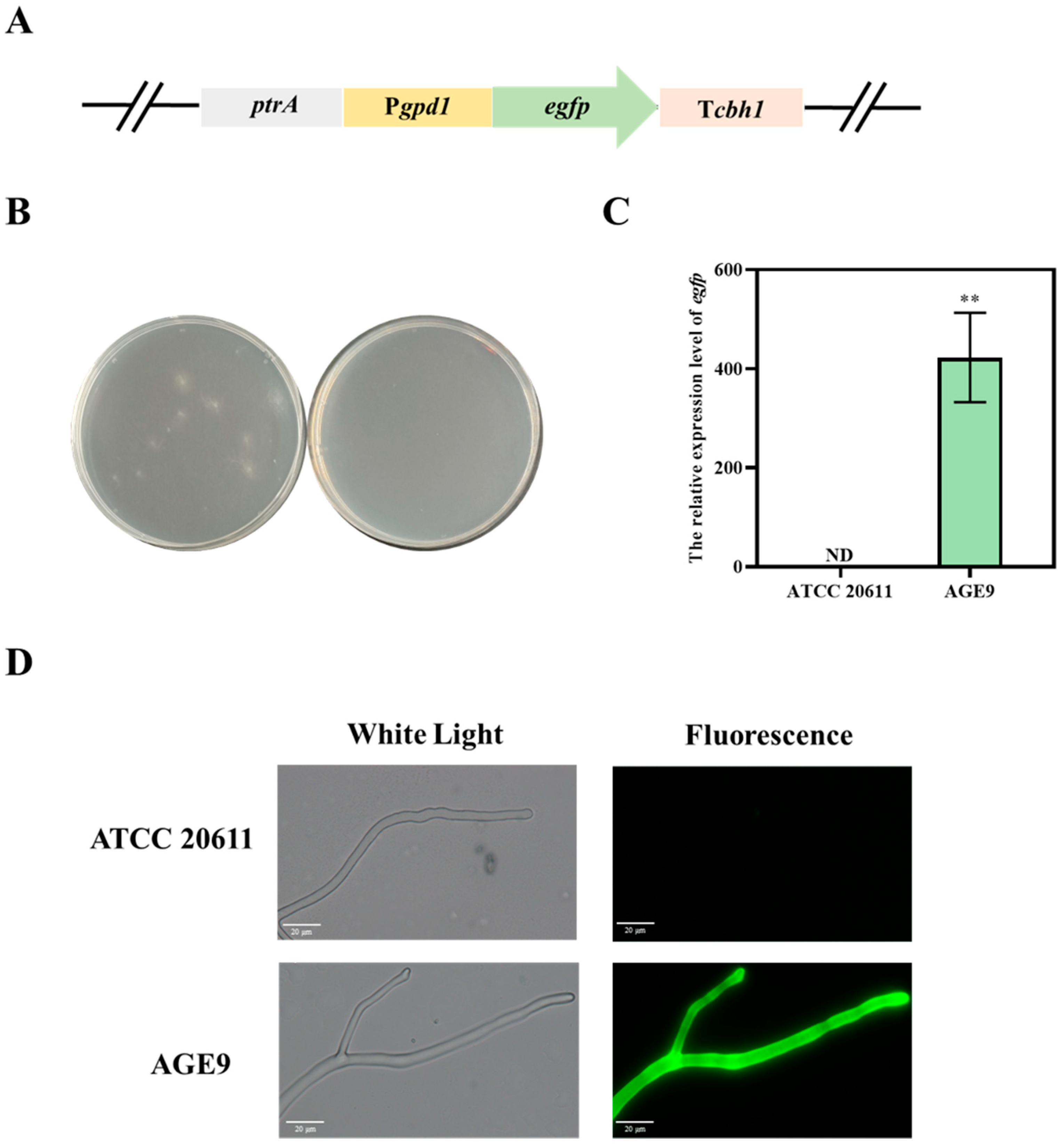
3.1. The T. reesei gpd1 Promoter-Driven EGFP Expression in A. niger ATCC 20611

In filamentous fungi, the promoter of glyceraldehyde-3-phosphate dehydrogenase-encoding gene (Pgpd) has usually been applied as an efficient constitutive promoter for the expression of heterologous proteins [17,18]. Here, we selected the promoter (Pgpd1) from T. reesei to express heterologous proteins in A. niger ATCC 20611. Firstly, the enhanced green fluorescent protein (EGFP) as a reporter protein was expressed under the control of the promoter Pgpd1. The gpd1 promoter-driven EGFP expression cassette was constructed by double-joint PCR, which contained the gpd1 promoter, the egfp gene and the trpC terminator (Figure 1A). The resistance gene ptrA was used as the selectable marker for transformation. Then, the expression cassette and the resistance gene were co-transformed into A. niger ATCC 20611. The transformant strains were selected on an MM plate with 0.3% pyrithiamine (Figure 1B). Subsequently, one of the putative transformants validated by PCR was named AGE9, which was used for the next analysis. RT-qPCR was used to detect the transcript level of egfp in AGE9 (Figure 1C). It was found that the gene of egfp was successfully transcribed in AGE9, while its transcription could not be detected in the parental strain A. niger ATCC 20611. Then, the spores of AGE9 were planted on the MM plates with coverslips and grown at 30 °C for 48 h to observe the fluorescence. It was shown that the transformant AGE9 was able to emit intense green fluorescence, while no fluorescence was detected in the parental strain (Figure 1D). These results demonstrate the adaptability of the T. reesei promoter Pgpd1 for protein expression in A. niger ATCC 20611.

3.2. The gpd1 Promoter-Driven β-Glucosidase Expression in A. niger ATCC 20611

β-glucosidase could hydrolyze cellobiose into glucose, which is a rate-limiting step in the hydrolytic saccharification of cellulose [19]. Meanwhile, β-glucosidase is a large molecular weight secreted protein with glycosylation sites, which could be used as a reporter protein [20]. Here, the T. reesei Pgpd1 promotor was used to drive the expression of β-glucosidase in A. niger ATCC 20611. Firstly, the gpd1 promoter-driven β-glucosidase expression cassette was constructed by double-joint PCR, which contained the gpd1 promoter, the signal peptides of cbh1 (SP), the bglA gene and the trpC terminator (Figure 2A). The ptrA gene was also used as the selectable marker. After genetic transformation, the transformants were selected on the MM plates with 0.3% pyrithiamine and validated by PCR. The putative transformants were inoculated on the CMC-esculin plates to detect the β-glucosidase secretion. The esculin could be degraded by β-glucosidase to glucose and aesculetin, which would interact with Fe3+ to form a black halo. It was found that the transformants AGB1 and AGB33 displayed wider black halos, while the parental strain A. niger ATCC 20611 showed a much smaller halo (Figure 2B,C). Moreover, RT-qPCR was used to detect the transcript level of bglA (Figure 2D). It was found that the gene of bglA was successfully transcribed in AGB1 and AGB33, while it could not be detected in A. niger ATCC 20611. The β-glucosidase activities of transformants were further measured after 4-day fermentation (Figure 2E). It was found that the β-glucosidase activity of AGB1 and AGB33 were 1.02 and 0.51 U/mL, respectively, while it was barely detectable in the parental strain A. niger ATCC 20611. These results suggest that the gpd1 promotor from T. reesei could successfully drive the β-glucosidase expression in A. niger ATCC 20611.

3.3. Detection of the ER Pressure in the gpd1 Promoter-Driven β-glucosidase Expression Strains

The production of heterologous proteins could lead to the accumulation of a large number of unfolded proteins in the endoplasmic reticulum, which exceeds the processing capacity of the endoplasmic reticulum and then trigger the UPR response to alleviate the stress and damage to a certain extent, restore the homeostasis of the endoplasmic reticulum [21]. Therefore, to investigate whether the expression of bglA in A. niger caused the ER pressure, the transcript levels of the genes encoding the protein folding molecular chaperone gene (bip1) and the disulfide bond isomerase (pdi1), which indicated the degree of the UPR, were detected by RT-qPCR (Figure 3A). Meanwhile, the transcript levels of the genes encoding the ubiquitin ligase enzymes (yos9 and der1), which indicated the degree of the ERAD, were also detected (Figure 3B). It was found that there were no remarkable differences in the transcript levels of pdi1, yos9 and der1 between the transformants and A. niger ATCC 20611, only that the transcript level of bip1 slightly increased. The results show that the T. reesei gpd1 promoter driven-β-glucosidase expression could not cause intense ER pressure, indicating the strength of the gpd1 promoter was not enough to accumulate β-glucosidase in the ER.

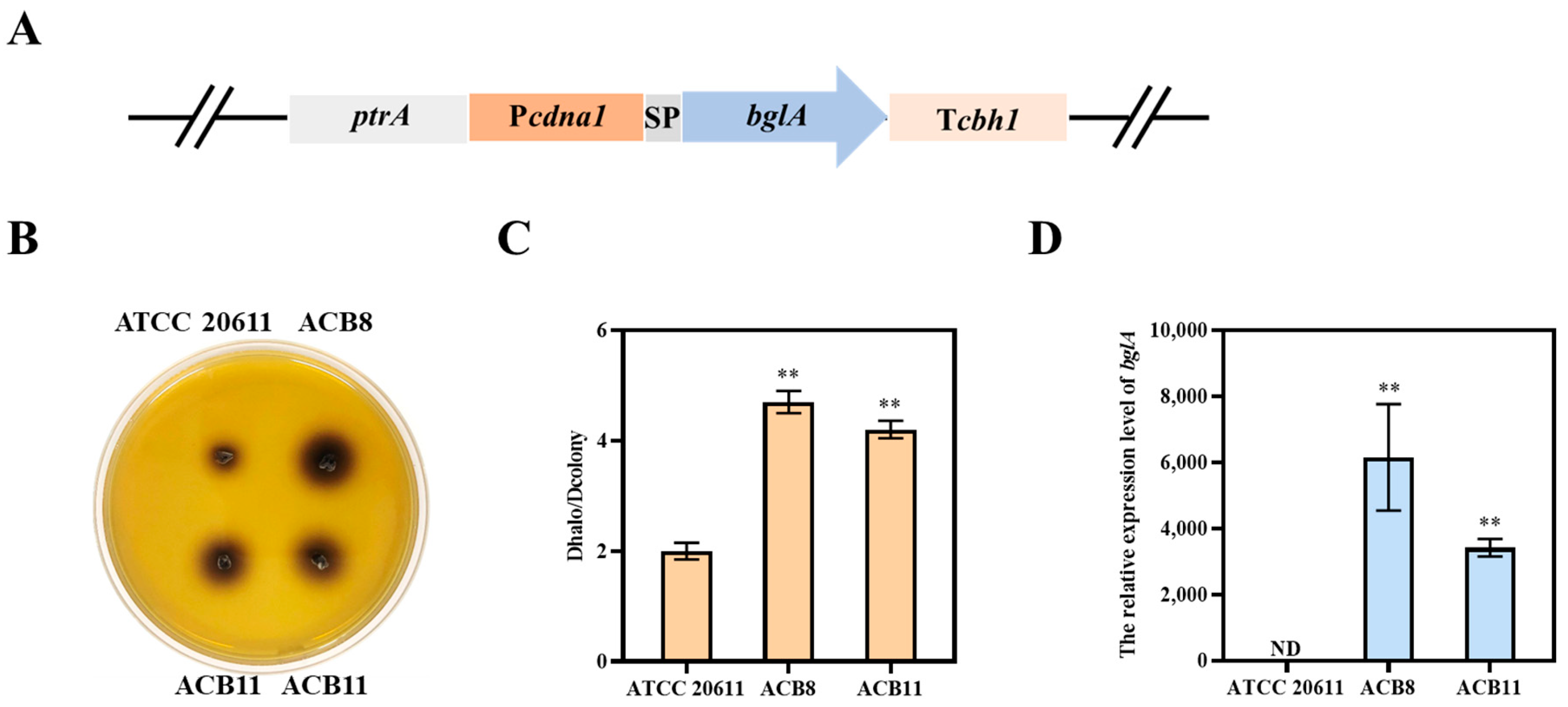
3.4. The cdna1 Promoter-Driven β-Glucosidase Expression in A. niger ATCC 20611

The use of strong promoters is an effective means of increasing the expression of heterologous proteins [22]. It was reported that the promoter Pcdna1, screened from the cDNA library of T. reesei, was stronger than the commonly used promoters Pgpd1 and Ptef1 [22]. And it has been used to drive the expression of uronate dehydrogenase, β-mannanase, SARS-CoV-2 neutralizing nanobody Nb20 in T. reesei [23,24,25]. Here, the promoter Pcdna1 was selected to drive β-glucosidase expression in A. niger ATCC 20611. Firstly, the Pcdna1-driven β-glucosidase expression cassette was constructed by double-joint PCR, which contained the cdna1 promoter, the signal peptides of cbh1 (SP), the bglA gene and the trpC terminator (Figure 4A). The resistance gene ptrA was used as the selectable marker. After transformation, the transformant strains were selected on the MM plates with 0.3% pyrithiamine. Then, the complete expression cassette was validated by PCR and the putative transformants were inoculated on the CMC-esculin plates to detect the β-glucosidase secretion. It was shown that the transformants ACB8 and ACB11 displayed wider black halos than the parental strain A. niger ATCC 20611 (Figure 4B,C). Furthermore, RT-qPCR was used to detect the transcript level of bglA (Figure 4D). It was found that the gene of bglA was successfully transcribed in ACB8 and ACB11, while it could not be detected in A. niger ATCC 20611. Moreover, the β-glucosidase activities of transformants were further measured after 4-day fermentation (Figure 5A). It was found that the β-glucosidase activity of ACB8 and ACB11 were 15.2 U/mL and 5.6 U/mL, respectively, while it was barely detectable in A. niger ATCC 20611. These results suggest that the cdna1 promotor from T. reesei could successfully drive the high-level expression of β-glucosidase in A. niger ATCC 20611. Subsequently, the fermentation supernatant was determined by SDS-PAGE assay (Figure 5B). The transformants ACB8 and ACB11 showed clear and specific bands at around 120 kDa, consistent with the theoretical size of β-glucosidase. And this band was further confirmed to be β-glucosidase by MS identification. Moreover, the total protein concentration in the fermentation supernatant was measured by Bradford’s method (Figure 5C). It was found that the total extracellular protein content of ACB8 and ACB11 were 1.2 and 0.9 mg/mL, respectively, while that of A. niger ATCC 20611 was 0.18 mg/mL. It was estimated that β-glucosidase secreted represented more than 85% of the total extracellular protein. Sun et al. in 2021 reported the production of β-glucosidase using the promoter Pcdna1 in T. reesei, in which the specific enzyme activity of β-glucosidase in the seventh-day fermentation broth of T. reesei QVB-1 was 18.6 U/mg, with the enzyme production rate reached up to 2.66 U/mg/d [20]. In this study, the specific enzyme activity of β-glucosidase in the fourth day of fermentation broth of A. niger ACB8 was 12.7 U/mg (the enzyme activity was 15.2 U/mL and the extracellular protein content was 1.2 mg/mL), with the enzyme production rate reached up to 3.17 U/mg/d. These results demonstrate that the promoter Pcdna1 could drive β-glucosidase expression not only in T. reesei but also in A. niger and that A. niger ACB8 had a faster growth rate and higher β-glucosidase production efficiency compared with T. reesei QVB-1, indicating the superiority of A. niger ATCC 20611 as a protein expression system.
In addition, the transcript levels of the genes bip1, pdi1, yos9 and der1 were detected by RT-qPCR (Figure 6A,B). It was found that the transcript levels of bip1, pdi1, yos9 and der1 in the ACB8 and ACB 11 were all to a certain extent higher than those in A. niger ATCC 20611, indicating that the expression of β-glucosidase under the control of the promotor Pcdna1 caused the certain ER-pressure. These results demonstrate that the T. reesei promoter Pcdna1 could drive the high-level expression of β-glucosidase with a low background of protein secretion in A. niger and the strength of the promoter Pcdna1 was sufficient to saturate the expressed proteins in the ER.

3.5. Enzymatic Property of β-Glucosidase in A. niger ACB8

To test the property of the β-glucosidase heterologously expressed in A. niger ATCC 20611, we detected the effects of temperature and pH on the activity and stability of β-glucosidase in A. niger ACB8. T. reesei QVB-1. T. reesei QVB-1 expressed β-glucosidase under the control of the promoter Pcdna1, as the control [20]. Firstly, β-glucosidase activity was measured at different temperatures (Figure 7A). It was found that the β-glucosidase in A. niger ACB8 showed the maximal activity at 60 °C and was active in a temperature range of 20–70 °C. After preincubation of the fermentation broth of A. niger ACB8 and the T. reesei QVB-1 at 50, 60 and 70 °C for 0.5 h to 10 h, the residual activities were determined at pH 4.8 and 50 °C (Figure 7B,C). It was found that the β-glucosidase in A. niger ACB8 was highly stable at temperatures of 50 °C and 60 °C, which retained 85% relative activity at 50 °C after 10 h and 50% relative activity at 60 °C after 2 h. However, the enzyme was inactive at 70 °C after 0.5 h. Then, β-glucosidase activity was measured in different pH ranges of 3.0–8.0 (Figure 7D). It was found that the β-glucosidase of A. niger ACB8 had high stability in acid conditions and its optimum pH was 4.5. After preincubation of the fermentation broth of the A. niger ACB8 and the T. reesei QVB-1 without substrate in citric acid buffer of different pH in 4 °C for 24 h, the residual activities were determined at pH 4.8 and 50 °C (Figure 7E). It was found that the β-glucosidase in A. niger ACB8 was highly stable in acid conditions, which could remain at more than 44% relative activity after incubation in citric acid buffer of pH 3.0–6.0 for 24 h and maintain 31% in pH 8. The effects of temperature and pH on the activity and stability of β-glucosidase in T. reesei QVB-1 were essentially the same as that of ACB8. The results show that the activity and stability of the cdna1 promoter-driven β-glucosidase in A. niger ACB8 were consistent with that of T. reesei, indicating that the heterologous promoter-driven protein expression in A. niger ATCC 20611 could not result in changes in enzymatic property. Meanwhile, the β-glucosidase under the control of the T. reesei promoter Pcdna1 was a moderately acidic β-glucosidase, suggesting that it had potential for application in cellulose hydrolysis.

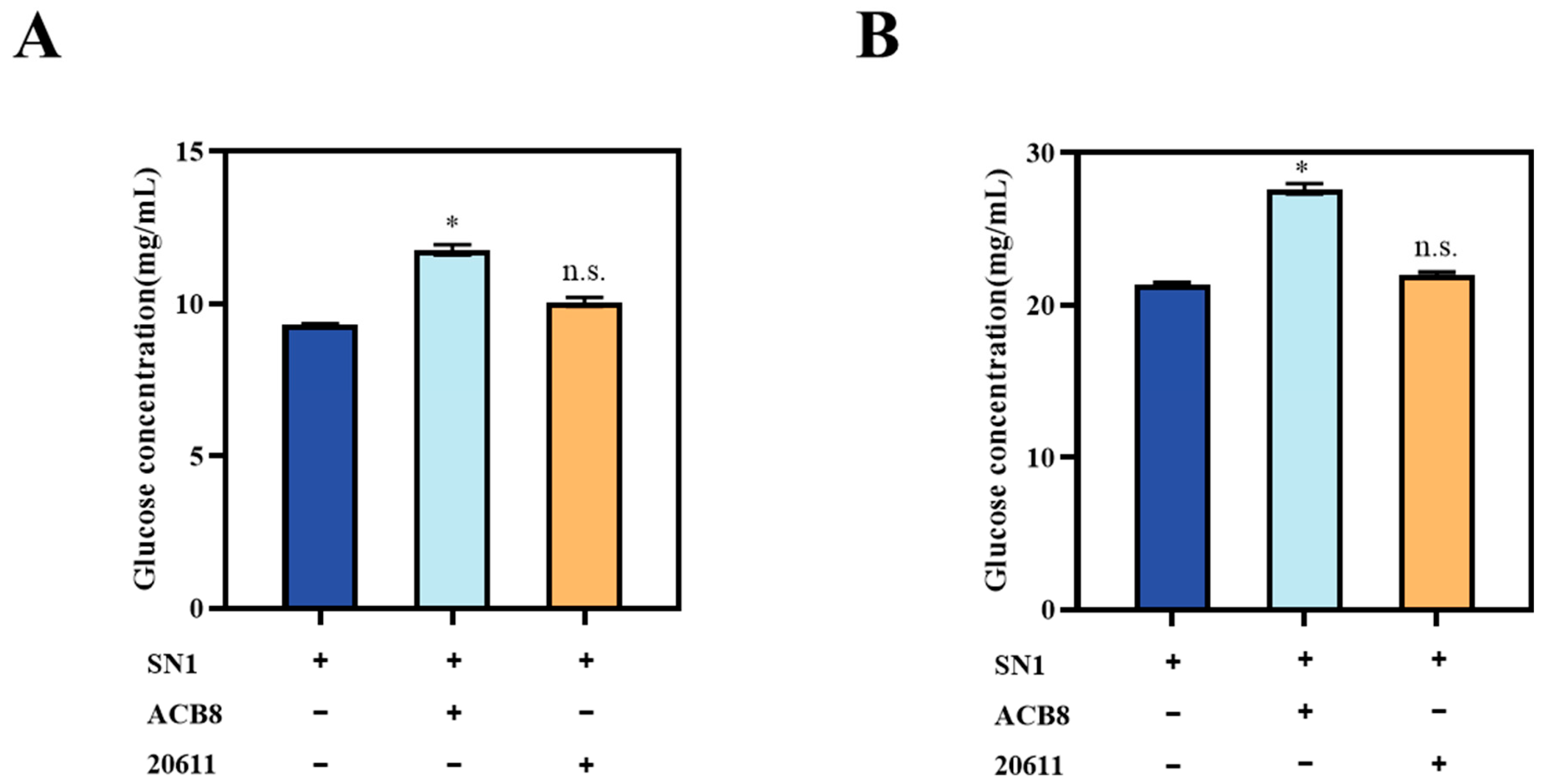
3.6. Saccharification of the Corncob Residues by Supplementing the β-glucosidase to the Cellulase Mixture of T. reesei

The filamentous fungus T. reesei is widely used for the production of cellulases, in which the β-glucosidase represents only 1% of the cellulase mixture, resulting in the lack of BGL becoming the rate-limiting step for effective hydrolysis of the cellulose substrate [11]. To improve the saccharification ability of the cellulase mixture of T. reesei, the fermentation broth of A. niger ACB8, which contains β-glucosidase under the control of the T. reesei promoter Pcdna1, was added to the cellulase mixture of T. reesei SN1 according to the ratio of the FPA to β-glucosidase activity of 1:1 for saccharification of acid pretreated corncob residues (ACR) and delignified corncob residues (DCR). As shown in Figure 8A, in the saccharification of the acid-pretreated corncob residues, the glucose released by the cellulase mixture of T. reesei SN1 supplemented with the fermentation broth of A. niger ACB8 was 11.78 mg/mL (corresponding to 33.86% cellulose conversion), which was 26.21% higher than that of T. reesei SN1 (9.33 mg/mL, corresponding to 26.83% cellulose conversion) after 48 h. While in the saccharification of the delignified pretreated corncob residues (Figure 8B), the glucose released by the cellulase mixture of T. reesei SN1 supplemented with the fermentation broth of A. niger ACB8 was 27.64 mg/mL (corresponding to 75.74% cellulose conversion), which was 29.51% higher than that of T. reesei SN1 (21.35 mg/mL, corresponding to 58.48% cellulose conversion) after 48 h. And the saccharification with the addition of the equal volume of fermentation broth of A. niger ATCC 20611 as the control and the results show that the glucose released had no significant difference from that of T. reesei SN1 (Figure 8A,B). Taken together, the results demonstrate that the β-glucosidase of A. niger ACB8 significantly enhanced the ability of the cellulase system of T. reesei for highly efficient biomass hydrolysis.

4. Discussion

A. niger ATCC 20611, as an industrial strain for fructooligosaccharides production, has its main enzyme protein β-fructofuranosidase localized in the cell wall, resulting in a pure protein background [2]. Therefore, A. niger ATCC 20611 is expected to be a potentially excellent protein expression host. Meanwhile, the use of robust promoters is essential for the construction of protein expression systems [3]. In this study, we established an A. niger ATCC 20611 protein expression secretion system driven by the strong constitutive promoter Pcdna1 from T. reesei for the high-level production of high-purity β-glucosidase, which was used to increase the rate of cellulose degradation.
The process of gene expression begins with transcription. And the transcription level of genes is largely determined by promoters, which determine the mode and level of expression of the recombinant protein. Therefore, it is crucial for the construction of protein expression systems by using robust promoters. The approaches obtaining robust promotors include screening by traditional biochemical and constructing synthetic hybrid promoters [26,27]. Erden et al. attempted to express recombinant proteins in P. pastoris using heterologous yeast promoters and the results demonstrate that the expression pattern of the heterologous promoters in P. pastoris was similar to that of their native host [28]. Based on RNA-seq data, Zhang et al. used 18 promoters selected from GS115 genome to drive the expression of reporter proteins in P. pastoris and found that the methanol-induced promoter pFDH1 was an alternative to pAOX1 [29]. It was reported that the constitutive promoter Pcdna1 was identified from the cDNA libraries in T. reesei [7]. It is generally considered the strongest constitutive promoter in T. reesei [8]. Additionally, it is only found in T. reesei and differs from the commonly used promoters of the gene encoding glyceraldehyde-3-phosphate dehydrogenase Pgpd1 and the gene encoding translation elongation factor Ptef1. The promoters Pgpd1 and Ptef1 are relatively conservative and widely exist in different microorganisms. In our study, the β-glucosidase activity in the transformant ACB8 under the control of Pcdna1 was 15.2 U/mL, which was a 13.9-fold increase from that of the transformant AGB1 under the control of the promoter Pgpd1 (1.02 U/mL). The results demonstrate that using the Pcdna1 promoter of T. reesei could effectively enhance the expression level of heterologous proteins in A. niger, which indicated that the driving pattern of the T. reesei-specific promoter Pcdna1 is probably universal in filamentous fungi.
Lignocellulose biomass is the largest renewable resource on Earth and it can be converted into second-generation biofuel ethanol [30]. This renewable energy source is considered a promising substitute for fossil fuels [31]. The cellulase mixtures secreted by T. reesei can effectively degrade cellulose. The process of cellulase hydrolysis is characterized by a synergistic effect between different enzyme components. However, in the cellulase mixture, the β-glucosidase accounts for only about 1%, which is a rate-limiting step for effective hydrolysis of the cellulose substrate. Moreover, enzyme purification increases the time and cost [32,33]. Consequently, it is of great importance to adjust the content of key enzyme components in the cellulase mixture in order to improve the cellulose degradation rate. It was reported that the T. reesei Δvib1 demonstrated a low background of cellulase and protease secretion [20]. Then, the T. reesei QVB-1 was constructed to express β-glucosidase using the T. reesei Δvib1 as the host strain. The β-glucosidase activity in the seventh-day fermentation broth of T. reesei QVB-1 was 18.6 U/mg, with the enzyme production rate reaching up to 2.66 U/mg/d. In this study, the β-glucosidase secreted represented more than 85% of the total extracellular protein in the transformant A. niger ACB8. And the specific enzyme activity of β-glucosidase in the fourth day of fermentation broth of A. niger ACB8 was 12.7 U/mg, with the enzyme production rate reached up to 3.17 U/mg/d. These results demonstrate that A. niger ATCC 20611 has a fast growth rate and a low extracellular protein background and it had obvious advantages in pure β-glucosidase production to improve cellulose degradation rate when compared with T. reesei.
The β-glucosidase was stable at low pH, which makes it suitable for cellulose hydrolysis [34]. The saccharification reaction conditions for the pretreated corn cob residues were 50 °C and a pH of 4.8 [14]. In our study, the β-glucosidase in A. niger ACB8 was highly stable at temperatures of 50 °C, which retained 85% relative activity at 50 °C after 10 h and had high stability in acid condition, remaining more than 44% relative activity after incubation in citric acid buffer of pH 3.0–6.0 for 24 h (Figure 7D). To optimize cellulose hydrolysis, a fermentation broth of ACB8 was added to the cellulase mixture from T. reesei SN1. The glucose released from the saccharification system was 11.78 mg/mL and 27.64 mg/mL after 48 h when the acid-pretreated corncob residues and delignified pretreated corncob residues were used as the substrates for saccharification, respectively. And the saccharification efficiency increased by 26.21% and 29.51% compared with that of T. reesei SN1 alone after 48 h, respectively (Figure 8A,B). Ma et al. expressed the β-glucosidase gene bgl1 from P. decumbens in T. reesei Rut-C30 and purified β-glucosidase with a HiTrap column from the fermentation broth [13]. The addition of purified pBGL1 to T. reesei cellulase preparation could facilitate cellulose saccharification and the promotion was more remarkable with further adding pBGL1. Taken together, the successful expression of β-glucosidase under the control of the cdna1 promoter Pcdna1 in A. niger ACB8 showed the applicability of heterologous promoter in A. niger ATCC 20611.
In conclusion, the robust promoter Pcdna1 from T. reesei was successfully applied in the construction of a protein expression system in A. niger ATCC 20611. And the production level of β-glucosidase driven by the promoter Pcdna1 in A. niger ATCC 20611 reached 15.2 U/mL, representing a 13.9-fold increase compared with that of the promotor Pgpd1. Furthermore, the β-glucosidase in A. niger ACB8 exhibited high stability under the temperature and pH conditions of cellulose degradation. Moreover, the β-glucosidase accounted for 85% of the total extracellular protein in the fermentation broth of A. niger ACB8. The efficiency of saccharification of different pretreated corncob residues could be increased with the addition of the fermentation supernatant of A. niger ACB8 to the cellulase system of T. reesei. Therefore, the high-level expression of β-glucosidase in A. niger ATCC 20611 using the T. reesei promoter Pcdna1 has enhanced the efficiency of cellulose degradation and facilitated the effective utilization of lignocellulose, which was of great importance for the conversion of renewable biomass resources.

Supplementary Materials

The following supporting information can be downloaded at: https://www.mdpi.com/article/10.3390/fermentation10090461/s1, Table S1: Primers used in this study.

Author Contributions

J.C., J.W. and Y.Z. conceived the work and drafted the manuscript. J.W., J.C., Z.L., L.W. and P.L. performed the experiments and analyzed the data. Y.Z. and H.L. designed the work and revised the manuscript. All authors have read and agreed to the published version of the manuscript.

Funding

This work was supported by grants from the National Key R&D Program of China (No. 2021YFC2101300; No. 2018YFA0900500), the SKLMT Frontiers and Challenges Project (No. SKLMTFCP-2023-02) and the Taishan Industrial Experts Program.

Institutional Review Board Statement

Not applicable.

Informed Consent Statement

Not applicable.

Data Availability Statement

The original contributions presented in the study are included in the article and Supplementary Material, further in-quiries can be directed to the corresponding authors.

Acknowledgments

The authors thank Jingyao Qu and Zhifeng Li of the Core Facilities for Life and Environmental Sciences, State Key Laboratory of Microbial Technology of Shandong University, for their help and guidance in the qRT-PCR and protein analysis.

Conflicts of Interest

The authors declare no conflict of interest.

References

  1. Sangeetha, P.T.; Ramesh, M.N.; Prapulla, S.G. Recent trends in the microbial production, analysis and application of fructooligosaccharides. Trends Food. Sci. Technol. 2005, 16, 442–457. [Google Scholar] [CrossRef]
  2. Zhang, J.; Liu, C.; Xie, Y.; Li, N.; Ning, Z.; Du, N.; Huang, X.; Zhong, Y. Enhancing fructooligosaccharides production by genetic improvement of the industrial fungus Aspergillus niger ATCC 20611. J. Biotechnol. 2017, 249, 25–33. [Google Scholar] [CrossRef] [PubMed]
  3. Kluge, J.; Terfehr, D.; Kück, U. Inducible promoters and functional genomic approaches for the genetic engineering of filamentous fungi. Appl. Microbiol. Biotechnol. 2018, 102, 6357–6372. [Google Scholar] [CrossRef]
  4. Wan, X.; Wang, L.; Chang, J.; Zhang, J.; Zhang, Z.; Li, K.; Sun, G.; Liu, C.; Zhong, Y. Effective synthesis of high-content fructooligosaccharides in engineered Aspergillus niger. Microb. Cell Fact. 2024, 23, 76. [Google Scholar] [CrossRef]
  5. Wang, L.; Xie, Y.; Chang, J.; Wang, J.; Liu, H.; Shi, M.; Zhong, Y. A novel sucrose-inducible expression system and its application for production of biomass-degrading enzymes in Aspergillus niger. Biotechnol. Biofuels Bioprod. 2023, 16, 23. [Google Scholar] [CrossRef]
  6. Adnan, M.; Ma, X.; Olsson, S.; Wang, J.; Liu, G. Promoter regulation and genetic engineering strategies for enhanced cellulase expression in Trichoderma reesei. Microbiol. Res. 2022, 259, 127011. [Google Scholar] [CrossRef]
  7. Nakari-Set, L.T.; Penttil, M. Production of Trichoderma reesei cellulases on glucose-containing media. Appl. Environ. Microb. 1995, 61, 3650–3655. [Google Scholar] [CrossRef]
  8. Uzbas, F.; Sezerman, U.; Hartl, L.; Kubicek, C.P.; Seiboth, B. A homologous production system for Trichoderma reesei secreted proteins in a cellulase-free background. Appl. Microbiol. Biotechnol. 2012, 93, 1601–1608. [Google Scholar] [CrossRef] [PubMed]
  9. Chen, M.; Qin, Y.; Liu, Z.; Liu, K.; Wang, F.; Qu, Y. Isolation and characterization of a β-glucosidase from Penicillium decumbens and improving hydrolysis of corncob residue by using it as cellulase supplementation. Enzym. Microb. Technol. 2010, 46, 444–449. [Google Scholar] [CrossRef]
  10. Bischof, R.H.; Ramoni, J.; Seiboth, B. Cellulases and beyond: The first 70 years of the enzyme producer Trichoderma reesei. Microb. Cell Fact. 2016, 15, 106. [Google Scholar] [CrossRef]
  11. Dashtban, M.; Qin, W. Overexpression of an exotic thermotolerant β-glucosidase in Trichoderma reesei and its signifcant increase in cellulolytic activity and saccharifcation of barley straw. Microb. Cell Fact. 2012, 11, 63. [Google Scholar] [CrossRef]
  12. Chen, M.; Zhao, J.; Xia, L. Enzymatic hydrolysis of maize straw polysaccharides for the production of reducing sugars. Carbohyd. Polym. 2008, 71, 411–415. [Google Scholar] [CrossRef]
  13. Ma, L.; Zhang, J.; Zou, G.; Wang, C.; Zhou, Z. Improvement of cellulase activity in Trichoderma reesei by heterologous expression of a beta-glucosidase gene from Penicillium decumbens. Enzym. Microb. Techol. 2011, 49, 366–371. [Google Scholar] [CrossRef]
  14. Qian, Y.; Zhong, L.; Hou, Y.; Qu, Y.; Zhong, Y. Characterization and Strain Improvement of a Hypercellulytic Variant, Trichoderma reesei SN1, by Genetic Engineering for Optimized Cellulase Production in Biomass Conversion Improvement. Front. Microbiol. 2016, 7, 1349. [Google Scholar] [CrossRef] [PubMed]
  15. Kilaru, S.; Schuster, M.; Cannon, S.; Steinberg, G. Optimised red- and green-fluorescent proteins for live cell imaging in the industrial enzyme-producing fungus Trichoderma reesei. Fungal Genet. Biol. 2020, 138, 103366. [Google Scholar] [CrossRef] [PubMed]
  16. Liu, K.; Lin, X.; Yue, J.; Li, X.; Fang, X.; Zhu, M.; Lin, J.; Qu, Y.; Xiao, L. High concentration ethanol production from corncob residues by fed-batch strategy. Bioresour. Technol. 2010, 101, 4952–4958. [Google Scholar] [CrossRef] [PubMed]
  17. Madhavan, A.; Pandey, A.; Sukumaran, R.K. Expression system for heterologous protein expression in the filamentous fungus Aspergillus unguis. Bioresour. Technol. 2017, 245, 1334–1342. [Google Scholar] [CrossRef]
  18. Li, J.; Wang, J.; Wang, S.; Xing, M.; Yu, S.; Liu, G. Achieving efficient protein expression in Trichoderma reesei by using strong constitutive promoters. Microb. Cell. Fact. 2012, 11, 84. [Google Scholar] [CrossRef]
  19. Sørensen, A.; Lübeck, M.; Lübeck, P.S.; Ahring, B.K. Fungal beta-glucosidases: A bottleneck in industrial use of lignocellulosic materials. Biomolecules 2013, 3, 612–631. [Google Scholar] [CrossRef]
  20. Sun, Y.; Qian, Y.; Zhang, J.; Yao, C.; Wang, Y.; Liu, H.; Zhong, Y. Development of a novel expression platform for heterologous protein production via deleting the p53-like regulator Vib1 in Trichoderma reesei. Enzym. Microb. Technol. 2022, 155, 109993. [Google Scholar] [CrossRef]
  21. Yang, S.; Song, L.; Wang, J.; Zhao, J.; Tang, H.; Bao, X. Engineering Saccharomyces cerevisiae for efficient production of recombinant proteins. Eng. Microb. 2024, 4, 100122. [Google Scholar] [CrossRef]
  22. Zhou, S.; Du, G.; Kang, Z.; Li, J.; Chen, J.; Li, H.; Zhou, J. The application of powerful promoters to enhance gene expression in industrial microorganisms. World J. Microbiol. Biotechnol. 2017, 33, 23. [Google Scholar] [CrossRef] [PubMed]
  23. Paasikallio, T.; Huuskonen, A.; Wiebe, M.G. Scaling up and scaling down the production of galactaric acid from pectin using Trichoderma reesei. Microb. Cell Fact. 2017, 16, 119. [Google Scholar] [CrossRef] [PubMed]
  24. Jiang, S.; Wang, Y.; Liu, Q.; Zhao, Q.; Gao, L.; Song, X.; Li, X.; Qu, Y.; Liu, G. Genetic engineering and raising temperature enhance recombinant protein production with the cdna1 promoter in Trichoderma reesei. Bioresour. Bioprocess 2022, 9, 113. [Google Scholar] [CrossRef]
  25. Zhang, J.; Li, J.; Gao, L.; Waghmare, P.R.; Qu, J.; Liu, G. Expression of a SARS-CoV-2 neutralizing nanobody in Trichoderma reesei. Sheng Wu Gong Cheng Xue Bao 2022, 38, 2250–2258. [Google Scholar] [CrossRef]
  26. Keren, L.; Zackay, O.; Lotan Pompan, M.; Barenholz, U.; Dekel, E.; Sasson, V.; Aidelberg, G.; Bren, A.; Zeevi, D.; Weinberger, A.; et al. Promoters maintain their relative activity levels under different growth conditions. Mol. Syst. Biol. 2013, 9, 701. [Google Scholar] [CrossRef]
  27. Fitz, E.; Wanka, F.; Seiboth, B. The Promoter Toolbox for Recombinant Gene Expression in Trichoderma reesei. Front. Bioeng. Biotech. 2018, 6, 135. [Google Scholar] [CrossRef]
  28. Erden-Karaoğlan, F.; Karaoğlan, M. Applicability of the heterologous yeast promoters for recombinant protein production in Pichia pastoris. Appl. Microbiol. Biot. 2022, 106, 7073–7083. [Google Scholar] [CrossRef]
  29. Zhang, Y.; Wang, S.; Lu, L.; Zhang, C.; Cai, F.; Lin, Y.; Huang, Y. Isolation and evaluation of strong endogenous promoters for the heterologous expression of proteins in Pichia pastoris. World J. Microbiol. Biotechnol. 2022, 38, 226. [Google Scholar] [CrossRef]
  30. Singhania, R.R.; Patel, A.K.; Sukumaran, R.K.; Larroche, C.; Pandey, A. Role and significance of beta-glucosidases in the hydrolysis of cellulose for bioethanol production. Bioresour. Technol. 2013, 127, 500–507. [Google Scholar] [CrossRef]
  31. Gusakov, A.V. Cellulases and hemicellulases in the 21st century race for cellulosic ethanol. Biofuels 2013, 4, 567–569. [Google Scholar] [CrossRef]
  32. Li, J.; Gao, K.; Secundo, F.; Mao, X. Biochemical characterization of two β-N-acetylglucosaminidases from Streptomyces violascens for efficient production of N-acetyl-d-glucosamine. Food Chem. 2021, 364, 130393. [Google Scholar] [CrossRef]
  33. Kamaruddin, N.; Storms, R.; Mahadi, N.M.; Illias, R.M.; Bakar, F.D.A.; Murad, A.M.A. Reduction of extracellular proteases increased activity and stability of heterologous protein in Aspergillus niger. Arab. J. Sci. Eng. 2018, 43, 3327–3338. [Google Scholar] [CrossRef]
  34. Yang, W.; Su, Y.; Wang, R.; Zhang, H.; Jing, H.; Meng, J.; Zhang, G.; Huang, L.; Guo, L.; Wang, J.; et al. Microbial production and applications of beta-glucosidase—A review. Int. J. Biol. Macromol. 2024, 256, 127915. [Google Scholar] [CrossRef] [PubMed]
Figure 1. Construction of the gpd1 promoter-driven EGFP expression strains of A. niger ATCC 20611: (A) Schematic representation of the egfp expression under the control of gpd1 promoter. The resistance gene ptrA was used as the reporter gene. (B) The transformant strains expressing EGFP under the control of gpd1 promoter were selected on an MM plate with 0.3% PT (the left). The plate diagram on the right showed the growth of A. niger ATCC 20611 on a MM plate with 0.3% PT as the control. (C) The transcript levels of egfp in the transformant AGE9 and the parental strain A. niger ATCC 20611 were detected by RT–qPCR at 24 h. The relative expression was the level of transcripts normalized to that of actin gene. (D) Detection of the fluorescence in the mycelium of A. niger AGF9-1 and the parental strain ATCC 20611 at 48 h by light and fluorescent microscopy. Error bars indicate the standard deviation (SD) of three biological replicates. Student’s t-test: ** p < 0.01. ND, not detected.
Fermentation 10 00461 g001
Figure 2. Construction of the gpd1 promoter-driven β-glucosidase (BGLA) expression strains of A. niger ATCC 20611: (A) Schematic representation of the bglA expression cassette, which contained the promotor of gpd1 (Pgpd1), the signal peptides of cbh1 (SP), the β-glucosidase-encoding gene (bglA) and the terminator of cbh1 (Tcbh1). (B) Detection of the β-glucosidase activities of transformants on the CMC-esculin plates. The parental strain ATCC 20611 was used as control. (C) The ratios of halo diameter to colony diameter of the transformants in (B). (D) The transcript levels of bglA in transformants AGB1, AGB33 and the parental strain ATCC 20611 at 24 h by RT–qPCR. The relative expression was the level of transcripts normalized to that of actin gene. (E) Detection of β-glucosidase activity using p-Nitrophenyl-β-d-glucopyranoside (pNPG) as the substrate of the cdna1 promoter-driven β-glucosidase (BGLA) expression strains and the parental strain ATCC 20611 after 4-day fermentation. Error bars indicate the standard deviation (SD) of three biological replicates. Student’s t-test: ** p < 0.01. ND, not detected.
Fermentation 10 00461 g002
Figure 3. Transcript analysis of endoplasmic reticulum-associated genes (bip1, pdi1 yos9 and der1) by RT-qPCR. The transcript levels of UPR—(A) and ERAD—(B) related genes of transformants AGB1, AGB33 and the parental strain ATCC 20611. The relative expression was the level of transcripts normalized to that of the actin gene. Error bars indicate the standard deviation (SD) of three biological replicates. Student’s t-test: ** p < 0.01, * p < 0.05. n.s., no significant differences.
Fermentation 10 00461 g003
Figure 4. Construction of the cdna1 promoter-driven β-glucosidase (BGLA) expression strains of A. niger ATCC 20611: (A) Schematic representation of the bglA expression under the control of gpd1 promoter, which contained the promotor gpd1 (Pcdna1), the signal peptides of cbh1 (SP), the β-glucosidase-encoding gene (bglA) and the terminator of cbh1 (Tcbh1). (B) Detection of β-glucosidase activities of transformant on the CMC-esculin plate. The parental strain ATCC 20611 as control. (C) The ratios of halo diameter to colony diameter. The data assessed the β-glucosidase production capacity. Analysis of the expression level of bglA. (D) The transcript levels of bglA in transformants ACB8, ACB11 and the parental strain ATCC 20611 at 24 h by RT–qPCR. The relative expression was the level of transcripts normalized to that of the actin gene. Error bars indicate the standard deviation (SD) of three biological replicates. Student’s t-test: ** p < 0.01. ND, not detected.
Fermentation 10 00461 g004
Figure 5. Analysis of the expression level of bglA: (A) Detection of β-glucosidase activity using p-Nitrophenyl-β-d-glucopyranoside (pNPG) as the substrate of the cdna1 promoter-driven β-glucosidase (BGLA) expression strains and the parental strain ATCC 20611 after 4-day fermentation. (B) SDS PAGE showing secreted BGLA expression. (C) Detection of extracellular total protein concentration by Bradford’s method. Error bars indicate the standard deviation (SD) of three biological replicates. Student’s t-test: ** p < 0.01, * p < 0.05.
Fermentation 10 00461 g005
Figure 6. Transcript analysis of endoplasmic reticulum-associated genes (bip1, pdi1 yos9 and der1) by RT-qPCR: The transcript levels of UPR—(A) and ERAD—(B) related genes of transformants ACB8, ACB11 and the parental strain ATCC 20611. The relative expression was the level of transcripts normalized to that of actin. Error bars indicate the standard deviation (SD) of three biological replicates. Student’s t-test: ** p < 0.01, * p < 0.05.
Fermentation 10 00461 g006
Figure 7. Enzymatic characterization of BGLA in A. niger ACB 8. The BGLA in T. reesei QVB-1 was the control: (A) Optimal temperature. The activity of BGLA was detected in the temperature range of 30–80 °C at pH 4.8. (B) and (C) Thermostability. After preincubation of the fermentation broths of the ACB8 and QVB-1 at 50, 60 and 70 °C for 0.5 h to 10 h, respectively, the residual activities were determined at pH 4.8 and 50 °C and the initial activity at pH 4.8 and 50 °C was defined as 100%. (D) Optimal pH. The activity of BGLA was detected at 50 °C in the pH range 3 to 8. (E) pH stability. After preincubation of the fermentation broths of the ACB8 and QVB-1 in citric acid buffer of different pH for 24 h in 4 °C, respectively, the residual activities were determined at pH 4.8 and 50 °C and the initial activity at pH 4.8 and 50 °C was defined as 100%.
Fermentation 10 00461 g007
Figure 8. Saccharification of different pretreated corncob: (A) Glucose released from saccharification of acid-pretreated corncob residues by T. reesei SN1 with the addition of the fermentation broth of A. niger ACB8 for 48 h. (B) Glucose released from the saccharification of delignified corncob residues by T. reesei SN1 adding the fermentation broth of ACB8 for 48 h. Saccharification by adding the fermentation broth of the parental strain A. niger ATCC 20611 was used as the control. Error bars indicate the standard deviation (SD) of three biological replicates. Student’s t-test: * p < 0.05. n.s., no significant differences.
Fermentation 10 00461 g008
Disclaimer/Publisher’s Note: The statements, opinions and data contained in all publications are solely those of the individual author(s) and contributor(s) and not of MDPI and/or the editor(s). MDPI and/or the editor(s) disclaim responsibility for any injury to people or property resulting from any ideas, methods, instructions or products referred to in the content.

