Article

Lactic Fermentation of Broccoli (Brassica oleracea var. italica) to Enhance the Antioxidant and Antiproliferative Activities

by
Daniela Iga-Buitrón
1,
Edgar Torres-Maravilla
2,
Luis G. Bermúdez-Humaran
2,*,
Juan A. Ascacio-Valdes
1,
Raúl Rodríguez-Herrera
1,
Cristóbal N. Aguilar
1 and
Adriana C. Flores-Gallegos
1,*
1
Food Research Department, School of Chemistry, Universidad Autónoma de Coahuila, Blvd. Venustiano Carranza S/N, Colonia República, Saltillo 25280, Coahuila, Mexico
2
MICALIS Institut, INRAE, Univ Paris-Saclay, Equipe Interactions des Micro-Organismes Commensaux et Probiotiques avec l’Hôte (ProbiHôte), 78350 Jouy-en-Josas, France
*
Authors to whom correspondence should be addressed.
Fermentation 2023, 9(2), 122; https://doi.org/10.3390/fermentation9020122
Submission received: 31 October 2022 / Revised: 2 January 2023 / Accepted: 16 January 2023 / Published: 27 January 2023
(This article belongs to the Special Issue Plant-Based Fermented Foods and Civilization Diseases)

Abstract

:
Lactic acid bacteria (LAB) have been used for centuries to produce fermented foods. Cruciferous vegetables contain large amounts of health-promoting compounds such as glucosinolates (GLSs) and phenolics. GLSs and phenolics have been linked to antioxidant, anticancer, and immunosuppressive effects. However, it has been reported that some LAB strains are able to metabolize and enhance the activities and amounts of biomolecules through decarboxylation and/or reduction activities, with positive impacts on human diet and colorectal cancer (CRC) prevention. In the present work, the bioprocessing of broccoli by lactic fermentation was evaluated to produce a functional food using both spontaneous and induced fermentation (Levilactobacillus brevis and Lactococcus lactis as starter co-culture). Changes in the proximal composition, GLSs, and phenolic content as well as the antioxidant, antiproliferative, and immunosuppressive effect of the fermented product were evaluated in in vitro cellular models to validate their potential in CRC chemoprevention. The results demonstrated that fermented broccoli extracts increased the antioxidant activity in Caco2 cells and inhibited the proliferation of HT29 and HT116 cell lines in a concentration-dependent manner, with the best results on day 6 at a concentration of 600 µg/mL. Our findings also provide evidence that fermented broccoli could have an anti-inflammatory effect.

1. Introduction

Each year, 8.2 million people die of cancer. Colorectal cancer (CRC) is the third most prevalent form of cancer worldwide, and it usually manifests itself in elderly people [1,2]. Most cases of colorectal cancer have no family history [3]. However, many studies have found that certain extrinsic factors such as diet, obesity, smoking, alcohol intake, and the consumption of processed and red meat represent modifiable risks factors linked to colorectal carcinoma. Approaches to reduce CRC incidence and mortality have included primary prevention strategies such as dietary changes or physical activity increment [4]. Chemoprevention is the use of pharmacological or natural agents to treat the precancerous condition and to inhibit the initiation of tumorigenesis. Non-steroidal anti-inflammatory drugs, vitamins, and antioxidants have been used as chemopreventive agents [5]. Phytochemicals of plants such as glucosinolates (GLSs), isothiocyanates (ITCs), indoles, flavonoids, and phenolic compounds are sources of natural bioactive metabolites that act as anticancer molecules. These phytochemical compounds are present in the cruciferous vegetable group, including broccoli, cauliflower, cabbage, radish, and Brussels sprouts [6,7]. Nevertheless, many of these metabolites must undergo an enzymatic or microbial conversion into their bioactive form. For example, GLSs need to be hydrolyzed by myrosinase in their biologically active products, such as ITC and indoles [8,9]. Although these vegetables can be eaten fresh, they are usually cooked for consumption [10]. However, it has been demonstrated that cooking methods such as boiling and steam boiling lead to a negative effect on the myrosinase biological activity [11], resulting in physicochemical property changes as well as partially or totally inhibiting the formation of health-promoting compounds such as GLSs, polyphenols, or vitamin C [12]. As a result, GLSs must undergo hydrolysis to their breakdown products only by the gut microbiota, which is characterized by a very low efficiency [13]. On the other hand, it has been reported that fermentation has shown a potentially beneficial production of breakdown products [14] since it favors the phenolics and GLS hydrolysis to several beneficial breakdown products [15]. Lactic acid bacteria (LAB) have an important role in food fermentation since they contribute to sensory characteristics, preservative effects, and probiotic properties [16]; they constitute a small portion of the autochthonous microbiota of raw vegetables (2.0 -2.4 log CFU/g) [17]. When positive conditions of anaerobiosis, water activity, salt concentration, and temperature occur, raw vegetables may be subjected to spontaneous lactic acid fermentation. However, the selection of a starter culture offers an advantage by contributing to maintaining or increasing the antioxidant activities, to the complete degradation of GLSs, and to increased contents of health-promoting compounds [13,18]. It has been reported that some strains, such as Levilactobacillus brevis, Limosilactobacillus fermentum, and Lactiplantibacillus plantarum [19], are able to metabolize and improve the activities and amounts of biomolecules, such as phenolic compounds and GLSs, through decarboxylation and/or reduction activities, with positive impacts on human diets and CRC prevention [7,16]. Broccoli is one of the main dietary sources of phenolic compounds that have been shown to have several effects in health, such as scavenging free radicals and the inhibition of human low-density lipoprotein oxidation [20]. In addition, it is a good source of GLSs, fibers, vitamins, and minerals [19]. It is normally consumed after going through a thermal treatment process, which can cause the degradation or leaching of GLSs and phenolics [12], and unlike other cruciferous vegetables, such as cabbage, its fermentation has not been widely studied. Therefore, its bioprocessing through lactic fermentation represents an interesting strategy to produce a functional food. In the present work, both spontaneous and induced fermentation with an inoculum composed of lactic acid strains previously isolated from agave sap in the working group and their antioxidant and antiproliferative effects on in vitro cellular models were evaluated as a strategy for CRC prevention.

2. Materials and Methods

2.1. Raw Material

Broccoli heads were obtained from a local market in Saltillo, Coahuila, Mexico, in the winter of 2018. Broccoli was cut, washed with tap water, and sanitized in an aqueous solution of a commercial disinfectant for 10 min (BacDyn plus, 0.5 mL/L). Then, it was dried on paper towels and ground in a food processor (Hamilton Beach) to obtain homogeneous small pieces about 2–4 mm thick. Both stems and florets were used to perform the fermentation process.

2.2. Starter Culture Preparation

Levilactobacillus brevis (3M1) and Lactococcus lactis (3M8) strains, belonging to the Food Research Department of Autonomous University of Coahuila and previously isolated from agave sap, were growth at 37 °C in Man-Rogosa and Sharpe (MRS) broth (DIFCO) overnight. Lactic acid bacteria (LAB) cells were harvested by collecting cell pellets after centrifugation at 6000 rpm for 6 min at 4 °C and then washed twice with a sterile 0.9% saline solution. The starter culture used in the fermentation process was a mix that contained equal proportions of both strains (1 × 10⁸ CFU/mL).

2.3. Fermentation Process

The fermentation of broccoli was performed using 70 g of raw material in 45 mL of a 6% (w/v) brine solution using commercial non-refined salt [21]. Broccoli and brine were transferred to sterile jars (150 mL) for the lactic acid fermentation to occur. Fermentation was accomplished spontaneously (s-FB) by the indigenous microbiota present in raw broccoli or induced using the co-culture of Levilactobacillus brevis (3M1) and Lactococcus lactis (3M8) strains (induced fermentation; i-FB). Each fermentation was carried out in three parallel jars and incubated at room temperature (23 °C) for 10 days. During the fermentation, the vegetable material with the brine was sampled aseptically every 48 h. Samples were stored at −80 °C, freeze-dried, milled to a fine powder, and stored in the dark until the preparation of the extracts.

2.4. Enumeration of Microorganisms

During fermentation, brine samples were collected every two days. Ten-fold dilutions were made in a 0.9% saline solution, plated onto MRS agar, and incubated at 37 °C for 48 h.

2.5. Chemical Composition

2.5.1. Determination of pH and Total Titratable Acidity

The pH values of the samples were recorded using a pH meter (Metler-Toledo, Shanghai, China). The pH was measured directly in the samples immediately after opening the jars. For the total titratable acidity (TA), NaOH (0.1 N) was used with 10 mL of brine sample according to the standard procedures of AOAC (1984). As an indicator for titration, 2 drops of 1% phenolphthalein (C20H14O4) were used. TA was expressed as a gram equivalent of lactic acid per liter, using 0.009 as the acid factor for lactic acid.

2.5.2. Proximal Analysis

The proximal composition was determined according to the methods of the AOAC. The moisture content was determined by the gravimetric method (AOAC 925.45). The ash content was determined (AOAC 920.181). The protein content was determined using the micro-Kjeldahl method (AOAC 978.02), using 6.25 as the conversion factor.

2.6. Extraction Procedures

Briefly, a modified extraction procedure from Salas-Millán [21] was used. First, 400 mg of the freeze-dried broccoli powder was extracted using 2 mL of a 70% (v/v) aqueous methanol solution. The solutions were sonicated for 5 min and centrifuged at 4000 rpm for 6 min at 4 °C. The supernatant was taken, and a 1:50 dilution was made with a 70% aqueous methanol solution and stored at −20 °C. All extracts were prepared in duplicate and were used for total phenolic content, GLS analysis, antioxidant capacity, and IL-8 immunomodulation assays. For the cellular antioxidant activity (CAA) assay [22] and proliferation by Sulforhodamine (SRB) assay [23] on cancer cell lines, 5 mg were taken with 200 μL of dimethyl sulfoxide (DMSO, Sigma). On the day of the experiment, samples were diluted from 0 to 600 μg/mL in serum-free culture media; the final sample solutions contain <2% DMSO.

2.7. Glucosinolate Analysis (HPLC/ESI)

Analyses using reverse-phase high-performance liquid chromatography were performed on a Varian HPLC system including an autosampler (Varian ProStar 410, Poway, CA, USA), a ternary pump (Varian ProStar 230I, Poway, CA, USA), and a Photo Diode Array (PDA) detector (Varian ProStar 330, Poway, CA, USA). Samples (5 µL) were injected onto a Denali C18 column (150 mm × 2.1 mm, 3µm, Grace, Poway, CA, USA). The oven temperature was maintained at 30 °C. The eluents used were acetonitrile (solvent A) and formic acid (0.2% v/v; solvent B). The following gradient was applied: initial, 5% B; 0–10 min, 35% B; 10–15 min, 40% B; 15–16 min, 50% B; 16–25 min, 100% B; and 25–45 min, 5% B. The flow rate was maintained at 0.2 mL/min, and elution was monitored at 227, 254, 280, and 330 nm. The quantification was carried out with the construction of sinigrin and I3C standard curves.

2.8. Identification and Quantification of Phenolic Compounds

2.8.1. Total Hydrolyzable Polyphenols

The hydrolyzed phenolic content in the extracts was determined using the commercial reagent Folin–Ciocalteu method described by Wong et al. [24], using gallic acid as the standard. Briefly, a 1:200 dilution from the extract was prepared with 70% methanol. The reaction occurred with 20 µL of each sample, 20 µL of Folin–Ciocalteu reagent, and 20 µL of a sodium carbonate solution. Finally, the solution was diluted with 125 µL of distilled water, and the absorbance was measured in an EPOCH plate reader at 760 nm. The results were expressed as mg of gallic acid equivalent per g of dry weight.

2.8.2. Total Condensed Polyphenols

The total condensed polyphenols were assessed using the HCL-butanol method according to Shay et al. [25]; the standard curve was performed with catechin. Briefly, 500 μL of sample extracts were transferred into screw cap test tubes. Then, 3 mL of a 1:10 HCl-butanol solution was added to the mixture and 100 μL of a 1:9 ferric solution. The tubes were mixed and placed in a water bath at 100 °C for 1 h and then cooled at room temperature. The absorbance was measured at 460 nm (EPOCH plate reader), and the results were expressed as mg of catechin equivalent per g of dry weight.

2.8.3. HPLC-ESI-MS for Qualitative Analysis of Polyphenols

For the qualitative analysis of polyphenols, reverse-phase high-performance liquid chromatography was performed on a Varian HPLC system including an autosampler (Varian ProStar 410, Poway, CA, USA), a ternary pump (Varian ProStar 230I, Poway, CA, USA), and a PDA detector (Varian ProStar 330, Poway, CA, USA). A liquid chromatography ion trap mass spectrometer (Varian 500-MS IT Mass Spectrometer, Poway, CA, USA) equipped with an electrospray ion source was also used. Samples (5 µL) were injected onto a Denali C18 column (150 mm × 2.1 mm, 3 µm, Grace, Poway, CA, USA). The oven temperature was maintained at 30 °C. The eluents were formic acid (0.2%, v/v; solvent A) and acetonitrile (solvent B). The following gradient was applied: initial, 3% B; 0–5 min, 9% B linear; 5–15 min, 16% B linear; and 15–45 min, 50% B linear. The column was then washed and reconditioned. The flow rate was maintained at 0.2 mL/min, and elution was monitored at 245, 280, 320, and 550 nm. The whole effluent (0.2 mL/min) was injected into the source of the mass spectrometer without splitting. All MS experiments were carried out in the negative mode ([M-H]−1). Nitrogen was used as a nebulizing gas, and helium was used as a damping gas. The ion source parameters were a spray voltage of 5.0 kV and a capillary voltage and temperature of 90.0 V and 350 °C, respectively. Data were collected and processed using MS Workstation software (V 6.9). Samples were first analyzed in full scan mode and were acquired in the m/z range of 50–2000 [26].

2.9. Antioxidant Activity of Broccoli Extracts

This study used three chemical methods: the sequestration capacity of radical 2,2-dyphenyl-1-picrylhydrazyl (DPPH), ferric reduction antioxidant power (FRAP), and 2,2-azino-bis-3-ethylbenzothiazoline-6 sulfonic acid (ABTS) assays, and the results were expressed in µM of Trolox equivalent per g of sample.

2.9.1. DPPH Radical Scavenging Activity

The DPPH· assay was carried out according to the methodology reported by Molyneux [27]. The test samples were measured in terms of hydrogen-donating or radical-scavenging ability. The reaction mixture contained 7 µL of the extracts and 193 µL of a 60 µM DPPH radical solution. The microplate was placed in the dark for 30 min, and the absorbance was measured at 517 nm.

2.9.2. FRAP Assay

The determination of the antioxidant activity through the reduction of ferric (FRAP) was based on the methodology reported by Benzie and Strain [28]. A 10 mM 2,4,6-tripyridyl-S-triazine (TPTZ) in a 40 mM HCl solution was mixed with a 20 mM FeCl3 solution and a 0.3 M sodium acetate buffer in 1:1:10 (v/v/v) proportions. Then, 10 µL of each sample was placed in a microplate, 290 µL of the mixture solution was added to each sample, the reaction took place in the dark for 15 min, and the absorbance was measured at 593 nm.

2.9.3. ABTS Assay

The ABTS assay was carried out according to the methodology reported by Re et al. [29]. For this assay, 1 mL of a 2.45 mM potassium persulfate solution was mixed with 1 mL of a 7 mM 2,2-azino bis-(3-etilbenzotiazolin-6-sulfonate) solution. The mixture was placed in the dark for 12 h. The mixture was diluted with ethanol until it had an absorbance of 0.7 at 734 nm. Briefly, 5 µL of each sample was placed on a microplate, 95 µL of the mixture solution were added to each sample, the reaction was took place in the dark for 1 min, and the absorbance was measured at 734 nm.

2.10. Cellular Antioxidant Activity Assay

Human colon cancer cell line Caco-2 cells were seeded at a density of 6 × 10⁴ cells per well on a 96-well microplate in 100 μL of growth medium/well. The outside wells of the plate were not used, as there was much more variation from them than from the inner wells. Twenty-four hours after seeding, the growth medium was removed, and the cells were washed with phosphate-buffered saline (PBS). Triplicate treatments were treated for 1 h with 100 μL of fermented broccoli extracts plus 25 μM 2′,7′-dichlorofluorescein diacetate (DCFH-DA) dissolved in treatment medium. Next, cells were washed with 100 μL of PBS. Then, 600 μM 2,2′-azobis (2-amidinopropane) dihydrochloride (ABAP, Sigma) was applied to the cells in 100 μL of Hanks’ Balanced Salt Solution (HBSS, Sigma), and the 96-well microplate was placed into a Tecan plate-reader at 37 °C. Emission at 538 nm was measured with excitation at 485 nm every 5 min for 1 h. Each plate included triplicate controls and blanks. A commercial capsule of indole-3-carbinol with cruciferous vegetables (100 mg, Solaray®) was used as a control reference. Control wells contained cells treated with a DCFH-DA antioxidant; blank wells contained cells treated with dye and HBSS without an oxidant [22].

2.11. Proliferation (SRB) Assay on Cancer Cell Lines

Cell Culture. All the cell lines were purchased from ATCC. The cancer cell lines HT29, HTC116, and Caco2 were grown in high-glucose Dulbecco’s Modified Eagle’s Medium (DMEM) supplemented with 10% (vol/vol) fetal bovine serum (FBS), 2 mM L-glutamine, 50 U/ml penicillin, and 50 mg/mL streptomycin in a humidified atmosphere containing 5% CO2. The mouse (C57BL/6) lung tumor cell line TC-1 (generated by transduction with a retroviral vector harboring HPV-16 E6/E7 genes and a retrovirus expressing activated human oncogene c-Ha-ras [30]) was grown in RPMI 1640 medium (Lonza, Switzerland) supplemented with 10% heat-inactivated FBS, 50 U/mL penicillin, 50 U/mL streptomycin (Lonza, Levallois-Perret, France), 0.4 mg/mL G418 sulfate (Sigma, France), and 0.2 mg/mL hygromycin (Sigma, France) in a 5% CO2 atmosphere.
SRB assay. At 24 h before stimulation, the cells were seeded on 96-well microplates at 2 × 10⁴ cells per well. Concentrations of 0, 75, 150, 300, and 600 µg/mL were tested for freeze-dried fermented broccoli samples; 5-flouroracil (5-FU, Roche, Italy) was used as positive control at a 1 mM final concentration, and a commercial capsule of indole-3-carbinol with cruciferous vegetables (100 mg, Solaray ®) and indole-3-carbinol (Sigma) were used as control references. The cells were fixed in 5% trichloroacetic acid (TCA) for 1 h at 4 °C and washed four times in distilled water. The microplates were then dehydrated at room temperature, stained in 100 µL of 0.057% (wt/vol) SRB powder/distilled water per well, washed 4 times in 0.1% acetic acid, and re-dehydrated at room temperature. The stained cells were lysed in 10 mM Tris-buffer, the optical density (OD) was measured at 510 nm, and the antiproliferation percentage was calculated [23].

2.12. IL-8 Immunomodulation

HT-29 cells were cultured in 24-well culture plates in DMEM (Lonza, Switzerland) supplemented with 10% heat-inactivated fetal bovine serum (FBS) and 1% glutamine at 37 °C in a 10% CO2/air atmosphere. Media were changed every day. Experiments were initiated on day 7 after seeding, when cells were confluent (~1.83 × 10⁶ cells/well). Twenty-four hours before co-culture (day 6), the culture medium was changed to a medium with 5% heat-inactivated FBS, 1% glutamine without antibiotics, and 50 μL of broccoli extracts (10% final concentration (600 μg/mL)) in a total volume of 500 μL; PBS-7% methanol (3562 pg/mL) was used as control. Cells were simultaneously stimulated with recombinant human TNF-α (5 ng/mL, Peprotech, USA) for 6 h at 37 °C/10% CO2. After co-incubation, cell supernatants were collected and frozen at −80 °C until further analysis of IL-8 concentrations using ELISA (Biolegend, USA) [31].

2.13. Statistical Analysis

All fermentations were carried out in triplicate. Student’s t-test was performed on the data obtained every 48 h. Statistical analyses were performed using GraphPad Software (San Diego, CA, USA), and an ANOVA was performed; p < 0.05 was considered to be statistically significant.

3. Results and Discussion

3.1. Growth of Bacteria during Fermentation

Spontaneous broccoli fermentation (s-FB) was produced utilizing the endogenous microbiota of the broccoli. On the other hand, a starter culture was used to perform the induced fermentation of broccoli (i-FB) using Levilactobacillus brevis (3M1) and Lactococcus lactis (3M8), which were previously isolated from agave sap and have previously shown resistance to simulated gastrointestinal tract conditions and antiproliferative activity vs. colon cancer cell lines (HT29 and HTC 116). As shown in Figure 1, the native population of microorganisms, mainly lactic acid bacteria, was <1 log CFU/mL in s-FB, while in i-FB it was ~7.7 log CFU/mL, a significant statistical difference. Di Cagno et al. [17] have reported LAB counts between 2.0 and 4.0 log CFU/g as part of the normal microbiota of raw vegetables. However, from day 2 to day 8, no significant statistical differences were observed between the two fermentations, which reached the maximum values on day 2 of fermentation for both treatments (8.91 and 8.84 for s-FB and i-FB, respectively). After this maximum growth, a significant decrease was observed from day 4 (~8 log CFU/mL); between both fermentations, there was a significant statistical difference on day 10, where greater growth was observed in s-FB (7.77 log CFU/mL) compared to i-FB (7.31 log CFU/mL). In a study by Shokri et al. [32] with broccoli puree fermented with a mixed culture (Leuconostoc mesenteroides (BF1 and BF2; 1:1) and Lactiplantibacillus plantarum (B1)), maximum counts of 7.99 log CFU/g were also achieved. Salas-Millán et al. [21] also reported a significant increase in the LAB count after three days of spontaneous fermentation of broccoli stalks without dressing or dressed with garlic or mustard, reaching 8.04–8.50 log CFU/g. Regarding the spontaneous fermentation of broccoli, Chen et al. [33] reported the presence of Weisella, Lactococcus, and Lactobacillus as the most common genera in both fresh broccoli and Yan-tsai-chin (fermented broccoli stems), with the potential to produce a bacteriocin-like inhibitory substance (BLIS). In addition to contributing to the flavor, aroma, and texture of fermented products, the LAB present in fermented products also lower the pH, effectively promoting their quality and safety [34].

3.2. Changes in pH and Titratable Acidity during Fermentation

pH is a critical indicator during fermentation. decreasing pH values mainly occur due to the lactic acid formed by the degradation product of the LAB [13]. Fermentation was monitored by pH measures and the quantification of TA (g/L). As shown in Figure 2a, the initial pH values were ~5.8 for both treatments (s-FB and i-FB). After 2 days, pH decreased rapidly to 4.53 and 3.99, respectively, indicating the onset of fermentation. At day 4 of fermentation, the pH values of i-FB were significantly lower than s-FB, and this was maintained until the end of fermentation (day 10). During the fermentation process, the pH values were significantly different between the treatments (s-FB and i-FB) as well as different between the different days within the same treatment; however, from day 2 onwards, the target pH of less than 4.6 was achieved in order to promote the safety of the fermented food. Shokri et al. [32] reported a decrease in the pH of fermented broccoli floret purees from 6.8 (initial) to 3.8 (fermentation end point). In addition, they studied the effect of a pretreatment of the florets with heat or thermosonication (TS), finding that this value was reached after 24.1 h for the untreated control samples and at shorter times of 8.25 and 9.9 h for the thermal and TS pretreatments, respectively. Similar results were found by Salas-Millán et al. [21], who reported an initial pH of 7.85 and TA < 0.01 g/100 mL, which decreased on the third day of fermentation (4.73–4.96), followed by a second reduction on the sixth day (4.20–4.30), with no significant changes until the end of the experiment. Rapid acidification during fermentation is expected due to the increase in the production of organic acids, such as lactic and acetic acid; this is an important parameter since it minimizes the influence of bacteria of decomposition and contributes to good hygienic conditions of fermented foods [35]. The behavior of the lactic acid production in broccoli that was fermented spontaneously (s-FB) or induced (i-FB) (Figure 2b) was correlated with pH, as we expected. TA increased significantly from day 0 to day 2 in both treatments, reaching the highest production at day 8 of fermentation for both treatments. However, the total acidity was significantly higher in i-FB (13.26 ± 0.5 g/L) than in s-FB (11.85 ± 0.5 g/L) due to the metabolic activities of the lactic acid bacteria used. These results are similar to those reported by Shokri et al. [32] in broccoli purees, where values of 10.9, 11.7, and 13.1 g/L were achieved for non-pretreated, thermal pretreated, and TS pretreated broccoli, respectively.

3.3. Chemical Composition

The chemical compositions of both fermentation treatments are shown in Table 1. During spontaneous (s-FB) and induced (i-FB) fermentation, the moisture content remained the same between the different days. Reis dos Ramos et al. [35] reported the same values for moisture content in fresh broccoli: about 90.7%. Moreover, the moisture content coincided with the values reported by the National Database for Standard Reference [36], where a water content of 89.3 g/100 g was reported for broccoli. The ash content was significantly (p < 0.05) different for s-FB and i-FB, except on day 4. Regarding the s-FB ash contents, samples were significantly (p < 0.05) different, except on day 6. Otherwise, on day 2 the highest ash content was found for the i-FB samples (2.57 g/per 100 g of sample). No statistically significant results were found for the protein content during fermentation for the treatments. Compared to Nuñez-Gastélum, Moreno, and Lopez-Cervantes [37], the protein content in fermented broccoli was higher than in fresh broccoli. Moreover, protein content can vary between broccoli species and crop seasons. Despite no changes being found in protein quantities, fermentation can contribute to modifications in the bioavailability of peptides and the amino acid contents in foods [18].

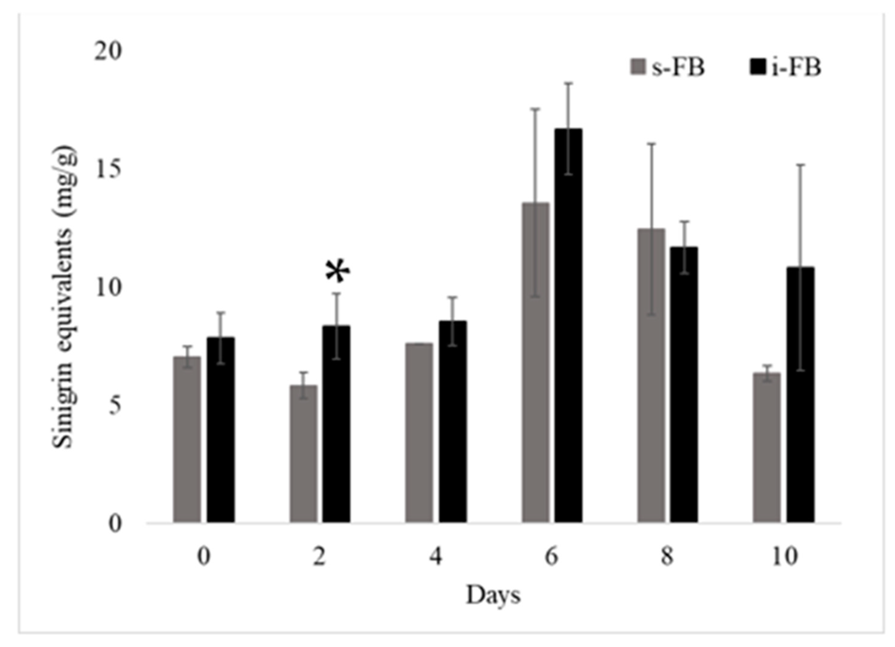
3.4. Glucosinolate Content Changes

Broccoli is a rich source of phytochemicals, including glucosinolates, which constitute the rich secondary metabolites in sulfur derivatives of sugars or amino acids, and their hydrolysis products (isothiocyanates) are responsible for the beneficial health effects through antioxidant and antiproliferative activity [38]. The changes in the number of bioactive compounds of fermented extracts during treatments were compared. Figure 3 and Figure 4 present the contents of the GLS sinigrin and the GLS breakdown product indole-3-carbinol in the two different fermentations. Similar values of sinigrin were obtained for s-FB and i-FB at the different fermentation times. A significant difference was found between fermentations only on day two of fermentation, in which i-FB had a higher value than s-FB. We found the highest content of sinigrin on day 6 of fermentation for both treatments. However, it was not statistically different from days 0, 2, 4, 8, or 10 of fermentation. In the case of the glucosinolate breakdown product indole-3-carbinol, the highest content was found in the s-FB on day 10. However, there was no significant difference among the other days from the same treatment. Moreover, no significant difference was found among the different fermentation days of i-FB. Baenas et al. [9] reported that after 7 and 14 days of storage at 5 or 10 °C, individual and total glucosinolates decreased in broccoli sprouts. Several clinical trials related broccoli intake to its bioactivity in relation to cancer, oxidative stress, and inflammation [39]; López-Chillón et al. [40] demonstrated a decrease in chronic inflammation in overweight subjects with a daily dietary ration of 120 mg of glucosinolates.

3.5. Effect of Fermentation and Starter Culture on Total Phenolic Content and Antioxidant Activity of Broccoli

Food matrices are composed of a complex of compounds that have functional groups with different polarities and chemical behaviors. The tests to determine the antioxidant capacity have peculiarities in their ways of assessing such activity. For example, ABTS considers lipophilic and hydrophilic compounds, DPPH is more sensitive to hydrophilic compounds, and FRAP is a method that is more stable and easily reproducible [41]. To evaluate the influence of fermentation on the antioxidant activity and the amount of phenolic content, in vitro methods were used, as described in Table 2. For antioxidant activities, three different in vitro antioxidant activity methods were compared. For the DPPH method, the antioxidant effect of the broccoli extract remained until the last day of fermentation (day 10), with a higher antioxidant activity on day 4 of fermentation with the inoculated treatment (23.2 ±3.33 µM TE/g of sample; s-FB). Nonetheless, there was no significant difference for this method between the different treatments. The naturally fermented broccoli sample from day 6 had the highest antioxidant activity (53.14 ±2.85 µM TE/g of sample; FB) when evaluated using the ABTS method. However, there was no significant difference in the s-FB from day 6 (46.71 ±3.57 µM TE/g of sample; s-FB). The antioxidant activity measured using the FRAP method was the lowest compared to those measured using the DPPH and ABTS methods.
In addition to the glucosinolates, broccoli is also rich in phenolic compounds, which are highly reactive molecules, and culinary processes such as boiling or steaming can affect their contents [12]. The fermentation process can lead to the production of enzymes that are used as a tool for the release of phenolic compounds and the production of new compounds [42]. Table 3 presents the contents of condensed phenols and hydrolyzed phenols. The obtained results showed that the s-FB (day 0) had a higher content of condensed phenols, but there were no differences with days 2, 4, 6, and 10 of the FB treatments. The contents of hydrolyzed phenols remained until day 10 of fermentation. The contents of total polyphenols in the present work were similar to that reported by Salas-Millán et al. [21] in broccoli stems before fermentation; however, in that work, this content increased up to 2.5 times after the fermentation process in treatments that were seasoned with garlic or mustard, demonstrating that the intrinsic polyphenols of these seeds were transferred to the fermented food. Therefore, the use of spices in the fermented product developed in the present work could improve the phytochemical content and profile. Notwithstanding the differences in TPC, the antioxidant activity evaluated by the ABTS method in the present work was similar to that found by Salas-Millán et al. Radošević et al. [12] indicated that processes such as blanching and boiling lead to reductions in the total phenolic content and antioxidant activity of fresh broccoli. Storage conditions such as temperature and time directly affect the constituents of this plant product [9]. The results of this study showed that with the fermentation process the antioxidant activity of the broccoli extracts was maintained, as were the phenolic compounds of the plant extracts.

3.6. Identification of Phenolic Compounds

Broccoli is one of the main dietary sources of flavonoids [16]. Approximately 60 different compounds were detected during the fermentation period in each treatment. The greatest diversity of polyphenols was detected on day 6 of fermentation for both treatments (Table 3). Sinapic, ferulic, caffeic, and p-coumaric acids were linked to flavonoid-glycoside molecules and have been reported to have several human health effects such as scavenging free radicals, antioxidant activities, antidiabetic, anti-hypertensive, antibacterial, and neuroprotective properties, among others [43]. Further, quercetin is known to possess antiproliferative activity in colon cancer cell lines [43]. Similar compounds were identified in raw stem samples by Salas-Millán et al. [21], who reported eleven phenolic compounds, mainly hydroxycinnamic acids and flavonoids, which they classified into caffeic acid derivatives (chlorogenic acid and rosmarinic acid), sinapic acid derivatives (sinapic acid, sinapic acid hexose, 1-sinapoyl-2-feruloyl-gentiobiose, and 1,2-disinapoyl-gentiobiose), ferulic acid derivatives (4-O-feruloyl quinic acid), and flavonoids (kaempferol-3-O-diglucoside-7-O-glucoside, quercetin-3-O-diglucoside-7-O-glucoside, kaempferol 3-O-diglucoside, and quercetin-3-O-diglucoside). These compounds were also previously reported by Thomas et al. [44].

3.7. Cellular Antioxidant Activity (CAA) Assay Results from Caco2 Cells

The CAA assay was performed using Caco2 cells, as previously described by Kellett et al. [22]. Concentrations of fermented broccoli ranging from 0 to 600 μg/mL were used. The CAA unit, the index of antioxidant activity, increased in a concentration-dependent manner; the strength of inhibition strongly followed a curvilinear pattern as the effect tapered off at higher antioxidant concentrations. The antioxidant effect was increased by up to 80 CAA units when 600 ug/mL was tested for all treatments including the commercial mixture of cruciferous vegetables as a reference (capsule). Nevertheless, a reduction of 50 CAA units was observed in the fresh/non-fermented broccoli when a concentration of 300 μg/mL was used. For the other treatments, the antioxidant effect remained at the same level. The same behavior was observed for the 150 and 75 μg/mL concentrations (Figure 5). The increase in the antioxidant effect on fermented broccoli may be due to the release of phytochemicals during fermentation. Broccoli is characterized by possessing antioxidant compounds such as phenolic compounds, including flavonoids and acylated derivatives (p-coumaric, caffeic acid, ferulic acid, and sinapic acid), carotenoids, and vitamin C, that have been reported to contribute to antioxidant activity [45]. Polyphenols have been demonstrated to possess multidirectional antioxidant activities by removing free radicals and reactive oxygen species, acting as complexing agents for iron and copper, inhibiting the activity of enzymes involved in the formation of reactive oxygen species, and blocking enzymatic and non-enzymatic lipid peroxidation [46,47]. Under optimal process conditions, the bacteria can contribute to the functionality of the vegetables through their enzymes. The cells promote the synthesis of several metabolites and the release of functional compounds found in the broccoli matrix. Septembre-Malaterre, Remize, and Poucheret [18] suggested the conversion of these compounds by the esterase activity of LAB, which catalyze the hydrolysis of ester groups, improving the bioavailability of phenolic acids and flavonoids. In addition, phenolic acid decarboxylase enzymes may influence the decreases in some phenolics during the fermentation process.

3.8. Proliferation Assay

In the present work, spontaneous and induced fermentation were used as tools for the elaboration of a functional food as a strategy to assist in the chemoprevention of CRC. Figure 6 shows the antiproliferative effect of fermented broccoli (s-FB and i-FB) on human colon adenocarcinoma cells. A range of concentrations (0, 75, 150, 300, and 600 µg/mL) were tested. At concentrations of 75 and 150 µg/mL, no antiproliferative effect was observed in any treatments. The antiproliferative effect of fermented broccoli (s-FB and i-FB) was better appreciated in HT29 cells than in HT116 cells, both at 24 and 48 h of incubation at a concentration of 600 µg/mL. The highest percentage of inhibition of proliferation in HT116 cells for s-FB was observed on day 6 at 600 µg/mL (Figure 6b). On the other hand, in the HT29 cells, the proliferation inhibition of s-FB observed an inhibition up to 60% in almost all the treatments (days 0, 2, 4, and 6) at 48 h of incubation at a concentration of 600 µg/mL (Figure 6a), which had a similar behavior to the 5-FU positive control (5-flouroracil, a drug used in cancer treatment, Roche, Italy). For i-FB, the highest antiproliferative effect was observed on day zero in the HT29 cell lines at 48 h at 600 µg/mL (Figure 6c). At a concentration of 600 µg/mL, all tested treatments reduced the proliferation significantly (p > 0.05) in comparison to the PBS control. However, the antiproliferative effect of fermented broccoli was lower than that of indole-3-carbinol (Sigma), which is a breakdown product of the glucosinolate glucobrassicin, which reduced the proliferation by 85%. The antiproliferative effect of broccoli could be due to the metabolites that are present, such as glucosinolates and the inactive forms such as the sulforaphane compounds derived from glucosinolates. The cells may inhibit cancer cells through cell arrest (G2-M); the upregulation of proapoptotic genes such as caspase8-p21, p53, and Bax; and the downregulation of the antiapoptotic genes Bcl-2 and Hsp90 [48].

3.9. IL-8 Immunomodulation

Inflammation plays a critical role in the development and progression of various diseases. Interleukin 8 (IL-8) is a chemokine of the CXC family. The main function of IL-8 is to attract and activate polymorphonuclear leukocytes, with the consequent release of bioactive lipids, proteases, and reactive oxygen intermediates (ROS). In the HT29 model, a compound that increases the secretion of IL-8 can be considered proinflammatory, and a compound that inhibits its secretion is considered anti-inflammatory. Therefore, we investigated whether the broccoli extracts on day 6 of s-FB and i-FB had the ability to suppress or inhibit IL-8 secretion in HT29 cells stimulated with TNFα to induce the release of IL-8. As shown in Figure 7a) (non- TNFα), it was observed that the compounds did not induce the production of IL-8, and therefore they were not pro-inflammatory compounds since IL-8 remained at its basal levels (~120 pg/mL). On the other hand, in the HT29 cells stimulated with TNFα for 6 h (Figure 7b), it was observed that the fermented extracts on day 6 significantly (p < 0.05) reduced the production of IL-8 compared to the unfermented extract and the control group (methanol 7%) by ~3500 pg/mL and ~2500 pg/mL, respectively, indicating that during the fermentation there is a release not only of antioxidant compounds but also anti-inflammatories. Lee et al. [49] found that sinigrin, a GLS of cruciferous vegetables that is present in broccoli, significantly suppressed the production of TNF-α, IL-6, IL-1β, and IL-8 in RWW 264.7 cells. Another study reported lower circulating TNF-α, IL-6, and IL-1β concentrations in serum from women with higher intakes of cruciferous vegetables. However, they reported lower concentrations of TNFα. Navarro et al. [49] suggested that many circulating inflammation markers are modifiable by diet.

4. Conclusions

Fermented plant foods can be employed as chemopreventives due to the presence of various nutrients such as fibers, vitamins, minerals, and phytochemicals. In particular, lactic fermentation can enhance the health benefits of these foods. In the present study, glucosinolates and phenolic compounds from broccoli were evaluated during a 10-day fermentation. The highest sinigrin content was detected on day 6 of fermentation; however, both spontaneous and induced fermentation preserved the contents of phenolic compounds and maintained their antioxidant activities. An evaluation of biological activity using a cellular antioxidant assay in the Caco2 cell line confirmed the protective effect against induced oxidative stress. The highest antioxidant activity was detected with fermentation extracts on day 6. In addition, broccoli extracts reduced the proliferation of colon cancer cell lines and had an anti-inflammatory effect, demonstrating that the lactic fermentation of broccoli is useful for the development of functional foods from this cruciferous vegetable.

Author Contributions

Conceptualization, A.C.F.-G., L.G.B.-H. and E.T.-M.; methodology, D.I.-B., E.T.-M. and J.A.A.-V.; software, D.I.-B., A.C.F.-G. and E.T.-M.; validation, A.C.F.-G., D.I.-B. and E.T.-M.; formal analysis, A.C.F.-G. and E.T.-M.; investigation, A.C.F.-G., D.I.-B. and E.T.-M.; resources, A.C.F.-G. and L.G.B.-H.; data curation, A.C.F.-G., D.I.-B. and E.T.-M.; writing—original draft preparation, D.I.-B.; writing—review and editing, D.I.-B., E.T.-M., A.C.F.-G., R.R.-H., C.N.A., L.G.B.-H. and J.A.A.-V.; visualization, A.C.F.-G.; supervision, A.C.F.-G., J.A.A.-V., E.T.-M. and L.G.B.-H.; project administration, A.C.F.-G.; funding acquisition, A.C.F.-G. All authors have read and agreed to the published version of the manuscript.

Funding

This research was funded by Universidad Autonoma De Coahuila, grant number CGEPI-UADEC-C01-2019-66, with the project entitled Desarrollo de cultivos iniciadores con propiedades probióticas para la fermentación de crucíferas.

Institutional Review Board Statement

Not applicable.

Informed Consent Statement

Not applicable.

Data Availability Statement

Not applicable.

Acknowledgments

D.I.B. thanks the National Council of Science and Technology (CONACYT) of Mexico for its postgraduate scholarship grant (grant number: 860997).

Conflicts of Interest

The authors declare no conflict of interest.

References

  1. Howlader, N.; Noone, A.; Krapcho, M. SEER Cancer Statistics Review, 1975–2011; National Cancer Institute, Bethesda: Rockville, MD, USA, 2014. [Google Scholar]
  2. Benarba, B.; Meddah, B.; Hamdani, H. Cancer incidence in North West Algeria (Mascara) 2000–2010: Results from a population-based cancer registry. EXCLI J. 2014, 13, 709–723. [Google Scholar] [PubMed]
  3. Brenner, H.; Kloor, M.; Pox, C.P. Colorectal cancer. Lancet 2014, 383, 1490–1502. [Google Scholar] [CrossRef] [PubMed]
  4. Johnson, C.M.; Wei, C.; Ensor, J.E.; Smolenski, D.J.; Amos, C.I.; Levin, B.; Berry, D.A. Meta-analyses of colorectal cancer risk factors. Cancer Causes Control. 2013, 24, 1207–1222. [Google Scholar] [CrossRef] [PubMed] [Green Version]
  5. American Cancer Society. Treatment of Colon Cancer, by Stage. 2016. Available online: https://www.cancer.org/cancer/colon-rectal-cancer/treating/by-stage-colon.html# (accessed on 23 October 2022).
  6. Divisi, D.; Di Tommaso, S.; Salvemini, S.; Garramone, M.; Crisci, R. Diet and cancer. Acta Biomed. 2006, 77, 118–123. [Google Scholar] [PubMed]
  7. Selma, M.V.; Espin, J.C.; Tomas-Barberan, F.A. Interaction between phenolics and gut microbiota: Role in human health. Food Chem. 2009, 57, 6485–6501. [Google Scholar] [CrossRef]
  8. Ho, C.L.; Tan, H.Q.; Chua, K.J.; Kang, A.; Lim, K.H.; Ling, K.L.; Yew, W.S.; Lee, Y.S.; Thiery, J.P.; Chang, M.W. Engineered commensal microbes for diet-mediated colorectal-cancer chemoprevention. Nat. Biomed. Eng. 2018, 2, 27–37. [Google Scholar] [CrossRef] [PubMed]
  9. Baenas, N.; Gómez-Jodar, I.; Moreno, D.A.; García-Viguera, C.; Periago, P.M. Broccoli and radish sprouts are safe and rich in bioactive phytochemicals. Postharvest Biol. Technol. 2017, 127, 60–67. [Google Scholar] [CrossRef]
  10. Lafarga, T.; Bobo, G.; Viñas, I.; Zudaire, L.; Simó, J.; Aguiló-Aguayo, I. Steaming and sous-vide: Effects on antioxidant activity, vitamin C, and total phenolic content of Brassica vegetables. Int. J. Gastron. Food Sci. 2018, 13, 134–139. [Google Scholar] [CrossRef] [Green Version]
  11. Sosińska, E.; Obiedziński, M.W. Effect of processing on the content of glucobrassicin and its degradation products in broccoli and cauliflower. Food Control. 2011, 22, 1348–1356. [Google Scholar] [CrossRef]
  12. Radošević, K.; Srček, V.G.; Bubalo, M.C.; Brnčić, S.R.; Takács, K.; Redovniković, I. Assessment of glucosinolates, antioxidative and antiproliferative activity of broccoli and collard extracts. J. Food Compos. Anal. 2017, 61, 59–66. [Google Scholar] [CrossRef]
  13. Palani, K.; Harbaum-Piayda, B.; Meske, D.; Keppler, J.K.; Bockelmann, W.; Heller, K.J.; Schwarz, K. Influence of fermentation on glucosinolates and glucobrassicin degradation products in sauerkraut. Food Chem. 2016, 190, 755–762. [Google Scholar] [CrossRef] [PubMed]
  14. Peñas, E.; Pihlava, J.M.; Vidal-Valverde, C.; Frías, J. Influence of fermentation conditions of Brassica oleracea L. var. capitata on the volatile glucosinolate hydrolysis compounds of sauerkrauts. LWT—Food Sci. Technol. 2012, 48, 16–23. [Google Scholar] [CrossRef] [Green Version]
  15. Filannino, P.; Bai, Y.; Di Cagno, R.; Gobbetti, M.; Gänzle, M.G. Metabolism of phenolic compounds by Lactobacillus spp. during fermentation of cherry juice and broccoli puree. Food Microbiol. 2015, 46, 272–279. [Google Scholar] [CrossRef]
  16. Xiong, T.; Li, X.; Guan, Q.; Peng, F.; Xie, M. Starter culture fermentation of Chinese sauerkraut: Growth, acidification and metabolic analyses. Food Control. 2014, 41, 122–127. [Google Scholar] [CrossRef]
  17. Di Cagno, R.; Coda, R.; De Angelis, M.; Gobbetti, M. Exploitation of vegetables and fruits through lactic acid fermentation. Food Microbiol. 2013, 33, 10–1016. [Google Scholar] [CrossRef]
  18. Septembre-Malaterre, A.; Remize, F.; Poucheret, P. Fruits and vegetables, as a source of nutritional compounds and phytochemicals: Changes in bioactive compounds during lactic fermentation. Food Res. Int. 2018, 104, 86–99. [Google Scholar] [CrossRef] [PubMed]
  19. Luang-In, V.; Albaser, A.A.; Nueno-Palop, C.; Bennett, M.H.; Narbad, A.; Rossiter, J.T. Glucosinolate and Desulfo-glucosinolate Metabolism by a Selection of Human Gut Bacteria. Curr. Microbiol. 2016, 73, 442–451. [Google Scholar] [CrossRef] [PubMed]
  20. Serra, A.T.; Duarte, R.O.; Bronze, M.R.; Duarte, C.M. Identification of bioactive response in traditional cherries from Portugal. Food Chem. 2011, 125, 318–325. [Google Scholar] [CrossRef]
  21. Salas-Millán, J.A.; Aznar, A.; Conesa, E.; Conesa-Bueno, A.; Aguayo, E. Functional food obtained from fermentation of broccoli by-products (stalk): Metagenomics profile and glucosinolate and phenolic compounds characterization by LC-ESI-QqQ-MS/MS. LWT 2022, 169, 113915. [Google Scholar] [CrossRef]
  22. Kellett, M.E.; Greenspan, P.; Pegg, R.B. Modification of the cellular antioxidant activity (CAA) assay to study phenolic antioxidants in a Caco-2 cell line. Food Chem. 2018, 125, 318–325. [Google Scholar] [CrossRef] [PubMed]
  23. Orellana, E.A.; Kasinski, A.L. Sulforhodamine B (SRB) assay in cell culture to investigate cell proliferation. Bio-Protoc. 2016, 6, e1984. [Google Scholar] [CrossRef] [Green Version]
  24. Wong-Paz, J.E.; Muñiz-Márquez, D.B.; Aguilar-Zárate, P.; Rodríguez-Herrera, R.; Aguilar, C.N. Microplate Quantification of Total Phenolic Content from Plant Extracts Obtained by Conventional and Ultrasound Methods. Phytochem. Anal. 2014, 25, 439–444. [Google Scholar] [CrossRef]
  25. Shay, P.E.; Trofymow, J.A.; Constabel, C.P. An improved butanol-HCl assay for quantification of water-soluble, acetone:methanol-soluble, and insoluble proanthocyanidins (condensed tannins). Plant Methods 2017, 13, 63. [Google Scholar] [CrossRef] [PubMed]
  26. Ascacio-Valdés, J.A.; Aguilera-Carbó, A.F.; Buenrostro, J.J.; Prado-Barragán, A.; Rodríguez-Herrera, R.; Aguilar, C.N. The complete biodegradation pathway of ellagitannins by Aspergillus niger in solid-state fermentation. J. Basic Microbiol. 2016, 56, 329–336. [Google Scholar] [CrossRef] [PubMed]
  27. Molyneux, P. The use of the stable free radical diphenylpicrylhydrazyl (DPPH) for estimating antioxidant activity. Songklanakarin J. Sci. Technol. 2004, 26, 211–219. [Google Scholar]
  28. Benzie, I.F.; Strain, J.J. The ferric reducing ability of plasma (FRAP) as a measure of “antioxidant power”: The FRAP assay. Anal. Biochem. 1996, 239, 70–76. [Google Scholar] [CrossRef] [PubMed] [Green Version]
  29. Re, R.; Pellegrini, N.; Proteggente, A.; Pannala, A.; Yang, M.; Rice-Evans, C. Antioxidant activity applying an improved ABTS radical cation decolorization assay. Free Radic. Biol. Med. 1999, 26, 1231–1237. [Google Scholar] [CrossRef]
  30. Lin, K.Y.; Guarnieri, F.G.; Staveley-O’Carroll, K.F.; Levitsky, H.I.; August, J.T.; Pardoll, D.M.; Wu, T.C. Treatment of established tumors with a novel vaccine that enhances major histocompatibility class II presentation of tumor antigen. Cancer Res. 1996, 56, 21–26. [Google Scholar]
  31. Kechaou, N.; Chain, F.; Gratadoux, J.-J.; Blugeon, S.; Bertho, N.; Chevalier, C.; Le Goffic, R.; Courau, S.; Molimard, P.; Marc Chatel, J. Identification of One Novel Candidate Probiotic Lactobacillus plantarum Strain Active against Influenza Virus Infection in Mice by a Large-Scale Screening. Appl. Environ. Microbiol. Am. Soc. Microbiol. 2013, 79, 1491–1499. [Google Scholar] [CrossRef] [Green Version]
  32. Shokri, S.; Jegasothy, H.; Hliang, M.M.; Augustin, M.A.; Terefe, N.S. Thermosonication of Broccoli Florets Prior to Fermentation Increases Bioactive Components in Fermented Broccoli Puree. Fermentation 2022, 8, 236. [Google Scholar] [CrossRef]
  33. Chen, Y.S.; Liou, M.S.; Ji, S.H.; Yu, C.R.; Pan, S.F.; Yanagida, F. Isolation and characterization of lactic acid bacteria from yan-tsai-shin (fermented broccoli stems), a traditional fermented food in Taiwan. J. Appl. Microbiol. 2013, 115, 125–132. [Google Scholar] [CrossRef] [PubMed]
  34. Kopermsub, P.; Yunchalard, S. Identification of lactic acid bacteria associated with the production of plaasom, a traditional fermented fish product of Thailand. Int. J. Food Microbiol. 2010, 138, 200–204. [Google Scholar] [CrossRef]
  35. Reis dos Ramos, L.C.; de Oliveira, V.R.; Hagen, M.E.; Jablonski, A.; Flôres, S.H.; de Oliveira Rios, A. Effect of cooking on the concentration of bioactive compounds in broccoli (Brassica oleracea var. Avenger) and cauliflower (Brassica oleracea var. Alphina F1) grown in an organic system. Food Chem. 2015, 172, 770–777. [Google Scholar] [CrossRef] [PubMed]
  36. National Database for Standard Reference. 2015. Available online: https://data.nal.usda.gov/dataset/usda-national-nutrient-database-standard-reference-legacy-release (accessed on 23 October 2022).
  37. Nuñez-Gastélum, J.A.; Moreno, C.R.; Lopez-cervantes, J. Biochemical composition and physicochemical properties of broccoli flours. Int. J. Food Sci. Nutr. 2009, 60, 163–173. [Google Scholar] [CrossRef]
  38. Bachiega, P.; Salgado, J.M.; de Carvalho, J.E.; Ruiz AL, T.; Schwarz, K.; Tezotto, T.; Morzelle, M.C. Antioxidant and antiproliferative activities in different maturation stages of broccoli (Brassica oleracea Italica) biofortified with selenium. Food Chem. 2016, 190, 771–776. [Google Scholar] [CrossRef] [Green Version]
  39. Marino, M.; Martini, D.; Venturi, S.; Tucci, M.; Porrini, M.; Riso, P.; Del Bo, C. An Overview of registered clinical trials on glucosinolates and human health: The current situation. Front. Nutr. 2021, 8, 798. [Google Scholar] [CrossRef] [PubMed]
  40. López-Chillón, M.T.; Carazo-Díaz, C.; Prieto-Merino, D.; Zafrilla, P.; Moreno, D.A.; Villaño, D. Effects of long-term consumption of broccoli sprouts on inflammatory markers in overweight subjects. Clin. Nutr. 2019, 38, 745–752. [Google Scholar] [CrossRef] [PubMed]
  41. Dey, T.B.; Chakraborty, S.; Jain, K.K.; Sharma, A.; Kuhad, R.C. Antioxidant phenolics and their microbial production by submerged and solid state fermentation process: A review. Trends Food Sci. Technol. 2016, 53, 60–74. [Google Scholar] [CrossRef]
  42. Olsen, H.; Grimmer, S.; Aaby, K.; Saha, S.; Borge GI, A. Antiproliferative effects of fresh and thermal processed green and red cultivars of curly kale (Brassica oleracea L. convar. acephala var. sabellica). J. Agric. Food Chem. 2012, 60, 7375–7383. [Google Scholar] [CrossRef]
  43. Pandi, A.; Kalappan, M.V. Pharmacological and therapeutic applications of Sinapic acid—An updated review. Mol. Biol. Rep. 2021, 48, 3733–3745. [Google Scholar] [CrossRef]
  44. Thomas, M.; Badr, A.; Desjardins, Y.; Gosselin, A.; Angers, P. Characterization of industrial broccoli discards (Brassica oleracea var. italica) for their glucosinolate, polyphenol and flavonoid contents using UPLC MS/MS and spectrophotometric methods. Food Chem. 2018, 245, 1204–1211. [Google Scholar] [CrossRef] [PubMed]
  45. Moreno, D.A.; Carvajal, M.; López-Berenguer, C.; García-Viguera, C. Chemical and biological characterisation of nutraceutical compounds of broccoli. J. Pharm. Biomed. 2006, 41, 1508–1522. [Google Scholar] [CrossRef] [PubMed]
  46. Moreno, D.A.; Carvajal, M.; López-Berenguer, C.; García-Viguera, C. Broccoli by-products improve the nutraceutical potential of gluten-free mini sponge cakes. Food Chem. 2017, 267, 170–177. [Google Scholar] [CrossRef]
  47. Lee, H.W.; Lee, C.G.; Rhee, D.K.; Um, S.H.; Pyo, S. Sinigrin inhibits production of inflammatory mediators by suppressing NF- κ B/MAPK pathways or NLRP3 in flammasome activation in macrophages. Int. Immunopharmacol. 2017, 45, 163–173. [Google Scholar] [CrossRef] [PubMed]
  48. Zhang, J.; Ming, C.; Zhang, W.; Okechukwu, P.N.; Morak-Młodawska, B.; Pluta, K.; Jeleń, M.; Akim, A.M.; Ang, K.P.; Ooi, K.K. 10H-3,6-Diazaphenothiazine induces G2/M phase cell cycle arrest and caspase-dependent apoptosis and inhibits cell invasion of A2780 ovarian carcinoma cells through the regulation of NF-κB and (BIRC6-XIAP) complexes. Drug Des. Devel Ther. 2017, 11, 3045–3063. [Google Scholar] [CrossRef] [Green Version]
  49. Navarro, S.L.; Schwarz, Y.; Song, X.; Wang, C.-Y.; Chen, C.; Trudo, S.P.; Kristal, A.R.; Kratz, M.; Eaton, D.L.; Lampe, J.W. Cruciferous Vegetables Have Variable Effects on Biomarkers of Systemic Inflammation in a Randomized Controlled Trial in Healthy Young Adults. J. Nutr. Nutr. Immunol. 2014, 144, 1850–1857. [Google Scholar] [CrossRef] [Green Version]
Figure 1. Changes in the microbial growth during the fermentation of broccoli. s-FB: spontaneous fermentation; i-FB: induced fermentation. Error bars represent standard deviations of the means.
Fermentation 09 00122 g001
Figure 2. (a) Change in pH value of broccoli fermented spontaneously (s-FB) and by starter cultures (i-FB) over 10 days. (b) Quantification of lactic acid of broccoli fermented spontaneously (s-FB) and by starter cultures (i-FB) over 10 days. Error bars represent the standard deviations of three replicates.
Fermentation 09 00122 g002
Figure 3. Quantification of sinigrin in fermented broccoli. s-FB: spontaneous fermentation; i-FB: induced fermentation (co-culture) *: significant difference (p < 0.05).
Fermentation 09 00122 g003
Figure 4. Quantification of I3C in fermented broccoli. s-FB: spontaneous fermentation; i-FB: induced fermentation (co-culture). *: significant difference (p < 0.05).
Fermentation 09 00122 g004
Figure 5. Cellular antioxidant activity (CAA) assay of fresh broccoli (upper-left corner), a commercial capsule of indole-3-carbinol with cruciferous vegetables (100 mg, Solaray ®) (upper right), and spontaneous (lower-left corner) or induced fermented broccoli (lower-right corner) extracts in Caco2 cells.
Fermentation 09 00122 g005
Figure 6. Antiproliferative effects of fermented broccoli on HT26 and HT116 cell lines at 24 and 48 h (600 µg/mL). (a) Antiproliferative effect of s-FB on HT29 cells, (b) antiproliferative effect of s-FB on HT11629 cells, (c) antiproliferative effect of i-FB on HT29 cells, and (d) antiproliferative effect of i-FB on HT116 cells. 5FU (5-fluoroacil); I3C (indole-3-carbinol, Sigma); Capsule (Commercial capsule of I·C with cruciferous vegetables, Solaray ®).
Fermentation 09 00122 g006
Figure 7. Effects of fermented broccoli extracts on IL-8 production by HT-29 cells. (a) No stimulation and (b) stimulated with TNFα. Cytokine production after co-incubation of fermented broccoli extracts and HT-29 cells for 6 h was analyzed using ELISA. The results are expressed as the percentage of induction of the HT-29/TNFα ±SEM. Asterisks (*) indicate significant differences compared to the Control group (p < 0.05). Control (PBS-7% methanol) = 3562 pg/mL.
Fermentation 09 00122 g007
Table 1. Chemical composition of freeze-dried fermented broccoli.
Fermentation DayMoisture (%)Ash (g/100 g of Sample)Protein (g/100 g of Sample)
s-FB
090.47 ± 0.462.49 ± 0.1819.91 ± 2.06
290.72 ± 0.242.42 ± 0.1026.90 ± 3.29
490.17 ± 0.532.57 ± 0.1123.36 ± 2.93
690.16 ± 1.002.58 ± 0.1227.38 ± 1.46
890.85 ± 0.662.38 ± 0.1425.55 ± 1.04
1090.92 ± 0.242.24 ± 0.1825.15 ± 1.34
i-FB
090.20 ± 0.862.49 ± 0.0124.56 ± 3.84
291.33 ± 0.382.57 ± 0.2324.92 ± 1.21
491.36 ± 2.272.49 ± 0.2027.71 ±7.86
691.15 ± 0.732.27 ± 0.0926.49 ± 1.62
891.27 ± 0.542.32 ± 0.1124.32 ± 3.50
1091.03 ± 0.842.51 ± 0.0428.32 ± 1.65
Data are expressed as the means ± standard derivations of assays in triplicate.
Table 2. Antioxidant activities and phenolic compound contents of fermented broccoli.
Fermentation DayDPPH (µM of Trolox Equivalent mg−1)ABTS (µM of Trolox Equivalent mg−1)FRAP (µM of Trolox Equivalent mg−1)Condensed Phenols (mg of Catechin g−1)Hydrolyzed Phenols (mg of Gallic Acid g−1)
0s-FB84.7 ± 5.32148.54 ± 17.1229.54 ± 7.4321.75 ± 0.83 *23.88 ± 0.34 *
i-FB71.38 ± 7.99166.66 ± 5.7034.19 ± 3.2526.33 ± 2.08 *24.90 ± 0 *
2s-FB74.04 ± 7.99189.49 ± 5.735.81 ± 3.4824.25 ± 0.8324 ± 0.45
i-FB68.72 ± 2.66158.10 ± 19.9731.63 ± 0.2323.0 ± 0.4123.43 ± 0.11
4s-FB78.04 ± 6.65183.78 ± 17.1216.30 ± 4.41 *25.08 ± 2.524.45 ± 0.22 *
i-FB92.69 ± 13.31158.10 ± 8.5639.07 ± 6.73 *23.0 ± 1.2523.54 ± 0.22
6s-FB68.72 ± 0.0212.32 ± 11.4121.88 ± 3.01 *24.66 ± 0.41 *23.54 ± 0.22
i-FB75.37 ± 14.64186.64 ± 14.2631.21 ± 4.64 *14.66 ± 4.58 *23.31 ± 0.22
8s-FB84.70 ± 0.0183.78 ± 17.1227.92 ± 2.5522.16 ± 1.2523.65 ± 0.34
i-FB72.71 ± 14.64166.66 ± 11.4125.59 ± 2.0920.91 ± 023.20 ± 0.11
10s-FB78.04 ± 9.32146.68 ± 19.97 *17.0 ± 3.71 *24.25 ± 0.83 *23.72 ± 0.22 *
i-FB74.04 ± 2.66209.47 ± 14.26 *32.61 ± 6.96 *22.16 ± 0.41 *23.20 ± 0.11 *
*: significant difference (p < 0.05).
Table 3. Phenolic compounds identified on day 6 of fermentation.
Retention TimeCompoundFamilyMolecular Mass
[M-H]
s-FB
21.1033-Caffeoylquinic acidHydroxycinnamic acids352.9
25.875-5′-Dehydrodiferulic acidMethoxycinnamic acid dimers384.9
26.85Ferulic acid 4-O-glucosideMethoxycinnamic acids354.8
27.4035-8′-Dehydrodiferulic acidMethoxycinnamic acid dimers384.8
27.33Feruloyl glucoseMethoxycinnamic acids354.9
27.5865-5′-Dehydrodiferulic acidMethoxycinnamic acid dimers384.9
30.646(+)-GallocatechinCatechins305.9
31.44(-)-EpigallocatechinCatechins306
32.84Quercetin 3-O-xylosyl-glucuronideFlavonols608.9
32.417Quercetin 3-O-galactoside 7-O-rhamnosideFlavonols609
34.29Caffeic acid 4-O-glucosideHydroxycinnamic acids339.9
40.28d-viniferinStilbene dimers455.1
40.761,2,2'-TrisinapoylgentiobioseMethoxycinnamic acids958.8
40.46p-Coumaric acid 4-O-glucosideHydroxycinnamic acids325
41.627SesaminolLignans369.9
43.368Feruloyl tartaric acid (isomero)Methoxycinnamic acids325.1
43.903Feruloyl tartaric acid (isomero)Methoxycinnamic acids325
46.97Feruloyl tartaric acidMethoxycinnamic acids325.1
47.18p-Coumaroyl tyrosineHydroxycinnamic acids327.1
i-FB
5.96BisdemethoxycurcuminCurcuminoids306.9
6.058ScopoletinHydroxycoumarins190.9
7.073PyrogallolOther polyphenols127.9
20.8291-Caffeoylquinic acidHydroxycinnamic acids353
26.5783-Caffeoylquinic acidHydroxycinnamic acids354.9
26.1984-Caffeoylquinic acidHydroxycinnamic acids354.9
27.328Caffeic acid 3-sulfate 258.9
27.693LariciresinolLignans358.9
31.339Apigenin galactoside-arabinosideFlavones563
32.876Caffeic acid 4-O-glucosideHydroxycinnamic acids339.9
32.923Quercetin 3-O-xylosyl-glucuronideFlavonols609
35.532Pedunculagin II 784.8
39.934d-ViniferinStilbene dimers455.2
40.2091,2,2'-TrisinapoylgentiobioseMethoxycinnamic acids958.9
40.4261,2,2'-Trisinapoylgentiobiose (isomero)Methoxycinnamic acids958.8
40.6981,2'-Disinapoyl-2-feruloylgentiobioseMethoxycinnamic acids928.9
40.872SesaminolLignans369.9
46.18p-Coumaroyl tyrosineHydroxycinnamic acids327.1
Disclaimer/Publisher’s Note: The statements, opinions and data contained in all publications are solely those of the individual author(s) and contributor(s) and not of MDPI and/or the editor(s). MDPI and/or the editor(s) disclaim responsibility for any injury to people or property resulting from any ideas, methods, instructions or products referred to in the content.

