Article

In Vitro Mycotoxin Decontamination by Saccharomyces cerevisiae Strains Isolated from Bovine Forage

by
Victor Farias Moebus
1,*,†,
Leonardo de Assunção Pinto
2,†,
Felipe Braz Nielsen Köptcke
3,†,
Kelly Moura Keller
4,†,
Marcos Aronovich
5,† and
Luiz Antonio Moura Keller
6,†
1
Higiene Veterinária e Tecnologia de Produtos de Origem Animal, Universidade Federal Fluminense, Niterói 24230-321, Rio de Janeiro, Brazil
2
Biotecnologia Vegetal e Bioprocessos, Universidade Federal do Rio de Janeiro, Rio de Janeiro 21941-590, Rio de Janeiro, Brazil
3
Pharmacy Course, Universidade Federal Fluminense, Niterói 24241-000, Rio de Janeiro, Brazil
4
Departamento de Medicina Veterinária Preventiva, Escola de Veterinária, Universidade Federal de Minas Gerais, Belo Horizonte 31270-901, Minas Gerais, Brazil
5
Empresa de Pesquisa Agropecuária do Estado do Rio de Janeiro (PESAGRO-RIO), Niterói 24120-191, Rio de Janeiro, Brazil
6
Veterinary Medicine Course, Universidade Federal Fluminense, Niterói 24230-321, Rio de Janeiro, Brazil
*
Author to whom correspondence should be addressed.
These authors contributed equally to this work.
Fermentation 2023, 9(7), 585; https://doi.org/10.3390/fermentation9070585
Submission received: 7 June 2023 / Revised: 14 June 2023 / Accepted: 19 June 2023 / Published: 22 June 2023
(This article belongs to the Special Issue Health and Bioactive Compounds of Fermented Foods and By-Products)

Abstract

:
Aflatoxin B1 (AFB1) and Zearalenone (ZEN) are among the most common and important mycotoxin contaminants in agricultural products, with AFB1 comprising a liver carcinogen and ZEN responsible for reproductive dysfunctions. As mycotoxins are heat-stable, their removal is carried out mainly by anti-mycotoxin additives. This includes the yeast Saccharomyces cerevisiae. In this context, this study aimed to evaluate the in vitro detoxification of AFB1 and ZEN at pH 3 and 6 by three S. cerevisiae strains isolated from bovine forage, coded LL74, LL08, and LL83, determining the adsorption and biotransformation capacities of each strain. The yeast were freeze-dried and added, in triplicate, at 0, 0.5, 1.0, 1.5, and 2.0 mg mL−1 to a static gastrointestinal model. Final mycotoxin concentrations were determined by HPLC-FL. The evaluated strains exhibited high mycotoxin adsorption rates (20–55%), especially the LL08 strain, although low biotransformation, both equivalent to a commercial strain. The results indicate that pH does not interfere in AFB1 detoxification, while the use of 2.0 mg mL−1 of the LL83 S. cerevisiae strain led to higher ZEN adsorption at pH 3. The investigated strains indicate the possibility for use in in vivo conditions and high potential for commercial applications, with LL08 as the most promising strain.

1. Introduction

Mycotoxins (MT) are produced by the secondary metabolism of fungi and are toxic to both humans and animals through inhalation, contact, and ingestion. Mycotoxin contamination is a recurring foodstuff problem, occurring in the field and/or during the harvest, storage, processing, or feeding stages [1], with some of the most common mycotoxins in agricultural products comprising aflatoxins (AF), zearalenone (ZEN), deoxynivalenol (DON), fumonisins (FM), and ochratoxin A (OTA) [2].
Aflatoxins are highly toxic, stable, and resistant molecules, being detectable at various levels of the production chain, representing one of the major food safety concerns worldwide. Aflatoxins alone represented 20.9% of food and feed notifications in Europe since 2002. The four most noteworthy AF are B1, B2, G1, and G2, all of which exhibit toxic effects [3,4].
Despite being reported worldwide, the occurrence of AF in food, feed and milk is a major problem in tropical and subtropical regions, mainly in developing countries, where climatic conditions favor fungal growth and AF production. Requiring the implementation of food quality control programs to avoid this risk [5,6,7].
The AFB1 is considered the most potent of then, being responsible to several physiological damages to purification and metabolization organs. The AFB1 also can be associate to the occurring of liver neoplasis, being considered the most potent natural liver carcinogen for some species [8,9,10].
Lactating animals continuously exposed to AFB1 ingestion may also produce a metabolite excreted in milk, aflatoxin M1 (AFM1). The AMF1 is associated to liver cancer and other acute pathologies when ingested by humans [5,8,9,11].
Zearalenone is a heat-stable, nonsteroidal estrogenic compound produced by many Fusarium species, with F. graminearum as the most noteworthy in this regard [12,13]. Food ZEN contamination has been reported worldwide, especially in temperate climates. Zearalenone concentrations are usually quite low in grains in the field, increasing under 30–40% moisture level storage conditions. Biologically, ZEN and its derivatives exhibit estrogenic activity, leading to reproductive dysfunctions in intoxicated animals [12,13].
Numerous strategies have been proposed to counteract toxic mycotoxin effects, including the use of anti-mycotoxin additives (AMA) such as inorganic and organic binding agents. Anti-mycotoxin additives comprise a group of products able to neutralize, adsorb and inactivate mycotoxins in the gastrointestinal tract of animals, and their effects are determined by their components and mechanism of action [14].
Many AMA focusing on mycotoxin removal have been developed, most based on inorganic binders capable of removing a high number of mycotoxins. The use of microorganisms displaying performance improvement properties, however, especially Saccharomyces cerevisiae, has increased in recent years due to beneficial effects associated with their ingestion, leading to increased dry matter intake and milk production and reduced risks for ruminal acidosis, as well as probiotic effects [15,16].
Numerous studies have demonstrated the AMA capacity of S. cerevisiae, due to its cell walls composition, resulting in mycotoxin adsorption. The cell wall compositions of this yeast play a major role in mycotoxin binding, exhibiting a strain-dependent performance also sensitive to in vivo conditions, such as animal gastrointestinal tract pH variations [17,18,19,20]. These components are essential for the selection of strains employed in commercial AMA, optimized for potent adsorbent effects without altering beneficial effects.
The genotypic variation of each S. cerevisiae strain can, however, alter these components, affecting mycotoxin adsorption efficiency. Studies focused on the genetic improvement of this yeast and the discovery of wild strains with high adsorptive potential have increased to support the development of new commercial AMA and improve already marketed AMA [21,22]. Industries can apply these strains to raw milk that would otherwise be discarded due to mycotoxin contamination exceeding established limits. The same strains may also optimize the fermentation process required in dairy product production, inserting probiotics both beneficial and attractive to consumers in the final product [23].
In this context, the present study aimed to determine the adsorption and biodegradation capacities of three S. cerevisiae strains isolated from bovine forage with regard to AFB1 and ZEN detoxification compared to a commercial strain.

2. Materials and Methods

2.1. Biological Material

Three S. cerevisiae strains isolated from bovine forage, coded S. cerevisiae LL74, LL08, and LL83, were investigated. The strains were deposited at the Mycology and Mycotoxin Laboratory culture collection belonging to the Universidade Federal de Minas Gerais (LAMICO-UFMG) and the National University of Río Cuarto (Córdoba, Argentina) collection center.
A commercial S. cerevisiae strain (coded CS) was used as the control, applied at the recommended dose of 2.0 mg mL−1 (2 mg g−1 per feed, with a cell count ≥ 107 CFU g−1).
The S. cerevisiae strains were propagated at 30 °C for two days in centrifuge tubes containing 10 mL of YPD broth (1% yeast extract, 2% peptone, and 2% glucose). The optical densities of the samples were determined at 600 nm and adjusted to 2.0 with sterile distilled water. All strains were maintained active in the YPD broth throughout the study period and preserved at 4 °C.

2.2. Mycotoxin Saccharomyces cerevisiae Adsorption and Desorption Assessments

The Saccharomyces cerevisiae strains were tested in YPD broth supplemented with 1261 μg mL−1 of each mycotoxin (MT) (Sigma-Aldrich®, Missouri, EUA), as YPD + AF and YPD + ZEN. Centrifuge tubes containing 5 mL of YPD + MT were inoculated in triplicate in 0.1 mL of the inoculum. Negative controls were prepared using 0.1 mL of sterile distilled water.
The yeast were first cultivated in 250 mL YPD at 30 °C for 48 h as requested in point 3 using an orbital shaker (150 rpm). The culture broths were then centrifuged at 10,000 rpm for 10 min, the supernatants were discarded and the pellets frozen at −80 °C and freeze-dried, obtaining dried cells.
Mycotoxin yeast cell wall adsorption was confirmed employing a static gastrointestinal model. The simulation solutions were composed by a physiological solution and enzymes, namely 125 mmol L−1 NaCl, 7 mmol L−1 KCl, 45 mmol L−1 NaHCO3 and 3 g L−1 pepsin (porcine gastric mucuos, 800–2500 U mg−1) at pH 3 for the gastric simulation and 0.5% bile (w/v), 1 mg mL−1 trypsin type IX-S (13,000–20,000 BAEE U mg−1), and 1 mg mL−1 α-chymotrypsin type II (pancreas, ≥40 U mg−1) at pH 6 for the intestinal simulation. The reaction solutions were prepared at the time of use and supplemented with 1261 μg mL−1 of each MT evaluated.
Considering the recommended dose for using commercial products that have a yeast concentration ≥ 107 CFU g−1 (2.0 mg mL−1), the inoculum concentrations used in the assay corresponded to 0%, 25%, 50%, 75% and 100% of the recommended dose (0, 0.5, 1.0, 1.5 and 2.0 mg mL−1). The freeze-dried yeast powder was added to reaction tubes and incubated at 37 °C for 1 h (150 rpm). After incubation period, the solutions were then centrifuged at 10,000 rpm for 10 min, the supernatants collected and the MT concentrations determined by HPLC-FL as described in Section 2.3. The MT yeast cell adsorption percentages were calculated using Equation (1) [15].
A d s o r p t i o n % = 1 - M T   c o n c e n t r a t i o n   i n   t h e   s u p e r n a t a n t M T   c o n c e n t r a t i o n   i n   t h e   p o s i t i v e   c o n t r o l × 100
The desorption assay, was proceeded washed the yeast pellet with YPD and resuspended. The solutions were then centrifuged at 10,000 rpm for 10 min, the supernatants collected and the desorbed MT concentrations was determined by HPLC-FL as described in Section 2.3. The MT yeast cell desorption percentages were calculated using Equation (1) [15].
A variance analysis (ANOVA) was performed on the data by applying Fisher’s LSD test and used to compare the adsorption means of the two pH conditions employing the PROC GLM SAS package (SAS Institute, Cary, NC, USA).

2.3. Mycotoxin Extraction and Detection

The HPLC system comprised a Varian Prostar 210® pump (Varian-Agilent®, Palo Alto, CA, USA), a Varian Prostar 410® autosampler (Varian-Agilent, Palo Alto, CA, USA), and a Jasco FP-920® fluorescence detector. The instrument and chromatographic data were managed by a Varian 850-MIB® data system interface (Varian-Agilent®, Palo Alto, CA, USA) and a Galaxie® chromatography data system (Varian-Agilent®, Palo Alto, CA, USA), respectively.
The AFB1 detection process was conducted employing a Spherisorb ODS2 Column® (150 mm × 4.6 mm, 5 µm) (Waters®, Milford, MA, USA, EUA) [24]. The best chromatographic conditions comprised a mixture of methanol:acetonitrile:ultrapure water (2:2:6 v/v/v) as the mobile phase under isocratic elution conditions at 1 mL min−1 with a 50 µL injection volume and 50 °C column oven temperature. Fluorescence detection was set at 365 nm and 445 nm excitation and emission wavelengths. The AFB1 retention time was 20 min under these conditions. Calibration curves were prepared with AFB1 standards ranging from 0.084 to 20.376 mg g−1 (Sigma-Aldrich®, St. Louis, MO, USA, EUA).
Concerning ZEN [15], samples were eluted at 1.0 mL min−1 for 23 min under isocratic conditions using methanol:water:acetic acid (65:35:1, v/v/v). Chromatographic separations were performed at 30 °C on a C18 reversed-phase YMC-Pack ODS-AQ® (YMC®, Kyoto, Japan) analytical column (250 × 4.6 mm I.D., 5 µm) fitted with a pre-column employing the same stationary phase. A 50 μL injection volume was applied and the fluorescence detection was performed at λexc = 236 nm, λem = 418 nm, and gain = 1000. Recorded retention times for ZEN were approximately 21 min. Calibration curves were prepared with ZEN standards (Sigma-Aldrich®, St. Louis, MO, USA, EUA) at a concentration between 0.25 and 2.0 μg mL−1.

2.4. Saccharomyces cerevisiae Mycotoxin Biotransformation

Biotransformed MT concentrations were determined by the difference between adsorption and desorption assay values, according to Equation (2):
M T b = M T i - M T a d s - M T d e s
where MTb is the biotransformed mycotoxin concentration following the incubation period, MTi is the initial mycotoxin percentage present in the assay [25], MTads is the adsorbed mycotoxin concentration and MTdes is the desorbed mycotoxin concentration. A variance analysis (ANOVA) was performed by applying Fisher’s LSD test and used to compare the means of biotransformed mycotoxins at the two investigated pH values using the PROC GLM SAS package (SAS Institute, Cary, NC, USA).

3. Results and Discussion

The yeast results at different concentrations and conditions obtained in the present study are discussed as both adsorptive potential and biotransformation potential. All investigated strains exhibited high mycotoxin adsorption rates but a lower biotransformation capability concerning AF and ZEN.

3.1. Adsorptive Potential

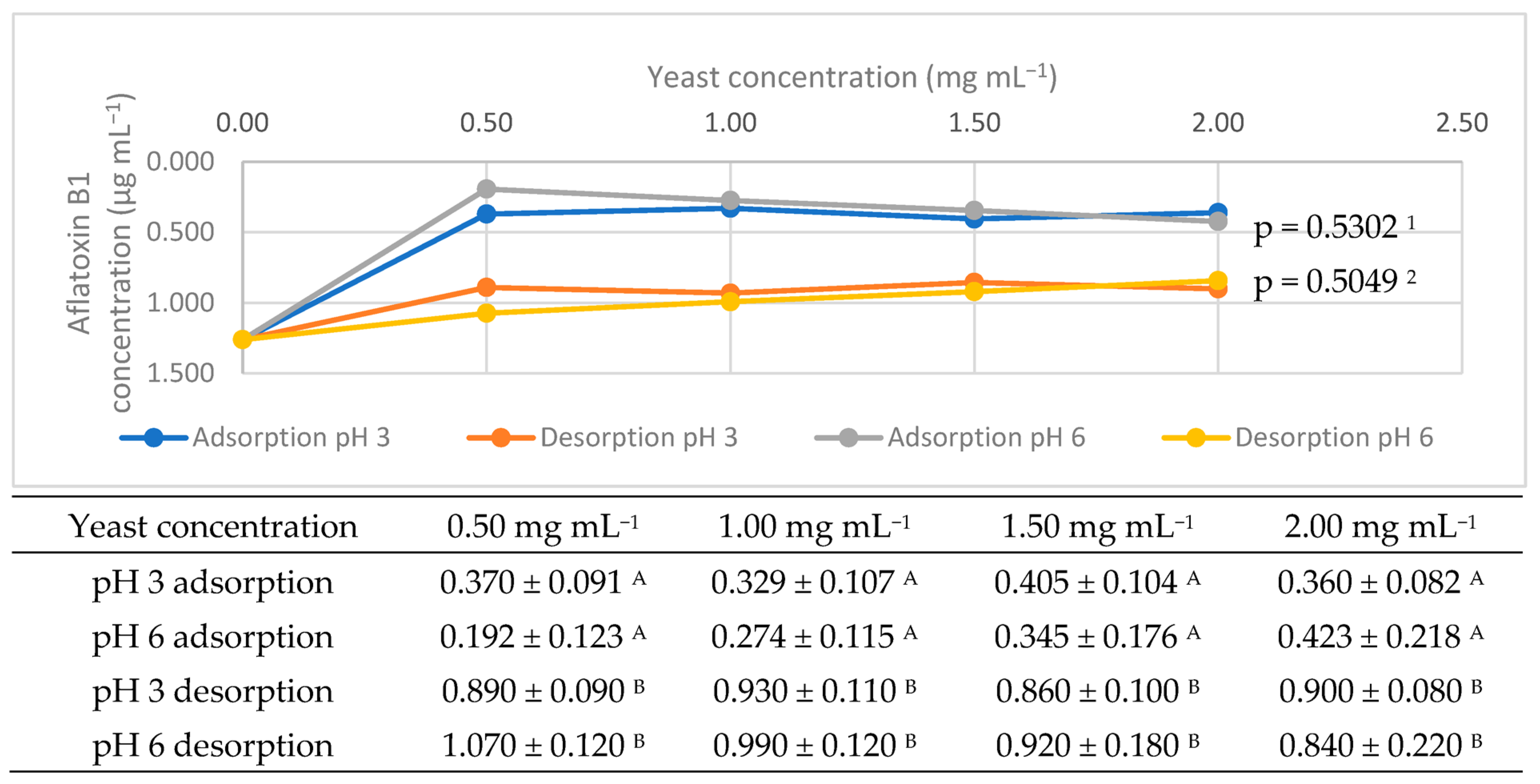
The adsorption capacities of each strain following the incubation period are depicted in Figure 1, Figure 2, Figure 3, Figure 4, Figure 5 and Figure 6.
Gradual increases in mycotoxin adsorption rates with increasing yeast cells counts were noted, albeit with no significant difference for AFB1 (p > 0.05). With regard to ZEN, only the LL83 strain applied at 2.0 mg mL−1 was significantly different when comparing both pH values (p < 0.05).
Mycotoxin adsorption occurs through hydrophobic and electrostatic interactions between the metabolite and the binding sites formed by the yeast cell constituents, such as weak hydrogen bonds and van der Waals bonds. Thus, more cells lead to more binding sites and, consequently, higher MT adsorption rates [23,26].
The differences observed between the evaluated yeast strains is due to innate variations among yeast species, due to a highly diversified cell wall composition. Consequently, the adsorption capacities of the different strains may vary under the same conditions [23]. Adsorption rates can also be affected by yeast cell wall thickness, as yeast with thick cell walls presents more surface binding sites when cultivated in certain growth media [21,26].
Besides adsorptive effects associated with cell wall components, other cellular mechanisms related to mycotoxin removal are also noteworthy, such as MT biotransformation catalyzed by complex chemical reactions. This reinforces that models that jointly consider all these factors are required for adequate AMA product development protocol and optimization [27].
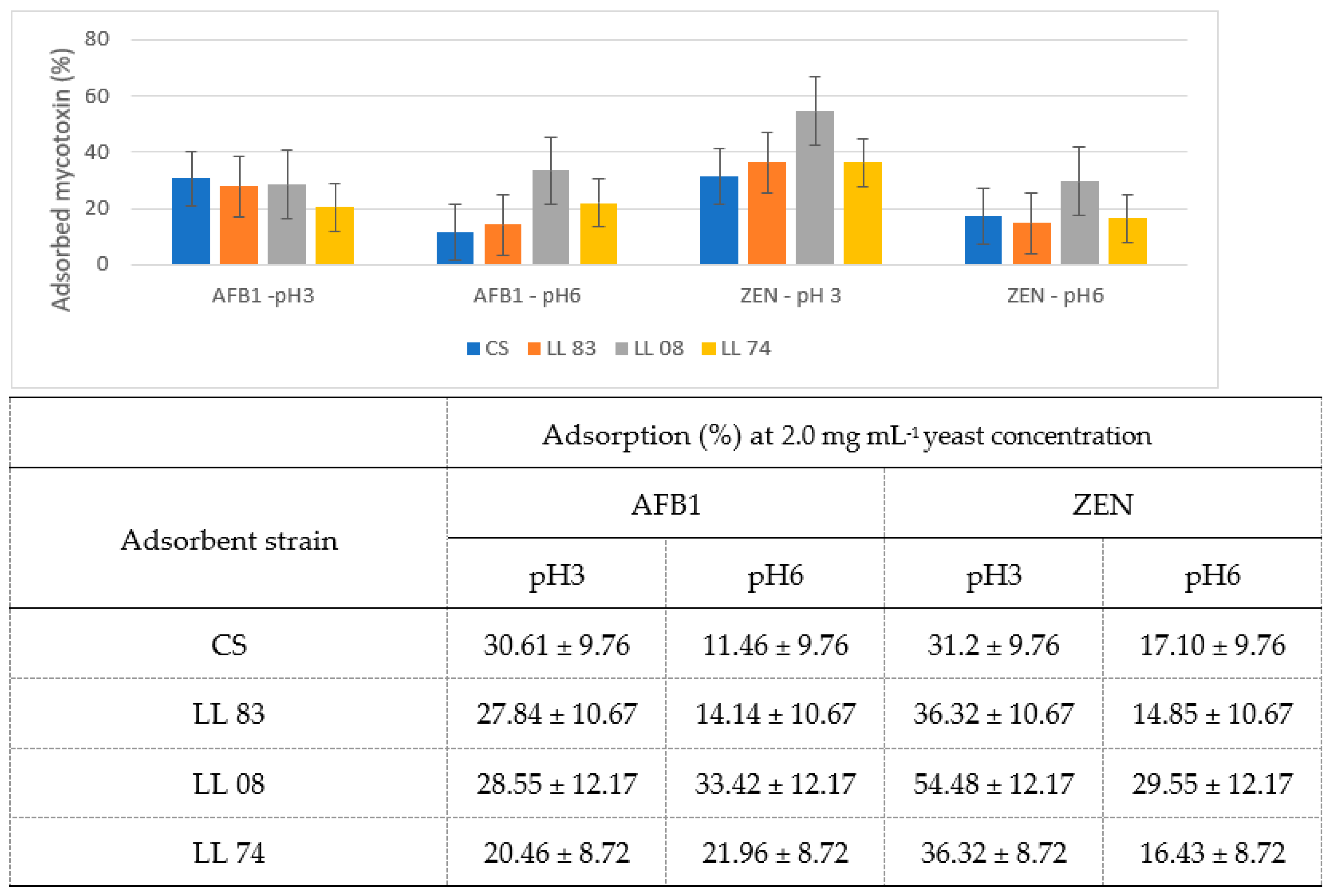
Figure 7 compares the commercial and isolated S. cerevisiae strains applied at 2.0 mg mL−1 for each pH condition evaluated herein.
Better results are generally expected for commercial strains, as they are optimized to adsorb mycotoxins in both acidic and neutral conditions and can act throughout the entire gastrointestinal tract. However, the isolated strains evaluated herein exhibited similar adsorption rates at both pH values when compared to the applied CS, albeit with no significant difference (p > 0.05).
The LL 08 strain obtained better results than the CS in all evaluated conditions, with only a slightly lower AFB1 adsorption at pH 3, albeit with no significant difference (p > 0.05). The other obtained adsorption values were higher than the CS, especially ZEN adsorption at pH 3, which was 57% higher than CS, (p value = 0.0045).
Yeast cell walls are composed of polysaccharides with an outer layer consisting of strongly glycosylated mannoproteins and an inner layer comprising β-1,6- and β-1,3-glucan chains linked to chitin and mannoproteins [18,20]. Some studies have indicated that the alkaline-insoluble β-1,3-glucan forms a complex structure with many ZEN binding sites, while the alkaline-soluble β-1,6-glucan potentiates this bond [27,28]. These parameters are considered when yeast are used as commercial adsorbent.
The wild strains investigated herein display similar adsorption rates to the applied CS, although studies aiming to identify, quantify, and optimize selected microorganisms and their β-glucan concentrations are required for commercial applications. Several studies have focused on gene regulation procedures to optimize yeast strains, leading to a greater number of cell wall binding sites for each specific mycotoxin [22].
The adsorption range of mycotoxins was higher at pH 3 compared to pH 6. The literature reports adsorption rates ranging from 20.46 to 54.48%, where yeast cell wall adsorption is favored under acidic conditions [15,29]. The solubilization of β-glucans under different pH conditions alters his structure and the conformational energy to create new interactions. pH value variations alter the flexibility of these interactions, leading to higher flexibility under neutral conditions compared to more acidic conditions. The decreasing flexibility noted herein at pH 3.0 increased ZEN adsorption values when compared to neutral and alkaline conditions [28,30]. However, as noted for LL08 adsorption rates at pH 6, interactions are more flexible and effective for AFB1, due to its physicochemical characteristics. This reinforces the need for studies dedicated to cell wall component enhancements to achieve more flexible products capable of adsorbing aflatoxins under different conditions.
In the present study, ZEN adsorption rates were higher than AFB1 rates. As the reaction conditions were the same, this difference can be explained based on the physicochemical characteristics of each investigated mycotoxin. The most important physicochemical parameter associated to adsorption is the oil/water partition coefficient (logP), where higher log values result in higher hydrophobicity. The logP values for AFB1 and ZEN are 1.23 and 3.60 respectively [31,32]. As ZEN is highly hydrophobic, it is expected to exhibit greater interaction with strains containing a high number of membrane binding sites, as observed in the present study.
Several inorganic-based compounds have been employed to assess aflatoxin adsorptive effects, and the literature indicates that aluminosilicates, bentonites, and zeolites exhibit high in vitro aflatoxin adsorption capabilities [33,34]. Concerning AFB1, adsorption values between 51.94% and 100% for at pH 3 and pH 6 have been reported when comparing the effectiveness of inorganic adsorbents [34,35,36]. The wild strains investigated herein exhibit equivalent effects to CS, thus considered moderately efficient adsorbents compared to inorganic adsorbents.
Despite the observed effectiveness differences, the use of live microorganisms has been the focus of mycotoxin mitigation in several assessments, due to their probiotic effect and mycotoxin biotransformation associated with microorganism intake [15]. In this regard, recent studies have focused on the optimization of mycotoxin mitigation in dairy cattle by the association of organic and inorganic adsorbents, to enhance AFB1 removal and concomitantly reduce AFM1 concentrations in milk [37,38,39,40].

3.2. Biotransformation Potential

Some studies have highlighted the ability of yeast in metabolizing mycotoxins into other compounds. Herein, however, none of the evaluated strains exhibited significant mycotoxin biotransformation potential under the evaluated conditions when compared with the commercial strain.
Higher conversion rates of up to 69–71% have been reported from initial mycotoxin concentrations following longer incubation periods (approximately 48–96 h) [15,41]. The incubation period of the present study, however, was of no more than 24 h, even though the enzymatic synthesis peak of some yeast takes place at longer periods of time, observed by the non-significant relationship between yeast concentrations and pH (p ≥ 0.05).
Although no significant biotransformations were observed herein, this effect cannot be neglected due to the microorganism’s enzymatic complex used for mycotoxin detoxification [21,41].
Microorganism mycotoxin biotransformations capabilities are an advantage compared to inorganic adsorbents, as other beneficial effects for target animal are also noted. Furthermore, inorganic compounds can present adverse effects linked to the absorption of micronutrients and other essential compounds [42,43,44].
The biotransformation assay applied herein may present false-positive results, mainly from chemical hydrolysis due to prolonged MT exposure or the presence of certain components in the reaction medium [45]. Therefore, assays using known derivative standards to correctly evaluate mycotoxin biotransformation by yeast are required [15,46,47].

4. Conclusions

The isolation of microorganisms from nature is a promising alternative for the development of new anti-mycotoxin additives with probiotic functions, reinforcing the idea of employing additives of biological origin. In this regard, the S. cerevisiae strains evaluated herein exhibited excellent adsorptive capacity for AFB1 and ZEN under gastrointestinal conditions. All strains displayed high potential for commercial applications, especially strain LL08, and further studies are required for their optimization for use in livestock. Although no significant biotransformation was observed, this effect should not be discarded when selecting microorganisms with anti-mycotoxin potential, as many microorganisms can convert mycotoxins into less toxic metabolites in the long term, which is important for the development of new products. Since microorganisms exhibit lower adsorptive capacity compared to inorganic adsorbents, commercial products should be developed by focused on genomic selection of biotransformation enzymes aiming at mycotoxin detoxification. Eliminating mycotoxins using additives is, however, not enough to ensure feed safety, and mechanisms to control and select quality raw materials should also be applied to improve product quality and animal health.

Author Contributions

Conceptualization, V.F.M., L.d.A.P. and F.B.N.K.; Methodology, V.F.M., L.d.A.P., F.B.N.K., K.M.K., M.A. and L.A.M.K.; Validation, K.M.K., M.A. and L.A.M.K.; Formal analysis, V.F.M. and L.A.M.K.; Investigation, V.F.M., L.d.A.P., F.B.N.K. and L.A.M.K.; Resources, K.M.K., M.A. and L.A.M.K.; Data curation, V.F.M. and L.d.A.P.; Writing—original draft preparation, V.F.M., L.d.A.P. and L.A.M.K.; Writing—review and editing, F.B.N.K., K.M.K., M.A. and L.A.M.K.; Supervision, V.F.M., L.d.A.P. and L.A.M.K.; Project administration, V.F.M., L.d.A.P., M.A. and L.A.M.K.; Funding acquisition, L.A.M.K. All authors have read and agreed to the published version of the manuscript.

Funding

This research was funded by FAPERJ-Fundação de Amparo à Pesquisa do Estado do Rio de Janeiro, Process SEI 260003/002531/2021 and SEI 260003/003283/2022.

Institutional Review Board Statement

Not applicable.

Informed Consent Statement

Not applicable.

Data Availability Statement

Not applicable.

Acknowledgments

The authors would like to thank to FAPERJ-Fundação de Amparo à Pesquisa do Estado do Rio de Janeiro for scholarship and funding. Thanks are also due to the post-graduation course Higiene Veterinária e Tecnologia de Produtos de Origem Animal of the University Federal Fluminense. the Departamento de Tecnologia de Alimentos at the same university (MTA-UFF), to the Mycology and Mycotoxin Laboratory of the at the Universidade Federal de Minas Gerais (LAMICO-UFMG).

Conflicts of Interest

The authors declare no conflict of interest.

References

  1. Peles, F.; Sipos, P.; Kovács, S.; Győri, Z.; Pócsi, I.; Pusztahelyi, T. Biological control and mitigation of aflatoxin contamination in commodities. Toxins 2021, 1, 104. [Google Scholar] [CrossRef]
  2. Yang, C.; Song, G.; Lim, W. Effects of mycotoxin-contaminated feed on farm animals. J. Hazard. Mater. 2020, 389, 122087. [Google Scholar] [CrossRef]
  3. Eskola, M.; Kos, G.; Elliott, C.T.; Hajšlová, J.; Mayar, S.; Krska, R. Worldwide contamination of food-crops with mycotoxins: Validity of the widely cited ‘FAO estimate’of 25. Crit. Rev. Food Sci. Nutr. 2020, 60, 2773–2789. [Google Scholar] [CrossRef] [PubMed]
  4. Bhardwaj, K.; Meneely, J.P.; Haughey, S.A.; Dean, M.; Wall, P.; Zhang, G.; Baker, B.; Elliott, C.T. Risk assessments for the dietary intake aflatoxins in food: A systematic review (2016–2022). Food Control 2023, 149, 109687. [Google Scholar] [CrossRef]
  5. Keller, L.A.M.; Aronovich, M.; Keller, K.M.; Castagna, A.A.; Cavaglieri, L.R.; Rosa, C.A.R. Incidence of Mycotoxins (AFB1 and AFM1) in Feeds and Dairy Farms from Rio de Janeiro State, Brazil. Veterinary. Medicine 2016, 1, 29–35. [Google Scholar]
  6. Kabak, B. Aflatoxins in foodstuffs: Occurrence and risk assessment in Turkey. J. Food Compost. Anal. 2021, 96, 103734. [Google Scholar] [CrossRef]
  7. Conteçotto, A.C.T.; Pante, G.C.; Castro, J.C.; Souza, A.A.; Lini, R.S.; Romoli, J.C.Z.; Abreu Filho, B.A.; Mikcha, J.M.G.; Mossini, S.A.G.; Machinski Junior, M. Occurrence, exposure evaluation and risk assessment in child population for aflatoxin M1 in dairy products in Brazil. Food Chem. Toxicol. 2021, 148, 111913. [Google Scholar] [CrossRef]
  8. Pitt, J.I. Toxigenic fungi and mycotoxins. Br. Med. J. 2000, 56, 184–192. [Google Scholar]
  9. Benkerroum, N. Chronic and acute toxicities of aflatoxins: Mechanisms of action. Int. J. Environ. Res. Public Health. 2020, 17, 423. [Google Scholar] [CrossRef] [Green Version]
  10. Wang, Y.; Liu, F.; Zhou, X.; Liu, M.; Zang, H.; Liu, X.; Shan, A.; Feng, X. Alleviation of Oral Exposure to Aflatoxin B1-Induced Renal Dysfunction, Oxidative Stress, and Cell Apoptosis in Mice Kidney by Curcumin. Antioxidants 2022, 11, 1082. [Google Scholar] [CrossRef]
  11. Forsythe, S.J. Microbiologia da Segurança Alimentar; ArtMed: Porto Alegre, Brazil, 2002; 422p. [Google Scholar]
  12. Rai, A.; Das, M.; Tripathi, A. Occurrence and toxicity of a fusarium mycotoxin, ZENralenone. Crit. Rev. Food Sci. Nutr. 2019, 60, 2710–2729. [Google Scholar] [CrossRef] [PubMed]
  13. Yu, H.; Zhang, J.; Chen, Y.; Zhu, J. Zearalenone and Its Masked Forms in Cereals and Cereal-Derived Products: A Review of the Characteristics, Incidence, and Fate in Food Processing. J. Fungi 2022, 8, 976. [Google Scholar] [CrossRef]
  14. Luo, Y.; Liu, X.; Yuan, L.; Li, J. Complicated interactions between bio-adsorbents and mycotoxins during mycotoxin adsorption: Current research and future prospects. Trends Food Sci. Technol. 2020, 96, 127–134. [Google Scholar] [CrossRef]
  15. Keller, L.A.M.; Abrunhosa, L.; Keller, K.M.; Rosa, C.A.R.; Cavaglieri, L.R.; Venâncio, A. ZENralenone and its derivatives α-ZENralenol and β-ZENralenol decontamination by Saccharomyces cerevisiae strains isolated from bovine forage. Toxins 2015, 7, 3297–3308. [Google Scholar] [CrossRef] [Green Version]
  16. Uyeno, Y.; Shigemori, S.; Shimosato, T. Effect of probiotics/prebiotics on cattle health and productivity. Microbes Environ. 2015, 30, 126–132. [Google Scholar] [CrossRef] [PubMed] [Green Version]
  17. Cecchini, F.; Morassut, M.; Saiz, J.C.; Moruno, E.G. Anthocyanins enhance yeast’s adsorption of Ochratoxin A during the alcoholic fermentation. Eur. Food Res. Technol. 2019, 245, 309–314. [Google Scholar] [CrossRef]
  18. Goncalves, B.L.; Rosim, R.E.; de Oliveira, C.A.F.; Corassin, C.H. The in vitro ability of different Saccharomyces cerevisiae-based products to bind aflatoxin B1. Food Control 2015, 47, 298–300. [Google Scholar] [CrossRef]
  19. Oliveira, A.A.; Keller, K.M.; Deveza, M.V.; Keller, L.A.M.; Dias, E.O.; Martini-Santos, B.J.; Rosa, C.A.R. Effect of three different anti-mycotoxin additives on broiler chickens exposed to aflatoxin B1. Arch. Med. Vet. 2015, 47, 175–183. [Google Scholar] [CrossRef] [Green Version]
  20. Petruzzi, L.; Corbo, M.R.; Sinigaglia, M.; Bevilacqua, A. Ochratoxin A removal by yeasts after exposure to simulated human gastrointestinal conditions. J. Food Sci. 2016, 81, 2756–2760. [Google Scholar] [CrossRef]
  21. Luo, Y.; Wang, J.G.; Liu, B.; Wang, Z.L.; Yuan, Y.H.; Yue, T.L. Effect of yeast cell morphology, cell wall physical structure and chemical composition on patulin adsorption. PLoS ONE 2015, 10, e0136045. [Google Scholar] [CrossRef] [Green Version]
  22. Luo, Y.; Liu, X.J.; Liu, Y.; Han, Y.Q.; Li, J. Exogenous calcium ions enhance patulin adsorption capability of Saccharomyces cerevisiae. J. Food Prot. 2019, 82, 1390–1397. [Google Scholar] [CrossRef]
  23. Pfliegler, W.P.; Pusztahelyi, T.; Pócsi, I. Mycotoxins-prevention and decontamination by yeasts. J. Basic Microbiol. 2015, 55, 805–818. [Google Scholar] [CrossRef]
  24. Silva, F.C.; Chalfoun, S.M.; Batista, L.R.; Santos, C.; Lima, N. Taxonomia polifásica para identificação de Aspergillus seção flavi: Uma revisão. Rev. Eletrônica Sala Aula Foco 2015, 1, 18–40. [Google Scholar] [CrossRef]
  25. Bovo, F.; Corassin, C.H.; Rosim, R.E.; Oliveira, C.A.F. Efficiency of Lactic Acid Bacteria Strains for Decontamination of Aflatoxin M1 in Phosphate Buffer Saline Solution and in Skimmed Milk. Food Bioprocess Technol. 2013, 6, 2230–2234. [Google Scholar] [CrossRef]
  26. Pereyra, C.M.; Gil, S.; Cristofolini, A.; Bonci, M.; Makita, M.; Monge, M.P.; Montenegro, M.A.; Cavaglieri, L.R. The production of yeast cell wall using an agroindustrial waste influences the wall thickness and is implicated on the aflatoxin B1 adsorption process. Food Res. Int. 2018, 111, 306–313. [Google Scholar] [CrossRef]
  27. Aazami, M.H.; Nasri, M.H.F.; Mojtahedi, M.; Mohammadi, S.R. In vitro aflatoxin B1 binding by the cell wall and (13)-β-d-glucan of baker’s yeast. J. Food Prot. 2018, 81, 670–676. [Google Scholar] [CrossRef]
  28. Hamza, Z.; El-Hashash, M.; Aly, S.; Hathout, A.; Soto, E.; Sabry, B.; Ostroff, G. Preparation and characterization of yeast cell wall beta-glucan encapsulated humic acid nanoparticles as an enhanced aflatoxin B1 binder. Carbohydr. Polym. 2019, 203, 185–192. [Google Scholar] [CrossRef]
  29. Prapapanpong, J.; Udomkusonsri, P.; Mahavorasirikul, W.; Choochuay, S.; Tansakul, S. In vitro studies on gastrointestinal monogastric and avian models to evaluate the binding efficacy of mycotoxin adsorbents by liquid chromatography-tandem mass spectrometry. J. Adv. Vet. Anim. Res. 2019, 6, 125–132. [Google Scholar] [CrossRef]
  30. Martínez, J.; Hernández-Rodríguez, M.; Méndez-Albores, A.; Téllez-Isaías, G.; Jiménez, E.M.; Nicolás-Vázquez, M.I.; Ruvalcaba, R.M. Computational Studies of Aflatoxin B1 (AFB1): A Review. Toxins 2023, 15, 153. [Google Scholar] [CrossRef]
  31. NCBI. National Center for Biotechnology Information. PubChem Compound Summary for CID 15558498, Aflatoxin M1. 2022a. Available online: https://pubchem.ncbi.nlm.nih.gov/compound/Aflatoxin-M1 (accessed on 24 March 2023).
  32. NCBI. National Center for Biotechnology Information. PubChem Compound Summary for CID 5281576, ZENralenone. 2022b. Available online: https://pubchem.ncbi.nlm.nih.gov/compound/ZENralenone (accessed on 24 March 2023).
  33. Holanda, D.M.; Kim, S.W. Mycotoxin Occurrence, Toxicity, and Detoxifying Agents in Pig Production with an Emphasis on Deoxynivalenol. Toxins 2021, 13, 171. [Google Scholar] [CrossRef]
  34. Vila-Donat, P.; Marín, S.; Ramos, A.J. A review of the mycotoxin adsorbing agents, with an emphasis on their multi-binding capacity, for animal feed decontamination. Food Chem. Toxicol. 2018, 114, 246–259. [Google Scholar] [CrossRef] [Green Version]
  35. Ahn, J.Y.; Kim, J.; Cheong, D.H.; Hong, H.; Jeong, J.Y.; Kim, B.G. An In Vitro Study on the Efficacy of Mycotoxin Sequestering Agents for Aflatoxin B1, Deoxynivalenol, and Zearalenone. Animals 2022, 12, 333. [Google Scholar] [CrossRef] [PubMed]
  36. Yiannikouris, A.; Apajalahti, J.; Kettunen, H.; Ojanperä, S.; Bell, A.N.W.; Keegan, J.D.; Moran, C.A. Efficient Aflatoxin B1 Sequestration by Yeast Cell Wall Extract and Hydrated Sodium Calcium Aluminosilicate Evaluated Using a Multimodal In-Vitro and Ex Vivo Methodology. Toxins 2021, 13, 24. [Google Scholar] [CrossRef] [PubMed]
  37. Jiang, Y.; Ogunade, I.M.; Kim, D.H.; Li, X.; Pech-Cervantes, A.A.; Arriola, K.G.; Oliveira, A.S.; Driver, J.P.; Ferraretto, L.F.; Staples, C.R.; et al. Effect of adding clay with or without a Saccharomyces cerevisiae fermentation product on the health and performance of lactating dairy cows challenged with dietary aflatoxin B1. J. Dairy Sci. 2018, 101, 3008–3020. [Google Scholar] [CrossRef] [PubMed]
  38. Maki, C.R.; Thomas, A.D.; Elmore, S.E.; Romoser, A.A.; Harvey, R.B.; Ramirez, H.A.; Phillips, T.D. Effects of calcium montmorillonite clay and aflatoxin exposure on dry matter intake, milk production, and milk composition. J. Dairy Sci. 2016, 99, 1039–1046. [Google Scholar] [CrossRef] [Green Version]
  39. Rodrigues, R.O.; Rodrigues, R.O.; Ledoux, D.R.; Rottinghaus, G.E.; Borutova, R.; Averkieva, O.; Mcfadden, T.B. Feed additives containing sequestrant clay minerals and inactivated yeast reduce aflatoxin excretion in milk of dairy cows. J. Dairy Sci. 2019, 102, 6614–6623. [Google Scholar] [CrossRef] [Green Version]
  40. Xiong, J.L.; Wang, Y.M.; Zhou, H.L.; Liu, J.X. Effects of dietary adsorbent on milk aflatoxin M1 content and the health of lactating dairy cows exposed to long-term aflatoxin B1 challenge. J. Dairy Sci. 2018, 101, 8944–8953. [Google Scholar] [CrossRef] [Green Version]
  41. Li, P.; Su, R.; Yin, R.; Lai, D.; Wang, M.; Liu, Y.; Zhou, L. Detoxification of Mycotoxins through Biotransformation. Toxins 2020, 12, 121. [Google Scholar] [CrossRef] [Green Version]
  42. Barrientos-Velázquez, A.L.; Arteaga, S.; Dixon, J.B.; Deng, Y. The effects of pH, pepsin, exchange cation, and vitamins on aflatoxin adsorption on smectite in simulated gastric fluids. Appl. Clay Sci. 2016, 120, 17–23. [Google Scholar] [CrossRef]
  43. Elliot, C.T.; Connolly, L.; Kolawole, O. Potential adverse effects on animal health and performance caused by the addition of mineral adsorbents to feeds to reduce mycotoxin exposure. Mycotoxin Res. 2019, 36, 115–126. [Google Scholar] [CrossRef] [Green Version]
  44. Wang, G.; Miao, Y.; Sun, Z.; Zheng, S. Simultaneous adsorption of aflatoxin B1 and zearalenone by mono- and di-alkyl cationic surfactants modified montmorillonites. J. Colloid Interface Sci. 2018, 511, 67–76. [Google Scholar] [CrossRef]
  45. Wang, J.; Xie, Y. Review on microbial degradation of ZENralenone and aflatoxins. Grain Oil Sci. Technol. 2020, 3, 117–125. [Google Scholar] [CrossRef]
  46. Fruhauf, S.; Novak, B.; Nagl, V.; Hackl, M.; Hartinger, D.; Rainer, V.; Labudová, S.; Adam, G.; Aleschko, M.; Moll, W.D.; et al. Biotransformation of the Mycotoxin ZENralenone to Its Metabolites Hydrolyzed ZENralenone (HZEN) and Decarboxylated Hydrolyzed ZENralenone (DHZEN) Diminishes Its Estrogenicity In Vitro and In Vivo. Toxins 2019, 11, 481. [Google Scholar] [CrossRef] [PubMed] [Green Version]
  47. Ji, C.; Fan, Y.; Zhao, L. Review on biological degradation of mycotoxins. Anim. Nutr. 2016, 2, 127–133. [Google Scholar] [CrossRef] [PubMed]
Figure 1. Adsorption levels and statistical comparisons between AFB1 adsorption and desorption rates by the LL83 Saccharomyces cerevisiae strain under in different pH conditions. 1 Adsorption p value according to the LSD Fisher test comparison at pH 3 and 6; 2 Desorption p value according to the LSD Fisher test comparison at pH 3 and 6; A and B represent differences according to the LSD Fisher test comparison. AFB1 concentrations are expressed in µg mL−1.
Fermentation 09 00585 g001
Figure 2. Adsorption levels and statistical comparison between AFB1 adsorption and desorption rates by the LL08 Saccharomyces cerevisiae strain under different pH conditions. 1 Adsorption p value according to the LSD Fisher test comparison at pH 3 and 6; 2 Desorption p value according to the LSD Fisher test comparison at pH 3 and 6; A and B represent differences according to the LSD Fisher test comparison. AFB1 concentrations are expressed in µg mL−1.
Fermentation 09 00585 g002
Figure 3. Adsorption levels and statistical comparison between AFB1 adsorption and desorption rates by the LL74 Saccharomyces cerevisiae strain under different pH conditions. 1 Adsorption p value according to the LSD Fisher test comparison at pH 3 and 6; 2 Desorption p value according to the LSD Fisher test comparison at pH 3 and 6; A and B represent differences according to the LSD Fisher test comparison. AFB1 concentrations are expressed in µg mL−1.
Fermentation 09 00585 g003
Figure 4. Adsorption levels and statistical comparison between ZEN adsorption and desorption rates by the LL83 Saccharomyces cerevisiae strain under different pH conditions. 1 Adsorption p value according to the LSD Fisher test comparison at pH 3 and 6; 2 Desorption p value according to the LSD Fisher test comparison at pH 3 and 6; A, a, B and b represent differences according to the LSD Fisher test comparison. ZEN concentrations are expressed in µg mL−1.
Fermentation 09 00585 g004
Figure 5. Adsorption levels and statistical comparison between ZEN adsorption and desorption rates by the LL08 Saccharomyces cerevisiae strain under different pH conditions. 1 Adsorption p value according to the LSD Fisher test comparison at pH 3 and 6; 2 Desorption p value according to the LSD Fisher test comparison at pH 3 and 6; A and B represent differences according to the LSD Fisher test comparison. ZEN concentrations are expressed in µg mL−1.
Fermentation 09 00585 g005
Figure 6. Adsorption levels and statistical comparison between ZEN adsorption and desorption rates by the LL74 Saccharomyces cerevisiae strain under different pH conditions. 1 Adsorption p value according to the LSD Fisher test comparison at pH 3 and 6; 2 Desorption p value according to the LSD Fisher test comparison at pH 3 and 6; A and B represent differences according to the LSD Fisher test comparison. ZEN concentrations are expressed in µg mL−1.
Fermentation 09 00585 g006
Figure 7. Mycotoxin adsorption of commercial and isolated S. cerevisiae strains at 2.0 mg mL−1 under gastric and intestinal simulation conditions at pH 3 and 6. p value > 0.05.
Fermentation 09 00585 g007
Disclaimer/Publisher’s Note: The statements, opinions and data contained in all publications are solely those of the individual author(s) and contributor(s) and not of MDPI and/or the editor(s). MDPI and/or the editor(s) disclaim responsibility for any injury to people or property resulting from any ideas, methods, instructions or products referred to in the content.