Article

Exploring the Production of Secondary Metabolites from a Halophyte Tetragonia tetragonoides through Callus Culture

1
Department of Horticulture, Chungnam National University, 99 Daehak-ro, Yuseong-gu, Daejeon 34134, Republic of Korea
2
Department of Smart Agriculture Systems, Chungnam National University, Daejeon 34134, Republic of Korea
3
Department of Crop Science, Graduate School, Chungnam National University, Daejeon 34134, Republic of Korea
4
College of Veterinary Medicine, Chungnam National University, Daejeon 34134, Republic of Korea
5
Department of Agricultural Machinery Engineering, Graduate School, Chungnam National University, Daejeon 34134, Republic of Korea
*
Author to whom correspondence should be addressed.
Current address: Floriculture Research Division, National Institute of Horticultural and Herbal Science, Rural Development Administration, Wanju 55365, Republic of Korea.
Horticulturae 2024, 10(3), 244; https://doi.org/10.3390/horticulturae10030244
Submission received: 30 January 2024 / Revised: 21 February 2024 / Accepted: 29 February 2024 / Published: 3 March 2024
(This article belongs to the Special Issue Innovative Micropropagation of Horticultural and Medicinal Plants)

Abstract

:
Considering Korea’s gradual shift toward an aging society, consumer interest in compounds with physiological benefits, including antioxidant and anticancer effects, has surged. This study explored the potential of Tetragonia tetragonoides (Pall.) Kunze, commonly known as New Zealand Spinach (NZS), a halophyte with reported health benefits, including efficacy in treating gastrointestinal diseases, high blood pressure, diabetes, and obesity. This study also introduced a novel callus culture system for NZS, allowing for the rapid in vitro production of secondary metabolites. Optimal callus induction (100%) and biomass production (0.416 g) were achieved by adding 2.0 mg·L−1 6-BA (6-Benzylaminopurine) and 0.5 mg·L−1 2,4-D (2,4-Dichlorophenoxyacetic acid) among five auxin and cytokinin combinations. Two distinct callus types, TGC [TDZ (Thidiazuron)-supplemented Green Callus] and TNYC [TDZ + NAA (Naphthalene acetic acid)-supplemented Yellow Callus], were identified, each with unique characteristics. The calli showed total phenolic and flavonoid contents comparable to those of NZS leaves grown in the greenhouse. An expression analysis of six genes (CHS, CHI, F3H, F3′H, FLS, and DFR) involved in the kaempferol biosynthesis revealed an enhanced flavonoid biosynthesis-related gene expression in TGC, emphasizing its potential for compound production. GC-MS analysis identified distinct compound profiles in TGC and TNYC, with 2,3-butanediol and succinic acid being the predominant compounds among the nine and forty-four components, respectively. These calli offer a stable supply of functional compounds and present an environmentally sustainable solution. The derived callus culture system is anticipated to contribute to the development of healthy functional foods or pharmaceuticals from halophyte NZS.

1. Introduction

Recently, as Korea has gradually become an aging society, consumers’ interest in compounds with physiological activities, such as antioxidant and anticancer effects, is increasing. Plants produce and accumulate a variety of secondary metabolites to respond and defend themselves against biological stress as well as abiotic stress such as environmental stress [1,2,3,4]. These metabolites, renowned for their antioxidant properties, possess diverse anticancer and antibacterial properties. Not only have they found industrial applications in chemicals, spices, food additives, and medicines, but they also serve as vital resources [5,6].
However, the production of secondary metabolites in plants may be limited because of slow growth and endangered plant species [7]. Recently, secondary metabolite production through cell culture has attracted attention because it can be performed in large quantities quickly without being affected by external weather [8,9,10]. Additionally, calli grow faster than seedlings and have shorter biosynthetic cycles. Therefore, they can produce secondary metabolites in a shorter time than plants [11]. Anjum et al. [12] reported that 1.0 mg·L−1 of NAA was treated to induce the callus of Linum usitatissimum L. followed by lignans and neo-lignans production. And Adil et al. [13] reported that high TPC and TFC were identified in the callus of Cnidium officalale Makino and that the content of phthalides was higher than that of seeding plants. In addition, 45 aromatic compounds, including rosemaryic acid, were identified in a callus derived from the leaves of Rosmarinus openis L., and calli treated with 200 μM of melatonin doubled the rosemaryic acid content [14].
Tetragonia tetragonoides (Pall.) Kunze is a conditional salt plant, commonly known as New Zealand Spinach (NZS). This plant grows naturally on sandy soil or coastal cliffs in New Zealand, Australia, Japan, and the Republic of Korea [15]. NZS has leaves and has a soft taste, with a texture similar to that of regular spinach. NZS is used to treat stomach cancer, gastritis, gastric ulcers, and hyper acidosis and to improve food intake [16]. In addition, anti-inflammatory, anti-blood pressure, and antioxidant agents have been reported [17,18,19,20]. In addition, extracts from NZS have been shown to enhance hippocampal insulin signaling, reduce insulin resistance, and enhance gut microbiota and memory function in amyloid-β-infused mice in an Alzheimer’s disease animal model [21]. These findings confirm that NZS extract can provide a variety of health benefits.
In this study, we explored methods to produce beneficial secondary metabolites from NZS using callus cultures. Hormonal conditions were established to effectively induce callus growth on NZS leaves. Furthermore, we assessed the potential of the selected callus as an extractant raw material for functional compounds using the total phenolic compound (TPC) and total flavonoid (TFC) measurements and GC-MS screening. The results of this study contribute to the development of this industry with a raw material for healthy functional foods, pharmaceuticals, and cosmetics.

2. Materials and Methods

2.1. Combinations of Plant Growth Regulators Used to Induce Callus from NZS Leaves

To investigate the effects of plant growth regulators (PGRs) (Duchefa, Haarlem, Netherlands) on callus induction, explants were cultured in MS medium containing different levels of auxins (IAA, NAA, and 2,4-D) and cytokinins (zeatin and 6-BA) (Figure S1). The explants were prepared from the apical site to the first and second nodes of in vitro-grown NZS, with leaves sized approximately 0.5 cm × 0.5 cm. Ten explants were placed in the medium for each treatment, and the procedure was repeated three times. We placed the prepared implants in the medium and measured the induction rate and fresh weight of the callus after four weeks. Additionally, to obtain calli of various colors and shapes, calli derived from the optimal media were transferred to MS medium supplemented with TDZ alone or a combination of TDZ and NAA. The concentrations of TDZ and NAA were set based on previous reports indicating a high induction rate of calli or sprouts in the Aizoaceae family [22]. Ultimately, we selected a vigorously growing green hard tissue callus in a medium containing 2.5 mg·L−1 TDZ, named TGC. Simultaneously, we chose a yellow hard tissue callus labeled TNYC, grown on a medium with 2.5 mg·L−1 TDZ and 0.5 mg·L−1 NAA.

2.2. Explant Material and In Vitro Culture Conditions

The mature seeds, collected from the seashore of Jeonnam Goheung-Gun, Republic of Korea, were stored at 4 °C until used in the experiment. The seeds were sterilized through dipping in 70% (v/v) ethanol for 1 min and in a 2% sodium hypochlorite solution for 20 min, and then washed with sterile distilled water three times. Germination was performed on 1/2 MS (Duchefa, Haarlem, Netherlands) medium supplemented with 30 g·L−1 sucrose (Duchefa, Haarlem, Netherlands) and 7 g·L−1 plant agar (Duchefa, Haarlem, The Netherlands). Plantlets established from 8-week-old seedlings were used as explant sources (Figure 1). Before the medium was autoclaved at 120 °C for 20 min, the pH was adjusted to 5.7. All the cultures were incubated in a growth room at 25 ± 1 °C under a 16 h light/8 h dark photoperiod, using an irradiance of 20 μmol·m−2·s−1 from white fluorescent lamps (Osram GmbH, Munich, Germany).

2.3. Measuring Total Phenol Content (TPC) and Total Flavonoid Content (TFC) of Selected Callus

To obtain the extracts of the callus grown in vitro and leaves grown in greenhouses, they were extracted with 80% ethanol for three hours at 85 °C and filtered through a 0.45 μm PTFE syringe filter (Hyundai micro, Seoul, Republic of Korea).
The TPC was determined using the Folin–Ciocaltzeu method [23]. A standard solution was prepared with concentrations of 13.25, 62.5, 125, 250, 500, and 1000 mg·L−1 from gallic acid (Sigma Chemical, St. Louis, MO, USA). Subsequently, 0.05 mL of each solution was mixed with 0.25 mL of Folin–Ciocalteu solvent (Sigma Chemical, St. Louis, MO, USA), followed by the addition of 2.5% Na2CO3 (0.5 mL). The mixture was stored in the dark for 40 min, and the extract was measured three times.
The TFC was analyzed using the aluminum chloride colorimetric method. Standard solutions were prepared at concentrations of 13.25, 125, 250, and 500 mg·L−1 using Quercetin (Sigma Chemical Co., St. Louis, MO, USA). In the measurement, 0.02 mL of 10% aluminum nitrate and 0.02 mL of 1 M potassium acetate were added to 0.02 mL of the extract, and then 80% ethanol was added to make a total volume of 1 mL. After incubating the mixture at room temperature for 40 min, absorbance was measured at 415 nm.

2.4. Analysis of Gene Expression Involved in the Biosynthesis of Flavonoids

Three major compounds of the kaempferol backbone were detected in NZS, as reported previously [20]. Thus, we investigated the expression levels of genes, including chalcone synthase (CHS), chalcone isomerase (CHI), flavanone 3-hydroxylase (F3H), flavonoid 3′-hydroxylase (F3′H), flavonol synthase (FLS), and dihydroflavonol 4-reductase (DFR), involved in the biosynthesis of kaempferol. The expression patterns of the six genes from the selected calli were compared with those of seedlings grown in the greenhouse.
The total RNA was extracted using an RNeasy mini kit (Qiagen, Hilden, Germany) after powdering each portion (leaf, stem, and root) of the NZS and callus using liquid nitrogen. Subsequently, approximately 1 μg of cDNA was synthesized from the extracted RNA using the PrimeScript 1st Strand cDNA Synthesis Kit (Takara Bio, Shiga, Japan).
Kaempferol biosynthesis-related genes were identified by searching the Kyoto Encyclopedia of Genes and Genomes pathway database. Gene-specific primers were designed to amplify products ranging from 200 to 550 bp (Table S1). The amplification conditions included 35 cycles of denaturation for 30 s at 94 °C, annealing for 30 s at 60 °C, extension for 30 s at 72 °C, and a final extension for 10 min at 72 °C. PCR was performed using a TaKaRa PCR Thermal Cycler Dice® gradient (Takara Bio, Shiga, Japan). The RT-PCR products were digested on a 1.2% agarose gel, and the electrophoretic bands were visualized under UV fluorescence using gel documentation.

2.5. Phytochemical Analysis of Selected Callus via GC-MS

To detect phytochemicals in the two selected calli, we prepared extracts and profiled them using gas chromatography–mass spectrometry (GC-MS) according to the manufacture’s manual. Callus extracts were prepared through soaking in 70% ethanol for 24 h at room temperature, followed by centrifugation at 13,000 rpm for 30 min to collect the supernatants. The freeze-dried powder obtained from the supernatant was dissolved in 100% ethanol for further analysis.
For GC-MS analysis, an HP-1MS capillary column (30 m × 320 μm × 1 μm film thickness, Agilent, Santa Clara, CA, USA) was utilized, with an MS (5975 inert Mass Selective Detector, Agilent) serving as the detector. The GC oven temperature was initially set at 70 °C for 1 min, and then increased from 70 °C to 180 °C at 10 °C/min, followed by an increase from 180 °C to 220 °C at 5 °C/min, maintaining at 220 °C for 1 min. Subsequently, the temperature was further raised from 220 °C to 300 °C at 25 °C/min, and a post time was held at 320 °C for 1 min. The total run time for the GC-MS analysis was 24.20 min. Helium was used as the carrier gas at a flow rate of 1 ml/min. The sample inlet was operated in splitless mode, and 1 μL of the sample was injected using an Autosampler (7683B Series injector, Agilent, Santa Clara, CA, USA). The syringe used for the autosampler had a volume of 10 μL. To minimize contamination-related errors, the syringe was washed thrice with acetone before injection, the sample was pumped thrice for collection, and washed thrice with acetone after injection. To reduce errors due to residual amounts in the column during sample analysis, the sample was analyzed once and subsequently cleaned once with acetone.

2.6. Statistical Analysis

The statistical analysis was performed using one-way analysis of variance (ANOVA) and Duncan’s multiple range test for the comparison of mean values using the SPSS statistical software Version 20.0 (IBM, New York, NY, USA).

3. Results

3.1. Effect of PGR on Callus Induction

In this study, leaf explants were placed in MS medium containing cytokinin and auxin to determine the effects of the PGRs on callus induction through NZS. When zeatin was used, the callus induction speed and fresh weight decreased in the order of 2,4-D > NAA > IAA (Table 1). In particular, calli induced in the MS medium with NAA showed root development (Figure S1). When 6-BA was used, the callus induction rate and fresh weight were higher in the 2,4-D treatment than in the NAA treatment (Table 2).
In media containing 6-BA and 2,4-D, the callus induction (0.851) and fresh weight (0.770) were significantly positively correlated with 2,4-D (p < 0.01). Ultimately, in media with 2.0 mg·L−1 6-BA and 0.5 mg·L−1 2,4-D, the maximum callus induction and fresh weight reached 100% and 0.416 ± 0.019 g, respectively. These results highlight the importance of specific PGR combinations in optimizing callus induction and growth in NZS.

3.2. Total Phenolic and Total Flavonoid Concentration of Selected Callus

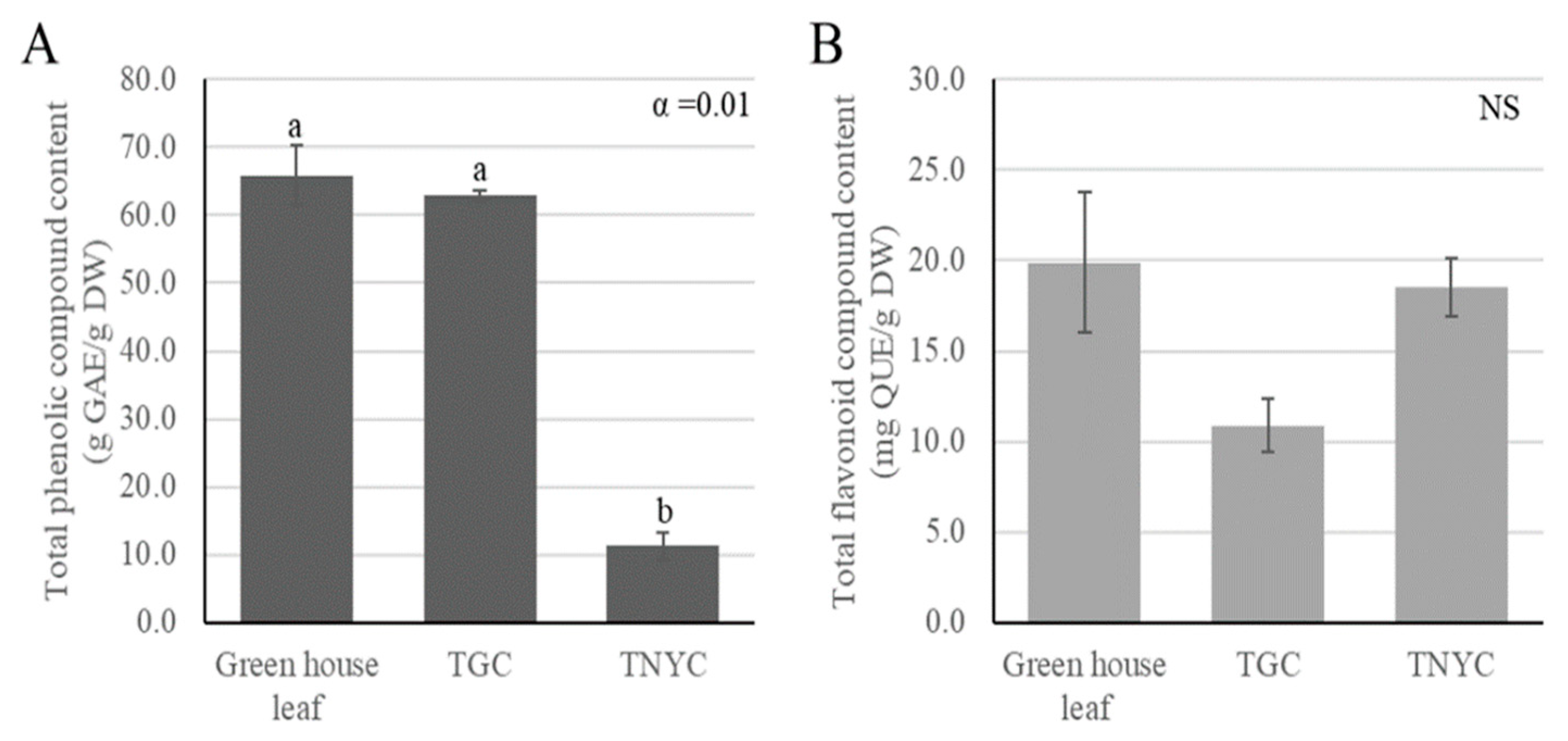
Following callus induction in the optimized medium, the cultured calli were subsequently cultured in a medium containing TDZ alone or a combination of TDZ and NAA (Figure 2). The two selected callus species were labeled TGC (induced in the medium with TDZ) and TNYC (induced in the medium with TDZ and NAA). Selected calli were used to measure the TPC and TFC to determine the secondary metabolite content. The TPC of TGC was measured at 62.9 g GAE/g DW, showing no significant difference when compared with the leaves of NZS grown in the greenhouse (Figure 3). In contrast, the TNYC calli displayed approximately six-times-lower content than that of the NZS leaves, indicating a significant difference (p < 0.01). The total flavonoid content did not differ significantly between the treatment groups. The measured values were 19.9, 10.9, and 18.5 g QE/g DW for the NZS leaves grown in the greenhouse, TGC, and TNYC, respectively.

3.3. Comparison of Gene Expressions Related to Flavonoid Biosynthesis

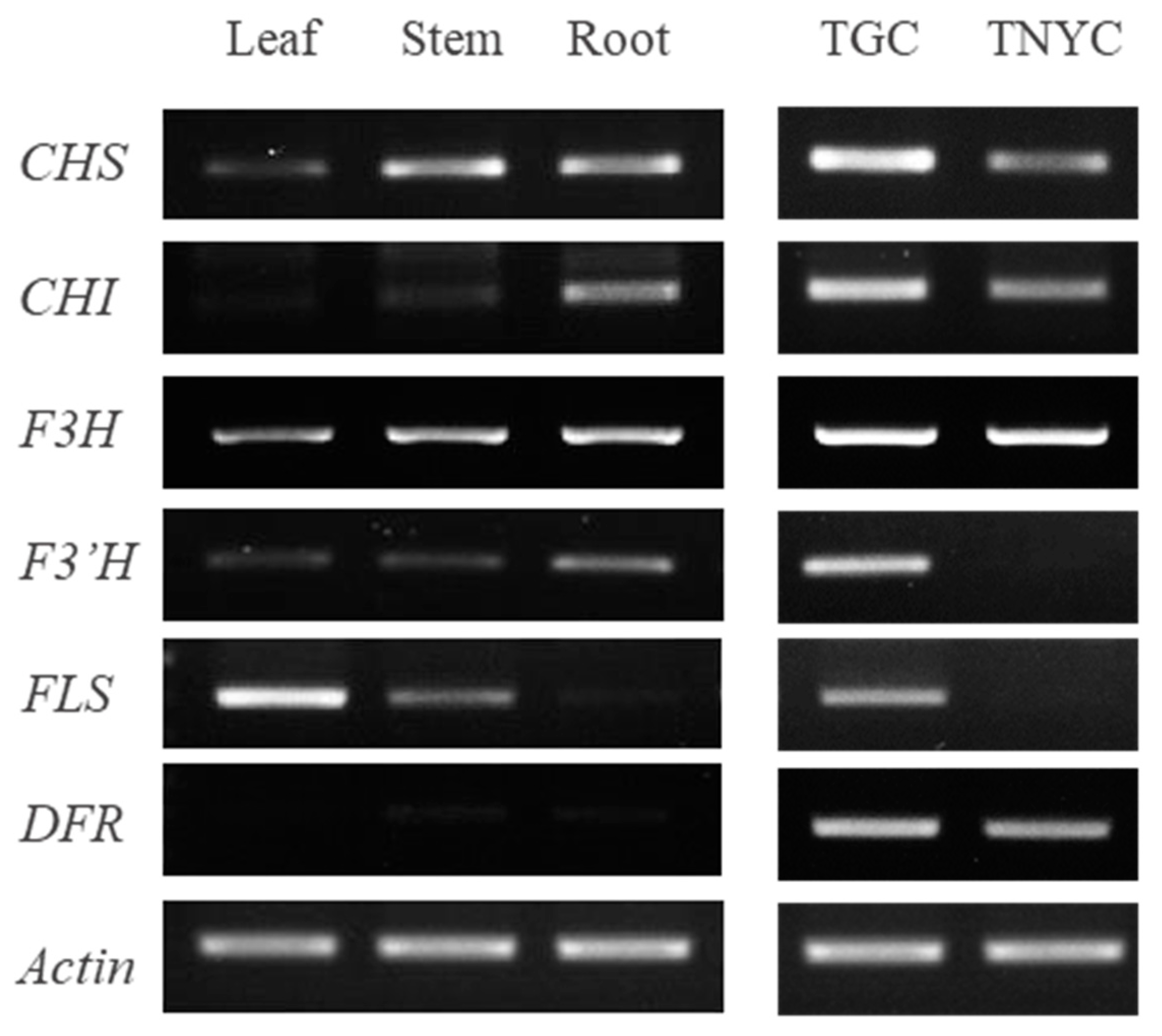
Lee et al. [24] identified the major compounds in NZS as 6-methoxykaempferol-3-O-β-d-glucosyl (1‴→2″)-β-d-glucopyranoside (1), 6-methoxykaempferol-3-O -β-d-glucosyl (1‴→2″)-β-d-glucopyranosyl-(6′′′′-caffeoyl)-7-O-β-d-glucopyranoside (2), and 6,4 ′-dimethoxykaempferol-3-O-β-d-glucosyl (1‴→2″)-β-d-glucopyranosyl-(6′′′′-caffeoyl)-7-O-β-d-glucopyranoside (3). We examined the expressions of the genes involved in kaempferol biosynthesis to confirm the production of these major compounds in the selected calli. The genes targeted chalcone synthase (CHS), chalcone isomerase (CHI), flavanone 3-hydroxylase (F3H), flavonoid 3′-hydroxylase (F3′H), flavonol synthase (FLS), and dihydroflavonol 4-reductase (DFR), and their expressions were compared. The RT-PCR results demonstrate the elevated production of kaempferol, indicated by low expression levels of F3′H and high expression levels of F3H and FLS in the leaves of NZS (Figure 4). In contrast, the roots exhibited minimal FLS expression, implying the predominant production and accumulation of flavonoids. In TGC, all six flavonoid biosynthesis-related genes showed elevated expression levels. TNYC displayed notably low expression levels of F3′H and FLS. In particular, high DFR expression levels of DFR were confirmed in both callus types, suggesting the production of a more diverse array of flavonoids in calli than in plants.

3.4. Analysis of the Phytochemicals of TGC and TNYC

We extracted callus components using 70% ethanol and analyzed the compounds in each extract via GC-MS. Nine components were detected in TGC, whereas TNYC exhibited a higher number of 44 detected components (Figures S2 and S3). There was a substantial difference in the composition of volatile compounds between the two types of calli. In TGC, the predominant components were 2,3-Butanediol (68.6%), cytidine (20.3%), and dimethylamine (5.6%). Conversely (Table 3), TNYC showed a higher abundance of butanedioic acid (23.1%); 3-piperidinol (14.1%); ethylamine, N-ethyl-nitroso (8.2%); and 1,3-propanediol, 2-ethyl-2-(hydroxymethyl)- (6.4%) (Table 4).

4. Discussion

In this study, we introduced callus culture techniques to identify the functional compounds in NZS. To obtain abundant compounds from calli within a short period, it is essential to establish effective callus induction conditions and select fast-growing calli. PGRs play crucial roles in plant cell growth under laboratory conditions. Therefore, in this study, we investigated the effect of PGRs on callus induction from NZS leaves and their subsequent effects on callus growth by examining induction rates and biomasses.
The PGR used in in vitro cultures generally depends on the type of explant and is strongly correlated with the amount of endogenous PGRs [25]. Auxins containing NAA and 2,4-D increase the vacuole size, thereby promoting the formation of smooth and friability calli [26,27]. In particular, 2,4-D is effective at low concentrations and exhibits high stability even at high temperatures, making it widely applicable for callus induction in various plant species [28,29]. Rad et al. [30] reported that the medium containing 0.5 mg·L−1 2,4-D and 2 mg·L−1 6-BA exhibited the most effective induction of calli in Digitalis purpurea L. These results suggest that the callus induction rate tends to increase with increasing concentrations of 2,4-D in the medium. We found that NZS callus induction increased effectively when 2,4-D was added compared to that in controls without 2,4-D. We observed a significant increase in the NZS callus induction when 2,4-D was added compared to the control conditions without 2,4-D. Additionally, a maximal callus induction was observed in the medium supplemented with 2.0 mg·L−1 6-BA and 0.5 mg·L−1 2,4-D. Consequently, our results demonstrate a trend similar to that observed in previous studies.
Furthermore, we selected calli with good growth by culturing them on media with TDZ alone or a combination of TDZ and NAA. TDZ induces callus formation and facilitates somatic embryo development. It plays a crucial role in the initiation and proliferation of calli, shoots, and roots [31,32,33]. However, it is noteworthy that increasing the concentration of TDZ may promote the growth of calli and plants but may result in browning. A careful selection of the TDZ concentration is essential because it can stimulate endogenous ethylene production [34,35]. Singh et al. [36] achieved optimal callus growth with 1.2 mg·L−1 TDZ alone in Santalum album L. Srivastava et al. [37] induced up to 85.56% callus using 9.1 μM of TDZ and 0.5 μM of NAA. In our study, we successfully obtained calli with 2.5 mg·L−1 TDZ, which were non- browning and had excellent growth.
Ultimately, we selected two distinct callus types based on the differences in color and shape. In general, a friable callus is required for cell differentiation cultures. Friable calli are easy to separate and facilitate cell proliferation and are a necessary factor for mass production. We did not select friable calli in the present study, but we additionally compared the growth of calli under different medium conditions. For TNYC, placing 1 g of calli in 50 mL liquid medium resulted in approximately eight- and two-fold increases in fresh and dry weights after five weeks, respectively. In addition, when 0, 1, 3, and 5% sucrose was added to the liquid medium, the fresh weight increased eight folds without a change in the browning in the 5% sucrose-added group (data not shown). Through the results of these further studies, we were able to assess the applicability of TNYC to liquid cultures and obtain supplemental information about growth conditions.
The TPC and TFC of the callus extract were measured to assess the functional potential of the calli. Numerous studies have reported on the antioxidant activity and effectiveness of callus extracts. However, the contents and activities of functional compounds differ depending on the culture conditions and methods used for plant callus culture. In this study, the TPC of the TGC was measured to be 62.9 g GAE/g DW. The TNYC showed 11.3 g GAE/g DW, which was lower than that of the TDZ treatment group. Upon comparing TPC and TFC after callus induction with various PGR combinations in Lallemantia Iberica, the TDZ single treatment group exhibited the highest total phenol (3.93 ± 0.018 mgPYRO/g) and flavonoid (51.27 ± 0.036 mgQUE/g) contents. Mok et al. [38] reported that TDZ inhibits nitrate transport through the stimulation of endogenous cytokinin biosynthesis or through its involvement in metabolism. Nitrate inhibition stimulates secondary metabolites [39]. Considering previous studies, we expected that the biosynthesis of secondary metabolites would be promoted in calli induced using a single TDZ treatment. Moreover, auxins and cytokinins may interact to exhibit synergistic and antagonistic effects, and thus have lower content in mixed treatments compared to single treatments [40]. Hence, variations in TPC and TFC among the selected calli may be attributed to this phenomenon.
In our previous experiments, we observed no statistically significant differences in TPC and TFC between leaves and calli. Consequently, we investigated whether the main compounds in the kaempferol backbone are also biosynthesized in calli. To achieve this, we compared and examined the expressions of six genes involved in the kaempferol and quercetin biosynthesis in callus and plant tissues. The Kaempferol and quercetin productions require dihydrokaempferol and dihydroquercetin, respectively. Furthermore, dihydrokaempferol acts as a substrate for F3′H to produce dihydroquercetin. Finally, FLS converts dihydrokaempferol into kaempferol, and dihydroquercetin into quercetin [41]. Therefore, the production of kaempferol or quercetin was expected to depend on the expression ratios of F3′H and FLS. According to the experimental results, there was a clear difference in the gene expression patterns between the two callus types. TGC exhibited high expressions for all six genes, whereas TNYC had low expressions for F3′H and FLS. Consequently, the two callus types were expected to produce different metabolites.
Plant color results from the accumulation of metabolites (pigments) [42]. In particular, it has been reported that yellow color occurs because of differences in flavonoid or carotenoid content [43]. Therefore, we expected TNYC (yellow) to have a higher flavonoid content than TGC (green) during callus screening. However, the total flavonoid content did not differ significantly between the groups (Figure 3). Therefore, we compared the expressions of flavonoid biosynthesis-related genes. The gene expression patterns of TNYC were high in the expressions of F3H and DFR and low in the expressions of F3′H and FLS (Figure 4). Therefore, the accumulation of dihydroflavonol was expected to be higher in the TNYC than in the other groups. F3H is also an important enzyme involved in anthocyanin and dihydroflavonol [44]. Thus, the high expression of DFR, which regulates anthocyanin accumulation, together with the high expression of F3H, indicates the potential for anthocyanin synthesis in TNYC. Therefore, further studies on the anthocyanin content are required. For TGC, the strong expression of CHS and the high expressions of the five genes indicate the rapid progression of the synthesis and conversion of various flavonoids. As there was no difference in the flavonoid content of greenhouse-grown NZS and TGC, it is expected that various flavonoids will be synthesized in TGC.
Finally, we performed a GC-MS analysis of the secondary metabolite profiles of the two calli. As a result, 9 and 44 components were detected in TGC and TNYC, respectively. None of these components matched the main compounds in NZS. Differences between the secondary metabolite profiles of in vitro and native plants have been revealed in previous reports. In addition, when cultured in vitro, the types and contents of the compounds differed depending on the growth conditions. Locally cultivated Mentha arvensis L. produces essential oils composed of 30 or more components, whereas in vitro-cultivated plant compounds differ depending on culture conditions. Pulegone and isopulegone have been isolated from calli [45]. This pattern has also been observed in Lallemantia iverica. When explants were treated with six different concentrations of TDZ, 55 components were detected and distributed variably among each treatment group [35]. In Justicia gendarussa [46], PGRs exert a significant influence on the production and accumulation of secondary metabolites as well as the growth and development of plants [47]. In the present study, we identified a novel compound, NZS, in the two types of calli selected under different growth conditions.
Among the compounds detected, 2,3-Butanediol (2,3-BD), which is the most abundant in TGC and is used in various chemical synthesis processes, has substantial industrial value in the pharmaceutical, cosmetic, perfume, wetting agent, and food industries [48,49,50]. Additionally, 2,3-BD promotes plant growth and enhances resistance to disease and drought stress by strengthening cell walls [51,52]. Traditionally, 2,3-BD has been produced through chemical processes using petroleum; however, biological processes using microorganisms have recently attracted attention because of their low production costs and minimal waste. In the TGC selected for this study, a high 2,3-Butanediol (2,3-BD) content of 68.6% was detected, suggesting the potential utility of this callus as a new production material. The second most abundant compound in the TGC was cytidine, constituting 20.3% of the composition. Forced swimming experiments, conducted to evaluate the antidepressant effects of cytidine, demonstrated a strong antidepressant effect [53]. Additionally, cytidine stimulates the synthesis of cytidine 5′-diphosphocholine(citicoline). Citicoline has been shown to enhance the production of membrane phospholipids in both cellular and animal models, and it has demonstrated a capacity to reduce cocaine withdrawal symptoms in mice [54,55].
The most prominent compound detected in the TNYC was butanedioic acid (23.1%), also known as succinic acid. Succinic acid is widely used in various industries, including food additives, detergents, spices, cement additives, and antimicrobials. In particular, the various pharmacological effects of succinic acid, such as anti-inflammatory and antibacterial effects, have been reported in many studies [56,57,58]. The demand for succinic acid is steadily increasing and is projected to reach 768 million metric tons by 2025 [59]. Traditionally, succinic acid has been produced through chemical pathways; however, in recent years, there has been an emphasis on biological production methods because of concerns about nonrenewable resources and environmental issues stemming from greenhouse gas emissions. Consequently, the TNYC selected in this study has potential as material capable of producing succinic acid, a crucial industrial chemical.

5. Conclusions

In this study, we utilized callus culture technology to detect and analyze the functional compounds in NZS. The initial optimization of induction conditions with PGRs, particularly the addition of 2.0 mg·L−1 6-BA and 0.5 mg·L−1 2,4-D, resulted in a maximum callus induction rate (100%) and biomass (0.416 ± 0.019 g). Subsequently, two distinct callus types, TGC and TNYC, were selected based on their color and shape variations. The TPC and TFC contents of the selected calli showed no statistically significant differences when compared to leaves grown in greenhouses. Furthermore, the gene involved in the biosynthesis of kaempferol, the backbone of NZS’s main compound of NZS, displayed different expression patterns in the two callus types, suggesting the potential synthesis of flavonoids distinct from those in NZS seedlings.
A GC-MS analysis revealed that 2,3-butanediol (2,3-BD) and succinic acid were the most abundant compounds in the TGC and TNYC, respectively. The current global chemical industry is pursuing biologically based production processes to address the impact of climate change. The calli selected in this study not only provide an effective method for the stable supply of functional compounds but also offer a sustainable solution without causing environmental issues. Moreover, calli containing diverse compounds are expected to serve as high-value raw materials in the industrial sector.

Supplementary Materials

The following supporting information can be downloaded from https://www.mdpi.com/article/10.3390/horticulturae10030244/s1: Figure S1: Appearance of calli induced from NZS leaves after four weeks in the medium; Figure S2: Phytochemical profile of TGC extracts via gas chromatography–mass spectrometry; Figure S3: Phytochemical profile of TNYC extracts via gas chromatography–mass spectrometry; Table S1: Primers used in this study.

Author Contributions

Conceptualization, G.-J.L.; investigation, K.Y.L., D.-H.N., Y.J. and M.A.G.; data curation, K.Y.L., D.-H.N., Y.J. and M.A.G.; formal analysis, K.Y.L., D.-H.N. and Y.J.; funding acquisition, S.U.P., J.C., S.-O.C. and G.-J.L.; methodology, K.Y.L. and G.-J.L.; project administration, G.-J.L.; writing—original draft, K.Y.L., D.-H.N., Y.J. and M.A.G.; writing—review and editing, S.U.P., J.C., S.-O.C. and G.-J.L. All authors have read and agreed to the published version of the manuscript.

Funding

This work was supported by the National Research Foundation of Korea Grant 2021, funded by the Korean Government (MOE).

Data Availability Statement

Data are contained within the article or Supplementary Material.

Conflicts of Interest

The authors declare no conflicts of interest regarding the publication of this article.

References

  1. Karakas, F.P.; Turker, A.U. Improvement of shoot proliferation and comparison of secondary metabolites in shoot and callus cultures of Phlomis armeniaca by LC-ESI-MS/MS analysis. In Vitro Cell. Dev. Biol.-Plant. 2016, 52, 608–618. [Google Scholar] [CrossRef]
  2. Bennett, R.N.; Wallsgrove, R.M. Secondary metabolites in plant defense mechanisms. New Phytol. 1994, 127, 617–633. [Google Scholar] [CrossRef] [PubMed]
  3. Wink, M. Plant secondary metabolism: Diversity, function and its evolution. Nat. Prod. Commun. 2008, 3, 1934578X0800300801. [Google Scholar] [CrossRef]
  4. Van Loon, L.C.; Rep, M.; Pieterse, C.M. Significance of inducible defense-related proteins in infected plants. Annu. Rev. Phytopathol. 2006, 44, 135–162. [Google Scholar] [CrossRef] [PubMed]
  5. Balandrin, M.F.; Klocke, J.A.; Wurtele, E.S.; Bollinger, W.H. Natural plant chemicals: Sources of industrial and medicinal materials. Science 1985, 228, 1154–1160. [Google Scholar] [CrossRef] [PubMed]
  6. Ashraf, M.A.; Iqbal, M.; Rasheed, R.; Hussain, I.; Riaz, M.; Arif, M.S. Environmental stress and secondary metabolites in plants: An overview. In Plant Metabolites and Regulation under Environmental Stress; Academic Press: Cambridge, MA, USA, 2018; pp. 153–167. [Google Scholar]
  7. Yue, W.; Ming, Q.L.; Lin, B.; Rahman, K.; Zheng, C.J.; Han, T.; Qin, L.P. Medicinal plant cell suspension cultures: Pharmaceutical applications and high-yielding strategies for the desired secondary metabolites. Crit. Rev. Biotechnol. 2016, 36, 215–232. [Google Scholar] [CrossRef]
  8. Steward, F.C.; Mapes, M.O.; Mears, K. Growth and organized development of cultured cells. II. Organization in cultures grown from freely suspended cells. Am. J. Bot. 1958, 45, 705–708. [Google Scholar] [CrossRef]
  9. Plasson, C.; Michel, R.; Lienard, D.; Saint-Jore-Dupas, C.; Sourrouille, C.; March, G.G.D.; Gomord, V. Production of recombinant proteins in suspension–cultured plant cells. In Recombinant Proteins from Plants: Methods and Protocols; Humana: New York, NY, USA, 2009; pp. 145–161. [Google Scholar]
  10. Lee, K.Y.; Shin, J.Y.; Ahn, M.S.; Kim, S.J.; An, H.R.; Kim, Y.J.; Kwon, O.H.; Lee, S.Y. Callus Derived from Petals of the Rosa hybrida Breeding Line 15R-12-2 as New Material Useful for Fragrance Production. Plants 2023, 12, 2986. [Google Scholar] [CrossRef]
  11. Dörnenburg, H.; Knorr, D. Strategies for the improvement of secondary metabolite production in plant cell cultures. Enzyme Microb. Technol. 1995, 17, 674–684. [Google Scholar] [CrossRef]
  12. Anjum, S.; Abbasi, B.H.; Hano, C. Trends in accumulation of pharmacologically important antioxidant-secondary metabolites in callus cultures of Linum usitatissimum L. Plant Cell Tissue Organ Cult. 2017, 129, 73–87. [Google Scholar] [CrossRef]
  13. Adil, M.; Ren, X.; Kang, D.I.; Thi, L.T.; Jeong, B.R. Effect of explant type and plant growth regulators on callus induction, growth and secondary metabolites production in Cnidium officinale Makino. Mol. Biol. Rep. 2018, 45, 1919–1927. [Google Scholar] [CrossRef]
  14. Coskun, Y.; Duran, R.E.; Kilic, S. Striking effects of melatonin on secondary metabolites produced by callus culture of rosemary (Rosmarinus officinalis L.). Plant Cell Tissue Organ Cult. 2019, 138, 89–95. [Google Scholar] [CrossRef]
  15. Wilson, C.; Lesch, S.M.; Grieve, C.M. Growth stage modulates salinity tolerance of New Zealand Spinach (Tetragonia tetragonioides, Pall) and Red Orach (Atriplex hortensis L.). Ann. Bot. 2000, 85, 501–509. [Google Scholar] [CrossRef]
  16. Kato, M.; Takeda, T.; Ogihara, Y.; Shimu, M.; Nomura, T.; Tomita, Y. Studies on the structure of polysaccharide from Tetragonia tetragonioides. Chem. Pharm. Bull. 1985, 33, 3675–3680. [Google Scholar] [CrossRef]
  17. Choi, H.J.; Kang, J.S.; Choi, Y.W.; Jeong, Y.K.; Joo, W.H. Inhibitory activity on the diabetes related enzymes of Tetragonia tetragonioides. KSBB J. 2008, 23, 419–424. [Google Scholar]
  18. Lee, M.A.; Choi, H.J.; Kang, J.S.; Choi, Y.W.; Joo, W.H. Antioxidant activities of the solvent extracts from Tetragonia tetragonioides. J. Life Sci. 2008, 18, 220–227. [Google Scholar] [CrossRef]
  19. Choi, H.J.; Yee, S.T.; Kwon, G.S.; Joo, W.H. Anti-inflammatory and anti-tumor effects of Tetragonia tetragonoides extracts. Microbiol. Biotechnol. Lett. 2015, 43, 391–395. [Google Scholar] [CrossRef]
  20. Choi, H.J.; Yee, S.T.; Joo, W.H. Antidiabetic activity of polysaccharide extract from Tetragonia tetragonoides in streptozotocin-induced diabetic mice. J. Life Sci. 2017, 27, 579–583. [Google Scholar] [CrossRef]
  21. Kim, D.S.; Ko, B.S.; Ryuk, J.A.; Park, S. Tetragonia tetragonioides protected against memory dysfunction by elevating hippocampal amyloid-β deposition through potentiating insulin signaling and altering gut microbiome composition. Int. J. Mol. Sci. 2020, 21, 2900. [Google Scholar] [CrossRef]
  22. Sunagawa, H.; Agarie, S.; Umemoto, M.; Makishi, Y.; Nose, A. Effect of urea-type cytokinins on the adventitious shoots plant, Mesembryanthemum crystallinum. Plant Prod. Sci. 2007, 10, 47–56. [Google Scholar] [CrossRef]
  23. Kumazawa, S.; Hamasaka, T.; Nakayama, T. Antioxidant activity of propolis of various geographic origins. Food Chem. 2004, 84, 329–339. [Google Scholar] [CrossRef]
  24. Lee, Y.S.; Kim, S.H.; Yuk, H.J.; Lee, G.J.; Kim, D.S. Tetragonia tetragonoides (Pall.) Kuntze (New Zealand Spinach) prevents obesity and hyperuricemia in high-fat diet-induced obese mice. Nutrients 2018, 10, 1087. [Google Scholar] [CrossRef] [PubMed]
  25. Centeno, M.L.; Rodríguez, A.; Feito, I.; Fernández, B. Relationship between endogenous auxin and cytokinin levels and morphogenic responses in Actinidia deliciosa tissue cultures. Plant Cell Rep. 1996, 16, 58–62. [Google Scholar] [CrossRef] [PubMed]
  26. Gueye, B.; Morcillo, F.; Collin, M.; Gargani, D.; Overvoorde, P.; Aberlenc-Bertossi, F.; Tranbarger, T.; Sane, D.; Tregear, J.W.; Borgel, A.; et al. Acquisition of callogenic capacity in date palm leaf tissues in response to 2,4-D treatment. Plant Cell Tissue Organ Cult. 2009, 99, 35–45. [Google Scholar] [CrossRef]
  27. Berejsza-Wysecki, W.; Hrazdin, G. Establishment of callus and cell suspension cultures of raspberry (Rubus idaeus cv. Royalty). Plant Cell Tissue Organ Cult. 1994, 37, 213–216. [Google Scholar] [CrossRef]
  28. Gaj, M.D. Factors influencing somatic embryogenesis induction and plant regeneration with particular reference to Arabidopsis thaliana (L.) Heynh. Plant Growth Regul. 2004, 43, 27–47. [Google Scholar] [CrossRef]
  29. George, E.F.; Hall, M.A.; De Klerk, G.J. Plant Propagation by Tissue Culture, 3rd ed.; Springer: Dordrecht, The Netherlands, 2008; 501p. [Google Scholar]
  30. Rad, M.M.; Abdossi, V.; Moradi, P.; Rakhshandehroo, F.; Mehrafarin, A. Phytochemical changes of Digitalis purpurea L. in response to polyamines and methyl jasmonate application in callus culture. J. Plant Biochem. Biotechnol. 2022, 31, 310–319. [Google Scholar] [CrossRef]
  31. Linden, L.; Riikonen, A. Effects of 6-benzylaminopurine, thidiazuron and type of explant on in vitro shoot development of Acer platanoides L. Propag. Ornam. Plants 2006, 6, 201–204. [Google Scholar]
  32. Jones, M.P.; Yi, Z.; Murch, S.J.; Saxena, P.K. Thidiazuron-induced regeneration of Echinacea purpurea L.: Micropropagation in solid and liquid culture systems. Plant Cell Rep. 2007, 26, 13–19. [Google Scholar] [CrossRef]
  33. Wang, J.; Bao, M.Z. Plant regeneration of pansy (Viola wittrockiana) ‘Caidie’ via petiole-derived callus. Sci. Hortic. 2007, 111, 266–270. [Google Scholar] [CrossRef]
  34. Suttle, J.C. Involvement of ethylene in the action of the cotton defoliant thidiazuron. Plant Physiol. 1985, 78, 272–276. [Google Scholar] [CrossRef]
  35. Pourebad, N.; Motafakkerazad, R.; Kosari-Nasab, M.; Farsad Akhtar, N.; Movafeghi, A. The influence of TDZ concentrations on in vitro growth and production of secondary metabolites by the shoot and callus culture of Lallemantia iberica. Plant Cell Tissue Organ Cult. 2015, 122, 331–339. [Google Scholar] [CrossRef]
  36. Singh, C.K.; Raj, S.R.; Jaiswal, P.S.; Patil, V.R.; Punwar, B.S.; Chavda, J.C.; Subhash, N. Effect of plant growth regulators on in vitro plant regeneration of sandalwood (Santalum album L.) via organogenesis. Agrofor. Syst. 2016, 90, 281–288. [Google Scholar] [CrossRef]
  37. Srivastava, S.; Krishna, R.; Sinha, R.P.; Singh, M. TDZ-induced plant regeneration in Brassica oleracea L. var. botrytis: Effect of antioxidative enzyme activity and genetic stability in regenerated plantlets. In Vitro Cell. Dev. Biol.-Plant. 2017, 53, 598–605. [Google Scholar]
  38. Mok, M.C.; Martin, R.C.; Mok, D.W.S. Cytokinins: Biosynthesis metabolism and perception. In Vitro Cell. Dev. Biol.-Plant. 2000, 36, 102–107. [Google Scholar] [CrossRef]
  39. Sakakibara, H. Cytokinins: Activity, biosynthesis, and translocation. Annu. Rev. Plant Biol. 2006, 57, 431–449. [Google Scholar] [CrossRef] [PubMed]
  40. Coenen, C.; Lomax, T.L. Auxin—Cytokinin interactions in higher plants: Old problems and new tools. Trends Plant Sci. 1997, 2, 351–356. [Google Scholar] [CrossRef] [PubMed]
  41. Crozier, A.; Jaganath, I.B.; Clifford, M.N. Dietary phenolics: Chemistry, bioavailability and effects on health. Nat. Prod. Rep. 2009, 26, 1001–1043. [Google Scholar] [CrossRef] [PubMed]
  42. Mol, J.; Grotewold, E.; Koes, R. How genes paint flowers and seeds. Trends Plant Sci. 1998, 3, 212–217. [Google Scholar] [CrossRef]
  43. Deng, J.; Chen, S.; Yin, X.; Wang, K.; Liu, Y.; Li, S.; Yang, P. Systematic qualitative and quantitative assessment of anthocyanins, flavones and flavonols in the petals of 108 lotus (Nelumbo nucifera) cultivars. Food Chem. 2013, 139, 307–312. [Google Scholar] [CrossRef] [PubMed]
  44. Cheynier, V.; Comtem, G.; Davies, K.M.; Lattanzio, V.; Martens, S. Plant phenolics: Recent advances on their biosynthesis, genetics, and ecophysiology. Plant Physiol. Biochem. 2013, 72, 1–20. [Google Scholar] [CrossRef]
  45. Phatak, S.V.; Heble, M.R. Organogenesis and terpenoid synthesis in Mentha arvensis. Fitoterapia 2002, 73, 32–39. [Google Scholar] [CrossRef]
  46. Wahyuni, D.K.; Huda, A.; Faizah, S.; Purnobasuki, H.; Wardojo, B.P.E. Effects of light, sucrose concentration and repetitive subculture on callus growth and medically important production in Justicia gendarussa Burm. f. Plant Biotechnol. Rep. 2020, 27, e00473. [Google Scholar]
  47. Richard, C.; Lescot, M.; Inzé, D.; De Veylder, L. Effect of auxin, cytokinin, and sucrose on cell cycle gene expression in Arabidopsis thaliana cell suspension cultures. Plant Cell Tissue Organ Cult. 2002, 69, 167–176. [Google Scholar] [CrossRef]
  48. Garg, S.K.; Jain, A. Fermentative production of 2,3-butanediol: A review. Bioresour. Technol. 1995, 51, 103–109. [Google Scholar] [CrossRef]
  49. Celińska, E.; Grajek, W. Biotechnological production of 2,3-butanediol—Current state and prospects. Biotechnol. Adv. 2009, 27, 715–725. [Google Scholar] [CrossRef] [PubMed]
  50. Syu, M.J. Biological production of 2,3-butanediol. Appl. Microbiol. Biotechnol. 2001, 55, 10–18. [Google Scholar] [CrossRef] [PubMed]
  51. Liu, X.J.; Ma, Y.; Shi, Y.; Ma, H.L. 2,3-Butanediol induces brown blotch resistance in creeping bentgrass by strengthening cell wall structure and promoting lignin synthesis of precursor phenolic acid. Acta Physiol. Plant. 2023, 45, 40. [Google Scholar] [CrossRef]
  52. Yasmin, H.; Rashid, U.; Hassan, M.N.; Nosheen, A.; Naz, R.; Ilyas, N.; Sajjad, M.; Azmat, A.; Alyemeni, M.N. Volatile organic compounds produced by Pseudomonas pseudoalcaligenes alleviated drought stress by modulating defense system in maize (Zea mays L.). Physiol. Plant. 2021, 172, 896–911. [Google Scholar] [CrossRef] [PubMed]
  53. Carlezon, W.A., Jr.; Pliakas, A.M.; Parow, A.M.; Detke, M.J.; Cohen, B.M.; Renshaw, P.F. Antidepressant-like effects of cytidine in the forced swim test in rats. Biol. Psychiatry 2002, 51, 882–889. [Google Scholar] [CrossRef] [PubMed]
  54. Knapp, S.; Wurtman, R.J. Enhancement of free fatty acid incorporation into phospholipids by choline plus cytidine. Brain Res. 1999, 822, 52–59. [Google Scholar] [CrossRef] [PubMed]
  55. Markou, A.; Hauger, R.L.; Koob, G.F. Desmethylimipramine attenuates cocaine withdrawal in rats. Psychopharmacology 1992, 109, 305–314. [Google Scholar] [CrossRef] [PubMed]
  56. Radkowski, M.; Zdrodowska, B.; Gomółka-Pawlicka, M. Effect of succinic acid on elimination of Salmonella in chicken meat. J. Food Prot. 2018, 81, 1491–1495. [Google Scholar] [CrossRef] [PubMed]
  57. Nissen, M.D.; Lau, E.T.; Cabot, P.J.; Steadman, K.J. Baltic amber teething necklaces: Could succinic acid leaching from beads provide anti-inflammatory effects? BMC Complement. Altern. Med. 2019, 19, 162. [Google Scholar] [CrossRef]
  58. Harber, K.J.; de Goede, K.E.; Verberk, S.G.; Meinster, E.; de Vries, H.E.; van Weeghel, M.; de Winther, M.P.; Van den Bossche, J. Succinate is an inflammation-induced immunoregulatory metabolite in macrophages. Metabolites 2020, 10, 372. [Google Scholar] [CrossRef]
  59. Global Succinic Acid Market Analysis & Trends [2013–2017]—Industry Forecast to 2025: $1.76 Billion Growth Opportunities/Investment Opportunities—Research and Markets. Available online: https://ceo.ca/@newswire/global-succinic-acid-market-analysis-trends-2013-2017 (accessed on 6 March 2017).
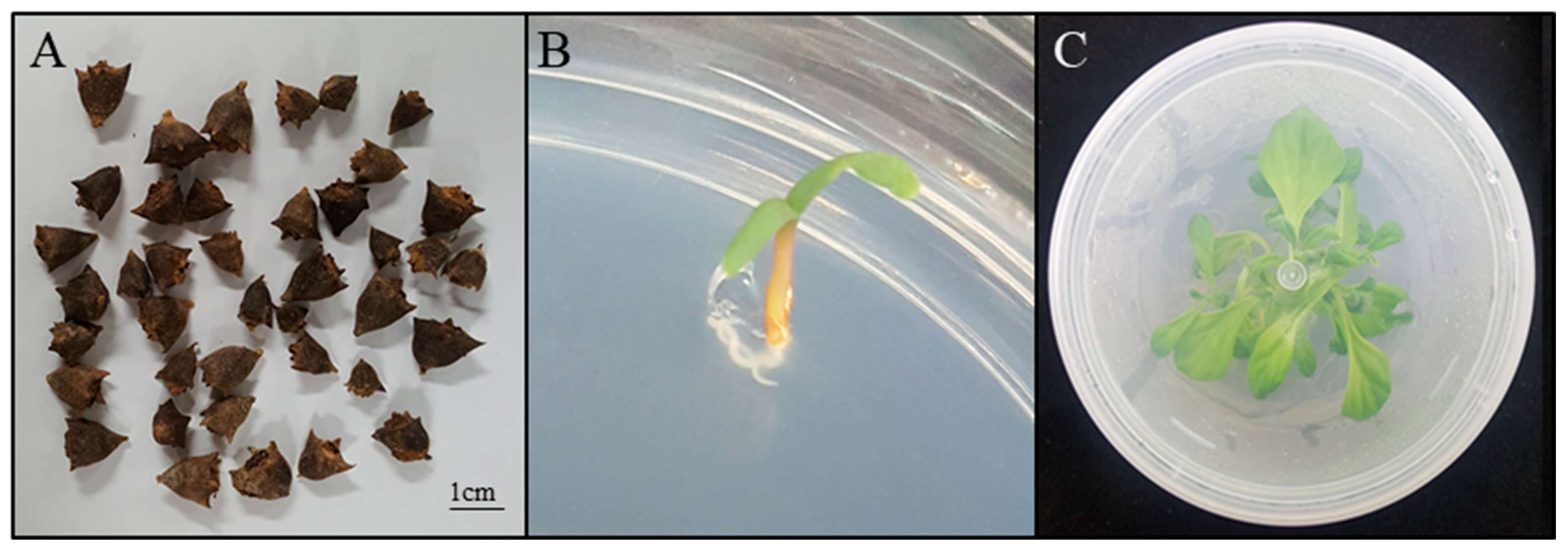
Figure 1. Tissue culture seedlings of NZS used for the induction of callus in this study. (A) Seeds of NZS collected in native habitat. (B) Young seedling germinated from seed. (C) Seedlings grown in vitro for eight weeks.
Horticulturae 10 00244 g001
Figure 2. Two types of calli selected after induction from the leaves of NZS. (A) Hard tissue and green callus induced in MS medium with 2.5 mg·L−1 TDZ (TGC). (B) Hard tissue and yellow callus induced in MS medium with 2.5 mg·L−1 TDZ and 0.5 mg·L−1 NAA (TNYC).
Horticulturae 10 00244 g002
Figure 3. The contents of total phenolics (A) and flavonoids (B) in the selected two types of calli. Groups sharing the same letter do not exhibit statistically significant differences, as determined by Duncan’s test. NS: not significant.
Horticulturae 10 00244 g003
Figure 4. Comparison of the relative expression levels of genes involved in flavonoid synthesis between the seedlings of NZS and selected calli via semi-quantitative RT-PCR analysis. CHS: chalcone synthase, CHI: chalcone isomerase, F3H: flavanone 3-hydroxylase, F3′H: flavonoid 3′-hydroxylase, FLS: flavonol synthase, DFR: dihydroflavonol 4-reductase.
Horticulturae 10 00244 g004
Table 1. Induction rates and fresh weights of calli in MS medium with various concentrations of zeatin and auxin (IAA, NAA, 2,4-D).
Plant Growth Regulators
(mg·L−1)
Callus
Induction (%) Z
Fresh
Weight (g) Z
ZeatinIAANAA2,4-D
0.50.5 0.0 ± 0.0 b0.046 ± 0.002 ij
0.51.0 0.0 ± 0.0 b0.064 ± 0.002 g–j
0.51.5 0.0 ± 0.0 b0.065 ± 0.004 g–j
0.52.0 60.0 ± 0.0 ab0.041 ± 0.003 ij
1.00.5 75.0 ± 2.5 ab0.053 ± 0.001 h–j
1.01.0 0.0 ± 0.0 b0.054 ± 0.003 h–j
1.01.5 0.0 ± 0.0 b0.058 ± 0.001 h–j
1.02.0 0.0 ± 0.0 b0.069 ± 0.005 g–j
1.50.5 65.0 ± 2.5 ab0.045 ± 0.004 ij
1.51.0 25.0 ± 2.5 b0.055 ± 0.004 h–j
1.51.5 25.0 ± 2.5 b0.058 ± 0.005 h–j
1.52.0 0.0 ± 0.0 b0.055 ± 0.003 h–j
2.00.5 0.0 ± 0.0 b0.044 ± 0.001 ij
2.01.0 0.0 ± 0.0 b0.037 ± 0.000 j
2.01.5 0.0 ± 0.0 b0.039 ± 0.001 ij
2.02.0 0.0 ± 0.0 b0.055 ± 0.004 h–j
0.5 0.5 75.0 ± 7.5 ab0.070 ± 0.004 g–j
0.5 1.0 80.0 ± 10.0 ab0.117 ± 0.010 c–j
0.5 1.5 95.0 ± 2.5 ab0.081 ± 0.006 e–j
0.5 2.0 90.0 ± 0.0 ab0.207 ± 0.034 a–c
1.0 0.5 65.0 ± 2.5 ab0.095 ± 0.013 d–j
1.0 1.0 70.0 ± 5.0 ab0.057 ± 0.003 h–j
1.0 1.5 100.0 ± 0.0 ab0.081 ± 0.006 e–j
1.0 2.0 90.0 ± 0.0 ab0.075 ± 0.004 f–j
1.5 0.5 65.0 ± 7.5 ab0.090 ± 0.005 d–j
1.5 1.0 65.0 ± 7.5 ab0.124 ± 0.007 c–j
1.5 1.5 70.0 ± 15.0 ab0.093 ± 0.011 d–j
1.5 2.0 45.0 ± 17.5 ab0.035 ± 0.003 j
2.0 0.5 45.0 ± 17.5 ab0.087 ± 0.010 d–j
2.0 1.0 70.0 ± 15.0 ab0.076 ± 0.001 f–j
2.0 1.5 65.0 ± 12.5 ab0.057 ± 0.001 h–j
2.0 2.0 95.0 ± 2.5 ab0.148 ± 0.007 a–g
0.5 0.560.0 ± 20.0 ab0.176 ± 0.014 a–e
0.5 1.0100.0 ± 0.0 ab0.081 ± 0.004 e–j
0.5 1.575.0 ± 2.5 ab0.088 ± 0.009 d–j
0.5 2.085.0 ± 2.5 ab0.044 ± 0.002 ij
1.0 0.565.0 ± 17.5 ab0.172 ± 0.002 a–f
1.0 1.0100.0 ± 0.0 a0.180 ± 0.008 a–d
1.0 1.5100.0 ± 0.0 a0.234 ± 0.009 a
1.0 2.0100.0 ± 0.0 a0.133 ± 0.010 c–j
1.5 0.5100.0 ± 0.0 a0.228 ± 0.027 ab
1.5 1.0100.0 ± 0.0 a0.139 ± 0.009 c–j
1.5 1.5100.0 ± 0.0 a0.076 ± 0.001 f–j
1.5 2.0100.0 ± 0.0 a0.060 ± 0.004 h–j
2.0 0.5100.0 ± 0.0 a0.161 ± 0.013 a–g
2.0 1.0100.0 ± 0.0 a0.112 ± 0.002 d–j
2.0 1.5100.0 ± 0.0 a0.237 ± 0.004 a
2.0 2.0100.0 ± 0.0 a0.072 ± 0.002 g–j
Z Means followed by the same letter are not significantly different from each other (p < 0.001), as determined using Duncan’s multiple range test (DMRT).
Table 2. Induction rates and fresh weights of calli in MS medium with various concentrations of 6-BA auxin (NAA, 2,4-D).
Plant Growth Regulators
(mg·L−1)
Callus
Induction (%) Z
Fresh
Weight (g) Z
6-BANAA2,4-D
0.00.0 0.0 ± 0.0 d0.014 ± 0.001 d
0.00.5 0.0 ± 0.0 d0.016 ± 0.001 d
0.01.0 0.0 ± 0.0 d0.017 ± 0.001 d
0.01.5 0.0 ± 0.0 d0.013 ± 0.001 d
1.00.0 40.0 ± 2.9 bc0.215 ± 0.005 d
1.00.5 16.7 ± 1.7 b–d0.026 ± 0.003 d
1.01.0 0.0 ± 0.0 b–d0.013 ± 0.001 d
1.01.5 0.0 ± 0.0 cd0.015 ± 0.001 d
1.50.0 26.7 ± 1.7 b–d0.211 ± 0.004 d
1.50.5 10.0 ± 2.9 d0.023 ± 0.002 d
1.51.0 10.0 ± 5.0 b–d0.026 ± 0.003 d
1.51.5 26.7 ± 7.3 cd0.035 ± 0.001 d
2.00.0 16.7 ± 3.3 b–d0.139 ± 0.001 d
2.00.5 3.3 ± 1.7 d0.019 ± 0.001 d
2.01.0 3.3 ± 1.7 b–d0.020 ± 0.004 d
2.01.5 46.7 ± 13.0 b0.047 ± 0.007 d
0.0 0.00.0 ± 0.0 d0.013 ± 0.001 d
0.0 0.586.7 ± 3.3 b0.060 ± 0.003 cd
0.0 1.0100.0 ± 0.0 b–d0.026 ± 0.015 d
0.0 1.5100.0 ± 0.0 b–d0.021 ± 0.004 d
1.0 0.046.7 ± 3.3 a0.124 ± 0.005 bc
1.0 0.5100.0 ± 0.0 a0.221 ± 0.001 b
1.0 1.0100.0 ± 0.0 a0.211 ± 0.003 b
1.0 1.5100.0 ± 0.0 a0.207 ± 0.006 b
1.5 0.033.3 ± 4.4 a0.220 ± 0.002 b
1.5 0.5100.0 ± 0.0 a0.344 ± 0.011 a
1.5 1.0100.0 ± 0.0 a0.337 ± 0.003 a
1.5 1.5100.0 ± 0.0 a0.392 ± 0.034 a
2.0 0.020.0 ± 7.6 a0.210 ± 0.004 b
2.0 0.5100.0 ± 0.0 a0.416 ± 0.019 a
2.0 1.0100.0 ± 0.0 a0.380 ± 0.027 a
2.0 1.5100.0 ± 0.0 a0.375 ± 0.021 a
Z Means followed by the same letter are not significantly different from each other (p < 0.001), as determined using Duncan’s multiple range test (DMRT).
Table 3. Classification and content (%) of phytochemicals identified via GC-MS analysis in TGC.
Peak No. R.T (min) z Identified Metabolites Area (%) y
17.58252,3-Butanediol 68.55
29.3225Ethanol 5.544
39.3779Cyclopentasiloxane, decamethyl- -
411.8558Dimethylamine 5.647
517.3404Cytidine 20.258
623.05865,6,8,9-TETRAMETHOXY-2-METHYLPEPERO (3,4,5-JK)-9,10-DIHYDROPHENANTHRACENE -
723.3906Silane, 1,4-phenylenebis[trimethyl- -
823.6858Silicone grease, Siliconfett -
923.7903Silicone grease, Siliconfett -
z,y means the time each substance stays in the column (R.T) and the calculated area of each peak (Area), respectively.
Table 4. Classification and content (%) of phytochemicals identified via GC-MS analysis in TNYC.
Peak No.R.T (min) zIdentified MetabolitesArea (%) y
14.5327Butanedioic acid23.053
24.7971Ethanol1.855
35.4305Ethanol-
46.156Ethyl 4-(N-(2-nitro)benzylidene)amino-benzoate3.528
56.2052Cyclotetrasiloxane, octamethyl-3.519
67.21355,6,8,9-TETRAMETHOXY-2-METHYLPEPERO (3,4,5-JK)-9,10-DIHYDROPHENANTHRACENE1.605
77.4165Propanoic acid, 3-(acetylthio)-2-methyl-3.473
87.6316Thioacetonitrile4.078
98.7384N-(Methoxycarbonylmethyl)-N-ethylnitrosamine4.666
108.96591-PROPANOL-O-D2.314
119.25496-Aza-5,7,12,14-tetrathiapentacene-
129.7714Cyclopentasiloxane, decamethyl-1.243
1310.374Ammonia1.726
1410.47856-Aza-5,7,12,14-tetrathiapentacene1.243
1510.706(S)-N-(Ethoxycarbonyl)valine-
1611.48692,4,5-Trioxoimidazolidine1.588
1711.56682,3,4-Tridesoxy-5,6:8,9-di-O-isopropyliden-3-methyl-4-nitro-.beta.-D-manno-4-nonulo-4,7-furanosononitrile5.062
1811.76351,3,5-Triazine, hexahydro-1,3,5-trimethyl-2.855
1912.04021H-Imidazole-4-carboxamide, 5-amino--
2012.7104Ethanamine, N-ethyl-N-nitroso-8.237
2112.858L-Alanine, N-glycyl-2.35
2213.25152,3-Dihydro-3,5-dihydroxy-6-methyl-4H-pyran-4-one-
2314.15542-Furancarboxaldehyde, 5-(hydroxymethyl)-2.104
2415.06541-(2-Adamantylidene)semicarbazide-
2515.98772-Methoxy-4-vinylphenol1.529
2616.71933-Piperidinol14.137
2716.75013-Piperidinol-
2817.35881,3-Propanediol, 2-ethyl-2-(hydroxymethyl)-6.378
2917.6971-Propanol, 3-amino--
3019.6031E-1-phenylbutetene-
3119.7383E-1-phenylbutetene-
3219.78753,27-Dioxa-2,28-disilanonacosane, 2,2,4,28,28-pentamethyl-0.854
3319.9597Phenol, 2,2′-[(1-methyl-1,2-ethanediyl)bis(nitrilomethylidyne)]bis-0.854
3421.1095Urea-
3521.47844-HYDROXY-3-NITROCOUMARIN0.938
3621.66284-HYDROXY-3-NITROCOUMARIN-
3721.8166Phenol, 2,2′-[(1-methyl-1,2-ethanediyl)bis(nitrilomethylidyne)]bis--
3822.9115,6,8,9-TETRAMETHOXY-2-METHYLPEPERO(3,4,5-JK)-9,10-DIHYDROPHENANTHRACENE-
3923.13245,6,8,9-TETRAMETHOXY-2-METHYLPEPERO(3,4,5-JK)-9,10-DIHYDROPHENANTHRACENE-
4023.22465,6,8,9-TETRAMETHOXY-2-METHYLPEPERO(3,4,5-JK)-9,10-DIHYDROPHENANTHRACENE-
4123.42755,6,8,9-TETRAMETHOXY-2-METHYLPEPERO(3,4,5-JK)-9,10-DIHYDROPHENANTHRACENE-
4223.57511,1,1,3,5,5,5-Heptamethyltrisiloxane-
4323.60581,3-Bis(trimethylsilyl)benzene0.854
4423.7657Silicone grease, Siliconfett0.854
z,y means the time each substance stays in the column (R.T) and the calculated area of each peak (Area), respectively.
Disclaimer/Publisher’s Note: The statements, opinions and data contained in all publications are solely those of the individual author(s) and contributor(s) and not of MDPI and/or the editor(s). MDPI and/or the editor(s) disclaim responsibility for any injury to people or property resulting from any ideas, methods, instructions or products referred to in the content.