Article

Enrichment of Rabbit Primitive Hematopoietic Cells via MACS Depletion of CD45+ Bone Marrow Cells

by
Jaromír Vašíček
1,2,*,
Andrej Baláži
1,
Miroslav Bauer
1,3,
Andrea Svoradová
1,
Mária Tirpáková
2,4,
Ľubomír Ondruška
1,
Vladimír Parkányi
1,
Alexander V. Makarevich
1 and
Peter Chrenek
1,2
1
Institute of Farm Animal Genetics and Reproduction, Research Institute for Animal Production Nitra, NPPC, Hlohovecká 2, 951 41 Lužianky, Slovakia
2
Department of Biochemistry and Biotechnology, Faculty of Biotechnology and Food Science, Slovak University of Agriculture in Nitra, Tr. A. Hlinku 2, 949 76 Nitra, Slovakia
3
Department of Botany and Genetics, Faculty of Natural Sciences, Constantine the Philosopher University in Nitra, Nábrežie mládeže 91, 949 74 Nitra, Slovakia
4
AgroBioTech Research Center, Slovak University of Agriculture in Nitra, Tr. A. Hlinku 2, 949 76 Nitra, Slovakia
*
Author to whom correspondence should be addressed.
Magnetochemistry 2021, 7(1), 11; https://doi.org/10.3390/magnetochemistry7010011
Submission received: 29 November 2020 / Revised: 29 December 2020 / Accepted: 5 January 2021 / Published: 13 January 2021
(This article belongs to the Special Issue Magnetic Cell Separation)

Abstract

:
Hematopoietic stem and progenitor cells (HSC/HPCs) of human or few animal species have been studied for over 30 years. However, there is no information about rabbit HSC/HPCs, although they might be a valuable animal model for studying human hematopoietic disorders or could serve as genetic resource for the preservation of animal biodiversity. CD34 marker is commonly used to isolate HSC/HPCs. Due to unavailability of specific anti-rabbit CD34 antibodies, a novel strategy for the isolation and enrichment of rabbit HSC/HPCs was used in this study. Briefly, rabbit bone marrow mononuclear cells (BMMCs) were sorted immunomagnetically in order to remove all mature (CD45+) cells. The cells were depleted with overall purity about 60–70% and then cultured in a special medium designed for the expansion of CD34+ cells. Quantitative Polymerase Chain Reaction (qPCR) analysis confirmed the enrichment of primitive hematopoietic cells, as the expression of CD34 and CD49f increased (p < 0.05) and CD45 decreased (p < 0.001) at the end of culture in comparison to fresh BMMCs. However, cell culture still exhibited the presence of CD45+ cells, as identified by flow cytometry. After gating on CD45 cells the MHCI+MHCIICD38+CD49f+CD90CD117 phenotype was observed. In conclusion, rabbit HSC/HPCs might be isolated and enriched by the presented method. However, further optimization is still required.

1. Introduction

Bone marrow (BM) is recognized as the major hematopoietic organ that is located inside the bones representing about 5% of human body [1] and producing new blood cells [2]. In general, BM consists of the bone marrow mesenchymal stem cells (BM-MSCs) that are responsible for the regeneration of bone tissue and quiescent hematopoietic stem cells (HSCs), which produce different types of immune cells necessary for the proper immune answer [3,4]. HSCs are multipotent and self-renewal ancestors of all blood cell types [5,6,7] that differentiate into the hematopoietic progenitor cells (HPCs; myeloid and lymphoid precursors). Thus, HSC/HPCs could give rise to either myeloid lineage cells such as erythrocytes, granulocytes, macrophages, monocytes, and platelets, or lymphoid lineage cells as B and T lymphocytes and natural killer (NK) cells [8] and so maintain the homeostasis of the hematopoietic system.
Over the years, HSCs have become of great importance to the treatment of human hematological disorders. These cells are mainly obtained from BM, mobilized peripheral blood, and umbilical cord blood [9] by the enrichment of CD34 positive cells using magnetic-activated cell sorting (MACS). The MACS technique has become a standard procedure for the selection of abundant or even rare cells from various biological sources of different species [10] based on the immunological principle of antigen-antibody binding or other chemical interactions of the specific protein binding affinity (e.g., streptavidin-biotin complex or binding of annexin V to phosphatidylserine, etc.).
At present, CD34 is still the most often used marker to identify or isolate human HSCs, although several other unique markers have been identified to be present on them [11]. On the other hand, no marker, even CD34 or other reagents, exists that could be used for the identification or isolation of rabbit HSC/HPCs thus making them unavailable for the study of HSC transplantation or for stem cell banking [12]. However, animal HSCs of other species (mouse, swine, cattle, sheep, or dog) are normally used in biomedicine [13,14,15,16,17], there is currently a demand for small animal models besides mouse, such as rabbit, that could be used for the regenerative medicine applications. Rabbit is closer to human than mouse in terms of genetics [18] and, moreover, rabbits have many hereditary diseases commonly occurring in the human population [19]. Hence, all these facts indicate that rabbit itself is a valuable animal model not only for basic research, but rabbit HSC/HPCs might be an interesting biological model for hematopoietic stem cell therapies, disease modeling, or pharmaceutical screening, etc. In addition, these cells might be cryopreserved in stem cell banks as valuable genetic resources in order to preserve the biodiversity of domestic farm animals. Unfortunately, the insufficient knowledge about rabbit HSCs does not allow using them for these goals [12].
Nevertheless, aside from the positive selection (e.g., direct sorting of CD34+ cells), the MACS method offers also negative selection of targeted cells by the depletion of unwanted cells from the heterogeneous cell population [20]. As HSCs do not express lineage commitment markers that are typical for the mature hematopoietic cells (lymphoid and myeloid lineage cells and their committed precursors) [21], they could be possibly enriched by the depletion of mature lineage positive cells [22]. Our preliminary results [22] indicated that this sorting strategy might be used until other rabbit HSCs-specific marker (CD34 or other) is discovered.
Therefore, the aim of this study was to enrich the cell population expressing CD34, as a promising rabbit HSC/HPCs marker, by the depletion of mature hematopoietic cells from the bone marrow and their expansion in a specific culture medium.

2. Results

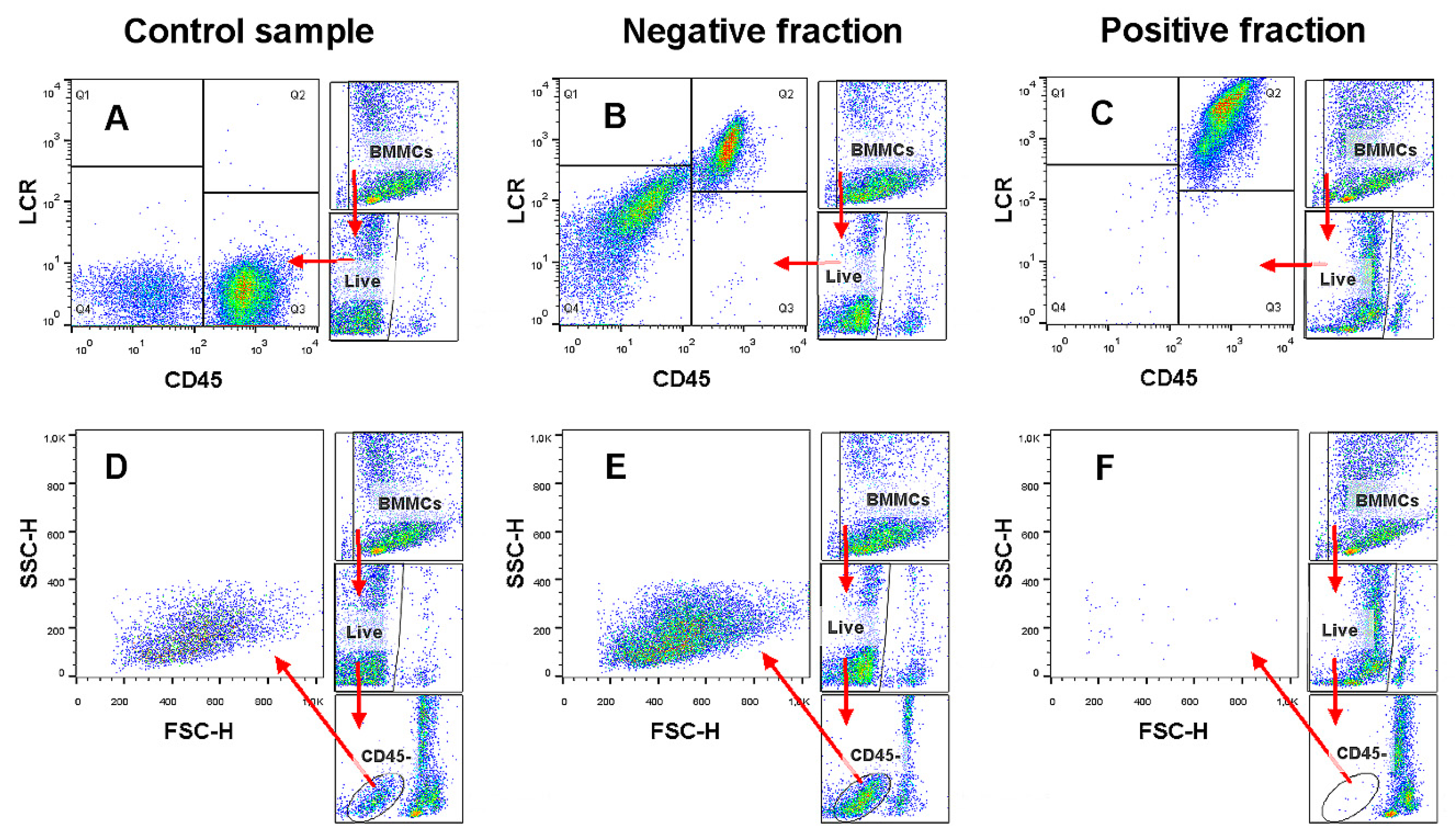
2.1. Magnetic-Activated Cell Sorting (MACS) Depletion of CD45+ Cells

Freshly isolated bone marrow mononuclear cells (BMMCs) were MACS sorted using two sorting strategies: positive selection of CD45+ cells (Posselds) or depletion of CD45+ cells (Depl025) in order to obtain pure CD45 cell fraction. The yield of MASC sorting in negative fractions was about 1–3% of cells from the initial heterogeneous BMMCs population (Table S1). The efficiency of both sorting procedures, evaluated as the number of CD45+ and microbeads (LCR+) bound cells (Figure 1), is shown in Figure 2. The proportion of CD45+ cells significantly (p < 0.001) decreased from ~80% to ~35% (Posselds) or to ~43% (Depl025) in negative fractions. Interestingly, percentage of cells with bound microbeads (CD45+LCR+) in negative fractions (Posselds) corresponded with the number of CD45+ cells, whereas a minimum of cells with microbeads were found in negative fractions after depletion using Depl025 program. This discrepancy might be due to insufficient microbeads binding affinity or failure in antibody and/or microbeads staining. The positive fractions contained more than 90% of CD45+ cells. However, the removal of CD45+ cells using both strategies evidently requires other optimization in order to obtain higher cell purity, even though at the cost of lower cell yield.

2.2. Expansion of CD45 Depleted Cells

As the sorting efficiencies of both used strategies were similar, the results from the following analysis were evaluated altogether independently of the used sorting strategy.

2.2.1. Cell Proliferation

Negatively sorted (CD45) cells were cultured for 2–3 days in a special medium that was developed to expand the number of human CD34+ cells. The number of cultured cells was significantly (p < 0.05) increased almost two-fold at the end of culture, which was also confirmed by a light microscope observation (Figure 3).

2.2.2. Flow-Cytometric Analysis of the CD45 Cultured Cells

At the end of the culture, cells were harvested and analyzed for their specific membrane marker expression by flow cytometry. Since about 30–40% of cells still expressed CD45 antigen at the time of cell seeding, it would be expected that particular percentage of cultured cells could remain CD45 positive at the end of culture. Hence, a specific flow-cytometric evaluation strategy was designed in order to analyze the phenotype of the truly CD45 negative cells (Figure 4).
In general, almost 70% of cultured cells still expressed the CD45 protein (Figure 5). Moreover, the majority of them (>65%) were positive for MHCI, MHCII, CD38, and CD49f. On the other hand, cells were dim positive for CD90 or negative for CD117. Overall cell viability reached about 70%, which may indicate the dying of non-hematopoietic cells during culture. The phenotype of CD45 gated cells slightly differ from the whole cell culture, since nearly 90% of cells expressed MHCI, while the expression of MHCII was obviously missing. Those cells were also positive for CD38 and CD49f, while negative for CD45, CD90, and CD117.

2.2.3. Real-Time Polymerase Chain Reaction (PCR) Analysis

Changes in the relative mRNA expression of typical membrane markers and transcription factors before and after MACS sorting as well as at the end of culture were analyzed by a qPCR method. Relative expression of some markers (CD34 and CD49f) increased either insignificantly after depletion (negative fractions) or significantly (p < 0.05) after expansion of cells in a special culture medium (Figure 6). On the other hand, significant decrease in the relative expression of CD45 (p < 0.01 and p < 0.001), CD90 (p < 0.01 and p < 0.001) and MHCII (p < 0.05 and p < 0.01) was observed after depletion and subsequent culture of cells, respectively. Expression of E-cadherin decreased significantly (p < 0.01) after depletion as well as the expression of CD172a (p > 0.05), whereas expression of both markers increased after cell culture, though insignificantly. A significant rise in the expression of CD117 (c-kit) was observed after the depletion of CD45 cells, while its expression decreased to the basal value after cell culture. No changes in the relative expression were noticed for CD9, CD38, and MHCI markers.
Concerning transcription factors, we found a significantly increased expression of GATA1 and GATA2 factors after depletion (negative fractions) in comparison to control samples (p < 0.001 and p < 0.05, respectively), although expression of both factors reached the basal values at the end of culture (Figure 7). On the other hand, GATA3 expression significantly decreased after cell depletion or even after culture (p < 0.001). No significant differences were observed in the expression of pluripotency markers (OCT4, SOX2, and SOX17) except NANOG, where significant decrease was observed in the cultured cells (p < 0.001).

3. Discussion

In general, cells can be enriched using two basic procedures, positive or negative selection (depletion). The first mentioned procedure is based on a specific and known marker (antigen) that can be labeled with an antibody or other protein with high affinity and subsequently isolated using flow cytometry or immunomagnetic sorting [20]. However, despite the fact that this procedure is quite easy and can reach high purity of targeted cells, the agent bound to the cells might alter their functions in vitro or even in vivo [23]. For this reason, the negative selection by the depletion (removal) of all unwanted cells from the heterogeneous mixture is much more desirable than direct positive selection [20]. Optimized protocols for MACS negative isolation of wanted cells from the desired organs are shown to be easy to use with a sufficient yield and purity and gentle to the cells during the whole process [23]. Therefore, these methods are considered to be a gold-standard for the immunological studies [20].
Here, we present for the first time, a novel promising strategy for the isolation and enrichment of rabbit primitive hematopoietic stem and progenitor cells using the MACS technology and depletion strategy. A similar technique that is based on the removal of cells with lineage commitment markers has been already used in mice for the purification of hematopoietic stem and progenitor cells [24,25,26]. Although we used only one marker (CD45) for the depletion of mature bone marrow cells, it might be sufficient, as the CD45 antigen (known as a leukocyte common antigen) is expressed on almost all hematopoietic cells except mature erythrocytes [27]. Thus, labeling the cells with anti-CD45 antibody might result in the removal of the majority of differentiated hematopoietic cells. However, the purity of cells (CD45) after MACS sorting was in the range of 60–70% (Figure 1). Hence, some optimization of the MACS process is still required in order to gain samples of high purity. On the other hand, the column capacities of the MACS instruments might be thoroughly obeying especially when a highly expressed antigen, as CD45, is used. Nevertheless, a great variability in the purity of CD34+ human samples (from 3 to 84%) enriched by positive MACS selection was also observed in our previous study [12], thus raising a question about the use of a method combining several MACS selection techniques.
BM cell samples depleted for CD45+ cells were cultured in special culture medium with the cytokine supplementation that was designed to expand the number of human hematopoietic cells, especially CD34+ cells. This supplemented medium has already been used for the expansion of human HSC/HPCs [28,29]. In our study, we also successfully expanded the number of seeded CD45-depleted cells almost twice (Figure 3). Moreover, the producer declares on the product’s website that this culture method can provide about 35-fold increase in the total cell number and about 14-fold increase in the number of CD34+ human cells after 7 days of culture [30]. Interestingly, a notable number of adherent cells was observed in the rabbit BM cultures (figure not shown), which might be some type of differentiated hematopoietic cells or even non-hematopoietic cells, since also CD45 negative mesenchymal stem cells or other mature cells could be presented within the negatively sorted fraction of the bone marrow.
The presented method of isolation and expansion of rabbit HSC/HPCs seems to be really promising, as the reduction in the CD45 antigen expression and overexpression of CD34 and CD49f antigen was observed in the cultured cells compared to control samples (Figure 6). However, almost 70% of cultured cells still expressed CD45 antigen, as was demonstrated by flow cytometric analysis (Figure 5). In addition, the majority of the cultured cells also expressed MHCI, MHCII, CD38, and CD49f, and were dim positive for CD90 (only 6%) or negative for CD117. To identify the phenotypic expression of primitive hematopoietic cells, a gate around the CD45 cells was created. Those cells possessed the MHCI+MHCIICD38+CD49f+CD90CD117 phenotype. It was known that human HSCs can be distinguished within the heterogeneous pool of cells as the lineage-negative cells with the expression of CD90 and CD34 [31]. Then, additional markers for the enrichment of long-term HSCs that are capable of self-renewal were identified: CD38 [32], CD45RA [33], and CD49f [34]. Moreover, besides the human HSCs, the CD49f antigen expression has been reported in human embryonic stem cells (ESc) [35], more recently also in human BM-MSCs [36], as well as in several other stem cell types [37], thus making this antigen a promising stemness marker also for rabbit. Additionally, it has been reported that 10% of cells with the LinCD34+CD38CD90+CD45RACD49f+ phenotype had long-term repopulating capacity in mouse models [34]. Nevertheless, another research group observed that the majority of CD34+ human HSC/HPCs also co-expressed CD38, and the same was reported for the CD133+ cells [38], although CD133 is also considered as a marker of human primitive HSCs. On the other hand, Porada et al. [11] identified the ovine HSC/HPCs as the CD34+CD45+ cells. Porcine CD34+ BMMCs were characterized by the lack of lineage markers and the presence of CD90. On the other hand, they were heterogeneous for MHCI, MHCII, and CD172a. This might indicate that different subsets exist within the CD34+ cells [39], since the same heterogeneity has been reported for porcine CD117/c-kit+ HSCs, which seemed to be primitive in vitro and also in vivo [40].
According to our qPCR analysis (Figure 6), rabbit HSC/HPCs certainly expressed CD34, CD38, CD49f, and MHCI and might also express CD9, E-cadherin, CD172a, and even CD45 or CD117. Moreover, several various cell subsets of rabbit HSC/HPCs that differ in their phenotype may exist, as has been already mentioned above for human, mouse, and swine. Since markers, like CD9 and E-cadherin, have been noticed in human ESc [41], it could be interesting to monitor their expression in rabbit primitive stem cells. CD117 (c-kit) is a transmembrane tyrosine kinase, which is very important for the regulation of early hematopoietic development. In the adult BM, the CD117 protein is expressed in both the long-term-repopulating HSCs and the more differentiated progenitor cells. Therefore, c-kit is a valuable marker for their identification [42,43]. Expression of CD117 in rabbit cell samples in the present study significantly increased after the depletion of CD45 (Figure 6), thus indicating the successful enrichment of primitive hematopoietic cells, whereas its expression noticeably decreased in the cell culture. This might be due to the expansion of various CD34+ cell subsets with different expression of CD117. Unfortunately, flow cytometry did not reveal significant expression of CD117 in the CD45 gated cells, however the specificity and/or affinity of the used rabbit polyclonal anti-c-kit antibody is doubtful when it is used for rabbit cells.
The family of GATA transcription factors has been reported to be regulators of genetic expression in hematopoietic cells [44]. GATA1 is important for normal primitive and also definitive erythropoiesis and is, therefore, expressed in the erythroid and mast cells as well as in megakaryocytes at high levels. GATA2 is similarly expressed in hematopoietic progenitors, such as early erythroid and mast cells and megakaryocytes, but also in embryonic stem cells [45]. Moreover, high levels of GATA2 were found in murine HSCs [46]. On the contrary, expression of GATA3 is restricted to the T-lymphoid cells and some non-hematopoietic cells, such as embryonic stem cells [47]. Moreover, for the regulation of stem cell pluripotency and their undifferentiated state other transcription factors are very important, such as OCT4, NANOG, and SOX2, which have been reported in embryonic as well as adult stem cells [48]. SOX17 is a typical marker of fetal HSCs, however, can be expressed also by the adult HSCs [49].
Our further qPCR analyses revealed overexpression of GATA1 and GATA2 factors and the reduction of GATA3 expression in cell samples depleted for CD45 cells (Figure 7), which might again indicate a positive enrichment of primitive HSCs. On the contrary, a decrease in the GATA1 and GATA2 expression was noticed at the end of the cell culture, while the GATA3 expression remained unchanged. Furthermore, there were no differences observed in the relative expression of pluripotency factors, except a significant decrease in NANOG expression at the end of the culture. However, true expression of all transcription factors might be masked by the presence of heterogeneous mixture of CD45+ mature hematopoietic cells in the cell culture as was confirmed by flow cytometric analysis.
In summary, according to the presented results further enhancements of the MACS sorting, e.g., longer bead incubation in combination with the use of higher bead concentrations and/or continuous shaking, or additional purification of depleted fractions with manual MACS instruments are required in order to obtain pure population of rabbit HSC/HPCs that could be properly analyzed for the expressed phenotype. Additionally, longer cell culture (for 4–5 days) with the supplementation of UM729 might increase the hematopoietic cell expansion and thus higher number of cells can be yielded for subsequent analysis. Finally, the differentiation capacity of the isolated and cultured cells should be also evaluated in the future in order to confirm the hematopoietic potential of rabbit HSC/HPCs.

4. Materials and Methods

4.1. Animals

In this study, clinically healthy New Zealand white (NZW) rabbits (n = 8) at the age of 3–8 months reared as described previously [50] were used. The treatment of the animals was approved by the Ministry of Agriculture and Rural Development of the Slovak Republic no. SK U 18016 in accordance with the ethical guidelines presented in Slovak Animal Protection Regulation (RD 377/12), which conforms to the Code of Ethics of the EU Directive 2010/63/EU for animal experiments.

4.2. Experimental Design

The experimental procedure was designed in order to magnetically deplete the mature hematopoietic (CD45+) cells from the heterogeneous mixture of freshly isolated bone marrow mononuclear cells (BMMCs). Briefly, BMMCs from rabbits were isolated using Biocoll solution (Biochrom, Berlin, Germany) and density gradient centrifugation as described previously [51]. At least 108 and more isolated cells were then used for the indirect immunomagnetic sorting under sterile conditions. Cells were firstly stained with anti-rabbit CD45 monoclonal antibody (L12/201, mouse IgG1; Bio-Rad, Hercules, CA, USA), then co-labeled with anti-mouse IgG1 MicroBeads (Miltenyi Biotec, Bergisch Gladbach, Germany) and sorted by AutoMACS Pro Separator (Miltenyi Biotec, Bergisch Gladbach, Germany) using two sorting strategies (Posselds and Depl025) as described previously [22]. According to the producer’s manual, Posselds is double column program for positive selection of cells with loading rate of 1 mL/min; whereas, Depl025 is a single column depletion program with loading rate of 0.25 mL/min.

4.3. MACS Sorting Efficiency and Sample Purity

The efficiency of the used MACS strategies was confirmed by the co-staining of control (unsorted) cell samples and sorted samples (negative and positive fractions) with APC-conjugated labeling check reagent (LCR; Miltenyi Biotec, Bergisch Gladbach, Germany) and another clone of anti-rabbit CD45 monoclonal antibody (ISC18A, mouse IgG2a; WSU, Pullman, WA, USA) as described previously [22]. To analyze the overall cell viability as well as to exclude the cell debris and dead cells from the analysis, 7-AAD (eBioscience, Wien, Austria) was used. A FACSCalibur flow cytometer (BD Biosciences, San Jose, CA, USA) was used to measure at least 25,000 cells (events) per each sample. Obtained data were analyzed using FlowJo™ Software v10.7 (BD Biosciences, San Jose, CA, USA). The sample purity and the sorting efficiency were determined in the negative fractions as the proportion of CD45 single positive (CD45+) cells and CD45/LCR double positive (CD45+LCR+) cells, respectively.

4.4. Expansion of CD45 Depleted Rabbit Hematopoietic Cells

Sorted cells from negative fractions (CD45 depleted cells) were diluted at the cell density of 1–1.5 × 10⁵ mL in a serum-free StemSpan™ SFEM II medium containing 10% of CD34+ Expansion Supplement (Stemcell Technologies, Vancouver, BC, Canada) and 0.5% of penicillin/streptomycin mixture (Life Technologies, Bratislava, Slovak Republic). This special commercial medium is designated for the in vitro culture and expansion of human hematopoietic cells especially CD34+ cells from different biological sources. Cell cultures were incubated at 37 °C in a 5% CO2 humidified atmosphere for at least 2–3 days according to the producer’s manual. At the end of the culture, cells were harvested by centrifugation for 10 min at 488× g and at 20 °C. In order to determine the cell proliferation rate, cell concentration was counted using an EVE™ automated cell counter (NanoEntek, Seoul, Korea). The changes in cell proliferation during the culture were monitored using Zeiss Primovert phase-contrast microscope (Carl Zeiss Slovakia, Bratislava, Slovakia).

4.5. Phenotypic Analysis of the CD45 Cultured Cells

The phenotype of the cells, which were cultured in special expansion medium, was determined by flow cytometry. Briefly, the specific monoclonal or polyclonal antibodies against several membrane markers, which were either fluorochrome conjugated or purified, were used to stain cultured cells according to the producer’s manual. Prior to the main cell staining, purified (unconjugated) antibodies were firstly conjugated with a proper fluorochrome (FITC, PE, or APC) using LYNX Rapid Conjugation Kits (Bio-Rad, Hercules, CA, USA). The antibodies used for flow cytometry are summarized in the Table 1. Labeled cells were analyzed using flow cytometer in the same way as mentioned above.

4.6. Real-Time PCR Analysis

The changes in the relative mRNA expression of several membrane markers (CD9, CD34, CD38, CD45, CD49f, CD90, CD172a, CDH1 (E-cadherin), c-kit (CD117), MHCI, and MHCII) and transcription factors (GATA1, GATA2, GATA3, NANOG, OCT4, SOX2, and SOX17) among the control and sorted samples (negative and positive fractions) as well as cultured cells, were monitored using quantitative real-time PCR (qPCR). Briefly, isolation of total RNA from samples was performed as described previously [22]. The gene-specific primers, which have been either already published or designed de novo using the Primer-BLAST at NCBI website [52], were used in this study (Table 2). qPCR was performed as described previously [53] with some modifications. The amplification protocol was as follows: initial denaturation and activation of hot-start DNA polymerase at 95 °C for 7 min followed by 40 cycles of denaturation at 95 °C for 10 s, annealing at 60 °C for 10 s, and extension at 72 °C for 10 s in Rotor-Gene 6000 (Corbett Research, Sydney, Australia). All samples were amplified in duplicates from the same RNA preparation and the mean value was considered. A melting curve analysis was performed within temperature range of 72–95 °C to check the specificity of PCR products. Standard curves were generated for all genes using a serial dilution of template cDNA from control cells (fresh BMMCs). A relative quantification of gene expression to housekeeping gene beta 2-microglobulin (B2M) was calculated using the threshold (CT) values and PCR reaction efficiencies according to Pfaffl [54].

4.7. Statistical Analysis

Data obtained from analyses were evaluated using GraphPad Prism version 9.0.0 for Windows (GraphPad Software, San Diego, CA, USA) with two-way ANOVA (Tukey test) for MACS efficiency, t-test for cell proliferation and one-way ANOVA (Fisher’s least significant difference (LSD) test). Results are expressed as the mean ± SD. p-values at p < 0.05 were considered as statistically significant.

5. Conclusions

As far as we know, a novel promising strategy for the isolation and enrichment of rabbit primitive hematopoietic stem and progenitor cells using MACS technology and depletion strategy is presented here for the first time. This strategy allows obtaining enriched population of rabbit bone marrow CD45 cells with the purity about 60–70%. Moreover, these cells can be expanded using special culture medium, which might increase the number of CD34+ cells. According to the flow cytometric and qPCR analyses rabbit HSC/HPCs could have CD45CD34+MHCI+MHCIICD38+CD49f+CD90CD117 phenotype. Additionally, relative mRNA expression of CD9, E-cadherin, CD117, and CD172a, as well as several transcription factors, was observed within the cultured cell population. However, the true phenotype of primitive HSC/HPCs could be masked by the presence of CD45+ hematopoietic cells in the cell culture. Therefore, further optimization of this method is still required for the purpose of obtaining pure CD45 cell population, which might reach the full phenotype and potential of rabbit hematopoietic stem and progenitor cells.

Supplementary Materials

The following are available online at https://www.mdpi.com/2312-7481/7/1/11/s1, Table S1: The yield of MACS sorting.

Author Contributions

J.V.: Conceptualization, methodology, formal analysis, investigation, resources, data curation, writing—original draft preparation, writing—review and editing, supervision, project administration, funding acquisition. A.B.: investigation, writing—review and editing. M.B.: methodology, formal analysis, investigation, data curation. A.S.: investigation, writing—review and editing. M.T.: investigation, writing—review and editing. Ľ.O.: resources. V.P.: resources. A.M.: writing—review and editing. P.C.: writing—review and editing, supervision. All authors have read and agreed to the published version of the manuscript.

Funding

This research was funded by the Slovak Research and Development Agency, grant number APVV-14-0348 and APVV-18-0146 and by the Scientific Grant Agency of the Ministry of Education, Science, Research and Sport of the Slovak Republic and Slovak Academy of Sciences, grant number VEGA 1/0160/18.

Institutional Review Board Statement

The study was conducted according to the guidelines of the Declaration of Helsinki, and approved by the Ministry of Agriculture and Rural Development of the Slovak Republic no. SK U 18016 (31.11.2016) in accordance with the ethical guidelines presented in Slovak Animal Protection Regulation (RD 377/12).

Data Availability Statement

The data presented in this study are available in article and supplementary material.

Acknowledgments

Authors would like to thank to Ján Pecho and Igor Matušica for their technical assistance.

Conflicts of Interest

The authors declare no conflict of interest.

References

  1. Hindorf, C.; Glatting, G.; Chiesa, C.; Linden, O.; Flux, G.; Committee, E.D. EANM Dosimetry Committee guidelines for bone marrow and whole-body dosimetry. Eur. J. Nucl. Med. Mol. Imaging 2010, 37, 1238–1250. [Google Scholar] [CrossRef]
  2. Greer, J.P.; Arber, D.A.; Glader, B.E.; List, A.F.; Means, R.M.; Rodgers, G.M. Wintrobe’s Clinical Hematology; Lippincott Williams & Wilkins: Philadelphia, PA, USA, 2018; p. 2346. [Google Scholar]
  3. Zhao, E.; Xu, H.; Wang, L.; Kryczek, I.; Wu, K.; Hu, Y.; Wang, G.; Zou, W. Bone marrow and the control of immunity. Cell. Mol. Immunol. 2012, 9, 11–19. [Google Scholar] [CrossRef] [PubMed] [Green Version]
  4. Tencerova, M.; Kassem, M. The Bone Marrow-Derived Stromal Cells: Commitment and Regulation of Adipogenesis. Front. Endocrinol. 2016, 7, 127. [Google Scholar] [CrossRef] [Green Version]
  5. Weissman, I.L. Stem cells: Units of development, units of regeneration, and units in evolution. Cell 2000, 100, 157–168. [Google Scholar] [CrossRef] [Green Version]
  6. Kondo, M.; Wagers, A.J.; Manz, M.G.; Prohaska, S.S.; Scherer, D.C.; Beilhack, G.F.; Shizuru, J.A.; Weissman, I.L. Biology of hematopoietic stem cells and progenitors: Implications for clinical application. Annu. Rev. Immunol. 2003, 21, 759–806. [Google Scholar] [CrossRef] [PubMed]
  7. Bryder, D.; Rossi, D.J.; Weissman, I.L. Hematopoietic stem cells: The paradigmatic tissue-specific stem cell. Am. J. Pathol. 2006, 169, 338–346. [Google Scholar] [CrossRef] [Green Version]
  8. Kawamoto, H.; Katsura, Y. A new paradigm for hematopoietic cell lineages: Revision of the classical concept of the myeloid-lymphoid dichotomy. Trends Immunol. 2009, 30, 193–200. [Google Scholar] [CrossRef]
  9. Watt, S.M.; Austin, E.; Armitage, S. Cryopreservation of hematopoietic stem/progenitor cells for therapeutic use. Methods Mol. Biol. 2007, 368, 237–259. [Google Scholar] [CrossRef]
  10. Bosio, A.; Huppert, V.; Donath, S.; Hennemann, P.; Malchow, M.; Heinlein, U.A. Isolation and enrichment of stem cells. Adv. Biochem. Eng. Biotechnol. 2009, 114, 23–72. [Google Scholar] [CrossRef]
  11. Porada, C.D.; Harrison-Findik, D.D.; Sanada, C.; Valiente, V.; Thain, D.; Simmons, P.J.; Almeida-Porada, G.; Zanjani, E.D. Development and characterization of a novel CD34 monoclonal antibody that identifies sheep hematopoietic stem/progenitor cells. Exp. Hematol. 2008, 36, 1739–1749. [Google Scholar] [CrossRef] [Green Version]
  12. Vašíček, J.; Shehata, M.; Schnabl, S.; Hilgarth, M.; Hubmann, R.; Jäger, U.; Bauer, M.; Chrenek, P. Critical assessment of the efficiency of CD34 and CD133 antibodies for enrichment of rabbit hematopoietic stem cells. Biotechnol. Prog. 2018, 34, 1278–1289. [Google Scholar] [CrossRef]
  13. Goodell, M.A.; Brose, K.; Paradis, G.; Conner, A.S.; Mulligan, R.C. Isolation and functional properties of murine hematopoietic stem cells that are replicating in vivo. J. Exp. Med. 1996, 183, 1797–1806. [Google Scholar] [CrossRef] [Green Version]
  14. Emery, D.W.; Sablinski, T.; Arn, J.S.; LeGuern, C.; Sachs, D.H. Bone marrow culture and transduction of stem cells in a miniature swine model. Blood Cells 1994, 20, 498–502. [Google Scholar]
  15. Jeong, D.K.; Sharma, N.; Kim, J.N.; Sodhi, S.; Oh, S. Optimization of Techniques for Isolation and Identification of Bovine Hematopoietic Stem Cells from Peripheral Blood. Reprod. Dev. Biol. Suppl. 2012, 36, 138. [Google Scholar]
  16. Oppenheim, S.M.; Moyer, A.L.; BonDurant, R.H.; Rowe, J.D.; Anderson, G.B. Evidence against humoral immune attack as the cause of sheep-goat interspecies and hybrid pregnancy failure in the doe. Theriogenology 2001, 55, 1567–1581. [Google Scholar] [CrossRef]
  17. Fang, J.; Jensen, E.S.; Boudreaux, M.K.; Du, L.M.; Hawkins, T.B.; Koukouritaki, S.B.; Cornetta, K.; Wilcox, D.A. Platelet gene therapy improves hemostatic function for integrin alphaIIbbeta3-deficient dogs. Proc. Natl. Acad. Sci. USA 2011, 108, 9583–9588. [Google Scholar] [CrossRef] [Green Version]
  18. Houdebine, L.M.; Fan, J. Rabbit Biotechnology: Rabbit Genomics, Transgenesis, Cloning and Models; Springer: Cham, The Netherlands, 2009; p. 136. [Google Scholar] [CrossRef]
  19. Carneiro, M.; Afonso, S.; Geraldes, A.; Garreau, H.; Bolet, G.; Boucher, S.; Tircazes, A.; Queney, G.; Nachman, M.W.; Ferrand, N. The genetic structure of domestic rabbits. Mol. Biol. Evol. 2011, 28, 1801–1816. [Google Scholar] [CrossRef] [Green Version]
  20. Hasenberg, M.; Kohler, A.; Bonifatius, S.; Borucki, K.; Riek-Burchardt, M.; Achilles, J.; Mann, L.; Baumgart, K.; Schraven, B.; Gunzer, M. Rapid immunomagnetic negative enrichment of neutrophil granulocytes from murine bone marrow for functional studies in vitro and in vivo. PLoS ONE 2011, 6, e17314. [Google Scholar] [CrossRef]
  21. Gallacher, L.; Murdoch, B.; Wu, D.M.; Karanu, F.N.; Keeney, M.; Bhatia, M. Isolation and characterization of human CD34(-)Lin(-) and CD34(+)Lin(-) hematopoietic stem cells using cell surface markers AC133 and CD7. Blood 2000, 95, 2813–2820. [Google Scholar] [CrossRef]
  22. Vašíček, J.; Baláži, A.; Parkányi, V.; Bauer, M. Different MACS sorting strategies for the enrichment of Lin-(CD34+ CD45-) hematopoietic progenitor cells: Preliminary study. Ann. Univ. Paedagog. Crac. Stud. Nat. 2018, 3, 83–89. [Google Scholar] [CrossRef]
  23. Gunzer, M.; Weishaupt, C.; Planelles, L.; Grabbe, S. Two-step negative enrichment of CD4+ and CD8+ T cells from murine spleen via nylon wool adherence and an optimized antibody cocktail. J. Immunol. Methods 2001, 258, 55–63. [Google Scholar] [CrossRef]
  24. Cheng, P.; Zlobin, A.; Volgina, V.; Gottipati, S.; Osborne, B.; Simel, E.J.; Miele, L.; Gabrilovich, D.I. Notch-1 regulates NF-kappaB activity in hemopoietic progenitor cells. J. Immunol. 2001, 167, 4458–4467. [Google Scholar] [CrossRef] [Green Version]
  25. Cheng, P.; Corzo, C.A.; Luetteke, N.; Yu, B.; Nagaraj, S.; Bui, M.M.; Ortiz, M.; Nacken, W.; Sorg, C.; Vogl, T.; et al. Inhibition of dendritic cell differentiation and accumulation of myeloid-derived suppressor cells in cancer is regulated by S100A9 protein. J. Exp. Med. 2008, 205, 2235–2249. [Google Scholar] [CrossRef]
  26. Ema, H.; Morita, Y.; Yamazaki, S.; Matsubara, A.; Seita, J.; Tadokoro, Y.; Kondo, H.; Takano, H.; Nakauchi, H. Adult mouse hematopoietic stem cells: Purification and single-cell assays. Nat. Protoc. 2006, 1, 2979–2987. [Google Scholar] [CrossRef]
  27. Nakano, A.; Harada, T.; Morikawa, S.; Kato, Y. Expression of leukocyte common antigen (CD45) on various human leukemia/lymphoma cell lines. Acta Pathol. Jpn. 1990, 40, 107–115. [Google Scholar] [CrossRef]
  28. Wu, X.; Dao Thi, V.L.; Huang, Y.; Billerbeck, E.; Saha, D.; Hoffmann, H.H.; Wang, Y.; Silva, L.A.V.; Sarbanes, S.; Sun, T.; et al. Intrinsic Immunity Shapes Viral Resistance of Stem Cells. Cell 2018, 172, 423–438.e25. [Google Scholar] [CrossRef]
  29. Agudelo, D.; Duringer, A.; Bozoyan, L.; Huard, C.C.; Carter, S.; Loehr, J.; Synodinou, D.; Drouin, M.; Salsman, J.; Dellaire, G.; et al. Marker-free coselection for CRISPR-driven genome editing in human cells. Nat. Methods 2017, 14, 615–620. [Google Scholar] [CrossRef]
  30. StemSpan™ CD34+ Expansion Supplement (10X). Available online: https://www.stemcell.com/stemspan-cd34-expansion-supplement-10x.html (accessed on 27 November 2020).
  31. Baum, C.M.; Weissman, I.L.; Tsukamoto, A.S.; Buckle, A.M.; Peault, B. Isolation of a candidate human hematopoietic stem-cell population. Proc. Natl. Acad. Sci. USA 1992, 89, 2804–2808. [Google Scholar] [CrossRef] [Green Version]
  32. Bhatia, M.; Wang, J.C.; Kapp, U.; Bonnet, D.; Dick, J.E. Purification of primitive human hematopoietic cells capable of repopulating immune-deficient mice. Proc. Natl. Acad. Sci. USA 1997, 94, 5320–5325. [Google Scholar] [CrossRef] [Green Version]
  33. Majeti, R.; Park, C.Y.; Weissman, I.L. Identification of a hierarchy of multipotent hematopoietic progenitors in human cord blood. Cell Stem Cell 2007, 1, 635–645. [Google Scholar] [CrossRef] [Green Version]
  34. Notta, F.; Doulatov, S.; Laurenti, E.; Poeppl, A.; Jurisica, I.; Dick, J.E. Isolation of single human hematopoietic stem cells capable of long-term multilineage engraftment. Science 2011, 333, 218–221. [Google Scholar] [CrossRef]
  35. Rodin, S.; Antonsson, L.; Niaudet, C.; Simonson, O.E.; Salmela, E.; Hansson, E.M.; Domogatskaya, A.; Xiao, Z.; Damdimopoulou, P.; Sheikhi, M.; et al. Clonal culturing of human embryonic stem cells on laminin-521/E-cadherin matrix in defined and xeno-free environment. Nat. Commun. 2014, 5, 3195. [Google Scholar] [CrossRef]
  36. Yang, Z.; Dong, P.; Fu, X.; Li, Q.; Ma, S.; Wu, D.; Kang, N.; Liu, X.; Yan, L.; Xiao, R. CD49f Acts as an Inflammation Sensor to Regulate Differentiation, Adhesion, and Migration of Human Mesenchymal Stem Cells. Stem Cells 2015, 33, 2798–2810. [Google Scholar] [CrossRef]
  37. Krebsbach, P.H.; Villa-Diaz, L.G. The Role of Integrin α6 (CD49f) in Stem Cells: More than a Conserved Biomarker. Stem Cells Dev. 2017, 26, 1090–1099. [Google Scholar] [CrossRef]
  38. Koutna, I.; Peterkova, M.; Simara, P.; Stejskal, S.; Tesarova, L.; Kozubek, M. Proliferation and differentiation potential of CD133+ and CD34+ populations from the bone marrow and mobilized peripheral blood. Ann. Hematol 2011, 90, 127–137. [Google Scholar] [CrossRef] [Green Version]
  39. Layton, D.S.; Strom, A.D.; O‘Neil, T.E.; Broadway, M.M.; Stephenson, G.L.; Morris, K.R.; Muralitharan, M.; Sandrin, M.S.; Ierino, F.L.; Bean, A.G. Development of an anti-porcine CD34 monoclonal antibody that identifies hematopoietic stem cells. Exp. Hematol. 2007, 35, 171–178. [Google Scholar] [CrossRef]
  40. Le Guern, A.C.; Giovino, M.A.; Abe, M.; Theodore, P.R.; Qi, J.; Down, J.D.; Sachs, D.H.; Sykes, M.; Yang, Y.G. Stem cell activity of porcine c-kit+ hematopoietic cells. Exp. Hematol. 2003, 31, 833–840. [Google Scholar] [CrossRef]
  41. Oka, M.; Tagoku, K.; Russell, T.L.; Nakano, Y.; Hamazaki, T.; Meyer, E.M.; Yokota, T.; Terada, N. CD9 is associated with leukemia inhibitory factor-mediated maintenance of embryonic stem cells. Mol. Biol. Cell 2002, 13, 1274–1281. [Google Scholar] [CrossRef] [Green Version]
  42. Briddell, R.A.; Broudy, V.C.; Bruno, E.; Brandt, J.E.; Srour, E.F.; Hoffman, R. Further phenotypic characterization and isolation of human hematopoietic progenitor cells using a monoclonal antibody to the c-kit receptor. Blood 1992, 79, 3159–3167. [Google Scholar] [CrossRef] [Green Version]
  43. Ratajczak, J.; Machalinski, B.; Majka, M.; Kijowski, J.; Marlicz, W.; Rozmyslowicz, T.; Ostrowski, M.; Ratajczak, M.Z. Evidence that human haematopoietic stem cells (HSC) do not reside within the CD34+KIT- cell population. Ann. Transplant. 1999, 4, 22–30. [Google Scholar]
  44. Tsai, F.Y.; Keller, G.; Kuo, F.C.; Weiss, M.; Chen, J.; Rosenblatt, M.; Alt, F.W.; Orkin, S.H. An early haematopoietic defect in mice lacking the transcription factor GATA-2. Nature 1994, 371, 221–226. [Google Scholar] [CrossRef]
  45. Briegel, K.; Lim, K.C.; Plank, C.; Beug, H.; Engel, J.D.; Zenke, M. Ectopic expression of a conditional GATA-2/estrogen receptor chimera arrests erythroid differentiation in a hormone-dependent manner. Genes Dev. 1993, 7, 1097–1109. [Google Scholar] [CrossRef] [Green Version]
  46. Akashi, K.; Traver, D.; Miyamoto, T.; Weissman, I.L. A clonogenic common myeloid progenitor that gives rise to all myeloid lineages. Nature 2000, 404, 193–197. [Google Scholar] [CrossRef]
  47. Ku, C.J.; Hosoya, T.; Maillard, I.; Engel, J.D. GATA-3 regulates hematopoietic stem cell maintenance and cell-cycle entry. Blood 2012, 119, 2242–2251. [Google Scholar] [CrossRef] [Green Version]
  48. Darr, H.; Benvenisty, N. Factors involved in self-renewal and pluripotency of embryonic stem cells. In Handbook of Experimental Pharmacology; Springer: Berlin/Heidelberg, Germany, 2006; pp. 1–19. [Google Scholar]
  49. He, S.; Kim, I.; Lim, M.S.; Morrison, S.J. Sox17 expression confers self-renewal potential and fetal stem cell characteristics upon adult hematopoietic progenitors. Genes Dev. 2011, 25, 1613–1627. [Google Scholar] [CrossRef] [Green Version]
  50. Kovac, M.; Vasicek, J.; Kulikova, B.; Bauer, M.; Curlej, J.; Balazi, A.; Chrenek, P. Different RNA and protein expression of surface markers in rabbit amniotic fluid-derived mesenchymal stem cells. Biotechnol. Prog. 2017, 33, 1601–1613. [Google Scholar] [CrossRef]
  51. Vašíček, J.; Kováč, M.; Baláži, A.; Kulíková, B.; Tomková, M.; Olexiková, L.; Čurlej, J.; Bauer, M.; Schnabl, S.; Hilgarth, M.; et al. Combined approach for characterization and quality assessment of rabbit bone marrow-derived mesenchymal stem cells intended for gene banking. New Biotechnol. 2020, 54, 1–12. [Google Scholar] [CrossRef]
  52. Ye, J.; Coulouris, G.; Zaretskaya, I.; Cutcutache, I.; Rozen, S.; Madden, T.L. Primer-BLAST: A tool to design target-specific primers for polymerase chain reaction. BMC Bioinform. 2012, 13, 134. [Google Scholar] [CrossRef] [Green Version]
  53. Kulikova, B.; Kovac, M.; Bauer, M.; Tomkova, M.; Olexikova, L.; Vasicek, J.; Balazi, A.; Makarevich, A.V.; Chrenek, P. Survivability of rabbit amniotic fluid-derived mesenchymal stem cells post slow-freezing or vitrification. Acta Histochem. 2019, 121, 491–499. [Google Scholar] [CrossRef]
  54. Pfaffl, M.W. A new mathematical model for relative quantification in real-time RT-PCR. Nucl. Acids Res. 2001, 29, e45. [Google Scholar] [CrossRef]
Figure 1. Flow cytometric strategy for the evaluation of magnetic-activated cell sorting (MACS) sorting efficiency. Firstly, live bone marrow mononuclear cells (BMMCs) were gated and then the number of single (CD45+) and double (CD45+LCR+) positive cells in control unsorted samples (A), negative (B), or positive (C) fractions was measured. Gating on CD45+ cells in control and both fractions (D, E, and F, respectively) shows the location of CD45 cells on FSC/SSC dot-plot as well as clear enrichment of these cells in negative fraction compared to control sample or positive fraction. BMMCs—bone marrow mononuclear cells, LCR—labelling check reagent.
Magnetochemistry 07 00011 g001
Figure 2. MACS sorting efficiency and purity of sorted cells. The numbers of single positive (CD45+) and double positive (CD45+LCR+) cells were compared among the control (unsorted) samples and both sorted fractions. CON—control sample, NEG—negative fraction, POS—positive fraction. The data from eight experiments are expressed as the mean ± SD; **—difference is statistically significant at p < 0.01; ***—difference is statistically significant at p < 0.001.
Magnetochemistry 07 00011 g002
Figure 3. Proliferation rate of the cultured CD45 cells. Cell number was increased almost two-fold during the culture. (A) Number of cells after seeding (B) and at the end of culture (C) observed under a Zeiss Primovert phase-contrast microscope (magnification at 100×; scale bar = 100 μm). The data from eight experiments are expressed as the mean ± SD; *—difference is statistically significant at p < 0.05.
Magnetochemistry 07 00011 g003
Figure 4. Evaluation strategy for the phenotyping of cultured CD45 cells. Firstly, live cultured cells were gated, then only CD45 cells were shown on FSC/SSC dot-plot (A) and cells from this region were analyzed for the expression of specific markers. (B) Blue: control, red: specific marker.
Magnetochemistry 07 00011 g004
Figure 5. Phenotype of MACS-depleted and cultured cells analyzed by flow cytometry. Cultured cells highly expressed almost all analyzed markers except CD90 and CD117, while maintaining the cell viability about 70% (A). Phenotype of CD45 gated cells was MHCI+MHCIICD38+CD49f+CD90CD117 (B). The data from six experiments are expressed as the mean ± SD.
Magnetochemistry 07 00011 g005
Figure 6. Relative expression of membrane markers analyzed in control (unsorted) samples, sorted fractions, and cultured cells. CON—control sample, NEG—negative fraction, POS—positive fraction, CULT—cultured cells. The data from five experiments are expressed as the mean ± SD; *—difference is statistically significant at p < 0.05; **—difference is statistically significant at p < 0.01; ***—difference is statistically significant at p < 0.001.
Magnetochemistry 07 00011 g006
Figure 7. Relative expression of transcription factors analyzed in the control (unsorted) samples, sorted fractions and cultured cells. CON—control sample, NEG—negative fraction, POS—positive fraction, CULT—cultured cells. The data from five experiments are expressed as the mean ± SD; *—difference is statistically significant at p < 0.05; **—difference is statistically significant at p < 0.01; ***—difference is statistically significant at p < 0.001.
Magnetochemistry 07 00011 g007
Table 1. List of primary antibodies used for flow cytometry.
MarkerHost/IsotypeCloneConjugateCompany
CD45mouse IgG2aISC18Apurified 1WSU
MHCImouse IgG2aH58Apurified 1WSU
MHCIImouse IgG2aTH14Bpurified 1WSU
CD38mouse IgG101purified 1Sino Biological
CD49frat IgG2aGoH3AF647BioLegend
CD90mouse IgG15E10FITCBD Biosciences
c-kit (CD117)rabbit IgGpolyclonalFITCBiorbyt
1 Purified antibodies were conjugated with a proper fluorochrome using LYNX Rapid Conjugation Kits.
Table 2. Gene-specific primers and size of PCR products.
GeneProduct Size (bp)Forward PrimerReverse PrimerReference
CD91585′-CAACAAATTCCACGTCATCG-3′5′-TTGAGGGGTACACCTTCCTG-3′XM_017343606.1 1
CD341555′-CTGAGGTTAGGGCTCAGTGC-3′5′-GGAGTAGCTCTGGTGGCTTG-3′[22]
CD381485′-CAACCTTGTGTGGTGTGGAG-3′5′-GACCGTACCACAGGCAGATT-3′NM_001082683.1 1
CD452625′-TACTCTGCCTCCCGTTG-3′5′-GCTGAGTGTCTGCGTGTC-3′[51]
CD49f1235′-AGGTACAGTGGTCGGTGAGC-3′5′-TTCAAAGTTGCTGTGCCAAG-3′XM_017342931.1 1
CD902935′-CTGCTGCTGCTCTCACTGTC-3′5′-ACAGAAGCAGCTTTGGGAAA-3′[51]
CD172a1625′-CCAGCATTCAGATTGGTCCT-3′5′-GTCCCATTCACTTCCTCTGC-3′XM_017341726.1 1
CDH1 (E-cadherin)1685′-GAATCCTGGCTCTGCTCATC-3′5′-GCTGGCTCAAGTCAAAGTCC-3′XM_002711639.3 1
c-kit (CD117)1895′-CCGGTGGACTCCAAGTTCTA-3′5′-GTAAACGTGGTGGGTGCTCT-3′NM_001329070.1 1
MHCI975′-AGTGGGAATTGTTGCTGGAG-3′5′-TCCTTTCCCATCTGAGCTGT-3′NM_001171270.2 1
MHCII1425′-CTGTGAGCACAGGAAGGTGA-3′5′-GGGTGGCTAGATGTCTGGAA-3′NM_001171118.1 1
GATA11645′-AAACGGGCAGGTACTCAGTG-3′5′-TTCCGAGTCTGGATTCCATC-3′XM_002719899.3 1
GATA21375′-GACAAGGACGGGGTCAAGTA-3′5′-TAGGAGGGGTAGGTGGGAAT-3′XM_008260327.2 1
GATA31185′-AAGGCAGGGAGTGTGTGAAC-3′5′-CGGTTCTGTCCGTTCATTTT-3′XM_002717361.3 1
NANOG1225′-GCCAGTCGTGGAGTAACCAT-3′5′-CTGCATGGAGGACTGTAGCA-3′[51]
OCT41495′-GAGGAGTCCCAGGACATGAA-3′5′-GTGGTTTGGCTGAACACCTT-3′[51]
SOX21525′-CAGCTCGCAGACCTACATGA-3′5′-TGGAGTGGGAGGAAGAGGTA-3′[51]
SOX171355′-CGTGTTGTTGCGTTTTGTTT-3′5′-CGGGGAACAAGTCTCTGAAG-3′XM_008255533.2 1
B2M1185′-ATTCACGCCCAATGATAAGG-3′5′-ATCCTCAGACCTCCATGCTG-3′[51]
1 NCBI Reference Sequence.
Publisher’s Note: MDPI stays neutral with regard to jurisdictional claims in published maps and institutional affiliations.

