Article

Comparison of 2D Optical Imaging and 3D Microtomography Shape Measurements of a Coastal Bioclastic Calcareous Sand

1
Department of Civil and Environmental Engineering, University of Massachusetts Dartmouth, Dartmouth, MA 02747, USA
2
Department of Civil and Urban Engineering, New York University, New York, NY 10012, USA
3
Department of Computer and Information Science, University of Massachusetts Dartmouth, Dartmouth, MA 02747, USA
4
Centre for Microscopy, Characterisation & Analysis, University of Western Australia, Crawley 6009, Australia
5
Department of Civil Engineering, City, University of London, London EC1V 0HB, UK
6
School for Marine Science and Technology, University of Massachusetts Dartmouth, Dartmouth, MA 02747, USA
*
Author to whom correspondence should be addressed.
J. Imaging 2022, 8(3), 72; https://doi.org/10.3390/jimaging8030072
Submission received: 31 December 2021 / Revised: 7 March 2022 / Accepted: 9 March 2022 / Published: 14 March 2022
(This article belongs to the Special Issue Recent Advances in Image-Based Geotechnics)

Abstract

:
This article compares measurements of particle shape parameters from three-dimensional (3D) X-ray micro-computed tomography (μCT) and two-dimensional (2D) dynamic image analysis (DIA) from the optical microscopy of a coastal bioclastic calcareous sand from Western Australia. This biogenic sand from a high energy environment consists largely of the shells and tests of marine organisms and their clasts. A significant difference was observed between the two imaging techniques for measurements of aspect ratio, convexity, and sphericity. Measured values of aspect ratio, sphericity, and convexity are larger in 2D than in 3D. Correlation analysis indicates that sphericity is correlated with convexity in both 2D and 3D. These results are attributed to inherent limitations of DIA when applied to platy sand grains and to the shape being, in part, dependent on the biology of the grain rather than a purely random clastic process, like typical siliceous sands. The statistical data has also been fitted to Johnson Bounded Distribution for the ease of future use. Overall, this research demonstrates the need for high-quality 3D microscopy when conducting a micromechanical analysis of biogenic calcareous sands.

1. Introduction

Advanced imaging techniques are used in the area of micromechanics to measure the shape parameters of soil grains on the micro-scale and correlate them to geotechnical properties on the macro-scale [1,2,3,4,5]. Shape parameters such as aspect ratio, convexity, and sphericity are commonly measured with two-dimensional (2D) dynamic image analysis (DIA) and 3-dimensional (3D) X-ray microtomography (μCT). DIA methods rely on imaging the 2D projection of a grain as it falls through the air [6,7,8,9]. This allows for a large number of particles to be sampled and low computational effort during analysis, but it does not measure the shape of the entire grain. In μCT, a series of radiographs of the sample are taken from multiple angles and then reconstructed into a 3D model. Variation in X-ray absorption between single grains or bulk soil and the background provides image contrast [10,11,12,13,14,15]. Although this method provides an accurate representation of the soil grains, it is limited in the number of grains it can image at once and is computationally more intensive. This is especially the case when watershed techniques are used to isolate individual grains for the μCT of bulk sand samples.
These imaging techniques have been especially useful for the study of problematic offshore calcareous sediments. These soils consist mainly of the calcium carbonate skeletal remains of marine microorganisms such as foraminifera, mollusk, coral, bryozoans and their bioclasts. Unlike typical siliceous sand, the shape of the calcareous sand grain is in part biologically driven. Grains in these sediments can consist wholly of shells or tests (e.g., shells of single-celled organisms) and their bioclasts. The complex shape of the grains in these calcareous sands contributes to their poor geotechnical behavior [16,17,18,19,20], such as pile running [21]. Although a few studies have examined the impact of 2D and 3D imaging techniques on the measurements of sand grains [22,23,24], there have been no direct comparisons of 2D DIA methods to 3D μCT of bulk samples. This paper investigates the impact of 2D DIA and 3D μCT imaging methods on the measured shape parameters, aspect ratio, convexity, and sphericity, of calcareous beach sand. In particular, the ability of the two techniques to measure the shape of biogenic platy and shelly features is examined.

2. Sand in Study

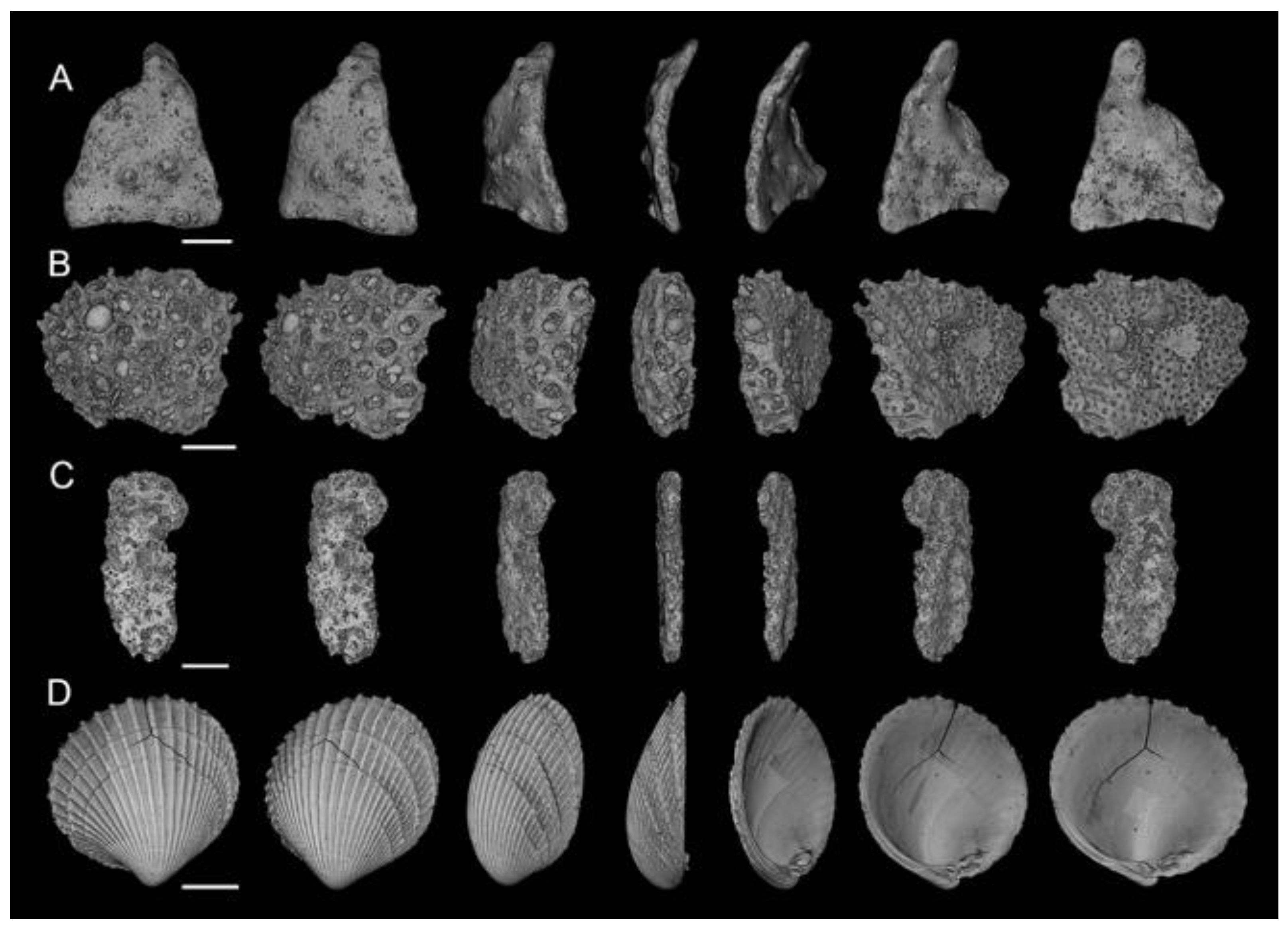
The Ledge Point coastal bioclastic sand used in this study was obtained from the coast of Ledge Point, WA, Australia (Figure 1) by a team from the University of Western Australia [25]. The sand has a carbonate content of 91% and consists largely of plate grains hollow foraminifera tests, mollusk shells, bryozoans, and their bioclasts. A selection of µCT scans of sand grains rotated through 180° from Ledge Point is presented in Figure 2. A summary of the site and geotechnical properties is provided in Table 1.

3. Microscopy Techniques

3.1. Dynamic Image Analysis (DIA)

Two-dimensional DIA for determining granular soil particle size and shape distribution has been shown to be feasible, repeatable, and accurate for many soils [7]. The method employs a high frame rate camera combined with a laser to image millions of individual particles in a short time. In this study a QICPIC (Sympatec, Clausthal-Zellerfeld, Germany) was employed to capture images of Ledge Point sand. The device consists of a vibratory feeding system, a dispersing system, and an imaging sensor.
The device operates as follows: (a) a 100 g specimen is poured into the device through a hopper (VIBRI) and sent to the disperser at a constant rate. (b) The specimen disperses as it falls through a 50 cm long fall shaft (GRADIS) towards the imaging sensor. A small amount of sand particles is transported through the GRADIS simultaneously, which maximizes particle separation. As a result, particle overlap is minimized, thereby eliminating the need for particle segmentation. (c) As particles pass through the image analyzer, particle shapes are captured at a frame rate of 175 frames per second with a 4 Megapixel (2336 × 1728) resolution, and the resulting image resolution is 4 µm/pixel. A more detailed description of the procedure is provided in [7]. For this study, the 100 g sample resulted in a sample size of 1,048,575 particles for the 2D DIA method.

3.2. X-ray Microtomography

X-ray micro-computed tomography (µCT) is a non-destructive imaging technique that relies on variations in the attenuation of X-rays as they pass through materials of differing density or mass to generate contrast in the resulting images. Multiple radiographs of the sample are taken from a range of angles, and this information is used to computationally reconstruct a 3D volume.
For the µCT of Ledge Point sediments, a 5 mm diameter plastic tube was filled with calcareous material, mounted onto the instrument stage, and then scanned using a Versa 520 XRM (Zeiss, Pleasanton, CA, USA). Imaging was conducted at 50 kV and 4 W using an LE3 source filter to minimize beam hardening and to improve contrast. Source sample and sample detector distances were set to −13 and 127 mm, respectively, which, in combination with the 0.4× objective lens and 2× camera binning, resulted in a final isotropic voxel resolution of 6.4 µm. Suitable image intensity was achieved using a 4 s exposure, and a total of 2501 X-ray projections were collected through 360° for each tomography.
Radiographs were automatically reconstructed using XRM Reconstructor (Zeiss, Pleasanton, CA, USA) using a default center shift and beam hardening corrections.

3.3. Three-Dimensional Watershed Segmentation

A watershed segmentation algorithm was used to segment the reconstructed µCT scans (Figure 3a). The watershed algorithm was developed by Kong and Fonseca [14] and adapted for branch recursive processing by Leonti et al. [26] to improve its computational speed. The preprocessing and segmentation steps follow.
The reconstructed µCT images, in Figure 3a, are first binarized using Otsu’s method [27]. The fully encompassed voids are filled. The scan is then partitioned into two images of the same size as the original, one containing objects smaller than a user-specified diameter, with the other containing everything else. Single-pixel artifacts from partitioning are removed, and the objects are filled.
The final preprocessing step before the watershed segmentation process is to split the image into overlapping sections through the y-axis. Subdividing the image allows for quicker, higher quality segmentation. The sections must overlap to avoid the hard boundaries that would result from them being disjointed and adversely affect segmentation.
The connected components of a single section are calculated. The connected components are disconnected regions of pixels that do not touch each other. The components are handled iteratively. The negated Euclidean distance map is calculated, which is the distance of any ‘set’ (1) pixel to the nearest ‘off’ (0) pixel. Next, the bring-up method proposed in [14] is used to dampen all local minima in the component (a mechanism for combining shapes when performing watershed segmentation). Finally, watershed segmentation is performed. If the component is split into two or more smaller regions, the segmentation process is recursively performed on each of those until watershed segmentation is constant. Once this termination case is reached, the original component is replaced by its segmented counterparts.
This process is performed on every section until the entire image is segmented. For the overlapping regions, only the latest segmented version is retained. For this study, the 5 mm diameter sample resulted in a sample size of 2325 for the 3D µCT method.

4. Shape Parameters

4.1. 2D Shape Parameters

The 2D shape parameters used for analysis of particles from the DIA imaging are summarized below and described in Table 2.
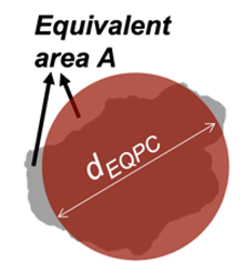
EQPC diameter (dEQPC) in 2D, dEQPC is the averaged parameter representing the diameter of a circle having an equivalent area to the particle under consideration:
d E Q P C = 4 × A π
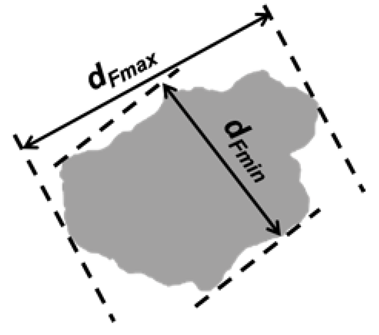
Feret diameters are the distance between two parallel tangents to the particle at an arbitrary angle [28]. Feret-max and Feret-min diameters (dFmax, dFmin) are the longest and shortest diameters from one particle image and are generally employed for describing the maximum and minimum dimensions of a particle.
Three types of particle shape descriptors were employed to quantify particle shape morphology (Table 2). The selected shape descriptors are thought to capture independent shape features [23]. The numerical value of shape descriptors ranges from 0.0 to 1.0, where a symmetrical particle, such as a sphere, approaches 1.0, while a highly irregular particle has descriptors approaching, but never reaching, 0.
Aspect ratio (AR2D) in 2D is defined as the ratio of the minimum and maximum Feret dimensions [28]:
A R 2 D = d F m i n d F m a x
Sphericity (S2D) in 2D has been defined by ISO 2008 [28] as the ratio of the perimeter of an area-equivalent circle to the real perimeter (P),
S 2 D = π × d E Q P C P
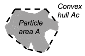
Convexity (Cx2D) in 2D is a measure of the overall concavity of a particle [28]. It is the ratio between the particle area (A) and the volume of the convex hull (Ac) in 2D shape analysis, as shown in Figure 3:
C x 2 D = A A c

4.2. 3D Shape Parameters

The 3D shape parameter used to analyze the particles from the segmented μCT scans are summarized below and described in Table 2.
ESD diameter (dESD) in 3D is the averaged parameter representing the diameter of a sphere having an equivalent volume to the particle under consideration:
d E S D = 6 V π 3
Feret-length, Feret-width, and Feret-thickness (dFlength, dFwidth, dFthickness) are generally employed for describing the longest, intermediate, and shortest dimensions of a soil particle [29,30]. The traditional definition employs three axes that are always perpendicular to each other. In 3D shape analysis, dFlength, dFwidth and dFthickness were obtained with principal coordinate analysis (PCA) through the regionprop3 and pcacov functions in MATLAB ver. R2021b.
Aspect ratio in 3D is defined from the ratio of the three Feret dimensions. In 3D, there are three different parameters that are typically calculated, including thickness-to-length ratio (AR3D), elongation index (EI), and flatness index (FI) [31]. AR3D is calculated as the ratio between dFthickness and dFlength, and FI = dFthickness/dFwidth, EI = dFwidth/dFlength. From this, AR3D = EIFI; so, only two of these parameters are independent. The definitions of AR3D, EI, and FI are adopted for reconstruction of µCT images, while in 2D DIA, aspect ratio is calculated as AR2D = dFmin/dFmax for each particle.
A R 3 D = d F t h i c k n e s s d F l e n g t h
F I = d F t h i c k n e s s d F w i d t h
E I = d F w i d t h d F l e n g t h
Sphericity (S3D) in 3D has been defined from [32] as the ratio of the surface area of a volume equivalent sphere to a surface area of a real particle (As):
S 3 D = π d E S D 2 A s
Convexity (Cx3D) in 3D is a measure of the overall concavity of a particle and taken from [28], but updated for particle with internal voids by [14]:
C x 3 D = V f i l l V c

5. Results

5.1. Particle Size Distribution

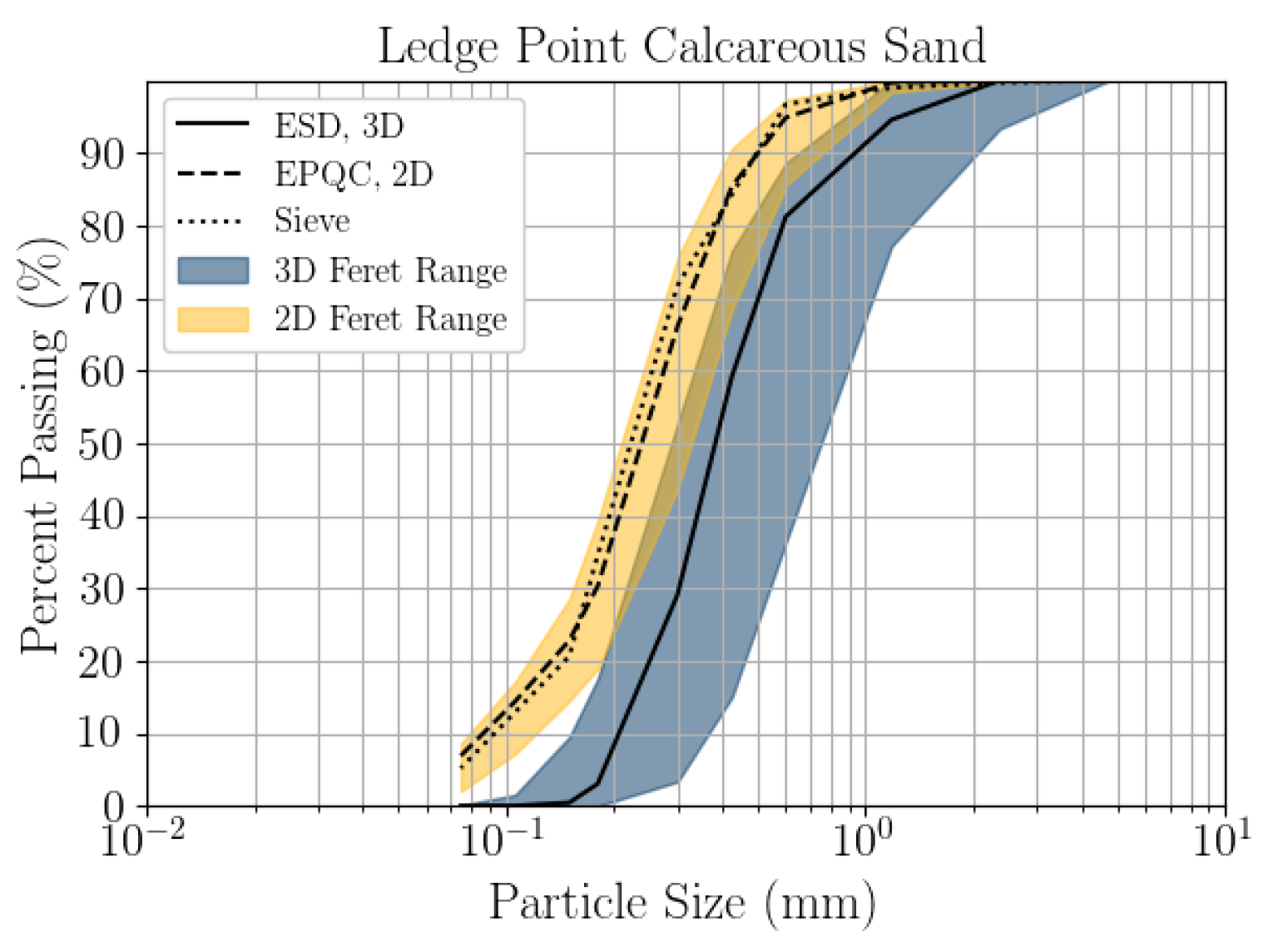
Particle size distributions have been compared between 2D DIA, 3D μCT, and mechanical sieve for the bioclastic coastal sand in Figure 4. The sieve was conducted in line with [33], with the sieve sizes listed in Table 3. The particle size distributions from microscopy analyses were created by setting the histogram bin edges to the sieve sizes listed in Table 3. The 2D EQPC diameter aligns well with the mechanical sieve, and the 2D Feret range (dFmindFmax) bounds the sieve. The 3D ESD significantly overestimates the mechanical sieve, the d50 of the sieve being 0.11 mm and the d50 of the ESD being 0.38 mm. The mechanical sieve falls entirely outside the 3D Feret range (dFthicknessdFlength). Although the mechanical sieve cannot be defined directly by a specific analytical description of grain shape, it is clear from Figure 3. that the 3D μCT is underestimating the quantity of grains below 0.2 mm in diameter. It is likely that the segmentation algorithms are unintentionally eliminating smaller grains. During this process, it is difficult to balance over segmentation and the preservation of naturally small grains. Note that the sieve analysis also lacks the precision to capture small particles accurately.
A key limitation of μCT is throughput. While the 3D segmentation of soil grains from μCT could be improved by higher resolution scanning, doing so would reduce the field of view, which would further reduce throughput. Continued development of these watershed techniques, or increased μCT throughput, could improve the 3D segmentation of soil grains from μCT in the future. The particle size distribution may have also been impacted by sample size. The 2D DIA sample size was over 450 times larger by particle count than the 3D μCT.

5.2. Shape Parameter Variation with Size

The relationship between particle size and shape parameters for 2D DIA and 3D μCT is compared by calculating the mean particle shape parameters (Figure 5). The particle size distribution is provided as a probability density plot (histogram) as is common in the field of geology. The mean particle shape parameters were calculated from bins where the edges were set at the sieve opening sizes listed in Table 3. This should represent the mean shape parameter being calculated for the material that would be accumulated on each sieve. The 2D DIA shape parameters AR2D and Cx2D are larger than the 3D μCT parameters across all sizes. For example, at 0.3 mm: AR2D = 0.66, AR3D = 0.44, Cx2D = 0.88, and Cx3D = 0.65. All 3D μCT parameters vary with size, with S3D changing the most. For example, at 0.3 mm: S3D = 0.63, and at 2.36 mm: S3D = 0.36. AR2D varies the most with size, and S2D shows Cx2D small variation with size. Below 0.2 mm, the 3D parameters could have been affected by the segmentation algorithm over-filtering the small grains.

5.3. Statistics of Particle Shape Parameter

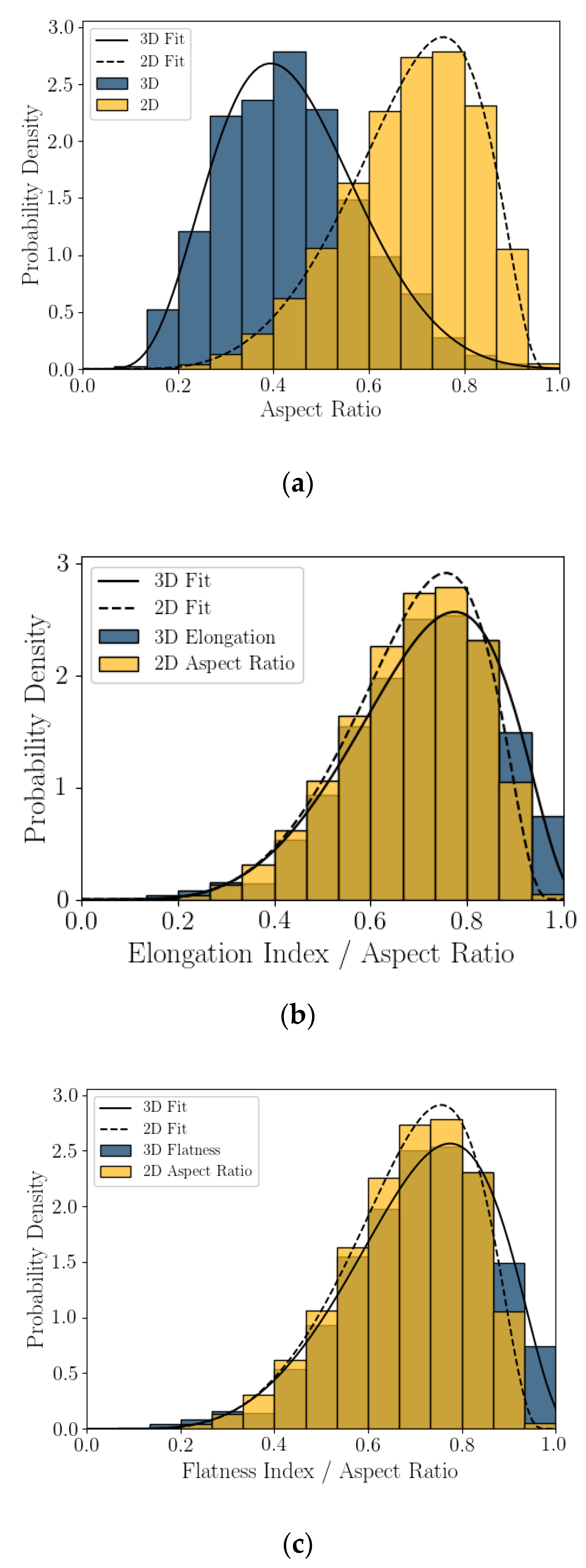
Probability density plots, presented as histograms, of the 2D DIA and 3D μCT shape parameters calculated from Feret dimensions (aspect ratio, elongation index, and flatness index) are presented in Figure 6. The mode of AR2D is nearly double that of AR3D and the two histograms skew in the opposite direction. It appears that the project AR2D value overestimates aspect ratio relative to AR3D for this coastal calcareous sand. Flatness compares well with 2D DIA aspect ratio, indicating that the projected section of the grains in DIA is better represented by the length and width of the 3D particles, which is reasonable for a platy or shelly particle. Finally, elongation index does not trend well with AR2D.
Probability density plots or histograms for 2D DIA and 3D μCT convexity and sphericity are presented in Figure 7. The modes of both Cx2D and S2D are larger than those of Cx3D and S3D, at 35% and 23% percent larger, respectively. The shape of the distributions varies between the two imaging techniques. The Cx2D and S2D histograms skew to the left (mean smaller than the median), while in 3D μCT the Cx3D and S3D histograms appear to be normally distributed. It appears that convexity and sphericity calculated from the projected shape of a calcareous sand grain is not an accurate representation of the real 3D shape. The measured values in 2D are significantly larger, dependent on grain size (Figure 5), and the shape of distributions are dissimilar.
The histograms of shape parameter for this coastal calcareous sediment are non-normal. This was also seen in [22,23] for the 2D DIA of Ledge Point and the Browse #1 hemipelagic calcareous sand. For this study, the Bounded Johnson Distributions [34] were fitted to all of the shape parameter histograms; fit lines are shown in Figure 6 and Figure 7. The four parameter Johnson family of distribution is identified by [35] as being useful for modeling non-normal geotechnical data. The Bounded Johnson Distribution limits the range of the random variable to 0 ≤ x ≤. 1, which matches the normalized definitions of the particle shape parameters. Its probability density function can be defined by
f ( y ) = δ 1 + ( x ξ λ ) 2 Φ ( γ + δ × l n ( x ξ   λ + ξ y   ) )
where Φ is the normal distribution probability density function, x is an independent random variable, δ and γ are fitting parameters, ξ is the location variable, and λ is the scaling variable.
The SciPy distribution fitting function [36] was used to obtain the four Johnson fitting parameters for the shape parameters AR2D, AR3D, EI, FI, Cx2D, Cx3D, S2D, and S3D for the Ledge Point bioclastic calcareous sand in Table 4.

5.4. Correlation of Particle Shape Parameters

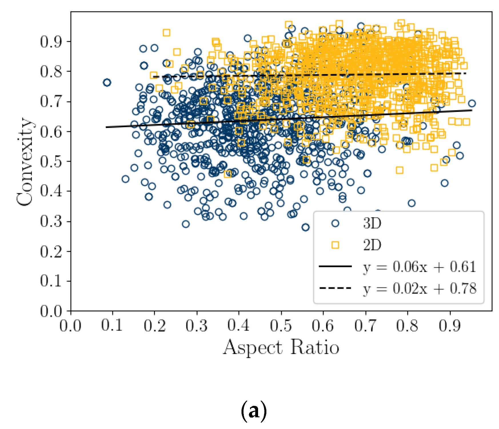
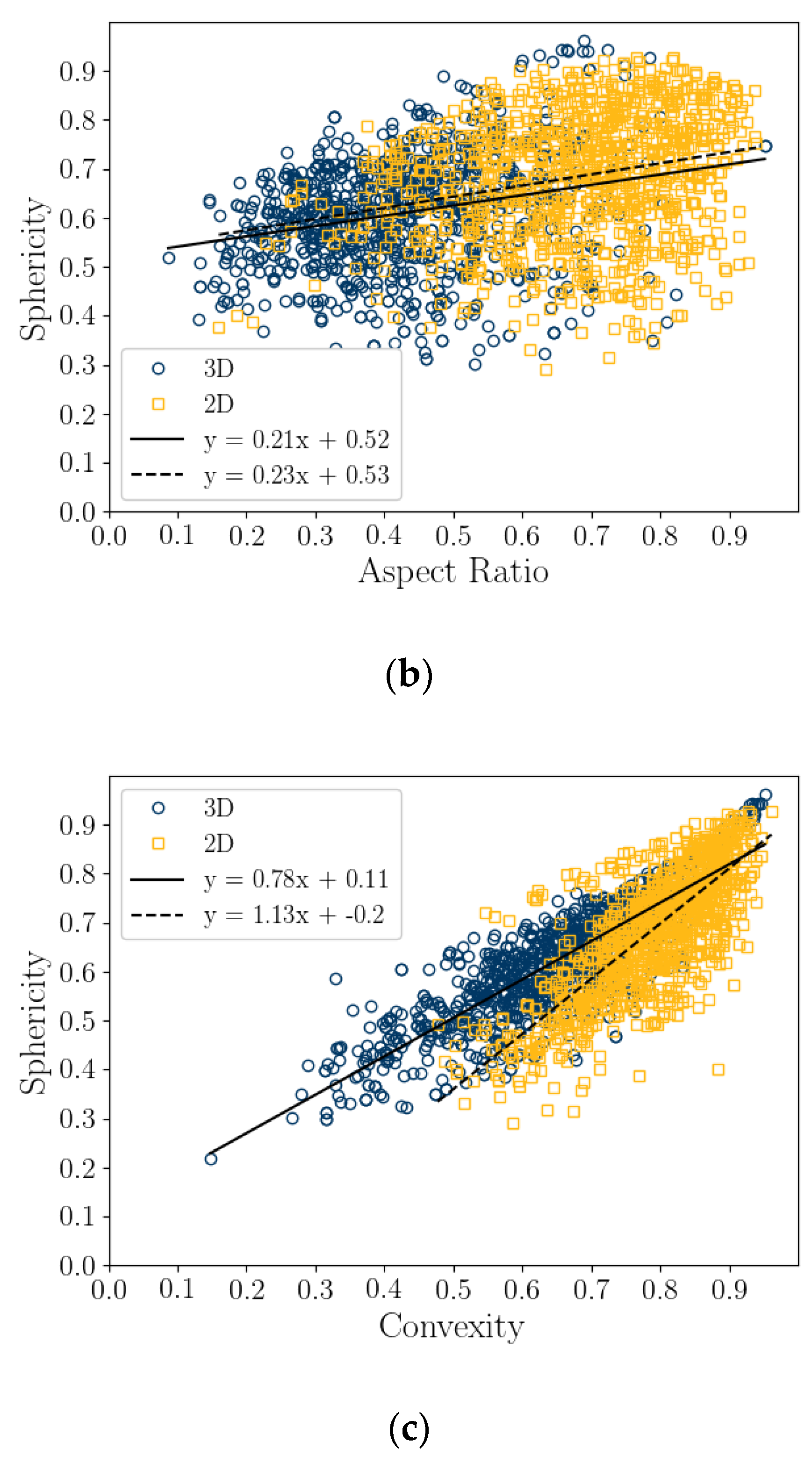
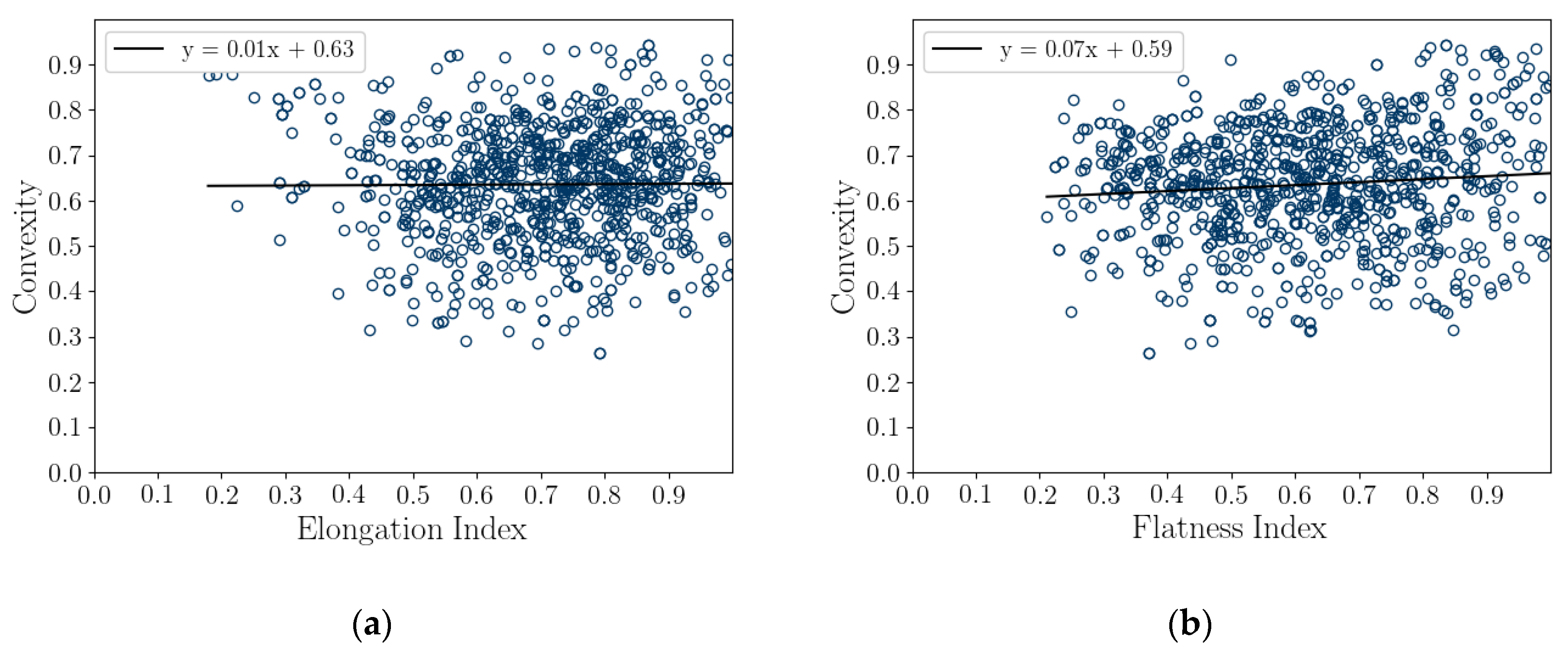
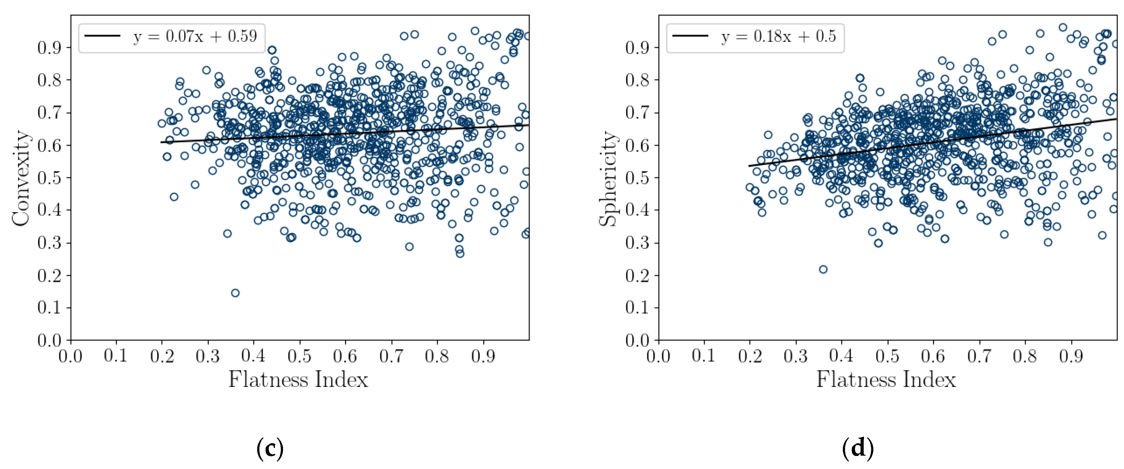
A correlation coefficient analysis was conducted on both the 2D DIA and 3D μCT shape parameters and average diameters, shown in Table 5 and Table 6, respectively. These results are also presented graphically as correlation plots of sphericity versus aspect ratio and convexity and convexity to aspect ratio for 1000 randomly selected particles (in Figure 8), while correlation plots comparing Cx3D and S3D versus elongation index and flatness index for 1000 randomly selected particles are presented in Figure 9. The strongest correlations of shape parameters are of S2D with Cx2D, 0.77, S3D with Cx, 0.85, and FI with AR3D, 0.72. These are associated with the tightest grouping of points in Figure 8 and Figure 9. It should be noted that AR3D = EIFI; so, only two of these parameters are independent, and the correlation of these parameters is reasonable.

6. Discussion

The sphericity, aspect ratio, and convexity measured with 2D DIA were significantly larger than that measured with 3D μCT; in particular, S2D was approximately 66% larger than S3D at a particle size of 2.36 mm (Figure 5). This difference appears to be a result of measuring shape parameters from the projection of platy particles such as those common in the Ledge Point coastal bioclastic calcareous sediment, shown in Figure 2.
A thought experiment can be used to examine the possible sets of shape parameters such as AR2D and S2D from platy calcareous sand grains. Consider a thin disc with a radius of one unit that rotates at an angle θ about the y-axis, as in Figure 10. As the disc rotates, its projected shape will go from a disc to a line. The 2D projected aspect ratio of the thin disc at any angle θ will be
A R d i s c = cos ( θ )
where ARdisc is the aspect ratio of the projected disc, and θ is the rotation of the disc about the y-axis (Figure 10).
The equivalent diameter of a circle with the same area as the project disc will be defined by
d E Q P C D i s c   = 4 × cos ( θ )
where dEQPC-Disc is the equivalent diameter of a circle with the same area as the projected disc.
The perimeter of the projected disc with a radius of one unit can be estimated using [37] the perimeter of an ellipse formula:
P = π [ 3 ( a + b ) ( 3 a + b ) ( a + 3 b ) ]
The sphericity of the projected disc can then be calculated from Equations (3), (12) and (13) with the major radius of the projected disc being a = 1.0 and the minor radius of the projected disc being b = cos (θ).
S d i s c = 4 × cos ( θ ) 3 ( 1 + cos ( θ ) ) ( 3 + cos ( θ ) ) ( 1 + 3 × cos ( θ ) )
where Sdisc is the sphericity of the projected disc.
The set of possible ARdisc and Sdisc for a disc with a radius of one unit rotated at an angle θ about the y-axis are plotted in Figure 11. It can be seen that both Sdisc and ARdisc follow a sinusoidal pattern. Sphericity has very wide peaks, with the value being close to one for nearly half of the set of angles.
The 2D DIA imaging of a grain falling in front of an imaging sensor at a random angle can be modelled by randomly sampling Equations (12) and (15) at 10,000 random angles θ about the y-axis and plotting the probability density, as in Figure 12. Rotation about the x-axis would follow the same formulations presented above due to the disc symmetry. Rotations about the z-axis will have no impact of the projected shape. The 2D DIA method process of measuring the shape parameters from the projection of grains clearly overestimates both aspect ratio and sphericity of platy particles, with sphericity being the most extreme, as in Figure 12. The mode of both parameters is approximately one, while both AR3D and S3D would approach zero for very thin discs (for a diameter to thickness ratio of 10:1 S3D = 0.46, which is similar to that of the larger diameter grains, shown in Figure 5c). This aligns well with the results of the 2D DIA and 3D μCT of the Ledge Point coastal bioclast calcareous sand (Figure 5, Figure 6 and Figure 7). In addition, this example demonstrates that any error in the 2D DIA is inherent to the method itself and will not be corrected through increasing sample size.
The measured convexity from 2D DIA was also significantly larger than that from 3D μCT for particle sizes above 0.2 mm. The impact of using a 2D projection to measure convexity is not as simple to mathematically model as aspect ratio or sphericity; however, a qualitative visual assessment of the mollusk shell, Particle D, in Figure 2 demonstrates how the 2D DIA method can overestimate convexity. Figure 2 presents the 3D μCT scans of a number of Ledge Point calcareous sand grains rotating about their vertical axis (relative to the figure orientation). It can be seen that the projected shape of the mollusk shell has a high convexity as it is rotated, but it is obvious that a shell of this nature is concave in 3D (a low value of convexity). A similar pattern holds for Particle A, which is a shell bioclast. It is therefore likely that the high measured value of convexity in the 2D DIA are the result of the microscopy technique itself.
Three-dimensional sphericity is likely correlated to convexity due to the biomorphology of the sand grains. Intact shells such as that in Figure 2d will have a low sphericity and convexity. As they break down through clastic processes their Feret dimensions should tend towards unity (likely due to the shell thickness) and as a result their sphericity and convexity will increase. Since new grains are constantly being introduced to the sand through biogenesis there will be large young shells with low sphericity and convexity and small older bioclasts with high sphericity and convexity in the system. This aligns well with the variations in Cx3D and S3D seen in Figure 5b,c.
The correlation of sphericity to convexity in 2D could be the result of 2D projection angle. For example, when considering the bioclast in Figure 2a angular orientation with the highest sphericity appears to have the highest convexity and vice versa. This would explain how Cx2D and S2D can be correlated (Figure 8c), but Cx2D does not show the same variation in size that S2D does (Figure 5b,c). The Correlation of sphericity with convexity in 2D DIA methods have also been shown by [22] and [24] for both siliceous and calcareous sands.
Li et al. [22] argued that 3D μCT should be used to assess the shape parameters of calcareous sediments in order to resolve internal voids within the grains and that DIA methods can result in dimensionality projection errors that make particles appear larger in projection (this type of error would have a minimum impact on thin platy particles). The angular projection error presented here for biogenic platy and shelly particles that results in a statistical increase in the measured aspect ratio and sphericity in DIA provides an additional reason for using μCT for measuring the shape parameters of complex calcareous sands.

7. Practical Significance of This Study

Calcareous sediments are known for being problematic soils [20,21]. One significant issue is they are known for being unclassifiable. Grain size distribution and calcium carbonate content are not good predictors of geotechnical behavior [38]. Clark and Walker [39] is the most commonly used geological classification scheme for calcareous sediments, but it does not provide direct insights into geotechnical behavior. Thus, there is an industry need for a standard geotechnical classification system for calcareous soils [38,40,41]. Their problematic behavior has been attributed to the unique shape of their grains [20,41]. This study demonstrates the need for researchers to carefully consider the imaging methods used to measure shape parameters and the biomorphology of soil grains when undertaking studies on the mechanical behavior and classification of calcareous sediments.

8. Conclusions

This article presents the results of a comparison study of 2D DIA and 3D μCT analyses of a coastal bioclastic calcareous sediment from Ledge Point, Western Australia. This study provides unique insights into state-of-the-art soil imaging techniques for quantifying the particle size and shape of bioclastic calcareous sands. The following conclusions from the study can be drawn:
  • For this calcareous sand, 2D DIA correlates better to the traditional sieve analysis than 3D μCT, as shown in Figure 4. The μCT analysis underestimates the number of fine sand grains below 0.2 mm relative to the sieve test. This is possibly due to the watershed algorithm used for segmenting the sand, which digitally removes smaller grains. Alternatively, it may be due to sampling error arising from the limited imaging volume captured by the μCT device compared with the 2D DIA technique.
  • The 2D DIA mean particle shape parameters aspect ratio, sphericity, and convexity with size were significantly larger (dependent on grain size) than those from 3D μCT (Figure 5).
  • The 3D μCT imaging technique is a more accurate method for measuring particle shape parameters of a bioclastic calcareous sand. When measured in 3D, the grains had a lower aspect ratio, AR3D vs. AR2D; had a lower convexity, Cx3D vs. Cx2D; and had a lower sphericity, S3D vs. S2D, as shown in Figure 6 and Figure 7. This agrees with the visual assessment of the randomly selected grains (Figure 2).
  • A simple analytic/statistical analysis of a disc rotating about a single axis indicates that 2D DIA inherently overestimates the aspect ratio and sphericity of platy particles, as shown in Figure 10, Figure 11 and Figure 12.
  • As demonstrated in Figure 2, 2D DIA is limited in its capabilities to accurately measure the convexity of platy bioclasts and shells. It is possible for a particle, such as that in Figure 2d, to be concave in 3D and its 2D projection not to be.
  • Non-normal Johnson Bounded distributions fit the histograms of 2D and 3D particle shape well (Figure 6 and Figure 7). The fitted Johnson variables have been provided in Table 4.
  • Sphericity (S2D and S3D) is correlated with convexity (Cx2D and Cx3D), aspect ratio (AR2D and AR3D), elongation index (EI), and flatness index (FI) (Figure 8 and Figure 9). This is likely due to the biogenic nature of the soil in the case of 3D measurements and the imaging method in 2D measurements.

Author Contributions

Conceptualization, R.D.B.; data collection and analysis, L.L., A.L. and J.S.; writing—original draft preparation, R.D.B., L.L., A.L. and J.S.; writing—review and editing, R.D.B., L.L., A.L., J.S., J.F., M.I. and I.V.; supervision, R.D.B., M.I., I.V. and C.H.P. All authors have read and agreed to the published version of the manuscript.

Funding

This project received funding from the National Science Foundation award number 1536542. Results are not reflective of positions of the National Science Foundation.

Institutional Review Board Statement

Not applicable.

Informed Consent Statement

Not applicable.

Data Availability Statement

Source data are available from the corresponding author upon reasonable request.

Acknowledgments

This project was also supported by the University of Massachusetts Dartmouth and the Geotechnical Women Faculty Project at Syracuse University. The authors also acknowledge the facilities and scientific and technical assistance of the Microscopy Australia at the Centre for Microscopy, Characterisation & Analysis, The University of Western Australia, a facility funded by the University, State, and Commonwealth Governments.

Conflicts of Interest

The authors declare no conflict of interest and certify that they have no affiliations with or involvement in any organization or entity with any financial interest or non-financial interest in the subject matter or materials discussed in this manuscript.

List of Notations

AParticle area
AsSurface area of a particle
AHalf the minimum Feret dimension
BHalf the maximum Feret dimension
AcArea of the convex hull
AR2DTwo-dimensional (2D) aspect ratio
AR3DThree-dimensional (3D) aspect ratio
ARdiscAspect ratio of the project area of a disc rotated about a single axis at angle, θ
Cx2D2D convexity
Cx3DThree-dimensional (3D) convexity
dEQPCDiameter of an equivalent circle having an area equal to that of the projected particle area
dEQPC-discDiameter of an equivalent circle having an area equal to that of the projected disc rotating about its vertical axis
dESDDiameter of an equivalent sphere having the same volume as the particle
dFlengthIn 3D, the longest Feret dimensions
dFmaxMaximum Feret dimension
dFminMinimum Feret dimension
dFthicknessIn 3D, the shortest Feret dimensions
dFwidthIn 3D, the intermediate Feret dimensions
EIElongation index
emaxMaximum void ratio
eminMinimum void ratio
FIFlatness index
PPerimeter
SaParticle surface area
S2D2D sphericity
S3D3D sphericity
SdiscSphericity of the project area of a disc rotated about a single axis at angle, θ
VcVolume of the convex hull
VfillVolume of the infilled particle, for porous and hollow particles
γFitting parameter of the Johnson SB distribution
δFitting parameter of the Johnson SB distribution
θAngle of rotation
λScale parameter of the Johnson SB distribution
ξLocation parameter of Johnson SB distribution

References

  1. Altuhafi, F.N.; Coop, M.R.; Georgiannou, V.N. Effect of particle shape on the mechanical properties of natural sands. J. Geotech. Geoenviron. Eng. ASCE 2016, 142, 145. [Google Scholar] [CrossRef]
  2. Cavarretta, I.; Coop, M.; O'Sullivan, C. The influence of particle characteristics on the behaviour of coarse grained soils. Géotechnique 2010, 60, 413–423. [Google Scholar] [CrossRef] [Green Version]
  3. Cho, G.-C.; Dodds, J.; Santamarina, J.C. Particle Shape Effects on Packing Density, Stiffness, and Strength: Natural and Crushed Sands. J. Geotech. Geoenviron. Eng. 2006, 132, 591–602. [Google Scholar] [CrossRef] [Green Version]
  4. Guida, G.; Sebastiani, D.; Casini, F.; Miliziano, S. Grain morphology and strength dilatancy of sands. Géotechnique Lett. 2019, 9, 245–253. [Google Scholar] [CrossRef]
  5. Zheng, J.; Hryciw, R.D. Soil Particle Size and Shape Distributions by Stereophotography and Image Analysis. Geotech. Test. J. 2017, 40, 317–328. [Google Scholar] [CrossRef]
  6. Altuhafi, F.; O’Sullivan, C.; Cavarretta, I. Analysis of an Image-Based Method to Quantify the Size and Shape of Sand Particles. J. Geotech. Geoenviron. Eng. 2013, 139, 1290–1307. [Google Scholar] [CrossRef]
  7. Li, L.; Iskander, M. Evaluation of Dynamic Image Analysis for Characterizing Granular Soils. Geotech. Test. J. 2019, 43, 1149–1173. [Google Scholar] [CrossRef]
  8. Wei, H.; Zhao, T.; Meng, Q.; Wang, X.; Zhang, B. Quantifying the Morphology of Calcareous Sands by Dynamic Image Analysis. Int. J. Géoméch. 2020, 20, 04020020. [Google Scholar] [CrossRef]
  9. White, D.J. PSD measurement using the single particle optical sizing (SPOS) method. Géotechnique 2003, 53, 317–326. [Google Scholar] [CrossRef]
  10. Cnudde, V.; Boone, M.N. High-resolution X-ray computed tomography in geosciences: A review of the current technology and applications. Earth-Sci. Rev. 2013, 123, 1–17. [Google Scholar] [CrossRef] [Green Version]
  11. Fonseca, J.; O’Sullivan, C.; Coop, M.R.; Lee, P.D. Non-invasive characterization of particle morphology of natural sands. Soils Found. 2012, 52, 712–722. [Google Scholar] [CrossRef]
  12. Hall, S.; Bornert, M.; Desrues, J.; Pannier, Y.; Lenoir, N.; Viggiani, G.; Bésuelle, P. Discrete and continuum analysis of localised deformation in sand using X-ray μCT and volumetric digital image correlation. Géotechnique 2010, 60, 315–322. [Google Scholar] [CrossRef]
  13. Beemer, R.D.; Bandini-Maeder, A.N.; Shaw, J.; Lebrec, U.; Cassidy, M.J. The granular structure of two marine carbonate sediments. In Proceedings of the 37th International Conference on Ocean, Offshore & Arctic Engineering, Madrid, Spain, 17–22 June 2018. [Google Scholar]
  14. Kong, D.; Fonseca, J. Quantification of the morphology of shelly carbonate sands using 3D images. Géotechnique 2018, 68, 249–261. [Google Scholar] [CrossRef] [Green Version]
  15. Li, L.; Beemer, R.D.; Iskander, M. Granulometry of Two Marine Calcareous Sands. J. Geotech. Geoenviron. Eng. 2021, 147, 04020171. [Google Scholar] [CrossRef]
  16. Beemer, R.D.; Bandini-Maeder, A.N.; Shaw, J.; Cassidy, M.J. Volumetric Particle Size Distribution and Variable Granular Density Soils. Geotech. Test. J. 2019, 43. [Google Scholar] [CrossRef]
  17. Beemer, R.D.; Sadekov, A.; Lebrec, U.; Shaw, J.; Bandini-Maeder, A.; Cassidy, M.J. Impact of Biology on Particle Crushing in Offshore Calcareous Sediments. In Geo-Congress 2019: Geotechnical Materials, Modeling, and Testing; ASCE: Philadelphia, PA, USA, 2019. [Google Scholar]
  18. Demars, K.R. Unique Engineering Properties and Compression Behavior of Deep-Sea Calcareous Sediments. In Geotechnical Properties, Behavior, and Performance of Calcareous Soils; ASTM STP 777; Demars, K., Chaney, R., Eds.; ASTM: Fort Lauderdale, FL, USA, 2009; pp. 97–112. [Google Scholar] [CrossRef]
  19. Golightly, C.; Hyde, A.F.L. Some fundamental properties of carbonate sands. In Engineering for Calcareous Sediments; Jewell, R.J., Andrews, D.C., Eds.; Balkema: Perth, Australia, 1988; pp. 69–78. [Google Scholar]
  20. Semple, R.M. Mechanical properties of carbonate soils. In Engineering for Calcareous Sediments; Jewell, R.J., Andrews, D.C., Eds.; Balkema: Perth, Australia, 1988; pp. 807–836. [Google Scholar]
  21. Khorshid, M.S.E.D. Development of geotechnical experience on the North West Shelf. Trans. Inst. Eng. Australia. 1990, 32, 11–21. [Google Scholar]
  22. Li, L.; Sun, Q.; Iskander, M. Efficacy of 3D dynamic image analysis for characterising the morphology of natural sands. Géotechnique 2022, 1–14. [Google Scholar] [CrossRef]
  23. Li, L.; Iskander, M. Comparison of 2D and 3D dynamic image analysis for characterization of natural sands. Eng. Geol. 2021, 290, 106052. [Google Scholar] [CrossRef]
  24. Rorato, R.; Arroyo, M.; Andò, E.; Gens, A. Sphericity measures of sand grains. Eng. Geol. 2019, 254, 43–53. [Google Scholar] [CrossRef]
  25. Sharma, S.S. Characterisation of cyclic behaviour of calcite cemented calcareous soils. Ph.D. Thesis, University of Western Australia, Perth, Australia, 2004. [Google Scholar]
  26. Leonti, A.; Fonseca, J.; Valova, I.; Beemer, R.; Cannistraro, D.; Pilskaln, C.; Deflorio, D.; Kelly, G. Optimized 3D Segmentation Algorithm for Shelly Sand Images. In proceeding of the 6th World Congress on Electrical Engineering and Computer Systems and Science (EECSS'20), Virtual Conference, 13–15 August 2020. [Google Scholar]
  27. Otsu, N. A Threshold selection method from gray-level histogram. IEEE Trans. Syst. Man Cybern. 1979, 9, 62–66. [Google Scholar] [CrossRef] [Green Version]
  28. ISO 9276–6. Representation of Results of Particle Size Analysis—Part 6: Descriptive and Quantitative Representation of Particle Shape and Morphology; ISO: Geneva, Switzerland, 2008. [Google Scholar]
  29. ASTM. Standard Practice for Characterization of Particles; ASTM F1877; ASTM: West Conshohocken, PA, USA, 2016. [Google Scholar]
  30. Krumbein, W.C. Measurement and Geological Significance of Shape and Roundness of Sedimentary Particles. J. Sediment. Res. 1941, 11. [Google Scholar] [CrossRef]
  31. Zingg, T. Beitrag zur Schotteranalyse. Ph.D. Thesis, ETH Zurich, Zürich, Switzerland, 1935. (In German). [Google Scholar]
  32. Wadell, H. Volume, Shape, and Roundness of Quartz Particles. J. Geol. 1935, 43, 250–280. [Google Scholar] [CrossRef]
  33. ASTM. ASTM D6913 Standard Test Methods for Particle-Size Distribution (Gradation) of Soils Using Sieve Analysis; ASTM International: West Conshohocken, PA, USA, 2017. [Google Scholar]
  34. Johnson, N.L. Systems of Frequency Curves Generated by Methods of Translation. Biometrika 1949, 36, 29. [Google Scholar] [CrossRef]
  35. Ching, J.; Phoon, K.-K. Constructing multivariate distributions for soil parameters. In Risk and Reliability in Geotechnical Engineering; Taylor & Francis: Boca Raton, FL, USA, 2015; pp. 3–76. [Google Scholar]
  36. Virtanen, P.; Gommers, R.; Oliphant, T.E.; Haberland, M.; Reddy, T.; Cournapeau, D.; Burovski, E.; Peterson, P.; Weckesser, W.; Bright, J.; et al. SciPy 1.0: Fundamental Algorithms for Scientific Computing in Python. Nat. Methods 2020, 17, 261–272. [Google Scholar] [CrossRef] [PubMed] [Green Version]
  37. Ramanujan, S. Modular Equations and Approximations to π. Q. J. Math. 1914, 45, 350–372. [Google Scholar]
  38. Dutt, R.; Ingram, W. Discussion of ‘Classification of Marine Sediments. J. Geotech. Eng. 1990, 116, 1288–1289. [Google Scholar] [CrossRef]
  39. Clark, A.R.; Walker, B.F. A proposed scheme for the classification and nomemclature for use in the engineering description on Middle Eastern sedimentary rocks. Géotechnique 1977, 27, 93–99. [Google Scholar] [CrossRef]
  40. Murff, J.D. Pile Capacity in Calcareous Sands: State if the Art. J. Geotech. Eng. 1987, 113, 490–507. [Google Scholar] [CrossRef]
  41. Watson, P.; Bransby, F.; Delimi, Z.L.; Erbrich, C.; Finnie, I.; Krisdani, H.; Meecham, C.; Randolph, M.; Rattley, M.; Silva, M.; et al. Foundation Design in Offshore Carbonate Sediments–Building on Knowledge to Address Future Challenges. In Proceedings of the from Research to Applied Geotechnics: Invited Lectures of the XVI Pan-American Conference on Soil Mechanics and Geotechnical Engineering (XVI PCSMGE), Cancun, Mexico, 17–20 November 2019. [Google Scholar]
Figure 1. Ledge Point calcareous sand site location.
Jimaging 08 00072 g001
Figure 2. Three-dimensional rendered X-ray µCT images demonstrating typical platy particles from Ledge Point rotated though 180°: (A) Bioclast, (B) Bryozoan, (C) Bioclast, and (D) mollusk shell.
Jimaging 08 00072 g002
Figure 3. Example of watershed processing of 3D μCT: (a) grayscale reconstruction of the µCT slice, (b) binary image of the µCT slice, and (c) fully segmented image of the µCT slice.
Jimaging 08 00072 g003
Figure 4. Particle size distribution for Ledge Point from 3D μCT and DIA.
Jimaging 08 00072 g004
Figure 5. Effect of particle size on the mean particle shape: aspect ratio (a), convexity (b), and sphericity (c). The particle size distribution is provided as a normalized probability density plot, histogram. The mean particle shape parameters were calculated from bins where the edges were set at the sieve opening sizes listed in Table 3.
Jimaging 08 00072 g005aJimaging 08 00072 g005b
Figure 6. Three shape parameters derived from Feret dimensions: aspect ratio (a), elongation index (b), and flatness index (c), with Johnson Bounded Distribution fitting.
Jimaging 08 00072 g006
Figure 7. Two-dimensional and 3D convexity (a) and sphericity (b), with Johnson Bounded Distribution fitting.
Jimaging 08 00072 g007
Figure 8. Correlation plots of 2D DIA and 3D μCT aspect ratio, convexity, and sphericity for 1000 randomly selected particles. Convexity versus aspect ratio (a), sphericity versus aspect ratio (b), and sphericity versus convexity (c).
Jimaging 08 00072 g008aJimaging 08 00072 g008b
Figure 9. Correlation plots of 3D Feret dimension shape parameter with sphericity and convexity for 1000 random grains. Convexity versus elongation (a), convexity versus flatness (b), sphericity versus elongation (c), and sphericity versus flatness (d).
Jimaging 08 00072 g009aJimaging 08 00072 g009b
Figure 10. Sketch of rotating disc that demonstrates change in aspect ratio and sphericity.
Jimaging 08 00072 g010
Figure 11. Aspect ratio, AR2D, and sphericity, S2D, of a disc rotated about an axis.
Jimaging 08 00072 g011
Figure 12. Particle shape parameter histograms of a disc rotated about an axis sampled at 10,000 random angles. (a) Aspect ratio and (b) sphericity.
Jimaging 08 00072 g012
Table 1. Ledge point sand site and geotechnical index properties (Sharma 2004).
NameLocationWater Depth
m
D50
µm
CaCO3
%
eminemax
Ledge PointLedge Point, WA,
Australia
0270910.901.21
Table 2. Two-dimensional and 3D shape parameter descriptions.
DIAFormula/Explanation µCTFormula/Explanation
Size DescriptorsEQPC
(dEQPC)
Diameter of circle with equivalent particle area 4 A π Jimaging 08 00072 i001ESD
(dESD)
Diameter of sphere with equivalent particle volume 6 V π 3 Jimaging 08 00072 i002
Feret-max
(dFmax)
Maximum dimension of a particle, aka. Maximum Feret diameter Jimaging 08 00072 i003dFlengthLongest axis Jimaging 08 00072 i004
Feret-min
(dFmin)
Minimum dimension of a particle, aka. Minimum Feret diameterdFwidthIntermediate axis
dFthicknessShortest axis
Shape DescriptorsAspect ratio
(AR2D)
Ratio of Feret-min to Feret-max d F m i n d F m a x AR3DRatio of shortest to longest axes d F t h i c k n e s s d F l e n g t h
Flatness
index
(FI)
Ratio of shortest to intermediate axes d F t h i c k n e s s d F w i d t h
Elongation index
(EI)
Ratio of intermediate to longest axes d F w i d t h d F l e n g t h
Sphericity
(S2D)
Ratio of the perimeter of a circle with equivalent area to the real particle perimeter π d E Q P C P Jimaging 08 00072 i005Sphericity
(S3D)
Ratio of the surface area of a volume equivalent sphere to the real particle surface area π d E S D 2 A s Jimaging 08 00072 i006
Convexity
(Cx2D)
Ratio between the particle area and the area of its convex hull A A C Jimaging 08 00072 i007Convexity
(Cx3D)
The ratio of the particle volume and the volume of its convex hull V f i l l V C Jimaging 08 00072 i008
Table 3. Mechanical sieve opening diameter and numerical bin edges.
Sieve Size (mm)
4.75
2.36
1.18
0.60
0.425
0.30
0.180
0.15
0.106
0.075
0.0
Table 4. Johnson Bounded Distribution fitting parameters.
Sphericity
(S2D or S3D)
Convexity
(Cx2D or Cx3D)
Aspect Ratio
(AR2D or AR3D)
Flatness
(FI)
Elongation
(EI)
2D Johnson Bounded Distribution
γ:−0.8053−2.2276−1.1976
δ:1.25941.66071.3674
ξ:0.18090.09990.0294
λ:0.79350.88720.9557
3D Johnson Bounded Distribution
γ:−2.102e6−4.33931.3406−0.0989−1.0627
δ:1.133e74.93031.88071.02471.4269
ξ:−2.92136−1.50050.00010.16030.0095
λ:5.348e63.03001.29160.89581.0573
Table 5. Two-dimensional shape parameter correlation coefficients.
EQPCAspect RatioConvexitySphericity
(dEQPC)(AR2D)(Cx2D)(S2D)
EQPC(dEQPC)1
Aspect ratio(AR2D)−0.121
Convexity(Cx2D)0.350.021
Sphericity(S2D)−0.060.240.771
Table 6. Three-dimensional shape parameter correlation coefficients.
ESDAspect RatioElongationFlatnessConvexitySphericity
dESD(AR3D)EIFI(Cx3D)(S3D)
ESD(dESD)1
Aspect ratio(AR3D)−0.071
ElongationEI0.030.471
FlatnessFI−0.100.72−0.251
Convexity(Cx3D)−0.230.080.010.091
Sphericity(S3D)−0.480.260.020.280.851
Publisher’s Note: MDPI stays neutral with regard to jurisdictional claims in published maps and institutional affiliations.