Abstract
This paper presents long-term stable multichannel recording of neural activity using novel intracortical floating probes implanted chronically in rat cortex. The novel flexible probe design approach allows recording of action potentials for at least 38 days after implantation. Furthermore the capability of the PEDOT: PSS coated microelectrodes for electrical stimulation is characterized in vitro and in an acute in vivo experiment. The in vitro results show a charge injection capacity of 2 mC/cm2 and the in vivo results demonstrate reproducible response of the neural network to charge injection up to 1 mC/cm2. The optical inspection of the explanted neural probe reveals sufficient stability of the PEDOT: PSS microelectrode coating for the acute microstimulation experiment. These preliminary results indicate the capability for long-term stable microstimulation.
1. Introduction
Bidirectional neural interfaces are fundamental tools for neuroscience and medicine. For medical applications these tools have to interface the neural network in the brain for several months and in the optimal case for several years. In recent years many research groups focused on different approaches to solve this “chronic challenge” [1]. To reduce the foreign body response due to the micromotion of the brain the mechanical coupling between the cortex and the skull has to be reduced to a minimum [2,3]. In our previous work [4] we developed a microfabrication process to enable a monolithical integration of the multichannel neural probes with highly flexible polyimide cables to reduce this mechanical coupling after chronic implantation of the probes. Furthermore the microelectrodes are coated with the long-term stable polymer PEDOT: PSS to reduce the microelectrode impedance and enable microstimulation [5]. In this paper new probe designs improved for electrical microstimulation purpose are presented and evaluated in vitro and in vivo.
2. Microfabrication of Floating Neural Probes
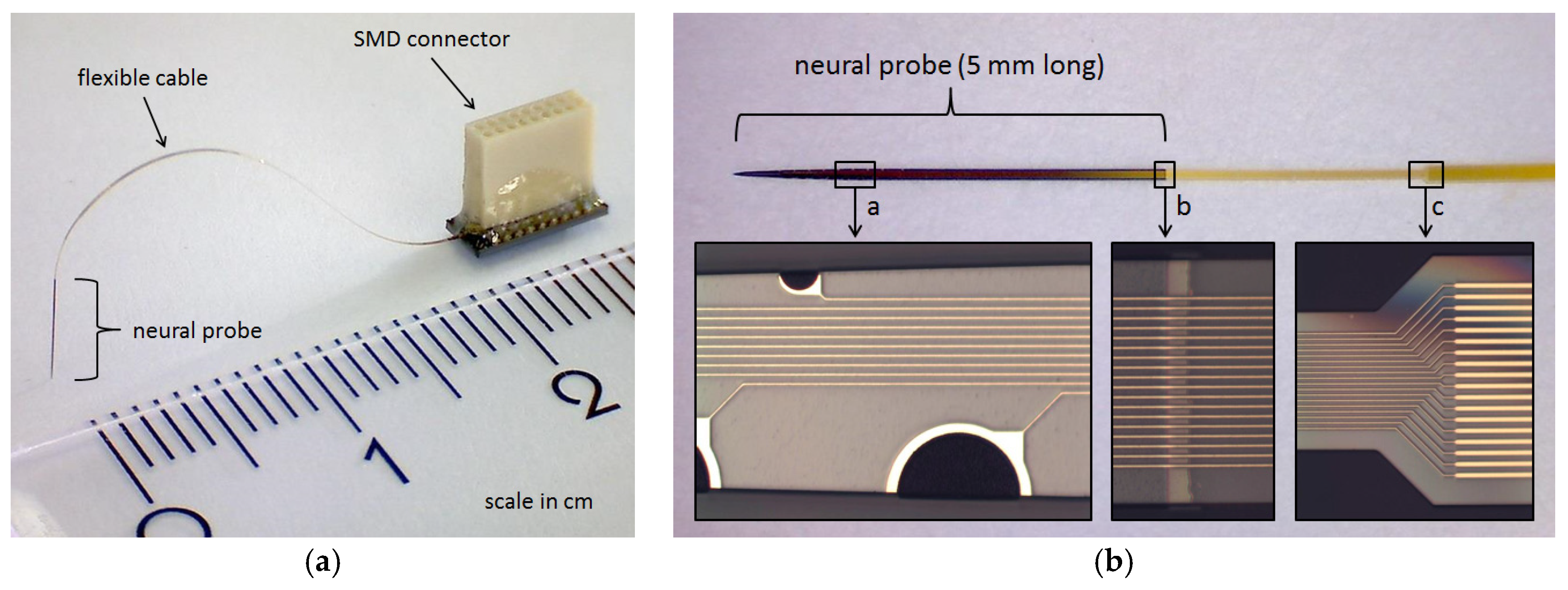
For the monolithical integration of a highly flexible electrical connection to the silicon stiffened neural probe a microfabrication process was developed previously [4]. Using the existing microfabrication process and some minor modifications new probes were fabricated carrying microelectrodes of geometric surface areas up to 4000 µm2 for intracortical microstimulation experiments (see Figure 1). Compared to the previous generation of these neural probes the electrical insulation stability of the used biocompatible polymer polyimide [6] could be further improved by increasing the adhesion strength between two polyimide layers that encapsulate the gold conductive paths on the probe shaft and flexible cable. This is achieved by a short O2 plasma treatment (30 s) using a reactive ion etching process directly prior to the second polyimide coating.

Figure 1.
(a) 5 mm long silicon stiffened multichannel neural probe (30 µm thick and maximum 130 µm wide) with a monolithically integrated 25 mm long highly flexible ribbon cable (10 µm thick PI) connected to a base with a soldered 0.64 mm pitch SMD Omnetics connector; (b) Top view of the multichannel neural probe connected to the flexible cable with enlarged view of (a) PEDOT: PSS coated gold microelectrodes of different geometric areas (160 µm2 to 4000 µm2); (b) transition of the silicon stiffened shaft to the flexible cable and (c) widening of the gold conductive paths from 2 µm to 5 µm widths.
3. Functional Characterization In Vitro and In Vivo
3.1. In Vitro Characterization
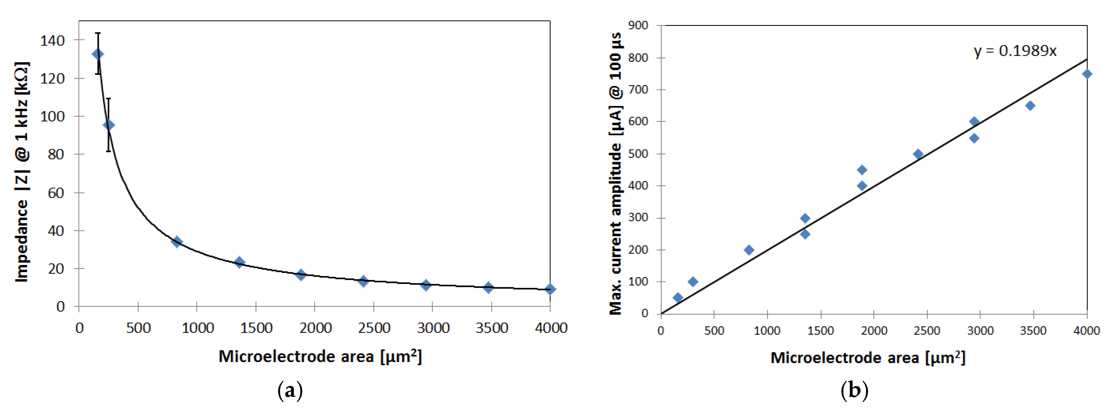
After the microfabrication process, soldering of the connector and PEDOT: PSS coating of the gold microelectrodes the functionality of the probes is evaluated in vitro using Ringer’s electrolyte solution. As the electrode double-layer capacity rises by increasing the geometric surface area, the total impedance of the microelectrodes decreases to a minimum of approx. 10 kΩ @ 1 kHz (see Figure 2a). For microstimulation purposes the charge injection capacity of the fabricated electrodes is essential. Bipolar current pulses were applied to PEDOT: PSS coated electrodes with different geometric surface areas to evaluate the maximum charge injection before electrolysis occurs (see Figure 2b). The linear regression reveals a charge injection capacity of 2 mC/cm2, which is comparable to in vitro results using PEDOT: PSS electrodes reported by Venkatraman et al. [7].
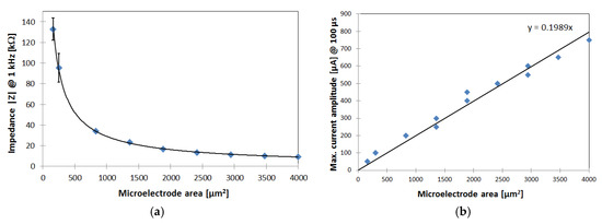
Figure 2.
(a) In Vitro impedance measurement results of PEDOT: PSS coated gold microelectrodes with different geometric areas on the silicon stiffened shaft (measurements were done in Ringer’s solution and platinum counter electrode); (b) In Vitro evaluation of the maximum current amplitude for bipolar 100 µs long current pulses in Ringer’s solution with PEDOT: PSS counter electrode, before electrolysis occurs. Linear regression results in a charge injection capacity of approx. 2 mC/cm2.
3.2. In Vivo Characterization
All procedures used in this study were performed in accordance with the guidelines for the welfare of experimental animals issued by the Federal Government of Germany, approved by local authorities and conformed to the guidelines of the National Institutes of Health for the care and use of laboratory animals.
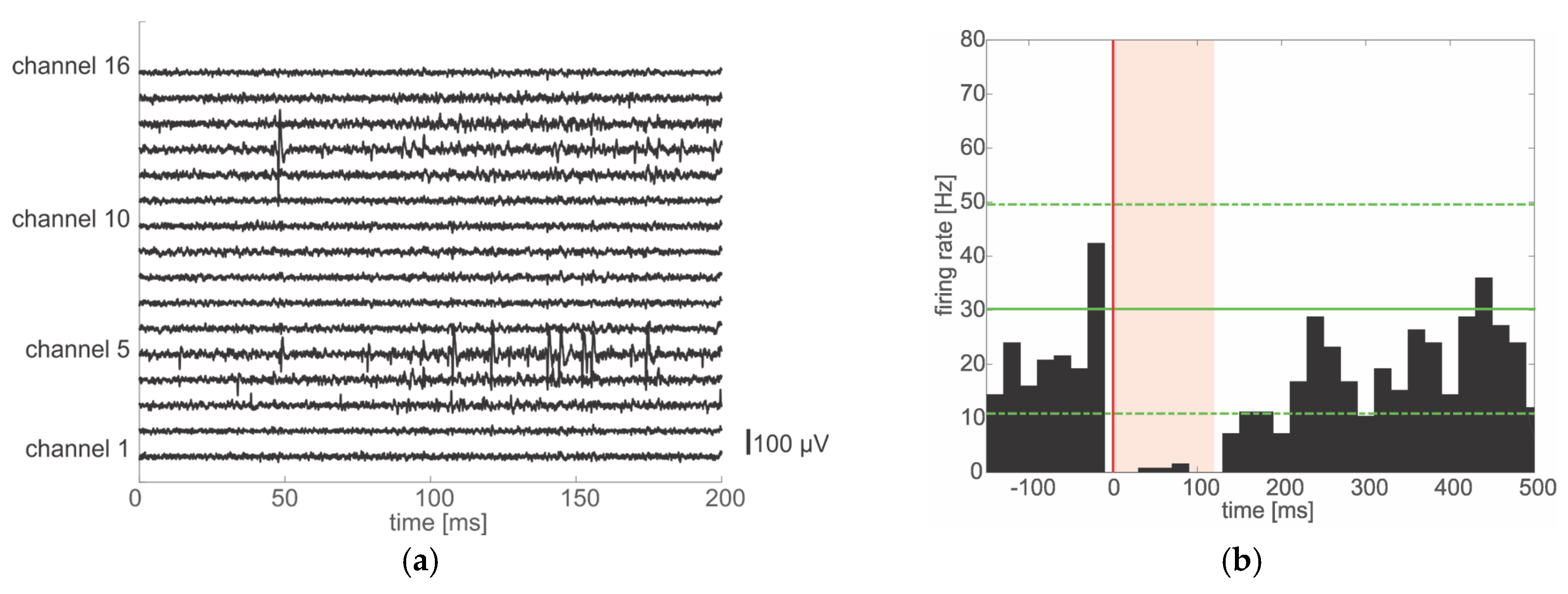
For the evaluation of the in vivo stability of intracortical neural recording and microstimulation the floating neural probes were implanted in rat cortex. The implantation procedure using a developed insertion tool is described in detail elsewhere [4]. Figure 3a shows data traces with recorded neural activity 38 days after chronic implantation of a 16 channel probe with only 160 µm2 microelectrodes. Action potentials can be clearly identified on several channels.

Figure 3.
(a) Data traces of 200 ms duration showing data recorded 38 days after chronic implantation of the multichannel neural probe. Single unit activity can be observed on several channels; (b) Microstimulation effects on neural activity: single bipolar pulses (20 µA per phase, 100 µs phase duration) were applied at one electrode while the others were recorded. (P)eris(S)timulus(T)ime(H)istogram shows a suppression of activity directly after the pulse (red line at time 0) for 120 ms (red shaded area) at one microelectrode with spiking activity. Recovery of activity may take up to 400–500 ms. The green line depicts the average firing rate before electrical stimulation. Broken green lines depict two times the standard deviation.
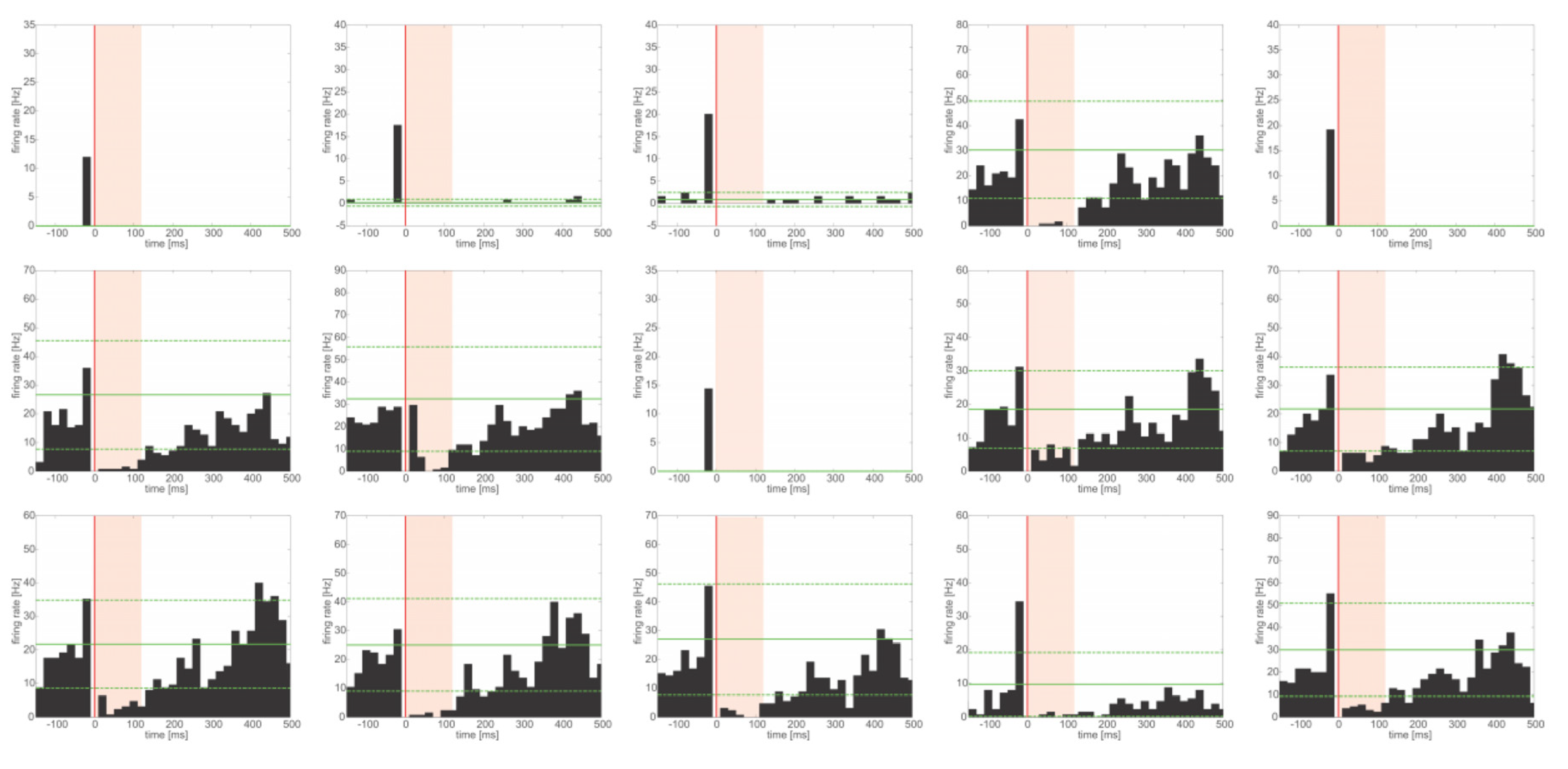
Figure 3b and Figure 4 show the response of the neural network to the application of a microstimulation pulse at a 2000 µm2 microelectrode in an acute experiment at one recorded microelectrode and at all recorded microelectrodes, respectively. The neural activity recorded with the non-stimulated electrodes of the probe is reproducible suppressed for 100 to 150 ms after the application of a single bipolar pulse of 20 µA current amplitude with a phase duration of 100 µs and interphase duration of 50 µs. These results verify the functionality of the neural probes for acute intracortical microstimulation.
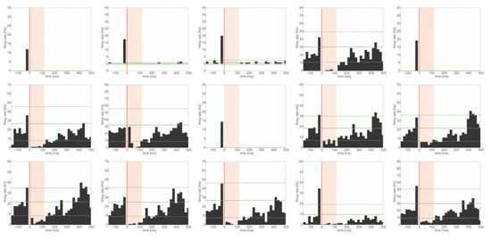
Figure 4.
Microstimulation effects on neural activity: single bipolar pulses (20 µA per phase, 100 µs phase duration) were applied at one electrode while the others were recorded. (P)eris(S)timulus(T)ime(H)istograms show a suppression of activity directly after the pulse (red line at time 0) for 100 to 150 ms (red shaded area) at all recorded microelectrodes with spiking activity. Recovery of activity may take up to 400–500 ms. The green line depicts the average firing rate before electrical stimulation. Broken green lines depict two times the standard deviation.
4. Conclusions and Outlook
The presented results show chronically recorded neural activity in rat cortex with high signal quality using novel multichannel floating neural probes. Furthermore the preliminary microstimulation results indicate the capability of the improved probes for long-term stable stimulation of the neural network in the brain. These promising findings are fundamental for chronic, bidirectional neural interfaces.
In the next step the microstimulation stability will be evaluated in vivo for chronically implanted neural probes.
Acknowledgments
This work was supported by the Priority Program SPP 1665 “Resolving and Manipulating Neuronal Networks in the Mammalian Brain—From Correlative to Causal Analysis” of the DFG (German Research Foundation, LA 1471/11-2 and KR 1844/2-2) and partially by the German Excellence Initiative via the Creative Unit “I-See—The Artificial Eye: Chronic Wireless Interface to the Visual Cortex”.
Conflicts of Interest
The authors declare no conflict of interest.
References
- Kook, G.; Lee, S.W.; Lee, H.C.; Cho, I.J.; Lee, H.J. Neural probes for chronic applications. Micromachines 2016, 7, 179. [Google Scholar] [CrossRef] [PubMed]
- Spencer, K.C.; Sy, J.C.; Falcón-Banchs, R.; Cima, M.J. A three dimensional in vitro glial scar model to investigate the local strain effects from micromotion around neural implants. Lab Chip 2017, 17, 795–804. [Google Scholar] [CrossRef] [PubMed]
- Luan, L.; Wei, X.; Zhao, Z.; Siegel, J.J.; Potnis, O.; Tuppen, C.A.; Lin, S.; Kazmi, S.; Fowler, R.A.; Holloway, S. Ultraflexible nanoelectronic probes form reliable, glial scar-free neural integration. Sci. Adv. 2017, 3, e1601966. [Google Scholar] [CrossRef] [PubMed]
- Schander, A.; Stemmann, H.; Tolstosheeva, E.; Roese, R.; Biefeld, V.; Kempen, L.; Kreiter, A.K.; Lang, W. Design and fabrication of novel multi-channel floating neural probes for intracortical chronic recording. Sens. Actuators A Phys. 2016, 247, 125–135. [Google Scholar] [CrossRef]
- Schander, A.; Teßmann, T.; Strokov, S.; Stemmann, H.; Kreiter, A.K.; Lang, W. In Vitro evaluation of the long-term stability of PEDOT: PSS coated microelectrodes for chronic recording and electrical stimulation of neurons. In Proceedings of the 2016 IEEE 38th Annual International Conference of the Engineering in Medicine and Biology Society (EMBC), Orlando, FL, USA, 16–20 August 2016; pp. 6174–6177. [Google Scholar]
- Hassler, C.; Boretius, T.; Stieglitz, T. Polymers for neural implants. J. Polym. Sci. Part B Polym. Phys. 2011, 49, 18–33. [Google Scholar] [CrossRef]
- Venkatraman, S.; Hendricks, J.; King, Z.A.; Sereno, A.J.; Richardson-Burns, S.; Martin, D.; Carmena, J.M. In Vitro and In Vivo evaluation of PEDOT microelectrodes for neural stimulation and recording. IEEE Trans. Neural Syst. Rehabil. Eng. 2011, 19, 307–316. [Google Scholar] [CrossRef] [PubMed]
Publisher’s Note: MDPI stays neutral with regard to jurisdictional claims in published maps and institutional affiliations. |
© 2017 by the authors. Licensee MDPI, Basel, Switzerland. This article is an open access article distributed under the terms and conditions of the Creative Commons Attribution (CC BY) license (https://creativecommons.org/licenses/by/4.0/).