Abstract
Hemodialysis (HD) is a life-sustaining extracorporeal blood purifying treatment for end-stage renal disease (ESRD) patients. However, this membrane-based therapy is associated with acute side effects, life-threatening chronic conditions, and unacceptably high morbidity and mortality rates. Numerous surface coatings have been developed to improve the blood compatibility of biomaterials. Heparin is a widely used anticoagulant substance that increases the clotting time and increases the membrane hemocompatibility in terms of platelet adhesion and protein adsorption and anti-clotting activity. However, using heparin is challenging due to its severe or life-threatening side effects such as heparin-induced thrombocytopenia (HIT), in addition to heparin induced thrombocytopenia and thrombosis (HITT). In addition, heparin is strongly electronegative and exhibits a binding affinity for the positive active sites of human serum proteins, which is an additional challenge. Consequently, covalently immobilized heparin would create a more charged surface to induce more blood–membrane interactions, and consequently more adsorbed human serum proteins and biochemical pathway activations, which can negatively affect dialysis patients. Therefore, the current critical review has thoroughly focused on different heparin HD membrane systems, the challenges of heparin-coated dialysis membranes, and the factors affecting its hemocompatibility, in addition to the methods that can be used to enhance its hemocompatibility. Furthermore, this review summarizes the advantages and disadvantages of heparin-grafted methods. Furthermore, the influence of the heparin-immobilization method on the hemocompatibility and performance of the HD membrane was comprehensively analyzed. Finally, we conclude with the future perspectives for the strategies toward the heparinization and heparin-like/mimicking modification of membrane surfaces.
1. Introduction
Hemodialysis (HD) is a life-sustaining extracorporeal blood purifying treatment for end-stage renal disease (ESRD) patients. However, blood–membrane interactions activate blood cells and trigger fibrinogen (FB) conformation, which promotes inflammation cascades. Complement activation results in inflammatory mediator products that cause allergic reactions during HD and can also lead to acute intradialytic pulmonary hypertension, chronic low-grade systemic inflammation, and leukocyte dysfunction [1,2,3]. Bio-incompatibility is also the major reason for albumin adsorption, platelet adhesion, and the production of bradykinin (white blood cells (WBCs) due to inflammation), blood clots, and thrombosis. Coagulation can be prevented with heparin, as the most efficient anticoagulant [4,5].
Several research efforts for hemodialysis membrane coatings have been developed to improve the blood compatibility of biomaterials [6,7,8,9]. Heparin is a widely used anticoagulant substance that increases the clotting time and increases the membrane hemocompatibility in terms of platelet adhesion and protein adsorption and anti-clotting activity. A heparin-coated membrane might unexpectedly reduce the concentration of pro-inflammatory cytokines [10]. Nineteen stable HD patients were first dialyzed with conventional membranes and enoxaparin as an anticoagulant, and then with the heparin-coated membrane without systemic anticoagulation. After the HD session with an Evodial dialyzer (heparin-grafted membrane), the plasma levels of the monocyte chemoattractant protein, endostatin, and activin A were 2–3-fold lower than with standard dialysis. Nevertheless, covalently immobilized heparin would create a more charged surface to induce more blood–membrane interactions, and consequently more adsorbed human serum proteins and biochemical pathway activations, which can negatively affect dialysis patients. Therefore, optimal anticoagulation remains a controversial issue for clinical practice, and use of anticoagulants may increase the uremic bleeding tendency.
Therefore, the current critical review has thoroughly focused on different heparin HD membrane systems, the challenges of heparin-coated dialysis membranes, factors affecting its hemocompatibility, in addition to the methods that can be used to enhance its hemocompatibility. Furthermore, this review summarizes the advantages and disadvantages of heparin-grafted methods. Furthermore, the influence of the heparin-immobilization method on the hemocompatibility and performance of the HD membrane was comprehensively analyzed. Finally, we conclude with the future perspectives for the strategies toward the heparinization and heparin-like/mimicking modification of membrane surfaces
2. Current Challenges of Heparin-Coated Dialysis Membranes
Heparin is a medication and naturally occurring glycosaminoglycan (i.e., a long linear polysaccharide consisting of repeating disaccharide units). The heparin structure is illustrated in Figure 1.
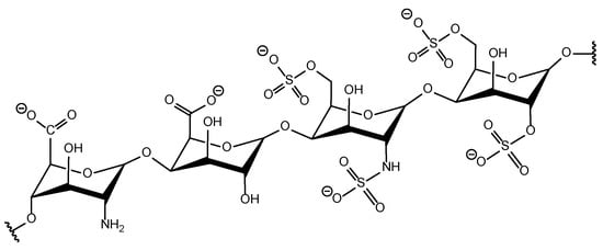
Figure 1.
The structure of heparin.
Heparin is able to interact with coagulation factors XIa, IXa, Xa, and IIa (thrombin), and has been widely used as an anticoagulant reagent since 1935. Although the use of heparin has some side effects such as heparin-induced thrombocytopenia, hypertriglyceridemia, anaphylaxis, bone mineral disease, hyperkalemia, catheter-related sepsis, etc.
The most severe side effect is heparin-induced thrombocytopenia (HIT), which results in blood clotting. There are basically two types of HIT. The most dangerous and potentially life-threatening form is type II HIT, which results in both bleeding and thromboembolic complications. The mechanism of HIT includes several steps. At first, heparin binds with blood platelets with the release of platelet factor 4 (PF4). This PF4, in turn, is able to interact with heparin, resulting in the formation of the heparin–PF4 complex, which triggers blood antibodies (see Figure 2). This interaction between antibodies and the heparin–PF4 complex provokes cascade reactions, resulting in more platelet aggregation, which causes severe thrombocytopenia and further bleeding complications. Moreover, when these heparin-triggered antibodies bind with endothelial cells, it often results in paradoxical thrombus formation with subsequent limb-threatening ischemia, or even fatal pulmonary emboli. Type II HIT usually occurs 5–12 days after heparin exposure, but can happen immediately in the case of re-exposure.
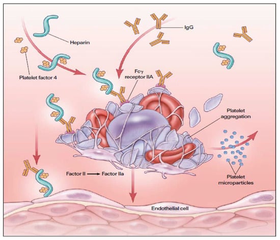
Figure 2.
The mechanism of heparin-induced thrombocytopenia (HIT) with further thrombosis Reprinted/adapted with permission from Ref. [1], 2012, Shen and Winkelmayer.
The above-mentioned complications arise when heparin is injected into the blood during HD procedure and/or HD circuit parts are covered with heparin. Thus, after performing 120 HD sessions, 73% of them ended up with multiple fiber clotting (Grade 3) or clotting of the dialyzer (Grade 4) (see Figure 3) [2].
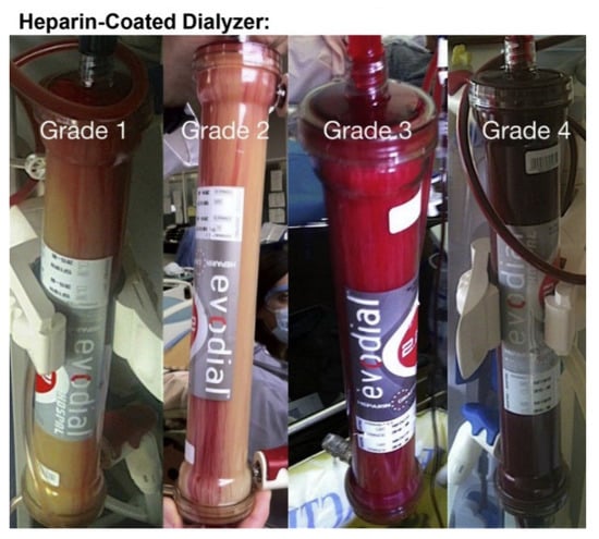
Figure 3.
Examples of different grades (by visual inspection) of the heparin-coated dialyzer membrane modules after performing HD sessions. (Grade 1: clean dialyzer; Grade 2: ~5% of fibers clotting; Grade 3: multiple fiber clotting; Grade 4: total clotting).
The opposite effect of heparin treatment as well as for all forms of anticoagulants is acute hemorrhaging [3]. Incidents of major bleeding during systematic heparinization are reported to occur with the rate of 7.3 to 16.7 per 100 person-years [4], causing major side effects such as osteopenia and drug intolerance aside from thrombocytopenia [5]. Even when heparinization is ceased, nearly a half of all patients that develop HIT experience thrombotic-related complications that result in a mortality rate up to 30% [6].
Furthermore, the use of heparin is also limited by its high price, activity degradation with time, and strong dependence on antithrombin, whose concentration broadly varies across patients with different diseases [7].
3. Factors Affecting Hemocompatibility of Heparin-Grafted Membranes
One of the main reasons responsible for these side effects of using heparin is believed to be associated with the surface negative charge that arises when heparin is applied to cover the HD membrane surface. The negative charge of heparin arises from the inequality in the positive and negative charge groups (see Figure 4).
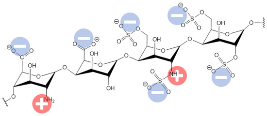
Figure 4.
The distribution of the charged groups in the heparin molecule.
At first glance, the negatively charged membrane has a benefit in terms of protein repulsion, since the main human serum proteins are also charged negatively due to their pI (isoelectric) values (see Table 1) being lower than the blood pH. Despite this, there are frequent observations where negatively charged proteins can adsorb on a negatively charged membrane surface (i.e., in the “wrong” region of their isoelectric points) [8,9,10,11]. This phenomenon can be explained by the presence of rather large charged areas in the protein molecule due to the high molecular weight, and hence the size of the protein molecule. Thus, the protein can position positively charged areas toward the membrane surface, resulting in electrostatic interaction, although the heparin-coated surfaces demonstrated a decreased affinity for particular blood proteins including C3, fibronectin, and fibrinogen [12]. The higher negative surface charge of the HD membrane indicates a high degree of hydrophilicity, which leads to the absorption of water from the blood circulatory system [11]. The dehydration of RBCs significantly contributes to the increased deformability and hemolysis of blood cells [11]. Nevertheless, the current trend in HD membrane development is to create near-zero charged surfaces to minimize any possible electrostatic interaction with human serum proteins or other molecules whose adsorption can provoke further cascade reactions and related undesired consequences. Thus, the above-mentioned heparin-induced thrombocytopenia results from the interaction of negatively charged heparin with positively charged platelet factor 4 (PF4) [13]. It should also be noted that roughness is a key in membrane morphology, which leads to blood cell rupture.

Table 1.
The isoelectric points of the main human proteins.
4. Methods for Enhancing Dialysis Membrane Hemocompatibility
All of the methods for improvements in the material biocompatibility applicable for membranes can be divided into two categories through achieving a bio-passive antifouling surface or bioactive surfaces.
4.1. Biopassive Antifouling Surface
This approach assumes the creation of surfaces that possess a minimal adsorption of proteins and blood cells, since this phenomenon is considered as the very first step for further thrombotic response, blood clotting, and biochemical cascade reaction, which result in severe health problems for HD patients. Although the aim of biopassive surfaces is to minimize triggering immune response reactions, the effectiveness of this approach for long-term applications is still a major concern. Hence, a bioinactive surface is hardly suitable for biomedical implants, but is a good option for short-term or single-use applications such as hemodialysis.
One example of a biopassive surface is a micropatterned surface that possesses superhydrophobic (SH) properties, resulting in antifouling behavior (so called “the lotus effect”). There are some reports, indicating good protein repulsion, decreased platelet adsorption, and reduced blood component activation [14,15]. The main concern regarding SH surfaces is the air pocket layer, which prevents liquid contact with the SH surface, which is able to provoke protein denaturation, causing coagulation cascade reactions and further distal thrombus formation [16]. Despite this concern, there has been a report showing a successful implementation of the SH surface for 8 days in the in vivo implant introduced into a sheep body without any sign of blood clotting or thrombosis [17].
Another example of a biopassive surface is a zwitterionic (ZW)-coated surface. Unlike SH surfaces, this type of surface is very hydrophilic. ZW molecules have an equal number of positive and negative charges, which results in near-zero total charge of the surface, thus minimizing the electrostatic interaction with proteins and other charged blood components. Moreover, the presence of charged moieties in ZW molecules creates a layer of bonded water molecules that creates obstacles for protein adsorption on the material surface, thus possessing antifouling properties [18,19]. These ZW molecules are now being developed for use to create the 3rd (the latest at the moment)-generation of HD membranes [20]. ZW coated materials have also been successfully used in implant applications, showing good bio- and hemocompatibility [21,22,23].
Another approach to create a biopassive surface utilizes the immobilization of specific physiological proteins on a material surface. In vivo experiments have demonstrated that protein adsorption on the surface of the nanoparticles results in the elimination of non-specific cellular uptake [24]. Moreover, the protein-covered surface being passive is believed to be associated, not with protein repellence as with other biopassive surface creation methods, but due to the affinity of the desired proteins due to surface functionalization [25]. However, the adsorbed proteins may change their conformation, revealing their active and antigenic sites, which may trigger cascade complement reactions and immune responses, resulting in undesired consequences [26,27]. Another issue that arises from using this approach is the Vroman effect (i.e., preliminary adsorbed proteins are able to be replaced with other proteins such as fibrinogen), which results in further platelet adhesion and blood clotting [28]. Even if protein is covalently immobilized on the material surface to prevent protein exchange, the adsorbed protein is subjected to degradation that also results in platelet adhesion with undesired further thrombotic events, which significantly limits of the long-term applications of this approach [29].
4.2. Bioactive Surfaces
This approach utilizes the immobilization of bioactive compounds that minimize the immune response by interacting with key blood components or by releasing bioactive compounds. Nitric oxide (NO) is a gaseous free radical molecule that attenuates the interaction of platelets to the adsorbed fibrinogen, von Willebrand factor, and other blood proteins, resulting in reduced platelet activation [30]. Moreover, this approach is based on the ability of the endothelial lining of blood vessel cells to synthesize and exhaust NO from L-arginine at an estimated flux of 0.5 to 4.0 × 10⁻¹⁰ mol/cm2/min [31]. NO producing materials depend on the NO generation triggering type including physiologically, enzymatic, chemical, and thermal activated [32,33,34,35]. Ex vivo experiments have demonstrated a significant reduction in the thrombus formation in human blood and the inhibition of platelet aggregation in platelet rich plasma [36]. Earlier in vivo experiments in developing NO releasing materials allowed for a reduction in the thrombus area up to six times in a 7 h rabbit ECC model [37]. Recent work with NO releasing materials indicated the viability of this approach for ECC applications, wherein the maintenance of 90% of the baseline platelet count was achieved in a 4 h rabbit model using polyvinylchloride (PVC) tubing [38].
Indeed, the most widely known substance used to prevent blood coagulation is heparin, which has been used as systemic anticoagulant since 1935. Despite reducing the thrombin activity, heparin-coated surfaces are often not able to suppress other procoagulant activity including platelet activation.
One of the main reasons responsible for the side effects of using heparin is believed to be associated with the surface negative charge that arises when heparin is applied to cover the HD membrane surface. To overcome this, heparin should be attached to a positively charged substrate, resulting in near-zero surface charge (see Figure 5).
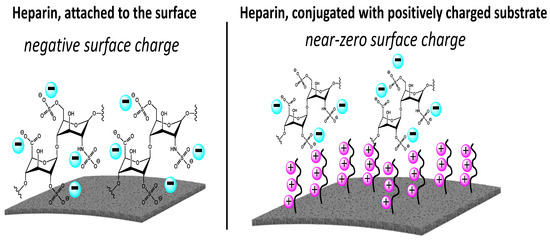
Figure 5.
Approaches to immobilize heparin on the membrane surface.
Using this approach, there have been several reports claiming the successful implementation of ionic complexes and near-zero charges in in vivo experiments, when no fibrosis was observed around the implanted foreign parts [21,39,40,41]. Furthermore, the above-mentioned complexes are reported to possess good hemocompatibility and near-zero adsorption of the platelets and fibrinogen, which is believed to be the first step in designing no-clotting HD membranes [40,42,43,44,45,46,47,48,49,50,51].
5. Recent Development of Heparin-Immobilized Dialysis Membranes
A hollow-fiber oxygenator membrane was covalently coated with nitrous acid degraded heparin using aqueous free radical activation and oxygenation and the coupling of a polyethyleneimine spacer [52]. This coating technology allowed for a heparin surface density of 3.5 μg/cm2 to be achieved. Protein adsorption was performed within 6 h. The binding mechanisms of the proteins were demonstrated to be changed for a heparin-coated surface that resulted in less protein adsorption, which was observed starting from 10 min of the experiment. However, complement system initiator proteins were detected to bind more to the heparinized membrane.
The Klotho (KL) gene was cross-linked with heparin to the acellular small intestinal submucosa (SIS) [53]. In vivo experiments demonstrated enhanced adhesion of the endothelial cells on the SIS membrane, resulting in increased patency rate, endothelialization, and smooth muscle regeneration, which indicates improved hemocompatibility. This approach of promoting endothelial cell growth on the implant surface demonstrates promising results, though at the moment, it is hardly applicable for the biocompatibility of hollow fiber-based HD membrane modules.
To simplify the membrane surface heparinization, a chitosan support layer was used [54]. Chitosan was directly added to the PES spinning solution, so further heparinization can be easily performed just by treating of hollow fibers with heparin solution. The authors proposed performing this procedure right before the HD session to minimize the immobilized heparin degradation. The resultant membranes were observed to possess a reduction in the thrombin–antithrombin complex, active complement component 3, and platelet factor 4 concentration, which indicates an improvement in hemocompatibility. Moreover, the above-mentioned modification with heparin increased the membrane flux (for pure water).
A similar approach in the simplification of heparin immobilization was used in another work [55]. Instead of chitosan, polydopamine (PDA) was used. It was demonstrated that aside from adding polydopamine in the bore spinning solution, it is also possible to coat PES hollow fiber membranes with PDA after membrane spinning, though it requires more steps compared to using PDA in the spinning process. Both ways resulted in obtaining a near-zero charged membrane, which is considered as a benefit. The resultant membranes are going to be used in hemocompatibility tests in future works.
Chitosan was also used for heparin immobilization on a thermo-responsive poly(N-isopropylacrylamide) (PNIPAM) polymer, that is, used for controlled drug release and tissue engineering [56]. Up to 10 layers of alternating layers of chitosan and heparin were created. To enhance the resultant complex stability layers were covalently cross-linked using 1-ethyl-3-(3-dimethylaminopropyl) carbodiimide (EDC) and N-hydroxysuccinimide (NHS), which resulted in a reduced surface charge that is associated with the minimization of heparin and chitosan loss. Moreover, the cross-linked complex demonstrated better biocompatibility in terms of the growth of C3H10T1/2 cells after 24 h of cultivation. However, an uncross-linked chitosan/heparin complex was reported to be successfully used as an implant for 5 months for vascular regeneration [57]. In this study, a decellularized scaffold (DCS) vascular path was coated with poly(vinyl alcohol) (PVA) with the further immobilization of chitosan/heparin via layer-by-layer (LbL) self-assembly. At the same time, the in vitro hemocompatibility test showed that the APPT time increased only to 45 s for the heparin/chitosan coated vascular path, whereas the PVA-coated DCS possessed a 26 s APPT time. The same situation appeared for the PRT, PT, and TT times when the heparin/chitosan complex increased these times to 25–40 s, though the unmodified material demonstrated 15–20 s.
The layer-by-layer self-assembly (LbL) technique was used to immobilize heparin with the oppositely charged dihydroxy-iron (DHI) on the bovine pericardial scaffold (BPS) surface [58]. This cycle was repeated up to 10 times to create a multi-layer structure with alternating charged layers. The resultant complex was not stable and was able to gradually release heparin, thus resulting in 30 days of anticoagulant activity in the in vitro tests. As was previously discussed, the presence of heparin in human blood is able to provoke undesired consequences such as HIT. Heparin dosage is always a critical point for HD sessions, thus additional research is necessary to develop this approach.
Carbon nanotubes were used to carry heparin on their surface with further incorporation into the polyurethane (PU) matrix [59]. The resultant material showed increased APPT time (more than 120 s compared to 20 s for non-heparinized material), reduced platelet adhesion, and increased hydrophilicity.
Heparin was also used to improve the hemocompatibility and hydrophilicity of the extreme hydrophobic nature of poly(ε-caprolactone) (PCL) to create vascular graft implants [60]. Heparin-coated PCL nanofibrous scaffolds were prepared using gamma irradiation and N-(3-dimethylaminopropyl)-N′-ethyl carbodiimide hydrochloride/N-hydroxysuccinimide reaction chemistry on a preliminary 2-aminoethyl methacrylate (AEMA) hydrochloride grafted surface. The resultant modification reduced the fibrinogen adsorption to 4 μg/mm2 (twice less compared with the unmodified PCL vascular graft). Moreover, improvement in the recovery of blood vessel function was observed for implanted heparin-coated vascular grafts into 24-month-old Sprague Dawley rats due to promoting the proliferation of endothelial cells and preventing thrombosis. The examples of using heparin through different approaches of its immobilization on various surfaces and the resulting outcomes are listed in Table 2.
Table 2.
The effects of the different approaches of heparin immobilization.
6. Conclusions
The development of antifouling and anti-clotting materials is of great importance for hemodialysis and biomedical applications. The current tendency in the development of biocompatible materials is to design a membrane with near-zero charge due to the immobilization of zwitterionic molecules or pseudo-zwitterionic complexes. Achieving a near-zero charge dialysis membrane will minimize any possible electrostatic interaction with human serum proteins or other molecules, whose adsorption can provoke further cascade reactions and related undesired consequences.
The possibilities of current chemistry allow us to synthesize tunable structures with the desired properties that are potentially capable of replacing heparin and providing ultimate hemocompatibility. At the same time, it is also possible to use heparin for the creation of complex conjugates that eliminate heparin drawbacks, although this approach seems to be less promising than the controlled synthesis of heparin-mimicking polymers.
Achieving a biopassive antifouling surface that possesses minimal adsorption of proteins and blood cells is urgently required, since this phenomenon is considered as the very first step for further thrombotic response, blood clotting, and biochemical cascade reaction, which result in severe health problems for HD patients. Though the aim of biopassive surfaces is to minimize triggering immune response reactions, the effectiveness of this approach for long-term applications is still a major concern. Hence, a bioinactive surface is hardly suitable for biomedical implants, but is a good option for short-term or single-use applications such as hemodialysis. On the other hand, a bioactive surface utilizes the immobilization of bioactive compounds that minimize the immune response by interacting with key blood components or by releasing bioactive compounds.
Furthermore, with the methods mentioned in our study, it is more suitable for modifying flat-sheet membranes. However, studies to date on the heparin or heparin-mimicking modification of hollow fibers are not sufficient or well-tested. Therefore, in future studies, much more attention should be paid to the surface modification of hollow fiber membranes.
A few studies have succeeded in the mimicking of heparin conformation, since the anticoagulant of heparin is not only derived from the chemical groups, but also because the specific conformation of heparin may also promote the binding of coagulant factors. Thus, with a further understanding of heparin, the ultimate goal should be to design advanced heparin-mimicking polymers with both mimicking groups and conformations. It is believed that this review will evoke more attention toward the design of heparinized and heparin-like/mimicking membranes and encourage future advancements of this emerging research field.
Author Contributions
Conceptualization, A.A; formal analysis, D.K.; investigation, A.A. and A.S.; visualization, D.K.; supervision, A.A.; project administration, A.A.; funding acquisition, A.A. All authors have read and agreed to the published version of the manuscript.
Funding
The manuscript received no external funding.
Institutional Review Board Statement
Amira Abdelrasoul, the principal investigator of the project, obtained Research Ethics Approval and Operational Approval to conduct this research from the Saskatchewan Health Authority. She is responsible for the regulatory approvals that pertain to this project, and for ensuring the project was conducted according to the governing law. All participants in this study signed a written informed consent, approved by the University of Saskatchewan Biomedical Research Ethics Board (Bio-REB).
Acknowledgments
The authors acknowledge the funding support from the Saskatchewan Research Health Foundation (SHRF). The authors are also grateful to the University of Saskatchewan (U of S) for all of the facilities provided to conduct this research in collaboration with the Saskatchewan Transplant Program at St. Paul’s Hospital, Saskatoon.
Conflicts of Interest
The authors declare no conflict of interest.
Abbreviations
| Abbreviation | Meaning |
| HIT | Heparin-induced thrombocytopenia |
| PF4 | Platelet factor 4 |
| HD | Hemodialysis |
| C3 | Complement component 3 |
| HSA | Human serum albumin |
| FB | Fibrinogen |
| TRF | Transferrin |
| SH | Superhydrophobic |
| ZW | Zwitterionic |
| NO | Nitric oxide |
| ECC | Extracorporeal circuits |
| PVC | Polyvinyl chloride |
| KL | The Klotho Gene |
| SIS | Small intestinal submucosa |
| PES | Polyethersulfone |
| PDA | Polydopamine |
| PNIPAM | Poly(N-isopropylacrylamide |
| EDC | 1-ethyl-3-(3-dimethylaminopropyl) carbodiimide |
| NHS | N-hydroxysuccinimide |
| DCS | Decellularized scaffold |
| PVA | Poly(vinyl alcohol) |
| LbL | Layer-by-layer |
| APTT | Activated partial thromboplastin time |
| PRT | Plasma recalcification time |
| PT | Prothrombin time |
| TT | Thrombin time |
| DHI | Dihydroxy-iron |
| BPSs | Bovine pericardial scaffolds |
| PU | Polyurethane |
| PCL | Poly(ε-caprolactone) |
| AEMA | 2-aminoethyl methacrylate |
| PTFE | Poly-tetrafluoroethylene |
| ACT | Activated clotting time |
| PFO | Pericapsular fibrotic overgrowth |
| PA | Pullulan acetate |
| PEG | Polyethylene glycol |
| PLL | Poly-L-lysine |
| BSA | Bovine serum albumin |
| PEI | Poly(ethyleneimine) |
| MES | Morpholinoethane sulfonic acid |
| PLA | Polylactic acid |
| VEGF165 | Vascular endothelial basic fibroblast growth factor |
| CH | Chitosan |
| Hep | Heparin |
| SDVG | Small-diameter vascular grafts |
| sNHS | N-hydroxysulfosuccinimide sodium salt |
| PSf | Polysulfone |
| PEC | Polyelectrolyte complex |
| PAN | Polyacrylonitrile |
| HAAb | Heparin-associated antiplatelet antibodies |
| AVG | Arteriovenous graft |
| PVDF | Poly-vinylidene fluoride |
| PAV | Polyallylamine |
| TA | Tannic acid |
| PEtOx | Poly(2-ethyl-2-oxazoline) |
| HUVEC | Human umbilical vein endothelial cells |
| Tyr | Tyrosinase |
| PMA | Phorbol 12-myristate 13-acetate |
| PMNLs | Polymorphonuclear leukocytes |
| DX | Dextran sulfate |
| DS | Dermatan sulfate |
| ES | Endothelial cell surface |
| HS | Heparan sulfate |
| AFNB | 4-azido-lfluoro-2-nitrobenzene |
| SBS | Styrene-butadiene-styrene |
| CDI | Carbonyldiimidazole |
| SS | Stainless steel |
| ChS | Chondroitin 6-sulfate |
| TPE | Thermoplastic elastomer |
| EDAC | 1-ethyl-3-(dimethyl-aminopropyl) carbodiimide hydrochloride |
| PE | Hydrophobic polyethylene |
| ECM | Extracellular matrix |
| SMC | Smooth muscle cells |
References
- Shen, J.I.; Winkelmayer, W.C. Use and safety of unfractionated heparin for anticoagulation during maintenance hemodialysis. Am. J. Kidney Dis. 2012, 60, 473–486. [Google Scholar] [CrossRef] [PubMed]
- Islam, M.S.; Hassan, Z.A.; Chalmin, F.; Vido, S.; Berrada, M.; Verhelst, D.; Donnadieu, P.; Moranne, O.; Esnault, V.L. Vitamin E-Coated and Heparin-Coated Dialyzer Membranes for Heparin-Free Hemodialysis: A Multicenter, Randomized, Crossover Trial. Am. J. Kidney Dis. 2016, 68, 752–762. [Google Scholar] [CrossRef] [PubMed]
- Harter, K.; Levine, M.; Henderson, S.O. Anticoagulation drug therapy: A review. West J. Emerg. Med. 2015, 16, 11–17. [Google Scholar] [CrossRef] [PubMed]
- van Rein, N.; Biedermann, J.S.; van der Meer, F.J.M.; Cannegieter, S.C.; Wiersma, N.; Vermaas, H.W.; Reitsma, P.H.; Kruip, M.; Lijfering, W.M. Major bleeding risks of different low-molecular-weight heparin agents: A cohort study in 12 934 patients treated for acute venous thrombosis. J. Thromb. Haemost. 2017, 15, 1386–1391. [Google Scholar] [CrossRef] [PubMed]
- Nelson-Piercy, C. Hazards of heparin: Allergy, heparin-induced thrombocytopenia and osteoporosis. Baillikres Clin. Obstet. Gynaecol. 1997, 11, 489–509. [Google Scholar] [CrossRef]
- Abdelrasoul, A.; Shoker, A. Induced Hemocompatibility of Polyethersulfone (PES) Hemodialysis Membrane using Polyvinylpyrrolidone: Investigation on Human Serum Fibrinogen Adsorption and Inflammatory Biomarkers Released. Chem. Eng. Res. Des. 2022, 177, 615–624. [Google Scholar] [CrossRef]
- Eduok, U.; Westphalen, H.; Abdelrasoul, A.; Shoker, A. Influence of UV-irradiation intensity and exposure duration on the hemobiocompatibility enhancement of a novel synthesized phosphobetaine zwitterions polyethersulfone clinical hemodialysis membranes. J. Biomed. Mater. Res. Part B Appl. Biomater. 2022, 110, 573–586. [Google Scholar] [CrossRef]
- Saadati, S.; Eduok, U.; Westphalen, H.; Abdelrasoul, A.; Shoker, A.; Choi, P.; Doan, H.; Ein-Mozaffari, F.; Zhu, N. In situ Synchrotron Imaging of Human Serum Proteins Interactions, Molecular Docking and Inflammatory Biomarkers of Hemocompatible Synthesized Zwitterionic Polymer Coated-Polyvinylidene Fluoride (PVDF) Dialysis Membranes. Surf. Interfaces 2021, 27, 101505. [Google Scholar] [CrossRef]
- Saadati, S.; Westphalen, H.; Eduok, U.; Abdelrasoul, A.; Shoker, A.; Choi, P.; Doan, H.; Ein-Mozaffari, F.; Zhu, N. Biocomptability Enhancement of Hemodialysis Membranes using Novel Zwitterionic: Experimental, in situ Synchrotron Imaging, Molecular Docking, and Clinical Inflammatory Biomarkers Investigations. Mater. Sci. Eng. C 2020, 117, 111301. [Google Scholar] [CrossRef]
- Westphalen, H.; Saadati, S.; Eduok, U.; Abdelrasoul, A.; Shoker, A.; Choi, P.; Doan, H.; Ein-Mozaffari, F. Case studies of clinical hemodialysis membranes: Influences of membrane morphology and biocompatibility on uremic blood-membrane interactions and inflammatory biomarkers. Sci. Rep. 2020, 10, 1–18. [Google Scholar]
- Abdelrasoul, A.; Shoker, A. Influence of Hydration Shell of Hemodialysis Clinical Membranes on Surrogate Biomarkers Activation in Uremic Serum of Dialysis Patients. Biomed. Eng. Adv. 2022, 4, 100049. [Google Scholar] [CrossRef]
- Bolten, S.; Rinas, S.; Scheper, T. Heparin: Role in protein purification and substitution with animal-component free material. Appl. Microbiol. Biotechnol. 2018, 102, 8647–8660. [Google Scholar] [CrossRef] [PubMed]
- Nguyen, T.H.; Xu, Y.; Brandt, S.; Mandelkow, M.; Raschke, R.; Strobel, U.; Delcea, M.; Zhou, W.; Liu, J.; Greinacher, A. Characterization of the interaction between platelet factor 4 and homogeneous synthetic low molecular weight heparins. J. Thromb. Haemost. 2020, 18, 390–398. [Google Scholar] [CrossRef] [PubMed]
- Abdelrasoul, A.; Westphalen, H.; Saadati, S.; Shoker, A. Hemodialysis biocompatibility mathematical models to predict the inflammatory biomarkers released in dialysis patients based on hemodialysis membrane characteristics and clinical practices. Sci. Rep. 2021, 11, 23080. [Google Scholar] [CrossRef]
- Ozkan, E.; Mondal, A.; Singha, P.; Douglass, M.; Hopkins, S.P.; Devine, R.; Garren, M.; Manuel, J.; Warnock, J.; Handa, H. Fabrication of Bacteria- and Blood-Repellent Superhydrophobic Polyurethane Sponge Materials. ACS Appl. Mater. Interfaces 2020, 12, 51160–51173. [Google Scholar] [CrossRef]
- Cieplak, M.; Allan, D.B.; Leheny, R.L.; Reich, D.H. Proteins at air-water interfaces: A coarse-grained model. Langmuir 2014, 30, 12888–12896. [Google Scholar] [CrossRef]
- Brancato, L.; Decrop, D.; Lammertyn, J.; Puers, R. Surface Nanostructuring of Parylene-C Coatings for Blood Contacting Implants. Materials 2018, 11, 1109. [Google Scholar] [CrossRef]
- Venault, A.; Wei, T.-C.; Shih, H.-L.; Yeh, C.-C.; Chinnathambi, A.; Alharbi, S.A.; Carretier, S.; Aimar, P.; Lai, J.-Y.; Chang, Y. Antifouling pseudo-zwitterionic poly(vinylidene fluoride) membranes with efficient mixed-charge surface grafting via glow dielectric barrier discharge plasma-induced copolymerization. J. Membr. Sci. 2016, 516, 13–25. [Google Scholar] [CrossRef]
- Cai, N.; Li, Q.; Zhang, J.; Xu, T.; Zhao, W.; Yang, J.; Zhang, L. Antifouling zwitterionic hydrogel coating improves hemocompatibility of activated carbon hemoadsorbent. J. Colloid. Interface Sci. 2017, 503, 168–177. [Google Scholar] [CrossRef]
- Mollahosseini, A.; Abdelrasoul, A.; Shoker, A. Latest advances in zwitterionic structures modified dialysis membranes. Mater. Today Chem. 2020, 15, 100227. [Google Scholar] [CrossRef]
- Nazari, S.; Abdelrasoul, A. Surface Zwitterionization of HemodialysisMembranesfor Hemocompatibility Enhancement and Protein-mediated anti-adhesion: A Critical Review. Biomed. Eng. Adv. 2022, 3, 100026. [Google Scholar] [CrossRef]
- Abdelrasoul, A.; Shoker, A. Investigations on the Impact of Hemodialysis Clinical Practices on Human Plasma Proteins Loss and von Willebrand factor. Kidney Int. Rep. 2022, 7, S258. [Google Scholar] [CrossRef]
- Venault, A.; Ye, C.C.; Lin, Y.C.; Tsai, C.W.; Jhong, J.F.; Ruaan, R.C.; Higuchi, A.; Chinnathambi, A.; Ho, H.T.; Chang, Y. Zwitterionic fibrous polypropylene assembled with amphiphatic carboxybetaine copolymers for hemocompatible blood filtration. Acta Biomater. 2016, 40, 130–141. [Google Scholar] [CrossRef] [PubMed]
- Schottler, S.; Becker, G.; Winzen, S.; Steinbach, T.; Mohr, K.; Landfester, K.; Mailander, V.; Wurm, F.R. Protein adsorption is required for stealth effect of poly(ethylene glycol)- and poly(phosphoester)-coated nanocarriers. Nat. Nanotechnol. 2016, 11, 372–377. [Google Scholar] [CrossRef]
- Schottler, S.; Landfester, K.; Mailander, V. Controlling the Stealth Effect of Nanocarriers through Understanding the Protein Corona. Angew. Chem. Int. Ed. Engl. 2016, 55, 8806–8815. [Google Scholar] [CrossRef]
- Tenzer, S.; Docter, D.; Kuharev, J.; Musyanovych, A.; Fetz, V.; Hecht, R.; Schlenk, F.; Fischer, D.; Kiouptsi, K.; Reinhardt, C.; et al. Rapid formation of plasma protein corona critically affects nanoparticle pathophysiology. Nat. Nanotechnol. 2013, 8, 772–781. [Google Scholar] [CrossRef]
- Deng, Z.J.; Liang, M.; Monteiro, M.; Toth, I.; Minchin, R.F. Nanoparticle-induced unfolding of fibrinogen promotes Mac-1 receptor activation and inflammation. Nat. Nanotechnol. 2011, 6, 39–44. [Google Scholar] [CrossRef]
- Szleifer, F.F.a.I. Kinetics and Thermodynamics of Protein Adsorption: A Generalized Molecular Theoretical Approach. Biophys. J. 2001, 80, 2568–2589. [Google Scholar]
- Sivaraman, B.; Latour, R.A. Time-dependent conformational changes in adsorbed albumin and its effect on platelet adhesion. Langmuir 2012, 28, 2745–2752. [Google Scholar] [CrossRef]
- Kristyn, S.; Bohl, J.L.W. Nitric oxide-generating polymers reduce platelet adhesion and smooth muscle cell proliferation. Biomaterials 2000, 21, 2273–2278. [Google Scholar]
- Tousoulis, D.; Kampoli, A.M.; Tentolouris Nikolaos Papageorgiou, C.; Stefanadis, C. The Role of Nitric Oxide on Endothelial Function. Curr. Vasc. Pharmacol. 2012, 10, 4–18. [Google Scholar] [CrossRef] [PubMed]
- Handa, H.; Brisbois, E.J.; Major, T.C.; Refahiyat, L.; Amoako, K.A.; Annich, G.M.; Bartlett, R.H.; Meyerhoff, M.E. In vitro and in vivo study of sustained nitric oxide release coating using diazeniumdiolate-oped poly(vinyl chloride) matrix with poly(lactide-co-glycolide) additive. J. Mater. Chem. B 2013, 1, 3578–3587. [Google Scholar] [CrossRef] [PubMed]
- Grossi, L.; Montevecchi, P.C. A Kinetic Study of S-Nitrosothiol Decomposition. Chem. Eur. 2002, 8, 380–387. [Google Scholar] [CrossRef]
- Frost, M.C.; Meyerhoff, M.E. Controlled Photoinitiated Release of Nitric Oxide from Polymer Films Containing S-Nitroso-N-acetyl-DL-penicillamine Derivatized Fumed Silica Filler. J. Am. Chem. Soc. 2004, 126, 1348–1349. [Google Scholar] [CrossRef]
- Broniowska, K.A.; Hogg, N. The chemical biology of S-nitrosothiols. Antioxid. Redox Signal 2012, 17, 969–980. [Google Scholar] [CrossRef]
- Roberts, T.R.; Neufeld, M.J.; Meledeo, M.A.; Cap, A.P.; Cancio, L.C.; Reynolds, M.M.; Batchinsky, A.I. A metal organic framework reduces thrombus formation and platelet aggregation ex vivo. J. Trauma Acute Care Surg. 2018, 85, 572–579. [Google Scholar] [CrossRef]
- Wo, Y.; Brisbois, E.J.; Wu, J.; Li, Z.; Major, T.C.; Mohammed, A.; Wang, X.; Colletta, A.; Bull, J.L.; Matzger, A.J.; et al. Reduction of Thrombosis and Bacterial Infection via Controlled Nitric Oxide (NO) Release from S-Nitroso-N-acetylpenicillamine (SNAP) Impregnated CarboSil Intravascular Catheters. ACS Biomater. Sci. Eng. 2017, 3, 349–359. [Google Scholar] [CrossRef]
- Douglass, M.E.; Goudie, M.J.; Pant, J.; Singha, P.; Hopkins, S.; Devine, R.; Schmiedt, C.W.; Handa, H. Catalyzed Nitric Oxide Release Via Cu Nanoparticles Leads to an Increase in Antimicrobial Effects and Hemocompatibility for Short Term Extracorporeal Circulation. ACS Appl. Bio Mater. 2019, 2, 2539–2548. [Google Scholar] [CrossRef]
- Vaithilingam, V.; Kollarikova, G.; Qi, M.; Larsson, R.; Lacik, I.; Formo, K.; Marchese, E.; Oberholzer, J.; Guillemin, G.J.; Tuch, B.E. Beneficial effects of coating alginate microcapsules with macromolecular heparin conjugates-in vitro and in vivo study. Tissue Eng. Part A 2014, 20, 324–334. [Google Scholar] [CrossRef]
- Bünger, C.M.; Gerlach, C.; Freier, T.; Schmitz, K.P.; Pilz, M.; Werner, C.; Jonas, L.; Schareck, W.; Hopt, U.T.; De Vos, P. Biocompatibility and surface structure of chemically modified immunoisolating alginate–PLL capsules. J. Biomed. Mater. Res. Part A 2003, 67, 1219–1227. [Google Scholar] [CrossRef]
- Chan, H.-M.; Erathodiyil, N.; Wu, H.; Lu, H.; Zheng, Y.; Ying, J.Y. Calcium cross-linked zwitterionic hydrogels as antifouling materials. Mater. Today Commun. 2020, 23, 100950. [Google Scholar] [CrossRef]
- Tsai, C.C.; Chang, Y.; Sung, H.W.; Hsu, J.C.; Chen, C.N. Effects of heparin immobilization on the surface characteristics of a biological tissue fixed with a naturally occurring crosslinking agent (genipin): An in vitro study. Biomaterials 2001, 22, 523–533. [Google Scholar] [CrossRef]
- Zhu, A.; Zhang, M.; Wu, J.; Shen, J. Covalent immobilization of chitosan/heparin complex with a photosensitive hetero-bifunctional crosslinking reagent on PLA surface. Biomaterials 2002, 23, 4657–4665. [Google Scholar] [CrossRef]
- Wang, J.; Chen, Y.; Liu, T.; Wang, X.; Liu, Y.; Wang, Y.; Chen, J.; Huang, N. Covalent co-immobilization of heparin/laminin complex that with different concentration ratio on titanium surface for selectively direction of platelets and vascular cells behavior. Appl. Surf. Sci. 2014, 317, 776–786. [Google Scholar] [CrossRef]
- Chen, Q.; He, Y.; Zhao, Y.; Chen, L. Intervening oxidative stress integrated with an excellent biocompatibility of hemodialysis membrane fabricated by nucleobase-recognized co-immobilization strategy of tannic acid, looped PEtOx brush and heparin. J. Membr. Sci. 2021, 625, 119174. [Google Scholar] [CrossRef]
- Yang, J.M.; Wang, M.C.; Hsu, Y.G.; Chang, C.H.; Lo, S.K. Preparation of heparin containing SBS-g-VP copolymer membrane for biomaterial usage. J. Membr. Sci. 1998, 138, 19–27. [Google Scholar] [CrossRef]
- Zhang, X.; Zhang, G.; Zhang, H.; Li, J.; Yao, X.; Tang, B. Surface immobilization of heparin and chitosan on titanium to improve hemocompatibility and antibacterial activities. Coll. Surf. B Biointerfaces 2018, 172, 338–345. [Google Scholar] [CrossRef]
- Liu, T.; Liu, Y.; Chen, Y.; Liu, S.; Maitz, M.F.; Wang, X.; Zhang, K.; Wang, J.; Wang, Y.; Chen, J.; et al. Immobilization of heparin/poly-(L)-lysine nanoparticles on dopamine-coated surface to create a heparin density gradient for selective direction of platelet and vascular cells behavior. Acta Biomater. 2014, 10, 1940–1954. [Google Scholar] [CrossRef]
- Drozd, N.N.; Lunkov, A.P.; Shagdarova, B.T.; Zhuikova, Y.V.; Il’ina, A.V.; Varlamov, V.P. Chitosan/heparin layer-by-layer coatings for improving thromboresistance of polyurethane. Surf. Interfaces 2022, 28, 101674. [Google Scholar] [CrossRef]
- Vaithilingam, V.; Steinkjer, B.; Ryan, L.; Larsson, R.; Tuch, B.E.; Oberholzer, J.; Rokstad, A.M. In Vitro and In Vivo Biocompatibility Evaluation of Polyallylamine and Macromolecular Heparin Conjugates Modified Alginate Microbeads. Sci. Rep. 2017, 7, 1–15. [Google Scholar] [CrossRef]
- Manabe, K.; Nara, H. Construction of stable biological albumin/heparin multilayers for elastic coatings on hydrophobic antithrombogenic artificial blood vessels. Tribol. Int. 2021, 156, 106843. [Google Scholar] [CrossRef]
- Große-Berkenbusch, K.; Avci-Adali, M.; Arnold, M.; Cahalan, L.; Cahalan, P.; Velic, A.; Maček, B.; Schlensak, C.; Wendel, H.-P.; Stoppelkamp, S. Profiling of time-dependent human plasma protein adsorption on non-coated and heparin-coated oxygenator membranes. Biomater. Adv. 2022, 156, 106843. [Google Scholar] [CrossRef] [PubMed]
- Zhao, P.; Fang, Q.; Gao, D.; Wang, Q.; Cheng, Y.; Ao, Q.; Wang, X.; Tian, X.; Zhang, Y.; Tong, H.; et al. Klotho functionalization on vascular graft for improved patency and endothelialization. Mater. Sci. Eng. C Mater. Biol. Appl. 2022, 133, 112630. [Google Scholar] [CrossRef] [PubMed]
- Rose, I.I.; Kather, M.; Roth, H.; Dünkelberg, H.; Rein, L.; Klimosch, S.N.; Schmolz, M.; Wessling, M. Single-step chitosan functionalized membranes for heparinization. J. Membr. Sci. 2022, 655, 120567. [Google Scholar] [CrossRef]
- Rose, I.I.; Roth, H.; Xie, J.; Hollmann, F.; Votteler, S.; Storr, M.; Krause, B.; Wessling, M. Chemistry in a spinneret—Polydopamine functionalized hollow fiber membranes. J. Membr. Sci. 2022, 648, 120324. [Google Scholar] [CrossRef]
- Lu, Y.T.; Zeng, K.; Fuhrmann, B.; Woelk, C.; Zhang, K.; Groth, T. Engineering of Stable Cross-Linked Multilayers Based on Thermo-Responsive PNIPAM-Grafted-Chitosan/Heparin to Tailor Their Physiochemical Properties and Biocompatibility. ACS Appl. Mater. Interfaces 2022, 14, 29550–29562. [Google Scholar] [CrossRef]
- Sun, G.; Li, Y.; Liu, C.; Jiang, X.; Yang, L.; He, L.; Song, S.; Zhang, J.; Shen, J.; Qiao, T. Chitosan-Heparin Polyelectrolyte Multilayer-Modified Poly(vinyl alcohol) Vascular Patches based on a Decellularized Scaffold for Vascular Regeneration. ACS Appl. Bio Mater. 2022, 5, 2928–2934. [Google Scholar] [CrossRef]
- Nguyen, M.T.N.; Tran, H.L.B. Heparinization of the bovine pericardial scaffold by layer-by-layer (LBL) assembly technique. J. Sci. Adv. Mater. Devices 2022, 7, 100405. [Google Scholar] [CrossRef]
- Dehghani, F.; Khorasani, M.T. Movahedi, M. Fabrication of polyurethane—Heparinized carbon nanotubes composite for heart valves application. Mater. Chem. Phys. 2022, 280, 125819. [Google Scholar] [CrossRef]
- Kim, S.E.; Jeong, S.I.; Shim, K.M.; Jang, K.; Park, J.S.; Lim, Y.M.; Kang, S.S. In Vivo Evaluation of Gamma-Irradiated and Heparin-Immobilized Small-Diameter Polycaprolactone Vascular Grafts with VEGF in Aged Rats. Polymers 2022, 14, 1265. [Google Scholar] [CrossRef]
- Zea, N.; Menard, G.; Le, L.; Bazan, H.; Sternbergh, W.C.; Smith, T. 48. Heparin Bonded PTFE Does Not Improve Hemodialysis Arteriovenous Graft Function. Ann. Vasc. Surg. 2015, 29, 645. [Google Scholar] [CrossRef]
- Borawski, J.; Naumnik, B.; Mysliwiec, M. Activation of hepatocyte growth factor/activin A/follistatin system during hemodialysis: Role of heparin. Kidney Int. 2003, 64, 2229–2237. [Google Scholar] [CrossRef] [PubMed]
- Baird, C.W.; Zurakowski, D.; Robinson, B.; Gandhi, S.; Burdis-Koch, L.; Tamblyn, J.; Munoz, R.; Fortich, K.; Pigula, F.A. Anticoagulation and pediatric extracorporeal membrane oxygenation: Impact of activated clotting time and heparin dose on survival. Ann. Thorac. Surg. 2007, 83, 912–919; discussion 919-20. [Google Scholar] [CrossRef] [PubMed]
- Luley, C.; Westphal, S.; Ambrosch, A.; Neumann, K.H. Anticoagulation with heparin during hemodialysis causes pronounced LDL alterations. In Proceedings of the 11th International Symposium on Atherosclerosis, Paris, France, 5–9 October 1997. [Google Scholar]
- Sung, W.J.; Na, K.; Bae, Y.H. Biocompatibility and interference eliminating property of pullulan acetate/polyethylene glycol/heparin membrane for the outer layer of an amperometric glucose sensor. Sens. Actuators B Chem. 2004, 99, 393–398. [Google Scholar] [CrossRef]
- Dumont, G. C0281 Heparin-induced thrombocytopenia (HIT) type 2 caused by preventive anticoagulation of the circuit of hemodialysis (HD): A case report. Thromb. Res. 2012, 130, S185. [Google Scholar] [CrossRef]
- Glick, D.; Dzierba, A.L.; Abrams, D.; Muir, J.; Eisenberger, A.; Diuguid, D.; Abel, E.; Agerstrand, C.; Bacchetta, M.; Brodie, D. Clinically suspected heparin-induced thrombocytopenia during extracorporeal membrane oxygenation. J. Crit. Care 2015, 30, 1190–1194. [Google Scholar] [CrossRef]
- Pham, P.T.; Miller, J.M.; Demetrion, G.; Lew, S.Q. Clotting by Heparin of Hemoaccess for Hemodialysis in an End-Stage Renal Disease Patient. Am. J. Kidney Dis. 1995, 25, 642–647. [Google Scholar] [CrossRef]
- Warkentin, T.E. Hemodialysis-associated acute systemic reactions and heparin-induced thrombocytopenia. Thromb. Res. 2012, 129, 405–406. [Google Scholar] [CrossRef]
- Doi, Y.; Koga, K.; Sugioka, S.; Inoue, Y.; Arisato, T.; Nishioka, K.; Ishihara, T.; Sugawara, A. Heparin-induced thrombocytopenia among incident hemodialysis patients anticoagulated with low molecular weight heparin: A single-center retrospective study. Nefrologia 2021, 41, 356–358. [Google Scholar] [CrossRef]
- Welp, H.; Ellger, B.; Scherer, M.; Lanckohr, C.; Martens, S.; Gottschalk, A. Heparin-induced thrombocytopenia during extracorporeal membrane oxygenation. J. Cardiothorac. Vasc. Anesth. 2014, 28, 342–344. [Google Scholar] [CrossRef]
- Yamamoto, S.; Koide, M.; Matsuo, M.; Suzuki, S.; Ohtaka, M.; Saika, S.; Matsuo, T. Heparin-Induced Thrombocytopenia in Hemodialysis Patients. Am. J. Kidney Dis. 1996, 28, 82–85. [Google Scholar] [CrossRef]
- Carrier, M.; Rodger, M.A.; Fergusson, D.; Doucette, S.; Kovacs, M.J.; Moore, J.; Kelton, J.G.; Knoll, G.A. Increased mortality in hemodialysis patients having specific antibodies to the platelet factor 4-heparin complex. Kidney Int. 2008, 73, 213–219. [Google Scholar] [CrossRef] [PubMed]
- Tanhehco, Y.C.; Cuker, A.; Rudnick, M.; Sachais, B.S. Investigation of a potential protective mechanism against heparin-induced thrombocytopenia in patients on chronic intermittent hemodialysis. Thromb. Res. 2013, 131, 244–248. [Google Scholar] [CrossRef] [PubMed]
- Andrianova, I.; Hayes, V.M.; Madeeva, D.; Litvinov, R.I.; Cines, D.B.; Poncz, M.; Weisel, J.W.; Rauova, L. Membrane Remodeling By Pathogenic Antibodies Underlies Monocyte Activation in Heparin-Induced Thrombocytopenia. Blood 2015, 126, 2244. [Google Scholar] [CrossRef]
- Matsuo, T.; Nakao, K.; Yamada, T.; Matsuo, O. A new thrombin inhibitor MD805 and thrombocytopenia encountered with heparin hemodialysis. Thromb. Res. 1986, 44, 247–251. [Google Scholar] [CrossRef]
- Prechel, M.M.; Jeske, W.P.; Walenga, J.M. Physiological changes in membrane-expressed platelet factor 4: Implications in heparin-induced thrombocytopenia. Thromb. Res. 2010, 125, e143–e148. [Google Scholar] [CrossRef]
- Ahmed, M.S.; Amin, T.; Amin, A.; Nashawi, M.; Haloot, J.; Fichardt, H.; Prasad, A.; Almomani, A. Valve-in-Valve Transcatheter Aortic-Valve Replacement with Basilica in a Patient with History of Evans Syndrome, Heparin Induced Thrombocytopenia and Hemodialysis. Is It Even Possible? J. Am. Coll. Cardiol. 2021, 77, 2357. [Google Scholar] [CrossRef]
- Ichinose, K.; Okamoto, T.; Tanimoto, H.; Yoshitake, A.; Tashiro, M.; Sakanashi, Y.; Kuwana, K.; Tahara, K.; Kamiya, M.; Terasaki, H. Comparison of a New Heparin-coated Dense Membrane Lung with Nonheparin-coated Dense Membrane Lung for Prolonged Extracorporeal Lung Assist in Goats. Artif. Organs 2004, 28, 993–1001. [Google Scholar] [CrossRef]
- Pekna, M.; Hagman, L.; Halden, E.; Nilsson, U.R.; Nilsson, B.; Thelin, S. Complement Activation During Cardiopulmonary Bypass: Effects of Immobilized Heparin. Ann. Thorac. Surg. 1994, 58, 421–424. [Google Scholar] [CrossRef]
- Ding, Y.; Yang, M.; Yang, Z.; Luo, R.; Lu, X.; Huang, N.; Huang, P.; Leng, Y. Cooperative control of blood compatibility and re-endothelialization by immobilized heparin and substrate topography. Acta Biomater. 2015, 15, 150–163. [Google Scholar] [CrossRef]
- Naumnik, B.; Pawlak, K.; Mysliwiec, M. Different effect of unfractionated heparin and enoxaparin on circulating proangiogenic factors during hemodialysis: A cross-over study. Cytokine 2007, 40, 98–104. [Google Scholar] [CrossRef] [PubMed]
- Kara, F.; Aksoy, E.A.; Yuksekdag, Z.; Aksoy, S.; Hasirci, N. Enhancement of antibacterial properties of polyurethanes by chitosan and heparin immobilization. Appl. Surf. Sci. 2015, 357, 1692–1702. [Google Scholar] [CrossRef]
- Perrenoud, I.A.; Rangel, E.C.; Mota, R.P.; Durrant, S.F.; Cruz, N.C.D. Evaluation of Blood Compatibility of Plasma Deposited Heparin-like Films and SF6 Plasma Treated Surfaces. Mater. Res. 2010, 13, 95–98. [Google Scholar] [CrossRef]
- Caracciolo, P.C.; Diaz-Rodriguez, P.; Ardao, I.; Moreira, D.; Montini-Ballarin, F.; Abraham, G.A.; Concheiro, A.; Alvarez-Lorenzo, C. Evaluation of human umbilical vein endothelial cells growth onto heparin-modified electrospun vascular grafts. Int. J. Biol. Macromol. 2021, 179, 567–575. [Google Scholar] [CrossRef] [PubMed]
- Tseng, P.Y.; Rele, S.M.; Sun, X.L.; Chaikof, E.L. Fabrication and characterization of heparin functionalized membrane-mimetic assemblies. Biomaterials 2006, 27, 2627–2636. [Google Scholar] [CrossRef]
- Wang, L.; Fang, F.; Liu, Y.; Li, J.; Huang, X. Facile preparation of heparinized polysulfone membrane assisted by polydopamine/polyethyleneimine co-deposition for simultaneous LDL selectivity and biocompatibility. Appl. Surf. Sci. 2016, 385, 308–317. [Google Scholar] [CrossRef]
- Kastellorizios, M.; Michanetzis, G.P.; Pistillo, B.R.; Mourtas, S.; Klepetsanis, P.; Favia, P.; Sardella, E.; d’Agostino, R.; Missirlis, Y.F.; Antimisiaris, S.G. Haemocompatibility improvement of metallic surfaces by covalent immobilization of heparin-liposomes. Int. J. Pharm. 2012, 432, 91–98. [Google Scholar] [CrossRef]
- Norbert Weber, H.P.W. Gerhard Ziemer Hemocompatibility of heparin-coated surfaces and the role of selective plasma protein adsorption. Biomaterials 2002, 23, 429–439. [Google Scholar] [CrossRef]
- Lin, W.C.; Liu, T.Y.; Yang, M.C. Hemocompatibility of polyacrylonitrile dialysis membrane immobilized with chitosan and heparin conjugate. Biomaterials 2004, 25, 1947–1957. [Google Scholar] [CrossRef]
- Jacob Kaleekkal, N. Heparin immobilized graphene oxide in polyetherimide membranes for hemodialysis with enhanced hemocompatibility and removal of uremic toxins. J. Membr. Sci. 2021, 623, 119068. [Google Scholar] [CrossRef]
- Humphries, J.E.; Kaplan, D.M.; Bolton, W.K. Heparin Skin Necrosis: Delayed Occurrence in a Patient on Hemodialysis. Am. J. Kidney Dis. 1991, 17, 233–236. [Google Scholar] [CrossRef]
- Mureebe, L.; Coats, R.D.; Silliman, W.R. Heparin-associated antiplatelet antibodies increase morbidity and mortality in hemodialysis patients. J. Vasc. Surg. 2005, 41, 560. [Google Scholar] [CrossRef][Green Version]
- Zea, N.; Menard, G.; Le, L.; Luo, Q.; Bazan, H.A.; Sternbergh, W.C., 3rd; Smith, T.A. Heparin-Bonded Polytetrafluorethylene Does Not Improve Hemodialysis Arteriovenous Graft Function. Ann. Vasc. Surg. 2016, 30, 28–33. [Google Scholar] [CrossRef] [PubMed]
- Evenepoel, P.; Dejagere, T.; Verhamme, P.; Claes, K.; Kuypers, D.; Bammens, B.; Vanrenterghem, Y. Heparin-coated polyacrylonitrile membrane versus regional citrate anticoagulation: A prospective randomized study of 2 anticoagulation strategies in patients at risk of bleeding. Am. J. Kidney Dis. 2007, 49, 642–649. [Google Scholar] [CrossRef] [PubMed]
- Yu, X.; Zhu, Y.; Zhang, T.; Deng, L.; Li, P.; Wang, X.; Hsiao, B.S. Heparinized thin-film composite membranes with sub-micron ridge structure for efficient hemodialysis. J. Membr. Sci. 2020, 599, 117706. [Google Scholar] [CrossRef]
- Lin, D.-J.; Lin, D.-T.; Young, T.-H.; Huang, F.-M.; Chen, C.-C.; Cheng, L.-P. Immobilization of heparin on PVDF membranes with microporous structures. J. Membr. Sci. 2004, 245, 137–146. [Google Scholar] [CrossRef]
- Guo, J.; Li, K.; Ning, C.; Liu, X. Improved cellular bioactivity by heparin immobilization on polycarbonate film via an aminolysis modification for potential tendon repair. Int. J. Biol. Macromol. 2020, 142, 835–845. [Google Scholar] [CrossRef]
- Begovac, P.C.; Thomson, R.C.; Fisher, J.L.; Hughson, A.; Gallhagen, A. Improvements in GORE-TEX vascular graft performance by Carmeda BioActive surface heparin immobilization. Eur. J. Vasc. Endovasc. Surg. 2003, 25, 432–437. [Google Scholar] [CrossRef]
- Zhao, J.; Chen, Y.; Yang, S.; Wu, S.; Zeng, R.; Wu, H.; Zhang, J.; Zha, Z.; Tu, M. Improving blood-compatibility via surface heparin-immobilization based on a liquid crystalline matrix. Mater. Sci. Eng. C Mater. Biol. Appl. 2016, 58, 133–141. [Google Scholar] [CrossRef]
- Kopp, R.; Mottaghy, K.; Kirschfink, M. Mechanism of Complement Activation During Extracorporeal Blood-Biomaterial Interaction: Effects of Heparin Coated and Uncoated Surfaces. Asaio J. 2002, 48, 598–605. [Google Scholar] [CrossRef]
- Tran, D.L.; Le Thi, P.; Lee, S.M.; Hoang Thi, T.T.; Park, K.D. Multifunctional surfaces through synergistic effects of heparin and nitric oxide release for a highly efficient treatment of blood-contacting devices. J. Control. Release 2021, 329, 401–412. [Google Scholar] [CrossRef] [PubMed]
- Sela, S.; Shurtz-Swirski, R.; Shapiro, G.; Nasser, L.; Hamzi, M.; Shasha, S.M.; Kristal, B. Oxidative stress during hemodialysis: Effect of heparin. Kidney Int. Suppl. 2001, 78, S159–S163. [Google Scholar] [CrossRef] [PubMed]
- Erdtmann, M.; Keller, R.; Baumann, H. Photochemical immobilization of heparin, dermatan sulphate, dextran sulphate and endothelial cell surface heparan sulphate onto cellulose membranes for the preparation of athrombogenic and antithrombogenic polymers. Biomaterials 1994, 15, 1043–1048. [Google Scholar] [CrossRef]
- Seifert, B.; Mihanetzis, G.; Groth, T.; Albrecht, W.; Richau, K.; Missirlis, Y.; Paul, D.; Von Sengbusch, G. Polyetherimide: A New Membrane-Forming Polymer for Biomedical Applications. Artif. Organs 2002, 26, 189–199. [Google Scholar] [CrossRef]
- Gao, A.; Liu, F.; Xue, L. Preparation and evaluation of heparin-immobilized poly (lactic acid) (PLA) membrane for hemodialysis. J. Membr. Sci. 2014, 452, 390–399. [Google Scholar] [CrossRef]
- Badr, I.H.; Gouda, M.; Abdel-Sattar, R.; Sayour, H.E. Reduction of thrombogenicity of PVC-based sodium selective membrane electrodes using heparin-modified chitosan. Carbohydr. Polym. 2014, 99, 783–790. [Google Scholar] [CrossRef]
- Laville, M.; Dorval, M.; Fort Ros, J.; Fay, R.; Cridlig, J.; Nortier, J.L.; Juillard, L.; Debska-Slizien, A.; Fernandez Lorente, L.; Thibaudin, D.; et al. Results of the HepZero study comparing heparin-grafted membrane and standard care show that heparin-grafted dialyzer is safe and easy to use for heparin-free dialysis. Kidney Int. 2014, 86, 1260–1267. [Google Scholar] [CrossRef]
- Huang, L.Y.; Yang, M.C. Surface immobilization of chondroitin 6-sulfate/heparin multilayer on stainless steel for developing drug-eluting coronary stents. Colloids Surf. B Biointerfaces 2008, 61, 43–52. [Google Scholar] [CrossRef]
- Wu, Y.-B.; Li, K.; Xiang, D.; Zhang, M.; Yang, D.; Zhang, J.-H.; Mao, J.; Wang, H.; Guo, W.-L. Surface immobilization of heparin on functional polyisobutylene-based thermoplastic elastomer as a potential artificial vascular graft. Appl. Surf. Sci. 2018, 445, 8–15. [Google Scholar] [CrossRef]
- Jiang, J.-H.; Zhu, L.-P.; Li, X.-L.; Xu, Y.-Y.; Zhu, B.-K. Surface modification of PE porous membranes based on the strong adhesion of polydopamine and covalent immobilization of heparin. J. Membr. Sci. 2010, 364, 194–202. [Google Scholar] [CrossRef]
- Yu, C.; Yang, H.; Wang, L.; Thomson, J.A.; Turng, L.S.; Guan, G. Surface modification of polytetrafluoroethylene (PTFE) with a heparin-immobilized extracellular matrix (ECM) coating for small-diameter vascular grafts applications. Mater. Sci. Eng. C Mater. Biol. Appl. 2021, 128, 112301. [Google Scholar] [CrossRef] [PubMed]


Publisher’s Note: MDPI stays neutral with regard to jurisdictional claims in published maps and institutional affiliations. |
© 2022 by the authors. Licensee MDPI, Basel, Switzerland. This article is an open access article distributed under the terms and conditions of the Creative Commons Attribution (CC BY) license (https://creativecommons.org/licenses/by/4.0/).